Pathogenic Epitopes of Autoantibodies in Pemphigus Reside in the Amino-Terminal Adhesive Region of...
24
Pathogenic Epitopes of Autoantibodies in Pemphigus Reside in the Amino-Terminal Adhesive Region of Desmogleins Which Are Unmasked by Proteolytic Processing of Prosequence Mariko Yokouchi 1 , Marwah Adly Saleh 1 , Keiko Kuroda 2 , Takahisa Hachiya 2 , John R. Stanley 3 , Masayuki Amagai 1 , and Ken Ishii 1 1 Department of Dermatology, Keio University School of Medicine, Tokyo, Japan 2 Medical and Biological Laboratories Co. Ltd, Nagoya, Japan 3 Department of Dermatology, University of Pennsylvania, Philadelphia, PA, USA Abstract Pemphigus targets desmogleins (Dsgs), which are thought to be synthesized as inactive precursor proteins with prosequences that are cleaved by substilisin-like proprotein convertases, such as furin, to yield mature adhesive molecules. We hypothesized that some pemphigus pathogenic antibodies (Abs), which presumably interfere with adhesion, only bind the mature form. A pathogenic and three non-pathogenic anti-Dsg1 monoclonal Abs (mAbs) isolated from a pemphigus foliaceus (PF) patient, were used for immunoprecipitation and ELISA of recombinant precursor and mature Dsg1. The pathogenic Ab binds mature Dsg1, whereas non-pathogenic Abs bind either only the precursor or both the precursor and mature Dsg1. Competition ELISA showed that the majority of PF sera target the same or nearby epitopes defined by the pathogenic anti-Dsg1 mAb that blocked >20% binding of 29 out of 40 PF sera. Furthermore, the immunoreactivity of 45 PF sera against the mature Dsg1 was 3.2 fold stronger than that against the precursor Dsg1 by ELISA. Similar results were observed in anti-Dsg3 Abs in 47 pemphigus vulgaris sera, suggesting that most pemphigus sera target epitopes that are unmasked by proteolytic processing. These findings support the idea that at least some pathogenic pemphigus autoantibodies induce the loss of cell adhesion by directly binding the trans- interaction site of Dsgs. INTRODUCTION Pemphigus is a tissue-specific autoimmune disease characterized by the loss of intercellular adhesion of keratinocytes because of the binding of autoantibodies to the cell surface (Stanley and Amagai, 2006). Pemphigus consists of two major subtypes, pemphigus foliaceus (PF) and pemphigus vulgaris (PV), in which autoantibodies target cadherin-type cell adhesion molecules, desmoglein 1 (Dsg1) and Dsg3, respectively. The autoantibodies are thought to block the cell adhesive function mediated by Dsgs, inducing blister formation in the skin and mucous membranes. © 2009 The Society for Investigative Dermatology Correspondence: Dr Ken Ishii, Department of Dermatology, Keio University School of Medicine, 35 Shinanomachi, Shinjuku, Tokyo, Japan. [email protected]. CONFLICT OF INTEREST Drs Ishii and Stanley have filed for a provisional patent on the Dsg1 mAbs described herein. Ms Kuroda and Dr Hachiya belong to Medical and Biological Laboratories Co. Ltd, who supply Dsg1 and Dsg3 ELISA kits. NIH Public Access Author Manuscript J Invest Dermatol. Author manuscript; available in PMC 2010 January 29. Published in final edited form as: J Invest Dermatol. 2009 September ; 129(9): 2156. doi:10.1038/jid.2009.61. NIH-PA Author Manuscript NIH-PA Author Manuscript NIH-PA Author Manuscript
Transcript of Pathogenic Epitopes of Autoantibodies in Pemphigus Reside in the Amino-Terminal Adhesive Region of...

Pathogenic Epitopes of Autoantibodies in Pemphigus Reside inthe Amino-Terminal Adhesive Region of Desmogleins Which AreUnmasked by Proteolytic Processing of Prosequence
Mariko Yokouchi1, Marwah Adly Saleh1, Keiko Kuroda2, Takahisa Hachiya2, John R.Stanley3, Masayuki Amagai1, and Ken Ishii11Department of Dermatology, Keio University School of Medicine, Tokyo, Japan2Medical and Biological Laboratories Co. Ltd, Nagoya, Japan3Department of Dermatology, University of Pennsylvania, Philadelphia, PA, USA
AbstractPemphigus targets desmogleins (Dsgs), which are thought to be synthesized as inactive precursorproteins with prosequences that are cleaved by substilisin-like proprotein convertases, such as furin,to yield mature adhesive molecules. We hypothesized that some pemphigus pathogenic antibodies(Abs), which presumably interfere with adhesion, only bind the mature form. A pathogenic and threenon-pathogenic anti-Dsg1 monoclonal Abs (mAbs) isolated from a pemphigus foliaceus (PF) patient,were used for immunoprecipitation and ELISA of recombinant precursor and mature Dsg1. Thepathogenic Ab binds mature Dsg1, whereas non-pathogenic Abs bind either only the precursor orboth the precursor and mature Dsg1. Competition ELISA showed that the majority of PF sera targetthe same or nearby epitopes defined by the pathogenic anti-Dsg1 mAb that blocked >20% bindingof 29 out of 40 PF sera. Furthermore, the immunoreactivity of 45 PF sera against the mature Dsg1was 3.2 fold stronger than that against the precursor Dsg1 by ELISA. Similar results were observedin anti-Dsg3 Abs in 47 pemphigus vulgaris sera, suggesting that most pemphigus sera target epitopesthat are unmasked by proteolytic processing. These findings support the idea that at least somepathogenic pemphigus autoantibodies induce the loss of cell adhesion by directly binding the trans-interaction site of Dsgs.
INTRODUCTIONPemphigus is a tissue-specific autoimmune disease characterized by the loss of intercellularadhesion of keratinocytes because of the binding of autoantibodies to the cell surface (Stanleyand Amagai, 2006). Pemphigus consists of two major subtypes, pemphigus foliaceus (PF) andpemphigus vulgaris (PV), in which autoantibodies target cadherin-type cell adhesionmolecules, desmoglein 1 (Dsg1) and Dsg3, respectively. The autoantibodies are thought toblock the cell adhesive function mediated by Dsgs, inducing blister formation in the skin andmucous membranes.
© 2009 The Society for Investigative DermatologyCorrespondence: Dr Ken Ishii, Department of Dermatology, Keio University School of Medicine, 35 Shinanomachi, Shinjuku, Tokyo,Japan. [email protected] OF INTERESTDrs Ishii and Stanley have filed for a provisional patent on the Dsg1 mAbs described herein. Ms Kuroda and Dr Hachiya belong toMedical and Biological Laboratories Co. Ltd, who supply Dsg1 and Dsg3 ELISA kits.
NIH Public AccessAuthor ManuscriptJ Invest Dermatol. Author manuscript; available in PMC 2010 January 29.
Published in final edited form as:J Invest Dermatol. 2009 September ; 129(9): 2156. doi:10.1038/jid.2009.61.
NIH
-PA Author Manuscript
NIH
-PA Author Manuscript
NIH
-PA Author Manuscript

The mechanism by which anti-Dsg autoantibodies induce the loss of keratinocyte cell adhesionis still a matter of discussion. One explanation is that of steric hindrance, in which pathogenicautoantibodies induce the loss of cell adhesion by directly interfering with the trans-interactionof Dsg. Another explanation is that the blister formation requires cellular responses, includinginternalization and degradation of Dsg, and intercellular signaling, such as p38MAPK, Rhofamily GTPase, c-myc, protein kinase C, and phospholipase C (Esaki et al., 1995; Berkowitzet al., 2005; Calkins et al., 2006; Waschke et al., 2006; Williamson et al., 2006)(review in(Sharma et al., 2007).
Although it was shown that anti-Dsg antibodies (Abs) are involved in blister formation inpemphigus, not all anti-Dsg Abs are pathogenic. Recent studies using monoclonal Abs (mAbs)from pemphigus mice model or from pemphigus patients’ lymphocytes by phage displayshowed that there are both pathogenic and non-pathogenic Abs (Tsunoda et al., 2003; Payneet al., 2005; Ishii et al., 2008). For example, we recently isolated a cohort of human anti-Dsg1mAbs as single-chain variable fragments (scFv) from a PF patient using phage display (Ishiiet al., 2008). Out of 67 unique mAbs isolated, only two anti-Dsg1 mAbs showed pathogenicactivity, as they induced blister formation in mouse and/or human skin, thereby indicating thatanti-Dsg Abs consist of pathogenic and non-pathogenic Abs. Determining factors forinfluencing Abs’ pathogenicity may be important for developing disease-specific therapy orcorrect evaluation for disease activity. Epitopes seem to be an important factor forpathogenicity. The pathogenic Abs tend to bind the amino-terminal extracellular domain ofDsgs that is predicted to form the trans-adhesive interface between cells, whereas the non-pathogenic Abs bind more membrane-proximal extracellular domains.
Cadherins, including Dsgs, are thought to be initially synthesized as inactive precursor proteinsin the endoplasmic reticulum, and transit through the cis-and trans-Golgi (Ozawa and Kemler,1990). Subsequently, they are processed by subtilisin-like proprotein convertases, such as furin,to become biologically active mature proteins, and assembled into adherence junctions ordesmosomes (Posthaus et al., 1998; Wahl et al., 2003) (Figure 9a). The proteolytic cleavagesites are well conserved among cadherins, although the size of the prosequences is differentbetween classic cadherins and Dsgs, that is, ~130 amino acids and ~25 amino acids,respectively (Koch et al., 2004). Previous studies on E-cadherin showed that, if this proteolyticcleavage is impaired, proE-cadherin integrates into the membrane but is not able to conferadhesiveness, as the prosequence may prevent successful interaction either by steric hindranceor by masking a recognition site (Ozawa and Kemler, 1990). Recent studies using nuclearmagnetic resonance revealed that the prosequence of N-cadherin is a folded domain that mayprevent the homophilic trans-interactions between the molecules (Koch et al., 2004).
In this study, we focused on the effect of proteolytic processing on the binding of autoantibodiesagainst Dsgs. We took advantage of the availability of pathogenic and non-pathogenic mAbs.We hypothesized that pathogenic Abs bind the epitopes that are masked by the prosequenceand unmasked by proteolytic processing, if pathogenic autoantibodies induce the loss of celladhesion by directly interfering with the trans-interaction of Dsg. We found that a pathogenicAb recognized the epitope on Dsg1 that was unmasked by proteolytic processing. Furthermore,the majority of anti-Dsg Abs binding in PV and foliaceus patients target the mature formcompared with the precursor form of Dsg. This finding is consistent with the pathogenic Absbinding to adhesive sites on Dsgs, perhaps directly interfering with their function.
RESULTSProteolytic cleavage of Dsg1 increases binding of a pathogenic PF scFv on ELISA assay
We previously reported the cloning of anti-Dsg1 mAbs from a PF patient (Ishii et al., 2008).Genetic analysis of the PF mAb library showed 67 unique mAbs based on V(D)J gene usage,
Yokouchi et al. Page 2
J Invest Dermatol. Author manuscript; available in PMC 2010 January 29.
NIH
-PA Author Manuscript
NIH
-PA Author Manuscript
NIH
-PA Author Manuscript

heavy- and light-chain combinations, and somatic hypermutation. These mAbs were encodedby a total of 12 variable VDJ genes, and also by a total of only five variable heavy-chain (VH)genes. Predictably, all clones were bound to Dsg1, as shown by immunoprecipitation. Butintriguingly, some clones immunoprecipitatied a baculovirus-produced recombinant Dsg1protein with a slightly greater apparent molecular weight on SDS-PAGE than other clones(Ishii et al., 2008). In addition, the Abs that bound only the higher molecular weight Dsg1polypeptide (3–97/1c, 3–94/O18O8) showed intracellular staining in the superficial layers ofthe epidermis using immunofluorescence (See Ishii et al., 2008, and Figure 1). As the insectcells transfected with recombinant baculovirus produce both a precursor and a mature proteinform of Dsg1 (Hanakawa et al., 2002), and as the precursor form is believed to be localized inthe endoplasmic reticulum to early Golgi (Wahl et al., 2003), we hypothesized that some mAbsbound the precursor form of Dsg1.
To test the hypothesis, we used furin, a human subtilisin-related proprotein convertase to cleavethe precursor form of Dsg1 to obtain a mature form. As previously mentioned, baculovirallyproduced recombinant Dsg1 contains both precursor form and mature form. To confirmwhether furin can cleave the precursor form of recombinant Dsg1 produced by the baculovirusexpression system, we directly added furin to the culture supernatant of Dsg1-EHisbaculoprotein. As shown in Figure 2a, Dsg1-EHis was detected as double bands, the higherand lower band were most likely the precursor form and mature form, respectively. After furintreatment, Dsg1-EHis was detected as a single band, the molecular weight of which was thesame as that of the lower band, indicating that furin cleaved almost all precursor Dsg1-EHisinto the mature form. As ELISA plates with baculovirus-produced Dsg1-EHis were expectedto contain both mature and precursor forms of Dsg1, we examined the change in reactivity ofmAbs against Dsg1 ELISA plates after treating them with furin (Figure 2b). We chose onepathogenic (3–30/3h) and three non-pathogenic PF mAbs (3–97/1c, 3–94/O18O8, 1–18/L12)(detailed in Table 1) as representative Abs. The reactivity of 1–18/L12, which recognizes alinear epitope on the extracellular domain 3 (EC3) of Dsg1, did not show any significantchange. However, the reactivity of 3–97/1c was decreased after treatment with furin, consistentwith the hypothesis that this Ab recognizes only the precursor form. The reactivity of thepathogenic Ab, 3–30/3h, was significantly increased after furin treatment, indicating thatpathogenic 3–30/3h bound to an epitope on Dsg1, which was more accessible or increased inamount after furin treatment.
A pathogenic scFv recognizes the mature, but not precursor, form of Dsg1There may be limitations for furin pretreatment of the ELISA plate coated with Dsg, becauseit may not lead to a complete cleavage of Dsg1 proteins. Therefore, we further confirmed theabove observations by analyzing the immunoreactivity of various anti-Dsg1 mAbs against botha precursor and a mature form of a recombinant extracellular region of Dsg1 usingimmunoprecipitation. To obtain a precursor form of Dsg1, we used a mutated recombinantDsg1, named precursor-FacX-Dsg1, in which the normal endoproteolytic cleavage site of Dsg1(R-Q-K-R) was replaced with the recognition site (I-E-G-R) of serum coagulation factor Xa(Figure 3a). Recombinant baculovirus containing the mutant precursor-FacX-Dsg1complementary DNA was infected into High Five-cultured insect cells, and precursor-FacX-Dsg1 was produced in culture supernatant without cleaving the prosequence. To generate themature form of Dsg1-EHis, named mature-Dsg1-EHis, we incubated the Dsg1-EHisbaculoprotein with furin. As shown in Figure 3b, precursor-FacX-Dsg1 and mature-Dsg1-EHiswere produced as a single band on immunoblots, and precursor-FacX-Dsg1 had a slightlyhigher molecular weight than mature-Dsg1-EHis.
Precursor-FacX-Dsg1 and mature-Dsg1 were immunoprecipitated with representative anti-Dsg1 mAbs or a control mAb (AM3–13) and detected on western blots by an anti-E-tag Ab.
Yokouchi et al. Page 3
J Invest Dermatol. Author manuscript; available in PMC 2010 January 29.
NIH
-PA Author Manuscript
NIH
-PA Author Manuscript
NIH
-PA Author Manuscript

The pathogenic mAb, 3–30/3h, immunoprecipitated only the mature form of Dsg1. Non-pathogenic mAbs, 3–97/1c and 3–94/O18O8, immunoprecipitated only the precursor form ofDsg1. Another non-pathogenic mAb, 1–18/L12, immunoprecipitated both the precursor andmature forms of Dsg1.
Taken together, these data show that a pathogenic anti-Dsg1 mAb recognized the mature form,but not the precursor form of Dsg1, whereas non-pathogenic mAb binds either only theprecursor form or both the precursor and mature Dsg1.
The majority of PF sera target the same or nearby epitopes defined by a pathogenic anti-Dsg1 mAb
Next, we examined whether other PF sera target epitopes defined by the isolated anti-Dsg1mAbs. To do so, we carried out competition ELISA to see the inhibition of the binding againstDsg1 of 40 PF sera by adding the mAbs (Figure 4). As competitors, we used the pathogenicmAb, 3–30/3h, and non-pathogenic mAbs, 3–97/1c, 3–94/O18O8, and 1–18/L12. The stronglypathogenic mAb 3–30/3h blocked >20 % binding of 29 out of 40 PF sera (73%). Furthermore,14 sera were blocked over 50% and seven sera were blocked over 80%. In contrast, non-pathogenic Abs (3–97/1c, 3–94/ O18O8) that recognized precursor Dsg1, blocked >20%binding against Dsg1 in 10 out of 40 (25%) PF patients’ sera. Four sera were blocked over50% binding by 3–97/1c and 3–94/O18O8. None of the 40 sera were blocked over 20% by 1–18/L12, which is a non-pathogenic Ab recognizing a linear epitope of the middle portion ofDsg1.
These data indicate that the pathogenic mAb isolated from one PF patient defines a majorpathogenic epitope targeted by most PF sera. On the other hand, the shared immunoreactivityamong PF sera against non-pathogenic epitopes defined by several scFv from this PF patientis less, although a certain proportion of anti-Dsg1 reactivity in some PF sera targets theprecursor form.
Next, we determined the finer epitope mapping of the pathogenic 3–30/3h usingimmunoprecipitation–immunoblot assay using a series of six Dsg1/Dsg3 and five Dsg1/Dsg2chimeric molecules ((Sekiguchi et al., 2001) and Chan et al.(manuscript in preparation)). Asshown in Figure 5, immunoprecipitation analysis using a domain-swapped molecule of Dsg1/Dsg2 or Dsg3 revealed that the epitope of 3–30/3h was mapped to amino acid 89–101 of theEC1 region of Dsg1.
Immunoreactivity against Dsgs in the majority of pemphigus sera is increased by proteolyticprocessing of precursor to mature Dsg
As much of the immunoreactivity of PF sera targeted the same or nearby epitopes defined bythe pathogenic mAb 3–30/3h, which recognized only the mature form of Dsg1, wehypothesized that most autoantibodies in the PF sera target the mature, compared with theprecursor, form of Dsg1. To verify this hypothesis, we generated ELISA plates coated withhighly purified mature or precursor Dsg1 to compare the immunoreactivity of multiplepemphigus sera against a precursor and a mature form of Dsg1.
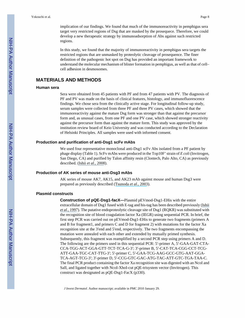
To obtain purified precursor Dsg1, recombinant Dsg1-EHis baculoprotein was passed throughan immunoadsorption column containing mAb 3–30/3h to eliminate mature Dsg1. To achievea purified mature form of Dsg1, recombinant Dsg1-EHis baculoprotein treated with furin waspassed through an immunoadsorption column containing mAb 3–97/1c to remove the precursorform (Figure 6a). Those purified recombinant baculoproteins were coated on ELISA plates togenerate purified mature Dsg1 and precursor Dsg1 ELISA plates. We confirmed that theseELISA plates contained either mature or precursor forms by immunoreactivities of anti-Dsg1
Yokouchi et al. Page 4
J Invest Dermatol. Author manuscript; available in PMC 2010 January 29.
NIH
-PA Author Manuscript
NIH
-PA Author Manuscript
NIH
-PA Author Manuscript

mAbs (Figure 6b). 3–30/3h reacted against the complete-mature Dsg1 plate in a dose-dependent manner, but did not react to the complete-precursor Dsg1. In contrast, 3–97/1cshowed dose-dependent reactivity to the complete-precursor plate, but showed no reactivity tothe complete-mature plate. 1–18/L12 exhibited similar reactivity against both plates.
We tested 45 PF sera and 20 mucocutaneous PV sera, which contained an anti-Dsg1 Ab inaddition to an anti-Dsg3 Ab. Strikingly, all PF sera, except two, showed stronger reactivityagainst the mature Dsg1 plate than against the precursor Dsg1 plate (Figure 6c). The averageand standard deviation of immunoreactivity, as determined by ELISA optical density (OD),against mature Dsg1 was 1.37 ± 0.32, whereas the average and standard deviation ofimmunoreactivity against the precursor form was 0.43 ± 0.23. Difference betweenimmunoreactivity against mature and precursor Dsg1 was statistically significant (paired t-test,P < 0.001). For PV sera that contained Dsg1 and Dsg3 Abs, all sera but two also reacted morestrongly to the mature Dsg1 ELISA plate than to the precursor ELISA plate (Figure 6d). Theaverage and standard deviation of the ELISA OD of PV sera against mature Dsg1 was 1.26 ±0.41, whereas the average and standard deviation against precursor Dsg1 was 0.31 ± 0.16,which was a statistically significant difference (P < 0.001). These findings show that themajority of pemphigus sera containing anti-Dsg1 Abs target epitopes that are unmasked byproteolytic processing.
As changes in the reactivity of anti-Dsg3 mAbs have also been observed on Dsg3 ELISA plateswith different ratios of precursor and mature Dsg3 (Sharma et al., 2009), we also tested thewell-characterized mouse anti-Dsg3 mAb AK series and PV sera on ELISA plates with purifiedmature or precursor Dsg3. To obtain precursor Dsg3, we used a mouse anti-Dsg3 mAb, AK23,obtained from PV model mice (Tsunoda et al., 2003). AK23, a pathogenic anti-Dsg3 mousemonoclonal Ab, targets an epitope on the adhesive interface of Dsg3 molecules and binds onlyto the mature form (Tsunoda et al., 2003). Recombinant Dsg3-EHis baculoprotein was passedthrough an immunoadsorption column containing AK23 mAb to eliminate mature Dsg3. Toobtain mature Dsg3, recombinant Dsg3-EHis baculoprotein was treated with furin (Figure 7a).We confirmed the accuracy of ELISA plates coated with the purified mature and precursorDsg3 by mouse anti-Dsg3 mAbs, AK23, and AK15 (Figure 7b). AK15 is an anti-Dsg3monoclonal Ab that recognizes a linear epitope in the middle portion of Dsg3, so that the Abshould bind both the mature and precursor forms of Dsg3. AK23 mAb showed dose-dependentreactivity to the complete-mature plate, while showing no reactivity to the complete-precursorplate. AK15 mAb exhibited similar binding capacity to both plates.
We tested 47 PV sera against these plates. All but five PV sera had stronger reactivity againstthe complete-mature plate than against the complete-precursor plate (Figure 7c). For PV sera,the average and standard deviation of immunoreactivity against mature Dsg3 was 0.74 ± 0.36,whereas the average and standard deviation of immunoreactivity against the precursor formwas 0.20 ± 0.13 (paired t-test, P < 0.001). These data suggest that the majority of anti-Dsg3immunoreactivity in PV also targets epitopes that are unmasked by proteolytic processing.
Titers measured by mature Dsg ELISA tend to reflect the disease activity more precisely thanthat by the precursor Dsg ELISA
Finally, we analyzed PF and PV cases for immunoreactivity against the mature and precursorforms of Dsg1 and Dsg3 over time, and compared them with clinical disease activity. First, weanalyzed three PF and three PV cases in which the immunoreactivity against the mature formwas stronger than that against the precursor form of Dsg because we considered them as typicalcases. As shown in Figure 8a–f, the immunoreactivity against both the mature and precursorDsg tended to fluctuate in parallel with disease activity. However, immunoreactivity againstthe mature form was a more sensitive indicator of disease activity in most patients, becausethe reactivity against the precursor was often minimal or negative, even at times of disease
Yokouchi et al. Page 5
J Invest Dermatol. Author manuscript; available in PMC 2010 January 29.
NIH
-PA Author Manuscript
NIH
-PA Author Manuscript
NIH
-PA Author Manuscript

activity (Figure 8a–d and f). We also analyzed two unusual cases that showed strongerreactivity against the precursor Dsg plate than against the mature form (Figure 8g and h). Inone case (Figure 8g), the ELISA reactivity against the precursor Dsg did reflect the diseaseactivity well, but in the other (Figure 8h), the reactivity against the precursor did not fall withdecreased disease activity, as did the reactivity against the mature protein. In summary,considering all the patients whom we tested (Figure 8), reactivity against the mature proteincorrelated well with disease activity, whereas reactivity against the precursor Dsg sometimesdid not.
DISCUSSIONDetailed characterization of monoclonal anti-Dsg1 Abs isolated from a PF patient revealedthat the patient had Abs against the mature and precursor forms of Dsg1. We also found thata pathogenic Dsg1 Ab binds only the mature form, whereas non-pathogenic ones bind only theprecursor form or bind both mature and precursor forms. These findings led us to explore theimmunoreactivity of multiple pemphigus sera against the mature and precursor forms of Dsg1and 3. Competition ELISA showed that much of the immunoreactivity in multiple PF serabinds to the same, or nearby, epitopes defined by the pathogenic Ab. ELISA using the matureand precursor Dsgs showed that much of the immunoreactivity in the majority of pemphigussera target the mature form of Dsg than with the precursor form. Furthermore, a longitudinalstudy of pemphigus patients showed that immunoreactivity against the mature Dsg reflects thedisease activity well. All these findings suggest that epitopes that are masked by theprosequence are very important in pathogenesis in pemphigus.
Although the pathogenic PF monoclonal anti-Dsg1 Ab, used in this study, which binds onlythe mature form of Dsg1, is dependent on the conformation of Dsg1 (Table 1), a pathogenicanti-Dsg Ab has been described that binds a non-conformational epitope (Yeh et al., 2006). Inaddition, it has been reported that IgG against non-conformational epitopes in pemphigus alsocorrelated with clinical disease activity (Muller et al. 2006,2008). Although it remains to bedetermined, it is not unreasonable to assume that the prosequence could block the binding ofpathogenic Abs against both conformational and non-conformational pathological epitopes.
High-resolution crystal structure analysis of the entire extracellular domain of C-cadherinprovided a mechanistic basis for intermolecular cadherin interactions (Boggon et al., 2002).The trans-adhesive interface is a two-fold symmetrical interaction, as the conserved tryptophan(W2) side chain at the amino-terminal end of the cadherin molecule from one cell inserts intothe hydrophobic pocket at the amino-terminal end of a cadherin molecule on an opposing cell.Recent cryo-electron tomography of vitreous sections from human epidermis visualized thethree-dimensional molecular architecture of desmosomal cadherins, which resembled the X-ray structure of C-cadherin (Al-Amoudi et al., 2007). Considering the similarity of amino-acidsequences between the ectodomains of classic cadherins and desmosomal cadherins, theyprobably share similar adhesive domains. Immunoblot analysis using domain-swappedmolecule of Dsg1/Dsg2 or Dsg3 revealed that the epitope of 3–30/3h was mapped to aminoacid 89–101 of the EC1 region of Dsg1 (Figure 5). When the amino-acid sequences of Dsg1were superimposed on the predicted structure of C-cadherin, the predicted residues for theadhesive interface of the W2 acceptor sites (hydrophobic pocket) were 24I, 38Y, 78C, 80A,89E, 91P, and 92L (Boggon et al., 2002). This putative W2-acceptor sites partially overlappedwith the presumed epitope of 3–30/3h, indicating that the epitope of 3–30/3h is located in apresumptive hydrophobic pocket of the adhesive interface of Dsg1. Furthermore, previousstudies suggested that the prosequence of E-cadherin blocks the N-terminal adhesive regionof cadherin to prevent self-aggregation during protein synthesis in the Golgi or endoplasmicstructure, and proteolytic processing creates the active form by conformational changes or byexposure of active binding sites (Ozawa and Kemler, 1990). In addition, the three-dimensional
Yokouchi et al. Page 6
J Invest Dermatol. Author manuscript; available in PMC 2010 January 29.
NIH
-PA Author Manuscript
NIH
-PA Author Manuscript
NIH
-PA Author Manuscript

structure study of the prosequence of N-cadherin showed that the proregion is a structure similarto the cadherin adhesive domain but lacking features for cadherin–cadherin interactions,suggesting that the prosequence might render interaction sites less accessible and also preventthe propagation of the interactions (Koch et al., 2004). Therefore, by analogy, pathogenic Absin pemphigus sera target regions of Dsgs that are functionally important and are unmasked byproteolytic processing. This finding is consistent with the hypothesis that the direct inhibitionof trans-interaction of Dsg by autoantibodies plays a primary role in inducing blister formationin the pemphigus. However, we cannot rule out an alternative hypothesis that Ab binding toexposed adhesive sites in Dsg somehow causes signaling inside the cells or internalization ofDsg, which leads to acantholysis. In any case, our results showed the importance of pemphigusAb binding to Dsgs after adhesive domains are exposed by its normal proteolytic processing(Figure 9b).
We should mention that not all pathogenic autoantibodies target the regions that are maskedby the prosequence. For example, we have identified another weakly pathogenic anti-Dsg1monoclonal Ab (3–7/1e) that did not cause blisters in mice, but induced slight blister formationin human skin (Ishii et al., 2008). This Ab bound both the mature and the precursor form ofDsg1. Epitope mapping using a domain-swapped molecule of Dsg1/Dsg2 revealed that theepitope resides in the EC2 domain of Dsg1 (data not shown). It may be possible that such Absblock the cis-interaction of Dsgs. The mechanism of blister formation by such Abs remains tobe determined.
Another major finding in this paper is that PF sera have a subset of autoantibodies against onlya precursor Dsg1. The frequency of PF sera that contains Abs against the precursor form ofDsg1 may be high because inhibition of ELISA in a previous paper showed that the bindingof the non-pathogenic scFvs against precursor Dsg1 was inhibited by four out of the six PFsera tested (Ishii et al., 2008). However, in almost all PF sera, only a small proportion of theirreactivity is against the precursor form of Dsg1, as shown by the competition ELISA of the PFsera by anti-Dsg1 scFvs (Figure 4), and by the finding that both PF and PV sera have strongerreactivity against the mature than against the precursor form of Dsg (Figure 6c and Figure 7c).The epitope bound by the scFv mAbs reacting exclusively against the precursor form of Dsg1could either be the prosequence itself or a conformational epitope on Dsg1 stabilized by theprosequence and lost in the mature protein. Further definition of this exact epitope awaitsadditional studies.
Cloned scFv that binds exclusively against the precursor Dsg1 shows weak intracellularstaining by indirect immunofluorescence, as opposed to the cell surface staining typical ofpemphigus. We think this staining is weak because there is probably little intracellularprecursor Dsg as most is efficiently processed to the mature cell surface form. We can detectthis weak staining because we have potent monoclonal scFv against precursor Dsg1, but suchstaining would be difficult or impossible to appreciate with polyclonal pemphigus sera thathave a predominant Ab response against the mature cell surface Dsg.
We do not know how these Abs against the precursor form are involved in the pathogenesis inPF. It may be possible that PF patients first develop Abs against the intracellular precursorDsg1, which may not normally be exposed to the immune system but is released at times ofkeratinocyte injury, and subsequently develop pathogenic Abs against mature Dsg1. Anotherpossibility is that Abs against the unprocessed form of Dsg1 are developed after PF is activeas a consequence of the damage to keratinocytes.
The findings we presented here have clinical implications. We showed that the disease activitywas more precisely correlated with the level of immunoreactivity against the mature form ofDsg by ELISA than that against the premature Dsg ELISA. There is another therapeutic
Yokouchi et al. Page 7
J Invest Dermatol. Author manuscript; available in PMC 2010 January 29.
NIH
-PA Author Manuscript
NIH
-PA Author Manuscript
NIH
-PA Author Manuscript
implication of our findings. We found that much of the immunoreactivity in pemphigus seratarget very restricted regions of Dsg that are masked by the prosequece. Therefore, we coulddevelop a new therapeutic strategy by immunoadsorption of Abs against such restrictedregions.
In this study, we found that the majority of immunoreactivity in pemphigus sera targets therestricted regions that are unmasked by proteolytic cleavage of prosequence. The finerdefinition of the pathogenic hot spot on Dsg has provided an important framework tounderstand the molecular mechanism of blister formation in pemphigus, as well as that of cell–cell adhesion in desmosomes.
MATERIALS AND METHODSHuman sera
Sera were obtained from 45 patients with PF and from 47 patients with PV. The diagnosis ofPF and PV was made on the basis of clinical features, histology, and immunofluorescencefindings. We chose sera from the clinically active stage. For longitudinal follow-up study,serum samples were collected from three PF and three PV cases, which showed that theimmunoreactivity against the mature Dsg form was stronger than that against the precursorform and, as unusual cases, from one PF and one PV case, which showed stronger reactivityagainst the precursor form than against the mature form. This study was approved by theinstitution review board of Keio University and was conducted according to the Declarationof Helsinki Principles. All samples were used with informed consent.
Production and purification of anti-Dsg1 scFv mAbsWe used four representative monoclonal anti-Dsg1 scFv Abs isolated from a PF patient byphage display (Table 1). ScFv mAbs were produced in the Top10F’ strain of E coli (Invitrogen,San Diego, CA) and purified by Talon affinity resin (Clontech, Palo Alto, CA) as previouslydescribed. (Ishii et al., 2008).
Production of AK series of mouse anti-Dsg3 mAbsAK series of mouse AK7, AK15, and AK23 mAb against mouse and human Dsg3 wereprepared as previously described (Tsunoda et al., 2003).
Plasmid constructsConstruction of pQE-Dsg1-facX—Plasmid pEVmod-Dsg1-EHis with the entireextracellular domain of Dsg1 fused with E-tag and his-tag has been described previously (Ishiiet al., 1997). The putative endoproteolytic cleavage site of Dsg1 (RQKR) was substituted withthe recognition site of blood coagulation factor Xa (IEGR) using sequential PCR. In brief, thefirst step PCR was carried out on pEVmod-Dsg1-EHis to generate two fragments (primers Aand B for fragment1, and primers C and D for fragment 2) with mutations for the factor Xarecognition site at the 3′end and 5′end, respectively. The two fragments encompassing themutation were annealed with each other and extended by mutually primed synthesis.Subsequently, this fragment was reamplified by a second PCR step using primers A and D.The following are the primers used in this sequential PCR: 5′-primer A, 5′-GAA-GAT-CTA-CCA-TGG-ACT-GGA-GTT-TCT-TCA-G-3′; 3′-primer B, 5′-CAT-TCA-CGG-CCT-TCG-ATT-GAA-TGC-CAT-TTG-3′; 5′-primer C, 5′-GAA-TCG-AAG-GCC-GTG-AAT-GGA-TCA-AGT-TCG-3′; 3′-primer D, 5′-CCG-GTC-GAC-ATG-TAC-ATT-GTC-TGA-TAA-C.The final PCR product containing the factor Xa recognition site was digested with an NcoI andSalI, and ligated together with NcoI-XhoI-cut pQE-trisystem vector (Invitrogen). Thisconstruct was designated as pQE-Dsg1-FacX (p330).
Yokouchi et al. Page 8
J Invest Dermatol. Author manuscript; available in PMC 2010 January 29.
NIH
-PA Author Manuscript
NIH
-PA Author Manuscript
NIH
-PA Author Manuscript

Production of recombinant baculoproteinsPrecursor-FacX-Dsg1-EHis—pQE-Dsg1-FacX was cotransfected with the Baculo-sapphire baculovirus DNA (Orbigen, San Diego, CA) into cultured insect Sf9 cells, andrecombinant viruses were obtained. For baculoprotein production, High Five cells (Invitrogen)cultured in a serum-free EX cell 405 medium (SAFC biosciences, Lenexa, KS) were infectedwith high-titer virus stock and incubated at 27°C for 3 days. Precursor-FacX-Dsg1-EHis wassecreted in the culture supernatant.
Mature-Dsg1-EHis—Dsg1-EHis baculoprotein was cleaved by incubation with proproteinconvertase furin (New England BioLabs, Beverly, MA). Typically, 50 µl of the culturesupernatant was incubated with two units of furin at room temperature for 15 hours.
Production of highly purified mature and precursor ELISA platesHighly purified precursor and mature Dsg1 plates—For obtaining complete-precursorDsg1-EHis, baculovirus-expressed recombinant Dsg1-Ehis, which contained both theprecursor and the mature form, was purified by TALON affinity resin (Clontech) andsubsequently adsorbed by a 3–30/3h mAb beads column (Figure 4a) to eliminate the matureform of Dsg1-EHis. For obtaining complete-mature Dsg1-EHis, recombinant Dsg1-EHisbaculoprotein was purified by TALON affinity resin, treated with furin (20 unitmg−1) for 16hours at room temperature in the manufacturer’s recommended buffer (New England Biolabs),and subsequently passed through an affinity column containing 3–97/1C mAb to absorb theprecursor form (Figure 4a). The purified complete-precursor Dsg1-EHis and complete-matureDsg1-EHis were immobilized on 96-well polystyrene plates (Maxisorb Immunoplate; Nunc,Wiesbaden, Germany) by coating each well with 2.5 µgml−1 of recombinant proteins at 4°Cfor 20 hours.
Highly purified precursor and mature Dsg3 plates—For obtaining complete-precursorDsg3-EHis, baculovirus-expressed recombinant Dsg3-Ehis, which contained both theprecursor and the mature form was purified by TALON affinity resin (Clontech) andsubsequently adsorbed by AK23 mAb-bound affinity column (Figure 4a) to eliminate themature form of Dsg3-EHis (Figure 5a). For obtaining complete-mature Dsg3-EHis, purifiedrecombinant Dsg3-EHis baculoprotein was treated with furin using the above protocol andagain purified on TALON affinity resin. Complete-precursor Dsg3-EHis and complete-matureDsg3-EHis were immobilized on 96-well polystyrene plates (Nunc).
For the longitudinal study to compare reactivity against mature and precursor Dsg ELISA withdisease activity, we measured OD of pemphigus sera at an approximate dilution (1:100–1:6,400).
Dsg1 scFv ELISAThe reactivity of scFv against human Dsg1 was measured by Dsg1 ELISA (Medical andBiological Laboratories Co. Ltd, Nagoya, Japan) using a horseradish peroxidase-conjugatedanti-HA (Hemagglutinin) monoclonal Ab (clone 3F10, 1:1,000 dilution, Roche DiagnosticsCorp., Basel, Switzerland) as a secondary Ab as described (Ishii et al., 2008).
To increase the ratio of the mature form on a standard Dsg1 ELISA plate, the plates werepretreated with 2U/ well of furin (New England Biolabs, Ipswich, MA) in Tris-buffered salinewith 1mM CaCl2 at room temperature for 5 hours.
Yokouchi et al. Page 9
J Invest Dermatol. Author manuscript; available in PMC 2010 January 29.
NIH
-PA Author Manuscript
NIH
-PA Author Manuscript
NIH
-PA Author Manuscript

Indirect immunofluorescenceImmunofluorescence for scFvs was carried out on human skin as previously described (Payneet al., 2005). Binding was detected using a rat monoclonal anti-HA Ab (3F10, 1:100 dilution,Roche Diagnostics) and thereafter using a Alexa Fluor 446-conjugated anti-rat IgG (1:200dilution, Invitrogen). A confocal image was obtained using a Zeiss LSM 510 confocalmicroscope (Carl Zeiss Meditec, Dublin, CA).
ImmunoblottingThe recombinant baculoproteins were size-fractionated by SDS-PAGE and transferred to anImmobilon-P membrane (Millipore, Bedford, MA), and detected using an anti-E-tag Ab(1:2,000 dilution, GE Healthcare Bio-Sciences, Piscataway, NJ), followed by alkalinephosphatase-conjugated goat anti-mouse IgG Abs (1:1,000 dilution, DPRA-Zymax, SanFrancisco, CA).
Immunoprecipitation–immunoblotting analysisBaculovirus-infected insect cell culture supernatants containing recombinant proteins wereincubated with anti-Dsg1 scFv mAbs for 30 minutes and then immunoprecipitated with anti-HA agarose (Sigma-Aldrich, St Louis, MO) at 4°C for 2 hours with gentle rotation. Afterwashing with Tris-buffered saline-calcium, the immunoprecipitates were resuspended in aLaemmli sample buffer, separated by SDS-PAGE, and transferred to an Immobilon-Pmembrane (Millipore). The membranes were probed with an anti-E-tag Ab (1:2,000 dilution,GE Healthcare Bio-Sciences) followed by alkaline phosphatase-conjugated goat anti-mouseIgG Abs (1:1,000 dilution, DPRA-Zymax). The recombinant proteins used in the epitopemapping of 3–30/3h mAb have been described previously ((Sekiguchi et al., 2001) and Chanet al. (manuscript in preparation)).
Inhibition ELISAPF sera were diluted to result in an OD450 reading of ~1.0 in the Dsg1 ELISA withoutcompetitors. The diluted PF sera, mixed with scFv mAbs, were analyzed by standard Dsg1ELISA (Medical and Biological Laboratories) developed using a horseradish peroxidase-conjugated anti-human Fab Ab. Inhibition was calculated according to the following formula:% inhibition = (1 − (OD T/B–OD N/B)/(OD T/Bc − OD N/Bc))*100, where T is the PF seratested, N is the normal control serum, B is the blocking monoclonal anti-Dsg1 scFvs, and Bcis the control scFv.
Abbreviations
Ab antibody
Dsg desmoglein
mAb monoclonal antibody
PF pemphigus foliaceus
PV pemphigus vulgaris
scFv single-chain variable fragment
AcknowledgmentsWe thank Dr Aimee Payne for helpful discussions, Mrs Minae Suzuki for immunofluorescence staining, and YoshikoFujii and Sakiko Kobayashi for technical assistance. This work was supported by Grants-in-Aid for Scientific Researchfrom the Ministry of Education, Culture, Sports, Science and Technology of Japan, the Health and Labour SciencesResearch Grants for Research on Measures for Intractable Diseases from Ministry of Health, Labor and Welfare of
Yokouchi et al. Page 10
J Invest Dermatol. Author manuscript; available in PMC 2010 January 29.
NIH
-PA Author Manuscript
NIH
-PA Author Manuscript
NIH
-PA Author Manuscript

Japan. These studies were also supported, in part, by grants (RO1-AR538807 and RO1-AR546265) from the NationalInstitute of Arthritis, Musculoskeletal, and Skin Diseases.
REFERENCESAl-Amoudi A, Diez DC, Betts MJ, Frangakis AS. The molecular architecture of cadherins in native
epidermal desmosomes. Nature 2007;450:832–837. [PubMed: 18064004]Berkowitz P, Hu P, Liu Z, Diaz LA, Enghild JJ, Chua MP, et al. Desmosome signaling. Inhibition of
p38MAPK prevents pemphigus vulgaris IgG-induced cytoskeleton reorganization. J Biol Chem2005;280:23778–23784. [PubMed: 15840580]
Boggon TJ, Murray J, Chappuis-Flament S, Wong E, Gumbiner BM, Shapiro L. C-cadherin ectodomainstructure and implications for cell adhesion mechanisms. Science 2002;296:1308–1313. [PubMed:11964443]
Calkins CC, Setzer SV, Jennings JM, Summers S, Tsunoda K, Amagai M, et al. Desmoglein endocytosisand desmosome disassembly are coordinated responses to pemphigus autoantibodies. J Biol Chem2006;281:7623–7634. [PubMed: 16377623]
Esaki C, Seishima M, Yamada T, Osada K, Kitajima Y. Pharmacologic evidence for involvement ofphospholipase C in pemphigus IgG-induced inositol 1,4,5-trisphosphate generation, intracellularcalcium increase, and plasminogen activator secretion in DJM-1 cells, a squamous cell carcinoma line.J Invest Dermatol 1995;105:329–333. [PubMed: 7665907]
Hanakawa Y, Schechter NM, Lin C, Garza L, Li H, Yamaguchi T, et al. Molecular mechanisms of blisterformation in bullous impetigo and staphylococcal scalded skin syndrome. J Clin Invest 2002;110:53–60. [PubMed: 12093888]
Ishii K, Amagai M, Hall RP, Hashimoto T, Takayanagi A, Gamou S, et al. Characterization ofautoantibodies in pemphigus using antigen-specific enzyme-linked immunosorbent assays withbaculovirus-expressed recombinant desmogleins. J Immunol 1997;159:2010–2017. [PubMed:9257868]
Ishii K, Lin C, Siegel DL, Stanley JR. Isolation of pathogenic monoclonal anti-desmoglein 1 humanantibodies by phage display of pemphigus foliaceus autoantibodies. J Invest Dermatol 2008;128:939–948. [PubMed: 18007588]
Koch AW, Farooq A, Shan W, Zeng L, Colman DR, Zhou MM. Structure of the neural (N-) cadherinprodomain reveals a cadherin extracellular domain-like fold without adhesive characteristics. Structure2004;12:793–805. [PubMed: 15130472]
Muller R, Svoboda V, Wenzel E, Gebert S, Hunzelmann N, Muller HH, et al. IgG reactivity against non-conformational NH-terminal epitopes of the desmoglein 3 ectodomain relates to clinical activity andphenotype of pemphigus vulgaris. Exp Dermatol 2006;15:606–614. [PubMed: 16842599]
Muller R, Svoboda V, Wenzel E, Muller HH, Hertl M. IgG against extracellular subdomains ofdesmoglein 3 relates to clinical phenotype of pemphigus vulgaris. Exp Dermatol 2008;17:35–43.[PubMed: 18095943]
Ozawa M, Kemler R. Correct proteolytic cleavage is required for the cell adhesive function of uvomorulin.J Cell Biol 1990;111:1645–1650. [PubMed: 2211831]
Payne AS, Ishii K, Kacir S, Lin C, Li H, Hanakawa Y, et al. Genetic and functional characterization ofhuman pemphigus vulgaris monoclonal autoantibodies isolated by phage display. J Clin Invest2005;115:888–899. [PubMed: 15841178]
Posthaus H, Dubois CM, Laprise MH, Grondin F, Suter MM, Muller E. Proprotein cleavage of E-cadherinby furin in baculovirus overexpression system: potential role of other convertases in mammaliancells. FEBS Lett 1998;438:306–310. [PubMed: 9827567]
Sekiguchi M, Futei Y, Fujii Y, Iwasaki T, Nishikawa T, Amagai M. Dominant autoimmune epitopesrecognized by pemphigus antibodies map to the N-terminal adhesive region of desmogleins. JImmunol 2001;167:5439–5448. [PubMed: 11673563]
Sharma P, Mao X, Payne AS. Beyond steric hindrance: the role of adhesion signaling pathways in thepathogenesis of pemphigus. J Dermatol Sci 2007;48:1–14. [PubMed: 17574391]
Sharma P, Choi EJ, Kuroda K, Hachiya T, Ishii K, Payne AS. Pathogenic anti-desmoglein monoclonalantibodies demonstrate variable ELISA activity due to preferential binding of mature versus
Yokouchi et al. Page 11
J Invest Dermatol. Author manuscript; available in PMC 2010 January 29.
NIH
-PA Author Manuscript
NIH
-PA Author Manuscript
NIH
-PA Author Manuscript

proprotein isoforms of desmoglein 3. J Invest Dermatol. 2009 advance online publication 12 March2009; doi:10.1038/jid.2009.41.
Stanley JR, Amagai M. Pemphigus, bullous impetigo, and the staphylococcal scalded-skin syndrome. NEngl J Med 2006;355:1800–1810. [PubMed: 17065642]
Tsunoda K, Ota T, Aoki M, Yamada T, Nagai T, Nakagawa T, et al. Induction of pemphigus phenotypeby a mouse monoclonal antibody against the amino-terminal adhesive interface of desmoglein 3. JImmunol 2003;170:2170–2178. [PubMed: 12574390]
Wahl JK III, Kim YJ, Cullen JM, Johnson KR, Wheelock MJ. N-cadherin-catenin complexes form priorto cleavage of the proregion and transport to the plasma membrane. J Biol Chem 2003;278:17269–17276. [PubMed: 12604612]
Waschke J, Spindler V, Bruggeman P, Zillikens D, Schmidt G, Drenckhahn D. Inhibition of Rho Aactivity causes pemphigus skin blistering. J Cell Biol 2006;175:721–727. [PubMed: 17130286]
Williamson L, Raess NA, Caldelari R, Zakher A, de Bruin A, Posthaus H, et al. Pemphigus vulgarisidentifies plakoglobin as key suppressor of c-Myc in the skin. Embo J 2006;25:3298–3309. [PubMed:16871158]
Yeh S, Cavacini LA, Bhol KC, Lin M, Kumar M, Duval M, et al. Pathogenic human monoclonal antibodyagainst desmoglein 3. Clin Immunol 2006;120:68–75. [PubMed: 16635589]
Yokouchi et al. Page 12
J Invest Dermatol. Author manuscript; available in PMC 2010 January 29.
NIH
-PA Author Manuscript
NIH
-PA Author Manuscript
NIH
-PA Author Manuscript

Figure 1. Confocal microscopy of anti-desmoglein 1 single-chain variable fragment (scFv) mAbson human skin3–30/3h and 1–18/L12 scFv antibodies stained the cell surface of keratinocytes throughouthuman epidermis. 3–97/1c and 3–94/O18O8 showed cytoplasmic staining in the superficiallayers of the epidermis. Scale bars: 50 µm.
Yokouchi et al. Page 13
J Invest Dermatol. Author manuscript; available in PMC 2010 January 29.
NIH
-PA Author Manuscript
NIH
-PA Author Manuscript
NIH
-PA Author Manuscript

Figure 2. Binding capacity of anti-desmoglein 1 (Dsg1) single-chain variable fragment (scFv) mAbsto the Dsg1 ELISA plate changes after furin treatment(a) Baculovirally produced recombinant Dsg1 (Dsg1-EHis), visualized by immunoblot usinganti-E-tag antibody. Dsg1-EHis was detected as double bands, which were considered to beprecursor and mature form of Dsg1-EHis (left lane). After treatment with furin protease, onlysingle band was observed (right lane). White arrowhead indicates the precursor form and blackarrowhead indicates the mature form. (b) Comparison of anti-Dsg1 scFv mAbs bindingsbetween furin-treated and Tris-buffered saline-calcium (TBS-Ca)-treated (control) Dsg1ELISA plates. The ratio was calculated according to the following formula: (optical density(OD) values obtained in furin-treated wells)/(OD values obtained in TBS-Ca-treated wells).Control ratio (ratio = 1) is shown by a broken line. The binding of 3–30/3h, a pathogenic mAb,was significantly increased to nearly twofold, whereas that of 3–97/1c, non-pathogenic mAbwas decreased. The binding of 1–18/L12 showed no significant change. AM3–13 is anirrelevant scFv and used as a control.
Yokouchi et al. Page 14
J Invest Dermatol. Author manuscript; available in PMC 2010 January 29.
NIH
-PA Author Manuscript
NIH
-PA Author Manuscript
NIH
-PA Author Manuscript

Figure 3. Binding of anti-desmoglein 1 (Dsg1) single-chain variable fragment (scFv) mAbs toprecursor-FacX-Dsg1 and mature Dsg1(a) Molecular structure of precursor-FacX-Dsg1-EHis and mature-Dsg1-EHis. To obtainprecursor form, the recognition sequence in precursor Dsg1 was substituted with that of bloodcoagulation factor Xa (R-Q-K-R → I-E-G-R) by endogenous proprotein convertases. To obtainmature form of Dsg1, recombinant Dsg1-EHis produced by baculovirus expression systemwas treated with proprotein convertase furin. (b) Precursor-FacX-Dsg1-EHis and mature-Dsg1-EHis recombinant baculoprotein were immunoprecipitated with representative anti-Dsg1 scFv antibodies or control scFv (AM3–13) and detected on an immunoblot by anti-E-tagantibody.
Yokouchi et al. Page 15
J Invest Dermatol. Author manuscript; available in PMC 2010 January 29.
NIH
-PA Author Manuscript
NIH
-PA Author Manuscript
NIH
-PA Author Manuscript

Figure 4. Competition ELISA of PF sera binding by anti-desmoglein 1 (Dsg1) single-chain variablefragment (scFv) mAbsCompetition ELISA of 40 PF sera was carried out to see if the binding against standard Dsg1ELISA plates of PF sera was blocked by adding anti-Dsg1scFv mAbs. 3–30/3h (shown inorange), 3–97/1c, 3–94/O18O8 (shown in green) and 1–18/L12 (shown in blue) were used ascompetitors. When >20% inhibition was considered as significant (shown in broken line), thebindings were blocked by pathogenic 3–30/3h mAb in 29 PF sera. 3–97/1c and 3–94/O18O8,which recognized the precursor form, blocked the binding in 10 PF sera.
Yokouchi et al. Page 16
J Invest Dermatol. Author manuscript; available in PMC 2010 January 29.
NIH
-PA Author Manuscript
NIH
-PA Author Manuscript
NIH
-PA Author Manuscript

Figure 5. Epitope mapping of pathogenic 3–30/3h mAbWild-type and domain-swapped extracellular domains of human desmoglein 1 (Dsg1) andDsg3, and of Dsg1 and Dsg2 produced by baculovirus expression system were used forimmunoprecipitation with 3–30/3h mAb. The molecular structure of domain-swappedmolecules is shown. The results of immunoprecipitation assays with 3–30/3h mAb are listedin the right panel.
Yokouchi et al. Page 17
J Invest Dermatol. Author manuscript; available in PMC 2010 January 29.
NIH
-PA Author Manuscript
NIH
-PA Author Manuscript
NIH
-PA Author Manuscript
Figure 6. Majority of anti-desmoglein 1 (Dsg1) antibody binding in PF and PV sera targets themature form more than the precursor form of Dsg1(a) Coomassie blue staining of purified recombinant baculoproteins used for precursor andmature Dsg1 ELISA plates. White arrowhead indicates the precursor form and black arrowheadindicates the mature form. (b) Binding curves of representative anti-Dsg1 scFv antibodies (3–30/3h, 3–97/1c, 1–18/L12) and control scFv (AM3–13) against mature and precursor Dsg1ELISA plate. (c) Dispersion of immunoreactivity of 45 PF sera between the mature andprecursor Dsg1 ELISA plate. Each closed square represents single patient. Note that most PFsera had stronger reactivity against mature Dsg1 plate than precursor Dsg1 plate. (d) Dispersionof immunoreactivity of 20 mucocutaneous PV sera between the mature and precursor Dsg1
Yokouchi et al. Page 18
J Invest Dermatol. Author manuscript; available in PMC 2010 January 29.
NIH
-PA Author Manuscript
NIH
-PA Author Manuscript
NIH
-PA Author Manuscript

ELISA plate. Each closed triangle represents single patient. Similar to PF patients, most PVsera had stronger reactivity against mature Dsg1 plate than against precursor Dsg1 plate.
Yokouchi et al. Page 19
J Invest Dermatol. Author manuscript; available in PMC 2010 January 29.
NIH
-PA Author Manuscript
NIH
-PA Author Manuscript
NIH
-PA Author Manuscript

Figure 7. Majority of anti-desmoglein 3 (Dsg3) antibody binding in PV sera targets the matureform more than the precursor form of Dsg1(a) Coomassie blue staining of purified recombinant baculoproteins used for precursor andmature Dsg1 ELISA plates. White arrowhead indicates the precursor form and black arrowhead indicates the mature form. (b) Binding curves of representative anti-Dsg3 mAbs (AK23,AK15) against mature and precursor Dsg3 ELISA plates. (c) Dispersion of immunoreactivityof 47 PV sera between the mature and precursor Dsg3 ELISA plates. Each closed squarerepresents single patient. Most PV sera showed stronger reactivity against mature Dsg3 platethan against precursor Dsg3 plate.
Yokouchi et al. Page 20
J Invest Dermatol. Author manuscript; available in PMC 2010 January 29.
NIH
-PA Author Manuscript
NIH
-PA Author Manuscript
NIH
-PA Author Manuscript

Figure 8. Time-course analysis of immunoreactivity against precursor and mature form ofdesmoglein (Dsg) in PF and PV patientsImmunoreactivities against precursor and mature form of Dsg were compared with the diseaseactivity over a time course. four PF patients (a, c, e, and g) and four PV patients (b, d, f, andh) were studied. Each serum was diluted up to 1:100 (a), 1:100 (b), 1:100 (c), 1:200 (d), 1:200(e), 1:100 (f), 1:6400 (g), and 1:100 (h). Clinical disease activity was subjectively assessed andthe disease activity in the first presentation was scored as 10. (0 (normal) to 10 (most severe)).three PF (a, c, and e) and three PV (b, d, and f) were randomly chosen from the PF and PVpatients’ sera, which showed stronger reactivity against the mature Dsg plate than against theprecursor Dsg plate. As unusual cases, a PF (g) and a PV case (h) who showed strongerreactivity against precursor-Dsg than against the mature-Dsg form were selected.
Yokouchi et al. Page 21
J Invest Dermatol. Author manuscript; available in PMC 2010 January 29.
NIH
-PA Author Manuscript
NIH
-PA Author Manuscript
NIH
-PA Author Manuscript

Figure 9. Proteolytic processing of the precursor form of desmogleins (Dsgs) and mAb binding tothe precursor and mature form of Dsg1(a) Dsgs are thought to be synthesized as inactive precursor Dsgs with prosequences in theendoplasmic reticulum. The prosequences are then removed by subtillisin-like proproteinconvertases before transporting them to the plasma membrane and mature Dsgs are assembledinto desmosomes. (b) Binding of anti-Dsg1 mAbs to the mature and the precursor form ofDsg1. The pathogenic mAb, 3–30/3h (shown in orange) binds to the mature Dsg1, but not theprecursor Dsg1. Some non-pathogenic mAbs, 3–97/1c and 3–94/O18O8 (shown in green), bindto the precursor Dsg1. A non-pathogenic mAb, 1–18/L12 (shown in blue), which recognizesthe middle portion of Dsg1, binds to both mature and precursor Dsg1. The pathogenic mAb
Yokouchi et al. Page 22
J Invest Dermatol. Author manuscript; available in PMC 2010 January 29.
NIH
-PA Author Manuscript
NIH
-PA Author Manuscript
NIH
-PA Author Manuscript

bind the region of adhesive interface of Dsg1, which are unmasked by proteolytic processing,perhaps blocking trans-interaction of Dsgs.
Yokouchi et al. Page 23
J Invest Dermatol. Author manuscript; available in PMC 2010 January 29.
NIH
-PA Author Manuscript
NIH
-PA Author Manuscript
NIH
-PA Author Manuscript

NIH
-PA Author Manuscript
NIH
-PA Author Manuscript
NIH
-PA Author Manuscript
Yokouchi et al. Page 24
Table 1
Epitopes and pathogenicity of anti-Dsg1 mAbs
Name of clones Epitope type Deduced epitope
Pathogenic antibody 3–30/3h Conformational aa 89–101 of Dsg1
Non-pathogenic antibodies 3–97/1c Conformational Precursor form of Dsg1
3–94/O18O8 Conformational Precursor form of Dsg1
1–18/L12 Linear EC3 of Dsg1
Abbreviations: aa, amino acid; Dsg, desmoglein; EC, extracellular domain.
J Invest Dermatol. Author manuscript; available in PMC 2010 January 29.