Chronic VEGF blockade worsens glomerular injury in the remnant kidney model

Anti-DNA auto-antibodies initiate experimental lupus nephritisby binding directly to the glomerular basement membrane inmice
Meera R. Krishnan, Ph.D.*, Congmiao Wang, M.D., Ph.D.*, and Tony N. Marion, Ph.D.Department of Microbiology, Immunology and Biochemistry, The University of Tennessee HealthScience CenterMeera R. Krishnan: [email protected]; Congmiao Wang: [email protected]
AbstractThe strongest serological correlate for lupus nephritis is antibody to double-stranded DNAalthough the mechanism by which anti-DNA antibodies initiate lupus nephritis is unresolved.Most recent reports indicate that anti-DNA must bind chromatin in the glomerular basementmembrane or mesangial matrix to form glomerular deposits. Here we determined whether directbinding of anti-DNA antibody to glomerular basement membrane is critical to initiate glomerularbinding of anti-DNA in experimental lupus nephritis. Mice were co-injected with IgG monoclonalantibodies or hybridomas with similar specificity for DNA and chromatin but different IgGsubclass and different relative affinity for basement membrane. Only anti-DNA antibodies thatbound basement membrane bound to glomeruli, activated complement, and induced proteinuriawhether injected alone or co-injected with a non-basement membrane-binding anti-DNA antibody.Basement membrane-binding anti-DNA antibodies co-localized with heparan sulfate proteoglycanin glomerular basement membrane and mesangial matrix but not with chromatin. Thus, directbinding of anti-DNA antibody to antigens in the glomerular basement membrane or mesangialmatrix may be critical to initiate glomerular inflammation. This may accelerate and exacerbateglomerular immune complex formation in human and murine lupus nephritis.
IntroductionThe contribution of anti-DNA antibody to glomerulonephritis in mouse (1) and human (2)systemic lupus erythematosus (SLE) is well established. Although anti-double-strandedDNA (dsDNA) antibody is the best serological correlate for lupus nephritis (3, 4), thefrequent lack of correlation between serum anti-dsDNA and glomerulonephritis is a longrecognized conundrum in the clinical evaluation of individual SLE patients (3, 5, 6). Thelack of correlation between anti-dsDNA and lupus nephritis within individual patients maybe a consequence of how anti-dsDNA antibodies bind in the glomerulus and initiateglomerulonephritis (6), a process not yet fully resolved (7). Mechanisms proposed to explainglomerular deposition of anti-DNA antibody include glomerular binding of soluble immunecomplexes of nucleosomes and IgG anti-DNA (2, 8–10), in situ formation of immune
Corresponding author: Tony N. Marion, Ph.D., Department of Microbiology, Immunology, and Biochemistry, University ofTennessee Health Science Center, 858 Madison Ave., Memphis, TN 38163, Tele: 901-448-6527, FAX: 901-448-6527,[email protected].*These authors contributed equally to the research.Present address: Congmiao Wang, M.D., Ph.D., Department of Rheumatology, Bei Fang Hospital, No. 5, Nan Men Cang, Dong-si-shi-tiao, Beijing, 100700, P.R. China
DisclosureThe authors declare no commercial financial support or competing financial interests.
NIH Public AccessAuthor ManuscriptKidney Int. Author manuscript; available in PMC 2013 January 01.
Published in final edited form as:Kidney Int. 2012 July ; 82(2): 184–192. doi:10.1038/ki.2011.484.
NIH
-PA Author Manuscript
NIH
-PA Author Manuscript
NIH
-PA Author Manuscript
complexes when anti-DNA antibody binds to chromatin that has bound to glomerularbasement membrane (GBM) or mesangial matrix (MM) (11–17), and direct binding of anti-DNA antibody that cross-reacts with GBM or cell surface antigens (18–25). Recentmorphologic studies (12–14, 16) have identified chromatin and IgG within the glomerularsubendothelial and subepithelial electron dense deposits (EDS) in nephritic kidneys fromlupus patients (26) and lupus-prone mice (27). The recent results were interpreted to indicatethat anti-DNA antibody could form glomerular deposits only when bound to chromatin ornucleosomes (28–30).
The present experiments were designed to test the hypothesis that initial glomerular bindingof anti-DNA antibody in lupus nephritis is a function of direct, cross-reactive binding toglomerular antigens, particularly in GBM or MM, and independent of DNA, nucleosomes,or chromatin. The experiments took advantage of a panel of anti-DNA monoclonalantibodies (mAbs) with similar relative affinities for DNA and chromatin but differentrelative affinities for basement membrane (BM) antigens in GBM and MM. Only anti-DNAmAbs that also bound BM antigens bound glomeruli in vivo and induced proteinuria.Glomerular binding of the anti-DNA mAbs was independent of DNA, nucleosomes, orchromatin. The results may explain why some anti-DNA mAbs are very effective atinducing lupus nephritis, but others are not. Similarly, the results may help to explain whySLE patients with similar serum anti-dsDNA antibody may have different susceptibility forlupus nephritis.
ResultsIn vitro binding of anti-DNA mAb to BM
Culture supernatants from 69 autoimmune anti-DNA mAbs from eight different (NZB ×NZW)F1 mice (BWF1) were randomly selected for analysis (Table 1). Total IgG andrelative affinity for binding to ssDNA, dsDNA, chromatin, and BM were quantified for eachsupernatant. The mAbs were stratified by relative affinity for BM into four differentspecificity groups (Table 1). There is a significant difference among the four specificitygroups for competitive binding to ssDNA and dsDNA and direct binding to BM but not fordirect binding to chromatin. There is a strong and highly significant correlation betweenbinding to BM and binding to dsDNA and a moderate, highly significant inverse correlationbetween binding to BM and binding to ssDNA. Anti-DNA mAbs that bound best to dsDNAare generally the mAbs that also bound best to BM. The correlation between BM andchromatin binding, although significant, was low compared to that for BM and dsDNA. Theresults indicate that mAbs with high relative affinity for dsDNA are more likely to bind BMthan mAbs with high relative affinity for ssDNA. The results also indicate that anti-DNAmAb binding to BM is unrelated to relative affinity for chromatin.
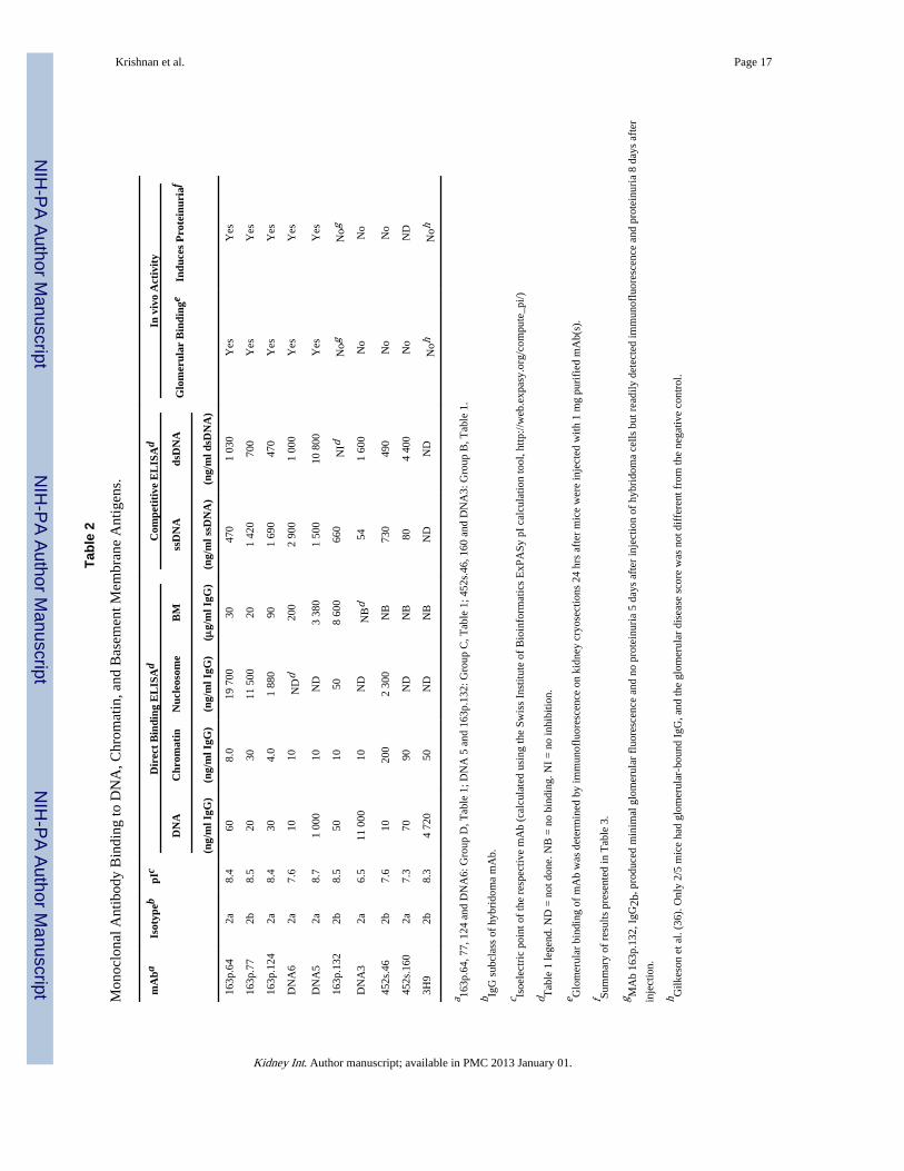
The correlations between mAb binding to DNA and chromatin versus their potential to bindBM were further confirmed with purified mAbs (Table 2). BM binding by purified mAbswas independent of relative binding affinity for dsDNA, chromatin, or nucleosomes since163p.132, 452s.160, DNA3, and 3H9 mAbs bound nucleosomes and/or chromatin with highrelative affinity but bound poorly or not at all to BM. MAb 452s.46 bound dsDNA with highrelative affinity but did not bind BM. DNA6 mAb bound chromatin similarly to 163p.132and DNA3 but unlike 163p.132 and DNA3, DNA6 also bound to BM. MAbs 163p.64, 163p.77, and 163p.124 had 20–650 fold higher relative affinity for BM than for nucleosomes.Binding to BM was also independent of mAb pI. These results further indicate that anti-DNA mAb binding to BM is correlated with dsDNA binding and to lesser extent chromatinbinding, but is independent of both for binding to BM.
Krishnan et al. Page 2
Kidney Int. Author manuscript; available in PMC 2013 January 01.
NIH
-PA Author Manuscript
NIH
-PA Author Manuscript
NIH
-PA Author Manuscript

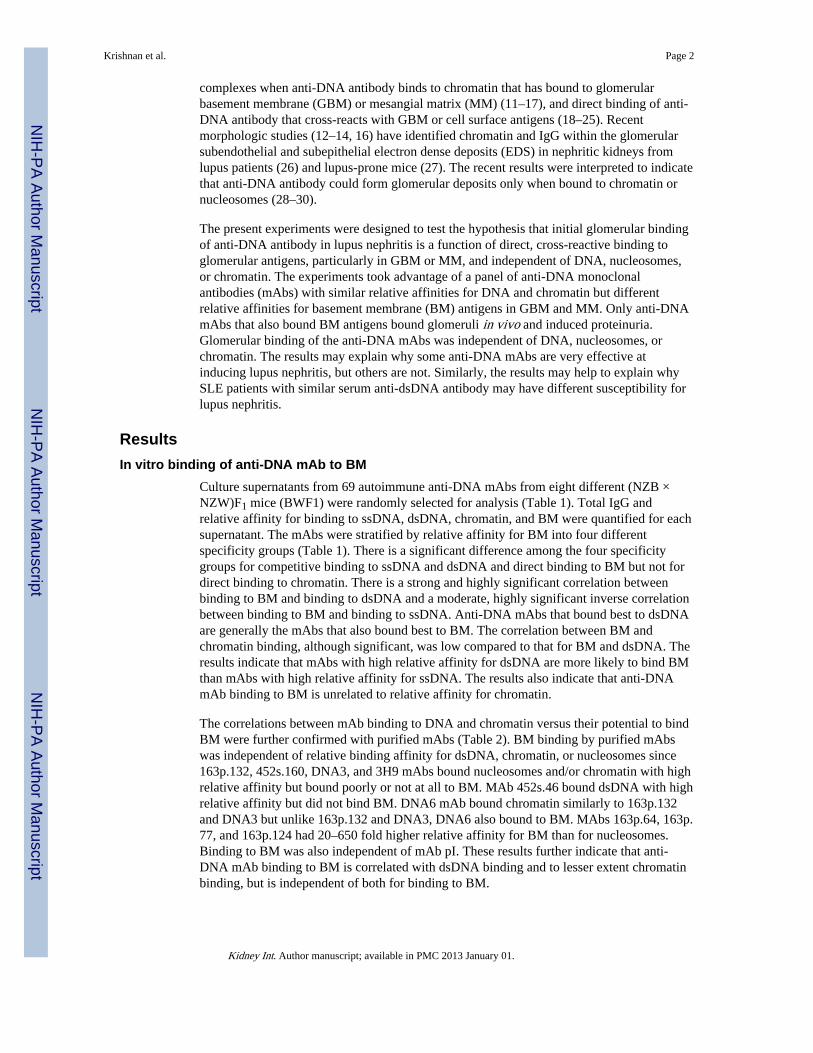
Since previous investigators had concluded that anti-dsDNA mAb binding to BM wasconsequential to nucleosome contamination of hybridoma supernatants and purified mAbs(10), we performed co-incubation assays to insure that differential binding of anti-dsDNAmAbs to BM was not simply a consequence of contaminating chromatin in some but not allhybridoma supernatants. When hybridoma supernatants of mAb pairs 163p.132 and 163p.124, 452s.46 and 163p.64, 163p.77 and DNA3, and 163p.77 and 452s.160 were assayed forbinding to BM, only the mAb that bound to BM in the individual assays, 163p.124, 163p.64,and 163p.77, bound to BM when co-incubated with a non BM-binding mAb (Fig. 1 andTable 2). MAb 163p132 does bind BM but with 100–500-fold less relative affinity thanmAbs 163p.64, 77, and 124. The results in Fig 1 corroborate the conclusion that anti-DNAmAb binding to BM is independent of dsDNA or chromatin.
MAb 163p.64 was tested by direct ELISA for binding to individual components of BM,including laminin, perlecan, entactin, and agrin. The mAb bound perlecan, entactin, andagrin (59, 250, and 220 ng IgG/ml, respectively, for 50% maximum binding) but notlaminin. The recombinant agrin did not include the amino-terminal extracellular matrixinteraction domains (R&D Systems). Binding to collagen IV was not tested. The resultsindicate that a BM binding mAb may also bind to some but not all of the individualcomponents of GBM.
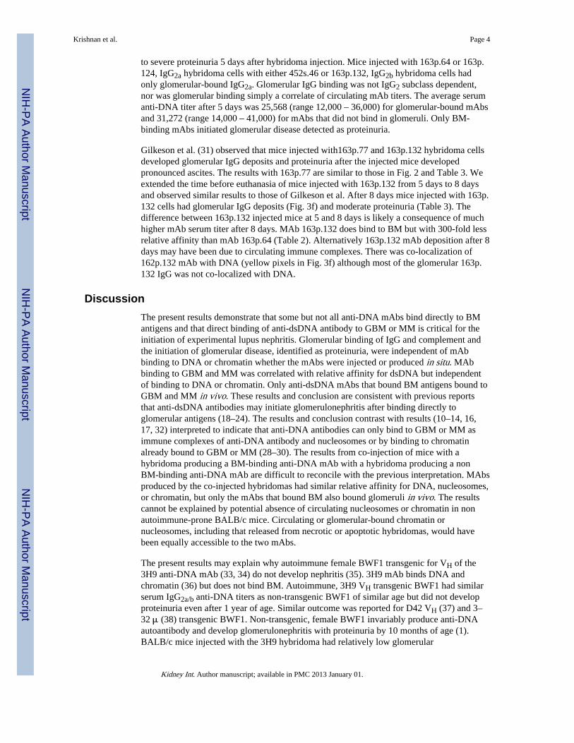
In vivo glomerular binding of anti-DNA mAbsSix purified mAbs were further tested for glomerular binding when injected into nonautoimmune-prone BALB/c mice alone or co-injected with a mAb with different BMbinding potential and different IgG subclass. The co-injected pairs were 163p.77, IgG2b with452s.160, IgG2a; 163p.64, IgG2a with 452s.46, IgG2b; and 163p.124, IgG2a with 163p.132,IgG2b (Table 2). The co-injection experiments were included to exclude the possibility thatco-purified chromatin or nucleosomes influenced glomerular binding (10). Only mAbs thatbound BM by ELISA, 163p77, 16p.64, and 163p.124, bound glomeruli in vivo wheninjected either alone or co-injected with a mAb of different IgG subclass (Table 2 and Fig.2). Glomerular binding was unrelated to relative affinity of the mAbs for DNA, chromatin,or mononucleosomes or to IgG subclass.
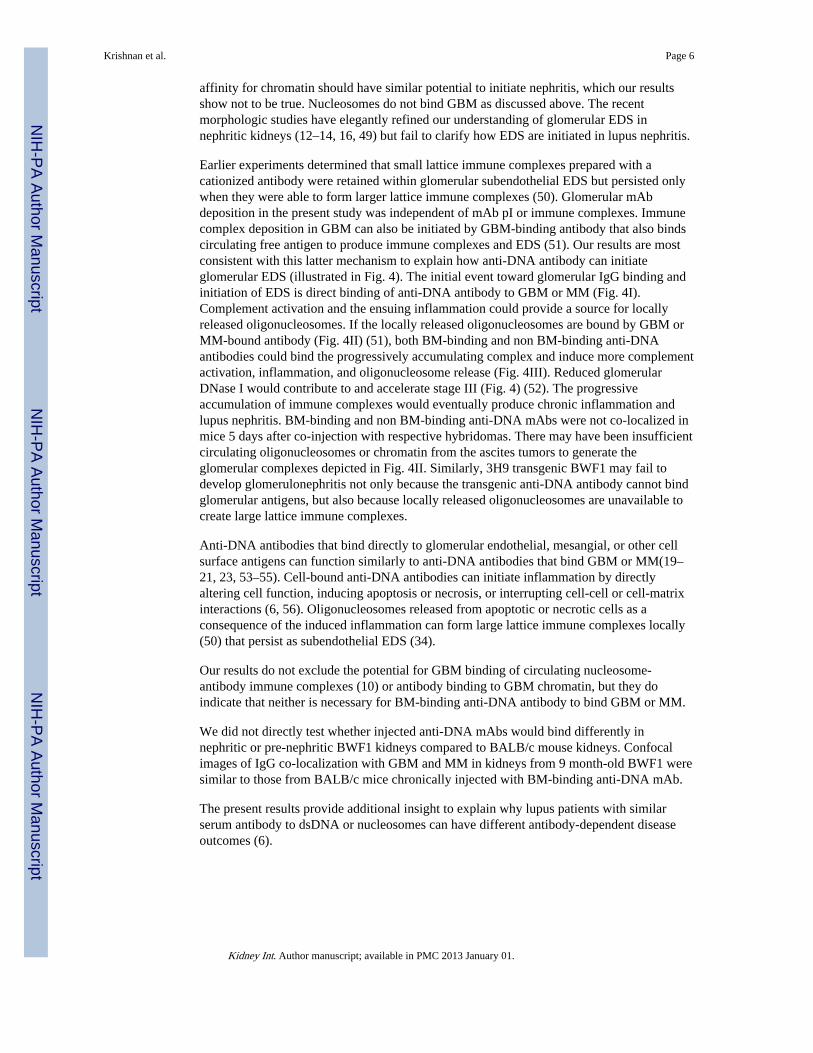
Confocal microscopy indicated that 163p.64 mAb chronically injected over a 3-monthperiod was co-localized with heparan sulfate proteoglycan (HSPG) in GBM and MM butminimally with chromatin (Fig. 3a). Glomerular IgG was also co-localized with HSPG andminimally with chromatin in autoimmune BWF1 kidneys (Fig. 3b). As expected, there wasno glomerular binding of anti-DNA mAb 452s.46 after similar 3-months chronic injection(Fig. 3c). Complement C3 was co-localized with mAb 163p.64 in glomeruli fromchronically injected mice (Fig. 3e). These results indicate that BM-binding anti-DNAantibodies also bind directly to MM and GBM antigens independently of DNA, chromatin,or nucleosomes and initiate complement activation. The small regions of chromatin and IgGco-localization and perlecan, chromatin, and IgG co-localization in the kidneys from BALB/c mice chronically injected with 163p.64 mAb (Fig. 3a) were more numerous in kidneysfrom autoimmune BWF1 (Fig. 3b). Those regions of co-localization may be the glomerularEDS identified by electron microscopy in kidneys from autoimmune BWF1 (13) and anti-DNA mAb-injected mice (11).
Only BM-binding anti-DNA mAbs induce proteinuria in non autoimmune-prone miceAscites tumors were induced in non-autoimmune BALB/c mice by injecting hybridoma cellseither individually or as co-injected pairs, one producing IgG2a and the other, IgG2b (Table3). Only mice injected with hybridomas producing mAbs that bound BM, 163p.64, 77, or124 or DNA 5 or 6, had glomerular-bound IgG of the expected IgG subclass and moderate
Krishnan et al. Page 3
Kidney Int. Author manuscript; available in PMC 2013 January 01.
NIH
-PA Author Manuscript
NIH
-PA Author Manuscript
NIH
-PA Author Manuscript
to severe proteinuria 5 days after hybridoma injection. Mice injected with 163p.64 or 163p.124, IgG2a hybridoma cells with either 452s.46 or 163p.132, IgG2b hybridoma cells hadonly glomerular-bound IgG2a. Glomerular IgG binding was not IgG2 subclass dependent,nor was glomerular binding simply a correlate of circulating mAb titers. The average serumanti-DNA titer after 5 days was 25,568 (range 12,000 – 36,000) for glomerular-bound mAbsand 31,272 (range 14,000 – 41,000) for mAbs that did not bind in glomeruli. Only BM-binding mAbs initiated glomerular disease detected as proteinuria.
Gilkeson et al. (31) observed that mice injected with163p.77 and 163p.132 hybridoma cellsdeveloped glomerular IgG deposits and proteinuria after the injected mice developedpronounced ascites. The results with 163p.77 are similar to those in Fig. 2 and Table 3. Weextended the time before euthanasia of mice injected with 163p.132 from 5 days to 8 daysand observed similar results to those of Gilkeson et al. After 8 days mice injected with 163p.132 cells had glomerular IgG deposits (Fig. 3f) and moderate proteinuria (Table 3). Thedifference between 163p.132 injected mice at 5 and 8 days is likely a consequence of muchhigher mAb serum titer after 8 days. MAb 163p.132 does bind to BM but with 300-fold lessrelative affinity than mAb 163p.64 (Table 2). Alternatively 163p.132 mAb deposition after 8days may have been due to circulating immune complexes. There was co-localization of162p.132 mAb with DNA (yellow pixels in Fig. 3f) although most of the glomerular 163p.132 IgG was not co-localized with DNA.
DiscussionThe present results demonstrate that some but not all anti-DNA mAbs bind directly to BMantigens and that direct binding of anti-dsDNA antibody to GBM or MM is critical for theinitiation of experimental lupus nephritis. Glomerular binding of IgG and complement andthe initiation of glomerular disease, identified as proteinuria, were independent of mAbbinding to DNA or chromatin whether the mAbs were injected or produced in situ. MAbbinding to GBM and MM was correlated with relative affinity for dsDNA but independentof binding to DNA or chromatin. Only anti-dsDNA mAbs that bound BM antigens bound toGBM and MM in vivo. These results and conclusion are consistent with previous reportsthat anti-dsDNA antibodies may initiate glomerulonephritis after binding directly toglomerular antigens (18–24). The results and conclusion contrast with results (10–14, 16,17, 32) interpreted to indicate that anti-DNA antibodies can only bind to GBM or MM asimmune complexes of anti-DNA antibody and nucleosomes or by binding to chromatinalready bound to GBM or MM (28–30). The results from co-injection of mice with ahybridoma producing a BM-binding anti-DNA mAb with a hybridoma producing a nonBM-binding anti-DNA mAb are difficult to reconcile with the previous interpretation. MAbsproduced by the co-injected hybridomas had similar relative affinity for DNA, nucleosomes,or chromatin, but only the mAbs that bound BM also bound glomeruli in vivo. The resultscannot be explained by potential absence of circulating nucleosomes or chromatin in nonautoimmune-prone BALB/c mice. Circulating or glomerular-bound chromatin ornucleosomes, including that released from necrotic or apoptotic hybridomas, would havebeen equally accessible to the two mAbs.
The present results may explain why autoimmune female BWF1 transgenic for VH of the3H9 anti-DNA mAb (33, 34) do not develop nephritis (35). 3H9 mAb binds DNA andchromatin (36) but does not bind BM. Autoimmune, 3H9 VH transgenic BWF1 had similarserum IgG2a/b anti-DNA titers as non-transgenic BWF1 of similar age but did not developproteinuria even after 1 year of age. Similar outcome was reported for D42 VH (37) and 3–32 μ (38) transgenic BWF1. Non-transgenic, female BWF1 invariably produce anti-DNAautoantibody and develop glomerulonephritis with proteinuria by 10 months of age (1).BALB/c mice injected with the 3H9 hybridoma had relatively low glomerular
Krishnan et al. Page 4
Kidney Int. Author manuscript; available in PMC 2013 January 01.
NIH
-PA Author Manuscript
NIH
-PA Author Manuscript
NIH
-PA Author Manuscript

immunofluorescence and disease scores compared with mice injected with 163p.77 or 163p.132 hybridomas (31). The majority of anti-DNA hybridomas from VH3H9 transgenic BWF1had VH3H9 H chains (39). Likely those mAbs could not bind BM and could not initiatedisease.
Essentially three experimental systems have described nucleosome-dependent glomerularbinding of anti-DNA antibodies. Schmiedeke et al. (32) and Termaat et al. (17) allowedsoluble DNA to bind to histones after the histones were perfused into kidneys or added toisolated glomeruli or GBM. Anti-DNA mAb bound to the immobilized DNA but not toGBM, histone-coated GBM, or DNA added to GBM. Although interesting, the experimentsdo not accurately reflect the physical chemical properties of intact nucleosomes, nor hownucleosomes or chromatin may interact with GBM or MM. Kramers et al. (10) reported thatpurified anti-DNA mAbs perfused into kidneys may only bind in glomeruli as immunecomplexes with histones or nucleosomes, presenting as example mAb 32. Nucleosomes inthe immune complexes were presumed to promote binding to GBM through histone-dependent charge interaction. Nucleosomes in physiological saline have a net negativecharge with more exposed acidic than basic regions (40, 41). The basic termini of H2B andH3 that protrude from the octamer cores through the DNA superhelix bind with the acidicpatches on the octamer surface of consecutive nucleosomes and with linker DNA toorganize the nucleosomes into chromatin (40, 42). Nucleosomal organization into chromatinprecludes surface availability of positive charge contributed by histones (41). The net chargeof the GBM lamina rara interna and externa initially accessible to chromatin or nucleosomesis anionic (43, 44) and unlikely to promote binding. Although nucleosomes bound isolatedcollagen IV, laminin (15), and agrin (16) on laboratory sensor chips, radiolabelednucleosomes (45, 46) were rapidly cleared from blood into the liver with insignificantlocalization to kidneys unless nucleosome injections were preceded by injection of solublehistones (45). DNA-anti-DNA immune complexes were likewise rapidly cleared from thecirculation (47, 48). Perfusion into the renal artery (10) would bypass initial circulation tothe liver. An alternative explanation for why mAb 32-nucleosome immune complexesbound GBM, but mAb alone did not, might be that the mAb 32 in nucleosome immunecomplexes had increased relative avidity for GBM. The mAb 32-nucleosome immunecomplexes were created at a 15:1 molar ratio of mAb to mononucleosome (10). Multipleunbound antibody combining sites in mAb 32-nucleosome immune complexes prepared inantibody excess may have created higher avidity of the complexes for GBM than mAb 32alone. The DNA, nucleosome, and BM binding characteristics of mAb 32 were similar tothose for mAb 163p.132 in the present study. MAb 163p.132 bound glomeruli only afterreaching a serum concentration of ~10 mg/ml. MAbs 163p.64 and 163p.77 that bind withhigh relative affinity to BM, both bound glomeruli at serum concentrations of ≤720 μg/ml.MAb 163p.132 binds BM but with low relative affinity. Alternatively, the additional 3 daysof 163p.132 hybridoma growth from 5 to 8 days may have produced sufficient chromatin ornucleosomes from dying cells to produce immune complexes, likely in mAb excess. Therewas more glomerular co-localization of DNA with 163p.132 mAb than with the BM-binding163p.64 mAb.
GBM-associated EDS in kidneys from nephritic BWF1 (13), nephritic lupus patients (12),and BALB/c mice chronically injected with an anti-DNA mAb (11) contained bothchromatin and IgG. The EDS chromatin was presumed to have originated from mesangialcells undergoing apoptosis (13). The released chromatin was presumed to bind GBM andpresent target antigens to chromatin-binding antibody. Caspace 3-positive mesangial cellswere detected in kidneys from nephritic but not pre-nephritic BWF1 (13), and chromatinwas never detected in EDS that did not also contain IgG (11, 13). Direct binding ofnucleosomes or chromatin to GBM was not tested. If chromatin binding to GBM determineswhen and where anti-DNA antibody binds GBM to initiate EDS, mAbs with similar relative
Krishnan et al. Page 5
Kidney Int. Author manuscript; available in PMC 2013 January 01.
NIH
-PA Author Manuscript
NIH
-PA Author Manuscript
NIH
-PA Author Manuscript
affinity for chromatin should have similar potential to initiate nephritis, which our resultsshow not to be true. Nucleosomes do not bind GBM as discussed above. The recentmorphologic studies have elegantly refined our understanding of glomerular EDS innephritic kidneys (12–14, 16, 49) but fail to clarify how EDS are initiated in lupus nephritis.
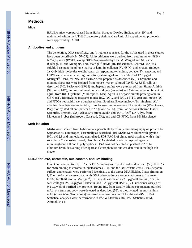
Earlier experiments determined that small lattice immune complexes prepared with acationized antibody were retained within glomerular subendothelial EDS but persisted onlywhen they were able to form larger lattice immune complexes (50). Glomerular mAbdeposition in the present study was independent of mAb pI or immune complexes. Immunecomplex deposition in GBM can also be initiated by GBM-binding antibody that also bindscirculating free antigen to produce immune complexes and EDS (51). Our results are mostconsistent with this latter mechanism to explain how anti-DNA antibody can initiateglomerular EDS (illustrated in Fig. 4). The initial event toward glomerular IgG binding andinitiation of EDS is direct binding of anti-DNA antibody to GBM or MM (Fig. 4I).Complement activation and the ensuing inflammation could provide a source for locallyreleased oligonucleosomes. If the locally released oligonucleosomes are bound by GBM orMM-bound antibody (Fig. 4II) (51), both BM-binding and non BM-binding anti-DNAantibodies could bind the progressively accumulating complex and induce more complementactivation, inflammation, and oligonucleosome release (Fig. 4III). Reduced glomerularDNase I would contribute to and accelerate stage III (Fig. 4) (52). The progressiveaccumulation of immune complexes would eventually produce chronic inflammation andlupus nephritis. BM-binding and non BM-binding anti-DNA mAbs were not co-localized inmice 5 days after co-injection with respective hybridomas. There may have been insufficientcirculating oligonucleosomes or chromatin from the ascites tumors to generate theglomerular complexes depicted in Fig. 4II. Similarly, 3H9 transgenic BWF1 may fail todevelop glomerulonephritis not only because the transgenic anti-DNA antibody cannot bindglomerular antigens, but also because locally released oligonucleosomes are unavailable tocreate large lattice immune complexes.
Anti-DNA antibodies that bind directly to glomerular endothelial, mesangial, or other cellsurface antigens can function similarly to anti-DNA antibodies that bind GBM or MM(19–21, 23, 53–55). Cell-bound anti-DNA antibodies can initiate inflammation by directlyaltering cell function, inducing apoptosis or necrosis, or interrupting cell-cell or cell-matrixinteractions (6, 56). Oligonucleosomes released from apoptotic or necrotic cells as aconsequence of the induced inflammation can form large lattice immune complexes locally(50) that persist as subendothelial EDS (34).
Our results do not exclude the potential for GBM binding of circulating nucleosome-antibody immune complexes (10) or antibody binding to GBM chromatin, but they doindicate that neither is necessary for BM-binding anti-DNA antibody to bind GBM or MM.
We did not directly test whether injected anti-DNA mAbs would bind differently innephritic or pre-nephritic BWF1 kidneys compared to BALB/c mouse kidneys. Confocalimages of IgG co-localization with GBM and MM in kidneys from 9 month-old BWF1 weresimilar to those from BALB/c mice chronically injected with BM-binding anti-DNA mAb.
The present results provide additional insight to explain why lupus patients with similarserum antibody to dsDNA or nucleosomes can have different antibody-dependent diseaseoutcomes (6).
Krishnan et al. Page 6
Kidney Int. Author manuscript; available in PMC 2013 January 01.
NIH
-PA Author Manuscript
NIH
-PA Author Manuscript
NIH
-PA Author Manuscript
MethodsMice
BALB/c mice were purchased from Harlan Sprague-Dawley (Indianapolis, IN) andmaintained within the UTHSC Laboratory Animal Care Unit. All experimental protocolswere approved by IACUC.
Antibodies and antigensThe generation, DNA specificity, and V-region sequences for the mAbs used in these studieshave been described (34, 57–59). All hybridomas were derived from autoimmune (NZB ×NZW)F1 mice (BWF1) except 3H9 (34) provided by Drs. M. Weigert and M. Radic(Chicago, IL and Memphis, TN). Matrigel® (BM) (BD Biosciences, Bedford, MA) is asoluble basement membrane matrix of laminin, collagen IV, HSPG, and entactin (nidogen1). Only high molecular weight bands corresponding to laminin, collagen IV, entactin, andHSPG were detected after high sensitivity staining of an SDS-PAGE of 12.5 μg ofMatrigel®. DNA, ssDNA, and dsDNA were prepared as described (58). Chromatin andmononucleosomes were isolated from mouse liver or cultured P3x63-Ag8.653 cells asdescribed (60). Perlecan (HSPG2) and heparan sulfate were purchased from Sigma-Aldrich(St. Louis, MO), and recombinant human nidogen (entactin) and C-terminal recombinant ratagrin, from R&D Systems, (Minneapolis, MN). Agrin is a heparin sulfate proteoglycan inGBM (61). Biotinylated goat anti-mouse IgG, IgG2a, and IgG2b; FITC-goat anti-mouse IgG;and FITC-streptavidin were purchased from Southern Biotechnology (Birmingham, AL);alkaline phosphatase-streptavidin, from Jackson Immunoresearch Laboratories (West Grove,PA); biotinylated rat anti-perlecan mAb (clone A7L6), from Lab Vision (Thermo FisherScientific, Fremont, CA); Alexa 546-strepatavidin and TO-PRO3® DNA dye, fromMolecular Probes (Invitrogen, Carlsbad, CA); and anti-C3-FITC, from BD Bioscience.
MAb isolationMAbs were isolated from hybridoma supernatants by affinity chromatography on protein G-Sepharose 4B (Invitrogen) essentially as described (10). MAbs were eluted with glycine-HCl, pH 2.8 and immediately neutralized. SDS-PAGE of eluted mAbs stained with a highsensitivity Coomassie (Biorad, Hercules, CA) yielded bands corresponding only toimmunoglobulin H and L polypeptides. DNA was not detected in purified mAbs byethidium bromide staining after agarose electrophoresis but was detected in the high salteluate.
ELISA for DNA, chromatin, nucleosome, and BM bindingDirect and competitive ELISAs for DNA binding were performed as described (59). ELISAsfor mAb binding to chromatin, nucleosomes, BM, and the BM constituents HSPG, heparansulfate, and entactin were performed identically to the direct DNA ELISA. Plates (ImmulonI, Thermo-Fisher) were coated with DNA, chromatin or mononucleosomes at 1 μg/wellDNA; 1/250 dilution of Matrigel®, ~5 μg/well, estimated as 2.8 μg/well laminin, 1.5 μg/well collagen IV, 0.4 μg/well entactin, and 0.25 μg/well HSPG (BD Bioscience assay); or0.2 μg/well of purified BM proteins. Bound IgG from serially diluted supernatant, purifiedmAb, or serum antibody were detected as described (59). A biotinylated rat anti-lamininmAb (clone A5) (Neomarkers) was used as a positive control for the anti-BM ELISA.Statistical analyses were performed with PASW Statistics 18 (SPSS Statistics, IBM,Armonk, NY).
Krishnan et al. Page 7
Kidney Int. Author manuscript; available in PMC 2013 January 01.
NIH
-PA Author Manuscript
NIH
-PA Author Manuscript
NIH
-PA Author Manuscript

In vivo glomerular binding of anti-DNA mAb and measurement of proteinuriaBALB/c mice, eight-to-twelve weeks old, were injected once intravenously with 1 mg of asingle, purified mAb or 1 mg each of two purified mAbs, one IgG2a, the other IgG2b.Twenty-four hours later injected mice were euthanized and their kidneys removed and snapfrozen in OCT embedding medium (Tissue-Tek, Miles Laboratories, Elkhart, IN). Serial oneμm cryosections were fluorescently stained with biotinylated goat anti-mouse IgG2a orIgG2b and FITC-streptavidin. In separate experiments, mice were chronically injected with100 μg per intraperitoneal (ip) injection of a single mAb twice weekly for 3 months orinjected ip with hybridoma cells 5–7 days after ip injection with 0.5 ml pristane (Sigma).The hybridoma injection consisted of 107 cells from one hybridoma or 107 cells each fromtwo hybridomas, one producing IgG2a and the other, IgG2b. Kidneys were removed andembedded for cryosection after 3 months chronic injection of purified mAb or 1–5 days afterhybridoma injection. Serial cryosections from the same kidney were stained for detection ofmouse IgG2a or IgG2b. For confocal microscopy 4–12 μm cryosections were stained withTO-PRO3 for DNA, goat anti-mouse IgG-FITC, and rat anti-perlecan and streptavidin Alexa546 or anti-C3-FITC and biotinylated goat anti-mouse IgG and streptavidin-Alexa 546.Confocal images were collected with a Zeiss LSM510 confocal microscope (Carl ZeissMicroimaging, Thornwood, NY). Proteinuria was measured with Ames Uristix (Miles)according to manufacturer’s instructions.
AcknowledgmentsThe research was supported by NIH NIAID grant AI26833, NIH NCRR grant RR301812, and a grant from theUTHSC Center of Excellence for Diseases of Connective Tissue.
The authors wish to acknowledge Ryle Holder for technical assistance in mAb purification, Tim Higgins for help infigure preparations, Dr. Michael Madaio for help in interpreting glomerular immunofluorescence, and theMicrobiology, Immunology, and Biochemistry Confocal Microscope Facility for assistance with confocalmicroscopy. We also acknowledge Drs. David Isenberg, Marc Monestier, Marko Radic, and Ole Petter Rekvig forcritical commentary on the manuscript.
Abbreviations
BWF1 (NZB × NZW)F1 mice
dsDNA native, double-stranded DNA
ssDNA denatured, single-stranded DNA
BM basement membrane
GBM glomerular basement membrane
mAb monoclonal antibody
MM glomerular mesangial matrix
EDS electron dense substance, electron dense region, electron dense deposit
HSPG heparan sulfate proteoglycan
References1. Andrews BS, Eisenberg RA, Theofilopoulos AN, et al. Spontaneous murine lupus-like syndromes.
Clinical and immunopathological manifestations in several strains. J Exp Med. 1978; 148:1198–215. [PubMed: 309911]
2. Koffler D, Schur PH, Kunkel HG. Immunological studies concerning the nephritis of systemic lupuserythematosus. J Exp Med. 1967; 126:607–24. [PubMed: 4168098]
Krishnan et al. Page 8
Kidney Int. Author manuscript; available in PMC 2013 January 01.
NIH
-PA Author Manuscript
NIH
-PA Author Manuscript
NIH
-PA Author Manuscript
3. Isenberg DA. Autoantibodies: markers of disease or pathogenic? Ann N Y Acad Sci. 1997;823:256–62. [PubMed: 9292052]
4. Manson JJ, Ma A, Rogers P, et al. Relationship between anti-dsDNA, anti-nucleosome and anti-alpha-actinin antibodies and markers of renal disease in patients with lupus nephritis: a prospectivelongitudinal study. Arthritis Res Ther. 2009; 11:R154. [PubMed: 19828047]
5. Gladman DD, Urowitz MB, Keystone EC. Serologically active clinically quiescent systemic lupuserythematosus: a discordance between clinical and serological features. Am J Med. 1979; 66:210–5.[PubMed: 218447]
6. Madaio MP. The role of autoantibodies in the pathogenesis of lupus nephritis. Semin Nephrol. 1999;19:48–56. [PubMed: 9952280]
7. Isenberg DA, Manson JJ, Ehrenstein MR, et al. Fifty years of anti-ds DNA antibodies: are weapproaching journey’s end? Rheumatology (Oxford). 2007; 46:1052–6. [PubMed: 17500073]
8. Dixon FJ, Oldstone MB, Tonietti G. Pathogenesis of immune complex glomerulonephritis of newzealand mice. J Exp Med. 1971; 134:65–71. [PubMed: 19867382]
9. Morioka T, Woitas R, Fujigaki Y, et al. Histone mediates glomerular deposition of small size DNAanti-DNA complex. Kidney Int. 1994; 45:991–7. [PubMed: 8007603]
10. Kramers C, Hylkema MN, van Bruggen MC, et al. Anti-nucleosome antibodies complexed tonucleosomal antigens show anti-DNA reactivity and bind to rat glomerular basement membrane invivo. J Clin Invest. 1994; 94:568–77. [PubMed: 8040312]
11. Fenton KA, Tommeras B, Marion TN, et al. Pure anti-dsDNA mAbs need chromatin structures topromote glomerular mesangial deposits in BALB/c mice. Autoimmunity. 2010; 43:179–88.[PubMed: 19835488]
12. Kalaaji M, Fenton KA, Mortensen ES, et al. Glomerular apoptotic nucleosomes are central targetstructures for nephritogenic antibodies in human SLE nephritis. Kidney Int. 2007; 71:664–72.[PubMed: 17332738]
13. Kalaaji M, Mortensen E, Jorgensen L, et al. Nephritogenic lupus antibodies recognize glomerularbasement membrane-associated chromatin fragments released from apoptotic intraglomerularcells. Am J Pathol. 2006; 168:1779–92. [PubMed: 16723695]
14. Mjelle JE, Kalaaji M, Rekvig OP. Exposure of chromatin and not high affinity for dsDNAdetermines the nephritogenic impact of anti-dsDNA antibodies in (NZBxNZW)F1 mice.Autoimmunity. 2009; 42:104–11. [PubMed: 19005880]
15. Mjelle JE, Rekvig OP, Fenton KA. Nucleosomes possess a high affinity for glomerular lamininand collagen IV and bind nephritogenic antibodies in murine lupus-like nephritis. Ann Rheum Dis.2007; 66:1661–8. [PubMed: 17504842]
16. Mjelle JE, Rekvig OP, Van Der Vlag J, et al. Nephritogenic antibodies bind in glomeruli throughinteraction with exposed chromatin fragments and not with renal cross-reactive antigens.Autoimmunity. 2011; 44:373–83. [PubMed: 21244336]
17. Termaat RM, Assmann KJ, Dijkman HB, et al. Anti-DNA antibodies can bind to the glomerulusvia two distinct mechanisms. Kidney Int. 1992; 42:1363–71. [PubMed: 1474767]
18. Faaber P, Rijke TP, van de Putte LB, et al. Cross-reactivity of human and murine anti-DNAantibodies with heparan sulfate. The major glycosaminoglycan in glomerular basementmembranes. J Clin Invest. 1986; 77:1824–30. [PubMed: 2940265]
19. Madaio MP, Carlson J, Cataldo J, et al. Murine monoclonal anti-DNA antibodies bind directly toglomerular antigens and form immune deposits. J Immunol. 1987; 138:2883–9. [PubMed:3553329]
20. Raz E, Brezis M, Rosenmann E, et al. Anti-DNA antibodies bind directly to renal antigens andinduce kidney dysfunction in the isolated perfused rat kidney. J Immunol. 1989; 142:3076–82.[PubMed: 2785132]
21. Raz E, Ben-Bassat H, Davidi T, et al. Cross-reactions of anti-DNA autoantibodies with cell surfaceproteins. Eur J Immunol. 1993; 23:383–90. [PubMed: 7679642]
22. Vlahakos DV, Foster MH, Adams S, et al. Anti-DNA antibodies form immune deposits at distinctglomerular and vascular sites. Kidney Int. 1992; 41:1690–700. [PubMed: 1501424]
Krishnan et al. Page 9
Kidney Int. Author manuscript; available in PMC 2013 January 01.
NIH
-PA Author Manuscript
NIH
-PA Author Manuscript
NIH
-PA Author Manuscript

23. D’Andrea DM, Coupaye-Gerard B, Kleyman TR, et al. Lupus autoantibodies interact directly withdistinct glomerular and vascular cell surface antigens. Kidney Int. 1996; 49:1214–21. [PubMed:8731084]
24. Sabbaga J, Line SRP, Potocnjak P, et al. A murine nephritogenic monoclonal anti-DNAautoantibody binds directly to mouse laminin, the major non-collagenous protein component of theglomerular basement membrane. Eur J Immunol. 1989; 19:137–43. [PubMed: 2784103]
25. Mostoslavsky G, Fischel R, Yachimovich N, et al. Lupus anti-DNA autoantibodies cross-react witha glomerular structural protein: a case for tissue injury by molecular mimicry. Eur J Immunol.2001; 31:1221–7. [PubMed: 11298348]
26. Farquhar MG, Vernier RL, Good RA. An electron microscope study of the glomerulus innephrosis, glomerulonephritis, and lupus erythematosus. J Exp Med. 1957; 106:649–60. [PubMed:13475621]
27. Channing AA, Kasuga T, Horowitz RE, et al. An ultrastructural study of spontaneous lupusnephritis in the NZB-BL-NZW mouse. Am J Pathol. 1965; 47:677–94. [PubMed: 5837733]
28. Fenton KA, Rekvig OP. A central role of nucleosomes in lupus nephritis. Ann N Y Acad Sci.2007; 1108:104–13. [PubMed: 17893976]
29. van Bavel CC, Fenton KA, Rekvig OP, et al. Glomerular targets of nephritogenic autoantibodies insystemic lupus erythematosus. Arthritis Rheum. 2008; 58:1892–9. [PubMed: 18576314]
30. van Bavel CC, van der Vlag J, Berden JH. Glomerular binding of anti-dsDNA autoantibodies: thedispute resolved? Kidney Int. 2007; 71:600–1. [PubMed: 17387307]
31. Gilkeson GS, Bernstein K, Pippen AM, et al. The influence of variable-region somatic mutationson the specificity and pathogenicity of murine monoclonal anti-DNA antibodies. Clin ImmunolImmunopathol. 1995; 76:59–67. [PubMed: 7606869]
32. Schmiedeke TM, Stockl FW, Weber R, et al. Histones have high affinity for the glomerularbasement membrane. Relevance for immune complex formation in lupus nephritis. J Exp Med.1989; 169:1879–94. [PubMed: 2732675]
33. Chen C, Nagy Z, Prak EL, et al. Immunoglobulin heavy chain gene replacement: a mechanism ofreceptor editing. Immunity. 1995; 3:747–55. [PubMed: 8777720]
34. Shlomchik MJ, Aucoin AH, Pisetsky DS, et al. The structure and function of anti-DNAautoantibodies derived from a single autoimmune mouse. Proc Natl Acad Sci USA. 1987;84:9150–4. [PubMed: 3480535]
35. Steeves MA, Marion TN. Tolerance to DNA in (NZB x NZW)F1 mice that inherit an anti-DNAV(H) as a conventional micro H chain transgene but not as a V(H) knock-in transgene. J Immunol.2004; 172:6568–77. [PubMed: 15153471]
36. Neeli I, Richardson MM, Khan SN, et al. Divergent members of a single autoreactive B cell cloneretain specificity for apoptotic blebs. Mol Immunol. 2007; 44:1914–21. [PubMed: 17084454]
37. Friedmann D, Yachimovich N, Mostoslavsky G, et al. Production of high affinity autoantibodies inautoimmune New Zealand Black/New Zealand white F1 mice targeted with an anti-DNA heavychain. J Immunol. 1999; 162:4406–16. [PubMed: 10201976]
38. Wellmann U, Letz M, Schneider A, et al. An Ig mu-heavy chain transgene inhibits systemic lupuserythematosus immunopathology in autoimmune (NZB x NZW)F1 mice. Int Immunol. 2001;13:1461–9. [PubMed: 11717187]
39. Steeves, MA. Tolerance and Autoimmunity in (NZB x NZW)F1 Mice Transgenic for Anti- DNAAntibody. Memphis: The University of Tennessee Health Science Center; 2005.
40. Luger K, Mader AW, Richmond RK, et al. Crystal structure of the nucleosome core particle at 2.8A resolution. Nature. 1997; 389:251–60. [PubMed: 9305837]
41. Materese CK, Savelyev A, Papoian GA. Counterion atmosphere and hydration patterns near anucleosome core particle. J Am Chem Soc. 2009; 131:15005–13. [PubMed: 19778017]
42. Schalch T, Duda S, Sargent DF, et al. X-ray structure of a tetranucleosome and its implications forthe chromatin fibre. Nature. 2005; 436:138–41. [PubMed: 16001076]
43. Kanwar YS, Farquhar MG. Anionic sites in the glomerular basement membrane. In vivo and invitro localization to the laminae rarae by cationic probes. J Cell Biol. 1979; 81:137–53. [PubMed:90048]
Krishnan et al. Page 10
Kidney Int. Author manuscript; available in PMC 2013 January 01.
NIH
-PA Author Manuscript
NIH
-PA Author Manuscript
NIH
-PA Author Manuscript

44. Caulfield JP, Farquhar MG. Distribution of annionic sites in glomerular basement membranes:their possible role in filtration and attachment. Proc Natl Acad Sci U S A. 1976; 73:1646–50.[PubMed: 1064037]
45. Gauthier VJ, Tyler LN, Mannik M. Blood clearance kinetics and liver uptake of mononucleosomesin mice. J Immunol. 1996; 156:1151–6. [PubMed: 8557992]
46. Rumore P, Muralidhar B, Lin M, et al. Haemodialysis as a model for studying endogenous plasmaDNA: oligonucleosome-like structure and clearance. Clin Exp Immunol. 1992; 90:56–62.[PubMed: 1395101]
47. Ben Chetrit E, Dunsky EH, Wollner S, et al. In vivo clearance and tissue uptake of an anti-DNAmonoclonal antibody and its complexes with DNA. Clin Exp Immunol. 1985; 60:159–68.[PubMed: 3874013]
48. Emlen W, Mannik M. Clearance of circulating DNA-anti-DNA immune complexes in mice. J ExpMed. 1982; 155:1210–5. [PubMed: 7061954]
49. Fenton K, Fismen S, Hedberg A, et al. Anti-dsDNA antibodies promote initiation, and acquiredloss of renal Dnase1 promotes progression of lupus nephritis in autoimmune (NZBxNZW)F1mice. PLoS One. 2009; 4:e8474. [PubMed: 20041189]
50. Gauthier VJ, Mannik M, Striker GE. Effect of cationized antibodies in performed immunecomplexes on deposition and persistence in renal glomeruli. J Exp Med. 1982; 156:766–77.[PubMed: 7108443]
51. Agodoa LY, Gauthier VJ, Mannik M. Antibody localization in the glomerular basement membranemay precede in situ immune deposit formation in rat glomeruli. J Immunol. 1985; 134:880–4.[PubMed: 3880792]
52. Seredkina N, Zykova SN, Rekvig OP. Progression of murine lupus nephritis is linked to acquiredrenal Dnase1 deficiency and not to up-regulated apoptosis. Am J Pathol. 2009; 175:97–106.[PubMed: 19528352]
53. Lafer EM, Rauch J, Andrzejewski C Jr, et al. Polyspecific monoclonal lupus autoantibodiesreactive with both polynucleotides and phospholipids. J Exp Med. 1981; 153:897–909. [PubMed:6972993]
54. Qing X, Pitashny M, Thomas DB, et al. Pathogenic anti-DNA antibodies modulate gene expressionin mesangial cells: involvement of HMGB1 in anti-DNA antibody-induced renal injury. ImmunolLett. 2008; 121:61–73. [PubMed: 18822317]
55. Qing X, Zavadil J, Crosby MB, et al. Nephritogenic anti-DNA antibodies regulate gene expressionin MRL/lpr mouse glomerular mesangial cells. Arthritis Rheum. 2006; 54:2198–210. [PubMed:16804897]
56. Madaio MP. Lupus autoantibodies 101: one size does not fit all; however, specificity influencespathogenicity. Clin Exp Immunol. 2003; 131:396–7. [PubMed: 12605690]
57. Krishnan MR, Marion TN. Structural similarity of antibody variable regions from immune andautoimmune anti-DNA antibodies. J Immunol. 1993; 150:4948–57. [PubMed: 8496596]
58. Marion TN, Lawton ARd, Kearney JF, et al. Anti-DNA autoantibodies in (NZB X NZW)F1 miceare clonally heterogeneous, but the majority share a common idiotype. J Immunol. 1982; 128:668–74. [PubMed: 7198664]
59. Tillman DM, Jou NT, Hill RJ, et al. Both IgM and IgG anti-DNA antibodies are the products ofclonally selective B cell stimulation in (NZB x NZW)F1 mice. J Exp Med. 1992; 176:761–79.[PubMed: 1512540]
60. Rekvig OP, Hannestad K. The specificity of human autoantibodies that react with both cell nucleiand plasma membranes: the nuclear antigen is present on core mononucleosomes. J Immunol.1979; 123:2673–81. [PubMed: 91640]
61. Raats CJ, Bakker MA, Hoch W, et al. Differential expression of agrin in renal basementmembranes as revealed by domain-specific antibodies. J Biol Chem. 1998; 273:17832–8.[PubMed: 9651386]
62. Gauthier VJ, Mannik M. Only the initial binding of cationic immune complexes to glomerularanionic sites is mediated by charge-charge interactions. J Immunol. 1986; 136:3266–71. [PubMed:3958495]
Krishnan et al. Page 11
Kidney Int. Author manuscript; available in PMC 2013 January 01.
NIH
-PA Author Manuscript
NIH
-PA Author Manuscript
NIH
-PA Author Manuscript
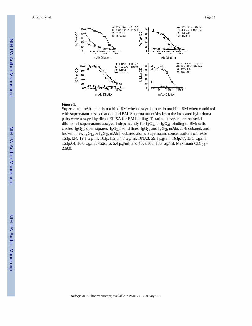
Figure 1.Supernatant mAbs that do not bind BM when assayed alone do not bind BM when combinedwith supernatant mAbs that do bind BM. Supernatant mAbs from the indicated hybridomapairs were assayed by direct ELISA for BM binding. Titration curves represent serialdilution of supernatants assayed independently for IgG2a or IgG2b binding to BM: solidcircles, IgG2a; open squares, IgG2b; solid lines, IgG2a and IgG2b mAbs co-incubated; andbroken lines, IgG2a or IgG2b mAb incubated alone. Supernatant concentrations of mAbs:163p.124, 12.1 μg/ml; 163p.132, 34.7 μg/ml; DNA3, 29.1 μg/ml; 163p.77, 23.5 μg/ml;163p.64, 10.0 μg/ml; 452s.46, 6.4 μg/ml; and 452s.160, 18.7 μg/ml. Maximum OD405 =2.600.
Krishnan et al. Page 12
Kidney Int. Author manuscript; available in PMC 2013 January 01.
NIH
-PA Author Manuscript
NIH
-PA Author Manuscript
NIH
-PA Author Manuscript
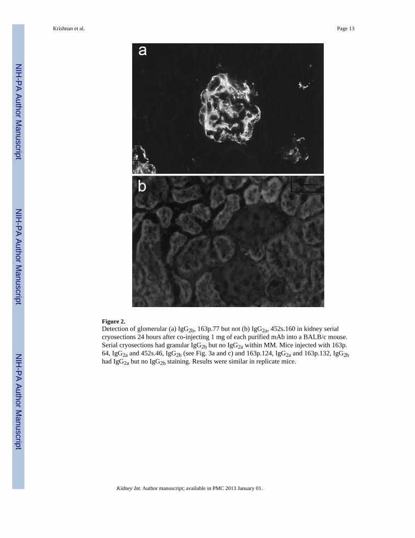
Figure 2.Detection of glomerular (a) IgG2b, 163p.77 but not (b) IgG2a, 452s.160 in kidney serialcryosections 24 hours after co-injecting 1 mg of each purified mAb into a BALB/c mouse.Serial cryosections had granular IgG2b but no IgG2a within MM. Mice injected with 163p.64, IgG2a and 452s.46, IgG2b (see Fig. 3a and c) and 163p.124, IgG2a and 163p.132, IgG2bhad IgG2a but no IgG2b staining. Results were similar in replicate mice.
Krishnan et al. Page 13
Kidney Int. Author manuscript; available in PMC 2013 January 01.
NIH
-PA Author Manuscript
NIH
-PA Author Manuscript
NIH
-PA Author Manuscript

Figure 3.Confocal micrographs of kidney cryosections from an autoimmune BWF1 mouse or BALB/c mice injected with purified anti-DNA mAb. a and e) 100 μg 163p.64 mAb twice weeklyfor 3 months; b) uninjected BWF1, c) 100 μg 452s.46 mAb twice weekly for 3 months; d)uninjected BALB/c; and f) BALB/c with 163p.132 hybridoma-induced ascites 8 days afterhybridoma injection. Images a and b show chromatin as red, perlecan in GBM and MM asdark blue, and IgG as green. Co-localization of IgG with HSPG is clearly identified asturquoise; co-localization of IgG with chromatin, yellow; and co-localization of IgG andchromatin with HSPG, white. The large white arrowheads in a and b indicate areas of IgG,chromatin, and perlecan co-localization. Small arrowheads indicate IgG and chromatin co-localization. Image e shows IgG as red and C3 as green with co-localization of 163p.64mAb and C3 as yellow. Confocal images a and b: 512 pxels2, 180 nm/pixel (92 μm2),optical sections collected at 0.6 μm intervals; c–f: 512 pixels2, 450 nm/pixel (230 μm2),optical sections collected at 0.8 μm intervals (c–e) and 0.5 μm (f). All images are fromoptical sections near the center of respective z-stacks. Replicate mice yielded similar results.
Krishnan et al. Page 14
Kidney Int. Author manuscript; available in PMC 2013 January 01.
NIH
-PA Author Manuscript
NIH
-PA Author Manuscript
NIH
-PA Author Manuscript
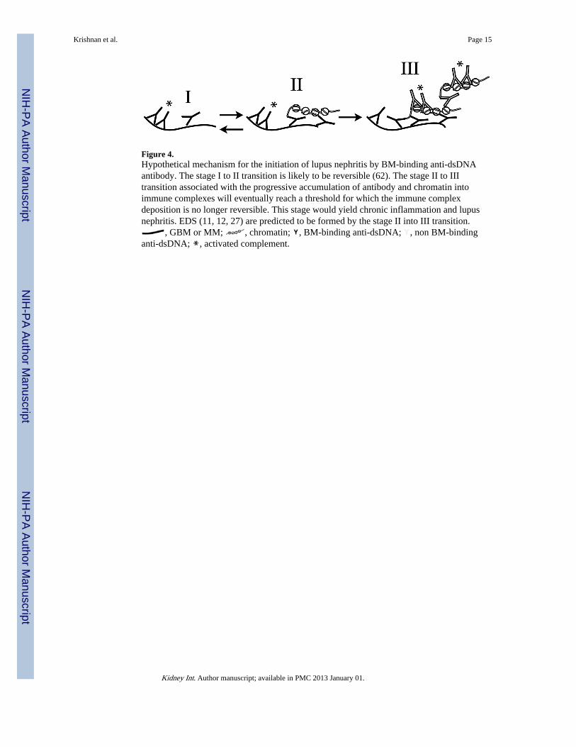
Figure 4.Hypothetical mechanism for the initiation of lupus nephritis by BM-binding anti-dsDNAantibody. The stage I to II transition is likely to be reversible (62). The stage II to IIItransition associated with the progressive accumulation of antibody and chromatin intoimmune complexes will eventually reach a threshold for which the immune complexdeposition is no longer reversible. This stage would yield chronic inflammation and lupusnephritis. EDS (11, 12, 27) are predicted to be formed by the stage II into III transition.
, GBM or MM; , chromatin; , BM-binding anti-dsDNA; , non BM-bindinganti-dsDNA; , activated complement.
Krishnan et al. Page 15
Kidney Int. Author manuscript; available in PMC 2013 January 01.
NIH
-PA Author Manuscript
NIH
-PA Author Manuscript
NIH
-PA Author Manuscript

NIH
-PA Author Manuscript
NIH
-PA Author Manuscript
NIH
-PA Author Manuscript
Krishnan et al. Page 16
Tabl
e 1
Spec
ific
ity o
f M
onoc
lona
l Ant
ibod
ies
Gro
upa
Num
ber
mA
bs
Com
peti
tive
EL
ISA
(ng
/ml c
ompe
tito
r)a
Dir
ect
EL
ISA
(ng
/ml I
gG)b
ssD
NA
dsD
NA
DN
AC
hrom
atin
BM
A14
111
± 8
4dN
I75
± 6
182
6 ±
904
NB
B21
560
± 3
515
770
± 2
580
6 73
0 ±
6 1
101
810
± 2
590
NB
C18
658
± 3
024
200
± 2
580
7 04
0 ±
5 5
6081
± 9
45
510
± 1
490
D16
957
± 4
691
570
± 6
9097
1 ±
1 9
9052
± 6
094
± 7
80
a Sixt
y-ni
ne m
Abs
wer
e st
ratif
ied
acco
rdin
g to
BM
bin
ding
into
(A
) N
B to
BM
(14
mA
b), (
B)
NB
to B
M b
ut b
indi
ng to
dsD
NA
(21
mA
b), (
C)
BM
bin
ding
with
≥ 1
,000
ng/
ml I
gG (
18 m
Abs
), a
nd (
D)
BM
bind
ing
with
≤ 1
,000
ng/
ml I
gG (
16 m
Abs
).
b ng/m
l com
petit
or is
the
amou
nt o
f ds
DN
A o
r ss
DN
A c
ompe
titor
req
uire
d to
pro
duce
50%
inhi
bitio
n of
mA
b bi
ndin
g to
sol
id p
hase
DN
A in
a c
ompe
titiv
e E
LIS
A (
24).
NI
= n
o in
hibi
tion
with
10,
000
ng/
ml c
ompe
titor
.
c ng/m
l mA
b th
at y
ield
s 50
% m
axim
um b
indi
ng in
a d
irec
t EL
ISA
. NB
= n
o bi
ndin
g w
ith ≥
10,0
00 n
g/m
l mA
b.
d The
val
ues
are
mea
ns ±
95%
con
fide
nce
inte
rval
s. A
NO
VA
am
ong
grou
ps f
or th
e ca
tego
ry o
f bi
ndin
g to
: ssD
NA
, p=
0.02
5; d
sDN
A, p
= 0
.033
; DN
A, p
= n
.s.;
chro
mat
in, p
= n
.s.;
BM
, p =
3.6
x 1
0−8 .
Lin
ear
regr
essi
on w
ith B
M-b
indi
ng a
s de
pend
ent v
aria
ble
(R2
= 0
.465
, p =
4.3
x 1
0−8 )
: Chr
omat
in, B
= 0
.381
and
β =
0.2
90, p
= 0
.002
98; s
sDN
A, B
= −
0.49
6 an
d β
= −
.301
, p =
0.0
022;
and
dsD
NA
, B =
0.60
6 an
d β
= 0
.423
, p =
0.0
0010
(B
= s
lope
and
β =
cor
rela
tion
coef
fici
ent,
PASW
Sta
tistic
s18)
.
Kidney Int. Author manuscript; available in PMC 2013 January 01.
NIH
-PA Author Manuscript
NIH
-PA Author Manuscript
NIH
-PA Author Manuscript
Krishnan et al. Page 17
Tabl
e 2
Mon
oclo
nal A
ntib
ody
Bin
ding
to D
NA
, Chr
omat
in, a
nd B
asem
ent M
embr
ane
Ant
igen
s.
mA
baIs
otyp
ebpI
cD
irec
t B
indi
ng E
LIS
Ad
Com
peti
tive
EL
ISA
dIn
viv
o A
ctiv
ity
DN
AC
hrom
atin
Nuc
leos
ome
BM
ssD
NA
dsD
NA
Glo
mer
ular
Bin
ding
eIn
duce
s P
rote
inur
iaf
(ng/
ml I
gG)
(ng/
ml I
gG)
(ng/
ml I
gG)
(μg/
ml I
gG)
(ng/
ml s
sDN
A)
(ng/
ml d
sDN
A)
163p
.64
2a8.
460
8.0
19 7
0030
470
1 03
0Y
esY
es
163p
.77
2b8.
520
3011
500
201
420
700
Yes
Yes
163p
.124
2a8.
430
4.0
1 88
090
1 69
047
0Y
esY
es
DN
A6
2a7.
610
10N
Dd
200
2 90
01
000
Yes
Yes
DN
A5
2a8.
71
000
10N
D3
380
1 50
010
800
Yes
Yes
163p
.132
2b8.
550
1050
8 60
066
0N
IdN
ogN
og
DN
A3
2a6.
511
000
10N
DN
Bd
541
600
No
No
452s
.46
2b7.
610
200
2 30
0N
B73
049
0N
oN
o
452s
.160
2a7.
370
90N
DN
B80
4 40
0N
oN
D
3H9
2b8.
34
720
50N
DN
BN
DN
DN
ohN
oh
a 163p
.64,
77,
124
and
DN
A6:
Gro
up D
, Tab
le 1
; DN
A 5
and
163
p.13
2: G
roup
C, T
able
1; 4
52s.
46, 1
60 a
nd D
NA
3: G
roup
B, T
able
1.
b IgG
sub
clas
s of
hyb
rido
ma
mA
b.
c Isoe
lect
ric
poin
t of
the
resp
ectiv
e m
Ab
(cal
cula
ted
usin
g th
e Sw
iss
Inst
itute
of
Bio
info
rmat
ics
ExP
ASy
pI
calc
ulat
ion
tool
, http
://w
eb.e
xpas
y.or
g/co
mpu
te_p
i/)
d Tab
le 1
lege
nd. N
D =
not
don
e. N
B =
no
bind
ing.
NI
= n
o in
hibi
tion.
e Glo
mer
ular
bin
ding
of
mA
b w
as d
eter
min
ed b
y im
mun
oflu
ores
cenc
e on
kid
ney
cryo
sect
ions
24
hrs
afte
r m
ice
wer
e in
ject
ed w
ith 1
mg
puri
fied
mA
b(s)
.
f Sum
mar
y of
res
ults
pre
sent
ed in
Tab
le 3
.
g MA
b 16
3p.1
32, I
gG2b
, pro
duce
d m
inim
al g
lom
erul
ar f
luor
esce
nce
and
no p
rote
inur
ia 5
day
s af
ter
inje
ctio
n of
hyb
rido
ma
cells
but
rea
dily
det
ecte
d im
mun
oflu
ores
cenc
e an
d pr
otei
nuri
a 8
days
aft
er
inje
ctio
n.
h Gilk
eson
et a
l. (3
6). O
nly
2/5
mic
e ha
d gl
omer
ular
-bou
nd I
gG, a
nd th
e gl
omer
ular
dis
ease
sco
re w
as n
ot d
iffe
rent
fro
m th
e ne
gativ
e co
ntro
l.
Kidney Int. Author manuscript; available in PMC 2013 January 01.

NIH
-PA Author Manuscript
NIH
-PA Author Manuscript
NIH
-PA Author Manuscript
Krishnan et al. Page 18
Tabl
e 3
Hyb
rido
mas
pro
duci
ng B
M-b
indi
ng m
Ab
indu
ce p
rote
inur
ia.
Hyb
rido
ma(
s) I
njec
ted
mA
b Is
otyp
eG
lom
erul
ar I
soty
peb
Day
scA
nti-
DN
A S
erum
Tit
erP
rote
inur
ia
(2a/
2b)d
(mg/
dl)e
163p
.64
2a2a
41
601/
<90
100
2a5
11 8
42/<
9030
0
163p
.77
2b2b
5<
90/3
6 00
010
0
163p
.124
2a2a
524
000
/<90
100
DN
A5
2a2a
536
000
/<90
100
DN
A6
2a2a
536
000
/<90
100
452s
.46
2bN
one
5<
90/2
8 02
4<
30
DN
A3
2aN
one
536
000
/<90
<30
163p
.132
2b~2
bf5
<90
/32
938
<30
2b8
<90
/>20
0 00
010
0
163p
.64
2a2a
42
578/
4 54
610
0
163p
.132
2b2a
524
704
/25
202
300
163p
.77
2b2b
536
000
/36
000
30
DN
A3
2a
163p
.124
2a2a
524
000
/24
000
100
163p
.132
2b
163p
.64
2a2a
512
001
/41
470
100
452s
.46
2b
a Ten
mic
e pe
r gr
oup
wer
e in
ject
ed w
ith th
e in
dica
ted
hybr
idom
as o
n da
y 0
and
mon
itore
d da
ily f
or p
rote
inur
ia. T
wo
mic
e pe
r gr
oup
wer
e te
rmin
ated
dai
ly. R
esul
ts a
re p
rese
nted
fro
m o
ne m
ouse
in e
ach
grou
p. S
imila
r re
sults
wer
e ob
tain
ed w
ith th
e ot
her
mou
se in
eac
h gr
oup
on th
e re
spec
tive
day.
b The
sub
clas
s of
IgG
det
ecte
d w
ithin
kid
ney
seri
al c
ryos
ectio
ns w
as d
eter
min
ed b
y im
mun
oflu
ores
cenc
e fr
om k
idne
ys e
xcis
ed o
n th
e in
dica
ted
days
aft
er h
ybri
dom
a in
ject
ion.
c The
num
ber
of d
ays
afte
r in
ject
ion
of h
ybri
dom
a ce
lls.
d Seru
m I
gG2a
and
IgG
2b a
nti-
DN
A ti
ters
wer
e de
term
ined
on
the
indi
cate
d da
ys a
fter
hyb
rido
ma
inje
ctio
n.
e Prot
einu
ria
mea
sure
d on
the
indi
cate
d da
ys a
fter
hyb
rido
ma
inje
ctio
n.
Kidney Int. Author manuscript; available in PMC 2013 January 01.
NIH
-PA Author Manuscript
NIH
-PA Author Manuscript
NIH
-PA Author Manuscript
Krishnan et al. Page 19f W
eak
imm
unof
luor
esce
nce
only
slig
htly
abo
ve b
ackg
roun
d. T
wo
mic
e w
ere
sepa
rate
ly in
ject
ed w
ith 1
63p.
132
in a
late
r ex
peri
men
t. Se
ra a
nd k
idne
ys w
ere
colle
cted
and
pro
tein
uria
was
mea
sure
d 8
days
afte
r hy
brid
oma
inje
ctio
n. S
ee F
ig. 3
f.
Kidney Int. Author manuscript; available in PMC 2013 January 01.
Copyright © 2022 FDOKUMEN