Laundering the proceeds of privatised violence: Angolan and DRC experiences
Upload
independentCategory
view
0download
0
Kidney International, Vol. 68 (2005), pp. 84–95
Citrullination preferentially proceeds in glomerular Bowman’scapsule and increases in obstructive nephropathy
DONGYUN FENG, TOSHIYUKI IMASAWA, TADASUKE NAGANO, MASARU KIKKAWA,KAORI TAKAYANAGI, TAKAKO OHSAWA, KYOICHI AKIYAMA, AKIHITO ISHIGAMI, TOSIFUSA TODA,TETSUYA MITARAI, TAKEO MACHIDA, and NAOKI MARUYAMA
Department of Molecular Pathology, Tokyo Metropolitan Institute of Gerontology, Tokyo, Japan; Department of BioactivityRegulation, Tokyo Metropolitan Institute of Gerontology, Tokyo, Japan; Department of Proteome Analysis, Tokyo MetropolitanInstitute of Gerontology, Tokyo, Japan; Department of Internal Medicine and Division of Immunopathology, Clinical ResearchCenter, Chiba-East National Hospital, Chiba, Japan; Division of Nephrology, Saitama Medical Center, Saitama Medical School,Saitama, Japan; and Department of Regulation Biology, Faculty of Science, Saitama University, Saitama, Japan
Citrullination preferentially proceeds in glomerular Bowman’scapsule and increases in obstructive nephropathy.
Background. Peptidylarginine deiminases (PADs) are agroup of posttranslational modification enzymes that citrulli-nate (deiminate) protein arginine residues, yielding citrullineresidues. Citrullination of arginine residues abolishes their pos-itive charge, markedly altering their structure. We undertookthis study to investigate the actions of PADs in the kidney.
Methods. In male rats, we ligated the unilateral ureter, thenanalyzed the obstructed and contralateral kidneys 1 week later.Controls were rats simultaneously given sham operations. Inanother experiment, we ligated unilateral ureters of eight rats,four of which received a ureter-bladder anastomosis 1 weeklater. These rats were subjected to histologic examinations5 weeks after unilateral ureteral obstruction (UUO).
Results. Reverse transcription-polymerase chain reaction(RT-PCR) revealed that, of PADs (type I, II, III, and IV),only PAD type II was expressed in kidneys. Western blot studyshowed that PAD type II expression and citrullinated proteincontent increased greatly in kidneys that underwent unilateralureteral ligation compared to that in contralateral or sham-operated kidneys. Immunohistochemical analyses revealed thatPAD type II was preferentially expressed by parietal epithelialcells and that only in Bowman’s capsule were proteins citrul-linated. Additionally, these PAD type II and citrullinated pro-teins in obstructed nephropathy were significantly attenuatedby the release of the obstruction. Proteome analysis revealedthat one of citrullinated proteins in the kidney should be actin.
Conclusion. This result indicates that PAD type II and citrul-linated proteins are suitable markers of Bowman’s capsule. Notonly are these markers preferentially expressed in Bowman’scapsules but their expression is also increased in damaged kid-neys by UUO, features that promise the further clarification ofkidney diseases.
Key words: obstructive nephropathy, Bowman’s capsule, parietal ep-ithelial cells, citrullination, peptidylarginine deiminases (PAD), post-translational modification.
Received for publication August 26, 2004and in revised form December 7, 2004, and January 15, 2005Accepted for publication January 28, 2005
C© 2005 by the International Society of Nephrology
Bowman’s capsule, the outer epithelial wall of theglomerular corpuscle, surrounds glomeruli’s loops andlobules. In normal conditions, parietal epithelial cells con-stituting Bowman’s capsule are simple, flat squamousstructures. However, the conformation of parietal epithe-lial cells variously changes in response to such pathologicstimuli as ischemia, hypertension, and inflammation. Theresult is thickening of the parietal epithelium, and as theseparietal epithelial cells proliferate, they become cuboidaland form the glomerular crescents that typify sometypes of glomerulonephritis. Although Bowman’s capsulefrequently develops this conformation in patients withkidney diseases, few studies have dealt with it. To date,protein gene product 9.5 is the only known specificmarker for Bowman’s capsule but this also localizes nervefibers around arteries [1].
Peptidylarginine deiminase (PADs) are posttrans-lational modification enzymes that convert proteinarginine to citrulline residues [2, 3]. Enzymatic citrullina-tion (deimination) abolishes the positive charge of pro-tein molecules inevitably causing significant alterations intheir structure and function [4, 5]. These PADs are distinctfrom nitric oxide synthetase (NOS) and arginine deimi-nase, which convert free arginine to citrulline. Mammalshave four types of PADs, designated as type I, type II,type III, and type IV [6]. Functionally, PADs have beenlinked with the pathogenesis of some diseases; for exam-ple, PAD type IV is the proposed cause of rheumatoidarthritis and multiple sclerosis [4, 7–9].
Multiple mammalian tissues contain PADs [10] andPAD expression has tissue specificity; for examples, al-though all PADs are present in epidermis [6], only PADtype II is exclusively expressed in brain [9]. Moreover,citrullination by all PADs shows a definite requirementfor Ca2+ [11]. Although the mRNA of PAD type II andtype IV was previously identified in the rat kidney [6],whether citrullinated proteins also occupy the kidneys
84

Feng et al: Citrullinated protein as a new marker of Bowman’s capsule 85
remains uncertain. Therefore, in this study, we used a ratmodel of kidneys damaged by unilateral ureteral obstruc-tion (UUO) to assess the expression of PADs and thepresence of citrullinated proteins. As a result, we presenthere the first report that specific markers are preferen-tially expressed in glomerular Bowman’s capsules of thekidney and that their expression increases in response tokidney damage.
METHODS
Animals
Male Sprague-Dawley rats (Charles River BreedingLaboratories, Wilmington, MA, USA), weighing 150 to180 g, were used in this study. Animals were fed stan-dard rodent chow and were given water ad libitum. Af-ter intraperitoneal administration of pentobarbital (5.0mg/kg body weight) (Dainippon Pharmaceutical Co., Ltd,Osaka, Japan), anesthetized animals underwent eitherleft proximal ureteral ligation (N = 6) or, simultane-ously, a sham operation (N = 6). One week later, theobstructed (left UUO) kidneys and the contralateral un-obstructed (right) kidneys as well as normal kidneysfrom sham-operated animals were harvested from theserats. Before removal, the kidneys were well perfusedwith approximately 100 mL of normal saline to removecirculating blood cell fractions or adherent cells. Bloodsamples were centrifuged (6000 rpm for 3 minutes) to sep-arate serum for biochemical measurements. Blood ureanitrogen (BUN) and serum creatinine were measured byurease-ultraviolet and enzymatic reaction, respectively(SRL, Inc., Tokyo Japan).
Ureter-bladder anastomosis
In another animal experiment, eight male Sprague-Dawley rats (150 to 180 g) were subjected to left UUO.Four rats of them underwent operation to make ureter-bladder anastomosis 1 week after the ureteral ligation.For building the anastomosis, a sterile and nontoxicpolyethylene tube (external diameter 1 mm) (PE-90)(Clay Adams Intradermic Inc., Sparks, MD, USA) wasinserted to dilated left ureter and the bladder and fixedsecurely by silk ligation. These rats were histologicallyanalyzed 5 weeks after the first UUO operation. Simul-taneously, the other four rats with UUO for 5 weeks wereexamined.
Reverse transcriptase-polymerase chain reaction(RT-PCR)
The kidney cortex was dissected and homogenizedin Isogen (Nippon Gene, Tokyo, Japan) to isolate totalRNA, followed by treatment with RNase-free DNaseI (Nippon Gene) to remove DNA contamination ac-cording to the supplier’s protocol. The amount of RNA
extracted was estimated by spectrophotometer. RT-PCRwas performed with a Takara mRNA Selective PCR Kit(Takara Shuzo Co., Ltd., Kyoto, Japan) according to themanufacturer’s instructions. RT was performed usingantisense oligonucleotide primers. The primers of ratPADs type I, type II, type II, type IV, and glyceraldehyde-3-phosphate dehydrogenase (GAPDH) were designedas described previously [6]. The sense and antisenseprimers were for rat PAD type I, 5′-AGGTATTAGAAGATGGTGGGGTAGG-3′ and 5′-CCCAACCTTCTCATCCCCCTTTA-3′ (expected size 631 bp) [12]; for ratPAD type II, 5′-ATTCAAGATAGACCAGGAGGACCAG-3′ and 5′-CAGAATAGGAAGGCCAGTGTCAGAA-3′ (expected size 428 bp) [10]; for rat PADtype III, 5′- CCTTGGCTTGTGCTTCCTATGGT-3′
and 5′-TCCCTCCCTTCTCCCAGTATGTG-3′ (ex-pected size 648 bp) [6]; for rat PAD type IV, 5′-CGCTCCTGGCAGCCTCCCTCGAGGA-3′ and 5′-CAGCATCTCTAAGCAGGACTGAGTT-3′ (expected size 205bp) [12]; and for rat GAPDH, 5′-GTGAAGGTCGGTGTGAACGGAT-3′ and 5′-GCCGCCTGCTTCACCACCTTCTT-3′ (expected size 788 bp) [6]. We also followedPCR conditions as reported with some modifications [6];30 minutes at 50◦C, and then 15 minutes at 95◦C, followedby 31 or 32 cycles at 94◦C for 30 seconds, 60◦C for 30seconds, and 72◦C for 1 minute, and a terminal extension(72◦C, 10 minutes). We used the same templates fromeach experimented kidney for RT-PCR. The conditionof templates was checked by amplification for GAPDH.A positive control for RT-PCR of PADs was obtainedfrom rat epidermis where all PADs are expressed [6].For RT-PCR of PAD type II, mRNA was amplifiedby mixing primers of PAD type II and GAPDH. PCRproducts were visualized by agarose gel electrophoresiswith ethidium bromide staining. Bands were quantifiedby densitometry. GAPDH mRNA was analyzed as aninternal control, and relative amounts of PAD type IIexpression were calculated.
Extraction of proteins and Western blotting
Specimens from the renal cortex containing ∼500mg of protein were sonicated in extraction buffer[20 mmol/L Tris-HCl, 150 mmol/L NaCl, 2 mmol/Lethylenediaminetetraacetic acid (EDTA), 1% NonidetP40, 50 mmol/L NaF, and 1 mmol/L Na3VO4] with pro-tease inhibitor cocktail (Roche, Mannheim, Germany)to extract proteins. Insoluble materials were removedby centrifugation at 15,000 rpm for 10 minutes. Thesupernatants of samples were incubated with thesample buffer [125 mmol/L Tris-HCl, pH 6.8, 20%glycerol, 10% 2-mercaptoethanol, 4% sodium dodecylsulfate (SDS), 0.005% bromophenol blue (BPB), and0.005% methylene blue] and heated in boiling water for5 minutes. Protein concentrations were estimated with

86 Feng et al: Citrullinated protein as a new marker of Bowman’s capsule
the BCA Protein Assay Reagent Kit (Pierce, Rockford,IL, USA). Aliquots (30 lg of crude kidney homogenate)were resolved by SDS-polyacrylamide gel electrophore-sis (PAGE) (10% gel) and transferred to polyvinyli-dene difluoride (PVDF) membranes (Millipore, Bed-ford, MA, USA). The membranes were blocked with5% nonfat dried milk in Tris-bufferd saline (100 mmol/LTris-HCl/1.4 mol/L NaCl, pH 7.5)/0.1% Tween-20(TBST) for 1 hour at room temperature before reactionwith the first antibodies. For detection of PAD type II, theblot was incubated with rabbit antirat PAD type II anti-serum (×2000) [13]. As a control, the other membranewas incubated with the same concentration of rabbitserum instead of anti PAD type II antibody. For detectionof deiminated proteins, we followed a previously reportedprotocol [14]. Briefly, citrulline residues on the blot werechemically modified by overnight incubation at 37◦C in0.0125% FeCl3, 2.3 mol/L H2SO4, 1.5 mol/L H3PO4,0.25% diacetyl monoxime, 0.125% antipyrine, and 0.25mol/L acetic acid (modification medium). As a con-trol, the other membrane was incubated in the mediumfree from diacetyl monoxime and antipyrine. The blotwas then incubated with a monospecific IgG to modi-fied citrulline (0.125 g/mL) [14]. After reaction with thefirst antibody, the bound antibodies were detected usinghorseradish peroxidase–conjugated goat anti-rabbit IgG(PAD type II ×5000, citrulline ×10000) (Bio-Rad Lab-oratories, Hercules, CA, USA) and enhanced cheminlu-minescence (ECL) Western blotting detection reagents(Amersham Biosciences Corp., Piscataway, NJ, USA) ac-cording to the manufacturer’s instructions. To visualizetotal proteins, Coomassie brilliant blue (CBB) stainingwas then performed with a Quick-CBB kit (Wako PureChemical Industries, Ltd., Osaka, Japan). The bands ofPAD type II proteins were quantified by densitometry.The intensity of each band was standardized by the meanof sham-operated group.
Light microscopy
One week after the operation, we performed histologicanalyses of the kidneys, which had been sufficiently per-fused for removal of circulating cells from the glomeruli.For light microscopy, tissue samples were then fixed with3% formalin-phosphate-buffered saline (PBS) and em-bedded in paraffin. After sectioning, the tissues werestained with periodic acid-methenamine-silver (PAM).
PAD staining
Tissue samples from the rats were embedded in22-oxacalcitriol (OCT) compound (Miles Scientific,Naperville, IL, USA), and quickly frozen in dry ice-acetone. Cryostat sections (4 lm) were fixed in cold ace-tone for 10 minutes. PAD type II was visible after staining
for the indirect fluorescent method. After being washedwith PBS, the sections were incubated with rabbit anti-PAD type II antiserum [13], followed by rinsing in PBSand incubation at 37◦C for 60 minutes with rhodamine-conjugated sheep antirabbit IgG antibody (ICN Pharma-ceuticals, Inc., Aurora, OH, USA). As controls, the tissueswere incubated with rabbit serum instead of the first an-tibody. The sections were rinsed again in PBS, mountedwith SlowFade antifade kits (Molecular Probes, Eugene,OR, USA). No positive staining occurred in control tis-sues. The degrees of PAD type II staining were calcu-lated by two methods. At first, we counted the numberof positive glomeruli and expressed them as the percent-age of all visible glomeruli. In addition, the area posi-tive for PAD type II in the whole Bowman’s capsule ofa glomerulus was scored semiquantitatively from 0 to 4:0, negative staining; 1, less than 25%; 2, 25% to 50%; 3,50% to 75%; and 4, 75% to 100%. We evaluated morethan 30 glomeruli a section and calculated mean value ofeach section.
Detection of citrullinated protein in situ
Immunostaining of citrullinated proteins was per-formed by using antimodified citrulline IgG monoclonalantibody and a Vectastain Elite ABC kit (PK-6101)(Vector Laboratories, Inc., Burlingame, CA, USA) asdescribed previously [15]. This antibody reacts with cit-rulline residues that are chemically modified. Briefly,cryosections were post-fixed with 2.5% glutaraldehydein PBS and then incubated in the modification medium(0.0125% FeCl3, 2.3 mol/L H2SO4, 1.5 mol/L H3PO4,0.25% diacetyl monoxime, 0.125% antipyrine, and 0.25mol/L acetic acid) at 37◦C overnight to modify citrullineresidues in situ. Control sections were incubated in themedium free from diacetyl monoxime and antipyrine. Af-ter washing with PBS, the sections were incubated with2% bovine serum albumin (BSA)-PBS at room tempera-ture for 30 minutes to block nonspecific reactions. Then,they were reacted with anti-modified citrulline antibodyat 37◦C for 2 hours. After washing with PBS, the sec-tions were next incubated with biotinylated antirabbitantibody (Vector Laboratories, Inc) at room temperaturefor 30 minutes. The sensitivity of this immunostaining wasamplified by Elite ABC mixture. Finally, we incubatedsections in a peroxidase substrate solution made frombuffered substrate for liquid diaminobenzidine (DAB)(Dako Corporation, Carpinteria, CA, USA) and liquidDAB chromogen (Dako Corporation) until the desiredstain intensity developed. Staining intensity of citrulli-nated proteins were analyzed by same ways as scoring ofPAD type II staining.
Feng et al: Citrullinated protein as a new marker of Bowman’s capsule 87
Preparation of protein and two-dimensionalgel electrophoresis
Proteins in the kidney were extracted with extrac-tion buffer (8.5 mol/L urea, 0.2% SDS, 2% Triton X-100, 3% 2-mercaptoethanol, 0.8% pharmalyte 3-10, 1mmol/L Na3VO4, and protease inhibitor cocktail), ho-mogenized and centrifuged for 10 minutes at 15,000 rpm.The cytosolic fraction (supernatant) was separated by twodimensional electrophoresis performed in an immobi-lized pH gradients (IPG)-isoelectric focusing (IEF)/SDS-PAGE system according to the standard protocolfound on the Web page http://proteome.tmig.or.jp/2D/2DE method.html with minor modification. In brief, IEFwas performed on IPG, pH 4 to 7, 180 mm (AmershamBiosciences AB, Uppsala, Sweden)] with CoolPhorestarmodel 3610 Horizontal IEF apparatus (Anatech, Tokyo,Japan). The IPG gels were incubated in rehydratingbuffer [6 mol/L urea, 2 mol/L thiourea, 13 mmol/L dithio-threitol (DTT), 0.8% pharmalyte 3-10, 2.5 mmol/L aceticacid, 0.0025% (wt/vol) orangeG, and 2% (vol/vol) Tri-ton X-100] overnight. The protein samples (100 lg) wereabsorbed in a small piece of filter paper and they were ap-plied near the cathode wick on the IPG gel. After 600 to700 V/hour at 20◦C of electrofocusing, the IPG gels wereincubated for 30 minutes in equilibration buffer [6 mol/Lurea, 33 mmol/L DTT, 25 mmol/L Tris-HCl (pH6.8), 2%(wt/vol) SDS, 0.0025% (wt/vol) BPB, and 30% (vol/vol)glycerol], followed by a 20-minute incubation in equili-bration buffer [25 mmol/L Tris-HCl, pH 6.8, 2% (wt/vol)SDS, 0.0025% (wt/vol) BPB, 30% (vol/vol) glycerol, and0.243 mol/L idoacetamide].
Separation of the second dimension was performed inprecast 7.5% SDS/polyacrylamide gel [Tris/Tricine, 200× 200mm (Anatech)] using the CoolPhoreStar SDS-PAGE Dual-200 vertical slab gel electrophoresis appa-ratus (Anatech). SDS-PAGE was carried out at 25 mAper gel for 5 hours. After electrophoresis, proteins werestained with Sypro ruby gel stain (Molecular Probes, Inc.)following the manufacturer’s protocol and visualized byMolecular Imager FX (Bio-Rad, Tokyo, Japan).
Mass spectrometry
Protein spots exicised from two-dimensional elec-trophoresed gels were destained and subjected to di-gestion with sequence-grade modified trypsin (SigmaChemical Co., St. Louis, MO, USA) at 30◦C overnight.The digested peptides was mixed with equal volumesof matrix (10 mg/mL a-cyano-4-hydroxy-trans-cinnamicacid in 50% acetonitrile, 40% methanol, and 0.1% tri-fluoroacetic acid) and applied to a matrix-assisted laserdesorption/ionization (MALDI) targed plate. Pepetidemass was analyzed using AXIMA CFR MALDI time-of-flight (MALDI-TOF) mass spectrometer (Shimadzu,Kyoto, Japan). The spectra were obtained in the positive-
Sha
m
UU
O
0
0.1
0.2
0.3
0.4
Cr,
mg/
dL
B
0
5
10
15
20
25
BU
N, m
g/dL
Sha
m
UU
O
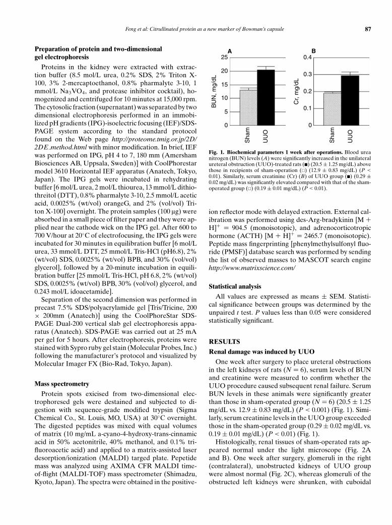
A
Fig. 1. Biochemical parameters 1 week after operations. Blood ureanitrogen (BUN) levels (A) were significantly increased in the unilateralureteral obstruction (UUO)-treated rats (�) (20.5 ± 1.25 mg/dL) abovethose in recipients of sham-operation (�) (12.9 ± 0.83 mg/dL) (P <
0.01). Similarly, serum creatinine (Cr) (B) of UUO group (�) (0.29 ±0.02 mg/dL) was significantly elevated compared with that of the sham-operated group (�) (0.19 ± 0.01 mg/dL) (P < 0.01).
ion reflector mode with delayed extraction. External cal-ibration was performed using des-Arg-bradykinin [M +H]+ = 904.5 (monoisotopic), and adrenocorticotropichormone (ACTH) [M + H]+ = 2465.7 (monoisotopic).Peptide mass fingerprinting [phenylmethylsulfonyl fluo-ride (PMSF)] database search was performed by sendingthe list of observed masses to MASCOT search enginehttp://www.matrixscience.com/
Statistical analysis
All values are expressed as means ± SEM. Statisti-cal significance between groups was determined by theunpaired t test. P values less than 0.05 were consideredstatistically significant.
RESULTS
Renal damage was induced by UUO
One week after surgery to place ureteral obstructionsin the left kidneys of rats (N = 6), serum levels of BUNand creatinine were measured to confirm whether theUUO procedure caused subsequent renal failure. SerumBUN levels in these animals were significantly greaterthan those in sham-operated group (N = 6) (20.5 ± 1.25mg/dL vs. 12.9 ± 0.83 mg/dL) (P < 0.001) (Fig. 1). Simi-larly, serum creatinine levels in the UUO group exceededthose in the sham-operated group (0.29 ± 0.02 mg/dL vs.0.19 ± 0.01 mg/dL) (P < 0.01) (Fig. 1).
Histologically, renal tissues of sham-operated rats ap-peared normal under the light microscope (Fig. 2Aand B). One week after surgery, glomeruli in the right(contralateral), unobstructed kidneys of UUO groupwere almost normal (Fig. 2C), whereas glomeruli of theobstructed left kidneys were shrunken, with cuboidal

88 Feng et al: Citrullinated protein as a new marker of Bowman’s capsule
Sham
UUO
Right Left
A B
DC
Fig. 2. Light microscopic findings 1 weekafter the operations. Under the light mi-croscope, periodic acid-methenamine-silver(PAM) staining of kidney sections 1 weekafter unilateral ureteral obstruction (UUO)or sham-operation (Sham) showed thatglomeruli and interstitium from right kidneys(A) and left kidneys (B) of sham-operated ratswere normal. Similarly, unobstructed rightkidneys from rats whose left kidneys receivedUUO had normal glomeruli (C). However,in UUO-treated left kidneys (D), not onlyinterstitial expansion was observed, but alsothickened Bowman’s capsules and cuboidalchanges of parietal epithelial cells (originalmagnifications ×200).
parietal epithelium and fibrosis around Bowman’s cap-sule (Fig. 2D). The expansion of interstitial area wasalso observed in the obstructed kidneys. These resultspresent a clear picture of surgically induced obstructivenephropathy.
PAD type II mRNA was detected in kidney samples
The expression of PAD mRNAs was assessed by RT-PCR using individual primers for rat PAD type I, type II,type III, and type IV. However, no mRNA of PAD typeI or type III was detected in samples from obstructed,contralateral or healthy control kidneys (Fig. 3B and C,lanes 1 to 4). Because mRNAs of the same samples fromexperimentally manipulated kidneys were amplified byprimers of GAPDH (Fig. 3A, upper bands of lanes 1 to 5),and positive controls from epidermis expressing all PADs(Fig. 3A to D, lane 5) were also amplified by primers ofPAD type I and type III, we verified that these sampleswere suitable for detection of the mRNA of interest. LikePAD types I and III, PAD type IV mRNA was seldomexpressed in obstructed, contralateral or healthy controlkidneys (Fig. 3D, lanes 1 to 4). Again, the suitability ofextracted mRNA and primers of PAD type IV (Fig. 3Ato D, lane 5) for these assays was confirmed. In contrastto the foregoing results in which PAD type I, III, and IVwere not expressed, PAD type II mRNA was detected inall samples from the cortexes of normal as well as dam-aged kidneys (Fig. 3A, lanes 1 to 4). Relative PAD type IImRNA expression (PAD type II/GAPDH) is the highestin the left kidney with the obstruction (Fig. 3E).
Amounts of PAD type II protein increased inobstructed kidneys
Next, expression of PAD type II was examined byWestern blot analysis. As shown in Figure 4, PAD typeII protein was only minimally detectable in kidneys ofsham-operated rats and the right contralateral kidneysof the UUO group (Fig. 4A, sham left and UUO right).However, PAD type II protein of approximately 70 kDwas observed in the left kidneys of the UUO group(Fig. 4A, UUO left). The specificity of rabbit anti-PADtype II antibody used to identify the protein was recon-firmed when no bands were detectable with rabbit serumused as a first antibody instead of anti-PAD type II anti-body (Fig. 4C). Additionally, the extracted proteins werestained by CBB (Fig. 4B and D). When protein bandswere quantified by densitometry, PAD type II protein wassignificantly increased in obstructed kidneys (Fig. 4E).
Obstructed kidneys developed large increases incitrullinated proteins
The expression of the PAD type II does not always cat-alyze citrullination, because this reaction requires Ca2+.However, Western blot analysis using specific antibodyagainst modified citrulline residues successfully yieldedbands of approximately 70 kD, 60 kD, and 40 kD, de-noting the presence of citrullinated proteins on chemi-cally treated membranes only in obstructed kidneys of theUUO group (Fig. 5A, UUO left). To the contrary, in thekidneys of sham-operated rats and contralateral kidneysof the UUO animals, no such bands appeared (Fig. 5A,sham left and UUO right). Because this antibody doesnot react with unmodified citrulline, we can certify the

Feng et al: Citrullinated protein as a new marker of Bowman’s capsule 89
GAPDH
PAD type II
PAD type I
PAD type III
PAD type IV
788 bp
428 bp
631 bp
648 bp
205 bp
1 2 3 4 5
0.2
0.40.6
0.8
1
Rel
ativ
e m
RN
A e
xpre
ssio
nPA
D ty
pe II
/GA
PD
H
Sha
m-le
ft
UU
O-r
ight
UU
O-le
ft
* P < 0.01** P < 0.05
*
***
E
Fig. 3. Expression of glyceraldehyde-3-phosphate dehydrogenase (GAPDH) (A),peptidylarginine deiminases (PAD) typeI (B), type II (A), type III (C), and typeIV (D) transcripts in rat kidneys. TotalRNA was isolated from the cortexes ofrat kidneys, and the same templates fromexperimented kidneys were amplified byreverse transcription-polymerase chainreaction (RT-PCR) using each primer. Lane1, right kidney with sham-operation; lane2, left kidney with sham-operation; lane 3,right (contralateral) kidney with unilateralureteral obstruction (UUO) treatment; lane4, left kidney (obstructed) kidney with UUO;lane 5, positive control from rat epidermis.Expected sizes of amplified sequences basedon primer design were 631 bp for PAD typeI, 428 bp for PAD type II, 648 bp for PADtype III, 205 bp for PAD type III, and 788bp for GAPDH. All PADs transcripts wereobserved in epidermal samples and all sam-ples were amplified by primers of GAPDH.PAD type II mRNA was detected only inkidney tissue, regardless of obstruction orlack thereof (A). PAD type I (B), III (C),and IV (D) mRNA were merely detected insamples from obstructed, contralateral, orhealthy control kidneys. Relative amount ofPAD type II mRNA expression (PAD typeII/GAPDH) is the highest in left kidneys withthe obstruction (E). Values are mean ± SEM.
specificity of this antibody by staining membranes with-out chemical treatment. As shown in Fig. 5C, no bandsemerged on the membrane without chemical modifica-tion. The extracted proteins were stained by CBB staining(Fig. 5B and D).
PAD type II protein was preferentially expressed in pari-etal epithelial cells of kidneys damaged by obstructionbut not in normal kidneys
By immunohistochemical analysis, PAD type II expres-sion was barely noticeable along the parietal epithelia ofnormal rat kidneys (Fig. 6A and B, Sham) or in unob-structed kidneys (Fig. 6C, UUO right). On the other hand,PAD type II expression was marked in parietal epitheliaof tissue undergoing obstructive nephropathy 1 week af-ter the UUO operation (Fig. 6D, UUO left). No PAD typeII expression was observed except on glomerular parietalepithelial cells. As shown in Table 1, 1 week after UUO,intensity of PAD type II expression in Bowman’s cap-sules and percentages of PAD type II–positive glomeruliwere significantly increased in left kidneys of UUO group(UUO left 1 week) compared with those of contralateralkidneys (UUO right 1 week) or sham-operated kidneys(Sham left).
Citrullinated protein is preferentially present alongBowman’s capsule
When we proceeded with staining to identify citrulli-nated proteins in situ, almost no citrullinated proteins ap-
peared in kidneys of sham-operated rats (Fig.7A and B).Sometimes, a few citrullinated proteins lay along Bow-man’s capsule in a very small number of glomeruli ofthe sham-operated group. Similarly, citrullinated proteinswere scarce in right contralateral kidneys of the UUOgroup (Fig. 7C). To the contrary, citrullinated proteinswere visible along Bowman’s capsule of the obstructedleft kidneys (Fig. 7D). When we stained rat kidney tissueswithout chemical treatment by the modification medium,no positive staining was seen at all (Fig. 7E to H). There-fore, the specificity of the antibody used was reconfirmedhistochemically, and this procedure showed that PADtype II and citrullinated proteins both lodged preferen-tially along Bowman’s capsules of damaged kidneys byUUO. One week after UUO, staining intensity for citrul-linated proteins in Bowman’s capsules and percentagesof positive glomeruli were markedly higher in the left ob-structed kidneys of UUO group than right unobstructedkidneys or sham-operated kidneys (Table 1).
Citrullination decreased by the release of ureteralobstruction
To investigate whether citrullination reflects kidneydamages by UUO, ureter-bladder anastomosis was made1 week after the UUO operation. Glomeruli of theleft kidneys with UUO for 5 weeks had cuboidal pari-etal epithelium and periglomerular fibrosis around Bow-man’s capsule (Fig. 8A). Four weeks after the release ofureteral obstruction, glomeruli returned to normal, andits parietal epithelium became flat (Fig. 8B). This light

90 Feng et al: Citrullinated protein as a new marker of Bowman’s capsule
97 kD
66 kD
45 kD
97 kD
66 kD
45 kD
Sha
m-le
ft
UU
O-r
ight
UU
O-le
ft
Sha
m-le
ft
UU
O-r
ight
UU
O-le
ft
Sha
m-le
ft
UU
O-r
ight
UU
O-le
ft
0
2
4
6E *
Fig. 4. Western blot for peptidylargininedeiminases (PAD) type II. A total 30 lg of iso-lated proteins from kidney cortexes was trans-ferred to polyvinylidine difluoride (PVDF)membranes. Bands positive for PAD type IIscarcely appeared in sham-operated (Sham-left) or unobstructed kidneys of unilateralureteral obstruction (UUO) group (UUO-right). In contrast, a clear band at approxi-mately 70 kD was observed in left kidneys 1week after UUO (UUO-left) (A). Thereafter,the membrane was stained by Coomassie bril-liant blue (CBB) (B). Although bands at ap-proximately 60 kD and 80 kD were more in-tense in left kidneys of the UUO group thanothers, intensities of other bands were simi-lar. When the membranes stained by rabbitserum as a first antibody instead of anti PADtype II antibody, no bands were visible (C).Therefore, the band in (A) is specific. Thatmembrane was stained by CBB (D). Whenprotein bands were quantified by densitom-etry, PAD type II amount was significantlyincreased in left obstructed kidneys (UUO-left) compared with that in sham-operatedkidneys (Sham-left) or contra-lateral kidneys(UUO-right) (Fig 4E). ∗P < 0.01 versus sham-operated kidneys and right contra-lateral kid-neys with UUO. Values are mean ± SEM.
66 kD
45 kD
66 kD
45 kD
Sha
m-le
ft
UU
O-r
ight
UU
O-le
ft
Sha
m-le
ft
UU
O-r
ight
UU
O-le
ft
Fig. 5. Western blot for detection of citrul-linated proteins. A total 30 lg of proteinsisolated from kidney cortex was transferredto polyvinylidine difluroride (PVDF) mem-branes. For detection, monoclonal antibodywas added to react chemically with modifiedcitrulline. One membrane was treated with themodification medium before the reaction withanti-modified citrulline antibody, and then cit-rullinated proteins were stained as describedin the Methods section (A), followed byCoomassie brilliant blue (CBB) staining (B).Citrullinated proteins were detected only inobstructed kidneys (UUO-left) (A). Anothermembrane was stained by same first antibodyand second antibody without chemical mod-ification as a control (C), followed by CBBstaining (D). No bands of citrullinated pro-teins were observed in the membrane withoutchemical modification (C), which indicatedspecific reaction of first antibody to chemicallymodified citrulline.

Feng et al: Citrullinated protein as a new marker of Bowman’s capsule 91
Sham
UUO
Right Left
Fig. 6. Peptidylarginine deiminases (PAD)type II expression on abnormal Bowman’scapsules. Kidney tissues were stained by rab-bit antirat PAD type II antiserum. (A) Rightkidney of sham-operated group. (B) Left kid-ney of sham-operated group. (C) Right kid-ney of unilateral ureteral obstruction (UUO)group. (D) Left kidney with ureteral obstruc-tion of UUO group. In both sides of kidneysin sham-operated group, PAD type II expres-sion was miniscule (A and B). In contralat-eral, unobstructed kidney (right), the expres-sion was also scarce (C). On the other hand,in left kidneys staining for PAD type II waspositive and located preferentially in parietalepithelial cells of Bowman’s capsules (D). G isa glomerulus (original magnifications ×200).
Table 1. Summary of histologic examinations (the semiquantitative values of staining intensity and percentages of positive glomeruli forpeptidylarginine deiminase (PAD) type II and citrullinated proteins
PAD II Citrullinated protein
Group Intensity % Intensity %
Sham left (N = 6) 0.63 ± 0.14 36.9 ± 3.7 0.13 ± 0.04 9.2 ± 2.9UUO right 1 week (N = 6) 0.76 ± 0.12 40.7 ± 4.5 0.08 ± 0.03 8.1 ± 2.7UUO left 1 week (N = 6) 3.64 ± 0.13a,c 100 ± 0a,c 1.71 ± 0.06a,c 92.1 ± 1.8a,c
UUO left 5 week (N = 4) 4.00 ± 0.00a,c 100 ± 0a,c 3.02 ± 0.24a,c,d 100 ± 0a,c,d
Bypass left 5 week (N = 4) 2.61 ± 0.22a,c,d,e 80.1 ± 3.8a,b,c,d,e 0.33 ± 0.06b,c,d,e 27.8 ± 6.0b,c,d,e
UUO is unilateral ureteral obstruction; Bypass left 5 weeks, the data from rats receiving ureter-bladder bypass 1 week after UUO and then living for 4 weeks. Thecalculation methods of staining intensity and percentages were explained in the Methods section. Values are mean ± SEM. Statistical significance between groups wasdetermined by the unpaired t test.
aP < 0.01 versus sham; bP < 0.05 versus sham; cP < 0.01 versus right contralateral kidneys of UUO group for 1 week (UUO right 1week); dP < 0.01 versus leftobstructed kidneys after 1-week ligation (UUO left 1weed); eP < 0.01 versus left obstructed kidneys after 5 weeks of UUO (UUO left 5 weeks).
microscopic study showed that the lesions in Bowman’scapsules with UUO were attenuated by the ureter-bladder anastomosis to release the ureteral obstruction.Five weeks after UUO operation, PAD type II expressionwas strongly observed in the whole parietal epithelial cells(Fig. 8C) (Table 1). This intense expression of PAD type IIwas significantly attenuated by the release of ureteral ob-struction (Fig. 8D) (Table 1). Citrullinated proteins werealso increased in Bowman’s capsules of obstructed kid-neys for 5 weeks (Fig. 8E) (Table 1). However, when weperformed the operation for release of UUO 1 week af-ter the ureteral ligation, the citrullinated proteins werediffusely decreased below those in kidneys with UUOfor 5 weeks. Furthermore, PAD type II and citrullinatedproteins were also significantly diminished in the leftkidneys of the group that underwent the ureter-bladderbypass operation even compared with those in the leftobstructed kidneys for 1 week, in which these pro-teins were already increased in Bowman’s capsules(Table 1).
Actin filaments should be citrullinated in the kidney
Next, two-dimensional electrophoresis was performedto investigate what proteins were citrullinated in thekidney. We obtained two spots which were citrullinated(Fig. 9). Their molecular weights were both approxi-mately 40 kD. The MALDI mass fingerprint spectrumsof these spots at 40 kD (Fig. 10A) were analyzed by theMASCOT search engine. The results showed that citrul-linated proteins at 40 kD were actins (Fig. 10B).
DISCUSSION
This report describes for the first time that PAD typeII and citrullinated proteins are suitable markers forBowman’s capsules, since their expression occurs pref-erentially in Bowman’s capsules of the renal glomeruliand increases markedly during the course of obstructivenephropathy.
For this study, we adapted a model of obstructive renaldamage by unilaterally ligating the left ureter (UUO) in

92 Feng et al: Citrullinated protein as a new marker of Bowman’s capsule
A E
F
G
HD
C
B
Right
Left
Right
Left
Sham
UUO
Modification (+) Modification (−)
Fig. 7. Detection of citrullinated protein insitu. Citrullinated proteins were detected bymodification of citrulline residues in tissuesfollowed by staining with anti modified cit-rulline antibody. (A and E) Right kidney ofsham-operated group. (B and F) Left kidneyof sham-operated group. (C and G) Right un-obstructed kidney of unilateral ureteral ob-struction (UUO) group. (D and H) Left ob-structed kidney of UUO group. When tissueswere stained after chemical modification (Ato D), both sides of sham-operated group andright kidney of UUO group did not show pos-itive staining for citrullinated proteins (A toC). Only in the left obstructed kidney of theUUO group was protein citrullination waspreferentially detected in Bowman’s capsule,which appeared like rings (D). When tissueswere not chemically modified (E to H), nopositive staining followed (original magnifica-tions ×200).
each of six rats and simultaneously performed sham op-erations as a control (N = 6). UUO increases fluid pres-sure in the urinary tract, thereby also increasing pressurein tubuli and Bowman’s space. This model has been usedoften to study the mechanisms of renal fibrosis [16]. In theglomeruli, UUO makes parietal epithelial cells of Bow-man’s capsule receive tension as mechanical stress. In thisstudy, UUO of the rat urinary tract caused renal failurewithin 1 week, as confirmed by increases of serum BUNand creatinine (Fig. 1). In addition, the obstructed kid-neys had thickening of Bowman’s capsules and cuboidalformation of the parietal epithelial cells (Fig. 2).
The results of RT-PCR to identify PADs indicated thatneither PAD type I nor type III was expressed in any kid-neys, whether normal, UUO operated, or sham operated(Fig. 3). Similarly, PAD type IV was rarely detected in anyof these kidneys (Fig. 3), and even this limited expressionof PAD type IV mRNA seemed to come from circulat-ing leukocytes left after presurgical perfusion, becausethese cells do express PAD type IV [17]. A marked dif-
ference was the expression of PAD type II mRNA foundin both right and left kidneys of the UUO group and thesham-operated group (Fig. 3). Therefore, we judged thatrenal cells express only type II of the four known PADtypes. When we measured the densities of bands and cal-culated relative amounts of PAD type II mRNA/GAPDHmRNA, amounts in the rats’ left obstructed kidneys weresignificantly higher than in any other samples of kid-neys from sham-operated rats or right kidneys from theUUO group (Fig. 3E). This increase of PAD type II inobstructed kidneys was confirmed by Western blot ofprotein levels (Fig. 4). Sham-operated and unobstructedkidneys had only negligible levels of PAD type II, whereasthe bands of PAD type II were readily detected at the ex-pected molecular weight in all obstructed kidneys fromthe UUO group. Therefore, PAD type II is expressed byrenal cells, and the amount increases after ureteral block-age (Fig. 4E). In addition, glomerular parietal epithelialcells of obstructed kidneys preferentially express PADtype II, but those of normal kidneys (Fig. 6). Because

Feng et al: Citrullinated protein as a new marker of Bowman’s capsule 93
Fig. 8. Representative sections stained forperiodic acid-Schiff (PAS) (A and B), pep-tidylarginine deiminases (PAD) type II (Cand D), and citrullinated protein (E and F)of rats that lived with unilateral ureteral ob-struction (UUO) for 5 weeks (A, C, andE) or that received operation to release theureteral obstruction by ureter-bladder bypass2 week after UUO operation and then livedfor 4 weeks (B, D, and F). Under light micro-scope, although glomeruli of obstructed kid-neys had thickened Bowman’s capsules andcuboidal changes of parietal epithelium (A),these changes returned to normal in rats re-ceiving bypass operation, in which parietal ep-ithelial cells were simple and flat squamous(B). PAD type II expression was stronglyobserved preferentially in parietal epithelialcells around the whole Bowman’s capsule ofrats with UUO (C). On the other hand, pari-etal epithelial cell of rats with the ureter-bladder bypass expressed much less PAD typeII (D). Similarly, citrullinated proteins wereclearly detected preferentially in Bowman’scapsules of rats with UUO for 5 weeks (E). Bythe ureter-bladder bypass, citrullinated pro-teins in Bowman’s capsules were apparentlydecreased (F).
20
25
37
50
75100
150
Mol
ecul
ar m
ass,
kD
SYPRO Ruby staining SYPRO Ruby staining
Staining forcitrullinated proteins
5pI 6
Fig. 9. Sypro ruby stained protein map ofrat kidney with unilateral ureteral obstruc-tion (UUO). A total of 100 lg protein wasseparated using 18 cm pH 4 to 7 immobilizedpH gradients (IPG) and 7.5% sodium dode-cyl sulfate-polyacrylamide gel electrophore-sis (SDS-PAGE). The proteins were resolvedby differential isoelectric point (pI) for thefirst dimensional running (x axis) and differ-ential molecular weight (MW) for the sec-ond dimension (y axis). Proteins were stainedwith sypro ruby gel stain. To detect citrul-linated protein, after sypro ruby staining oftwo-dimensional electrophoresis gel, proteinswere transferred onto polyvylidine difluoride(PVDF) membrane and incubated with anti-body. Two protein spots were detected (spot 1,2). Enlarged region of sypro ruby stained two-dimensional electrophoresis gel are in rightside. Sypro ruby staining is shown in the up-per side and staining for citrullinated proteinis shown in lower side.
the right unobstructed kidneys from UUO-treated ratsscarcely expressed PAD type II, the significant increaseof PAD type II only in the left obstructed kidneys (Table1) evidently arose from the mechanical stress of ureteralobstruction, not circulating factors.
PADs catalyze the citrullination reaction, which con-verts protein arginine to citrulline residues in a Ca2+-dependent manner [11]. Possibly related is a previous re-port that endothelin and platelet-activating factor (PAF)act on the parietal sheet of Bowman’s capsule by increas-ing intracellular concentrations of Ca2+ [18]. In addition,endothelin and PAF were induced by ureteral obstruction[16, 19]. On the basis of those reports, presumably in-tracellular Ca2+ concentrations rise in parietal epithelialcells of Bowman’s capsule after ureteral obstruction, and
this elevated concentration would be available to func-tion with PAD type II in the increased catalysis of pro-tein citrullination at that site (Fig. 7). Therefore, when weassessed our three groups of kidneys with Western blot-ting, citrullinated proteins were detected only in kidneyswith ureteral obstruction (Fig. 5). Although PAD type IIexpression was clearly detected in cytoplasms of parietalepithelial cells, we could not decide whether citrullinatedproteins were located in the parietal epithelium or thebasement membrane of Bowman’s capsules. In future,immunoelectron microscopy will reveal the location.
Our studies using UUO model for 1 week indicatedthat citrullination occurs preferentially in Bowman’s cap-sules of the renal glomeruli and increases markedlyduring the course of obstructive nephropathy. Next, to

94 Feng et al: Citrullinated protein as a new marker of Bowman’s capsule
681.9
665.9
671.9
656.0
1208.9
1790.81185.8
1200.6
1198.6
1953.9
0102030405060708090
100In
tens
ity, %
500 1000 1500 2000 2500 3000 3500 4000
Mass/charge
A
Protein identities and peptide masses
B
Protein SWISS-PROT.Accession no.
Mr (Da) pI Coverage Score Matched masses
Actin (spot 2) P60711 42052 5.29 45% 123 976.34 2155.95 2382.831198.61 1354.57 800.361953.88 2295.08 3199.251639.69 1629.70 1790.772093.88 1580.64 1036.53795.59 923.43
Fig. 10. Identification of protein spots bymatrix-assisted laser desorption/ionizationtime-of flight (MALDI/TOF) mass spec-trometry analysis. MALDI mass fingerprintspectrum of spot 2 was shown in the up-per (A). Peptide mass fingerprinting wasperformed using the MASCOT search en-gine (www.matrixscience.com) based on theSWISS-PROT database. Results of the searchare shown in the bottom. Protein spot 2 wasidentified as actin (B) and spot 1 was also actin[SWISS-PROT accession no. P60711 (data notshown)].
investigate whether expression of PAD type II and citrul-linated proteins reflects damages of Bowman’s capsulesby ureteral obstruction, a ureter-bladder anastomosis torelease the obstruction was made 1 week after UUO op-eration. Four weeks after the bypass, the appearance ofBowman’s capsules returned to normal under the lightmicroscope (Fig. 8B). On the other hand, Bowman’s cap-sules of kidneys 5 weeks after UUO were severely thick-ening and had cuboidal epithelial cells (Fig. 8A). By theureter-bladder bypass, PAD type II expression of pari-etal epithelial cells was significantly decreased comparedwith that in UUO kidneys not only for 5 weeks but also for1 week (Fig. 8) (Table 1). Similarly, citrullinated proteinswere also markedly attenuated in kidneys with the bypasscompared with those in kidneys with UUO for 5 weeksand for 1 week (Fig. 8) (Table 1). These results indicatethat besides being markers of Bowman’s capsules, PADtype II, and citrullinated proteins can reflect the damagesof Bowman’s capsules. In crescentic glomerulonephritis,Bowman’s capsules become similarly cuboidal. There-fore, we histologically examined rats that were inducedcrescentic glomerulonephritis by rabbit antiglomerularbasement membrane antibody to investigate whether cit-rullination in Bowman’s capsules participates in cuboidalformation of parietal epithelial cells. Our study showedthat PAD type II and citrullinated proteins were notdetected in their Bowman’s capsules (data not shown).Therefore, although it was speculated that increment ofcitrullination in Bowman’s capsules should specificallyoccur in response to mechanical stress to glomeruli, it re-mains to be solved what roles citrullination in the kidneyplays.
Western blotting (Fig. 5) located several sites of cit-rullinated proteins in the obstructed kidneys examinedhere. To explain the roles of citrullination in the kid-ney, we performed proteome analysis of these citrulli-nated proteins. By two-dimensional electrophoresis, twoprotein spots which were citrullinated were obtained(Fig. 9). Their molecular weights were approximately40 kD although main band was present at approximately60 kD rather than 40 kD in Western blotting for cit-rullinated proteins (Fig. 5). The analysis of mass finger-print spectra of these spots at 40 kD revealed that thesewere actins (Fig. 10). Although we wanted to identify the60 kD citrullinated protein, the protein could not be de-tected by two-dimensional electrophoresis (Fig. 9), prob-ably caused by technical difficulties. Therefore, we thinkthese proteome analyses are now incomplete. In future,60 kD citrullinated proteins should be identified to ex-plain further fundamental roles of citrullination in kid-neys. Previous reports described intermediate filamentsof cytoskeletal proteins such as keratin and vimentinthat were also citrullinated [20, 21]. Such citrullinationabolished the positive charge of protein molecules, in-evitably causing significant alteration in their structure[4, 5]. Therefore, if citrullinated proteins in Bowman’scapsule are cytoskeletal proteins, that would explain de-fense mechanism of Bowman’s capsule against mechani-cal stress to glomeruli.
Only one suitable marker of Bowman’s capsule hasbeen reported previously [1]. Our study described heregreatly extends that report by indicating that PADtype II and citrullinated proteins are preferentially ex-pressed in Bowman’s capsules and, additionally, that their

Feng et al: Citrullinated protein as a new marker of Bowman’s capsule 95
expression increases in damaged kidneys by ureteral ob-struction. These newfound markers of Bowman’s capsuleare expected to broaden the experimental possibilities forresearch on kidney diseases.
ACKNOWLEDGMENTS
We thank Phyllis Minick for critical editing of the manuscript. Wealso acknowledge Dr. A. Shimizu and Dr. Y. Masuda for kindly provid-ing tissue samples of rat anti-GBM nephritis. This study was supporteda grant from the Health Science Research Grants for ComprehensiveResearch on Aging and Health from Ministry of Health Labor and Wel-fare, Japan, and a grant-in-aid for Scientific Research from Ministry ofEducation, Science and Culture, grant 15790436 Japan to T.I.
Reprint requests to Toshiyuki Imasawa M.D., Ph.D., Department ofInternal Medicine and Division of Immunopathology, Clinical ResearchCenter, Chiba-East National Hospital, 673 Nitona, Chuoh, Chiba, 260–8712, Japan.E-mail: [email protected]
REFERENCES
1. SHIRATO I, ASANUMA K, TAKEDA Y, et al: Protein gene product 9.5 isselectively localized in parietal epithelial cells of Bowman’s capsulein the rat kidney. J Am Soc Nephrol 11:2381–2386, 2000
2. KUBILUS J, WAITKUS RF, BADEN HP: Partial purification and speci-ficity of an arginine-converting enzyme from bovine epidermis.Biochim Biophys Acta 615:246–251, 1980
3. ROGERS GE, SIMMONDS DH: Content of citrulline and other aminoacids in a protein of hair follicles. Nature 182:186–187, 1958
4. MOSCARELLO MA, PRITZKER L, MASTRONARDI FG, WOOD DD: Pep-tidylarginine deiminase: A candidate factor in demyelinating dis-ease. J Neurochem 81:335–343, 2002
5. STEINERT PM, PARRY DA, MAREKOV LN: Trichohyalin mechanicallystrengthens the hair follicle: Multiple cross-bridging roles in theinner root shealth. J Biol Chem 278:41409–41419, 2003
6. ISHIGAMI A, ASAGA H, OHSAWA T, et al: Peptidylarginine deiminasetype I, type II, type III and type IV are expressed in rat epidermis.Biomed Res 22:63–65, 2001
7. SCHELLEKENS GA, DE JONG BA, VAN DEN HOOGEN FH, et al: Citrullineis an essential constituent of antigenic determinants recognized byrheumatoid arthritis-specific autoantibodies. J Clin Invest 101:273–281, 1998
8. SUZUKI A, YAMADA R, CHANG X, et al: Functional haplotypes ofPADI4, encoding citrullinating enzyme peptidylarginine deiminase4, are associated with rheumatoid arthritis. Nat Genet 34:395–402,2003
9. ASAGA H, ISHIGAMI A: Protein deimination in the rat brain afterkainate administration: Citrulline-containing proteins as a novelmarker of neurodegeneration. Neurosci Lett 299:5–8, 2001
10. WATANABE K, SENSHU T: Isolation and characterization of cDNAclones encoding rat skeletal muscle peptidylarginine deiminase. JBiol Chem 264:15255–15260, 1989
11. TARCSA E, MAREKOV LN, ANDREOLI J, et al: The fate of trichohyalin.Sequential post-translational modifications by peptidyl-argininedeiminase and transglutaminases. J Biol Chem 272:27893–27901,1997
12. ISHIGAMI A, KURAMOTO M, YAMADA M, et al: Molecular cloning oftwo novel types of peptidylarginine deiminase cDNAs from retinoicacid-treated culture of a newborn rat keratinocyte cell line. FEBSLett 433:113–118, 1998
13. WATANABE K, AKIYAMA K, HIKICHI K, et al: Combined biochemicaland immunochemical comparison of peptidylarginine deiminasespresent in various tissues. Biochim Biophys Acta 966:375–383, 1988
14. SENSHU T, SATO T, INOUE T, et al: Detection of citrulline residues indeiminated proteins on polyvinylidene difluoride membrane. AnalBiochem 203:94–100, 1992
15. SENSHU T, AKIYAMA K, KAN S, et al: Detection of deiminatedproteins in rat skin: Probing with a monospecific antibody aftermodification of citrulline residues. J Invest Dermatol 105:163–169,1995
16. KLAHR S, MORRISSEY J: Obstructive nephropathy and renal fibrosis.Am J Physiol Renal Physiol 283:F861–F875, 2002
17. ASAGA H, NAKASHIMA K, SENSHU T, et al: Immunocytochemical lo-calization of peptidylarginine deiminase in human eosinophils andneutrophils. J Leukoc Biol 70:46–51, 2001
18. MARCHETTI J, MENETON P, LEBRUN F, et al: The parietal sheet ofBowman’s capsule of rat renal glomerulus: A target of endothelinand PAF. Am J Physiol 268:F1053–F1061, 1995
19. REYES AA, KLAHR S: Renal function after release of ureteral ob-struction: Role of endothelin and the renal artery endothelium. Kid-ney Int 42:632–638, 1992
20. SENSHU T, KAN S, OGAWA H, et al: Preferential deimination of ker-atin K1 and filaggrin during the terminal differentiation of humanepidermis. Biochem Biophys Res Commun 225:712–719, 1996
21. ASAGA H, YAMADA M, SENSHU T: Selective deimination of vi-mentin in calcium ionophore-induced apoptosis of mouse peri-toneal macrophages. Biochem Biophys Res Commun 243:641–646,1998
Copyright © 2022 FDOKUMEN