Arsenic trioxide preferentially induces nonapoptotic cell deaths as well as actin cytoskeleton...
9
Postepy Hig Med Dosw (online), 2014; 68 www.phmd.pl Original Article Postepy Hig Med Dosw (online), 2014; 68: 1492-1500 e-ISSN 1732-2693 Received: 2012.12.21 Accepted: 2014.10.08 Published: 2014.12.21 Summary The therapeutic effect of arsenic trioxide (ATO, As 2 O 3 ) has been investigated for many years. Ho- wever, the precise molecular mechanisms underlying the antitumor activity of ATO are still not fully understood, but seem to depend on cell types, dosage, and duration of exposure. The purpo- se of this study was to assess the actin cytoskeleton rearrangement during the cell death process induced by arsenic trioxide in the CHO AA8 cells. A better understanding the mechanisms of ATO- -action is likely to lead to more rational use of this drug either as monotherapies or in combination with other anticancer agents. The effect of ATO on actin cytoskeleton was studied in Chinese Hamster Ovary AA8 cell line. Actin was visualized by fluorescence microscopy and phalloidin conjugated to Alexa Fluor® 488. Mor- phological and ultrastructural alterations in the CHO AA8 cells were evaluated by using light and electron microscope, respectively. For quantitative measurement of cell death, Annexin V-Alexa Fluor® 488 and Propidium Iodide assay was performed. The vital staining of CHO AA8 cells with acridine orange was applied to detect the development of acidic vesicular organelles (AVOs). The performed experiments revealed a dose-dependent decrease in the cell survival. The morpho- logical and ultrastructural features acquired by the cells after ATO-treatment were considered as typical for autophagy and mitotic cell death. As was shown by acridine orange staining, arsenic trioxide treatment increased red fluorescence signals in dose-dependent manner, indicating the development of AVOs, a hallmark of autophagy. Low level of apoptosis was induced in the ATO- -treated CHO AA8 cells. Furthermore, the rearrangement of actin filaments associated with cell death process was also detected. The obtained results suggest that arsenic trioxide preferentially induces nonapoptotic cell deaths, autophagy and mitotic cell death, in p53-deficient CHO AA8 cells. Furthermore, the distinctive pat- terns of F-actin remodeling after As 2 O 3 treatment were associated with different modes of cell de- ath, confirming that cytoskeleton is a dynamic structure actively involved in the cell death process. arsenic trioxide • actin • CHO AA8 cell line • autophagy • mitotic catastrophe (mitotic cell death) Arsenic trioxide preferentially induces nonapoptotic cell deaths as well as actin cytoskeleton rearrangement in the CHO AA8 cell line* Trójtlenek arsenu indukuje nieapoptotyczne mechanizmy śmierci i rearanżację cytoszkieletu aktynowego w komórkach linii CHO AA8 Magdalena Izdebska * , , , , , , Anna Klimaszewska-Wiśniewska * , , , , , , Dawid Lewandowski , , , Jakub Marcin Nowak , , , Maciej Gagat , , Alina Grzanka , , *These authors contributed equally to this work Introduction: Results: Conclusions: Key words: Material and methods: * is study was supported by research task within the framework of the statutory activities no. 895 (Nicolaus Copernicus University in Torun, Collegium Medicum in Bydgoszcz, Poland). Authors’ Contribution: Study Design Data Collection Statistical Analysis Data Interpretation Manuscript Preparation Literature Search Funds Collection 1492
Transcript of Arsenic trioxide preferentially induces nonapoptotic cell deaths as well as actin cytoskeleton...
Postepy Hig Med Dosw (online), 2014; 68
www.phmd.plOriginal Article
Postepy Hig Med Dosw (online), 2014; 68: 1492-1500e-ISSN 1732-2693
Received: 2012.12.21Accepted: 2014.10.08Published: 2014.12.21
SummaryThe therapeutic effect of arsenic trioxide (ATO, As2O3) has been investigated for many years. Ho-wever, the precise molecular mechanisms underlying the antitumor activity of ATO are still not fully understood, but seem to depend on cell types, dosage, and duration of exposure. The purpo-se of this study was to assess the actin cytoskeleton rearrangement during the cell death process induced by arsenic trioxide in the CHO AA8 cells. A better understanding the mechanisms of ATO--action is likely to lead to more rational use of this drug either as monotherapies or in combination with other anticancer agents.
The effect of ATO on actin cytoskeleton was studied in Chinese Hamster Ovary AA8 cell line. Actin was visualized by fluorescence microscopy and phalloidin conjugated to Alexa Fluor® 488. Mor-phological and ultrastructural alterations in the CHO AA8 cells were evaluated by using light and electron microscope, respectively. For quantitative measurement of cell death, Annexin V-Alexa Fluor® 488 and Propidium Iodide assay was performed. The vital staining of CHO AA8 cells with acridine orange was applied to detect the development of acidic vesicular organelles (AVOs).
The performed experiments revealed a dose-dependent decrease in the cell survival. The morpho-logical and ultrastructural features acquired by the cells after ATO-treatment were considered as typical for autophagy and mitotic cell death. As was shown by acridine orange staining, arsenic trioxide treatment increased red fluorescence signals in dose-dependent manner, indicating the development of AVOs, a hallmark of autophagy. Low level of apoptosis was induced in the ATO--treated CHO AA8 cells. Furthermore, the rearrangement of actin filaments associated with cell death process was also detected.
The obtained results suggest that arsenic trioxide preferentially induces nonapoptotic cell deaths, autophagy and mitotic cell death, in p53-deficient CHO AA8 cells. Furthermore, the distinctive pat-terns of F-actin remodeling after As2O3 treatment were associated with different modes of cell de-ath, confirming that cytoskeleton is a dynamic structure actively involved in the cell death process.
arsenic trioxide • actin • CHO AA8 cell line • autophagy • mitotic catastrophe (mitotic cell death)
Arsenic trioxide preferentially induces nonapoptotic cell deaths as well as actin cytoskeleton rearrangement in the CHO AA8 cell line*
Trójtlenek arsenu indukuje nieapoptotyczne mechanizmy śmierci i rearanżację cytoszkieletu aktynowego w komórkach linii CHO AA8
Magdalena Izdebska* , , , , , , Anna Klimaszewska-Wiśniewska* , , , , , , Dawid Lewandowski , , , Jakub Marcin Nowak , , , Maciej Gagat , , Alina Grzanka , ,
*These authors contributed equally to this work
Introduction:
Results:
Conclusions:
Key words:
Material and methods:
* This study was supported by research task within the framework of the statutory activities no. 895 (Nicolaus Copernicus University in Torun, Collegium Medicum in Bydgoszcz, Poland).
Authors’ Contribution: Study Design Data Collection Statistical Analysis Data Interpretation Manuscript Preparation Literature Search Funds Collection
1492

1493
Izdebska M. et al. – Arsenic trioxide preferentially induces nonapoptotic cell deaths...
Author’s addresses: Prof. Alina Grzanka, Ph.D., Department of Histology and Embryology, Collegium Medicum in Bydgoszcz; Nicolaus Copernicus University in Toruń, 24 Karłowicza St., 85-092 Bydgoszcz, Poland; e-mail: [email protected]
IntroductIon
Arsenic trioxide (ATO, As2O3) is the chemotherapy agent approved by FDA for the treatment of relapsed or refrac-tory acute promyelocytic leukemia (APL). Furthermore, it is also in clinical trials of other hematologic and solid tumors [20,57]. The extensive research demonstrating that ATO acts through the variety of intracellular sig-nal transduction pathways to result in the inhibition of growth, the induction of cell death and the promotion of differentiation. However, the precise molecular mecha-nisms underlying the antitumor activity of As2O3 are still fully unknown, but seem to depend on cell types, dosage, and the duration of exposure [40]. Many reports have shown that ATO induces cell death mainly via apop-totic pathways [12,13,14,15,33]. However, it is becoming increasingly clear that ATO-treated cells can also die by nonapoptotic mechanisms, such as mitotic catastrophe, autophagy and necrosis [53,34,44]. ATO-induced apopto-sis is associated with the activation of caspase cascade, the inhibition of NFκB, the induction of oxidative stress, and the disruption of mitochondrial membrane potential. Moreover, changes in the expression of bcl-2, the activa-tion of JNK kinase, and the inhibition of telomerase may also contribute to the induction of apoptosis by arsenic trioxide [5,10,40].
Previous studies revealed that p53 mutations are involved in 50% of all human cancers and nearly all chemothera-peutic drugs kill cancer cells mainly by apoptosis, that usually depends upon intact p53 signaling [16,18,47]. Thus, it is also extremely important to explore the mecha-nisms of anticancer drugs action that lead to nonapop-totic cell death induction such as mitotic catastrophe, because this type of cell death is not related to p53 tu-mor suppressor gene activity [29,45,48]. Hence, the drugs which are able to trigger the different types of cell death, offer a great potential for cancer therapy.
The biological responses to As2O3 are likely to be partially caused by the modification of critical cysteine groups in cellular proteins [10,40]. Therefore, the proteins with a high cysteine content and accessible thiol groups are potent candidates for interactions with ATO. The cyto-skeleton, that contains proteins with a relatively high sulfhydryl content is known to be a cellular target for
arsenic trioxide [4,37,38,39]. Since various studies have implicated the actin cytoskeleton in both cell death and tumorigenesis, actin filaments constitute an attractive target for cancer therapy [17].
In the current study, the responses of CHO AA8 cells to arsenic trioxide were evaluated by characterizing cell vi-ability, mechanisms of cell death and F-actin rearrange-ment. The choice of p53-lack cell line in present investi-gation in the manner of determination of cell death type seems justified.
MaterIal and Methods
Cell culture and treatment
The Chinese hamster ovary cell line (CHO AA8) was kindly provided by Prof. M. Zdzienicka (Department of Molecular Cell Genetics, Collegium Medicum in Bydgoszcz, Nicolaus Copernicus University, Poland). CHO AA8 cells were grown at 37°C in a humidified 5% CO2 atmosphere in minimum essential medium eagle (Sigma-Aldrich) supplemented with 10% fetal bovine serum (FBS, Gibco) and the mix-ture of penicillin and streptomycin (Sigma-Aldrich) in concentration of 1ml/100ml medium. After 24 h of the culture, the cells were incubated with arsenic trioxide. ATO was added at doses of 1 μg/ml, 2 μg/ml and 3 μg/ml for 24 h. Control cells were cultured identically without ATO treatment. Cell viability was assessed by the trypan blue dye exclusion method.
Light microscopy
For the morphological analysis, the CHO AA8 cells grown on coverslips were fixed in 4% paraformaldehyde and then in-cubated with 0,1M glycine solution. The cells were stained with Mayer’s hematoxylin and rinsed under running tap water and dehydrated in a graded series of alcohols and xylenes. The preparations were observed using an Eclipse E800 microscope (Nikon) with NIS-Elements image analysis system and CCD camera (DS-5Mc-U1; Nikon).
Transmission electron microscopy
For the ultrastructural analysis, the CHO AA8 cells were fixed with 3,6% glutaraldehyde and moved to 0.1M caco-
Full-text PDF:
Word count:Tables:
Figures:References:
http://www.phmd.pl/fulltxt.php?ICID=1133098
3638–661
1494
Postepy Hig Med Dosw (online), 2014; tom 68: 1492-1500
dylate buffer (pH 7,4). Afterwards, the cells were post-fixed with 1% osmium tetroxide, dehydrated with an as-cending series of alcohols and acetones, and embedded in Epon 812. The polymerization of the resin occurred at 37°C for 24 h, and then at 65°C for 120 h. Selected parts of material were cut into ultra-thin sections by using Reichert OmU3 ultramicrotome and then coun-terstained with uranyl acetate and lead citrate. The ma-terial was examined using JEM 100 CX electron micro-scope (JEOL).
Image-Based Cytometer
For quantitative measurement of cell death, Tali® Apoptosis Assay Kit with Annexin V-Alexa Fluor® 488 and Propidium Iodide (Invitrogen) was used according to the manufacturer’s instructions. Briefly, the CHO AA8 cells were harvested from 6-well plates, centri-fuged and the cell pellets were resuspended in 100 μl of Annexin Binding Buffer and 5 μl Annexin V-Alexa Fluor® 488. After 20 min incubation at room tempera-ture in the dark, the cells were centrifuged at 300g for 5 min. The supernatants were discarded and the cell pellets were resuspended in 100 μl of Annexin Binding Buffer and 1 μl of propidium iodide. The incubation lasted for 5 min at room temperature in the dark. Pre-parated cells were analyzed with TaliTM-Image Based Cytometer. The double labeling procedure allows to distinguish early apoptotic (Annexin V – Alexa Fluor® 488+/PI-) from late apoptotic (Annexin V – Alexa Fluor® 488+/PI+) and necrotic cells (Annexin V – Alexa Fluor® 488-/PI+).
Fluorescence microscopy
For the immunofluorescence labeling of F-actin, the CHO AA8 cells were fixed with 4% paraformaldehyde, embed-ded in 0.1 M glycine solution and thereafter permeabi-lized with 0.1% Triton X-100. The cells were incubated with phalloidin conjugated to Alexa Fluor 488 (Invitro-gen, diluted 1:40) to enable the visualization of F-actin. The nuclei of the cells were labeled with 4’,6-diamidino-2-phenyloindole (DAPI; Sigma-Aldrich, St. Louis, Mis-souri, USA). Slides were mounted in Aqua-Poly/Mount (Polysciences) and analysed by using an Eclipse E800 mi-croscope with a Y-FL fluorescence attachment (Nikon), NIS-Elements 3.30 image analysis system and CCD camera (DS-5Mc-U1; Nikon).
Supravital cell staining with Acridine Orange (AO)
To detect the development of acidic vesicular organelles (AVOs), the vital staining of CHO AA8 cells with acridine orange was performed. Treated cells were stained with medium containing 1 μg/ml AO for 15 min at 37ºC, then washed twice in PBS and examined immediately with an Eclipse E800 microscope with a Y-FL fluorescence at-tachment (Nikon), NIS-Elements 3.30 image analysis sys-tem and CCD camera (DS-5Mc-U1; Nikon). The cytoplasm
and nucleus of AO-stained cells fluoresced bright green, whereas the acidic autophagic vacuoles fluoresced bright red as described previously [51,54]. Fluorescence inten-sity was measured with ImageJ software (NIH).
Statistical analysis
GraphPad Prism 5.0 (GraphPad Software) was used for the statistical analysis. In order to assess the statistically significant differences between control and ATO doses, the nonparametric Mann-Whitney U test was performed. Results were considered at p<0.05.
results
The viability of CHO AA8 cells after treatment with arsenic trioxide
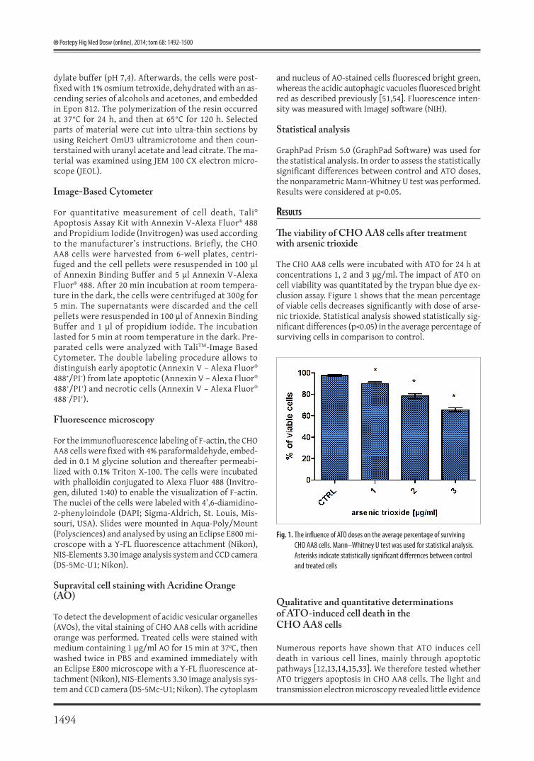
The CHO AA8 cells were incubated with ATO for 24 h at concentrations 1, 2 and 3 μg/ml. The impact of ATO on cell viability was quantitated by the trypan blue dye ex-clusion assay. Figure 1 shows that the mean percentage of viable cells decreases significantly with dose of arse-nic trioxide. Statistical analysis showed statistically sig-nificant differences (p<0.05) in the average percentage of surviving cells in comparison to control.
Qualitative and quantitative determinations of ATO-induced cell death in the CHO AA8 cells
Numerous reports have shown that ATO induces cell death in various cell lines, mainly through apoptotic pathways [12,13,14,15,33]. We therefore tested whether ATO triggers apoptosis in CHO AA8 cells. The light and transmission electron microscopy revealed little evidence
Fig. 1. The influence of ATO doses on the average percentage of surviving CHO AA8 cells. Mann–Whitney U test was used for statistical analysis. Asterisks indicate statistically significant differences between control and treated cells
1495
Izdebska M. et al. – Arsenic trioxide preferentially induces nonapoptotic cell deaths...
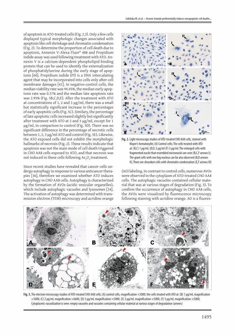
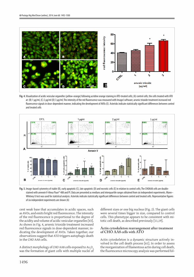
of apoptosis in ATO-treated cells (Fig. 2,3). Only a few cells displayed typical morphologic changes associated with apoptosis like cell shrinkage and chromatin condensation (Fig. 2). To determine the proportion of cell death due to apoptosis, Annexin V-Alexa Fluor® 488 and Propidium Iodide assay was used following treatment with ATO. An-nexin V is a calcium-dependent phospholipid binding protein that can be used to identify the externalization of phosphatidylserine during the early stage of apop-tosis [60]. Propidium iodide (PI) is a DNA intercalating agent that may be incorporated into cells only after cell membrane damages [41]. In negative-control cells, the median viability rate was 94.45%, the median early apop-tosis rate was 0.37% and the median late apoptosis rate was 2.95% (Fig. 5B,C,D,E). After the treatment with ATO at concentrations of 1, 2 and 3 μg/ml, there was a small but statistically significant increase in the percentages of early apoptotic cells (Fig. 5C). Similary, the percentage of late apoptotic cells increased slightly but significantly after treatment with ATO at 2 and 3 μg/ml, except for 1 μg/ml, in comparison to control (Fig. 5D). There was no significant difference in the percentage of necrotic cells between 1, 2, 3 μg/ml ATO and control (Fig. 5E). Likewise, the ATO exposed cells did not exhibit the morphologic hallmarks of necrosis (Fig. 2). These results indicate that apoptosis was not the main mode of cell death triggered in CHO AA8 cells exposed to ATO, and that necrosis was not induced in these cells following As2O3 treatment.
Since recent studies have revealed that cancer cells un-dergo autophagy in response to various anticancer thera-pies [36], therefore we examined whether ATO induces autophagy in CHO AA8 cells. Autophagy is characterized by the formation of AVOs (acidic vesicular organelles), which include autophagic vacuoles and lysosomes [34]. The activation of autophagy was determined with trans-mission electron (TEM) microscopy and acridine orange
(AO) labeling. In contrast to control cells, numerous AVOs were observed in the cytoplasm of ATO-treated CHO AA8 cells. The autophagic vacuoles contained cellular mate-rial that was at various stages of degradation (Fig. 3). To confirm the occurrence of autophagy in CHO AA8 cells, the AVOs were visualized by fluorescence microscopy following staining with acridine orange. AO is a fluores-
Fig. 2. Light microscopy studies of ATO-treated CHO AA8 cells, stained with Mayer’s hematoxylin; (A) Control cells; The cells treated with ATO at: (B,C) 1 μg/ml, (D,E) 2 μg/ml (F) 3 μg/ml; The enlarged cells with fragmented nuclei that resembled micronuclei are seen (B,C,F arrows I); The giant cells with one big nucleus can be also observed (B,D arrows II); There are shrunken cells with chromatin condensation (E,F arrows III)
Fig. 3. The electron microscopy studies of ATO-treated CHO AA8 cells; (A) control cells, magnification ×5000; the cells treated with ATO at: (B) 1 μg/ml, magnification ×5000, (C) 2 μg/ml, magnification ×6600, (D) 3 μg/ml, magnification ×5000, (E) 3 μg/ml, magnification ×5000, (F) 3 μg/ml, magnification ×5000; Cytoplasmic vacuolization is seen: empty vacuoles and vacuoles containing cellular material at various stages of degradation (arrows)
1496
Postepy Hig Med Dosw (online), 2014; tom 68: 1492-1500
different sizes or one big nucleus (Fig. 2). The giant cells were several times bigger in size, compared to control cells. This phenotype appears to be consistent with mi-totic cell death, as described previously [11,19].
Actin cytoskeleton rearrangement after treatment of CHO AA8 cells with ATO
Actin cytoskeleton is a dynamic structure actively in-volved in the cell death process [61]. In order to assess the reorganization of filamentous actin during cell death, the fluorescence microscopy analysis was performed fol-
Fig. 5. Image-based cytometry of viable (B), early apoptotic (C), late apoptotic (D) and necrotic cells (E) in relation to control cells; The CHOAA8 cells are double-stained with annexin V-Alexa Fluor® 488 and PI. Data are presented as medians and interquartile ranges obtained from six independent experiments. Mann–Whitney U test was used for statistical analysis. Asterisks indicate statistically significant differences between control and treated cells. Representative figures of six independent experiments are shown (A)
Fig. 4. Visualization of acidic vesicular organelles (yellow-orange) following acridine orange staining in ATO-treated cells; (A) control cells; the cells treated with ATO at: (B) 1 μg/ml, (C) 2 μg/ml (D) 3 μg/ml; The intensity of the red fluorescence was measured with ImageJ software; arsenic trioxide treatment increased red fluorescence signals in dose-dependent manner, indicating the development of AVOs (E). Asterisks indicate statistically significant differences between control and treated cells
cent weak base that accumulates in acidic spaces, such as AVOs, and emits bright red fluorescence. The intensity of the red fluorescence is proportional to the degree of the acidity and volume of acidic vesicular organelles [43]. As shown in Fig. 4, arsenic trioxide treatment increased red fluorescence signals in dose-dependent manner, in-dicating the development of AVOs. Taken together, our observations suggest that ATO triggers autophagic death in the CHO AA8 cells.
A distinct morphology of CHO AA8 cells exposed to As2O3 was the formation of giant cells with multiple nuclei of
1497
Izdebska M. et al. – Arsenic trioxide preferentially induces nonapoptotic cell deaths...
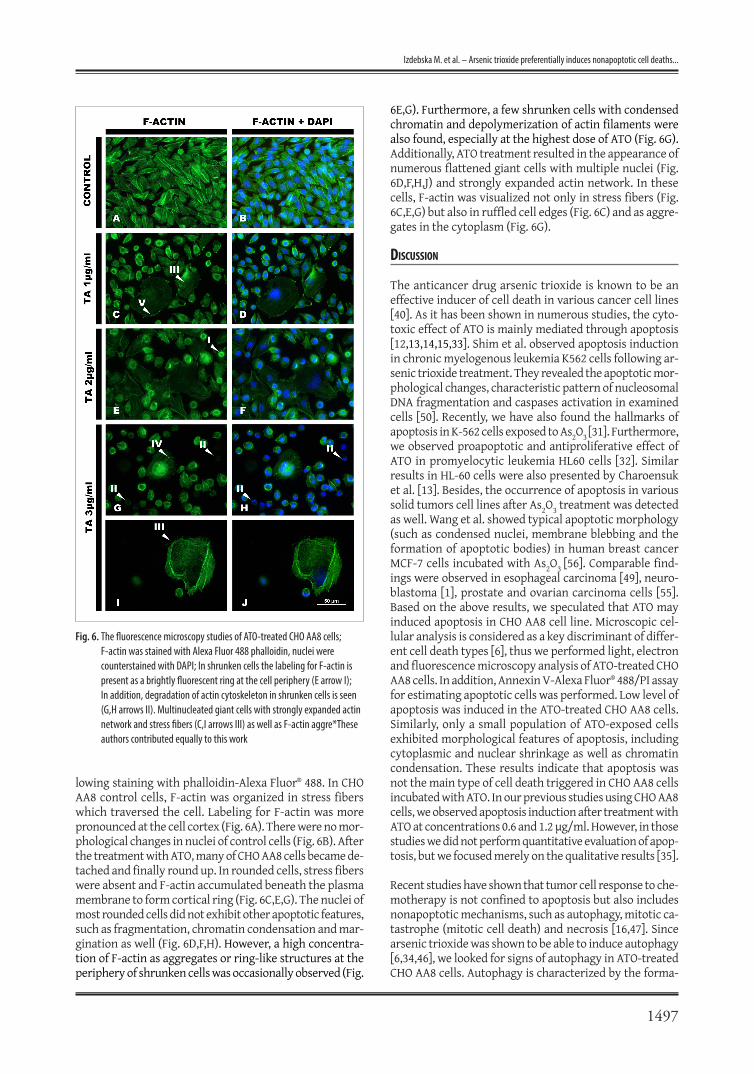
lowing staining with phalloidin-Alexa Fluor® 488. In CHO AA8 control cells, F-actin was organized in stress fibers which traversed the cell. Labeling for F-actin was more pronounced at the cell cortex (Fig. 6A). There were no mor-phological changes in nuclei of control cells (Fig. 6B). After the treatment with ATO, many of CHO AA8 cells became de-tached and finally round up. In rounded cells, stress fibers were absent and F-actin accumulated beneath the plasma membrane to form cortical ring (Fig. 6C,E,G). The nuclei of most rounded cells did not exhibit other apoptotic features, such as fragmentation, chromatin condensation and mar-gination as well (Fig. 6D,F,H). However, a high concentra-tion of F-actin as aggregates or ring-like structures at the periphery of shrunken cells was occasionally observed (Fig.
6E,G). Furthermore, a few shrunken cells with condensed chromatin and depolymerization of actin filaments were also found, especially at the highest dose of ATO (Fig. 6G). Additionally, ATO treatment resulted in the appearance of numerous flattened giant cells with multiple nuclei (Fig. 6D,F,H,J) and strongly expanded actin network. In these cells, F-actin was visualized not only in stress fibers (Fig. 6C,E,G) but also in ruffled cell edges (Fig. 6C) and as aggre-gates in the cytoplasm (Fig. 6G).
dIscussIon
The anticancer drug arsenic trioxide is known to be an effective inducer of cell death in various cancer cell lines [40]. As it has been shown in numerous studies, the cyto-toxic effect of ATO is mainly mediated through apoptosis [12,13,14,15,33]. Shim et al. observed apoptosis induction in chronic myelogenous leukemia K562 cells following ar-senic trioxide treatment. They revealed the apoptotic mor-phological changes, characteristic pattern of nucleosomal DNA fragmentation and caspases activation in examined cells [50]. Recently, we have also found the hallmarks of apoptosis in K-562 cells exposed to As2O3 [31]. Furthermore, we observed proapoptotic and antiproliferative effect of ATO in promyelocytic leukemia HL60 cells [32]. Similar results in HL-60 cells were also presented by Charoensuk et al. [13]. Besides, the occurrence of apoptosis in various solid tumors cell lines after As2O3 treatment was detected as well. Wang et al. showed typical apoptotic morphology (such as condensed nuclei, membrane blebbing and the formation of apoptotic bodies) in human breast cancer MCF-7 cells incubated with As2O3 [56]. Comparable find-ings were observed in esophageal carcinoma [49], neuro-blastoma [1], prostate and ovarian carcinoma cells [55]. Based on the above results, we speculated that ATO may induced apoptosis in CHO AA8 cell line. Microscopic cel-lular analysis is considered as a key discriminant of differ-ent cell death types [6], thus we performed light, electron and fluorescence microscopy analysis of ATO-treated CHO AA8 cells. In addition, Annexin V-Alexa Fluor® 488/PI assay for estimating apoptotic cells was performed. Low level of apoptosis was induced in the ATO-treated CHO AA8 cells. Similarly, only a small population of ATO-exposed cells exhibited morphological features of apoptosis, including cytoplasmic and nuclear shrinkage as well as chromatin condensation. These results indicate that apoptosis was not the main type of cell death triggered in CHO AA8 cells incubated with ATO. In our previous studies using CHO AA8 cells, we observed apoptosis induction after treatment with ATO at concentrations 0.6 and 1.2 μg/ml. However, in those studies we did not perform quantitative evaluation of apop-tosis, but we focused merely on the qualitative results [35].
Recent studies have shown that tumor cell response to che-motherapy is not confined to apoptosis but also includes nonapoptotic mechanisms, such as autophagy, mitotic ca-tastrophe (mitotic cell death) and necrosis [16,47]. Since arsenic trioxide was shown to be able to induce autophagy [6,34,46], we looked for signs of autophagy in ATO-treated CHO AA8 cells. Autophagy is characterized by the forma-
Fig. 6. The fluorescence microscopy studies of ATO-treated CHO AA8 cells; F-actin was stained with Alexa Fluor 488 phalloidin, nuclei were counterstained with DAPI; In shrunken cells the labeling for F-actin is present as a brightly fluorescent ring at the cell periphery (E arrow I); In addition, degradation of actin cytoskeleton in shrunken cells is seen (G,H arrows II). Multinucleated giant cells with strongly expanded actin network and stress fibers (C,I arrows III) as well as F-actin aggre*These authors contributed equally to this work

1498
Postepy Hig Med Dosw (online), 2014; tom 68: 1492-1500
tion of AVOs (acidic vesicular organelles), which include autophagic vacuoles and lysosomes. The development of AVOs is visualized and measured by vital staining of acri-dine orange [34,43]. In our studies, the transmission elec-tron microscopy was used to examine the occurrence of autophagy in the ATO-treated CHO AA8 cells, because TEM is considered the most convincing method for analysis of autophagy [3]. The large amount of AVOs were found in the cytoplasm of CHO AA8 cells exposed to As2O3. We ob-served a lot of empty vacuoles as well as vacuoles in differ-ent degrees of degradation of their content. Moreover, the performed AO-staining confirmed that As2O3 induced AVOs formation in CHO AA8 cells and revealed that this effect occurred in dose-dependent manner. The obtained results suggest that arsenic trioxide triggers autophagy in the CHO AA8 cells. Similar findings were presented by Kanzawa et al., who showed that the low concentration of ATO (2 μM) induced autophagic cell death, but not apoptosis in malig-nant glioma cell lines. Interestingly, they have also revealed that after exposure to low doses of ATO and bafilomycin A1 (an autophagy inhibitor), autophagy was inhibited and apoptosis occurred alternatively [34]. Furthermore, Bolt et al. suggest that in human lymphoblastoid cells autophagy is a key component in the cytotoxicity resulting from ar-senite exposure. They observed autophagic vacuoles for-mation, acidic vesicle fluorescence, and LC3 expression (an autophagosome marker) in examined cells treated with sodium arsenite at concentration of 6 μM. On the other hand, markers indicative of apoptosis (phosphatidylserine externalization, PARP cleavage, and sensitivity to caspase) were negative in arsenite-treated cells [6].
Cell death by mitotic catastrophe (MC) has recently been proposed [11,19,48]. MC was defined by Castedo et al. as a type of cell death occurring during mitosis that results from cellular damage as well as the combination of defi-cient cell-cycle checkpoints. However, among the scientists there was no consensus in defining mitotic catastrophe and the molecular mechanisms leading to MC are still largely unclear. Morphologically, mitotic cell death is character-ized by the formation of giant cells that contain nuclei with altered morphology, e.g. multiple nuclei or micronuclei [11]. Here, after the treatment of CHO AA8 cells with ATO, the appearance of numerous enlarged cells with one big nucleus or multiple nuclei of different sizes was seen. Re-cently, it has been proposed that arsenic compounds are mitotic disruptors causing mitotic abnormalities [52,53], which can lead to mitotic catastrophe as described previ-ously [11,19]. In the study shown by Yih et al., arsenite triggered the centrosome amplification, spindle multipo-larity and chromosome missegregation leading to mitotic arrest and mitotic cell death in CGL-2 cells [59]. The same authors have also revealed that sodium arsenite induced multinucleation, endoreduplication, aneuploidy and de-layed normal mitotic progression in human fibroblasts [58]. Importantly, accumulating evidences indicate that MC oc-curs preferentially in p53 deficient cells [29,45,48], whereas apoptosis is usually associated with functional wild-type p53 activity [40]. Ianzini et al. demonstrated that MC was induced in human tumor cells such as HeLa [30] and PC-3
cells [28], which are both impaired on their p53 function-ality. On the contrary, in the human glioblastoma U87MG cells with functional p53, MC was not observed [29]. More-over, Taylor et al., using TR9-7 cells (a model cell line with p53 exogenously regulated in a tetracycline-off expression system), showed that arsenite treatment triggered the in-cidence of mitotic catastrophe to a greater extent in p53-negative than in p53-positive cells. They concluded that p53 expression plays a pivotal role in preventing mitotic catastrophe induced by arsenite [52]. Based on the above results, we presume that the lack of functional p53 in CHO AA8 cell line potentiates mitotic cell death induction by arsenic trioxide. This result suggests, that ATO might be a beneficial drug for the treatment p53 deficient cancers, and supports conclusions presented by others [18,27].
Additionally, in the current study the reorganization of actin filaments after treatment of CHO AA8 cells with ATO were observed. Numerous studies have shown that actin cytoskeleton is a dynamic structure actively involved in the realization of cell death process [23,25,61]. The rearrange-ment of F-actin accompaning apoptosis was well docu-mented in many cell lines treated with various anticancer agents. Grzanka et al. suggested that there is a correlation between F-actin localization and apoptotic body formation during the apoptosis [23]. The same authors have also re-vealed the results where the F-actin was presented in the nucleus area during apoptosis and hypothesized that the function of nuclear F-actin may be involved in chromatin remodeling [24]. Besides, it has been documented that de-polymerization or cleavage of microfilament networks and other cytoskeletal elements (e.g. microtubules and lamins) also is required for major morphological changes that oc-cur with apoptosis [7,8]. Here, a high concentration of F-actin as aggregates or ring-like structures at the periphery of shrunken cells was occasionally observed. Moreover, a few shrunken cells with condensed chromatin as well as depolymerization of actin cytoskeleton was also found, especially at the highest dose of ATO. Based on the above-cited studies, we suggest that observed actin cytoskeleton rearrangement is required for the main morphological changes associated with apoptosis.
To date, less pharmacological research has been devoted to the study of actin remodeling during autophagy. Bur-sch et al. pointed that autophagic and apoptotic types of cell death exhibit different fates of cytoskeletal filaments. These authors used tryphostin A25-treated human colon cancer cells (HT29/HI1) as a model of apoptosis, whereas autophagy was induced by its known inducer, tamoxifen, in human breast cancer cell line (MCF-7). In early apoptotic cells, they observed actin depolymerization and degrada-tion of intermediate filaments. On the contrary, during au-tophagic cell death actin and intermediate filaments were redistributed, but largely preserved even beyond the stage of nuclear collapse. In autophagic cells, actin was found to be polymerized to filaments, which aggregated around the nucleus with star-like extension to the cell membrane [9]. Here, as it has been shown by phalloidin staining, the actin was still present in the form of filaments after the treat-
1499
Izdebska M. et al. – Arsenic trioxide preferentially induces nonapoptotic cell deaths...
ment of CHO AA8 cells with arsenic trioxide. We observed actin cytoskeleton rearrangement, but in the depolimer-ized state, actin was found only in the few cells exhibiting the morphological features of apoptosis. These observa-tions are consistent with those presented by Bursch et al [9]. The intact cytoskeleton has previously been shown to be necessary during autophagy. Aplin et al. revealed that intermediate and microfilaments are involved in the ini-tial formation of autophagosomes [2]. Likewise, microtu-bule was demonstrated to be required for fusion between autophagosomes and lysosomes to form autolysosomes [26,21], the degradative autophagic structures containing partially degraded cellular material [3].
In present study, we also noted strongly expanded actin network in cells with the phenotype resembling mitotic cell death. In these cells, F-actin was visualized not only in stress fibers but also in ruffled cell edges and as aggre-gates in the cytoplasm. Grzanka et al. have also reported
the appearance of giant multinucleated cells with extended network of fine microfilaments after exposure of CHO AA8 cells to doxorubicin [25]. Similar results were described by Nowak et al., in the non-small-cell lung cancer cells (A549) incubated with cotinine [42]. Likewise, Gonçalves et al. observed overexpression of the phalloidin staining-actin filaments in thyroid papillary carcinoma cells (TPC-1) undergoing mitotic catastrophe under the influence of rotenone [22].
In conclusion, our results showed that ATO reduced viabil-ity of CHO AA8 cells in dose dependent manner. These data also suggest that arsenic trioxide preferentially induces no-napoptotic cell deaths, autophagy and mitotic cell death, in p53-deficient CHO AA8 cells. Furthermore, the distinctive patterns of F-actin remodeling after As2O3 treatment were associated with different modes of cell death, confirming that cytoskeleton is a dynamic structure actively involved in the cell death process.
[1] Akao Y., Nakagawa Y., Akiyama K.: Arsenic trioxide induces apoptosis in neuroblastoma cell lines through the activation of caspase 3 in vitro. FEBS Lett., 1999; 455: 59-62
[2] Aplin A., Jasionowski T., Tuttle D.L., Lenk S., Dunn W.A.Jr.: Cytoskeletal elements are required for the formation and maturation of autophagic vacuoles. J. Cell Physiol., 1992; 153: 458-466
[3] Barth S., Glick D., Macleod K.F.: Autophagy: assays and artifacts. J Pathol., 2010; 221: 117-124
[4] Binet F., Cavalli H., Moisan E., Girard D.: Arsenic trioxide (AT) is a novel human neutrophil pro-apoptotic agent: effects of catalase on AT-induced apoptosis, degradation cytoskeletal proteins and de novo protein synthesis. Br. J. Haematol., 2006; 132: 349-358
[5] Bode A.M., Dong Z.: The paradox of arsenic: molecular mechanisms of cell transformation and chemotherapeutic effects. Crit. Rev. Oncol. Hematol., 2002; 42: 5-24
[6] Bolt A.M., Byrd R.M., Klimecki W.T.: Autophagy is the predominant process induced by arsenite in human lymphoblastoid cell lines. Toxi-col. Appl. Pharmacol., 2010; 244: 366-373
[7] Bonfoco E., Leist M., Zhivotovsky B., Orrenius S., Lipton S.A., Nico-tera P.: Cytoskeletal breakdown and apoptosis elicited by NO donors in cerebellar granule cells require NMDA receptor activation. J. Neuro-chem., 1996; 67: 2484-2493
[8] Brown S.B., Bailley K., Savill J.: Actin is cleaved during constitutive apoptosis. Biochem. J., 1996; 323: 233-237
[9] Bursch W., Hochegger K., Török L., Marian B., Ellinger A., Hermann R.S.: Autophagic and apoptotic types of programmed cell death ex-hibit different fates of cytoskeletal filaments. J. Cell Sci., 2000; 113: 1189-1198
[10] Carney D.A.: Arsenic trioxide mechanisms of action - looking be-yond acute promyelocytic leukemia. Leuk. Lymphoma, 2008; 49: 1846-1851
[11] Castedo M., Perfettini J.L., Roumier T., Andreau K., Medema R., Kroemer G.: Cell death by mitotic catastrophe: a molecular definition. Oncogene, 2004; 23: 2825-2837
[12] Cha Y., Park D.W., Lee C.H., Baek S.H., Kim S.Y., Kim J.R., Kim J.H.: Arsenic trioxide induces apoptosis in human colorectal adenocarcinoma HT-29 cells through ROS. Cancer Res. Treat., 2006; 38: 54-60
[13] Charoensuk V., Gati W.P., Weinfeld M., Le X.C.: Differential cytotoxic effects of arsenic compounds in human acute promyelocytic leukemia cells. Toxicol. Appl. Pharmacol., 2009; 239: 64-70
[14] Chen G.Q., Zhu J., Shi X.G., Ni J.H., Zhong H.J., Si G.Y., Jin X.L., Tang W., Li X.S., Xong S.M., Shen Z.X., Sun G.L., Ma J., Zhang P., Zhang T.D., Gazin C., Naoe T., Chen S.J., Wang Z.Y., Chen Z.: In vitro studies on cellular and molecular mechanisms of arsenic trioxide (As2O3) in the treatment of acute promyelocytic leukemia: As2O3 induces NB4 cell apoptosis with down regulation of Bcl-2 expression and modulation of PML-RAR α/PML proteins. Blood, 1996; 88: 1052-1061
[15] Chen M.J., Yang P.Y., Ye Y.Z., Hu D.N., Chen M.F.: Arsenic trioxide induces apoptosis in uveal melanoma cells through the mitochondrial pathway. Am J. Chin. Med., 2010; 38: 1131-1142
[16] De Bruin E.C., Medema J.P.: Apoptosis and nonapoptotic deaths in cancer development and treatment response. Cancer Treat. Rev., 2008; 34: 737-749
[17] Desouza M., Gunning P.W., Stehn J.R.: The actin cytoskeleton as a sensor and mediator of apoptosis. BioArchitecture, 2012; 2: 75-87
[18] Dong Z.: The molecular mechanisms of arsenic-induced cell trans-formation and apoptosis. Environ. Health Perspect., 2002; 110, Suppl. 5: 757-759
[19] Erenpreisa J., Cragg M.S.: Mitotic death: a mechanism of survival? A review. Cancer Cell Int., 2001; 1: 1
[20] Evens A.M., Tallman M.S., Gartenhaus R.B.: The potential of arsenic trioxide in the treatment of malignant disease: past, present, and future. Leukemia Res., 2004; 28: 891-900
[21] Fengsrud M., Roos N., Berg T., Liou W.L., Slot J.W., Seglen P.O.: Ul-trastructural and immunocytochemical characterization of autophagic vacuoles in isolated hepatocytes: effects of vinblastine and asparagine on vacuole distributions. Exp. Cell Res., 1995; 221: 504-519
[22] Gonçalvesa A.P., Máximo V., Limac J., Singhe K.K., Soares P., Videira A.: Involvement of p53 in cell death following cell cycle arrest and mi-totic catastrophe induced by rotenone. Biochim. Biophys. Acta, 2011; 1813: 492-499
[23] Grzanka A., Grzanka D., Orlikowska M.: Cytoskeletal reorganization during process of apoptosis induced by cytostatic drugs in K-562 and HL-60 leukemia cell lines. Biochem. Pharmacol., 2003; 66: 1611-1617
references

1500
Postepy Hig Med Dosw (online), 2014; tom 68: 1492-1500
[24] Grzanka A., Grzanka D., Orlikowska M.: Fluorescence and ultra-structural localization of actin distribution patterns in the nucleus of HL-60 and K-562 cell lines treated with cytostatic drugs. Oncol. Rep., 2004; 11: 765-770
[25] Grzanka D., Grzanka A., Izdebska M., Gackowska L., Stepien A., Marszalek A.: Actin reorganization in CHO AA8 cells undergoing mitotic catastrophe and apoptosis induced by doxorubicin. Oncol Rep., 2010; 23: 655-663
[26] Høyvik H., Gordon P.B., Berg T.O., Strømhaug P.E., Seglen P.O.: Inhibition of autophagiclysosomal delivery and autophagic lactolysis by asparagine. J. Cell Biol., 1991; 113: 1305-1312
[27] Huang C., Ma W.Y., Li J., Dong Z.: Arsenic induces apoptosis through a c-Jun NH2-terminal kinase-dependent, p53-independent pathway. Cancer Res., 1999; 59: 3053-3058
[28] Ianzini F., Bertoldo A., Gesie A., Kosmacek E.A., Nguyen T., Mack-ey M.A.: Adriamycin induces mitotic catastrophe in PC-3 human prostate cancer cells. 51st Annual Meeting of the Radiation Research Society. St. Louis, MO, April 24-27, 2004
[29] Ianzini F., Bertoldo A., Kosmacek E.A., Phillips S.T., Mackey M.A.: Lack of p53 function promotes radiation-induced mitotic catastro-phe in mouse embryonic fibroblast cells. Cancer Cell Int., 2006; 6: 11
[30] Ianzini F., Mackey M.A.: Spontaneous premature chromosome condensation and mitotic catastrophe following irradiation of HeLa S3 cells. Intl. J. Radiat. Biol., 1997; 72: 409-421
[31] Izdebska M., Grzanka A., Ostrowski M., Żuryń A., Grzanka D.: Effect of arsenic trioxide (Trisenox) on actin organization in K-562 erythroleukemia cells. Folia Histochem. Cytobiol., 2009; 47: 453-459
[32] Izdebska M., Grzanka D., Gackowska L., Żuryń A., Grzanka A.: The influence of Trisenox on actin organization in HL-60 cells. Cent. Eur. J. Biol., 2009; 4: 351-361
[33] Jing Y., Dai J., Chalmers-Redman R.M., Tatton W.G., Waxman S.: Arsenic trioxide selectively induces acute promyelocytic leukemia cell apoptosis via a hydrogen peroxide-dependent pathway. Blood, 1999; 94: 2102-2111
[34] Kanzawa T., Kondo Y., Ito H., Kondo S., Germano I.: Induction of autophagic cell death in malignant glioma cells by arsenic trioxide. Cancer Res., 2003; 63: 2103-2108
[35] Klimaszewska A., Izdebska M., Gagat M., Grzanka D., Grzanka A.: The influence of arsenic trioxide on the reorganization of the tubulin protein in CHO AA8 cell line. Med. Biol. Sci., 2011; 25: 17-23
[36] Kondo Y., Kanzawa T., Sawaya R., Kondo S.: The role of autophagy in cancer development and response to therapy. Nat. Rev. Cancer, 2005; 5: 726-734
[37] Li W., Chou I.N.: Effects of sodium arsenite on the cytoskeleton and cellular glutathione levels in cultured cells. Toxicol. Appl. Phar-macol., 1992; 114: 132-139
[38] Li Y.M., Broome J.D.: Arsenic targets tubulins to induce apoptosis in myeloid leukemia cells. Cancer Res., 1999; 59: 776-780
[39] Menzel D.B., Hamadeh H.K., Lee E., Meacher D.M., Said V., Ras-mussen R.E., Greene H., Roth R.N.: Arsenic binding proteins from human lymphoblastoid cells. Toxicol. Lett., 1999; 105: 89-101
[40] Miller W.H., Hyman M., Schiooer M., Lee J.S., Singer J., Wax-man S.: Mechanism of action of arsenic trioxide. Cancer Res., 2002; 62: 3893-3903
[41] Nicoletti I., Migliorati G., Pagliacci M.C., Grignani F., Riccardi C.: A rapid and simple method for measuring thymocyte apoptosis by propidium iodide staining and flow cytometer. J. Immunol. Meth-ods, 1991; 139: 271-276
[42] Nowak J.M., Grzanka A., Gagat M., Żuryń A.: The influence of cotinine on the non-small-cell lung cancer line A549. Postępy Hig. Med. Dośw., 2009; 63: 1-7
[43] Paglin S., Hollister T., Delohery T., Hackett N., McMahill M., Sphicas E., Domingo D., Yahalom J.: A novel response of cancer cells to radiation involves autophagy and formation of acidic vesicles. Cancer Res., 2001; 61: 439-444
[44] Pettersson H.M., Pietras A., Munksgaard Persson M., Karlsson J., Johansson L., Shoshan M.C. Påhlman S.: Arsenic trioxide is high-ly cytotoxic to small cell lung carcinoma cells. Mol. Cancer Ther., 2009; 8: 160-170
[45] Portugal J., Mansilla S., Bataller M.: Mechanisms of drug-induced mitotic catastrophe in cancer cells. Curr. Pharm. Des., 2010; 16: 69-78
[46] Qian W., Liu J., Jin J., Ni W., Xu W.: Arsenic trioxide induces not only apoptosis but also autophagic cell death in leukemia cell lines via up-regulation of Beclin-1. Leuk. Res., 2007; 31: 329-339
[47] Ricci M.S., Zong W.X.: Chemotherapeutic approaches for targe-ting cell death pathways. The Oncologist, 2006; 11: 342-357
[48] Roninson I.B., Broude E.V., Chang B.D.: If not apoptosis, then what? Treatment-induced senescence and mitotic catastrophe in tumor cells. Drug Resist. Update, 2001; 4: 303-313
[49] Shen Z.Y., Shen J., Cai W.J., Hong C., Zheng M.H.: The alteration of mitochondria is an early event of arsenic trioxide-induced apop-tosis in esophageal carcinoma cells. Int. J. Mol. Med., 2000; 5: 155-158
[50] Shim M.J., Kim H.J., Yang S.J., Lee I.S., Choi H.I., Kim T.: Arsenic trioxide induces apoptosis in chronic myelogenous leukemia K562 cells: possible involvement of p38 MAP kinase. Biochem. Mol. Biol., 2002; 35: 377-383
[51] Stankiewicz M., Jonas W., Hadas E., Cabaj W., Douch P.G.: Su-pravital staining of eosinophils. Int. J. Parasitol., 1996; 26: 445-446
[52] Taylor B.F., McNeely S.C., Miller H.L., Lehmann G.M., McCa-be M.J.Jr., States J.C.: p53 suppression of arsenite-induced mitotic catastrophe is mediated by p21CIP1/WAF1. J. Pharmacol. Exp. Ther., 2006; 318: 142-151
[53] Taylor B.F., McNeely S.C., Miller H.L., States J.C.: Arsenite-indu-ced mitotic death involves stress response and is independent of tubulin polymerization. Toxicol. Appl. Pharmacol., 2008; 230: 235-246
[54] Traganos F., Darzynkiewicz Z.: Lysosomal proton pump activity: supravital cell staining with acridine orange differentiates leukocyte subpopulations. Methods Cell Biol., 1994; 41: 185-194
[55] Uslu R., Sanli U.A., Sezgin C., Karabulut B., Terzioglu E., Omay S.B., Goker E.: Arsenic trioxide-mediated cytotoxicity and apopto-sis in prostate and ovarian carcinoma cell lines. Clin. Cancer Res., 2000; 6: 4957-4964
[56] Wang Y., Zhang Y., Yang L., Cai B., Li J., Zhou Y., Yin L., Yang L., Yang B.F., Lu Y.: Arsenic trioxide induces the apoptosis of human bre-ast cancer MCF-7 cells through activation of caspase-3 and inhibition of HERG channels. Exp. Ther. Med., 2011; 2: 481-486
[57] Waxman S., Anderson K.C.: History of the development of arse-nic derivatives in cancer therapy. Oncologist, 2001; 6: 3-10
[58] Yih L.H., Ho I.C., Lee T.C.: Sodium arsenite disturbs mitosis and induces chromosome loss in human fibroblasts. Cancer Res., 1997; 57: 5051-5059
[59] Yih L.H., Tseng Y.Y., Wu Y.C., Lee T.C.: Induction of centrosome amplification during arsenite-induced mitotic arrest in CGL-2 cells. Cancer Res., 2006; 66: 2098-2106
[60] Zhang G., Gurtu V., Kain S.R. Yan G.: Early detection of apop-tosis using a fluorescent conjugate of annexin V. BioTechniques, 1997; 23: 525-531
[61] Zitterbart K., Veselská R.: Effect of retinoic acid on the actin cy-toskeleton in HL-60 cells. Neoplasma, 2001; 48: 456-461
The authors have no potential conflicts of interest to declare.