Elucidation of a TRPC6-TRPC5 Channel Cascade That Restricts Endothelial Cell Movement
Upload
lmu-munichCategory
view
1download
0
Shankland and Jochen ReiserSever, Amit K. Dinda, Christian Faul, Stuart J.Kerjaschki, Phillip Ruiz, Stuart Dryer, Sanja Dietrich, Katherina Walz, DontschoSandeep Kumar Srivastava, Alexander C. Fernandez, Changkyu Gu, Cory Wilson,Pippin, Mehmet M. Altintas, Hao Yu, Isabel Andreas D. Kistler, Geetika Singh, Jeffrey complement-mediated glomerular diseaseTRPC6 protects podocytes duringSignal Transduction:
published online November 5, 2013J. Biol. Chem.
10.1074/jbc.M113.488122Access the most updated version of this article at doi:
.JBC Affinity SitesFind articles, minireviews, Reflections and Classics on similar topics on the
Alerts:
When a correction for this article is posted•
When this article is cited•
to choose from all of JBC's e-mail alertsClick here
http://www.jbc.org/content/early/2013/11/05/jbc.M113.488122.full.html#ref-list-1
This article cites 0 references, 0 of which can be accessed free at
at UN
IV O
F MIA
MI M
ILL
ER
SCH
OO
L O
F ME
DIC
INE
on Novem
ber 5, 2013http://w
ww
.jbc.org/D
ownloaded from
at U
NIV
OF M
IAM
I MIL
LE
R SC
HO
OL
OF M
ED
ICIN
E on N
ovember 5, 2013
http://ww
w.jbc.org/
Dow
nloaded from
at UN
IV O
F MIA
MI M
ILL
ER
SCH
OO
L O
F ME
DIC
INE
on Novem
ber 5, 2013http://w
ww
.jbc.org/D
ownloaded from
at U
NIV
OF M
IAM
I MIL
LE
R SC
HO
OL
OF M
ED
ICIN
E on N
ovember 5, 2013
http://ww
w.jbc.org/
Dow
nloaded from
at UN
IV O
F MIA
MI M
ILL
ER
SCH
OO
L O
F ME
DIC
INE
on Novem
ber 5, 2013http://w
ww
.jbc.org/D
ownloaded from
at U
NIV
OF M
IAM
I MIL
LE
R SC
HO
OL
OF M
ED
ICIN
E on N
ovember 5, 2013
http://ww
w.jbc.org/
Dow
nloaded from
at UN
IV O
F MIA
MI M
ILL
ER
SCH
OO
L O
F ME
DIC
INE
on Novem
ber 5, 2013http://w
ww
.jbc.org/D
ownloaded from
at U
NIV
OF M
IAM
I MIL
LE
R SC
HO
OL
OF M
ED
ICIN
E on N
ovember 5, 2013
http://ww
w.jbc.org/
Dow
nloaded from
at UN
IV O
F MIA
MI M
ILL
ER
SCH
OO
L O
F ME
DIC
INE
on Novem
ber 5, 2013http://w
ww
.jbc.org/D
ownloaded from
at U
NIV
OF M
IAM
I MIL
LE
R SC
HO
OL
OF M
ED
ICIN
E on N
ovember 5, 2013
http://ww
w.jbc.org/
Dow
nloaded from
at UN
IV O
F MIA
MI M
ILL
ER
SCH
OO
L O
F ME
DIC
INE
on Novem
ber 5, 2013http://w
ww
.jbc.org/D
ownloaded from
at U
NIV
OF M
IAM
I MIL
LE
R SC
HO
OL
OF M
ED
ICIN
E on N
ovember 5, 2013
http://ww
w.jbc.org/
Dow
nloaded from
at UN
IV O
F MIA
MI M
ILL
ER
SCH
OO
L O
F ME
DIC
INE
on Novem
ber 5, 2013http://w
ww
.jbc.org/D
ownloaded from
at U
NIV
OF M
IAM
I MIL
LE
R SC
HO
OL
OF M
ED
ICIN
E on N
ovember 5, 2013
http://ww
w.jbc.org/
Dow
nloaded from
at UN
IV O
F MIA
MI M
ILL
ER
SCH
OO
L O
F ME
DIC
INE
on Novem
ber 5, 2013http://w
ww
.jbc.org/D
ownloaded from
at U
NIV
OF M
IAM
I MIL
LE
R SC
HO
OL
OF M
ED
ICIN
E on N
ovember 5, 2013
http://ww
w.jbc.org/
Dow
nloaded from
at UN
IV O
F MIA
MI M
ILL
ER
SCH
OO
L O
F ME
DIC
INE
on Novem
ber 5, 2013http://w
ww
.jbc.org/D
ownloaded from
at U
NIV
OF M
IAM
I MIL
LE
R SC
HO
OL
OF M
ED
ICIN
E on N
ovember 5, 2013
http://ww
w.jbc.org/
Dow
nloaded from
at UN
IV O
F MIA
MI M
ILL
ER
SCH
OO
L O
F ME
DIC
INE
on Novem
ber 5, 2013http://w
ww
.jbc.org/D
ownloaded from
at U
NIV
OF M
IAM
I MIL
LE
R SC
HO
OL
OF M
ED
ICIN
E on N
ovember 5, 2013
http://ww
w.jbc.org/
Dow
nloaded from
at UN
IV O
F MIA
MI M
ILL
ER
SCH
OO
L O
F ME
DIC
INE
on Novem
ber 5, 2013http://w
ww
.jbc.org/D
ownloaded from
at U
NIV
OF M
IAM
I MIL
LE
R SC
HO
OL
OF M
ED
ICIN
E on N
ovember 5, 2013
http://ww
w.jbc.org/
Dow
nloaded from
at UN
IV O
F MIA
MI M
ILL
ER
SCH
OO
L O
F ME
DIC
INE
on Novem
ber 5, 2013http://w
ww
.jbc.org/D
ownloaded from
TRPC6 protects podocytes from complement-mediated injury
1
TRPC6 protects podocytes during complement-mediated glomerular disease*
Andreas D. Kistler1,2, Geetika Singh3, Jeffrey Pippin4, Mehmet M. Altintas5, Hao Yu1, Isabel C. Fernandez5, Changkyu Gu6, Cory Wilson7, Sandeep Kumar Srivasatava3, Alexander Dietrich8, Katherina Walz9, Dontscho Kerjaschki10, Phillip Ruiz11, Stuart Dryer7, Sanja Sever6, Amit K.
Dinda3, Christian Faul1, Stuart J. Shankland4, and Jochen Reiser5
1Department of Medicine, Division of Nephrology and Hypertension, University of Miami Miller School
of Medicine, Miami, FL, USA 2Division of Nephrology, University Hospital Zürich, Zürich, Switzerland
3Department of Pathology, All India Institute of Medical Sciences, New Delhi, India 4Department of Medicine, Division of Nephrology, University of Washington School of Medicine,
Seattle, WA, USA 5Department of Medicine, Rush University Medical Center, Chicago, IL, USA
6Nephrology Division, Department of Medicine, Harvard Medical School and Massachusetts General Hospital, Charlestown, MA, USA
7Department of Biology and Biochemistry, University of Houston, Houston, TX 8Experimental Pharmacotherapy, Walther-Straub-Institute of Pharmacology and Toxicology,
LM-University Munich, Germany 9John P. Hussman Institute for Human Genomics, Miller School of Medicine, University of Miami,
Miami, FL, USA 10Clinical Institute of Pathology, Medical University of Vienna, Vienna, Austria
11Department of Surgery, University of Miami, Miami, FL, USA
*Running Title: TRPC6 protects podocytes from complement-mediated injury To whom correspondence should be addressed: Jochen Reiser, Rush University Medical Center, Department of Medicine, 1735 W. Harrison St., Cohn Research Building, Suite: 724, Chicago, IL 60612
USA, Tel.: (312) 942-7080; Fax: (312) 942-0051; E-mail: [email protected] or Andreas D. Kistler, University Hospital Zürich, Rämistrasse 100, 8091 Zürich, Switzerland, Tel.: +41 44 255 13 89; Fax: +41 44 255 45 93 Keywords: Kidney; podocytes; calcium signaling; TRP channels; TRPC6; complement; CaMKII; membranous nephropathy; nephrotoxic serum nephritis Background: Activating mutations of the calcium channel TRPC6 lead to adult onset genetic kidney disease. Results: In acquired kidney diseases, increased TRPC6 expression protects kidney podocytes against complement-mediated injury. Conclusion: The effect -protective or nocuous- of TRPC6 in podocytes is context dependent. Significance: Pharmacologic inhibition of TRPC6 in acquired kidney disease may be detrimental. ABSTRACT Gain-of-function mutations in the calcium channel TRPC6 lead to autosomal dominant focal segmental glomerulosclerosis (FSGS) and podocyte expression of TRPC6 is increased in
some acquired human glomerular diseases, particularly in membranous nephropathy (MN). These observations led to the hypothesis that TRPC6 overactivation is deleterious to podocytes through pathological calcium signaling, both in genetic and acquired diseases. Here, we show that the effects of TRPC6 on podocyte function are context dependent. Overexpression of TRPC6 alone did not directly affect podocyte morphology and cytoskeletal structure. Unexpectedly however, overexpression of TRPC6 protected podocytes from complement-mediated injury, whereas genetic or pharmacological TRPC6 inactivation increased podocyte susceptibility to complement. Mechanistically, this effect was
http://www.jbc.org/cgi/doi/10.1074/jbc.M113.488122The latest version is at JBC Papers in Press. Published on November 5, 2013 as Manuscript M113.488122
Copyright 2013 by The American Society for Biochemistry and Molecular Biology, Inc.
TRPC6 protects podocytes from complement-mediated injury
2
mediated by Ca2+/calmodulin-dependent protein kinases II (CaMKII) activation. Podocyte-specific TRPC6 transgenic mice showed stronger CaMKII activation, reduced podocyte foot process (FP) effacement and reduced levels of proteinuria during nephrotoxic serum nephritis, whereas TRPC6 null mice exhibited reduced CaMKII activation and higher levels of proteinuria compared to wild type littermates. Human MN biopsy samples showed podocyte staining for active CaMKII, which correlated with the degree of TRPC6 expression. Together, these data suggest a dual and context dependent role of TRPC6 in podocytes where acute activation protects from complement-mediated damage, but chronic overactivation leads to FSGS.
Gain-of-function mutations in transient receptor potential channel 6 (TRPC6), a calcium-permeable cation channel expressed in kidney podocytes, have recently been identified as a cause of autosomal dominant focal segmental glomerulosclerosis (FSGS) (1-3). Interestingly, glomerular expression of wild-type TRPC6 was increased in acquired human and rodent glomerular diseases (4). This observation led to the hypothesis that increased TRPC6-mediated calcium signaling – either through hyperactive mutant channels or via overexpressed wild type channels – damages podocytes and causes glomerular disease (5). In support of this hypothesis, transgenic mice overexpressing either wild type or mutant TRPC6 selectively in podocytes developed glomerular lesions and proteinuria, although the phenotype was mild and had a late onset (6). The physiological function of podocyte TRPC6 remains largely elusive and little is known about the downstream mechanisms of TRPC6 in podocytes. In cardiac myocytes (7) and cultured podocytes (8, 9) TRPC6 has been shown to activate calcineurin. Since calcineurin activation in podocytes causes proteinuria via dephosphorylation and subsequent cathepsin L-mediated proteolysis of the actin-associated protein synaptopodin (10) we hypothesized that TRPC6 overactivity induces calcineurin activation and subsequent synaptopodin degradation in podocytes, leading to actin rearrangement that results in podocyte damage and proteinuria.
Among acquired glomerular diseases, the
highest levels of TRPC6 expression were found in membranous nephropathy (MN) (4). In MN, one of the most prevalent human glomerular diseases, sub-epithelial deposition of autoantibodies leads to characteristic morphological changes of the glomerular basement membrane (GBM) along with podocyte injury and proteinuria (11). Whereas tremendous advances have been recently made in the identification of auto-antigens involved in MN (12), the mechanism by which antibody deposition causes podocyte injury remain incompletely understood. A central role of complement activation in MN has been postulated for decades (11). In sublytic concentrations, the terminal complement components C5b-9 lead to cytoskeletal rearrangements in podocytes (13). Several cellular signaling pathways have been shown to be induced in podocytes by complement (14), among them phospholipase C activation leading to an increase in diacylglycerol (DAG) (15), which is a well-known endogenous activator of TRPC6 (16).
Given the above, we hypothesized that TRPC6 may be involved in the pathogenesis of podocyte injury during MN, potentially through a calcineurin-synaptopodin-cathepsin L-dependent pathway. Here, we overexpressed TRPC6 in cultured podocytes but were not able to detect any effects on synaptopodin expression levels or on the actin cytoskeleton. Unexpectedly, TRPC6 exhibited a CaMKII-dependent protective role in a model of complement-mediated podocyte injury. Mice with podocyte-specific overexpression of TRPC6 showed increased CaMKII activation and were less susceptible to nephrotoxic serum (NTS) nephritis, a rodent model of glomerulonephritis that is partially dependent on complement activation (17-21), whereas TRPC6-/- mice showed reduced CaMKII activation and higher degrees of proteinuria. These results suggest a dual role of TRPC6 in podocytes, providing protection from complement-mediated injury while inducing glomerulosclerosis upon chronic overactivity. EXPERIMENTAL PROCEDURES Reagents and antibodies − The following primary antibodies were used: polyclonal rabbit anti-TRPC6 (Alomone Labs); Alexa Fluor 488 and 594-conjugated phalloidin (Sigma); M2 monoclonal mouse anti FLAG (Sigma); polyclonal
TRPC6 protects podocytes from complement-mediated injury
3
goat anti synaptopodin (Santa Cruz Biotechnology; this antibody has been directly compared to our previously used rabbit polyclonal synaptopodin antibody (22), yielding identical results); polyclonal rabbit anti total and anti phospho(Thr286)-CaMKII (Cell Signaling #3361 and #3362) for Western Blot analysis and polyclonal rabbit anti phospho(Thr286)-CaMKIIα (Abcam ab5683) for immunohistochemistry; mouse monoclonal anti GAPDH (Abcam); mouse monoclonal anti paxillin (Millipore); rabbit anti total and phospho-ERK (Cell Signaling); rabbit polyclonal anti Neph1 (a kind gift of T. B. Huber, Albert-Ludwigs University Freiburg).
All chemicals were purchased from Sigma. Fresh frozen rat complement was purchased from Rockwell Sciences. Cobra venom factor (CVF) was purchased from Quidel.
Animal studies − Transgenic mice overexpressing wild type TRPC6 under the NPHS2 promoter have been previously described (6) and were backcrossed onto a pure C57BL/6 background since the original report. TRPC6-/- mice (23) were maintained on a mixed 129Sv/J x C57BL/6J background. For each experiment, five mice per group were used. The NTS mouse model was induced as described (24) in male mice, aged 8-10 weeks (TRPC6 tg) or 11-13 weeks (TRPC6-/-
). Briefly, mice were injected intraperitoneally with sheep anti-rabbit GBM antibody (10 mg/20 g body weight on 2 consecutive days). Urine was collected every other day starting on day 1 after the first injection. The "low dose" NTS nephritis model was induced by i.v. injection of 2 mg/20 g body weight of antibody and urine was collected at baseline and 4h after injection. CVF was injected intraperitoneally, 12 units/mouse each 24 and 16 hours before NTS injection. Urinary albumin excretion was determined by measurement of urinary albumin concentration by SDS-PAGE followed by Coomassie brilliant blue staining and densitometry, using internal BSA standards, and normalization to urinary creatinine concentration, measured using the alkaline picrate method (Cayman’s urinary creatinine assay kit). For the “high dose” NTS model, mice were sacrificed 7 days after NTS injection for histological and biochemical analysis. Immunohistochemistry and immunofluorescence − Human biopsy specimens from patients with
primary membranous nephropathy or focal segmental glomerulosclerosis and healthy human kidney tissue from tumor nephrectomy specimens were stained for TRPC6 and for active CaMKIIα using standard immunohistochemistry protocols. A semiquantitative grading system was used and the intensity of staining was graded from 0 to 4+. Intensity of 1+ was considered as 'weak' ; 2-3 + was considered as 'moderate' and 4+ was considered as 'strong' staining intensity. The glomerulus displaying maximum staining intensity in the biopsy was used for the purpose of grading. Location of the staining i.e. podocyte cytoplasm and/or foot processes was also noted. The biopsies were assessed blinded by two independent pathologists and graded semiquantitatively. In cases where there was a discrepancy in grade assigned, the biopsies were reviewed again by both pathologists and a consensus was reached.
Cultured cells were fixed in ice-cold 4% PFA in PBS buffer for 10 minutes, permeabilized in PBS containing 0.3% Triton X-100 for 10 minutes, blocked (2% FBS, 2% BSA and 0.2% fish-gelatin) and stained with appropriate primary and secondary antibodies.
Morphological kidney analysis – mouse kidneys were perfusion-fixed with 4% paraformaldehyde in phosphate-buffered saline and further processed for conventional histology (paraffin embedding) or transmission electron microscopy (post-fixation in diluted Karnowsky’s fixative, followed by Epon embedding). Immunoelectron microscopy of rat kidneys was done essentially as described earlier (25). Briefly, Ultrathin sections were blocked with 1% ovalbumin in PBS for one hour, followed by incubation with the indicated antibody and an anti-goat 10 nm gold conjugate (1:50).
Immunoblotting − Cells or tissue were lysed in RIPA (Boston BioProducts) or CHAPS (20 mM Tris, 500 mM NaCl, 0.5% CHAPS, pH 7.5) buffer containing protease and phosphatase inhibitor cocktail (Roche). Immunoblotting was performed according to standard protocols, using the Invitrogen’s NuPAGE Bis-Tris gel system and Immobilon-P PVDF membranes (Millipore) according to the manufacturer's instructions. Densitometric analysis was performed using Image J software (NIH).
TRPC6 protects podocytes from complement-mediated injury
4
Cell culture − Conditionally immortalized mouse and human podocytes were cultured as previously described (26, 27). HEK 293T obtained from ATC were cultured according to the suppliers’ instructions. All cell lines were regularly tested for mycoplasma infection by PCR. Lentiviral transduction of podocytes − For overexpression, N-terminal FLAG- and C-terminal EGFP-tagged wild type mouse and human TRPC6 cDNA was cloned into the VVPW lentiviral expression vector (kind gift of G. L. Gusella, Mount Sinai Hospital, New York). FLAG-tagged dominant negative mutations of TRPC6 (dnTRPC6) were generated by PCR-mediated mutagenesis replacing three highly conserved amino acids (L678-W680) for alanine residues (28). For knock down, we used the pLKO.1 vector (RNAi Consortium; supplied by Addgene). Eight shRNA sequences were tested and the two most efficient sequences were used for further studies (kd1: AATCGAGGACCAGCATACATG; kd8: CGTCCAAATCTCAGCCGTTTA). All constructs were confirmed by DNA sequencing. 80% confluent HEK 293T cells were transfected in antibiotic-free DMEM, 10% FBS with the VVPW or pLKO.1 plasmid and the two helper plasmids psPAX2 and pCMV-VSVG (both from Addgene) in a ratio of 3:2:1 (for VVPW) or 10:9:1 (for pLKO.1) using FuGENE, according to the manufacturers protocol. Control virus was produced using empty VVPW vector together with the same helper plasmids (for overexpression) or using pLKO.1 containing scramble shRNA (Addgene) for knock down. After 16 h, the medium was changed to DMEM, 10% FBS, containing Pen/Strep. Twenty four and 48 h thereafter, the virus containing cell culture supernatant was harvested, stored at 4°C, the 24 and 48 h collections were pooled, centrifuged (600 g x 5 min) and the supernatant filtered through a 0.45 µM filter, aliquoted and frozen at -80°C. Podocytes were transduced with lentivirus 8-10 days after induction of differentiation in the presence of 4 µg/ml polybrene for 16 hours, and used for experiments four days later. Sublytic complement assay − Differentiated conditionally immortalized mouse podocytes were pretreated with 2 mg/ml sheep anti-mouse podocyte IgG in complete growth medium for 30 min (37ºC) and washed once with medium,
followed by the addition of 2-4% rat complement in ice-cold complete medium and incubation at 37ºC for 60 min. The cells were then washed with ice-cold PBS and either fixed with 4% PFA in PBS for 15 min (cells grown on coverslips for immunofluorescence) or lysed in RIPA buffer containing protease and phosphatase inhibitors. The amount of antibody and complement used was titrated to achieve ca. 5% cell lysis (as visualized by trypan blue exclusion). For quantification of the sublytic complement effect on podocytes, the mean cell area was calculated by measuring total area occupied by cells divided by the number of cell nuclei for 6 fields of vision (10X magnification) per condition using Image J software and the reduction upon complement stimulation, as compared to cells not treated with complement, was calculated. Differentiated conditionally immortalized human podocytes were treated likewise with 4% rat complement after pre-incubation with growth medium containing 5% pooled serum from PLA2R antibody-positive MN patients.
Cell-surface protein isolation − Cell surface biotinylation assays were performed using the Pierce cell surface protein isolation kit, following the manufacturer's instructions, with the following modification: due to the strong adherence of differentiated podocytes, cells were washed after biotinylation and lysed directly in the dishes rather than scraping off cells and centrifuging.
Isolation and processing of mouse glomeruli − Mouse glomeruli were isolated using dynabead perfusion, as described (29). Glomeruli were lysed using plastic pestles in RIPA buffer containing protease and phosphatase inhibitor cocktails (50 µl per kidney).
Measurement of intracellular calcium − We used two different methods to measure podocyte calcium responses:
For calcium measurements in single cells that were used to validate functionality of overexpressed TRPC6 channels, we acquired images ratiometrically using Fura-2 optics, with an Olympus IX71 inverted fluorescence microscope and a 20X UAPO water immersion objective (NA = 0.70), optimized for UV excitation. A Lambda LS xenon lamp controlled by Lambda 10-3 controller (Sutter Instruments) provided
TRPC6 protects podocytes from complement-mediated injury
5
fluorescence excitation at 340 and 380 nm. We acquired fluorescence using an Andor iXon EM CCD camera (Andor Technology). We carried out imaging analysis using imaging workbench 6 (Indec Biosystems), with Fura-2 emission ratio (340/380) taken as representative of cytoplasmic Ca2+ concentration ([Ca2+]i). Stimuli were applied with the bathing solution. Changes in the 340/380 fluorescence emission ratio over time were analyzed in individual podocytes.
For quantifying the Ca-response to complement, we used the Fluo-4 Calcium Direct Assay Kit (Invitrogen). Differentiated cultured mouse podocytes were trypsinized 2 days after lentiviral transduction, re-plated in 96-well plates and cultured for 2 additional days before the experiment. Cells were then simultaneously preincubated with Fluo-4 and sheep anti mouse podocyte antibody as described above, washed and calcium signals were detected according to the manufacturer’s instructions upon addition of complement.
Electrophysiology − Methods for whole-cell recordings of currents through TRPC6 channels in immortalized mouse podocyte cell lines were made using standard methods described in detail previously (30-32). Briefly, the external solution in the recording chamber contained 150 mM NaCl, 5.4 mM CsCl, 0.8 mM MgCl2, 5.4 mM CaCl2, and 10 mM HEPES, pH 7.4. Pipette solutions in all experiments contained 10 mM NaCl, 125 mM CsCl, 6.2 mM MgCl2, 10 mM HEPES, and 10 mM EGTA, pH 7.2. The recording chamber was superfused by gravity feed at a constant flow rate (0.3 ml/min). To monitor TRPC6, currents were periodically evoked by ramp voltage commands (–80 to +80 mV over 2.5 sec) from a holding potential of -40 mV, before and during bath application of 50 µM OAG. Whole-cell currents were digitized and stored on hard disks for off-line analysis using PClamp™ v10.0 software (Molecular Devices). Currents at –80 mV and +80 mV were measured for statistical analysis.
Statistical analysis − Statistical analysis was performed by Student's t-test with the two-sided level of significance set at P<0.05. Data are reported as mean values +/- standard error of the means unless otherwise stated.
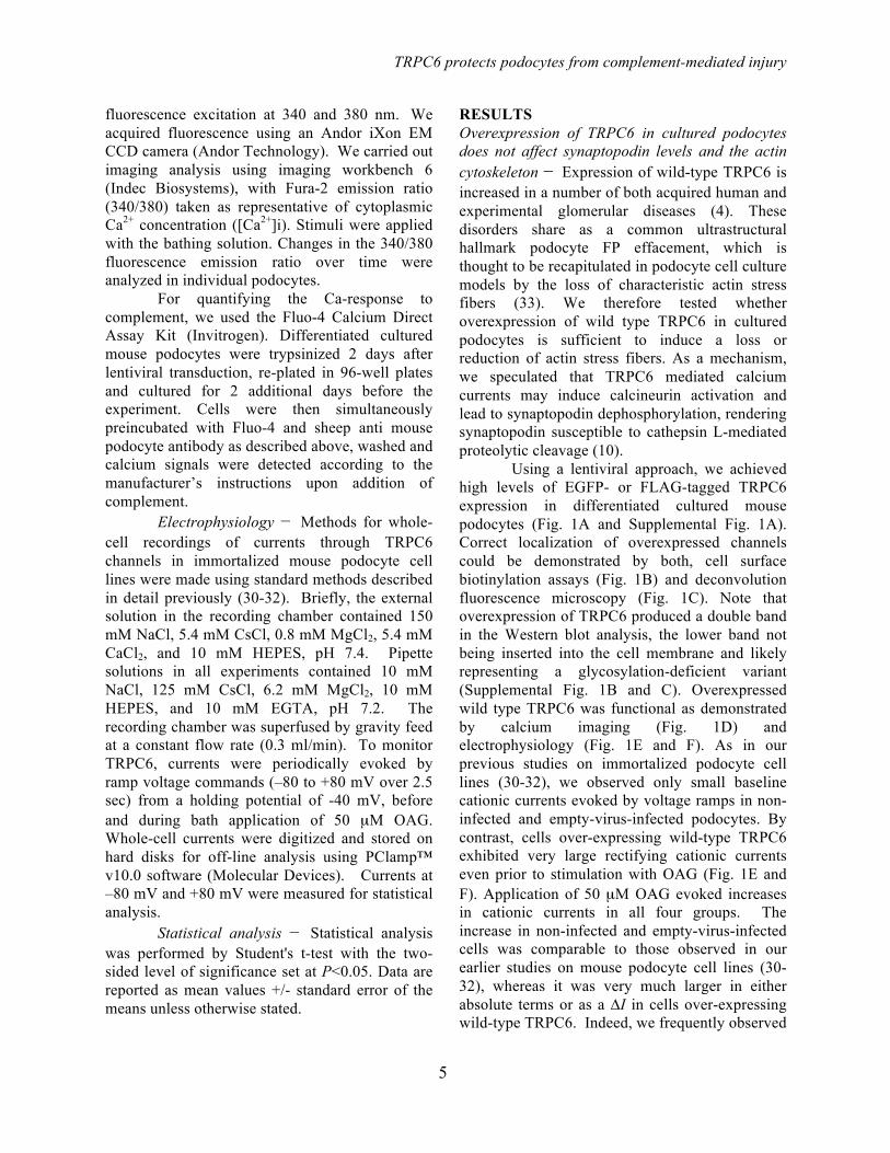
RESULTS Overexpression of TRPC6 in cultured podocytes does not affect synaptopodin levels and the actin cytoskeleton − Expression of wild-type TRPC6 is increased in a number of both acquired human and experimental glomerular diseases (4). These disorders share as a common ultrastructural hallmark podocyte FP effacement, which is thought to be recapitulated in podocyte cell culture models by the loss of characteristic actin stress fibers (33). We therefore tested whether overexpression of wild type TRPC6 in cultured podocytes is sufficient to induce a loss or reduction of actin stress fibers. As a mechanism, we speculated that TRPC6 mediated calcium currents may induce calcineurin activation and lead to synaptopodin dephosphorylation, rendering synaptopodin susceptible to cathepsin L-mediated proteolytic cleavage (10).
Using a lentiviral approach, we achieved high levels of EGFP- or FLAG-tagged TRPC6 expression in differentiated cultured mouse podocytes (Fig. 1A and Supplemental Fig. 1A). Correct localization of overexpressed channels could be demonstrated by both, cell surface biotinylation assays (Fig. 1B) and deconvolution fluorescence microscopy (Fig. 1C). Note that overexpression of TRPC6 produced a double band in the Western blot analysis, the lower band not being inserted into the cell membrane and likely representing a glycosylation-deficient variant (Supplemental Fig. 1B and C). Overexpressed wild type TRPC6 was functional as demonstrated by calcium imaging (Fig. 1D) and electrophysiology (Fig. 1E and F). As in our previous studies on immortalized podocyte cell lines (30-32), we observed only small baseline cationic currents evoked by voltage ramps in non-infected and empty-virus-infected podocytes. By contrast, cells over-expressing wild-type TRPC6 exhibited very large rectifying cationic currents even prior to stimulation with OAG (Fig. 1E and F). Application of 50 µM OAG evoked increases in cationic currents in all four groups. The increase in non-infected and empty-virus-infected cells was comparable to those observed in our earlier studies on mouse podocyte cell lines (30-32), whereas it was very much larger in either absolute terms or as a ∆I in cells over-expressing wild-type TRPC6. Indeed, we frequently observed
TRPC6 protects podocytes from complement-mediated injury
6
that OAG application caused those cells to die suddenly during the recording. By contrast, OAG-evoked currents were significantly smaller in cells expressing dnTRPC6 compared to all of the other groups, although they were still discernible (Fig. 1G).
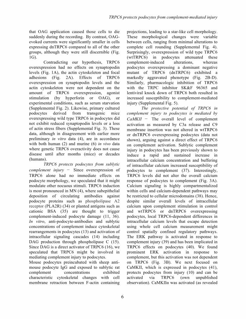
Contradicting our hypothesis, TRPC6 overexpression had no effects on synaptopodin levels (Fig. 1A), the actin cytoskeleton and focal adhesions (Fig. 2A). Effects of TRPC6 overexpression on synaptopodin levels and the actin cytoskeleton were not dependent on the amount of TRPC6 overexpression, agonist stimulation (by hyperforin or OAG), or experimental conditions, such as serum starvation (Supplemental Fig. 2). Likewise, primary cultured podocytes derived from transgenic mice overexpressing wild type TRPC6 in podocytes did not exhibit reduced synaptopodin levels or a loss of actin stress fibers (Supplemental Fig. 3). These data, although in disagreement with earlier more preliminary in vitro data (4), are in accordance with both human (2) and murine (6) in vivo data where genetic TRPC6 overactivity does not cause disease until after months (mice) or decades (humans).
TRPC6 protects podocytes from sublytic complement injury − Since overexpression of TRPC6 alone had no immediate effects on podocyte morphology, we speculated that it might modulate other nocuous stimuli. TRPC6 induction is most pronounced in MN (4), where subepithelial deposition of circulating antibodies against podocyte proteins such as phospholipase A2 receptor (PLA2R) (34) or planted antigens such as cationic BSA (35) are thought to trigger complement-induced podocyte damage (11, 36). In vitro, anti-podocyte-antibodies and sublytic concentrations of complement induce cytoskeletal rearrangements in podocytes (13) and activation of intracellular signaling cascades (14) including DAG production through phospholipase C (15). Since DAG is a direct activator of TRPC6 (16), we speculated that TRPC6 might be involved in mediating complement injury to podocytes. Mouse podocytes preincubated with sheep anti-mouse podocyte IgG and exposed to sublytic rat complement concentrations exhibited characteristic cytoskeletal changes with cell membrane retraction between F-actin containing
projections, leading to a star-like cell morphology. These morphological changes were variable between cells, ranging from minimal alterations to complete cell rounding (Supplemental Fig. 4). Surprisingly, overexpression of wild type TRPC6 (wtTRPC6) in podocytes attenuated these complement-induced alterations, whereas podocytes overexpressing a dominant negative mutant of TRPC6 (dnTRPC6) exhibited a markedly aggravated phenotype (Fig. 2B-D). Similarly, pharmacologic inhibition of TRPC6 with the TRPC inhibitor SK&F 96365 and lentiviral knock down of TRPC6 both resulted in increased susceptibility to complement-mediated injury (Supplemental Fig. 5).
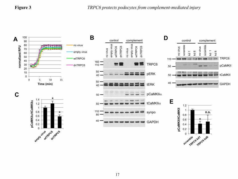
The protective potential of TRPC6 in complement injury to podocytes is mediated by CaMKII − The overall level of complement activation as measured by C3a release and C9 membrane insertion was not altered in wtTRPC6 or dnTRPC6 overexpressing podocytes (data not shown), arguing against a direct effect of TRPC6 on complement activation. Sublytic complement injury in podocytes has been previously shown to induce a rapid and sustained increase in intracellular calcium concentration and buffering of intracellular calcium increased susceptibility of podocytes to complement (37). Interestingly, TRPC6 levels did not alter the overall calcium response of podocytes to complement (Fig. 3A). Calcium signaling is highly compartmentalized within cells and calcium-dependent pathways may be restricted to cellular microdomains (38). Hence, despite similar overall levels of intracellular calcium upon complement stimulation in control and wtTRPC6 or dnTRPC6 overexpressing podocytes, local TRPC6-dependent differences in intracellular calcium levels that escape detection using whole cell calcium measurement might control spatially confined regulatory pathways. The ERK pathway is activated in response to complement injury (39) and has been implicated in TRPC6 effects on podocytes (40). We found prominent ERK activation in response to complement, but this activation was not dependent on TRPC6 (Fig. 3B). We next focused on CaMKII, which is expressed in podocytes (41), protects podocytes from injury (10) and can be activated via TRPC6 (own unpublished observation). CaMKIIα was activated (as revealed
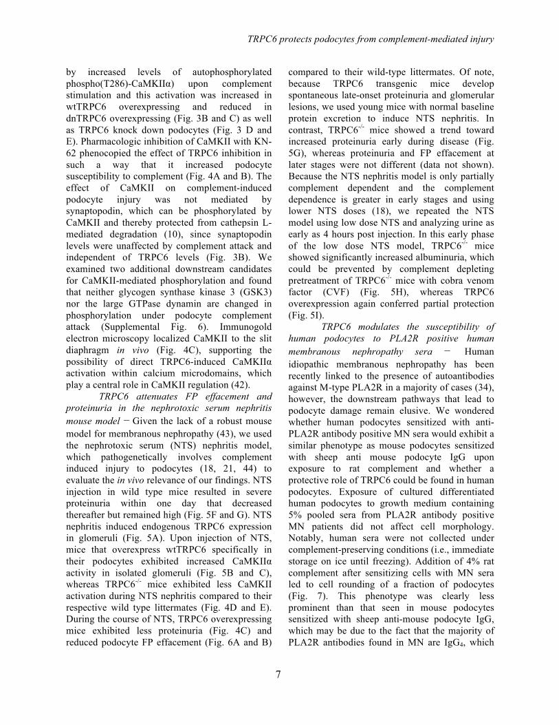
TRPC6 protects podocytes from complement-mediated injury
7
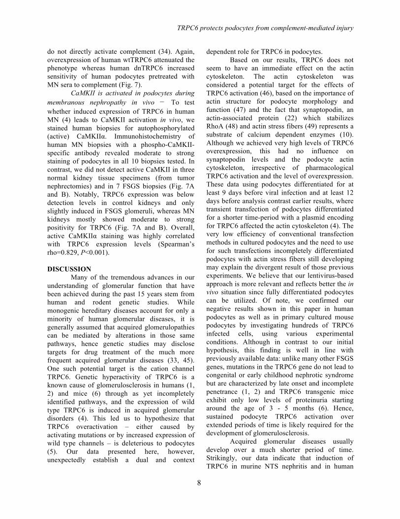
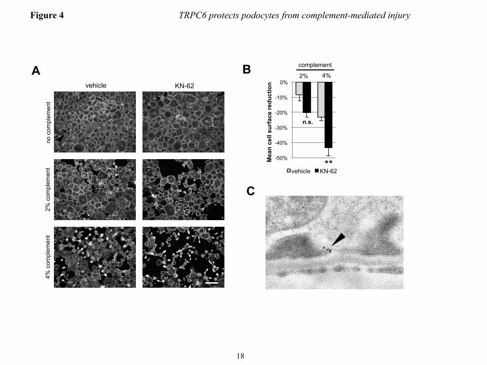
by increased levels of autophosphorylated phospho(T286)-CaMKIIα) upon complement stimulation and this activation was increased in wtTRPC6 overexpressing and reduced in dnTRPC6 overexpressing (Fig. 3B and C) as well as TRPC6 knock down podocytes (Fig. 3 D and E). Pharmacologic inhibition of CaMKII with KN-62 phenocopied the effect of TRPC6 inhibition in such a way that it increased podocyte susceptibility to complement (Fig. 4A and B). The effect of CaMKII on complement-induced podocyte injury was not mediated by synaptopodin, which can be phosphorylated by CaMKII and thereby protected from cathepsin L-mediated degradation (10), since synaptopodin levels were unaffected by complement attack and independent of TRPC6 levels (Fig. 3B). We examined two additional downstream candidates for CaMKII-mediated phosphorylation and found that neither glycogen synthase kinase 3 (GSK3) nor the large GTPase dynamin are changed in phosphorylation under podocyte complement attack (Supplemental Fig. 6). Immunogold electron microscopy localized CaMKII to the slit diaphragm in vivo (Fig. 4C), supporting the possibility of direct TRPC6-induced CaMKIIα activation within calcium microdomains, which play a central role in CaMKII regulation (42).
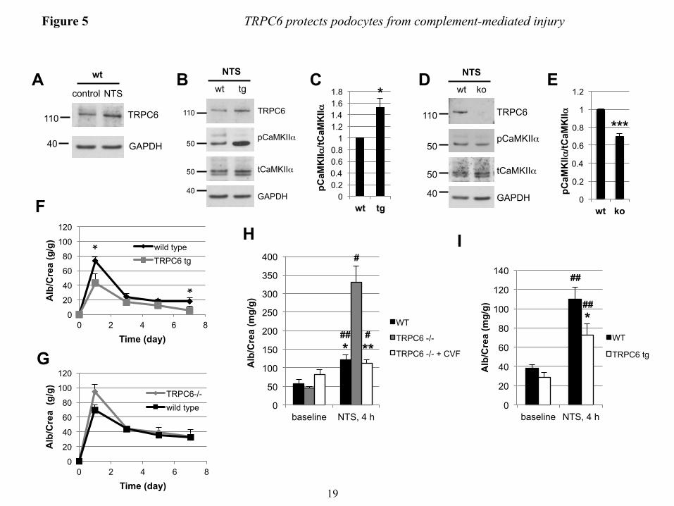
TRPC6 attenuates FP effacement and proteinuria in the nephrotoxic serum nephritis mouse model − Given the lack of a robust mouse model for membranous nephropathy (43), we used the nephrotoxic serum (NTS) nephritis model, which pathogenetically involves complement induced injury to podocytes (18, 21, 44) to evaluate the in vivo relevance of our findings. NTS injection in wild type mice resulted in severe proteinuria within one day that decreased thereafter but remained high (Fig. 5F and G). NTS nephritis induced endogenous TRPC6 expression in glomeruli (Fig. 5A). Upon injection of NTS, mice that overexpress wtTRPC6 specifically in their podocytes exhibited increased CaMKIIα activity in isolated glomeruli (Fig. 5B and C), whereas TRPC6-/- mice exhibited less CaMKII activation during NTS nephritis compared to their respective wild type littermates (Fig. 4D and E). During the course of NTS, TRPC6 overexpressing mice exhibited less proteinuria (Fig. 4C) and reduced podocyte FP effacement (Fig. 6A and B)
compared to their wild-type littermates. Of note, because TRPC6 transgenic mice develop spontaneous late-onset proteinuria and glomerular lesions, we used young mice with normal baseline protein excretion to induce NTS nephritis. In contrast, TRPC6-/- mice showed a trend toward increased proteinuria early during disease (Fig. 5G), whereas proteinuria and FP effacement at later stages were not different (data not shown). Because the NTS nephritis model is only partially complement dependent and the complement dependence is greater in early stages and using lower NTS doses (18), we repeated the NTS model using low dose NTS and analyzing urine as early as 4 hours post injection. In this early phase of the low dose NTS model, TRPC6-/- mice showed significantly increased albuminuria, which could be prevented by complement depleting pretreatment of TRPC6-/- mice with cobra venom factor (CVF) (Fig. 5H), whereas TRPC6 overexpression again conferred partial protection (Fig. 5I).
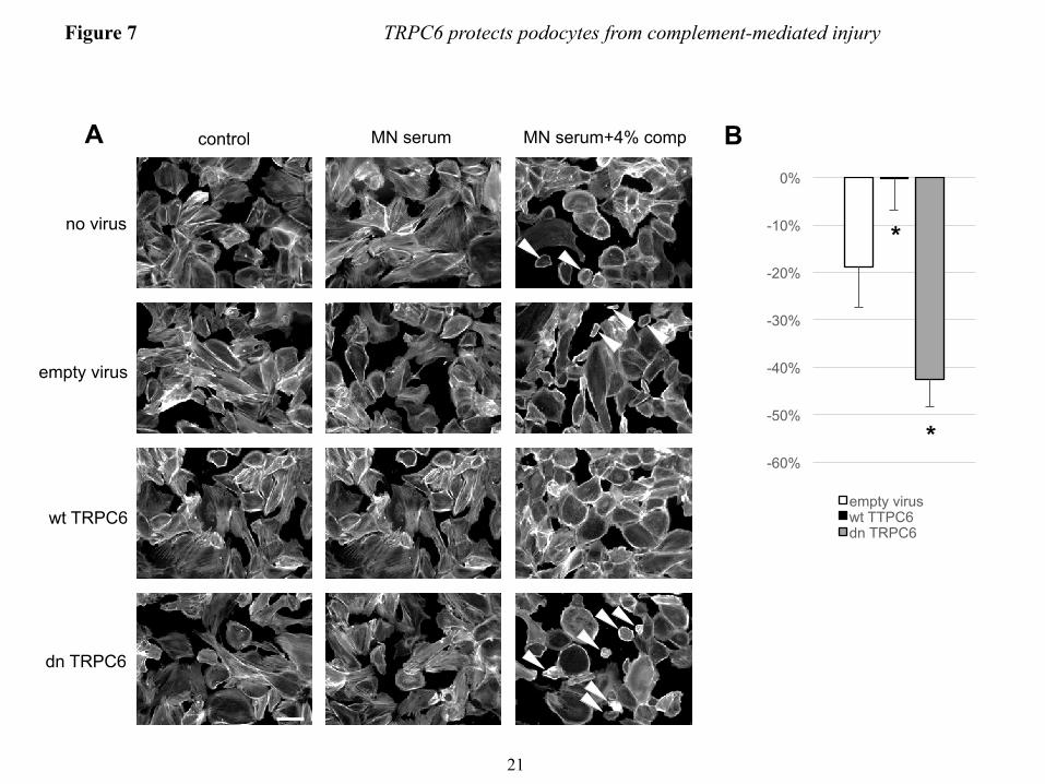
TRPC6 modulates the susceptibility of human podocytes to PLA2R positive human membranous nephropathy sera − Human idiopathic membranous nephropathy has been recently linked to the presence of autoantibodies against M-type PLA2R in a majority of cases (34), however, the downstream pathways that lead to podocyte damage remain elusive. We wondered whether human podocytes sensitized with anti-PLA2R antibody positive MN sera would exhibit a similar phenotype as mouse podocytes sensitized with sheep anti mouse podocyte IgG upon exposure to rat complement and whether a protective role of TRPC6 could be found in human podocytes. Exposure of cultured differentiated human podocytes to growth medium containing 5% pooled sera from PLA2R antibody positive MN patients did not affect cell morphology. Notably, human sera were not collected under complement-preserving conditions (i.e., immediate storage on ice until freezing). Addition of 4% rat complement after sensitizing cells with MN sera led to cell rounding of a fraction of podocytes (Fig. 7). This phenotype was clearly less prominent than that seen in mouse podocytes sensitized with sheep anti-mouse podocyte IgG, which may be due to the fact that the majority of PLA2R antibodies found in MN are IgG4, which
TRPC6 protects podocytes from complement-mediated injury
8
do not directly activate complement (34). Again, overexpression of human wtTRPC6 attenuated the phenotype whereas human dnTRPC6 increased sensitivity of human podocytes pretreated with MN sera to complement (Fig. 7).
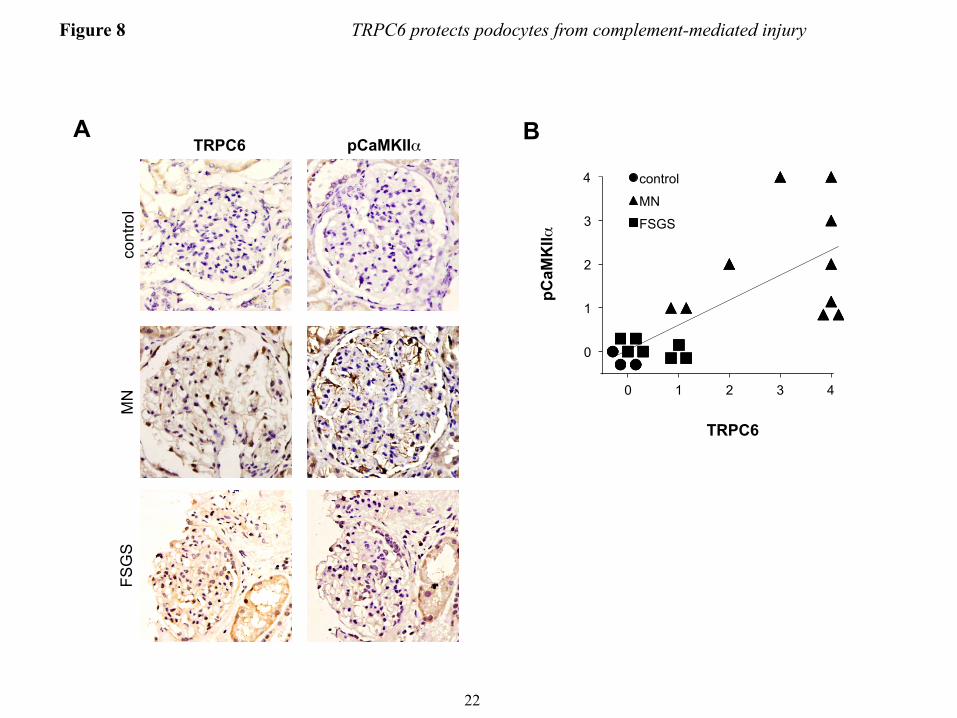
CaMKII is activated in podocytes during membranous nephropathy in vivo − To test whether induced expression of TRPC6 in human MN (4) leads to CaMKII activation in vivo, we stained human biopsies for autophosphorylated (active) CaMKIIα. Immunohistochemistry of human MN biopsies with a phospho-CaMKII-specific antibody revealed moderate to strong staining of podocytes in all 10 biopsies tested. In contrast, we did not detect active CaMKII in three normal kidney tissue specimens (from tumor nephrectomies) and in 7 FSGS biopsies (Fig. 7A and B). Notably, TRPC6 expression was below detection levels in control kidneys and only slightly induced in FSGS glomeruli, whereas MN kidneys mostly showed moderate to strong positivity for TRPC6 (Fig. 7A and B). Overall, active CaMKIIα staining was highly correlated with TRPC6 expression levels (Spearman’s rho=0.829, P<0.001). DISCUSSION
Many of the tremendous advances in our understanding of glomerular function that have been achieved during the past 15 years stem from human and rodent genetic studies. While monogenic hereditary diseases account for only a minority of human glomerular diseases, it is generally assumed that acquired glomerulopathies can be mediated by alterations in those same pathways, hence genetic studies may disclose targets for drug treatment of the much more frequent acquired glomerular diseases (33, 45). One such potential target is the cation channel TRPC6. Genetic hyperactivity of TRPC6 is a known cause of glomerulosclerosis in humans (1, 2) and mice (6) through as yet incompletely identified pathways, and the expression of wild type TRPC6 is induced in acquired glomerular disorders (4). This led us to hypothesize that TRPC6 overactivation – either caused by activating mutations or by increased expression of wild type channels – is deleterious to podocytes (5). Our data presented here, however, unexpectedly establish a dual and context
dependent role for TRPC6 in podocytes. Based on our results, TRPC6 does not
seem to have an immediate effect on the actin cytoskeleton. The actin cytoskeleton was considered a potential target for the effects of TRPC6 activation (46), based on the importance of actin structure for podocyte morphology and function (47) and the fact that synaptopodin, an actin-associated protein (22) which stabilizes RhoA (48) and actin stress fibers (49) represents a substrate of calcium dependent enzymes (10). Although we achieved very high levels of TRPC6 overexpression, this had no influence on synaptopodin levels and the podocyte actin cytoskeleton, irrespective of pharmacological TRPC6 activation and the level of overexpression. These data using podocytes differentiated for at least 9 days before viral infection and at least 12 days before analysis contrast earlier results, where transient transfection of podocytes differentiated for a shorter time-period with a plasmid encoding for TRPC6 affected the actin cytoskeleton (4). The very low efficiency of conventional transfection methods in cultured podocytes and the need to use for such transfections incompletely differentiated podocytes with actin stress fibers still developing may explain the divergent result of those previous experiments. We believe that our lentivirus-based approach is more relevant and reflects better the in vivo situation since fully differentiated podocytes can be utilized. Of note, we confirmed our negative results shown in this paper in human podocytes as well as in primary cultured mouse podocytes by investigating hundreds of TRPC6 infected cells, using various experimental conditions. Although in contrast to our initial hypothesis, this finding is well in line with previously available data: unlike many other FSGS genes, mutations in the TRPC6 gene do not lead to congenital or early childhood nephrotic syndrome but are characterized by late onset and incomplete penetrance (1, 2) and TRPC6 transgenic mice exhibit only low levels of proteinuria starting around the age of 3 - 5 months (6). Hence, sustained podocyte TRPC6 activation over extended periods of time is likely required for the development of glomerulosclerosis.
Acquired glomerular diseases usually develop over a much shorter period of time. Strikingly, our data indicate that induction of TRPC6 in murine NTS nephritis and in human
TRPC6 protects podocytes from complement-mediated injury
9
MN might in fact not cause podocyte damage but rather play a protective role against complement-induced damage. Thus, the overall effect of TRPC6 in podocytes likely depends on the presence or absence of complement and on whether its induction is transient or sustained. In fact, it is interesting that no physiological role for TRPC6 in podocytes has so far been shown, and TRPC6-/- mice do not exhibit a renal phenotype (50). Our data are the first to show a potential beneficial role of podocyte TRPC6, which upon prolonged stimulation may turn into a detrimental signal.
The effects of podocyte TRPC6 overexpression and knock out on the severity of NTS nephritis were limited, a fact that may be accounted for by a number of reasons. First, the NTS nephritis mouse model is only partially complement dependent, and particularly at high antibody doses, complement-independent pathways contribute to glomerular damage (18). Consequently, we observed a much more pronounced effect of TRPC6 deficiency in a low dose NTS nephritis model at a very early stage. Second, the absence of TRPC6 both in vitro and in vivo resulted in a roughly 30-50% reduction of CaMKII autophosphorylation. Hence, TRPC6-independent pathways might also contribute to CaMKII activation and autophosphorylation. Third, the TRPC6 - CaMKII pathway is most likely only one among others that regulate complement-mediated injury.
The overall intracellular calcium concentration in response to complement was not affected by TRPC6 levels to a detectable extent. Thus, the majority of overall calcium influx upon complement stimulation must be mediated by channels other than TRPC6, most likely through the pore forming membrane attack complex C5b-9. It is well known that calcium signaling is highly compartmentalized within cells and calcium-dependent pathways may be restricted to microdomains (38). Hence, despite similar overall levels of intracellular calcium upon complement stimulation in wtTRPC6 and dnTRPC6 overexpressing podocytes, local TRPC6-dependent differences in intracellular calcium levels that escape detection using whole cell calcium measurement might control spatially confined regulatory pathways. Likewise, the cellular effects of CaMKII activity largely depend
on its subcellular targeting and the resulting proximity to substrates, which is regulated by multi-site phosphorylation (42). Our immunogold EM-data showing subcellular localization of CaMKII in direct proximity to the slit diaphragm in vivo support the possibility of close functional interaction with TRPC6. The downstream effector pathways of CaMKII during complement injury in podocytes remain to be defined. We did not detect any effect on synaptopodin, which has been suggested as a target of CaMKII phosphorylation (10). However, podocytes express many known targets of CaMKII phosphorylation. In addition, CaMKII may act directly on the actin cytoskeleton (51). Furthermore, although our data provide evidence that CaMKII is involved in mediating the protective effects of TRPC6 on complement injury to podocytes, we cannot exclude the existence of additional pathways that mediate the downstream effects of TRPC6.
Our results have important potential clinical implications. First, they suggest CaMKII modulation as a potential therapeutic target in complement-mediated glomerular diseases. Second, and more importantly, they question whether TRPC6 represents an ideal drug target for the treatment of acquired glomerular diseases, such as MN, diabetic nephropathy, and non-genetic FSGS. Whether such pharmacological interventions would be beneficial or detrimental may critically depend on the pathophysiological context, including the presence or absence of complement activation. Acknowledgements
This work was supported in part by grants from the National Institute of Health (DK073495, DK089394 and DK101350 to JR). A.D.K. was supported by a grant of the Swiss National Science Foundation, a grant by the Swiss Society for Medical and Biological Scholarships and an Amgen-FROMO scholarship.
Human MN sera were kindly provided by David Salant and Laurence Beck (both from Boston University). We thank Madhusudhan Bhat, M.Sc. (All India Institute of Medical Sciences) for help with immunohistochemistry standardization and Alejandro Caicedo (University of Miami) for his assistance with calcium imaging.
TRPC6 protects podocytes from complement-mediated injury
10
References 1. Winn, M. P., Conlon, P. J., Lynn, K. L., Farrington, M. K., Creazzo. T., Hawkins, A. F., Daskalakis,
N., Kwan, S. Y., Ebersviller, S., Burchette, J. L., Pericak-Vance, M. A., Howell, D. N., Vance, J. M., and Rosenberg, P. B. (2005) A mutation in the TRPC6 cation channel causes familial focal segmental glomerulosclerosis. Science 308, 1801-1804
2. Reiser, J., Polu, K. R., Moller, C. C., Kenlan, P., Altintas, M. M., Wei, C., Faul, C., Herbert, S., Villegas, I., Avila-Casado, C., McGee, M., Sugimoto, H., Brown, D., Kalluri, R., Mundel, P., Smith, P. L., Clapham, D. E., and Pollak, M. R. (2005) TRPC6 is a glomerular slit diaphragm-associated channel required for normal renal function. Nat. Genet. 37, 739-744
3. Heeringa, S. F., Moller, C. C., Du, J., Yue, L., Hinkes, B., Chernin, G., Vlangos, C. N., Hoyer, P. F., Reiser, J., and Hildebrandt, F. (2009) A novel TRPC6 mutation that causes childhood FSGS. PLoS One 4, e7771
4. Moller, C. C., Wei, C., Altintas, M. M., Li, J., Greka, A., Ohse, T., Pippin, J. W., Rastaldi, M. P., Wawersik, S., Schiavi, S., Henger, A., Kretzler, M., Shankland, S. J., and Reiser, J. (2007) Induction of TRPC6 channel in acquired forms of proteinuric kidney disease. J. Am. Soc. Nephrol. 18, 29-36
5. Moller, C. C., Flesche, J., and Reiser, J. (2009) Sensitizing the slit diaphragm with TRPC6 ion channels. J. Am. Soc. Nephrol. 20, 950-953
6. Krall, P., Canales, C. P., Kairath, P., Carmona-Mora, P., Molina, J., Carpio, J. D., Ruiz, P., Mezzano, S. A., Li, J., Wei, C., Reiser, J., Young, J. I., and Walz, K. (2010) Podocyte-specific overexpression of wild type or mutant trpc6 in mice is sufficient to cause glomerular disease. PLoS One 5, e12859
7. Kuwahara, K., Wang, Y., McAnally, J., Richardson, J. A., Bassel-Duby, R., Hill, J. A., and Olson, E. N. (2006) TRPC6 fulfills a calcineurin signaling circuit during pathologic cardiac remodeling. J. Clin. Invest. 116, 3114-3126
8. Schlondorff, J., Del Camino, D., Carrasquillo, R., Lacey, V., and Pollak, M. R. (2009) TRPC6 mutations associated with focal segmental glomerulosclerosis cause constitutive activation of NFAT-dependent transcription. Am. J. Physiol. Cell. Physiol. 296, C558-569
9. Nijenhuis, T., Sloan, A. J., Hoenderop, J. G., Flesche, J., van Goor, H., Kistler, A. D., Bakker, M., Bindels, R. J., de Boer, R. A., Möller, C. C., Hamming, I., Navis, G., Wetzels, J. F., Berden, J. H., Reiser, J., Faul, C., and van der Vlag, J. (2011) Angiotensin II contributes to podocyte injury by increasing TRPC6 expression via an NFAT-mediated positive feedback signaling pathway. Am. J. Pathol. 179, 1719-1732
10. Faul, C., Donnelly, M., Merscher-Gomez, S., Chang, Y. H., Franz, S., Delfgaauw, J., Chang, J. M., Choi, H. Y., Campbell, K. N., Kim, K., Reiser, J., and Mundel, P. (2008) The actin cytoskeleton of kidney podocytes is a direct target of the antiproteinuric effect of cyclosporine A. Nat. Med. 14, 931-938
11. Glassock, R. J. (2010) The pathogenesis of idiopathic membranous nephropathy: a 50-year odyssey. Am. J. Kidney Dis. 56, 157-167
12. Glassock, R. J. (2012) The pathogenesis of membranous nephropathy: evolution and revolution. Curr. Opin. Nephrol. Hypertens. 21, 235-242
13. Topham, P. S., Haydar, S. A., Kuphal, R., Lightfoot, J. D., and Salant, D. J. (1999) Complement-mediated injury reversibly disrupts glomerular epithelial cell actin microfilaments and focal adhesions. Kidney Int. 55, 1763-1775
14. Cybulsky, A. V., Quigg, R. J., and Salant, D. J. (2005) Experimental membranous nephropathy redux. Am. J. Physiol. Renal Physiol. 289, F660-F671
15. Cybulsky, A. V., Salant, D. J., Quigg, R. J., Badalamenti, J., and Bonventre, J. V. (1989) Complement C5b-9 complex activates phospholipases in glomerular epithelial cells. Am. J. Physiol. 257, F826-F836
16. Hofmann, T., Obukhov, A. G., Schaefer, M., Harteneck, C., Gudermann, T., and Schultz, G. (1999) Direct activation of human TRPC6 and TRPC3 channels by diacylglycerol. Nature 397, 259-263
TRPC6 protects podocytes from complement-mediated injury
11
17. Quigg, R. J., Kozono, Y., Berthiaume, D., Lim, A., Salant, D. J., Weinfeld, A., Griffin, P., Kremmer, E., and Holers, V. M. (1998) Blockade of antibody-induced glomerulonephritis with Crry-Ig, a soluble murine complement inhibitor. J. Immunol. 160, 4553-4560
18. Hebert, M. J., Takano, T., Papayianni, A., Rennke, H. G., Minto, A., Salant, D. J., Carroll, M. C., and Brady, H. R. (1998) Acute nephrotoxic serum nephritis in complement knockout mice: relative roles of the classical and alternate pathways in neutrophil recruitment and proteinuria. Nephrol. Dial. Transplant. 13, 2799-2803
19. Lin, F., Emancipator, S. N., Salant, D. J., and Medof, M. E. (2002) Decay-accelerating factor confers protection against complement-mediated podocyte injury in acute nephrotoxic nephritis. Lab. Invest. 82, 563-569
20. Lin, F., Salant, D. J., Meyerson, H., Emancipator, S., Morgan, B. P., and Medof, M. E. (2004) Respective roles of decay-accelerating factor and CD59 in circumventing glomerular injury in acute nephrotoxic serum nephritis. J Immunol. 172, 2636-2642
21. Turnberg, D., Botto, M., Warren, J., Morgan, B. P., Walport, M. J., and Cook, H. T. (2003) CD59a deficiency exacerbates accelerated nephrotoxic nephritis in mice. J. Am. Soc. Nephrol. 14, 2271-2279
22. Mundel, P., Heid, H. W., Mundel, T. M., Kruger, M., Reiser, J., and Kriz, W. (1997) Synaptopodin: an actin-associated protein in telencephalic dendrites and renal podocytes. J. Cell Biol. 139, 193-204
23. Dietrich, A., Mederos y Schnitzler, M, Gollasch, M., Gross, V., Storch, U., Dubrovska, G., Obst, M., Yildirim, E., Salanova, B., Kalwa, H., Essin, K., Pinkenburg, O., Luft, F. C., Gudermann, T., and Birnbaumer, L. (2005) Increased vascular smooth muscle contractility in TRPC6-/- mice. Mol. Cell. Biol. 25, 6980-6989
24. Ophascharoensuk, V., Pippin, J. W., Gordon, K. L., Shankland, S. J., Couser, W. G., and Johnson, R. J. (1998) Role of intrinsic renal cells versus infiltrating cells in glomerular crescent formation. Kidney Int. 54, 416-425
25. Kerjaschki, D., Exner, M., Ullrich, R., Susani, M., Curtiss, L. K., Witztum, J. L., Farquhar, M. G., and Orlando, R. A. (1997) Pathogenic antibodies inhibit the binding of apolipoproteins to megalin/gp330 in passive Heymann nephritis. J. Clin. Invest. 100, 2303-2309
26. Mundel, P., Reiser, J., Zuniga Mejia Borja, A., Pavenstadt, H., Davidson, G. R., Kriz, W., and Zeller, R. (1997) Rearrangements of the cytoskeleton and cell contacts induce process formation during differentiation of conditionally immortalized mouse podocyte cell lines. Exp. Cell. Res. 236, 248-258
27. Saleem, M. A., O'Hare, M. J., Reiser, J., Coward, R. J., Inward, C. D., Farren, T., Xing, C. Y., Ni, L., Mathieson, P. W., and Mundel, P. (2002) A conditionally immortalized human podocyte cell line demonstrating nephrin and podocin expression. J. Am. Soc. Nephrol. 13, 630-638
28. Hofmann, T., Schaefer, M., Schultz, G., and Gudermann, T. (2002) Subunit composition of mammalian transient receptor potential channels in living cells. Proc. Natl. Acad. Sci. USA 99, 7461-7466
29. Takemoto, M., Asker, N., Gerhardt, H., Lundkvist, A., Johansson, B. R., Saito, Y., and Betsholtz, C. (2002) A new method for large scale isolation of kidney glomeruli from mice. Am. J. Pathol. 161, 799-805
30. Kim, E. Y., Anderson, M., and Dryer, S. E. (2012) Insulin increases surface expression of TRPC6 channels in podocytes: role of NADPH oxidases and reactive oxygen species. Am. J. Physiol. Renal Physiol. 302, F298-F307
31. Kim, E. Y., Anderson, M., Wilson, C., Hagmann, H., Benzing, T., and Dryer, S. E. (2013) NOX2 interacts with podocyte TRPC6 channels and contributes to their activation by diacylglycerol: Essential role of podocin in formation of this complex. Am. J. Physiol. Cell. Physiol. Epub ahead of print, doi:10.1152/ajpcell.00191.2013
32. Anderson, M., Kim, E. Y., Hagmann, H., Benzing, T., Dryer, S. E. (2013) Opposing effects of podocin on the gating of podocyte TRPC6 channels evoked by membrane stretch or diacylglycerol. Am. J. Physiol. Cell. Physiol. 305, C276-C289
TRPC6 protects podocytes from complement-mediated injury
12
33. Reiser, J., Gupta, V., and Kistler, A. D. (2010) Toward the development of podocyte-specific drugs. Kidney Int. 77, 662-668
34. Beck, L. H. Jr., Bonegio, R. G., Lambeau, G., Beck, D. M., Powell, D. W., Cummins, T. D., Klein, J. B., and Salant, D. J. (2009) M-type phospholipase A2 receptor as target antigen in idiopathic membranous nephropathy. N. Engl. J. Med. 361, 11-21
35. Debiec, H., Lefeu, F., Kemper, M. J., Niaudet, P., Deschênes, G., Remuzzi, G., Ulinski, T., and Ronco, P. (2011) Early-childhood membranous nephropathy due to cationic bovine serum albumin. N. Engl. J. Med. 364, 2101-2110
36. Cybulsky, A. V. (2011) Membranous nephropathy. Contrib. Nephrol. 169, 107-125 37. Cybulsky, A. V., Bonventre, J. V., Quigg, R. J., Lieberthal, W., and Salant, D. J. (1990) Cytosolic
calcium and protein kinase C reduce complement-mediated glomerular epithelial injury. Kidney Int. 38, 803-811
38. Laude, A. J., and Simpson, A. W. (2009) Compartmentalized signalling: Ca2+ compartments, microdomains and the many facets of Ca2+ signalling. FEBS J. 276, 1800-1816
39. Takano, T., and Cybulsky, A. V. (2000) Complement C5b-9-mediated arachidonic acid metabolism in glomerular epithelial cells: role of cyclooxygenase-1 and -2. Am. J. Pathol. 156, 2091-2101
40. Chiluiza, D., Krishna, S., Schumacher, V. A., and Schlondorff, J. (2013) Gain-of-function mutations in transient receptor potential C6 (TRPC6) activate extracellular-signal-regulated kinases Erk1/2. J. Biol. Chem. 288, 18407-18420
41. Giardino, L., Armelloni, S., Corbelli, A., Mattinzoli, D., Zennaro, C., Guerrot, D., Tourrel, F., Ikehata, M., Li, M., Berra, S., Carraro, M., Messa, P., and Rastaldi, M. P. (2009) Podocyte glutamatergic signaling contributes to the function of the glomerular filtration barrier. J. Am. Soc. Nephrol. 20, 1929-1940
42. Skelding, K. A., and Rostas, J. A. (2009) Regulation of CaMKII in vivo: the importance of targeting and the intracellular microenvironment. Neurochem. Res. 34, 1792-1804
43. Jefferson, J. A., Pippin, J. W., and Shankland, S. J. (2010) Experimental models of membranous nephropathy. Drug Discov. Today Dis. Models 7, 27-33
44. Quigg, R. J., He, C., Lim, A., Berthiaume, D., Alexander, J. J., Kraus, D., Holers, V. M. (1998) Transgenic mice overexpressing the complement inhibitor crry as a soluble protein are protected from antibody-induced glomerular injury. J. Exp. Med. 188, 1321-1331
45. Lavin, P. J., Gbadegesin, R., Damodaran, T. V., and Winn, M. P. (2008) Therapeutic targets in focal and segmental glomerulosclerosis. Curr. Opin. Nephrol. Hypertens. 17, 386-392
46. Schlondorff, J. S., and Pollak, M. R. (2006) TRPC6 in glomerular health and disease: what we know and what we believe. Semin. Cell. Dev. Biol. 17, 667-674
47. Faul, C., Asanuma, K., Yanagida-Asanuma, E., Kim, K., and Mundel, P. (2007) Actin up: regulation of podocyte structure and function by components of the actin cytoskeleton. Trends Cell. Biol. 17, 428-437
48. Asanuma, K., Yanagida-Asanuma, E., Faul, C., Tomino, Y., Kim, K., and Mundel, P. (2006) Synaptopodin orchestrates actin organization and cell motility via regulation of RhoA signalling. Nat. Cell Biol. 8, 485-491
49. Asanuma, K., Kim, K., Oh, J., Giardino, L., Chabanis, S., Faul, C., Reiser, J., and Mundel, P. (2005) Synaptopodin regulates the actin-bundling activity of alpha-actinin in an isoform-specific manner. J. Clin. Invest. 115, 1188-1198
50. Eckel, J., Lavin, P. J., Finch, E. A., Mukerji, N., Burch, J., Gbadegesin, R., Wu, G., Bowling, B., Byrd, A., Hall, G., Sparks, M., Zhang, Z. S., Homstad, A., Barisoni, L., Birbaumer, L., Rosenberg, P., and Winn, M. P. (2011) TRPC6 enhances angiotensin II-induced albuminuria. J. Am. Soc. Nephrol. 22, 526-535
51. Shen, K., Teruel, M. N., Subramanian, K., and Meyer, T. (1998) CaMKIIbeta functions as an F-actin targeting module that localizes CaMKIIalpha/beta heterooligomers to dendritic spines. Neuron 21, 593-606
TRPC6 protects podocytes from complement-mediated injury
13
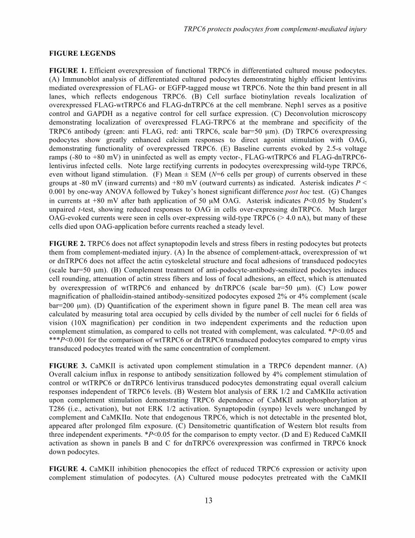
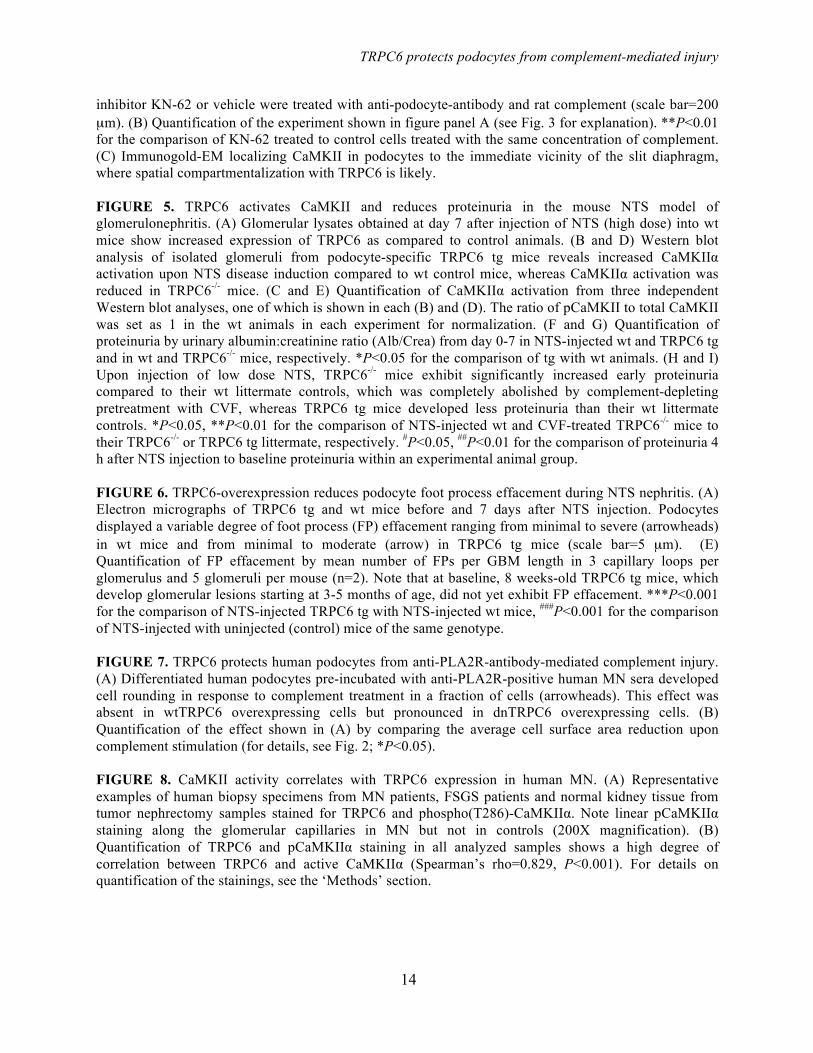
FIGURE LEGENDS FIGURE 1. Efficient overexpression of functional TRPC6 in differentiated cultured mouse podocytes. (A) Immunoblot analysis of differentiated cultured podocytes demonstrating highly efficient lentivirus mediated overexpression of FLAG- or EGFP-tagged mouse wt TRPC6. Note the thin band present in all lanes, which reflects endogenous TRPC6. (B) Cell surface biotinylation reveals localization of overexpressed FLAG-wtTRPC6 and FLAG-dnTRPC6 at the cell membrane. Neph1 serves as a positive control and GAPDH as a negative control for cell surface expression. (C) Deconvolution microscopy demonstrating localization of overexpressed FLAG-TRPC6 at the membrane and specificity of the TRPC6 antibody (green: anti FLAG, red: anti TRPC6, scale bar=50 µm). (D) TRPC6 overexpressing podocytes show greatly enhanced calcium responses to direct agonist stimulation with OAG, demonstrating functionality of overexpressed TRPC6. (E) Baseline currents evoked by 2.5-s voltage ramps (-80 to +80 mV) in uninfected as well as empty vector-, FLAG-wtTRPC6 and FLAG-dnTRPC6-lentivirus infected cells. Note large rectifying currents in podocytes overexpressing wild-type TRPC6, even without ligand stimulation. (F) Mean ± SEM (N=6 cells per group) of currents observed in these groups at -80 mV (inward currents) and +80 mV (outward currents) as indicated. Asterisk indicates P < 0.001 by one-way ANOVA followed by Tukey’s honest significant difference post hoc test. (G) Changes in currents at +80 mV after bath application of 50 µM OAG. Asterisk indicates P<0.05 by Student’s unpaired t-test, showing reduced responses to OAG in cells over-expressing dnTRPC6. Much larger OAG-evoked currents were seen in cells over-expressing wild-type TRPC6 (> 4.0 nA), but many of these cells died upon OAG-application before currents reached a steady level. FIGURE 2. TRPC6 does not affect synaptopodin levels and stress fibers in resting podocytes but protects them from complement-mediated injury. (A) In the absence of complement-attack, overexpression of wt or dnTRPC6 does not affect the actin cytoskeletal structure and focal adhesions of transduced podocytes (scale bar=50 µm). (B) Complement treatment of anti-podocyte-antibody-sensitized podocytes induces cell rounding, attenuation of actin stress fibers and loss of focal adhesions, an effect, which is attenuated by overexpression of wtTRPC6 and enhanced by dnTRPC6 (scale bar=50 µm). (C) Low power magnification of phalloidin-stained antibody-sensitized podocytes exposed 2% or 4% complement (scale bar=200 µm). (D) Quantification of the experiment shown in figure panel B. The mean cell area was calculated by measuring total area occupied by cells divided by the number of cell nuclei for 6 fields of vision (10X magnification) per condition in two independent experiments and the reduction upon complement stimulation, as compared to cells not treated with complement, was calculated. *P<0.05 and ***P<0.001 for the comparison of wtTRPC6 or dnTRPC6 transduced podocytes compared to empty virus transduced podocytes treated with the same concentration of complement. FIGURE 3. CaMKII is activated upon complement stimulation in a TRPC6 dependent manner. (A) Overall calcium influx in response to antibody sensitization followed by 4% complement stimulation of control or wtTRPC6 or dnTRPC6 lentivirus transduced podocytes demonstrating equal overall calcium responses independent of TRPC6 levels. (B) Western blot analysis of ERK 1/2 and CaMKIIα activation upon complement stimulation demonstrating TRPC6 dependence of CaMKII autophosphorylation at T286 (i.e., activation), but not ERK 1/2 activation. Synaptopodin (synpo) levels were unchanged by complement and CaMKIIα. Note that endogenous TRPC6, which is not detectable in the presented blot, appeared after prolonged film exposure. (C) Densitometric quantification of Western blot results from three independent experiments. *P<0.05 for the comparison to empty vector. (D and E) Reduced CaMKII activation as shown in panels B and C for dnTRPC6 overexpression was confirmed in TRPC6 knock down podocytes. FIGURE 4. CaMKII inhibition phenocopies the effect of reduced TRPC6 expression or activity upon complement stimulation of podocytes. (A) Cultured mouse podocytes pretreated with the CaMKII
TRPC6 protects podocytes from complement-mediated injury
14
inhibitor KN-62 or vehicle were treated with anti-podocyte-antibody and rat complement (scale bar=200 µm). (B) Quantification of the experiment shown in figure panel A (see Fig. 3 for explanation). **P<0.01 for the comparison of KN-62 treated to control cells treated with the same concentration of complement. (C) Immunogold-EM localizing CaMKII in podocytes to the immediate vicinity of the slit diaphragm, where spatial compartmentalization with TRPC6 is likely. FIGURE 5. TRPC6 activates CaMKII and reduces proteinuria in the mouse NTS model of glomerulonephritis. (A) Glomerular lysates obtained at day 7 after injection of NTS (high dose) into wt mice show increased expression of TRPC6 as compared to control animals. (B and D) Western blot analysis of isolated glomeruli from podocyte-specific TRPC6 tg mice reveals increased CaMKIIα activation upon NTS disease induction compared to wt control mice, whereas CaMKIIα activation was reduced in TRPC6-/- mice. (C and E) Quantification of CaMKIIα activation from three independent Western blot analyses, one of which is shown in each (B) and (D). The ratio of pCaMKII to total CaMKII was set as 1 in the wt animals in each experiment for normalization. (F and G) Quantification of proteinuria by urinary albumin:creatinine ratio (Alb/Crea) from day 0-7 in NTS-injected wt and TRPC6 tg and in wt and TRPC6-/- mice, respectively. *P<0.05 for the comparison of tg with wt animals. (H and I) Upon injection of low dose NTS, TRPC6-/- mice exhibit significantly increased early proteinuria compared to their wt littermate controls, which was completely abolished by complement-depleting pretreatment with CVF, whereas TRPC6 tg mice developed less proteinuria than their wt littermate controls. *P<0.05, **P<0.01 for the comparison of NTS-injected wt and CVF-treated TRPC6-/- mice to their TRPC6-/- or TRPC6 tg littermate, respectively. #P<0.05, ##P<0.01 for the comparison of proteinuria 4 h after NTS injection to baseline proteinuria within an experimental animal group. FIGURE 6. TRPC6-overexpression reduces podocyte foot process effacement during NTS nephritis. (A) Electron micrographs of TRPC6 tg and wt mice before and 7 days after NTS injection. Podocytes displayed a variable degree of foot process (FP) effacement ranging from minimal to severe (arrowheads) in wt mice and from minimal to moderate (arrow) in TRPC6 tg mice (scale bar=5 µm). (E) Quantification of FP effacement by mean number of FPs per GBM length in 3 capillary loops per glomerulus and 5 glomeruli per mouse (n=2). Note that at baseline, 8 weeks-old TRPC6 tg mice, which develop glomerular lesions starting at 3-5 months of age, did not yet exhibit FP effacement. ***P<0.001 for the comparison of NTS-injected TRPC6 tg with NTS-injected wt mice, ###P<0.001 for the comparison of NTS-injected with uninjected (control) mice of the same genotype. FIGURE 7. TRPC6 protects human podocytes from anti-PLA2R-antibody-mediated complement injury. (A) Differentiated human podocytes pre-incubated with anti-PLA2R-positive human MN sera developed cell rounding in response to complement treatment in a fraction of cells (arrowheads). This effect was absent in wtTRPC6 overexpressing cells but pronounced in dnTRPC6 overexpressing cells. (B) Quantification of the effect shown in (A) by comparing the average cell surface area reduction upon complement stimulation (for details, see Fig. 2; *P<0.05). FIGURE 8. CaMKII activity correlates with TRPC6 expression in human MN. (A) Representative examples of human biopsy specimens from MN patients, FSGS patients and normal kidney tissue from tumor nephrectomy samples stained for TRPC6 and phospho(T286)-CaMKIIα. Note linear pCaMKIIα staining along the glomerular capillaries in MN but not in controls (200X magnification). (B) Quantification of TRPC6 and pCaMKIIα staining in all analyzed samples shows a high degree of correlation between TRPC6 and active CaMKIIα (Spearman’s rho=0.829, P<0.001). For details on quantification of the stainings, see the ‘Methods’ section.
0.8
0.85
0.9
0.95
1
0 200 400 600
340/
380
Time (s)
wtTRPC6
empty virus
EG
FP
TRP
C6-
EG
FP
Em
pty
Viru
s
TRP
C6-
FLA
G
160
110
40
110 TRPC6
GAPDH
synpo
A
80
30
lysate surface
Em
pty
viru
s
wtT
RP
C6
dnTR
PC
6
Em
pty
viru
s
wtT
RP
C6
dnTR
PC
6
TRPC6
GAPDH
Neph1
B D 100 µM OAG
160
110
40
110
80
160
α FL
AG
α
TRP
C6
α FL
AG
α T
RP
C6
DA
PI
C
Figure 1 TRPC6 protects podocytes from complement-mediated injury
E F
G
15
-60%
-50%
-40%
-30%
-20%
-10%
0%
Mea
n ce
ll su
rfac
e ar
ea re
duct
ion
empty virus wt TRPC6 dn TRPC6
A B C
empty virus + 4% comp
wtTRPC6 + 4% comp
dnTRPC6 + 4% comp
empty virus
wtTRPC6
dnTRPC6
4% complement 2% complement
*
2% 4%
complement
phalloidin α paxillin DAPI
***
***
n.s.
Figure 2 TRPC6 protects podocytes from complement-mediated injury
D
empty virus
wtTRPC6
dnTRPC6
16
B
no v
irus
empt
y vi
rus
wtT
RP
C6
dnTR
PC
6
control complement
TRPC6
pERK
tERK
pCaMKIIα
tCaMKIIα
synpo
GAPDH
160
50
110
40 50
40 50
50
110 80
40
no v
irus
empt
y vi
rus
wtT
RP
C6
dnTR
PC
6
0 10 20 30 40 50 60 70 80 90
100
0 5 10 15
norm
aliz
ed R
FU
Time (min)
no virus
empty virus
wtTRPC6
dnTRPC6
A
C
0 0.2 0.4 0.6 0.8
1 1.2 1.4
*
*
pCaM
KIIα
/tCaM
KIIα
Figure 3 TRPC6 protects podocytes from complement-mediated injury
D
E
0
0.2
0.4
0.6
0.8
1
1.2
pCaM
KII/
tCaM
KII
*
no v
irus
scra
mbl
e
kd 1
kd 8
no v
irus
scra
mbl
e
kd 1
kd 8
pCaMKII
tCaMKII
GAPDH
50
50
40
control complement
110 TRPC6
n.s.
17
B
-50%
-40%
-30%
-20%
-10%
0%
Mea
n ce
ll su
rfac
e re
duct
ion
vehicle KN-62
A vehicle KN-62
no c
ompl
emen
t 2%
com
plem
ent
4% c
ompl
emen
t
2% 4%
complement
C
**
n.s.
Figure 4 TRPC6 protects podocytes from complement-mediated injury
18
control NTS
110 TRPC6
GAPDH 40
A
Figure 5 TRPC6 protects podocytes from complement-mediated injury
TRPC6
pCaMKIIα
tCaMKIIα
GAPDH
B wt tg
110
50
50
40
wt ko D
110
50
50
40
TRPC6
pCaMKIIα
tCaMKIIα
GAPDH
wt NTS NTS
pCaM
KIIα
/tCaM
KIIα
E C
pCaM
KIIα
/tCaM
KIIα
0
20
40
60
80
100
120
0 2 4 6 8
Alb
/Cre
a (g
/g)
Time (day)
wild type TRPC6 tg
*
* F
0
20
40
60
80
100
120
0 2 4 6 8
Alb
/Cre
a (g
/g)
Time (day)
TRPC6-/- wild type
G
0
50
100
150
200
250
300
350
400
baseline NTS, 4 h
Alb
/Cre
a (m
g/g)
WT
TRPC6 -/-
TRPC6 -/- + CVF
H
0
20
40
60
80
100
120
140
baseline NTS, 4 h
Alb
/Cre
a (m
g/g)
WT
TRPC6 tg
I
*
#
** ## #
* ##
##
0 0.2 0.4 0.6 0.8
1 1.2 1.4 1.6 1.8
wt tg 0
0.2
0.4
0.6
0.8
1
1.2
wt ko
*
***
19
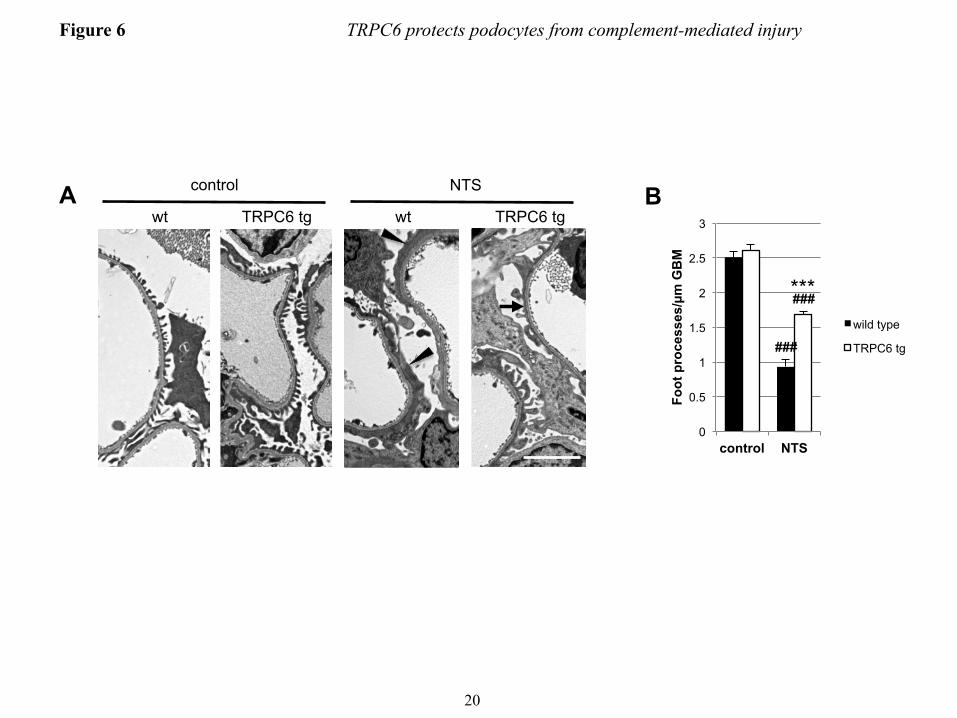
Figure 6 TRPC6 protects podocytes from complement-mediated injury
wt wt TRPC6 tg TRPC6 tg
control NTS
0
0.5
1
1.5
2
2.5
3
control NTS
Foot
pro
cess
es/µ
m G
BM
wild type
TRPC6 tg
B A
###
###
***
20
no virus
empty virus
wt TRPC6
dn TRPC6
control MN serum MN serum+4% comp
Figure 7 TRPC6 protects podocytes from complement-mediated injury
-60%
-50%
-40%
-30%
-20%
-10%
0%
empty virus wt TTPC6 dn TRPC6
B A
*
*
21
TRPC6 pCaMKIIα co
ntro
l M
N
A FS
GS
B
-1
0
1
2
3
4
5
-1 0 1 2 3 4 5
control
MN
FSGS
TRPC6
pCaM
KIIα
Figure 8 TRPC6 protects podocytes from complement-mediated injury
22
Copyright © 2022 FDOKUMEN