The directed differentiation of human iPS cells into kidney podocytes
9
The Directed Differentiation of Human iPS Cells into Kidney Podocytes Bi Song 1 , Alexandra M. Smink 1 , Christina V. Jones 1 , Judy M. Callaghan 2 , Stephen D. Firth 2 , Claude A. Bernard 1 , Andrew L. Laslett 3,4 , Peter G. Kerr 5 , Sharon D. Ricardo 1 * 1 Monash Immunology and Stem Cell Laboratories (MISCL), Monash University, Clayton, Victoria, Australia, 2 Monash Micro Imaging, Monash University, Clayton, Victoria, Australia, 3 CSIRO Materials Science and Engineering, Clayton, Victoria, Australia, 4 Department of Anatomy and Developmental Biology, Monash University, Clayton, Victoria, Australia, 5 Department of Medicine, Monash University, Monash Medical Centre, Clayton, Victoria, Australia Abstract The loss of glomerular podocytes is a key event in the progression of chronic kidney disease resulting in proteinuria and declining function. Podocytes are slow cycling cells that are considered terminally differentiated. Here we provide the first report of the directed differentiation of induced pluripotent stem (iPS) cells to generate kidney cells with podocyte features. The iPS-derived podocytes share a morphological phenotype analogous with cultured human podocytes. Following 10 days of directed differentiation, iPS podocytes had an up-regulated expression of mRNA and protein localization for podocyte markers including synaptopodin, nephrin and Wilm’s tumour protein (WT1), combined with a down-regulation of the stem cell marker OCT3/4. In contrast to human podocytes that become quiescent in culture, iPS-derived cells maintain a proliferative capacity suggestive of a more immature phenotype. The transduction of iPS podocytes with fluorescent labeled-talin that were immunostained with podocin showed a cytoplasmic contractile response to angiotensin II (AII). A permeability assay provided functional evidence of albumin uptake in the cytoplasm of iPS podocytes comparable to human podocytes. Moreover, labeled iPS-derived podocytes were found to integrate into reaggregated metanephric kidney explants where they incorporated into developing glomeruli and co-expressed WT1. This study establishes the differentiation of iPS cells to kidney podocytes that will be useful for screening new treatments, understanding podocyte pathogenesis, and offering possibilities for regenerative medicine. Citation: Song B, Smink AM, Jones CV, Callaghan JM, Firth SD, et al. (2012) The Directed Differentiation of Human iPS Cells into Kidney Podocytes. PLoS ONE 7(9): e46453. doi:10.1371/journal.pone.0046453 Editor: Maria Pia Rastaldi, Fondazione IRCCS Ospedale Maggiore Policlinico & Fondazione D’Amico per la Ricerca sulle Malattie Renali, Italy Received July 21, 2012; Accepted August 30, 2012; Published September 28, 2012 Copyright: ß 2012 Song et al. This is an open-access article distributed under the terms of the Creative Commons Attribution License, which permits unrestricted use, distribution, and reproduction in any medium, provided the original author and source are credited. Funding: This work was supported by the Alport Foundation of Australia and a Monash University Strategic Grant. CA Bernard is a recipient of an Erdi Fellowship in Neurological Diseases and funding from the Baker Foundation. Microscopy was performed with technical expertise from Monash Micro Imaging, Monash University. The funders had no role in study design, data collection and analysis, decision to publish, or preparation of the manuscript. Competing Interests: The authors have declared that no competing interests exist. * E-mail: [email protected] Introduction The epidemic of chronic kidney disease and end-stage renal failure represents a crisis for healthcare world-wide. Given the high morbidity of dialysis, its cost and the shortage of donor kidneys, there is an urgent need for further therapeutic options. Over two-thirds of patients with chronic kidney disease who progress to end-stage renal failure suffer from disorders that originate in the glomerulus, specifically podocyte injury leading to cell loss and proteinuria [1,2]. Podocytes are highly specialised cells with a complex cytoarchitecture consisting of tertiary foot processes that form the glomerular filtration barrier. The majority of glomerulopathies are a consequence of podocyte injury, resulting in the initiation and progression of fibrosis and impaired renal function [3,4]. In contrast to the regeneration of tubular epithelial cells that are rapidly repaired by intrinsic, proliferative expansion [5], the replacement of damaged glomerular podocytes remains a challenge. Podocyte precursors are derived from the metanephric mesenchyme during kidney development. Following maturation they establish their complex cell architecture and become highly terminally differentiated with a very limited regenerative capacity [6,7]. Likewise, primary cultures of human podocytes are difficult to maintain in culture due to their limited capacity to divide. Therefore, the reprogramming of adult cells to generate induced pluripotent stem (iPS) cells [8,9,10] with a high proliferative ability and broad differentiation capacity represents a major advance for both preclinical and clinical applications. iPS cells and their progeny will aid in understanding disease pathogenesis, screening new treatments, and offering possibilities for replacement cells to repair and regenerate damaged kidneys. However, due to the complexity of the developmental processes and kidney structure, there have been few successful reports showing differentiation of pluripotent cells to kidney progenitors. Cultured mouse embryonic stem (ES) have been reported to differentiate into intermediate mesoderm [11] and tubular cells [12,13,14] in vitro that is enhanced by activin A [15] and retinoic acid [11], however without evidence of integration into the structural development of glomeruli. Moreover, using human ES cells we have previously reported the directed differentiation of an enriched population of mesodermal kidney progenitors [16]. Recently we [17], and others [18] reported the successful generation of iPS cells from human kidney cells that are pluripotent and have long-term proliferative ability. Based on PLOS ONE | www.plosone.org 1 September 2012 | Volume 7 | Issue 9 | e46453
Transcript of The directed differentiation of human iPS cells into kidney podocytes
The Directed Differentiation of Human iPS Cells intoKidney PodocytesBi Song1, Alexandra M. Smink1, Christina V. Jones1, Judy M. Callaghan2, Stephen D. Firth2,
Claude A. Bernard1, Andrew L. Laslett3,4, Peter G. Kerr5, Sharon D. Ricardo1*
1Monash Immunology and Stem Cell Laboratories (MISCL), Monash University, Clayton, Victoria, Australia, 2Monash Micro Imaging, Monash University, Clayton, Victoria,
Australia, 3CSIRO Materials Science and Engineering, Clayton, Victoria, Australia, 4Department of Anatomy and Developmental Biology, Monash University, Clayton,
Victoria, Australia, 5Department of Medicine, Monash University, Monash Medical Centre, Clayton, Victoria, Australia
Abstract
The loss of glomerular podocytes is a key event in the progression of chronic kidney disease resulting in proteinuria anddeclining function. Podocytes are slow cycling cells that are considered terminally differentiated. Here we provide the firstreport of the directed differentiation of induced pluripotent stem (iPS) cells to generate kidney cells with podocyte features.The iPS-derived podocytes share a morphological phenotype analogous with cultured human podocytes. Following 10 daysof directed differentiation, iPS podocytes had an up-regulated expression of mRNA and protein localization for podocytemarkers including synaptopodin, nephrin and Wilm’s tumour protein (WT1), combined with a down-regulation of the stemcell marker OCT3/4. In contrast to human podocytes that become quiescent in culture, iPS-derived cells maintaina proliferative capacity suggestive of a more immature phenotype. The transduction of iPS podocytes with fluorescentlabeled-talin that were immunostained with podocin showed a cytoplasmic contractile response to angiotensin II (AII). Apermeability assay provided functional evidence of albumin uptake in the cytoplasm of iPS podocytes comparable tohuman podocytes. Moreover, labeled iPS-derived podocytes were found to integrate into reaggregated metanephric kidneyexplants where they incorporated into developing glomeruli and co-expressed WT1. This study establishes thedifferentiation of iPS cells to kidney podocytes that will be useful for screening new treatments, understanding podocytepathogenesis, and offering possibilities for regenerative medicine.
Citation: Song B, Smink AM, Jones CV, Callaghan JM, Firth SD, et al. (2012) The Directed Differentiation of Human iPS Cells into Kidney Podocytes. PLoS ONE 7(9):e46453. doi:10.1371/journal.pone.0046453
Editor: Maria Pia Rastaldi, Fondazione IRCCS Ospedale Maggiore Policlinico & Fondazione D’Amico per la Ricerca sulle Malattie Renali, Italy
Received July 21, 2012; Accepted August 30, 2012; Published September 28, 2012
Copyright: � 2012 Song et al. This is an open-access article distributed under the terms of the Creative Commons Attribution License, which permitsunrestricted use, distribution, and reproduction in any medium, provided the original author and source are credited.
Funding: This work was supported by the Alport Foundation of Australia and a Monash University Strategic Grant. CA Bernard is a recipient of an Erdi Fellowshipin Neurological Diseases and funding from the Baker Foundation. Microscopy was performed with technical expertise from Monash Micro Imaging, MonashUniversity. The funders had no role in study design, data collection and analysis, decision to publish, or preparation of the manuscript.
Competing Interests: The authors have declared that no competing interests exist.
* E-mail: [email protected]
Introduction
The epidemic of chronic kidney disease and end-stage renal
failure represents a crisis for healthcare world-wide. Given the
high morbidity of dialysis, its cost and the shortage of donor
kidneys, there is an urgent need for further therapeutic options.
Over two-thirds of patients with chronic kidney disease who
progress to end-stage renal failure suffer from disorders that
originate in the glomerulus, specifically podocyte injury leading to
cell loss and proteinuria [1,2]. Podocytes are highly specialised
cells with a complex cytoarchitecture consisting of tertiary foot
processes that form the glomerular filtration barrier. The majority
of glomerulopathies are a consequence of podocyte injury,
resulting in the initiation and progression of fibrosis and impaired
renal function [3,4]. In contrast to the regeneration of tubular
epithelial cells that are rapidly repaired by intrinsic, proliferative
expansion [5], the replacement of damaged glomerular podocytes
remains a challenge. Podocyte precursors are derived from the
metanephric mesenchyme during kidney development. Following
maturation they establish their complex cell architecture and
become highly terminally differentiated with a very limited
regenerative capacity [6,7].
Likewise, primary cultures of human podocytes are difficult to
maintain in culture due to their limited capacity to divide.
Therefore, the reprogramming of adult cells to generate induced
pluripotent stem (iPS) cells [8,9,10] with a high proliferative ability
and broad differentiation capacity represents a major advance for
both preclinical and clinical applications. iPS cells and their
progeny will aid in understanding disease pathogenesis, screening
new treatments, and offering possibilities for replacement cells to
repair and regenerate damaged kidneys. However, due to the
complexity of the developmental processes and kidney structure,
there have been few successful reports showing differentiation of
pluripotent cells to kidney progenitors. Cultured mouse embryonic
stem (ES) have been reported to differentiate into intermediate
mesoderm [11] and tubular cells [12,13,14] in vitro that is
enhanced by activin A [15] and retinoic acid [11], however
without evidence of integration into the structural development of
glomeruli. Moreover, using human ES cells we have previously
reported the directed differentiation of an enriched population of
mesodermal kidney progenitors [16].
Recently we [17], and others [18] reported the successful
generation of iPS cells from human kidney cells that are
pluripotent and have long-term proliferative ability. Based on
PLOS ONE | www.plosone.org 1 September 2012 | Volume 7 | Issue 9 | e46453
our established protocol using the directed differentiation of
human ES cells to kidney progenitors and extensive background
transcriptional profiling [16] we now report a reliable and efficient
method for differentiation of iPS cells into kidney podocyte
progenitors. The iPS-derived podocytes share cell morphology
similar to primary human podocytes including tertiary cytoplasmic
cell processes by scanning electron microscopy (SEM). By day 10
of differentiation the iPS-derived podocytes show protein localisa-
tion of the mesodermal and podocyte markers; Wilm’s tumour
protein (WT1), paired homeobox gene 2 (Pax-2), nephrin, podocin
and synaptopodin. qPCR confirmed that over the time course of
directed differentiation, the iPS-derived podocytes show an
upregulated expression of podocyte markers concurrent with
a down-regulation of the pluripotency gene, OCT3/4. The iPS-
derived podocytes have functional characteristics of podocytes in
their contractile response to angiotensin II (AII) and endocytosis of
albumin, and furthermore they integrate into WT1-positive
glomerular aggregates in developing kidneys.
Results
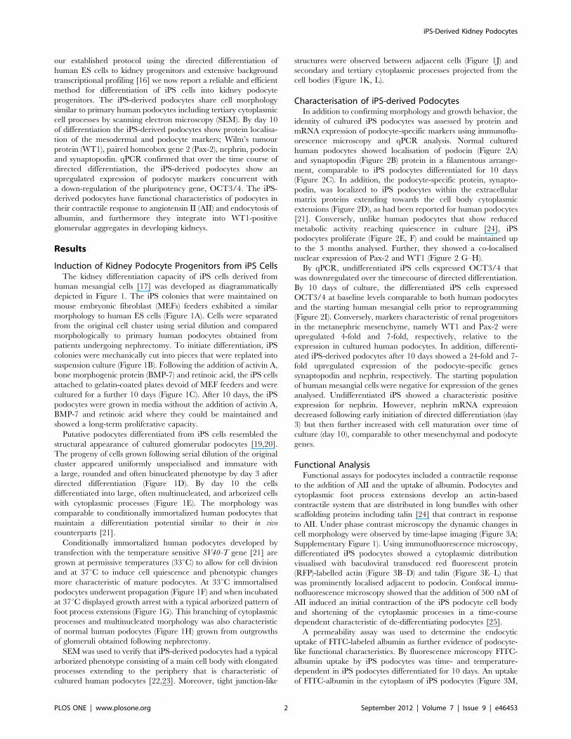
Induction of Kidney Podocyte Progenitors from iPS CellsThe kidney differentiation capacity of iPS cells derived from
human mesangial cells [17] was developed as diagrammatically
depicted in Figure 1. The iPS colonies that were maintained on
mouse embryonic fibroblast (MEFs) feeders exhibited a similar
morphology to human ES cells (Figure 1A). Cells were separated
from the original cell cluster using serial dilution and compared
morphologically to primary human podocytes obtained from
patients undergoing nephrectomy. To initiate differentiation, iPS
colonies were mechanically cut into pieces that were replated into
suspension culture (Figure 1B). Following the addition of activin A,
bone morphogenic protein (BMP-7) and retinoic acid, the iPS cells
attached to gelatin-coated plates devoid of MEF feeders and were
cultured for a further 10 days (Figure 1C). After 10 days, the iPS
podocytes were grown in media without the addition of activin A,
BMP-7 and retinoic acid where they could be maintained and
showed a long-term proliferative capacity.
Putative podocytes differentiated from iPS cells resembled the
structural appearance of cultured glomerular podocytes [19,20].
The progeny of cells grown following serial dilution of the original
cluster appeared uniformly unspecialised and immature with
a large, rounded and often binucleated phenotype by day 3 after
directed differentiation (Figure 1D). By day 10 the cells
differentiated into large, often multinucleated, and arborized cells
with cytoplasmic processes (Figure 1E). The morphology was
comparable to conditionally immortalized human podocytes that
maintain a differentiation potential similar to their in vivo
counterparts [21].
Conditionally immortalized human podocytes developed by
transfection with the temperature sensitive SV40-T gene [21] are
grown at permissive temperatures (33uC) to allow for cell division
and at 37uC to induce cell quiescence and phenotypic changes
more characteristic of mature podocytes. At 33uC immortalised
podocytes underwent propagation (Figure 1F) and when incubated
at 37uC displayed growth arrest with a typical arborized pattern of
foot process extensions (Figure 1G). This branching of cytoplasmic
processes and multinucleated morphology was also characteristic
of normal human podocytes (Figure 1H) grown from outgrowths
of glomeruli obtained following nephrectomy.
SEM was used to verify that iPS-derived podocytes had a typical
arborized phenotype consisting of a main cell body with elongated
processes extending to the periphery that is characteristic of
cultured human podocytes [22,23]. Moreover, tight junction-like
structures were observed between adjacent cells (Figure 1J) and
secondary and tertiary cytoplasmic processes projected from the
cell bodies (Figure 1K, L).
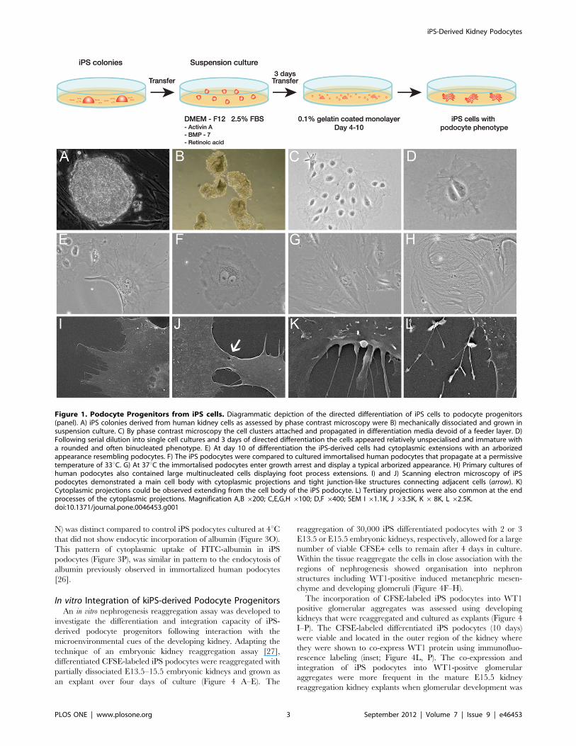
Characterisation of iPS-derived PodocytesIn addition to confirming morphology and growth behavior, the
identity of cultured iPS podocytes was assessed by protein and
mRNA expression of podocyte-specific markers using immunoflu-
orescence microscopy and qPCR analysis. Normal cultured
human podocytes showed localisation of podocin (Figure 2A)
and synaptopodin (Figure 2B) protein in a filamentous arrange-
ment, comparable to iPS podocytes differentiated for 10 days
(Figure 2C). In addition, the podocyte-specific protein, synapto-
podin, was localized to iPS podocytes within the extracellular
matrix proteins extending towards the cell body cytoplasmic
extensions (Figure 2D), as had been reported for human podocytes
[21]. Conversely, unlike human podocytes that show reduced
metabolic activity reaching quiescence in culture [24], iPS
podocytes proliferate (Figure 2E, F) and could be maintained up
to the 3 months analysed. Further, they showed a co-localised
nuclear expression of Pax-2 and WT1 (Figure 2 G–H).
By qPCR, undifferentiated iPS cells expressed OCT3/4 that
was downregulated over the timecourse of directed differentiation.
By 10 days of culture, the differentiated iPS cells expressed
OCT3/4 at baseline levels comparable to both human podocytes
and the starting human mesangial cells prior to reprogramming
(Figure 2I). Conversely, markers characteristic of renal progenitors
in the metanephric mesenchyme, namely WT1 and Pax-2 were
upregulated 4-fold and 7-fold, respectively, relative to the
expression in cultured human podocytes. In addition, differenti-
ated iPS-derived podocytes after 10 days showed a 24-fold and 7-
fold upregulated expression of the podocyte-specific genes
synaptopodin and nephrin, respectively. The starting population
of human mesangial cells were negative for expression of the genes
analysed. Undifferentiated iPS showed a characteristic positive
expression for nephrin. However, nephrin mRNA expression
decreased following early initiation of directed differentiation (day
3) but then further increased with cell maturation over time of
culture (day 10), comparable to other mesenchymal and podocyte
genes.
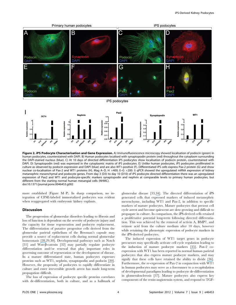
Functional AnalysisFunctional assays for podocytes included a contractile response
to the addition of AII and the uptake of albumin. Podocytes and
cytoplasmic foot process extensions develop an actin-based
contractile system that are distributed in long bundles with other
scaffolding proteins including talin [24] that contract in response
to AII. Under phase contrast microscopy the dynamic changes in
cell morphology were observed by time-lapse imaging (Figure 3A;
Supplementary Figure 1). Using immunofluorescence microscopy,
differentiated iPS podocytes showed a cytoplasmic distribution
visualised with baculoviral transduced red fluorescent protein
(RFP)-labelled actin (Figure 3B–D) and talin (Figure 3E–L) that
was prominently localised adjacent to podocin. Confocal immu-
nofluorescence microscopy showed that the addition of 500 nM of
AII induced an initial contraction of the iPS podocyte cell body
and shortening of the cytoplasmic processes in a time-course
dependent characteristic of de-differentiating podocytes [25].
A permeability assay was used to determine the endocytic
uptake of FITC-labeled albumin as further evidence of podocyte-
like functional characteristics. By fluorescence microscopy FITC-
albumin uptake by iPS podocytes was time- and temperature-
dependent in iPS podocytes differentiated for 10 days. An uptake
of FITC-albumin in the cytoplasm of iPS podocytes (Figure 3M,
iPS-Derived Kidney Podocytes
PLOS ONE | www.plosone.org 2 September 2012 | Volume 7 | Issue 9 | e46453
N) was distinct compared to control iPS podocytes cultured at 4uCthat did not show endocytic incorporation of albumin (Figure 3O).
This pattern of cytoplasmic uptake of FITC-albumin in iPS
podocytes (Figure 3P), was similar in pattern to the endocytosis of
albumin previously observed in immortalized human podocytes
[26].
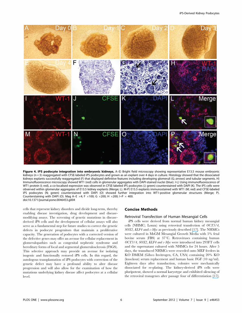
In vitro Integration of kiPS-derived Podocyte ProgenitorsAn in vitro nephrogenesis reaggregation assay was developed to
investigate the differentiation and integration capacity of iPS-
derived podocyte progenitors following interaction with the
microenvironmental cues of the developing kidney. Adapting the
technique of an embryonic kidney reaggregation assay [27],
differentiated CFSE-labeled iPS podocytes were reaggregated with
partially dissociated E13.5–15.5 embryonic kidneys and grown as
an explant over four days of culture (Figure 4 A–E). The
reaggregation of 30,000 iPS differentiated podocytes with 2 or 3
E13.5 or E15.5 embryonic kidneys, respectively, allowed for a large
number of viable CFSE+ cells to remain after 4 days in culture.
Within the tissue reaggregate the cells in close association with the
regions of nephrogenesis showed organisation into nephron
structures including WT1-positive induced metanephric mesen-
chyme and developing glomeruli (Figure 4F–H).
The incorporation of CFSE-labeled iPS podocytes into WT1
positive glomerular aggregates was assessed using developing
kidneys that were reaggregated and cultured as explants (Figure 4
I–P). The CFSE-labeled differentiated iPS podocytes (10 days)
were viable and located in the outer region of the kidney where
they were shown to co-express WT1 protein using immunofluo-
rescence labeling (inset; Figure 4L, P). The co-expression and
integration of iPS podocytes into WT1-positve glomerular
aggregates were more frequent in the mature E15.5 kidney
reaggregation kidney explants when glomerular development was
Figure 1. Podocyte Progenitors from iPS cells. Diagrammatic depiction of the directed differentiation of iPS cells to podocyte progenitors(panel). A) iPS colonies derived from human kidney cells as assessed by phase contrast microscopy were B) mechanically dissociated and grown insuspension culture. C) By phase contrast microscopy the cell clusters attached and propagated in differentiation media devoid of a feeder layer. D)Following serial dilution into single cell cultures and 3 days of directed differentiation the cells appeared relatively unspecialised and immature witha rounded and often binucleated phenotype. E) At day 10 of differentiation the iPS-derived cells had cytoplasmic extensions with an arborizedappearance resembling podocytes. F) The iPS podocytes were compared to cultured immortalised human podocytes that propagate at a permissivetemperature of 33uC. G) At 37uC the immortalised podocytes enter growth arrest and display a typical arborized appearance. H) Primary cultures ofhuman podocytes also contained large multinucleated cells displaying foot process extensions. I) and J) Scanning electron microscopy of iPSpodocytes demonstrated a main cell body with cytoplasmic projections and tight junction-like structures connecting adjacent cells (arrow). K)Cytoplasmic projections could be observed extending from the cell body of the iPS podocyte. L) Tertiary projections were also common at the endprocesses of the cytoplasmic projections. Magnification A,B 6200; C,E,G,H6100; D,F 6400; SEM I 61.1K, J 63.5K, K6 8K, L 62.5K.doi:10.1371/journal.pone.0046453.g001
iPS-Derived Kidney Podocytes
PLOS ONE | www.plosone.org 3 September 2012 | Volume 7 | Issue 9 | e46453
more established (Figure M–P). In sharp comparison, no in-
tegration of CFSE-labeled immortalized podocytes was evident
when reaggregated with embryonic kidney explants.
Discussion
The progression of glomerular disorders leading to fibrosis and
loss of function is dependent on the severity of podocyte injury and
the capacity for tissue regeneration and podocyte replacement.
The differentiation of putative progenitor cells derived from the
glomerular parietal epithelium of the Bowman’s capsule may
provide a source of replacement cells during normal glomerular
homeostasis [28,29,30]. Developmental pathways such as Notch
[31] and Wnt-b-catenin [32] may partially regulate podocyte
differentiation and/or renewal that play important roles in
governing outcome of regeneration or the development of disease.
In a mature differentiated state, human podocytes expresses
proteins such as WT1, nephrin, synaptopodin and podocin [20].
However, the propensity of podocytes to rapidly differentiate in
culture and enter irreversible growth arrest has made long-term
propagation difficult.
The loss of expression of podocyte specific proteins correlates
with de-differentiation, both in culture, and as a hallmark of
glomerular disease [33,34]. The directed differentiation of iPS
generated cells that expressed markers of induced metanephric
mesenchyme, including WT1 and Pax-2, in addition to specific
markers of mature podocytes. Mature podocytes that present cell
cycle arrest and become quiescent are slow growing and difficult to
propagate in culture. In comparison, the iPS-derived cells retained
a proliferative potential long-term following directed differentia-
tion. This was achieved by the removal of activin A, BMP7, and
retinoic acid from the culture medium after 10 days, however
while retaining the phenotypic expression of podocyte markers in
the iPS-derived podocytes.
The induced expression of WT1 target genes in podocyte
precursors may specifically activate cell cycle regulation leading to
the induction of mature podocyte markers [35]. Pax-2 co-
expression with WT1 has been reported in normal human parietal
podocytes that also express mature podocyte markers, and may
signify that these cells have retained the ability to divide [36].
Furthermore, the re-expression of Pax-2 in conjunction with WT1
in human podocytes may serve as a forerunner to a recapitulation
of developmental paradigms leading to podocyte de-differentiation
in glomerulosclerosis [37]. Mature podocytes also express key
components of the renin-angiotensin system, and respond to TGF-
Figure 2. iPS Podocyte Characterisation and Gene Expression. A) Immunofluorescence microscopy showed localisation of podocin (green) inhuman podocytes, counterstained with DAPI. B) Human podocytes localised with synaptopodin protein (red) throughout the cytoplasm surroundingthe DAPI-stained nucleus (blue). C) At 10 days of directed differentiation iPS podocytes show localisation of podocin protein, counterstained withDAPI. D) Synaptopodin (red) was expressed in the cytoplasmic matrix of iPS podocytes. E) Unlike human podocytes, iPS podocytes proliferated inculture as observed by podocin expression and DAPI (blue) and are also WT1-positive (F). Differentiated iPS cells express Pax-2 protein (G) and shownuclear co-localisation of Pax-2 and WT1 proteins (H). Mag A–D, H 6400; E–G 6200. I) qPCR showed the upregulated mRNA expression of kidneymetanephric mesenchymal and podocyte genes. From day 3 (D3) to day 10 (D10) of iPS podocyte directed differentiation there was an upregulatedexpression of Pax2 and WT1 and podocyte-specific markers synaptopodin and nephrin at comparable levels to primary human podocytes, butdifferent from the starting normal human mesangial cells (NHMC).doi:10.1371/journal.pone.0046453.g002
iPS-Derived Kidney Podocytes
PLOS ONE | www.plosone.org 4 September 2012 | Volume 7 | Issue 9 | e46453
b [38,39]. These pro-inflammatory mediators of fibrosis can
directly affect the podocyte leading to foot process contraction and
podocyte effacement. In response to TGF-b and AII, mature
podocytes undergo de-differentiation [25,40]. The resulting
flattening and contraction of cytoplasmic processes was also
observed in the iPS-derived podocytes in response to AII and was
associated with shortening of the extracellular matrix proteins
actin and talin.
There are many advantages of developing iPS cells as an
investigative strategy for patients with genetic and non-genetic
kidney disease that will provide a valuable tool to culture and
manipulate podocytes in vitro in order to understand their biology
and model human disease. We have previously reported that iPS
cells can be derived from human mesangial cells [17]. In addition,
epithelial cells obtained from the urine of kidney disease patients
may offer an obtainable source of kidney cells for generation of iPS
[18]. Several studies have reported genetic and epigenetic
transcriptional variation between iPS cultures [41,42,43] where
the cell type of origin may influence the capacity for in vitro
differentiation potential [44]. Indeed, DNA methylation states
change following directed differentiation, however aberrations in
epigenetic imprints are frequent in iPS colonies following cell
reprogramming where the epigenetic instabilities may persist [45].
Therefore, for future consideration transcriptional-based direct
reprogramming of kidney cells using a strategy of re-expressing key
developmental regulators may offer an alternative, as has been
shown in neurons [46,47], cardiomyocytes [48] and b-islet cells[49].
In the interim, the directed differentiation of iPS cells to
podocytes offers an unprecedented opportunity to generate human
Figure 3. Functional contractility and permeability. Live cell imaging was used to record the response of iPS podocytes to the addition of AII(See Movie S1). A) Phase contrast imaging of iPS podocytes following the addition of AII at time 0. The iPS podocytes were transduced with RFP-actin(B) and immunostained with the contractile protein, podocin (C). D) Merge image of actin (red) and podocin (green) with DAPI-stained nuclei (blue).E) Confocal immunofluorescence shows that iPS-podocytes transduced with RFP-talin (E) co-expressed podocin (F) at time 0. G) DAPI-stained nucleiand merged image are shown (H). After 6 hours in culture, the iPS podocytes were viable and display a contracted morphology in response to AII (I–L)where RFP-talin (red), podocin (green) and DAPI (blue) are shown. (M-N) By fluorescence microscopy, the iPS podocytes were able to uptake FITC-albumin (green) into the cytoplasm when cultured at 37uC, compared to iPS podocytes cultured at 4uC (O) that served as a control. (P) Immortalizedhuman podocytes also showed endocytosis of FITC-albumin in a similar morphological pattern. Mag A–D 6200; E–L, N-P 61000; M 6100.Abbreviations: angiotensin II (AII); red fluorescent protein (RFP).doi:10.1371/journal.pone.0046453.g003
iPS-Derived Kidney Podocytes
PLOS ONE | www.plosone.org 5 September 2012 | Volume 7 | Issue 9 | e46453
cells that represent kidney disorders and divide long-term, thereby
enabling disease investigation, drug development and disease-
modifying assays. The screening of genetic mutations in disease-
derived iPS cells and the development of cellular assays will also
serve as a fundamental step for future studies to correct the genetic
defects in podocyte progenitors that maintain a proliferative
capacity. The generation of podocytes with a corrected version of
the defective genes may offer an avenue for cellular replacement in
glomerulopathies such as congenital nephrotic syndrome and
hereditary forms of focal and segmental glomerulosclerosis (FSGS).
This selective approach may provide an avenue for isolating
isogenic and functionally restored iPS cells. In this regard, the
autologous transplantation of iPS podocytes with correction of the
genetic defect may have a profound ability to alter disease
progression and will also allow for the examination of how the
mutations underlying kidney disease affect podocytes at a cellular
level.
Concise Methods
Retroviral Transfection of Human Mesangial CellsiPS cells were derived from normal human kidney mesangial
cells (NHMC; Lonza) using retroviral transfection of OCT3/4,
SOX2, KLF4 and c-Myc as previously described [17]. The NHMCs
were cultured in MsGM Mesangial Growth Media with 5% fetal
bovine serum (FBS) at 37uC. Retroviruses containing human
OCT3/4, SOX2, KLF4 and c-Myc were introduced into 293FT cells
and the supernatant cultured with NHMCs for 24 hours. After 5
days, the transduced NHMCs were reseeded onto MEF feeders in
KO DMEM (Gibco Invitrogen, CA, USA) containing 20% KO
(knockout) serum replacement and human basic FGF (10 ng/ml).
Eighteen days after transduction, colonies were mechanically
dissociated for re-plating. The kidney-derived iPS cells were
pluripotent, showed a normal karyotype and exhibited silencing of
the retroviral transgenes after passage four of differentiation [17].
Figure 4. iPS podocyte integration into embryonic kidneys. A–E) Bright field microscopy showing representative E13.5 mouse embryonickidneys (n = 3) reaggregated with CFSE-labeled iPS podocytes and grown as an explant over 4 days in culture. Histology showed that the dissociatedkidneys explants successfully reaggregated (F) that displayed definitive features including developing glomeruli (G; arrows) and tubular segments. H)Immunofluorescence microscopy showed WT1 (red) cells in glomerular aggregates with DAPI-stained nuclei (blue). I-L) Using immunofluorescence ofWT1 protein (I; red), a co-localised expression was observed in CFSE-labeled iPS podocytes (J; green) counterstained with DAPI (K). The iPS cells wereobserved within glomerular aggregates of E13.5 kidney explants (Merge; L). M-P) E15.5 explants immunostained with WT1 (M; red) and CFSE-labelediPS podocytes (N; green) counterstained with DAPI (O) showed further integration into WT1-positive glomerular structures (Merge; P).Counterstaining with DAPI (O). Mag A–E64; F 6100; G 6200; H 6200; I–P6 400.doi:10.1371/journal.pone.0046453.g004
iPS-Derived Kidney Podocytes
PLOS ONE | www.plosone.org 6 September 2012 | Volume 7 | Issue 9 | e46453
Differentiation of iPS Cells to Podocyte ProgenitorsThe iPS colonies were mechanically cut into small pieces
approximately the same size, and were cultured in ultra low cluster
6-well plate (COSTAR) for 3 days in the differentiation medium
consisting of DMEM–F12 (Sigma) with 2.5% FBS, 100 mMnonessential amino acids, 100 mM beta mercaptoethanol with the
addition of 10 ng/ml of activin A, 15 ng/ml of BMP7, and
0.1 mM retinoic acid. The cells were transferred into 0.1% gelatin
pre-coated 10 cm tissue culture dishes for another 7–8 days in the
same medium before serial sub-passaging for characterization and
integration assays. At 10 days of differentiation the iPS podocytes
were fixed in 2.5% glutaraldehyde in cacodylate buffer and
processed for SEM visualization (Hitashi S570 microscope). For
the long-term maintenance of iPS podocytes, after 10 days of
directed differentiation the iPS-derived podocytes were grown in
DMEM-F12 media without the addition of 10 ng/ml of Activin A,
15 ng/ml of BMP7, and 0.1 mM retinoic acid where they were
able to maintain the morphological characteristics and functional
capacity.
Isolation and Labeling of Human PodocytesHuman kidneys were obtained from patients scheduled for
nephrectomy following written consent and approval from the
Southern Health Human Ethics Committee (approval #10179B),
Monash Medical Centre. Human podocytes were derived from
normal kidney tissue using a sieving method adapted from
isolation of mesangial cells [50]. Normal kidney tissue was minced
and passed through two mesh sieves (120 and 105m) using a series
of washes, centrifugation, syringe dissociation and resuspension in
DMEM-F12 media. Decapsulated glomeruli were grown as
explants for approximately 30 days. Human podocyte outgrowths
were easily identified by morphology and subsequently sub-
passaged and plated into chamber slides for immunofluorescence
staining and RNA extraction. The human podocytes were
confirmed to show positive protein and gene expression for
podocyte-specific markers podocin, synaptopodin and nephrin.
Immortalised podocytes grown at 33uC and 37uC and iPS-
derived podocytes were labelled using carboxyfluorescein diacetate
succinimidyl ester (CFSE; Life Technologies, Australia). Briefly,
cells were harvested, counted and incubated with 15 mMCFSE for
15 minutes at 37uC before centrifugation and washing in media.
To ensure complete integration of the CFSE probe the cells were
further incubated for 30 minutes at 37uC, washed with PBS and
resuspended in DMEM containing 10% FBS, 1% Pen/Strep, 1%
L-Glutamine and 1% Insulin Transferrin Selenium (Life Tech-
nologies).
Immunofluorescence MicroscopyImmunocytochemistry was performed as described previously
[17]. Differentiated iPS podocytes and human podocytes seeded
on chamber slides in 10% FBS medium were serum-starved
overnight, then fixed in 4% paraformaldehyde (PFA) for 10 min,
permeablized with 0.1% Triton X-100/PBS for 10 min and
incubated in blocking solution (4% normal goat serum/PBS) for
30 min. The cells were incubated with anti-nephrin, anti-
synaptopodin, anti-Pax-2, and anti-podocin (NPHS2; all from
Abcam, USA) and anti-WT1 (Santa Cruz, USA) at dilutions from
1:20–400 in blocking solution overnight at 4uC and incubated with
secondary antibodies (Alexa Fluor 488, 555 dilution 1:1000) in
PBS for 1 hr. Sections were counterstained with DAPI (1:10,000;
Life Technologies) and then mounted with Fluorescent Mounting
Medium (DakoCytomation, Denmark) and analysed with a Provis
AX70 (Olympus, Japan) or Nikon C1 confocal fluorescent
microscope (Nikon, Japan).
qPCRTotal RNA was extracted from undifferentiated iPS cells,
normal human podocytes obtained from primary culture, and iPS
podocytes at day 3 and day 10 of directed differentiation using
a pico pure RNA isolation kit (ARCTURUS). cDNA was
synthesized using SuperScript III first–strand synthesis system for
TR-PCR (Invitrogen). Quantitative real-time PCR (qPCR) was
performed using a platinum SYBR Green qPCR SuperMix-UDG
(Invitrogen). The threshold cycle (Ct) values were measured in
triplicate and normalized against the endogenous control b-actinand expression levels normalized against b-actin using primers
listed in Figure S1.
Cell Contractility and PermeabilityiPS podocytes at a seeding density of 0.56104 cells/well were
transduced using a Cell LightsTM (Life Technologies) intracellular
RFP-actin and RFP-talin. Cells were incubated with Cell LightsTM
2.0 reagent overnight before replacing with serum-free DMEM
media for 2 hours. The iPS podocytes were visualised every 15
minutes with/without the addition of AII (500 nM) using a Leica
AF6000 LX (Leica, Germany) live cell imaging system. Following
24 hours of live cell imaging (Movie S1) the control and AII-
treated iPS podocytes were fixed with 4% PFA and immunos-
tained with anti-podocin antibody before visualisation with a Nikon
C1 confocal microscope.
For the permeability assay, differentiated iPS podocytes were
cultured as described above, and, after differentiation the culture
medium was replaced with serum-free media with/without FITC-
labeled albumin (0.5 mg/ml; Abcam) and cultured at 37uC for 1
hour, in comparison to control cells cultured at 4uC. The cells
were fixed in 4% paraformaldehyde (PFA) and counterstained
with DAPI. Assays were performed in triplicate using separate cell
preparations.
Reaggregation AssayAll animal experiments were approved in advance by a Monash
University Animal Ethics Committee, which adheres to the
‘‘Australian Code of Practice for the Care and Use of Animals
for Scientific Purposes. Embryonic day (E) 13.5–15.5 embryos
were collected from time-mated pregnant C57BL6/J mice. For the
explants, 2 (E15.5) or 3 (E13.5) embryonic kidneys were trans-
ferred into Eppendorf tubes containing 30,000 CFSE-labelled cells
and gently mechanically disrupted using a 25 G needle before
centrifugation (3 minutes, 18 rcf) to form aggregates that were
transferred onto a floating polycarbonate membrane (3 mm pore
size; GE Water & Process Technologies, Australia) in DMEM
growth medium in a 24 well plate. The aggregates were incubated
(5% CO2, 37uC) for 4 days with a medium change after 48 hours.
Photomicrographs were taken daily to assess aggregate growth
(Olympus IX51 dissecting microscope). After 4 days of culture, the
kidney explants were fixed with 4% PFA for histological
examination with hematoxylin and eosin staining. CFSE-labeled
cells were visualised by immunofluorescence microscopy of frozen
sections (5 mm) that were immunostained with WT1 and counter-
stained with DAPI. Embryonic kidneys reaggregated with CFSE-
labeled immortalized podocytes were cultured over the same time
period as a control comparison.
Supporting Information
Figure S1 Table detailing the primers used for real-time quantitative PCR.
(DOC)
iPS-Derived Kidney Podocytes
PLOS ONE | www.plosone.org 7 September 2012 | Volume 7 | Issue 9 | e46453
Movie S1 Phase contrast microscopy using time-lapseimaging shows the dynamic changes in cell morphologyfollowing the addition of 500 nM of angiotensin II. Over
24 hours, the differentiated iPS podocytes showed a contractile
response and retraction of cytoplasmic processes following the
addition of angiotensin II, however without affecting cell viability.
(MOV)
Author Contributions
Conceived and designed the experiments: BS AMS CVJ JMC SDF CAB
ALL PGK SDR. Performed the experiments: BS AMS CVJ SDR.
Analyzed the data: BS AMS CVJ SDR. Contributed reagents/materials/
analysis tools: ALL PGK SDR. Wrote the paper: SDR.
References
1. Wiggins RC (2007) The spectrum of podocytopathies: a unifying view of
glomerular diseases. Kidney Int 71: 1205–1214.
2. Sato Y, Wharram BL, Lee SK, Wickman L, Goyal M, et al. (2009) Urine
podocyte mRNAs mark progression of renal disease. Journal of the American
Society of Nephrology : JASN 20: 1041–1052.
3. Abbate M, Zoja C, Remuzzi G (2006) How does proteinuria cause progressive
renal damage? J Am Soc Nephrol 17: 2974–2984.
4. Mundel P, Shankland SJ (2002) Podocyte biology and response to injury. J AmSoc Nephrol 13: 3005–3015.
5. Humphreys BD, Bonventre JV (2007) The contribution of adult stem cells to
renal repair. Nephrol Ther 3: 3–10.
6. Quaggin SE, Kreidberg JA (2008) Development of the renal glomerulus: goodneighbors and good fences. Development 135: 609–620.
7. Kriz W (2003) Progression of chronic renal failure in focal segmental
glomerulosclerosis: consequence of podocyte damage or of tubulointerstitialfibrosis? Pediatr Nephrol 18: 617–622.
8. Takahashi K, Yamanaka S (2006) Induction of pluripotent stem cells from
mouse embryonic and adult fibroblast cultures by defined factors. Cell 126: 663–676.
9. Park IH, Zhao R, West JA, Yabuuchi A, Huo H, et al. (2008) Reprogramming
of human somatic cells to pluripotency with defined factors. Nature 451: 141–146.
10. Takahashi K, Tanabe K, Ohnuki M, Narita M, Ichisaka T, et al. (2007)
Induction of pluripotent stem cells from adult human fibroblasts by definedfactors. Cell 131: 861–872.
11. Mae S, Shirasawa S, Yoshie S, Sato F, Kanoh Y, et al. (2010) Combination of
small molecules enhances differentiation of mouse embryonic stem cells intointermediate mesoderm through BMP7-positive cells. Biochemical and bio-
physical research communications 393: 877–882.
12. Kim D, Dressler GR (2005) Nephrogenic factors promote differentiation ofmouse embryonic stem cells into renal epithelia. Journal of the American Society
of Nephrology : JASN 16: 3527–3534.
13. Kobayashi T, Tanaka H, Kuwana H, Inoshita S, Teraoka H, et al. (2005) Wnt4-transformed mouse embryonic stem cells differentiate into renal tubular cells.
Biochemical and biophysical research communications 336: 585–595.
14. Steenhard BM, Isom KS, Cazcarro P, Dunmore JH, Godwin AR, et al. (2005)
Integration of embryonic stem cells in metanephric kidney organ culture.
Journal of the American Society of Nephrology : JASN 16: 1623–1631.
15. Morizane R, Monkawa T, Itoh H (2009) Differentiation of murine embryonic
stem and induced pluripotent stem cells to renal lineage in vitro. Biochemical
and biophysical research communications 390: 1334–1339.
16. Lin SA, Kolle G, Grimmond SM, Zhou Q, Doust E, et al. (2010)
Subfractionation of Differentiating Human Embryonic Stem Cell Populations
Allows the Isolation of a Mesodermal Population Enriched for IntermediateMesoderm and Putative Renal Progenitors. Stem Cells Dev.
17. Song B, Niclis JC, Alikhan MA, Sakkal S, Sylvain A, et al. (2011) Generation of
induced pluripotent stem cells from human kidney mesangial cells. Journal of theAmerican Society of Nephrology : JASN 22: 1213–1220.
18. Zhou T, Benda C, Duzinger S, Huang Y, Li X, et al. (2011) Generation of
induced pluripotent stem cells from urine. Journal of the American Society ofNephrology : JASN 22: 1221–1228.
19. Norgaard JO (1987) Rat glomerular epithelial cells in culture. Parietal or visceral
epithelial origin? Laboratory investigation; a journal of technical methods andpathology 57: 277–290.
20. Pavenstadt H, Kriz W, Kretzler M (2003) Cell biology of the glomerular
podocyte. Physiological reviews 83: 253–307.
21. Saleem MA, O’Hare MJ, Reiser J, Coward RJ, Inward CD, et al. (2002) A
conditionally immortalized human podocyte cell line demonstrating nephrin and
podocin expression. Journal of the American Society of Nephrology : JASN 13:630–638.
22. Economou CG, Kitsiou PV, Tzinia AK, Panagopoulou E, Marinos E, et al.
(2004) Enhanced podocalyxin expression alters the structure of podocyte basalsurface. Journal of cell science 117: 3281–3294.
23. Vaughan MR, Pippin JW, Griffin SV, Krofft R, Fleet M, et al. (2005) ATRA
induces podocyte differentiation and alters nephrin and podocin expression invitro and in vivo. Kidney international 68: 133–144.
24. Mundel P, Reiser J, Kriz W (1997) Induction of differentiation in cultured rat
and human podocytes. Journal of the American Society of Nephrology : JASN 8:697–705.
25. Herman-Edelstein M, Thomas MC, Thallas-Bonke V, Saleem M, Cooper ME,et al. (2011) Dedifferentiation of immortalized human podocytes in response to
transforming growth factor-beta: a model for diabetic podocytopathy. Diabetes
60: 1779–1788.
26. Eyre J, Ioannou K, Grubb BD, Saleem MA, Mathieson PW, et al. (2007) Statin-
sensitive endocytosis of albumin by glomerular podocytes. American journal of
physiology Renal physiology 292: F674–681.
27. Lusis M, Li J, Ineson J, Christensen ME, Rice A, et al. (2010) Isolation of
clonogenic, long-term self renewing embryonic renal stem cells. Stem Cell Res 5:
23–39.
28. Appel D, Kershaw D, Smeets B, Yuan G, Fuss A, et al. (2009) Recruitment of
Podocytes from Glomerular Parietal Epithelial Cells. Journal of the American
Society of Nephrology 20: 333–343.
29. Ronconi E, Sagrinati C, Angelotti ML, Lazzeri E, Mazzinghi B, et al. (2009)
Regeneration of Glomerular Podocytes by Human Renal Progenitors. Journal of
the American Society of Nephrology 20: 322–332.
30. Sagrinati C, Netti GS, Mazzinghi B, Lazzeri E, Liotta F, et al. (2006) Isolation
and characterization of multipotent progenitor cells from the Bowman’s capsule
of adult human kidneys. Journal of the American Society of Nephrology 17:
2443–2456.
31. Lasagni L, Ballerini L, Angelotti ML, Parente E, Sagrinati C, et al. (2010) Notch
activation differentially regulates renal progenitors proliferation and differenti-
ation toward the podocyte lineage in glomerular disorders. Stem Cells 28: 1674–
1685.
32. Shkreli M, Sarin KY, Pech MF, Papeta N, Chang W, et al. (2012) Reversible
cell-cycle entry in adult kidney podocytes through regulated control of
telomerase and Wnt signaling. Nature medicine 18: 111–119.
33. Mallipattu SK, Liu R, Zheng F, Narla G, Ma’ayan A, et al. (2012) Kruppel-Like
factor 15 (KLF15) is a key regulator of podocyte differentiation. The Journal of
biological chemistry.
34. He JC, Husain M, Sunamoto M, D’Agati VD, Klotman ME, et al. (2004) Nef
stimulates proliferation of glomerular podocytes through activation of Src-
dependent Stat3 and MAPK1,2 pathways. The Journal of clinical investigation
114: 643–651.
35. Palmer RE, Kotsianti A, Cadman B, Boyd T, Gerald W, et al. (2001) WT1
regulates the expression of the major glomerular podocyte membrane protein
Podocalyxin. Current biology : CB 11: 1805–1809.
36. Bariety J, Mandet C, Hill GS, Bruneval P (2006) Parietal podocytes in normal
human glomeruli. Journal of the American Society of Nephrology : JASN 17:
2770–2780.
37. Ohtaka A, Ootaka T, Sato H, Ito S (2002) Phenotypic change of glomerular
podocytes in primary focal segmental glomerulosclerosis: developmental
paradigm? Nephrology, dialysis, transplantation : official publication of the
European Dialysis and Transplant Association - European Renal Association 17
Suppl 9: 11–15.
38. Schiffer M, Bitzer M, Roberts IS, Kopp JB, ten Dijke P, et al. (2001) Apoptosis
in podocytes induced by TGF-beta and Smad7. The Journal of clinical
investigation 108: 807–816.
39. Ruster C, Bondeva T, Franke S, Tanaka N, Yamamoto H, et al. (2009)
Angiotensin II upregulates RAGE expression on podocytes: role of AT2
receptors. American journal of nephrology 29: 538–550.
40. Saleem MA, Zavadil J, Bailly M, McGee K, Witherden IR, et al. (2008) The
molecular and functional phenotype of glomerular podocytes reveals key features
of contractile smooth muscle cells. American journal of physiology Renal
physiology 295: F959–970.
41. Ohi Y, Qin H, Hong C, Blouin L, Polo JM, et al. (2011) Incomplete DNA
methylation underlies a transcriptional memory of somatic cells in human iPS
cells. Nature cell biology 13: 541–549.
42. Bock C, Kiskinis E, Verstappen G, Gu H, Boulting G, et al. (2011) Reference
Maps of human ES and iPS cell variation enable high-throughput character-
ization of pluripotent cell lines. Cell 144: 439–452.
43. Laurent LC, Ulitsky I, Slavin I, Tran H, Schork A, et al. (2011) Dynamic
changes in the copy number of pluripotency and cell proliferation genes in
human ESCs and iPSCs during reprogramming and time in culture. Cell stem
cell 8: 106–118.
44. Polo JM, Liu S, Figueroa ME, Kulalert W, Eminli S, et al. (2010) Cell type of
origin influences the molecular and functional properties of mouse induced
pluripotent stem cells. Nat Biotechnol 28: 848–855.
45. Nazor KL, Altun G, Lynch C, Tran H, Harness JV, et al. (2012) Recurrent
variations in DNA methylation in human pluripotent stem cells and their
differentiated derivatives. Cell stem cell 10: 620–634.
iPS-Derived Kidney Podocytes
PLOS ONE | www.plosone.org 8 September 2012 | Volume 7 | Issue 9 | e46453
46. Vierbuchen T, Ostermeier A, Pang ZP, Kokubu Y, Sudhof TC, et al. (2010)
Direct conversion of fibroblasts to functional neurons by defined factors. Nature
463: 1035–1041.
47. Han DW, Tapia N, Hermann A, Hemmer K, Hoing S, et al. (2012) Direct
reprogramming of fibroblasts into neural stem cells by defined factors. Cell stem
cell 10: 465–472.
48. Ieda M, Fu JD, Delgado-Olguin P, Vedantham V, Hayashi Y, et al. (2010)
Direct reprogramming of fibroblasts into functional cardiomyocytes by definedfactors. Cell 142: 375–386.
49. Zhou Q, Brown J, Kanarek A, Rajagopal J, Melton DA (2008) In vivo
reprogramming of adult pancreatic exocrine cells to beta-cells. Nature 455: 627–632.
50. Mene P, Stoppacciaro A (2009) Isolation and propagation of glomerularmesangial cells. Methods in molecular biology 466: 3–17.
iPS-Derived Kidney Podocytes
PLOS ONE | www.plosone.org 9 September 2012 | Volume 7 | Issue 9 | e46453