Expression and Function of C/EBP Homology Protein (GADD153) in Podocytes
13
Cardiovascular, Pulmonary and Renal Pathology Expression and Function of C/EBP Homology Protein (GADD153) in Podocytes Martin F. Bek,* Michael Bayer,* Barbara Mu ¨ ller, † Stefan Greiber, † Detlef Lang,* Albrecht Schwab, ‡ Christian August, § Erik Springer,* Rolf Rohrbach, ¶ Tobias B. Huber, † Thomas Benzing, † and Hermann Pavensta ¨dt* From the Department of Medicine,* Division of Nephrology and General Medicine, and the Department of Pathology, § University Clinic of Muenster, Mu ¨ nster; the Department of Physiology II, ‡ Mu ¨ nster University, Mu ¨ nster; and the Department of Medicine, † Division of Nephrology and General Medicine, and the Department of Pathology, ¶ University Clinic of Freiburg, Freiburg, Germany Podocytes are crucial for the permeability of the glo- merular filtration barrier. In glomerular disease , however , reactive oxygen species (ROS) may be in- volved in podocyte injury and subsequent protein- uria. Here , we describe ROS-dependent gene induc- tion in differentiated podocytes stimulated with H 2 O 2 or xanthine/xanthine-oxidase. Superoxide anions and H 2 O 2 increased mRNA and protein expression of GAS5 (growth arrest-specific protein 5) and CHOP (C/EBP homology protein). Cultured podocytes over- expressing CHOP showed increased generation of su- peroxide anions compared to controls. In addition , the expression of 3 / 1 integrins , crucial for cell- matrix interaction of podocytes , was down-regulated , leading to increased cell-matrix adhesion and cell dis- placement. The altered cell-matrix adhesion was an- tagonized by the ROS scavenger 1 ,3-dimethyl-2-thio- urea, and the increase in cell displacement could be mimicked by stimulating untransfected podocytes with puromycin, an inductor of ROS. We next per- formed immunohistochemical staining of human kid- ney tissue (normal , membranous nephropathy , focal segmental glomerulosclerosis , and minimal change nephropathy) as well as sections from rats with pu- romycin nephrosis , a model of minimal change ne- phropathy. CHOP was weakly expressed in podocytes of control kidneys but up-regulated in most protein- uric human kidneys and in rat puromycin nephrosis. Our data suggest that CHOP—via increased ROS gen- eration—regulates cell-matrix adhesion of podocytes in glomerular disease. (Am J Pathol 2006, 168:20 –32; DOI: 10.2353/ajpath.2006.040774) The podocyte plays a crucial role in the maintenance of the permeability properties of the glomerular filtration bar- rier. 1 Damage of the podocyte leads to proteinuria, which is a hallmark of almost all glomerular diseases. 2 The mechanisms by which podocytes are damaged are so far incompletely understood but reactive oxygen species (ROS) have been found to induce podocyte injury and proteinuria under a variety of circumstances. 3 In addition, direct intra-arterial infusion of H 2 O 2 causes proteinuria without a change in glomerular filtration rate, renal plasma flow, or structural changes of the glomerular fil- tration barrier. 4 Overproduction of ROS has been de- tected in several glomerular diseases including puromy- cin nephrosis, a model of minimal change disease; 5 Heyman nephritis, a model of membranous nephropathy; 6,7 and the Mpv17 / mouse, a model for steroid-resis- tant focal segmental sclerosis. 8 In these glomerulopa- thies, pretreatment of animals with ROS scavengers prevented foot process effacement and proteinuria. 5,7,9 In addition, in biopsies of patients with membranous ne- phropathy, oxidatively modified proteins are found in podocytes, mesangial cells, and basal membranes. 10 Because ROS seem to play a direct role in the damage of podocytes, it would be helpful to understand the cel- lular mechanisms by which ROS induce podocyte dam- age and proteinuria. Many cellular processes are accom- panied by changes in gene expression. Recently, we introduced an in vitro model in which genes that are differentially regulated by ROS could be identified by polymerase chain reaction (PCR)-based suppressive subtractive hybridization (PCR-SSH) in podocytes. 11 In Supported by the Deutsche Forschungsgemeinschaft (grant PA 483/5-1) and by Interdisziplina ¨ res Zentrum fu ¨ r Klinische Forschung Muenster (grant Pa2/108/04). Accepted for publication September 13, 2005. S. Greiber died in December 2003. Address reprint requests to Prof. Hermann Pavenstaedt, M.D., Chief of the Department of Medicine, Division of Nephrology, University Clinics Muenster, Albert-Schweitzer-Str. 33, 48149 Muenster, Germany. E-mail: [email protected]. American Journal of Pathology, Vol. 168, No. 1, January 2006 Copyright © American Society for Investigative Pathology DOI: 10.2353/ajpath.2006.040774 20
-
Upload
independent -
Category
Documents
-
view
0 -
download
0
Transcript of Expression and Function of C/EBP Homology Protein (GADD153) in Podocytes
Cardiovascular, Pulmonary and Renal Pathology
Expression and Function of C/EBP Homology Protein(GADD153) in Podocytes
Martin F. Bek,* Michael Bayer,* Barbara Muller,†
Stefan Greiber,† Detlef Lang,* Albrecht Schwab,‡
Christian August,§ Erik Springer,* Rolf Rohrbach,¶
Tobias B. Huber,† Thomas Benzing,† andHermann Pavenstadt*From the Department of Medicine,* Division of Nephrology and
General Medicine, and the Department of Pathology,§ University
Clinic of Muenster, Munster; the Department of Physiology II,‡
Munster University, Munster; and the Department of Medicine,†
Division of Nephrology and General Medicine, and the
Department of Pathology,¶ University Clinic of Freiburg,
Freiburg, Germany
Podocytes are crucial for the permeability of the glo-merular filtration barrier. In glomerular disease,however, reactive oxygen species (ROS) may be in-volved in podocyte injury and subsequent protein-uria. Here, we describe ROS-dependent gene induc-tion in differentiated podocytes stimulated with H2O2
or xanthine/xanthine-oxidase. Superoxide anionsand H2O2 increased mRNA and protein expression ofGAS5 (growth arrest-specific protein 5) and CHOP(C/EBP homology protein). Cultured podocytes over-expressing CHOP showed increased generation of su-peroxide anions compared to controls. In addition,the expression of �3/�1 integrins, crucial for cell-matrix interaction of podocytes, was down-regulated,leading to increased cell-matrix adhesion and cell dis-placement. The altered cell-matrix adhesion was an-tagonized by the ROS scavenger 1,3-dimethyl-2-thio-urea, and the increase in cell displacement could bemimicked by stimulating untransfected podocyteswith puromycin, an inductor of ROS. We next per-formed immunohistochemical staining of human kid-ney tissue (normal, membranous nephropathy, focalsegmental glomerulosclerosis, and minimal changenephropathy) as well as sections from rats with pu-romycin nephrosis, a model of minimal change ne-phropathy. CHOP was weakly expressed in podocytesof control kidneys but up-regulated in most protein-uric human kidneys and in rat puromycin nephrosis.Our data suggest that CHOP—via increased ROS gen-eration—regulates cell-matrix adhesion of podocytes
in glomerular disease. (Am J Pathol 2006, 168:20–32; DOI:
10.2353/ajpath.2006.040774)
The podocyte plays a crucial role in the maintenance ofthe permeability properties of the glomerular filtration bar-rier.1 Damage of the podocyte leads to proteinuria, whichis a hallmark of almost all glomerular diseases.2 Themechanisms by which podocytes are damaged are so farincompletely understood but reactive oxygen species(ROS) have been found to induce podocyte injury andproteinuria under a variety of circumstances.3 In addition,direct intra-arterial infusion of H2O2 causes proteinuriawithout a change in glomerular filtration rate, renalplasma flow, or structural changes of the glomerular fil-tration barrier.4 Overproduction of ROS has been de-tected in several glomerular diseases including puromy-cin nephrosis, a model of minimal change disease;5
Heyman nephritis, a model of membranous nephropathy;6,7 and the Mpv17�/� mouse, a model for steroid-resis-tant focal segmental sclerosis.8 In these glomerulopa-thies, pretreatment of animals with ROS scavengersprevented foot process effacement and proteinuria.5,7,9
In addition, in biopsies of patients with membranous ne-phropathy, oxidatively modified proteins are found inpodocytes, mesangial cells, and basal membranes.10
Because ROS seem to play a direct role in the damageof podocytes, it would be helpful to understand the cel-lular mechanisms by which ROS induce podocyte dam-age and proteinuria. Many cellular processes are accom-panied by changes in gene expression. Recently, weintroduced an in vitro model in which genes that aredifferentially regulated by ROS could be identified bypolymerase chain reaction (PCR)-based suppressivesubtractive hybridization (PCR-SSH) in podocytes.11 In
Supported by the Deutsche Forschungsgemeinschaft (grant PA 483/5-1)and by Interdisziplinares Zentrum fur Klinische Forschung Muenster(grant Pa2/108/04).
Accepted for publication September 13, 2005.
S. Greiber died in December 2003.
Address reprint requests to Prof. Hermann Pavenstaedt, M.D., Chief ofthe Department of Medicine, Division of Nephrology, University ClinicsMuenster, Albert-Schweitzer-Str. 33, 48149 Muenster, Germany. E-mail:[email protected].
American Journal of Pathology, Vol. 168, No. 1, January 2006
Copyright © American Society for Investigative Pathology
DOI: 10.2353/ajpath.2006.040774
20
these studies members of the growth arrest gene familywere identified. The family member CHOP (C/EBP homol-ogy protein) heterodimerizes with members of theC/EBP1 family of transcription factors.12 The expressionof CHOP is markedly induced by a variety of cellularstresses. The dimerization of CHOP with C/EBP inhibitsthe function of C/EBP by preventing its DNA binding tothe promoters of a subset of genes. In addition, theCHOP/C/EBP heterodimer binds to a specific DNA se-quence and stimulates the transcription of specificgenes.13 CHOP also heterodimerizes with other non-C/EBP subfamilies of basic region leucine zipper proteins,eg, activating transcription factor 3 and Jun/Fos. Severalfunctions have been proposed for CHOP. Heterotopicoverexpression of CHOP induces growth arrest in fibro-blasts,14 inhibits adipocyte differentiation,15 and inducesapoptosis in vitro.16 Here we show that CHOP is differen-tially induced by ROS in podocytes and that CHOP reg-ulates important properties of podocyte function.
Materials and Methods
Cell Culture
Conditionally immortalized mouse podocytes were cul-tured as reported elsewhere.17 In brief, podocytes weremaintained in RPMI 1640 medium (Life Technologies,Eggenstein, Germany) supplemented with 5% fetal calfserum (Biochrom, Berlin, Germany), 100 kU/L penicillin,and 100 mg/L streptomycin (Life Technologies). To prop-agate podocytes, cells were cultivated at 33°C on type Icollagen (permissive conditions) in culture medium sup-plemented with 10 U/ml recombinant interferon-�. To in-duce differentiation, podocytes were maintained on typeI collagen (Biochrom) at 37°C without supplementationwith interferon-� (nonpermissive conditions). Only differ-entiated podocytes between passages 10 and 16 wereused in our experiments. Cells were switched to mediathat contained 1% fetal calf serum 24 hours before theexperiments and then exposed to various treatments.
PCR-Based Suppressive SubtractiveHybridization (PCR-SSH)
PCR-SSH was performed according to the method re-cently described.11 In brief, RNA was isolated from con-trol cells and cells stimulated with superoxide anions(O2
�) generated from the xanthine/xanthine-oxidase re-action (X/XO; 50 �mol/L/5 or 50 mU/ml) for 4 hours. cDNAsynthesis using 1 �g of total RNA from each cell popu-lation was performed with the SMART PCR cDNA synthe-sis kit (Clone Tech, Palo Alto, CA) and subsequentlong-distance PCR according to the manufacturer’s in-structions. Virtual Northern analysis showed that cDNAfrom one clone hybridized to a 0.5-kb transcript that wasstrongly up-regulated in cells that had been treated withX/XO. The insert was then sequenced and identified aspart of the mouse GAS5 cDNA sequence (accession no.X59728).
Expression of GAS5 and CHOP mRNA inPodocytes
RNA preparation, reverse transcription, and polymerasechain reaction (PCR) amplification were performed ac-cording to the method described recently.18 In brief, totalRNA from cultured mouse podocytes was isolated withthe acid guanidinium-thiocyanate-phenol-chloroform ex-traction method, and the amount of RNA was measuredby spectrophotometry. For first strand synthesis, 0.2 �gof total RNA were mixed in 5� reverse transcription buffercontaining 0.5 mmol/L dNTP, 10 �mol/L random primers,10 mmol/L dithiothreitol, 4 U ribonuclease inhibitor, and20 U M-MLV reverse transcriptase (Promega, Mannheim,Germany) (reverse transcriptase was also omitted to con-trol for the amplification of contaminating DNA). Reversetranscription was performed at 42°C for 1 hour followedby 95°C for 5 minutes. PCR was performed in duplicatesin a total volume of 20 �l, each containing 4 �l of reversetranscription reaction and 16 �l of PCR master mixturewith 10 pmol each of sense and anti-sense primer and1.0 U Taq DNA polymerase. The cycle profile includeddenaturation for 60 seconds at 94°C, annealing for 60seconds at 63°C (CHOP), or 64°C (GAS5) and extensionfor 60 seconds at 72°C. In the experiments for the anal-ysis of the mRNA expression of CHOP, 30 cycles of PCRwere performed, for the analysis of GAS5, 33 cycles ofPCR were performed. The amplification products of 10 �lfrom each PCR reaction were separated on a 1.5% aga-rose gel, stained with ethidium bromide and visualized byUV irradiation. PCR amplification of reverse transcriptasereactions without reverse transcriptase revealed no PCRproduct, thereby excluding amplification of genomicDNA. All PCR products were sequenced to ensure iden-tity of the fragments with published sequences.
Semiquantitative Analysis
The amplification products of 10 �l of PCR product wereseparated on a 1.5% agarose gel, stained with ethidiumbromide, visualized with UV irradiation and digitally pho-tographed. Band densities were analyzed by imagingsoftware (Image Quant; Molecular Dynamics, Krefeld,Germany). The data were normalized to GAPDH/�-actinmRNA expression. The following primers were used:�-actin: sense 5�-TGTTACCAACTGGGACGACA-3�; anti-sense 5�-TCTCAGCTGTGGTGGTGAAG-3�; GAS5: sense5�-TGTGGCAAAGGAGGATGAAG-3�; anti-sense 5�-CC-AGGCACCTCAGAAACAAA-3�; CHOP: sense 5�-CACAT-CCCAAAGCCCTCG-3�; anti-sense 5�-CTCAGTCCCC-TCCTCAGC-3�.
Plasmids and Viral Vector Construction
The CHOP cDNA-containing plasmids used to generatethe retroviral transfer vectors have been described else-where.19 The plasmid pLXSN was kindly provided by D.Miller (Fred Hutchinson Cancer Research Center, Seattle,WA). The plasmids pMD-G, and pMD-gp were a kind giftof R. Mulligan (Harvard Medical School, Boston, MA). The
CHOP, ROS, and Cell Adhesion 21AJP January 2006, Vol. 168, No. 1
retroviral transfer vectors pLXSN-CHOP-IRES-gfp weregenerated using standard cloning techniques.
Cell Culture, Virus Production, and Transduction
The retroviral vector was produced by co-transfection ofHEK 293T cells with three plasmids (2.5 �g of pMD-G,7.5 �g of pMD-gp, and 10 �g of the retroviral transfervector) using the calcium phosphate method. The super-natant was harvested, centrifuged to remove cellular de-bris, and filtered. Cells were infected in the presence of 8�g/ml polybrene for 4 hours, and were selected 3 to 4days after viral transduction in neomycin (500 �g/ml) toachieve 100% positive cells. Virus transduction andtransgene expression were monitored by fluorescent mi-croscopy of the co-expressed GFP protein and Westernblot analysis.
Immunohistochemical Analysis
Fixation and preparation of tissue for immunohistochem-ical analyses were performed as recently described.20 Inbrief, human kidney biopsies or unaltered human kidneytissue from patients after carcinomectomy were incu-bated in cold (4°C) phosphate-buffered saline (PBS) fol-lowed by 5 ml of 4% paraformaldehyde. Thereafter, kid-ney samples were incubated for 24 hours at 4°C in 4%paraformaldehyde solution, embedded in paraffin, andcut into 2- to 4-�m-thick slices. Slices were deparaf-finized in xylol for 1 hour, gradually hydrated throughgraded alcohols (100 to 70%), and washed in deionizedwater. After incubation in 1% H2O2 for 30 minutes, sliceswere rehydrated with PBS, and antigen unmasking wasperformed by incubation of the slices in 10 �m citratebuffer (pH 6.0) for 10 minutes. Blocking was performedusing a 1% bovine serum albumin solution for 10 minutes.Thereafter, sections were incubated for 24 hours, in ahumidified chamber at 4°C, with antibodies againstCHOP (rabbit anti-GADD153; Santa Cruz Biotechnology,Santa Cruz, CA). The slices were washed extensively withPBS and incubated for 45 minutes with a secondaryantibody using a commercially available ABC kit (Vec-tastain rabbit peroxidase; Vector Laboratories, Burlin-game, CA). Slices were washed with PBS, incubated withavidin-biotin for 45 minutes, and stained with 3-amino-9ethylcarbazole or by using the Neufuchsin chromogen.Sections were examined in a blinded manner by twoindependent scientists with a conventional light micro-scope (Zeiss LSM 510). Negative controls were per-formed by elimination or heat denaturation of the primaryantibody.
Western Blotting
Cultured mouse podocytes were washed once with PBS,scraped, centrifuged (2000 � g, 4°C, 5 minutes), andhomogenized in ice-cold Tris-buffered saline containing2 mmol/L EDTA, 100 mmol/L NaCl, 20 mmol/L Tris, 2mmol/L EGTA, 2 mmol/L phenylmethyl sulfonyl fluoride, a
proteinase inhibitor cocktail (Roche Diagnostics, Mann-heim, Germany) and 1% Nonidet P-40. Samples wereresuspended in Laemmli sample buffer, boiled (5 min-utes), and subjected to sodium dodecyl sulfate-polyac-rylamide gel electrophoresis and transfer electrophore-sis. The transblots were probed with the primaryantibodies anti-CHOP, anti-�3 integrin, anti-�1 integrin,anti-�-actin, anti-�-tubulin (Santa Cruz Biotechnology,Santa Cruz, CA), or anti-�-dystroglycan (Biomol, Ham-burg, Germany), followed by peroxidase-labeled sec-ondary antibodies (sheep anti-mouse, donkey anti-rabbit;Amersham Pharmacia Biotech, Piscataway, NJ). Signalwas detected with chemiluminescence detection re-agents (Amersham Pharmacia Biotech). All transblotswere stained with Ponceau solution or reprobed withanti-�-actin antibodies or anti-�-tubulin antibodies toprove equal amounts of protein were loaded on themembrane.
Measurement of NADPH Oxidase Activity
Podocytes were rinsed twice with ice-cold PBS andscraped with KREBS solution (pH 7.35) containing 99mmol/L NaCl, 4.7 mmol/L KCl, 1.8 mmol/L CaCl2, 1.2mmol/L MgCl2, 25 mmol/L NaHCO3, 1.03 mmol/LK2HPO4, 20 mmol/L Na-HEPES, and 11.1 mmol/L glu-cose and centrifuged (200 � g, 4°C, 5 minutes). Thesupernatant was discarded and the pellet resuspendedin fresh KREBS buffer. The cell suspension (100 �l) wasadded to KREBS solution containing 5 �mol/L lucigeninand stimulated with 100 �mol/L NADPH. Biolumines-cence was measured with Lumat LB9501 (BertholdGmbH, Wildbad, Germany). Cells were subsequentlylysed and protein content measured by the Lowrymethod. To calculate the amount of superoxide pro-duced, total counts were analyzed by integrating the areaunder the signal curve. These values were comparedwith a standard curve that was generated by using xan-thine/xanthine oxidase as described.21 Superoxide gen-eration was expressed as nmol O2
� generated per mgcellular protein per minute.
Cell Adhesion Assay
Differentiated CHOP-overexpressing and control cellswere switched to media that contained 1% fetal calfserum 24 hours before the experiments. Cells weretrypsinized, centrifuged, and resuspended in RPMI me-dium without fetal calf serum. Cells were counted in aNeubauer chamber, plated on 96-well plates coated withcollagen IV (10,000 cells/well), and incubated for 1 to 5hours at 37°C. Thereafter, cells were washed with PBS,fixed, and stained with 0.2% crystal violet dissolved in20% methanol for 10 minutes at room temperature. Cellswere washed twice with PBS, solubilized with a 1% aque-ous sodium dodecyl sulfate solution, and the extinctionmeasured at 550 nm in an enzyme-linked immunosorbentassay reader (Thermolab System; Multiskan EX, Dreieich,Germany). In some experiments, cells were preincubatedwith the potent ROS scavenger 1,3-dimethyl-2-thiourea
22 Bek et alAJP January 2006, Vol. 168, No. 1
(DMTU) (10 mmol/L) and cell adhesion measured in thepresence of DMTU.
Cell Proliferation Assay
Proliferation assay was performed with untransfected dif-ferentiated podocytes stimulated with either H2O2 orX/XO using the CellTrace CFSE proliferation kit (Molecu-lar Probes, Leiden, The Netherlands) according to themanufacturer’s instructions. CFSE (carboxyfluoresceindiacetate, succinimidyl ester) is an amine-reactive dyethat is incorporated into cells and divided equally be-tween daughter cells during proliferation. The number ofcell divisions can be determined using flow cytometry tomeasure the mean fluorescence intensity.22 In brief, cellswere incubated with 10 �mol/L CFSE for 15 minutes at37°C and washed with PBS before culture. Thereaftereither H2O2 (250 �mol/L) or X/XO [50 �mol/L/(50 mU/ml)]was added to the culture medium for the indicated times.Proliferation was assessed by flow cytometry on aFACScan flow cytometer (Becton Dickinson, MountainView, CA).
Cell Displacement Experiments
Migration of individual podocytes was captured bymeans of time lapse video microscopy. Differentiatedpodocytes (CHOP-overexpressing, CHOP mock trans-fected, untransfected podocytes stimulated with 50�g/ml puromycin for 24 hours, and untransfected podo-cytes stimulated with vehicle for 24 hours) were seededon a collagen IV matrix, kept in culture medium with 1%fetal calf serum for 24 hours, and placed in a heatingchamber (37°C) on the stage of a phase contrast micro-scope (Axiovert 25, �32; Zeiss, Oberkochen, Germany).Migration was monitored for 245 minutes. Data acqui-sition was done with a video camera (Hamamatsu,Hersching, Germany) controlled by HiPic software(Hamamatsu). Images were taken in 25-minute intervalsand stored as stacks of TIFF files. The outlines of individ-ual cells were marked throughout the entire image stackwith Amira software (TGS, France; http://www.amiravis.com/). These segmentation data were used for furtherprocessing. Quantitative data analysis and calculation ofparameters were performed with JAVA programs devel-oped by us. Migration was determined as the movementof the cell center with time as described recently.23 Wecalculated the average speed of migrating cells (esti-mated from 25-minute time intervals applying a threepoint difference quotient) and their mean displacement,ie, the distance covered within 245 minutes.
Induction of Puromycin AminonucleosideNephrosis in Rats
Wistar rats (n � 6, �150 g) fed with standard rat chowand free access to water were randomly divided into anexperimental group (n � 3) and a control group (n � 3).
The rats in the experimental group received a singleintraperitoneal injection (15 mg/100 g) of puromycin ami-nonucleoside (PAN) (Sigma Chemical Co., St. Louis,MO), and the rats in the control group were given 2 ml ofnormal saline as described.24 Urine collections wereperformed daily to measure protein excretion. Ratswere sacrificed at day 5 and kidneys harvested forimmunohistochemistry.
Statistical Analyses
Data expressed as mean � SEM were analyzed by anal-ysis of variance for repeated measures when comparingwithin groups and one-way analysis of variance whencomparing among groups; Student’s t-test was used fora two-group comparison. P � 0.05 was consideredsignificant.
Results
To identify genes induced by ROS, we used a PCR-based cDNA subtractive hybridization strategy to gener-ate a cDNA library of genes that were differentially ex-pressed by ROS in podocytes. One hundred clones fromthe differentially expressed cDNA library were furtheranalyzed by differential screening, and 27 clones gener-ated an increased hybridization signal when hybridizedto RNA from X/XO-treated podocytes compared withRNA from control cells.11 Of these 27 clones, we de-tected a 0.5-kb transcript that was highly up-regulated inpodocytes that had been stimulated with ROS. ThiscDNA clone was sequenced and identified by BLASTanalysis as a 228-bp fragment of the mouse GAS5 cDNA.GAS5 belongs to the group of growth-sensitive genesrepresented by growth arrest-specific (GAS) and growtharrest and DNA damage (GADD) genes. Preliminaryanalysis of other members of this family (GAS2, GAS3,GAS5, GAS6, and CHOP) revealed that in addition toGAS5, only CHOP was differentially regulated by ROS(data for GAS2, GAS3, and GAS5 not shown). We there-fore further studied the function of GAS5 and CHOP inpodocytes.
O2� and H2O2 Increase GAS5 and CHOP
mRNA Expression in Podocytes
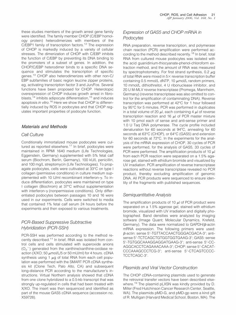
Reverse transcriptase (RT)-PCR studies showed thatpodocytes expressed mRNA for the growth arrestgenes GAS5 and CHOP (Figure 1). For GAS5, a doubleband representing two splice variants was found. Toconfirm the results found with the PCR-based cDNAsubtractive hybridization strategy, podocytes werestimulated with X/XO and the time-dependent inductionof GAS5 and CHOP mRNA expression was character-ized. In control cells only little mRNA expression forGAS5 and CHOP could be detected. Stimulation ofdifferentiated podocytes with X/XO for 1, 4, 12, 24, and48 hours induced a time-dependent significant in-
CHOP, ROS, and Cell Adhesion 23AJP January 2006, Vol. 168, No. 1
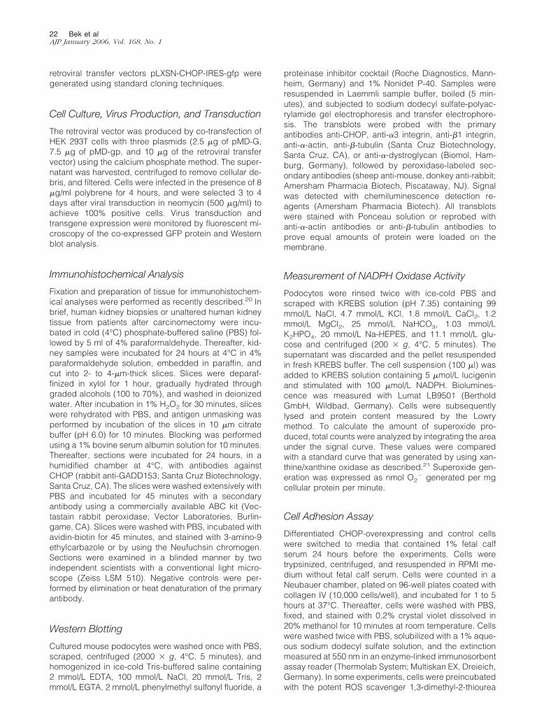
crease in GAS5 mRNA expression at 4 and 12 hoursand in CHOP mRNA expression at 1, 4, and 12 hours(Figure 1). Like X/XO, H2O2 (250 �mol/L) induced atime-dependent increase in CHOP mRNA expression(Figure 1). The response to H2O2 was concentration-dependent with a threshold concentration of 50 �mol/LH2O2 (Figure 2). To assure that quantification was ob-tained in the linear portion of the PCR amplificationcurve, cycle dependency of RT-PCR experiments wasshown (Figure 2). Pretreatment of podocytes with theROS scavenger N-acetylcysteine (10 mmol/L) for 30minutes completely inhibited the H2O2-induced in-crease in CHOP mRNA expression (Figure 2).
Recent data show that despite four alternative splicingpatterns for GAS5, no cellular protein is expressed.25,26
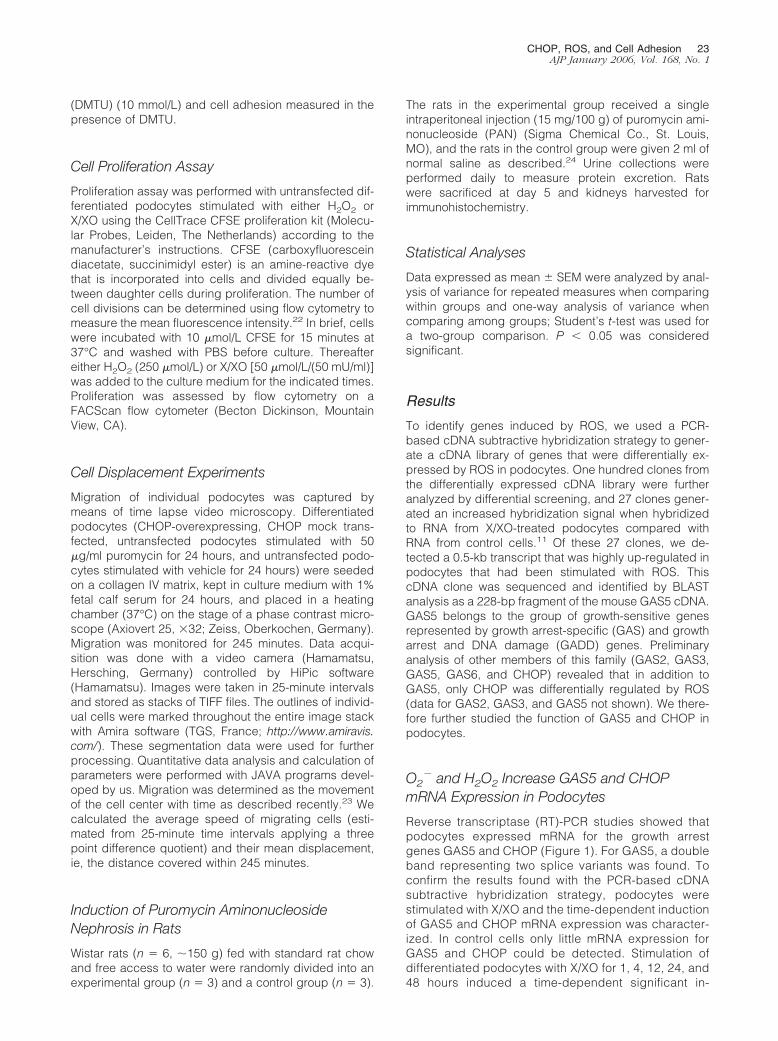
We therefore concentrated our efforts on the analysis ofCHOP protein expression. To test whether the increasedCHOP mRNA expression levels in response to ROS re-sulted in a subsequent increase in protein expression,CHOP protein was measured in H2O2-treated, xanthine/xanthine oxidase-treated, and control podocytes byWestern blotting. An �10-fold increase in H2O2-mediatedprotein expression and an approximately threefold in-crease in superoxide-mediated CHOP protein expressionwere detected after 24-hour stimulation with H2O2 orxanthine/xanthine oxidase, respectively (Figure 3).
Figure 1. GAS5 and CHOP mRNA is time dependently expressed in X/XOand H2O2-treated differentiated mouse podocytes. Time course for the in-duction of GAS5 and CHOP mRNA by X/XO and H2O2. Gas5 and CHOPmRNA induction was studied in control podocytes or in podocytes that havebeen incubated with X/XO (50 �mol/L/50 mU/ml) (A) or H2O2 (250 �mol/L)(B) for the indicated times. Experiments were performed by using RT or noRT in each set-up (RT� not shown). The densitometric analysis of mRNAexpression profiles for CHOP was normalized with GAPDH mRNA expres-sion. Studies were performed with primers derived from published mousecDNA sequences resulting in a 379-bp fragment for CHOP and two 344- and301-bp fragments for GAS5. Sequence analysis of the resulting amplificationproducts revealed sequence identity for the amplified fragments with pub-lished sequences (n � 3 to 15, values are means � SEM, *P � 0.05 versuscontrol, t-test).
Figure 2. Top left: Cycle dependency of RT-PCR experiments. Experimentswere performed to assure that quantification was obtained in the linearportion of the PCR amplification curve. Densitometric analysis of the bandsrevealed a linear correlation between band densities and amount of cycles(data not shown). In all experiments 21 cycles were used for �-actin and 30cycles for CHOP amplification. Top right: Concentration response curve ofthe effect of H2O2 on CHOP mRNA expression in podocytes (n � 3, valuesare means � SEM, *P � 0.05 versus 0 �mol/L H2O2, analysis of variance,Scheffe’s test). Bottom left: Effect of N-acetylcysteine on H2O2-mediatedincrease in CHOP mRNA expression. Podocytes were incubated with vehicle(Co), H2O2 (250 �mol/L), H2O2 (250 �mol/L) � n-ACC (10 mmol/L), orn-ACC (10 mmol/L) for 1 hour. N-acetylcysteine was added 60 minutesbefore stimulation with H2O2. The densitometric analysis of mRNA expres-sion profiles for CHOP by RT-PCR was normalized with �-actin mRNAexpression. Bottom right: Statistical analysis (n � 3, values are means �SEM, *P � 0.05 versus control, t-test).
24 Bek et alAJP January 2006, Vol. 168, No. 1
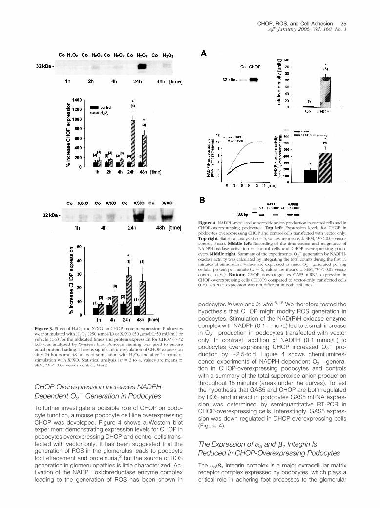
CHOP Overexpression Increases NADPH-Dependent O2
� Generation in Podocytes
To further investigate a possible role of CHOP on podo-cyte function, a mouse podocyte cell line overexpressingCHOP was developed. Figure 4 shows a Western blotexperiment demonstrating expression levels for CHOP inpodocytes overexpressing CHOP and control cells trans-fected with vector only. It has been suggested that thegeneration of ROS in the glomerulus leads to podocytefoot effacement and proteinuria,2 but the source of ROSgeneration in glomerulopathies is little characterized. Ac-tivation of the NADPH oxidoreductase enzyme complexleading to the generation of ROS has been shown in
podocytes in vivo and in vitro.6,18 We therefore tested thehypothesis that CHOP might modify ROS generation inpodocytes. Stimulation of the NAD(P)H-oxidase enzymecomplex with NADPH (0.1 mmol/L) led to a small increasein O2
� production in podocytes transfected with vectoronly. In contrast, addition of NADPH (0.1 mmol/L) topodocytes overexpressing CHOP increased O2
� pro-duction by �2.5-fold. Figure 4 shows chemilumines-cence experiments of NADPH-dependent O2
� genera-tion in CHOP-overexpressing podocytes and controlswith a summary of the total superoxide anion productionthroughout 15 minutes (areas under the curves). To testthe hypothesis that GAS5 and CHOP are both regulatedby ROS and interact in podocytes GAS5 mRNA expres-sion was determined by semiquantitative RT-PCR inCHOP-overexpressing cells. Interestingly, GAS5 expres-sion was down-regulated in CHOP-overexpressing cells(Figure 4).
The Expression of �3 and �1 Integrin IsReduced in CHOP-Overexpressing Podocytes
The �3/�1 integrin complex is a major extracellular matrixreceptor complex expressed by podocytes, which plays acritical role in adhering foot processes to the glomerular
Figure 3. Effect of H2O2 and X/XO on CHOP protein expression. Podocyteswere stimulated with H2O2 (250 �mol/L) or X/XO (50 �mol/L/50 mU/ml) orvehicle (Co) for the indicated times and protein expression for CHOP (�32kd) was analyzed by Western blot. Ponceau staining was used to ensureequal protein loading. There is significant up-regulation of CHOP expressionafter 24 hours and 48 hours of stimulation with H2O2 and after 24 hours ofstimulation with X/XO. Statistical analysis (n � 3 to 4, values are means �SEM, *P � 0.05 versus control, t-test).
Figure 4. NADPH-mediated superoxide anion production in control cells and inCHOP-overexpressing podocytes. Top left: Expression levels for CHOP inpodocytes overexpressing CHOP and control cells transfected with vector only.Top right: Statistical analysis (n � 5, values are means � SEM, *P � 0.05 versuscontrol, t-test). Middle left: Recording of the time course and magnitude ofNADPH-oxidase activation in control cells and CHOP-overexpressing podo-cytes. Middle right: Summary of the experiments. O2
� generation by NADPH-oxidase activity was calculated by integrating the total counts during the first 15minutes of stimulation. Values are expressed as nmol O2
� generated per mgcellular protein per minute (n � 6, values are means � SEM, *P � 0.05 versuscontrol, t-test). Bottom: CHOP down-regulates GAS5 mRNA expression inCHOP-overexpressing cells (CHOP) compared to vector-only transfected cells(Co). GAPDH expression was not different in both cell lines.
CHOP, ROS, and Cell Adhesion 25AJP January 2006, Vol. 168, No. 1
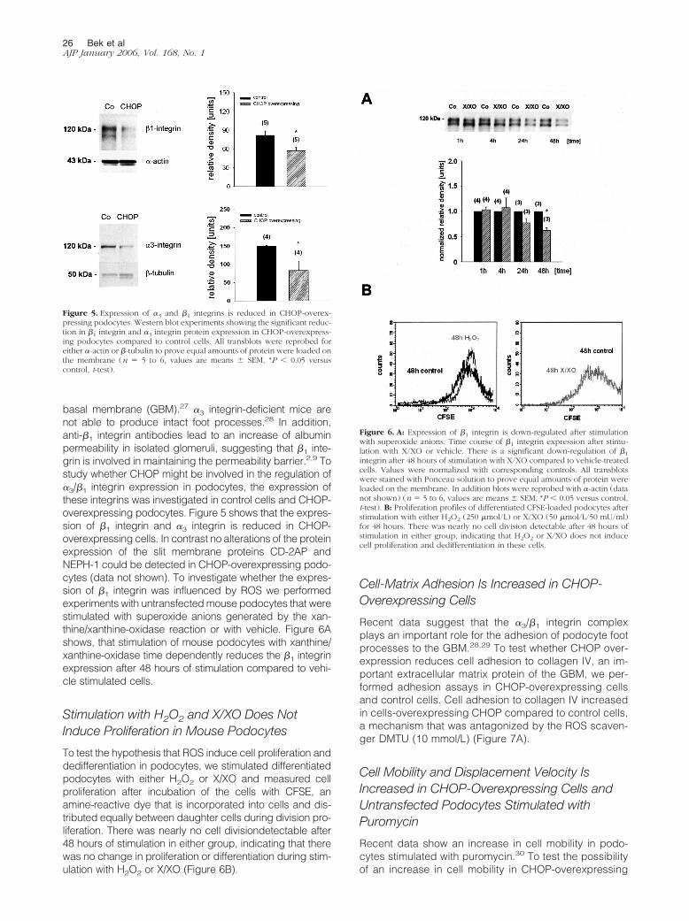
basal membrane (GBM).27 �3 integrin-deficient mice arenot able to produce intact foot processes.28 In addition,anti-�1 integrin antibodies lead to an increase of albuminpermeability in isolated glomeruli, suggesting that �1 inte-grin is involved in maintaining the permeability barrier.2,9 Tostudy whether CHOP might be involved in the regulation of�3/�1 integrin expression in podocytes, the expression ofthese integrins was investigated in control cells and CHOP-overexpressing podocytes. Figure 5 shows that the expres-sion of �1 integrin and �3 integrin is reduced in CHOP-overexpressing cells. In contrast no alterations of the proteinexpression of the slit membrane proteins CD-2AP andNEPH-1 could be detected in CHOP-overexpressing podo-cytes (data not shown). To investigate whether the expres-sion of �1 integrin was influenced by ROS we performedexperiments with untransfected mouse podocytes that werestimulated with superoxide anions generated by the xan-thine/xanthine-oxidase reaction or with vehicle. Figure 6Ashows, that stimulation of mouse podocytes with xanthine/xanthine-oxidase time dependently reduces the �1 integrinexpression after 48 hours of stimulation compared to vehi-cle stimulated cells.
Stimulation with H2O2 and X/XO Does NotInduce Proliferation in Mouse Podocytes
To test the hypothesis that ROS induce cell proliferation anddedifferentiation in podocytes, we stimulated differentiatedpodocytes with either H2O2 or X/XO and measured cellproliferation after incubation of the cells with CFSE, anamine-reactive dye that is incorporated into cells and dis-tributed equally between daughter cells during division pro-liferation. There was nearly no cell divisiondetectable after48 hours of stimulation in either group, indicating that therewas no change in proliferation or differentiation during stim-ulation with H2O2 or X/XO (Figure 6B).
Cell-Matrix Adhesion Is Increased in CHOP-Overexpressing Cells
Recent data suggest that the �3/�1 integrin complexplays an important role for the adhesion of podocyte footprocesses to the GBM.28,29 To test whether CHOP over-expression reduces cell adhesion to collagen IV, an im-portant extracellular matrix protein of the GBM, we per-formed adhesion assays in CHOP-overexpressing cellsand control cells. Cell adhesion to collagen IV increasedin cells-overexpressing CHOP compared to control cells,a mechanism that was antagonized by the ROS scaven-ger DMTU (10 mmol/L) (Figure 7A).
Cell Mobility and Displacement Velocity IsIncreased in CHOP-Overexpressing Cells andUntransfected Podocytes Stimulated withPuromycin
Recent data show an increase in cell mobility in podo-cytes stimulated with puromycin.30 To test the possibilityof an increase in cell mobility in CHOP-overexpressing
Figure 5. Expression of �3 and �1 integrins is reduced in CHOP-overex-pressing podocytes. Western blot experiments showing the significant reduc-tion in �1 integrin and �3 integrin protein expression in CHOP-overexpress-ing podocytes compared to control cells. All transblots were reprobed foreither �-actin or �-tubulin to prove equal amounts of protein were loaded onthe membrane (n � 5 to 6, values are means � SEM, *P � 0.05 versuscontrol, t-test).
Figure 6. A: Expression of �1 integrin is down-regulated after stimulationwith superoxide anions. Time course of �1 integrin expression after stimu-lation with X/XO or vehicle. There is a significant down-regulation of �1
integrin after 48 hours of stimulation with X/XO compared to vehicle-treatedcells. Values were normalized with corresponding controls. All transblotswere stained with Ponceau solution to prove equal amounts of protein wereloaded on the membrane. In addition blots were reprobed with �-actin (datanot shown) (n � 5 to 6, values are means � SEM, *P � 0.05 versus control,t-test). B: Proliferation profiles of differentiated CFSE-loaded podocytes afterstimulation with either H2O2 (250 �mol/L) or X/XO (50 �mol/L/50 mU/ml)for 48 hours. There was nearly no cell division detectable after 48 hours ofstimulation in either group, indicating that H2O2 or X/XO does not inducecell proliferation and dedifferentiation in these cells.
26 Bek et alAJP January 2006, Vol. 168, No. 1
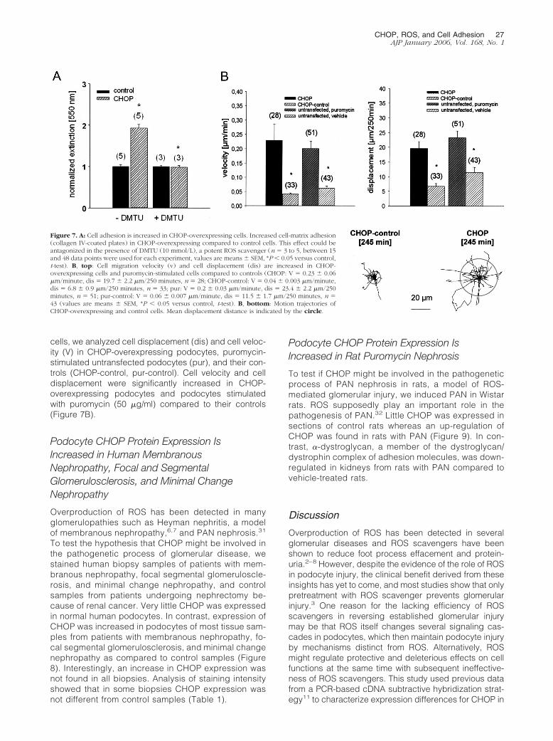
cells, we analyzed cell displacement (dis) and cell veloc-ity (V) in CHOP-overexpressing podocytes, puromycin-stimulated untransfected podocytes (pur), and their con-trols (CHOP-control, pur-control). Cell velocity and celldisplacement were significantly increased in CHOP-overexpressing podocytes and podocytes stimulatedwith puromycin (50 �g/ml) compared to their controls(Figure 7B).
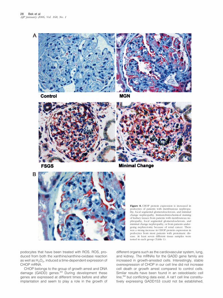
Podocyte CHOP Protein Expression IsIncreased in Human MembranousNephropathy, Focal and SegmentalGlomerulosclerosis, and Minimal ChangeNephropathy
Overproduction of ROS has been detected in manyglomerulopathies such as Heyman nephritis, a modelof membranous nephropathy,6,7 and PAN nephrosis.31
To test the hypothesis that CHOP might be involved inthe pathogenetic process of glomerular disease, westained human biopsy samples of patients with mem-branous nephropathy, focal segmental glomeruloscle-rosis, and minimal change nephropathy, and controlsamples from patients undergoing nephrectomy be-cause of renal cancer. Very little CHOP was expressedin normal human podocytes. In contrast, expression ofCHOP was increased in podocytes of most tissue sam-ples from patients with membranous nephropathy, fo-cal segmental glomerulosclerosis, and minimal changenephropathy as compared to control samples (Figure8). Interestingly, an increase in CHOP expression wasnot found in all biopsies. Analysis of staining intensityshowed that in some biopsies CHOP expression wasnot different from control samples (Table 1).
Podocyte CHOP Protein Expression IsIncreased in Rat Puromycin Nephrosis
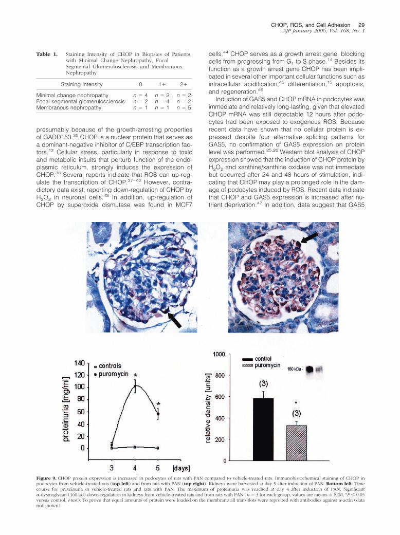
To test if CHOP might be involved in the pathogeneticprocess of PAN nephrosis in rats, a model of ROS-mediated glomerular injury, we induced PAN in Wistarrats. ROS supposedly play an important role in thepathogenesis of PAN.32 Little CHOP was expressed insections of control rats whereas an up-regulation ofCHOP was found in rats with PAN (Figure 9). In con-trast, �-dystroglycan, a member of the dystroglycan/dystrophin complex of adhesion molecules, was down-regulated in kidneys from rats with PAN compared tovehicle-treated rats.
Discussion
Overproduction of ROS has been detected in severalglomerular diseases and ROS scavengers have beenshown to reduce foot process effacement and protein-uria.2–8 However, despite the evidence of the role of ROSin podocyte injury, the clinical benefit derived from theseinsights has yet to come, and most studies show that onlypretreatment with ROS scavenger prevents glomerularinjury.3 One reason for the lacking efficiency of ROSscavengers in reversing established glomerular injurymay be that ROS itself changes several signaling cas-cades in podocytes, which then maintain podocyte injuryby mechanisms distinct from ROS. Alternatively, ROSmight regulate protective and deleterious effects on cellfunctions at the same time with subsequent ineffective-ness of ROS scavengers. This study used previous datafrom a PCR-based cDNA subtractive hybridization strat-egy11 to characterize expression differences for CHOP in
Figure 7. A: Cell adhesion is increased in CHOP-overexpressing cells. Increased cell-matrix adhesion(collagen IV-coated plates) in CHOP-overexpressing compared to control cells. This effect could beantagonized in the presence of DMTU (10 mmol/L), a potent ROS scavenger (n � 3 to 5, between 15and 48 data points were used for each experiment, values are means � SEM, *P � 0.05 versus control,t-test). B, top: Cell migration velocity (v) and cell displacement (dis) are increased in CHOP-overexpressing cells and puromycin-stimulated cells compared to controls (CHOP: V � 0.23 � 0.06�m/minute, dis � 19.7 � 2.2 �m/250 minutes, n � 28; CHOP-control: V � 0.04 � 0.003 �m/minute,dis � 6.8 � 0.9 �m/250 minutes, n � 33; pur: V � 0.2 � 0.03 �m/minute, dis � 23.4 � 2.2 �m/250minutes, n � 51; pur-control: V � 0.06 � 0.007 �m/minute, dis � 11.5 � 1.7 �m/250 minutes, n �43 (values are means � SEM, *P � 0.05 versus control, t-test). B, bottom: Motion trajectories ofCHOP-overexpressing and control cells. Mean displacement distance is indicated by the circle.
CHOP, ROS, and Cell Adhesion 27AJP January 2006, Vol. 168, No. 1
podocytes that have been treated with ROS. ROS, pro-duced from both the xanthine/xanthine-oxidase reactionas well as H2O2, induced a time-dependent expression ofCHOP mRNA.
CHOP belongs to the group of growth arrest and DNAdamage (GADD) genes.33 During development thesegenes are expressed at different times before and afterimplantation and seem to play a role in the growth of
different organs such as the cardiovascular system, lung,and kidney. The mRNAs for the GADD gene family areincreased in growth-arrested cells. Interestingly, stableoverexpression of CHOP in our cell line did not increasecell death or growth arrest compared to control cells.Similar results have been found in an osteoblastic cellline,34 but conflicting data exist. A rat1 cell line constitu-tively expressing GADD153 could not be established,
Figure 8. CHOP protein expression is increased inpodocytes of patients with membranous nephropa-thy, focal segmental glomerulosclerosis, and minimalchange nephropathy. Immunohistochemical stainingof kidney tissues from patients with membranous ne-phropathy, focal segmental glomerulosclerosis, andminimal change nephropathy, or from patients under-going nephrectomy because of renal cancer. Therewas a strong increase in CHOP protein expression inpodocytes from most patients with proteinuric dis-ease. At least seven different tissue samples weretested in each group (Table 1).
28 Bek et alAJP January 2006, Vol. 168, No. 1
presumably because of the growth-arresting propertiesof GADD153.35 CHOP is a nuclear protein that serves asa dominant-negative inhibitor of C/EBP transcription fac-tors.12 Cellular stress, particularly in response to toxicand metabolic insults that perturb function of the endo-plasmic reticulum, strongly induces the expression ofCHOP.36 Several reports indicate that ROS can up-reg-ulate the transcription of CHOP.37–42 However, contra-dictory data exist, reporting down-regulation of CHOP byH2O2 in neuronal cells.43 In addition, up-regulation ofCHOP by superoxide dismutase was found in MCF7
cells.44 CHOP serves as a growth arrest gene, blockingcells from progressing from G1 to S phase.14 Besides itsfunction as a growth arrest gene CHOP has been impli-cated in several other important cellular functions such asintracellular acidification,45 differentiation,15 apoptosis,and regeneration.46
Induction of GAS5 and CHOP mRNA in podocytes wasimmediate and relatively long-lasting, given that elevatedCHOP mRNA was still detectable 12 hours after podo-cytes had been exposed to exogenous ROS. Becauserecent data have shown that no cellular protein is ex-pressed despite four alternative splicing patterns forGAS5, no confirmation of GAS5 expression on proteinlevel was performed.25,26 Western blot analysis of CHOPexpression showed that the induction of CHOP protein byH2O2 and xanthine/xanthine oxidase was not immediatebut occurred after 24 and 48 hours of stimulation, indi-cating that CHOP may play a prolonged role in the dam-age of podocytes induced by ROS. Recent data indicatethat CHOP and GAS5 expression is increased after nu-trient deprivation.47 In addition, data suggest that GAS5
Table 1. Staining Intensity of CHOP in Biopsies of Patientswith Minimal Change Nephropathy, FocalSegmental Glomerulosclerosis and MembranousNephropathy
Staining intensity 0 1� 2�
Minimal change nephropathy n � 4 n � 2 n � 2Focal segmental glomerulosclerosis n � 2 n � 4 n � 2Membranous nephropathy n � 1 n � 1 n � 5
Figure 9. CHOP protein expression is increased in podocytes of rats with PAN compared to vehicle-treated rats. Immunohistochemical staining of CHOP inpodocytes from vehicle-treated rats (top left) and from rats with PAN (top right). Kidneys were harvested at day 5 after induction of PAN. Bottom left: Timecourse for proteinuria in vehicle-treated rats and rats with PAN. The maximum of proteinuria was reached at day 4 after induction of PAN. Significant�-dystroglycan (160 kd) down-regulation in kidneys from vehicle-treated rats and from rats with PAN (n � 3 for each group, values are means � SEM, *P � 0.05versus control, t-test). To prove that equal amounts of protein were loaded on the membrane all transblots were reprobed with antibodies against �-actin (datanot shown).
CHOP, ROS, and Cell Adhesion 29AJP January 2006, Vol. 168, No. 1
might regulate CHOP expression through snoRNA.26 Totest the hypothesis that GAS5 and CHOP can interact inpodocytes, GAS5 mRNA expression was determined inCHOP-overexpressing cells. GAS5 expression wasdown-regulated in CHOP-overexpressing cells suggest-ing a negative feedback mechanism of CHOP on GAS5expression.
CHOP protein could also be detected in podocytes inthe glomerulus, suggesting that it plays a role in podo-cyte function in vivo. As a consequence of the high de-gree of differentiation of podocytes, it has been shownthat they are unable to proliferate under normal condi-tions, and that in contrast to the proliferative capacity ofthe neighboring mesangial cells, the cell-cycle quies-cence in podocytes is tightly controlled.48 An escape ofthe podocyte from cell-cycle arrest leads to injury of theglomerular structure followed by a rapid deterioration ofkidney function, as shown by the deleterious course ofcollapsing nephropathy.49 Our data show that prolifera-tion in CHOP-overexpressing podocytes is not altered,but cell displacement is markedly increased. In addition,stimulation of untransfected podocytes with puromycinhad a similar effect. This effect has been shown before30
and might reflect the known migration tendency of podo-cytes in nephritic syndrome. Our data also indicate thatCHOP protein expression is increased in membranousnephropathy, focal segmental glomerulosclerosis, andminimal change disease suggesting a functional role ofCHOP in this disease. Interestingly, expression of CHOPwas not found in all biopsies studied. This might becaused by differences in the severity of disease, timecourse of the disease, or subforms of disease. Furtherstudies will be necessary to clarify this phenomenon.
To investigate possible functions of CHOP in podocytes,CHOP was constitutively overexpressed in a mouse cellline. In CHOP-overexpressing podocytes an increasedgeneration of O2
� could be detected. Podocytes are knownto be not only the target but also the source of ROS. InHeyman nephritis sublytic C5b-9 attack on podocytescauses up-regulation of the NADPH oxidoreductase en-zyme complex by podocytes, which is translocated to thecell surface.2 Subsequently, ROS are generated, reach theGBM matrix, and initiate lipid peroxidation and degradationof GBM collagen IV, leading to proteinuria.2 Therefore, in-duction of CHOP might—via activation of NADPH-depen-dent generation of superoxide anions—increase podocyteinjury and proteinuria. Recently, it has been demonstratedthat elevated CHOP expression results in an increased ROSproduction, reduction of cellular glutathione levels, anddown-regulation of BclII in fibroblasts.35
Attachment of podocytes to the glomerular basementmembrane is thought to be mediated in part by the �3/�1
integrin complex and by other cytoskeletal proteins in-cluding dystroglycan, actin, talin, vinculin, and �-acti-nin.49 The �3/�1 integrin complex plays an important rolefor kidney development and function.27,28 �3 integrinknockout mice show a disorganized GBM and podocyteswith immature foot processes.27 Cross-linking of �1 inte-grin by antibody binding has been shown to inhibit ad-hesion of podocytes in vitro and to increase glomerularpermeability for albumin.50,51 A reduced expression of
�3/�1 integrin on podocytes has been demonstrated inpodocytes of both humans and rats with diabetes52 andin PAN, an experimental model for human minimalchange disease.31 In the latter study treatment of animalswith phosphatidyl choline-bound superoxide dismutasereduced proteinuria and preserved the expression of �3
integrin expression in podocytes.31 Interestingly, in ourexperiments with rats treated with puromycin to inducePAN, up-regulation of CHOP was found exclusively inpodocytes. In addition, Western blot analysis of �3/�1
integrins in podocytes showed that the expression ofboth integrins is reduced in CHOP-overexpressing podo-cytes. In contrast the expression of CD2AP or NEPH-1,two important proteins of the podocyte slit diaphragm53
was not altered (data not shown). The data indicate thatCHOP may be a regulator of �3/�1 integrin expression inpodocytes and could play a role in the maintenance ofpodocyte adherence to the GBM. Down-regulation of�3/�1 integrins in podocytes might depend on extracel-lular ROS because stimulation of untransfected podo-cytes with xanthine/xanthine oxidase reduced �1 integrinexpression similar to the reduction seen in CHOP-over-expressing cells. Surprisingly, cell adhesion of CHOP-overexpressing cells was increased. This effect could beantagonized with the ROS scavenger DMTU. An in-creased cell-matrix adhesion of podocytes in the pres-ence of reduced �3 integrin expression has been shownvery recently in PAN, but the mechanism involved ispoorly understood.31 In the latter study, down-regulationof �3 integrin protected against PAN-induced podocytedetachment. It therefore might be possible, that CHOP—via down-regulation of integrins—inhibits the detachmentof podocyte foot processes in glomerular disease. Fur-ther studies will be necessary to differentiate betweendeleterious and beneficial effects of ROS on various cellfunctions in the glomerulus.
In summary the data indicate that podocytes regulateCHOP expression in response to ROS. The expression ofCHOP in podocytes is increased in some forms of pro-teinuric nephropathies and PAN. CHOP regulates impor-tant functions in podocytes: it increases NADPH-depen-dent generation of O2
�, it reduces the expression of�3/�1 integrins, and alters cell attachment and cell dis-placement. Thus, CHOP might play a critical role in theglomerular injury.
Acknowledgments
In remembrance of Stefan Greiber, a brilliant nephrologistand good friend, who died during the experimentation ofthis publication.
We thank C. Hupfer, P. Daemisch, and P. Kulick fortheir excellent technical assistance; and Peter Mundel,Albert Einstein College of Medicine, New York, NY, for thegenerous gift of podocyte cells.
References
1. Pavenstadt H: Roles of the podocyte in glomerular function. Am JPhysiol 2000, 278:F173–F179
30 Bek et alAJP January 2006, Vol. 168, No. 1
2. Kerjaschki D, Neale TJ: Molecular mechanisms of glomerular injury inrat experimental membranous nephropathy (Heymann nephritis).J Am Soc Nephrol 1996, 7:2518–2526
3. Gwinner W, Grone HJ: Role of reactive oxygen species in glomeru-lonephritis. Nephrol Dial Transplant 2000, 15:1127–1132
4. Yoshioka T, Ichikawa I, Fogo A: Reactive oxygen metabolites causemassive, reversible proteinuria and glomerular sieving defect withoutapparent ultrastructural abnormality. J Am Soc Nephrol 1991,2:902–912
5. Ricardo SD, Bertram JF, Ryan GB: Antioxidants protect podocyte footprocesses in puromycin aminonucleoside-treated rats. J Am SocNephrol 1994, 4:1974–1986
6. Neale TJ, Ullrich R, Ojha P, Poczewski H, Verhoeven AJ, KerjaschkiD: Reactive oxygen species and neutrophil respiratory burst cyto-chrome b558 are produced by kidney glomerular cells in passiveHeymann nephritis. Proc Natl Acad Sci USA 1993, 90:3645–3649
7. Shah SV: Evidence suggesting a role for hydroxyl radical in passiveHeymann nephritis in rats. Am J Physiol 1988, 254:F337–F344
8. Binder CJ, Weiher H, Exner M, Kerjaschki D: Glomerular overproduc-tion of oxygen radicals in Mpv17 gene-inactivated mice causes podo-cyte foot process flattening and proteinuria: a model of steroid-resis-tant nephrosis sensitive to radical scavenger therapy. Am J Pathol1999, 154:1067–1075
9. Grone HJ, Walli AK, Grone EF: The role of oxidatively modified li-poproteins in lipid nephropathy. Contrib Nephrol 1997, 120:160–175
10. Grone HJ, Grone EF, Malle E: Immunohistochemical detection ofhypochlorite-modified proteins in glomeruli of human membranousglomerulonephritis. Lab Invest 2002, 82:5–14
11. Greiber S, Muller B, Daemisch P, Pavenstadt H: Reactive oxygenspecies alter gene expression in podocytes: induction of granulocytemacrophage-colony-stimulating factor. J Am Soc Nephrol 1996,13:86–95
12. Ron D, Habener JF: CHOP, a novel developmentally regulated nu-clear protein that dimerizes with transcription factors C/EBP and LAPand functions as a dominant-negative inhibitor of gene transcription.Genes Dev 1992, 6:439–453
13. Wang XZ, Kuroda M, Sok J, Batchvarova N, Kimmel R, Chung P,Zinszner H, Ron D: Identification of novel stress-induced genesdownstream of CHOP. EMBO J 1998, 17:3619–3630
14. Barone MV, Crozat A, Tabaee A, Philipson L, Ron D: CHOP(GADD153) and its oncogenic variant, TLS-CHOP, have opposingeffects on the induction of G1/S arrest. Genes Dev 1994, 8:453–464
15. Batchvarova N, Wang XZ, Ron D: Inhibition of adipogenesis bythe stress-induced protein CHOP (Gadd153). EMBO J 1995,14:4654–4661
16. Matsumoto M, Minami M, Takeda K, Sakao Y, Akira S: Ectopic ex-pression of CHOP (GADD153) induces apoptosis in M1 myeloblasticleukemia cells. FEBS Lett 1996, 395:143–147
17. Mundel P, Reiser J, Zuniga Mejia BA, Pavenstadt H, Davidson GR,Kriz W, Zeller R: Rearrangements of the cytoskeleton and cell con-tacts induce process formation during differentiation of conditionallyimmortalized mouse podocyte cell lines. Exp Cell Res 1997,236:248–258
18. Greiber S, Munzel T, Kastner S, Muller B, Schollmeyer P, PavenstadtH: NAD(P)H oxidase activity in cultured human podocytes: effects ofadenosine triphosphate. Kidney Int 1998, 53:654–663
19. Nickel C, Benzing T, Sellin L, Gerke P, Karihaloo A, Liu ZX, CantleyLG, Walz G: The polycystin-1 C-terminal fragment triggers branchingmorphogenesis and migration of tubular kidney epithelial cells. J ClinInvest 2002, 109:481–489
20. Bek MJ, Wahle S, Muller B, Benzing T, Huber TB, Kretzler M, CohenC, Busse-Grawitz A, Pavenstadt H: Stra13, a prostaglandin E2-in-duced gene, regulates the cellular redox state of podocytes. FASEBJ 2003, 17:682–684
21. Bek MJ, Reinhardt HC, Fischer KG, Hirsch JR, Hupfer C, Dayal E,Pavenstadt H: Up-regulation of early growth response gene-1 via theCXCR3 receptor induces reactive oxygen species and inhibits Na�/K�-ATPase activity in an immortalized human proximal tubule cellline. J Immunol 2003, 170:931–940
22. Holcombe H, Mellman I, Janeway Jr CA, Bottomly K, Dittel BN: Theimmunosuppressive agent 15-deoxyspergualin functions by inhibit-ing cell cycle progression and cytokine production following naive Tcell activation. J Immunol 2002, 169:4982–4989
23. Schwab A, Wulf A, Schulz C, Kessler W, Nechyporuk-Zloy V, Romer
M, Reinhardt J, Weinhold D, Dieterich P, Stock C, Hebert SC: Sub-cellular distribution of Ca2� sensitive K� channels in migrating cells.J Cell Physiol 2006, 206:86–94
24. Guan N, Ding J, Deng J, Zhang J, Yang J: Key molecular eventsin puromycin aminonucleoside nephrosis rats. Pathol Int 2004,54:703–711
25. Raho G, Barone V, Rossi D, Philipson L, Sorrentino V: The gas 5 geneshows four alternative splicing patterns without coding for a protein.Gene 2000, 256:13–17
26. Smith CM, Steitz JA: Classification of gas5 as a multi-small-nucleolar-RNA (snoRNA) host gene and a member of the 5�-terminal oligopy-rimidine gene family reveals common features of snoRNA host genes.Mol Cell Biol 1998, 18:6897–6909
27. Kreidberg JA, Symons JM: Integrins in kidney development, function,and disease. Am J Physiol 2000, 279:F233–F242
28. Kreidberg JA, Donovan MJ, Goldstein SL, Rennke H, Shepherd K,Jones RC, Jaenisch R: Alpha 3 beta 1 integrin has a crucial role inkidney and lung organogenesis. Development 1996, 122:3537–3547
29. Adler S: Integrin receptors in the glomerulus: potential role in glomer-ular injury. Am J Physiol 1992, 262:F697–F704
30. Reiser J, Oh J, Shirato I, Asanuma K, Hug A, Mundel TM, Honey K,Ishidoh K, Kominami E, Kreidberg JA, Tomino Y, Mundel P: Podocytemigration during nephrotic syndrome requires a coordinated inter-play between cathepsin L and a3 integrin. J Biol Chem, 2004,279:34827–34832
31. Kojima K, Matsui K, Nagase M: Protection of alpha(3) integrin-medi-ated podocyte shape by superoxide dismutase in the puromycinaminonucleoside nephrosis rat. Am J Kidney Dis 2000, 35:1175–1185
32. Johnson RJ, Lovett D, Lehrer RI, Couser WG, Klebanoff SJ: Role ofoxidants and proteases in glomerular injury. Kidney Int 1994,45:352–359
33. Fleming JV, Fontanier N, Harries DN, Rees WD: The growth arrestgenes gas5, gas6, and CHOP-10 (gadd153) are expressed in themouse preimplantation embryo. Mol Reprod Dev 1997, 48:310–316
34. Pereira RC, Delany AM, Canalis E: CCAAT/enhancer binding proteinhomologous protein (DDIT3) induces osteoblastic cell differentiation.Endocrinology 2004, 145:1952–1960
35. McCullough KD, Martindale JL, Klotz LO, Aw TY, Holbrook NJ:Gadd153 sensitizes cells to endoplasmic reticulum stress by down-regulating Bcl2 and perturbing the cellular redox state. Mol Cell Biol2001, 21:1249–1259
36. Wang XZ, Ron D: Stress-induced phosphorylation and activation ofthe transcription factor CHOP (GADD153) by p38 MAP kinase. Sci-ence 1996, 272:1347–1349
37. Pomerance M, Carapau D, Chantoux F, Mockey M, Correze C, Fran-con J, Blondeau JP: CCAAT/enhancer-binding protein-homologousprotein expression and transcriptional activity are regulated by 3�,5�-cyclic adenosine monophosphate in thyroid cells. Mol Endocrinol2003, 17:2283–2294
38. Lovat PE, Oliverio S, Corazzari M, Ranalli M, Pearson AD, Melino G,Piacentini M, Redfern CP: Induction of GADD153 and Bak: novelmolecular targets of fenretinide-induced apoptosis of neuroblastoma.Cancer Lett 2003, 197:157–163
39. Ikeyama S, Wang XT, Li J, Podlutsky A, Martindale JL, KokkonenG, van Huizen R, Gorospe M, Holbrook NJ: Expression of thepro-apoptotic gene gadd153/chop is elevated in liver with agingand sensitizes cells to oxidant injury. J Biol Chem 2003,278:16726 –16731
40. Jeong JK, Stevens JL, Lau SS, Monks TJ: Quinone thioether-mediated DNA damage, growth arrest, and gadd153 expression inrenal proximal tubular epithelial cells. Mol Pharmacol 1996,50:592–598
41. Patton GW, Paciga JE, Shelley SA: NR8383 alveolar macrophagetoxic growth arrest by hydrogen peroxide is associated with inductionof growth-arrest and DNA damage-inducible genes GADD45 andGADD153. Toxicol Appl Pharmacol 1997, 147:126–134
42. Luethy JD, Fargnoli J, Park JS, Fornace Jr AJ, Holbrook NJ: Isolationand characterization of the hamster gadd153 gene. Activation ofpromoter activity by agents that damage DNA. J Biol Chem 1990,265:16521–16526
43. Paschen W, Mengesdorf T, Althausen S, Hotop S: Peroxidative stressselectively down-regulates the neuronal stress response activatedunder conditions of endoplasmic reticulum dysfunction. J Neurochem2001, 76:1916–1924
CHOP, ROS, and Cell Adhesion 31AJP January 2006, Vol. 168, No. 1
44. Guo G, Yan-Sanders Y, Lyn-Cook BD, Wang T, Tamae D, Ogi J,Khaletskiy A, Li Z, Weydert C, Longmate JA, Huang TT, Spitz DR,Oberley LW, Li JJ: Manganese superoxide dismutase-mediated geneexpression in radiation-induced adaptive responses. Mol Cell Biol2003, 23:2362–2378
45. Sok J, Wang XZ, Batchvarova N, Kuroda M, Harding H, Ron D:CHOP-dependent stress-inducible expression of a novel form of car-bonic anhydrase VI. Mol Cell Biol 1999, 19:495–504
46. Zinszner H, Kuroda M, Wang X, Batchvarova N, Lightfoot RT, RemottiH, Stevens JL, Ron D: CHOP is implicated in programmed cell deathin response to impaired function of the endoplasmic reticulum. GenesDev 1998, 12:982–995
47. Fleming JV, Hay SM, Harries DN, Rees WD: Effects of nutrient depri-vation and differentiation on the expression of growth-arrest genes(gas and gadd) in F9 embryonal carcinoma cells. Biochem J 1998,330:573–579
48. Kriz W: Progressive renal failure—inability of podocytes to replicate
and the consequences for development of glomerulosclerosis. Neph-rol Dial Transplant 1996, 11:1738–1742
49. Barisoni L, Kriz W, Mundel P, D’Agati V: The dysregulated podocytephenotype: a novel concept in the pathogenesis of collapsing idio-pathic focal segmental glomerulosclerosis and HIV-associated ne-phropathy. J Am Soc Nephrol 1999, 10:51–61
50. Kerjaschki D: Caught flat-footed: podocyte damage and the molec-ular bases of focal glomerulosclerosis. J Clin Invest 2001,108:1583–1587
51. Adler S, Chen X: Anti-Fx1A antibody recognizes a beta 1-integrin onglomerular epithelial cells and inhibits adhesion and growth. Am JPhysiol 1992, 262:F770–F776
52. Chen HC, Chen CA, Guh JY, Chang JM, Shin SJ, Lai YH: Alteringexpression of alpha3beta1 integrin on podocytes of human and ratswith diabetes. Life Sci 2000, 67:2345–2353
53. Tryggvason K, Wartiovaara J: Molecular basis of glomerular perm-selectivity. Curr Opin Nephrol Hypertens 2001, 10:543–549
32 Bek et alAJP January 2006, Vol. 168, No. 1