Functional and molecular alterations of the glomerular barrier in long-term diabetes in mice
10
ARTICLE Functional and molecular alterations of the glomerular barrier in long-term diabetes in mice M. Jeansson & A. Björnson Granqvist & J. Sörensson Nyström & B. Haraldsson Received: 20 December 2005 / Accepted: 2 May 2006 / Published online: 26 July 2006 # Springer-Verlag 2006 Abstract Aims/hypothesis Despite the fact that diabetic nephropathy is an increasingly common disorder that may lead to uraemia, the underlying mechanisms are still poorly understood and there is no specific therapy. To clarify whether long-term diabetes alters glomerular size- or charge-selectivity or both, we studied non-obese diabetic mice for up to 40 weeks. Materials and methods During the study period, spot urine was collected and blood pressure measured. At weeks 10 and 40, the right kidney was isolated and perfused at 8°C to inhibit tubular function, allowing for analysis of glomerular selectivity with albumin and Ficoll clearance. The left kidney was removed for further investigation using electron microscopy and molecular biology. Real-time PCR with low-density arrays was done to evaluate renal cortex mRNA expression of proteoglycans and other components in the glomerular barrier. After 40 weeks of diabetes, kidneys showed morphological changes typical of diabetic complications. Results At 40 weeks, the fractional clearance for negatively charged albumin was three times higher in the diabetic animals (0.0160) than in controls (0.0051, p<0.001), while fractional clearance for neutral Ficoll 35.5 Å with a Stokes Einstein radius similar to that of albumin was unaffected. In addition, protein and mRNA levels for versican and decorin were downregulated after 40 weeks of diabetes. Conclusions/interpretation We conclude that glomerular charge- but not size-selectivity was impaired in the diabetic animals with proteinuria. Also, glomerular components such as versican, decorin and fibromodulin were found to be downregulated after 40 weeks of diabetes. Keywords Charge-selectivity . Cooled isolated perfused kidney . Decorin . Diabetic nephropathy . Versican Abbreviations cIPK cooled isolated perfused kidney D10 10 weeks of diabetes D40 40 weeks of diabetes GBM glomerular basement membrane LDA low-density array NOD non-obese diabetic Introduction Worldwide, diabetic nephropathy is the most common cause of chronic kidney disease. Albuminuria is a hallmark of diabetic nephropathy, reflecting impairment of the glomerular capillary wall. Under normal conditions, the glomerular barrier restricts the passage of macromolecules such as albumin, while it is highly permeable to water and small solutes. The underlying mechanisms leading to proteinuria are still poorly understood, although the importance of the podocyte for glomerular integrity has been clearly documented [1–3]. Consequently, there is no specific therapy for diabetic nephropathy. Diabetologia (2006) 49:2200–2209 DOI 10.1007/s00125-006-0319-z M. Jeansson (*) : A. B. Granqvist : J. S. Nyström : B. Haraldsson Renal Center, Department of Nephrology, Institute of Internal Medicine, Sahlgrenska Academy, Bruna Straket 16, SE-413 45 Gothenburg, Sweden e-mail: [email protected]
Transcript of Functional and molecular alterations of the glomerular barrier in long-term diabetes in mice

ARTICLE
Functional and molecular alterations of the glomerularbarrier in long-term diabetes in mice
M. Jeansson & A. Björnson Granqvist &J. Sörensson Nyström & B. Haraldsson
Received: 20 December 2005 /Accepted: 2 May 2006 /Published online: 26 July 2006# Springer-Verlag 2006
AbstractAims/hypothesis Despite the fact that diabetic nephropathyis an increasingly common disorder that may lead touraemia, the underlying mechanisms are still poorlyunderstood and there is no specific therapy. To clarifywhether long-term diabetes alters glomerular size- orcharge-selectivity or both, we studied non-obese diabeticmice for up to 40 weeks.Materials and methods During the study period, spot urinewas collected and blood pressure measured. At weeks 10and 40, the right kidney was isolated and perfused at 8°C toinhibit tubular function, allowing for analysis of glomerularselectivity with albumin and Ficoll clearance. The leftkidney was removed for further investigation using electronmicroscopy and molecular biology. Real-time PCR withlow-density arrays was done to evaluate renal cortexmRNA expression of proteoglycans and other componentsin the glomerular barrier. After 40 weeks of diabetes,kidneys showed morphological changes typical of diabeticcomplications.Results At 40 weeks, the fractional clearance for negativelycharged albumin was three times higher in the diabeticanimals (0.0160) than in controls (0.0051, p<0.001), whilefractional clearance for neutral Ficoll 35.5 Å with a StokesEinstein radius similar to that of albumin was unaffected. In
addition, protein and mRNA levels for versican and decorinwere downregulated after 40 weeks of diabetes.Conclusions/interpretation We conclude that glomerularcharge- but not size-selectivity was impaired in the diabeticanimals with proteinuria. Also, glomerular componentssuch as versican, decorin and fibromodulin were found tobe downregulated after 40 weeks of diabetes.
Keywords Charge-selectivity . Cooled isolated perfusedkidney . Decorin . Diabetic nephropathy . Versican
AbbreviationscIPK cooled isolated perfused kidneyD10 10 weeks of diabetesD40 40 weeks of diabetesGBM glomerular basement membraneLDA low-density arrayNOD non-obese diabetic
Introduction
Worldwide, diabetic nephropathy is the most commoncause of chronic kidney disease. Albuminuria is a hallmarkof diabetic nephropathy, reflecting impairment of theglomerular capillary wall. Under normal conditions, theglomerular barrier restricts the passage of macromoleculessuch as albumin, while it is highly permeable to water andsmall solutes. The underlying mechanisms leading toproteinuria are still poorly understood, although theimportance of the podocyte for glomerular integrity hasbeen clearly documented [1–3]. Consequently, there is nospecific therapy for diabetic nephropathy.
Diabetologia (2006) 49:2200–2209DOI 10.1007/s00125-006-0319-z
M. Jeansson (*) :A. B. Granqvist : J. S. Nyström :B. HaraldssonRenal Center, Department of Nephrology,Institute of Internal Medicine, Sahlgrenska Academy,Bruna Straket 16,SE-413 45 Gothenburg, Swedene-mail: [email protected]

It is generally agreed that the glomerular barrier has bothsize- and charge-selective properties. However, the structuresresponsible for the permselective properties are under debate.For a long time the glomerular basement membrane (GBM)has been considered to be the main barrier for filtration. Morerecently, focus has shifted to the podocytes, since severalnovel proteins in the podocyte slit diaphragm have beenfound to be important for the barrier function [1–3]. Inaddition, we have suggested a role for the glomerularendothelial cell glycocalyx [4–10], an idea supported alsoby others [11, 12].
Previous studies have suggested that proteinuria indiabetes originates from alteration of either size- [13–16]or charge-selectivity [13, 17–21] or both [22, 23]. Much ofthe controversy over this issue may be due to methodolog-ical differences and limitations. Thus, in vivo studies ofproteinuria in patients or animals all have the majordisadvantage that proteins can be reabsorbed, secreted ordegraded in the tubules. Consequently, the protein concen-tration in the final urine may differ considerably from thatof primary urine. To overcome these limitations, one mayuse the inert polysaccharides dextran or Ficoll as markersinstead of albumin. Alternatively, the tubular processes maybe inhibited by toxins [24], but such drugs seem to affectpermeability per se [25]. At present, one of the mostreliable estimates of glomerular filtration is obtained byusing the cooled isolated perfused kidney (cIPK) model,either in rats [4, 8, 26] or in mice [9].
We have recently shown that human glomerular endo-thelial cells produce several different proteoglycans [10]and that hyaluronan and/or chondroitin sulphate in theglomerular endothelial cell glycocalyx are important tomaintain charge-selective properties in the barrier [9]. Indiabetic nephropathy, studies have suggested that decreasedsynthesis of heparan sulphate proteoglycans [27–30] suchas perlecan [31] is the reason for altered barrier function.Perlecan and agrin are the major proteoglycans in theGBM, both carrying heparan sulphate. However, genetical-ly modified mice lacking heparan sulphate chains onperlecan did not develop proteinuria [32]. Thus, studiesconcerning the role of proteoglycans in normal and diabetickidneys are still incomplete. The aim of this study was toassess functional, molecular and morphological changes ofthe glomerular barrier in non-obese diabetic (NOD) miceafter 10 and 40 weeks of diabetes.
Materials and methods
Animals and animal treatment
Experiments were performed in female NOD mice (M&B,Stensved, Denmark), a strain that spontaneously develops
insulin-dependent diabetes mellitus. The mice were kept onstandard chow and had free access to water prior to theexperiments. The local ethics review committee approvedthe experiments.
Diabetic NOD mice, aged 10–12 weeks at arrival, wereobserved for 10 weeks (D10) and 40 weeks (D40) whilereceiving insulin s.c. via implants (Linbit; Linshin Canada,ON, Canada) to maintain their blood glucose between 20and 33 mmol/l. At the end of the study, the left kidney wasremoved for electron microscopy and molecular biology,and the right kidney subjected to the cIPK procedure. NODmice at 25 weeks of age, which had not yet developeddiabetes at the breeders (blood glucose <10 mmol/l), wereused as controls and observed 1 week prior to the cIPKstudy.
Urine samples were collected once for controls or everythird week for diabetic mice, and blood glucose (One TouchUltra; Johnson & Johnson, Stockholm, Sweden) and mouseweight were recorded. Systolic blood pressure was mea-sured with a tail-cuff (Harvard Apparatus, Edenbridge, UK)on two consecutive days every third week.
Urinary albumin and creatinine
Albumin was determined in spot urine using ELISA(Bethyl Laboratories, Montgomery, TX, USA) with mousealbumin (ICN Biomedicals, Irvine, CA, USA) as standard.All urine samples were corrected for the urinary creatinineconcentration measured with a creatinine assay (SigmaDiagnostics, Sigma-Aldrich, Dorset, UK).
Isolated perfused kidneys at 8°C (cIPK)
Anaesthesia was induced and continued by inhalation ofisoflurane (2–3% v/v; Isoflurane; Pharmacia & Upjohn,Stockholm, Sweden) mixed with air (∼1 l/min) in anisoflurane vaporiser (Ohmeda Isotec 5; Simtec Engineering,Askim, Sweden). The body temperature of the mouse waskept at 37°C. The carotid artery was cannulated with a PE-10 cannula connected to a pressure transducer (PVBMedizintechnik GmbH, Kirchenseeon, Germany) for regis-tration of arterial pressure. The mouse was eviscerated andthe intestines removed. A cannula (PE-25) was put into thebladder for collection of urine. The left renal vein andartery were ligated and the left kidney was removed formolecular biology and electron microscopy. The aorta andcaval vein were clamped distal to the renal arteries and theaorta was cannulated in a retrograde direction with a T-tube(PE-25), connected to a pressure transducer. The clamp wasremoved, allowing perfusion of the right kidney by meansof a pulsatile pump (Ismatec IPC, Zurich, Switzerland). Theaorta was then ligated proximal to the renal arteries and thecaval vein was opened for venous outflow distal to the renal
Diabetologia (2006) 49:2200–2209 2201

arteries. The animal was killed by cutting the heartventricles open. After a short period of equilibration, urinesamples were collected and weighed. Great care was takennot to touch the kidneys and to provide adequate perfusionwith either blood or perfusate during the preparationprocedure. The temperature of the perfusate was kept at8°C in order to inhibit tubular function as well as energyconsumption. Arterial pressure and urine weight changeswere monitored by computer using AcqKnowledge v 3.7.3(Biopac Systems Inc., Goleta, CA, USA) computer software.
Perfusate for cIPK
Perfusate was prepared using a modified Tyrode solutionwith human serum albumin (18 g/l; Immuno, Vienna,Austria), 51Cr-EDTA and FITC-labelled Ficoll in the sizerange 12–70 Å. The solution had the following composi-tion: 113 mmol/l NaCl, 4.3 mmol/l KCl, 2.5 mmol/l CaCl2,0.8 mmol/l MgCl2, 25.5 mmol/l NaHCO3, 0.5 mmol/lNaH2PO4, 5.6 mmol/l glucose, 0.9 mmol/l nitroprusside(Merck, Darmstadt, Germany), 10 mg/l furosemide,200 mg/l FITC-labelled Ficoll (TdB Consultancy AB,Uppsala, Sweden), 0.16 MBq/l 51Cr-EDTA (AmershamPharmacia Biotech, Bucks, UK). The perfusate (pH 7.4)was protected from light and gassed with 5% CO2 in O2.
cIPK data analysis
Perfusate and urine samples from cIPK experiments wereanalysed for 51Cr-EDTA in a gamma counter (CobraAutoGamma Counting systems; Packard Instrument Com-pany, Meriden, CT, USA) and human serum albumin by animmunoturbidimetric assay (Triolab, Mölndal, Sweden).Glomerular filtration rate was calculated from the urine:perfusate concentration ratios for 51Cr-EDTA multiplied byurine flow. Fractional clearance for a solute is given by itsurine concentration divided by plasma concentration cor-rected for the residual tubular reabsorption (approximately1.15) using 51Cr-EDTA or the smallest Ficolls.
Analysis of Ficoll
The fractional clearance for different radii of FITC-Ficoll wascalculated by subjecting perfusate and urine samples to gelfiltration (TSK-gel G4000PWXL; Tosoh Bioscience, Stuttgart,Germany) and detection of fluorescence (Dionex fluorimeterRF-2000; Dionex Softron, Gynkotek, Germering, Germany)using Chromeleon (Gynkotek) software. A 0.05 mol/l phos-phate buffer with 0.15 mol/l NaCl (pH 7.0) was used aseluent. A volume of 5 μl from each sample was analysed at anexcitation wavelength of 492 nm and an emission wavelengthof 560 nm. The flow rate (0.5 ml/min) and the sampling
frequency (1/s) were maintained constant during the analysisand so were pressure (∼2 MPa) and temperature (8°C).
The gel-membrane model
According to the gel-membrane model, the glomerularbarrier is simplified to one charge-selective barrier (gel) andone size-selective barrier (membrane) in series. The gel isin contact with plasma and contains fixed negative charges,reducing the concentration of anionic solutes, such asalbumin. The membrane exerts size-selective properties.The concentrations of a solute in the urine will depend onthe effects of these two barriers as outlined below. Size-selective properties can be described by using a two-poremodel with experimental fractional clearances for Ficollswith a molecular radius range of 12–70 Å. In brief, theexchange was estimated using the following parameters: thefunctional small- and large-pore radii, the large-porefraction of the glomerular filtrate, and the unrestricted areadivided by diffusion distance [33]. By using a non-linearregression analysis and a previously defined set ofphysiological equations [8], model parameters were fittedto the experimental fractional clearances for neutral Ficolls(12–70 Å). Non-linear flux equations were used to calculatenet fluxes of fluid and solutes for the small- and large-porepathways individually. For calculation of charge-selectivity,the fractional clearances for the negatively charged albuminand its neutral counterpart of the same Stokes Einsteinradius, Ficoll 35.5 Å, were compared. For further details oncalculations, see Ohlson et al. [8].
Real-time PCR
RNA was prepared from fresh frozen renal cortex using theQiagen mini kit (Roche Diagnostics, Bromma, Sweden).The concentration and quality of the RNAwere evaluated bythe Agilent 2100 bioanalyser (Nano LabChip; AgilentTechnologies, Waldbronn, Germany). Synthesis of cDNAwas carried out using 1 μg of the RNA in an avianmyeloblastosis virus reverse transcriptase (AMV RT) bufferwith AMV RT, dNTP (deoxy-CTP, -GTP, -TTP and -ATP),random hexamers and RNase inhibitor (all reagents fromRoche Diagnostics) in a final volume of 20 μl. The reactionconditions were 5 min at 25°C and 50 min at 42°C, followedby 5 min at 70°C. The mRNA level of each target gene(Table 1) was quantified by real-time PCR on an ABI Prism7900 Sequence Detection system (Taqman; AppliedBiosystems, Foster City, CA, USA) using the low-densityarray (LDA) or 96-well plates (for TGF-β1). The PCR wascarried out in a reaction mix containing 50 ng sample cDNAand Taqman universal PCR master mix (containing MgCl2,dUTP, dATP, dCTP, dGTP, Taq Gold polymerase andAmpEraseUNG). The AmpEraseUNG was activated before
2202 Diabetologia (2006) 49:2200–2209

the denaturing step by heating for 2 min at 50°C. Sampleswere denatured at 95°C for 10 min and then subjected to 40cycles of two-step PCR, 15 s at 97°C and 1 min at 60°C.The LDA detects 23 genes in one run in duplicate, includingendogenous controls (18S and β-actin) and was designedincluding primer and probes for the genes summarised inTable 1. All LDA samples were run in duplicates giving amean from four reactions for each gene. All samples wereaveraged from the corrections with the two endogenouscontrols (18S and β-actin). The comparative ΔΔCT methodof relative quantification was used to calculate for differ-ences in gene expression using the software for ABI Prism7900 Sequence Detection system (Applied Biosystems).Samples run in 96-well plates were amplified simultaneouslyin triplicate in one assay run. Standard curves werecomputed for all genes (TGF-β1 and β-actin) from a seriesof dilutions from 3.125 through to 200 ng (seven concen-trations). For each sample, the amount of TGF-β1 and β-actin was determined from the corresponding standard curve.
Protein expression
Protein concentrations in renal cortex lysates were deter-mined using a BCA Protein Assay Reagent kit (Pierce,Rockford, IL, USA). Protein samples for versican detectionwere pretreated with enzymes (all from Sigma-Aldrich):chondroitinase ABC (0.1 U/sample), heparitinase I (10U/sample) and heparitinase III (1 U/sample) in ABC-buffer(100 mmol/l Tris-acetic acid, 10 mmol/l EDTA, 3 mmol/lCaCl2, pH 7.3) for 14 h at 37°C, in order to removeglycosaminoglycan chains. Protein lysates were separatedon NuPAGE 4–12% Bis–Tris gels (Novex, San Diego, CA,
USA) for decorin, podocin and TGF-β1 and on NuPAGE3–8% Tris-acetate gels (Novex) for versican. After electro-phoresis the proteins were transferred to polyvinylidenedifluoride membranes (decorin, podocin and TGF-β1) ornitrocellulose membrane (versican). Membranes wereblocked and immunological detection was done using thefollowing primary antibodies: rabbit anti-versican (kind giftfrom Dr D. Zimmerman, Department of Pathology, Zurich,Switzerland), rabbit anti-podocin (Sigma-Aldrich), rabbitanti-decorin, rabbit anti-TGF-β1 and rabbit anti-GAPDH(all from Abcam Ltd, Cambridge, UK) as loading control.Horseradish peroxidase-conjugated anti-rabbit IgG (Amer-sham) was used to visualise immunoreactive bands withenhanced chemoluminescence (ECL plus; Amersham) in aCCD camera (LAS1000; Fujifilm, Tokyo, Japan).
Immunohistochemistry was performed on acetone-fixed4-μm fresh-frozen tissue sections. Sections for versicanstaining were pretreated with 0.5 U/ml chondroitinase ABC(Sigma-Aldrich). Double-staining was performed withprimary antibodies towards versican (Chemicon Europe,Hants, UK) and decorin as described above, together withthe endothelial cell marker goat anti-von Willebrand factor(Santa Cruz Biotechnology Inc., Santa Cruz, CA, USA).
Morphological estimations
GBM thickness was measured in electron micrographswhere the thickness was defined as the distance between thepodocyte foot process and the corresponding endothelialcell. To ensure proper cross-sectioning, a slit diaphragmbetween the foot processes had to be visible as well as asingle layer of endothelial cells. Sclerotic areas were
Table 1 Targets analysed byreal-time PCR
↓downregulated expression;– unchanged expressionCS Chondroitin sulphate,DS dermatan sulphate,GPI glycosyl-phosphatidyl-inositol, HS heparan sulphate,KS keratin sulphate, PGproteoglycan, SLRP smallleucine-rich proteoglycan,CSPG chondroitin sulphateproteoglycan, HSPG heparansulphate proteoglycan
Coded protein Accession no. Protein description Change compared with control
D10 D40
18S – Endogenous controlβ-Actin NM_007393 Endogenous controlBiglycan NM_007542 CS/DS, SLRP, secreted – –Decorin NM_007833 CS/DS, SLRP, secreted – ↓Fibromodulin NM_021355 KS, SLRP, secreted – ↓Versican D28599 CSPG, secreted ↓ ↓Perlecan M77174 HS/CSPG, secreted – –Glypican-1 NM_016696 HSPG, GPI-anchored – –Glypican-4 NM_008150 HSPG, GPI-anchored – –Syndecan-1 NM_011519 HSPG, membrane-bound – –Syndecan-4 NM_011521 HSPG, membrane-bound – –Nephrin NM_019459 Podocyte slit – –Podocin NM_130456 Podocyte slit – –PKCδ NM_011103 Protein kinase C – –MMP-9 NM_013599 Matrix metalloprotease-9 – –TGF-β1 NM_011577 Transforming growth factor β1 – –
Diabetologia (2006) 49:2200–2209 2203

visualised with periodic acid-Schiff staining on 4-μmcryosections on light microscopy. A microscopic imagefor each glomerulus was evaluated in Adobe Photoshop7.0. The glomerular interface was marked, giving theglomerular area, and the sclerotic area was estimated bycolour tracing.
Statistics
Results are presented as means and 95% CIs. Variables weretested for uneven distribution (skewness >2 SDs) and unequalvariance. Logarithmic values were used for variables withuneven distribution, i.e. GFR, fractional clearance foralbumin, Ficoll 35.5 Å and Ficoll 55 Å, large-pore fractionof the glomerular filtrate, glomerular area, sclerosis/glomer-ulus, GBM thickness, albumin:creatinine ratio, and mRNAexpression. Statistical comparisons were made with controlsusing one-way ANOVA and a post-hoc Dunnett test. A valuep<0.05 was considered statistically significant.
Results
General
The mice had a mean body weight of 26.9 g (26.0–27.8),29.2 g (28.4–29.9) and 28.9 g (27.3–30.4) for controls, D10and D40, respectively. No increase in systolic blood
pressure over time could be seen. Diabetic animals had asystolic blood pressure of ~140 mmHg throughout thestudy (Fig. 1). The blood glucose was 12.4 mmol/l (7.5–17.3), 24.3 mmol/l (20.5–28.1), and 24.5 mmol/l (22.8–26.1) for controls, D10 and D40, respectively.
Diabetic mice showed a significant increase in glomer-ular surface area and periodic acid-Schiff-positive stainingas estimated with light microscopy. Using transmissionelectron microscopy the GBM thickness was measured andfound to be significantly increased in both diabetic groupscompared with controls (p<0.001). In addition, D40increased the kidney weight significantly (p<0.01). Dataregarding glomerular morphology are summarised inTable 2 and GFRs from the cIPK are described in Table 3.
Permselectivity of the glomerular barrier
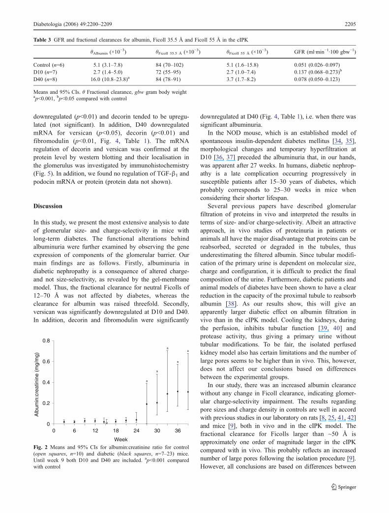
Albuminuria was constant and similar for controls anddiabetic animals until week 24. Thereafter, diabetic animalsshowed a significant increase in albumin:creatinine ratio(p<0.001, Fig. 2).
The fractional clearance for albumin in the cIPK wasfound to be significantly increased at D40 (p<0.001,Table 3). There was no difference in fractional clearancesfor Ficoll 35.5 Å, the neutral counterpart of albumin,compared with controls (Table 3). The fractional clearancesfor all Ficoll sizes (12–70 Å) are shown in Fig. 3. To furtherdescribe the glomerular barrier, mathematical estimationswere performed using the data above and the fractionalclearances for all Ficoll sizes (12–70 Å) in a gel-membranemodel, including two-pore analysis and calculations ofglomerular charge density. The two-pore analysis showedno increase in small- or large-pore radii in diabetes, thusreflecting unaltered size-selectivity (Table 4). The radiuswas 46.2–46.8 Å for the small pore and 110–173 Å for theless frequent large pore. There was an approximately 40%reduction in charge density in the glomerular barrier from41.4 mEq/l (30.1–52.8) in controls to 25.6 mEq/l (20.6–30.7) in the D40 group (p<0.001, Table 4).
Expression of mRNA and proteins
Real-time PCR revealed a significant downregulation in therenal cortex of several genes in diabetic compared withcontrol animals. At D10, versican mRNA was significantly
100
110
120
130
140
150
160
170
0 6 12 18 24 30 36
Week
Sys
tolic
blo
od p
ress
ure
(mm
Hg)
Fig. 1 Means and 95% CIs for systolic blood pressure estimated withtail-cuff for control (open squares, n=10) and diabetic (black squares,n=7–23) mice. Until week 9 both D10 and D40 are included
Table 2 Diabetic nephropathy
Glomerular area (μm2) Sclerosis/glomerulus (%) GBM thickness (nm) Kidney weight/body weight (%)
Control 4,966 (4,658–5,294) 0.002 (0.0003–0.008) 169 (164–175) 0.82 (0.71–0.94)D10 5,428 (5,173–5,695) 0.160 (0.084–0.305)a 189 (180–198)a 0.95 (0.83–1.07)D40 6,142 (5,855–6,444)a 0.599 (0.413–0.869)a 248 (233–264)a 1.04 (0.95–1.13)a
Means and 95% CIs. ap<0.001 compared with control
2204 Diabetologia (2006) 49:2200–2209
downregulated (p<0.01) and decorin tended to be upregu-lated (not significant). In addition, D40 downregulatedmRNA for versican (p<0.05), decorin (p<0.01) andfibromodulin (p<0.01, Fig. 4, Table 1). The mRNAregulation of decorin and versican was confirmed at theprotein level by western blotting and their localisation inthe glomerulus was investigated by immunohistochemistry(Fig. 5). In addition, we found no regulation of TGF-β1 andpodocin mRNA or protein (protein data not shown).
Discussion
In this study, we present the most extensive analysis to dateof glomerular size- and charge-selectivity in mice withlong-term diabetes. The functional alterations behindalbuminuria were further examined by observing the geneexpression of components of the glomerular barrier. Ourmain findings are as follows. Firstly, albuminuria indiabetic nephropathy is a consequence of altered charge-and not size-selectivity, as revealed by the gel-membranemodel. Thus, the fractional clearance for neutral Ficolls of12–70 Å was not affected by diabetes, whereas theclearance for albumin was raised threefold. Secondly,versican was significantly downregulated at D10 and D40.In addition, decorin and fibromodulin were significantly
downregulated at D40 (Fig. 4, Table 1), i.e. when there wassignificant albuminuria.
In the NOD mouse, which is an established model ofspontaneous insulin-dependent diabetes mellitus [34, 35],morphological changes and temporary hyperfiltration atD10 [36, 37] preceded the albuminuria that, in our hands,was apparent after 27 weeks. In humans, diabetic nephrop-athy is a late complication occurring progressively insusceptible patients after 15–30 years of diabetes, whichprobably corresponds to 25–30 weeks in mice whenconsidering their shorter lifespan.
Several previous papers have described glomerularfiltration of proteins in vivo and interpreted the results interms of size- and/or charge-selectivity. Albeit an attractiveapproach, in vivo studies of proteinuria in patients oranimals all have the major disadvantage that proteins can bereabsorbed, secreted or degraded in the tubules, thusunderestimating the filtered albumin. Since tubular modifi-cation of the primary urine is dependent on molecular size,charge and configuration, it is difficult to predict the finalcomposition of the urine. Furthermore, diabetic patients andanimal models of diabetes have been shown to have a clearreduction in the capacity of the proximal tubule to reabsorbalbumin [38]. As our results show, this will give anapparently larger diabetic effect on albumin filtration invivo than in the cIPK model. Cooling the kidneys, duringthe perfusion, inhibits tubular function [39, 40] andprotease activity, thus giving a primary urine withouttubular modifications. To be fair, the isolated perfusedkidney model also has certain limitations and the number oflarge pores seems to be higher than in vivo. This, however,does not affect our conclusions based on differencesbetween the experimental groups.
In our study, there was an increased albumin clearancewithout any change in Ficoll clearance, indicating glomer-ular charge-selectivity impairment. The results regardingpore sizes and charge density in controls are well in accordwith previous studies in our laboratory on rats [8, 25, 41, 42]and mice [9], both in vivo and in the cIPK model. Thefractional clearance for Ficolls larger than ~50 Å isapproximately one order of magnitude larger in the cIPKcompared with in vivo. This probably reflects an increasednumber of large pores following the isolation procedure [9].However, all conclusions are based on differences between
Table 3 GFR and fractional clearances for albumin, Ficoll 35.5 Å and Ficoll 55 Å in the cIPK
θAlbumin (×10−3) θFicoll 35.5 Å (×10−3) θFicoll 55 Å (×10−3) GFR (ml·min−1·100 gbw−1)
Control (n=6) 5.1 (3.1–7.8) 84 (70–102) 5.1 (1.6–15.8) 0.051 (0.026–0.097)D10 (n=7) 2.7 (1.4–5.0) 72 (55–95) 2.7 (1.0–7.4) 0.137 (0.068–0.273)b
D40 (n=8) 16.0 (10.8–23.8)a 84 (78–91) 3.7 (1.7–8.2) 0.078 (0.050–0.123)
Means and 95% CIs. θ Fractional clearance, gbw gram body weightap<0.001, bp<0.05 compared with control
0
0.2
0.4
0.6
0.8
0 6 12 18 24 30 36
Week
Alb
umin
:cre
atin
ine
(mg/
mg)
a
a
a
a
a
Fig. 2 Means and 95% CIs for albumin:creatinine ratio for control(open squares, n=10) and diabetic (black squares, n=7–23) mice.Until week 9 both D10 and D40 are included. ap<0.001 comparedwith control
Diabetologia (2006) 49:2200–2209 2205

the three experimental groups subjected to the sameprocedure. Therefore, the changes observed in the diabeticanimals reflect true alterations of the glomerular barrier.
Proteinuria in diabetes has been suggested to originatefrom alteration in either size- [13–16] or charge-selectivity[13, 17–21] or both [22, 23]. As mentioned above, our datasupport the view that the charge density is reduced indiabetic nephropathy. There was a tendency for reducedsize-selectivity but the increased number of large pores didnot reach statistical significance. There are several reasonsfor the discrepancy in the literature regarding charge- andsize-selectivity in diabetic nephropathy. Firstly, it may bedue to differences in methodology, since measurements ofglomerular permeability in vivo and in vitro have limi-tations as discussed above. Secondly, there may be speciesdifferences or differences due to the mechanisms behind thediabetes. Finally, the studies differ in severity of diseaseand time of observation. In the present long-term study inmice, proteinuria was caused by a reduced glomerularcharge density with no significant change in size-selectivity.
The second objective of this study was to couple theobserved functional changes to molecular alterations. Thus,we investigated two podocyte-specific proteins (nephrin
and podocin) together with proteoglycan core proteins,TGF-β1, protein kinase C and matrix metalloprotease-9.The reason for our interest in proteoglycans is that theyseem to play a crucial role in the microvascular permeabil-ity in all organs, including the kidney [9, 43]. One of themost prominent changes induced by diabetes was thedecreased expression of versican, decorin and fibromodulin.Versican is an extracellular matrix proteoglycan withchondroitin sulphate chains that are able to bind hyaluronicacid [44]. We have recently found it to be expressed byhuman glomerular endothelial cells [10] and versican is mostlikely a component of the endothelial cell glycocalyx.Furthermore, in previous studies we have shown thatdigestion of hyaluronic acid and/or chondroitin sulphate inthe glomerular endothelial cell glycocalyx increases thefractional clearance for albumin [9, 43]. Thus, downregula-tion of the highly negatively charged versican may causealtered glomerular endothelial cell glycocalyx compositionand reduced charge-selectivity. However, versican was alsodownregulated at D10, where the fractional clearance foralbumin was unaltered. Thus, further studies are needed toexplore the role of versican in diabetic nephropathy andother glomerular disorders.
0.001
0.01
0.1
1
0 10 20 30 40 50 60 70
Stokes Einstein radius (Å)
Fra
ctio
nal c
lear
ance
Fig. 3 Means and 95% CIs for neutral Ficolls for controls (white squares, n=6), D10 (grey squares, n=7) and D40 (black squares, n=8). Onlyevery tenth value and one bar for the 95% CI are shown for the sake of clarity. Please note the logarithmic scale
Table 4 Glomerular permselectivity estimated with the gel-membrane model, which includes two-pore analysis and calculation of charge density
Small-pore radius (Å) Large-pore radius (Å) fL (%) A0/ΔX
(×10−3 cm)Charge density (mEq/l)
Control (n=6) 46.8 (45.6–47.9) 167 (119–228) 0.42 (0.18–0.96) 21.0 (0–43.9) 41.4 (30.1–52.8)D10 (n=7) 46.3 (45.2–47.4) 108 (86–134)a 0.68 (0.37–1.25) 45.8 (22.0–69.6) 47.1 (41.8–52.4)D40 (n=8) 46.2 (45.4–47.0) 118 (86–161) 0.62 (0.38–1.01) 19.4 (11.2–27.6) 25.6 (20.6–30.7)b
Means and 95% CIs. A0/ΔX Unrestricted pore area divided by diffusion distance, fL Large-pore fraction of the glomerular filtrateap<0.05, bp<0.001 compared with control
2206 Diabetologia (2006) 49:2200–2209
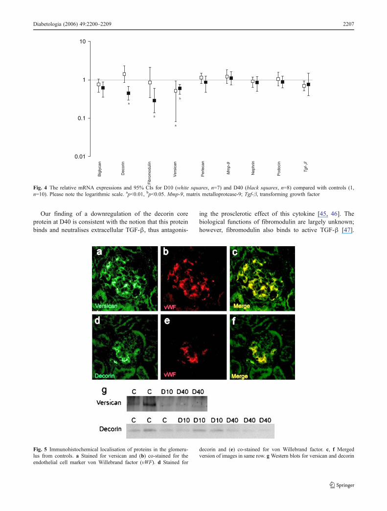
Our finding of a downregulation of the decorin coreprotein at D40 is consistent with the notion that this proteinbinds and neutralises extracellular TGF-β, thus antagonis-
ing the prosclerotic effect of this cytokine [45, 46]. Thebiological functions of fibromodulin are largely unknown;however, fibromodulin also binds to active TGF-β [47].
0.01
0.1
1
10
Pod
ocin
Nep
hrin
Mm
p-9
Per
leca
n
Ver
sica
n
Fib
rom
odul
in
Dec
orin
Big
lyca
n
Tgf
-
b
a
a
a
β
Fig. 4 The relative mRNA expressions and 95% CIs for D10 (white squares, n=7) and D40 (black squares, n=8) compared with controls (1,n=10). Please note the logarithmic scale. ap<0.01, bp<0.05. Mmp-9, matrix metalloprotease-9; Tgf-β, transforming growth factor
Fig. 5 Immunohistochemical localisation of proteins in the glomeru-lus from controls. a Stained for versican and (b) co-stained for theendothelial cell marker von Willebrand factor (vWF). d Stained for
decorin and (e) co-stained for von Willebrand factor. c, f Mergedversion of images in same row. g Western blots for versican and decorin
Diabetologia (2006) 49:2200–2209 2207

Previous studies have reported an upregulation of decorinmRNA in the early phase (1–6 weeks) of diabetes in mice[48]. Indeed, at D10 we observed a tendency towards anupregulation of decorin (Fig. 4) and the changed decorinexpression corresponds with changes in the fractionalclearance for albumin. However, data in the literatureregarding decorin expression in long-term diabetes areincomplete. In this study, we could not find any changes inmRNA and protein levels of TGF-β1. Therefore, in theseanimals, proteinuria and GBM thickening seem to bemediated through a different pathway than TGF-β.
In summary, this study reveals that diabetic nephropathyalters the composition of the glomerular barrier, thusleading to reduced charge-selectivity and proteinuria.Versican is markedly downregulated in diabetic animals.In diabetes with proteinuria, versican, decorin and fibro-modulin were found to be downregulated. Our findingssupport the idea that proteinuria in diabetic nephropathy isdue primarily to altered charge-selectivity. This suggeststhat diabetic nephropathy may be treated by interfering withproteoglycan synthesis and degradation.
Acknowledgements Technical assistance by E. Roos is gratefullyacknowledged. This study was supported by the Swedish MedicalResearch Council 9898, the Knut and Alice Wallenberg ResearchFoundation, the IngaBritt and Arne Lundbergs Research Foundation,the National Association for Kidney Diseases, the Swedish DiabetesAssociation Research Foundation, the John and Brit WennerströmsResearch Foundation and Sahlgrenska University Hospital GrantLUA-7545.
References
1. Shih NY, Li J, Cotran R, Mundel P, Miner JH, Shaw AS (2001)CD2AP localizes to the slit diaphragm and binds to nephrin via anovel C-terminal domain. Am J Pathol 159:2303–2308
2. Ruotsalainen V, Ljungberg P, Wartiovaara J et al (1999) Nephrinis specifically located at the slit diaphragm of glomerularpodocytes. Proc Natl Acad Sci 96:7962–7967
3. Roselli S, Gribouval O, Boute N et al (2002) Podocin localizes inthe kidney to the slit diaphragm area. Am J Pathol 160:131–139
4. Sörensson J, Ohlson M, Lindström K, Haraldsson B (1998)Glomerular charge selectivity for horseradish peroxidase andalbumin at low and normal ionic strengths. Acta Physiol Scand163:83–91
5. Haraldsson B, Sorensson J (2004) Why do we not all haveproteinuria? An update of our current understanding of theglomerular barrier. News Physiol Sci 19:7–10
6. Hjalmarsson C, Johansson BR, Haraldsson B (2004) Electronmicroscopic evaluation of the endothelial surface layer ofglomerular capillaries. Microvasc Res 67:9–17
7. Ciarimboli G, Hjalmarsson C, Bokenkamp A, Schurek HJ,Haraldsson B (2003) Dynamic alterations of glomerular chargedensity in fixed rat kidneys suggest involvement of endothelialcell coat. Am J Physiol Renal Physiol 285: F722–F730
8. Ohlson M, Sörensson J, Haraldsson B (2001) A gel-membranemodel of glomerular charge and size selectivity in series. Am JPhysiol Renal Physiol 280:F396–F405
9. Jeansson M, Haraldsson B (2003) Glomerular size and chargeselectivity in the mouse after exposure to glucosaminoglycan-degrading enzymes. J Am Soc Nephrol 14:1756–1765
10. Bjornson A, Moses J, Ingemansson A, Haraldsson B, Sorensson J(2005) Primary human glomerular endothelial cells produceproteoglycans, and puromycin affects their posttranslationalmodification. Am J Physiol Renal Physiol 288:F748–F756
11. Avasthi PS, Koshy V (1988) Pathophysiology and clinicalrelevance of proteinuria: glomerular endothelial glycocalyx.Contrib Nephrol 68:104–113
12. Deen WM (2004) What determines glomerular capillary perme-ability? J Clin Invest 114:1412–1414
13. Deckert T, Kofoed-Enevoldsen A, Vidal P, Norgaard K, AndreasenHB, Feldt-Rasmussen B (1993) Size- and charge selectivity ofglomerular filtration in type 1 (insulin-dependent) diabetic patientswith and without albuminuria. Diabetologia 36:244–251
14. Friedman S, Jones HW 3rd, Golbetz HV, Lee JA, Little HL,Myers BD (1983) Mechanisms of proteinuria in diabetic nephrop-athy. II. A study of the size-selective glomerular filtration barrier.Diabetes 32(Suppl 2):40–46
15. Nakamura Y, Myers BD (1988) Charge selectivity of proteinuriain diabetic glomerulopathy. Diabetes 37:1202–1211
16. Tomlanovich S, Deen WM, Jones HW 3rd, Schwartz HC, MyersBD (1987) Functional nature of glomerular injury in progressivediabetic glomerulopathy. Diabetes 36:556–565
17. Bangstad HJ, Kofoed-Enevoldsen A, Dahl-Jorgensen K, HanssenKF (1992) Glomerular charge selectivity and the influence ofimproved blood glucose control in type 1 (insulin-dependent)diabetic patients with microalbuminuria. Diabetologia 35:1165–1169
18. Pietravalle P, Morano S, Cristina G et al (1991) Charge selectivityof proteinuria in type I diabetes explored by Ig subclass clearance.Diabetes 40:1685–1690
19. Deckert T, Feldt-Rasmussen B, Djurup R, Deckert M (1988)Glomerular size and charge selectivity in insulin-dependentdiabetes mellitus. Kidney Int 33:100–106
20. Scandling JD, Myers BD (1992) Glomerular size-selectivity andmicroalbuminuria in early diabetic glomerular disease. Kidney Int41:840–846
21. Lemley KV, Blouch K, Abdullah I et al (2000) Glomerularpermselectivity at the onset of nephropathy in type 2 diabetesmellitus. J Am Soc Nephrol 11:2095–2105
22. Andersen S, Blouch K, Bialek J, Deckert M, Parving HH, MyersBD (2000) Glomerular permselectivity in early stages of overtdiabetic nephropathy. Kidney Int 58:2129–2137
23. Torffvit O, Rippe B (1999) Size and charge selectivity of theglomerular filter in patients with insulin-dependent diabetesmellitus: urinary immunoglobulins and glycosaminoglycans.Nephron 83:301–307
24. Osicka TM, Pratt LM, Comper WD (1996) Glomerular capillary wallpermeability to albumin and horseradish peroxidase. Nephrology2:199–212
25. Ohlson M, Sörensson J, Haraldsson B (2000) Glomerular size andcharge selectivity in the rat as revealed by FITC-Ficoll andalbumin. Am J Physiol Renal Physiol 279:F84–F91
26. Johnsson E, Haraldsson B (1992) An isolated perfused rat kidneypreparation designed for assessment of glomerular permeabilitycharacteristics. Acta Physiol Scand 144:65–73
27. Vernier RL, Steffes MW, Sisson-Ross S, Mauer SM (1992)Heparan sulfate proteoglycan in the glomerular basement mem-brane in type 1 diabetes mellitus. Kidney Int 41:1070–1080
28. Tamsma JT, van den Born J, Bruijn JA et al (1994) Expression ofglomerular extracellular matrix components in human diabeticnephropathy: decrease of heparan sulphate in the glomerularbasement membrane. Diabetologia 37:313–320
29. Reddi AS, Ramamurthi R, Miller M, Dhuper S, Lasker N (1991)Enalapril improves albuminuria by preventing glomerular loss of
2208 Diabetologia (2006) 49:2200–2209

heparan sulfate in diabetic rats. Biochem Med Metab Biol45:119–131
30. Wu VY, Wilson B, Cohen MP (1987) Disturbances in glomerularbasement membrane glycosaminoglycans in experimental diabe-tes. Diabetes 36:679–683
31. Conde-Knape K (2001) Heparan sulfate proteoglycans in exper-imental models of diabetes: a role for perlecan in diabetescomplications. Diabetes Metab Res Rev 17:412–421
32. Rossi M, Morita H, Sormunen R et al (2003) Heparan sulfatechains of perlecan are indispensable in the lens capsule but not inthe kidney. EMBO J 22:236–245
33. Rippe B, Haraldsson B (1994) Transport of macromoleculesacross microvascular walls: the two-pore theory. Physiol Rev74:163–219
34. Makino S, Kunimoto K, Muraoka Y, Mizushima Y, Katagiri K,Tochino Y (1980) Breeding of a non-obese, diabetic strain ofmice. Jikken Dobutsu 29:1–13
35. Craighead JE (1980) Experimental models of juvenile onset(insulin-dependent) diabetes mellitus. Monogr Pathol 21:166–176
36. Maeda M, Yabuki A, Suzuki S, Matsumoto M, Taniguchi K,Nishinakagawa H (2003) Renal lesions in spontaneous insulin-dependent diabetes mellitus in the nonobese diabetic mouse: acutephase of diabetes. Vet Pathol 40:187–195
37. Doi T, Hattori M, Agodoa LY et al (1990) Glomerular lesions innonobese diabetic mouse: before and after the onset of hypergly-cemia. Lab Invest 63:204–212
38. Hryciw DH, Lee EM, Pollock CA, Poronnik P (2004) Molecularchanges in proximal tubule function in diabetes mellitus. Clin ExpPharmacol Physiol 31:372–379
39. Esmann M, Skou JC (1988) Temperature-dependencies of variouscatalytic activities of membrane-bound Na+/K+-ATPase from ox
brain, ox kidney and shark rectal gland and of C12E8-solubilizedshark Na+/K+-ATPase. Biochim Biophys Acta 944:344–350
40. Charnoch JS, Doty DM, Russel JC (1971) The effect oftemperature on the activity of Na+/K+-ATPase. Arch BiochemBiophys 142:633–637
41. Sörensson J, Ohlson M, Haraldsson B (2001) A quantitativeanalysis of the glomerular charge barrier in the rat. Am J PhysiolRenal Physiol 280:F646–F656
42. Hjalmarsson C, Ohlson M, Haraldsson B (2001) Puromycin amino-nucleoside damages the glomerular size barrier with minimal effectson charge density. Am J Physiol Renal Physiol 281:F503–F512
43. Jeansson M, Haraldsson B (2006) Morphological and functionalevidence for an important role of the endothelial cell glycocalyx inthe glomerular barrier. Am J Physiol Renal Physiol 290:F111–F116
44. Iozzo RV (1998) Matrix proteoglycans: from molecular design tocellular function. Annu Rev Biochem 67:609–652
45. Border WA, Noble NA, Yamamoto T, Tomooka S, Kagami S(1992) Antagonists of transforming growth factor-beta: a novelapproach to treatment of glomerulonephritis and prevention ofglomerulosclerosis. Kidney Int 41:566–570
46. Border WA, Noble NA, Yamamoto T et al (1992) Natural inhibitorof transforming growth factor-beta protects against scarring inexperimental kidney disease. Nature 360:361–364
47. Hildebrand A, Romaris M, Rasmussen LM et al (1994) Interactionof the small interstitial proteoglycans biglycan, decorin andfibromodulin with transforming growth factor beta. Biochem J302:527–534
48. Mogyorosi A, Ziyadeh FN (1998) Increased decorin mRNA indiabetic mouse kidney and in mesangial and tubular cells culturedin high glucose. Am J Physiol 275:F827–F832
Diabetologia (2006) 49:2200–2209 2209