Failing to ponder? delusion-prone individuals rush to conclusions
Upload
independentCategory
view
0download
0
Brain Research 885 (2000) 251–261www.elsevier.com/ locate /bres
Research report
Structural alterations of tight junctions are associated with loss ofpolarity in stroke-prone spontaneously hypertensive rat blood–brain
barrier endothelial cellsa ,*,1 b,c ,1 b d aAndrea Lippoldt , Uwe Kniesel , Stefan Liebner , Hubert Kalbacher , Torsten Kirsch ,
b eHartwig Wolburg , Hermann Hallera ¨ ¨Max-Delbruck-Center for Molecular Medicine, Robert-Rossle-Strasse 10, 13092 Berlin, Germany
b ¨ ¨Institute of Pathology, University of Tubingen, Tubingen, GermanycInstitute of Zoology, University of Stuttgart–Hohenheim, Stuttgart, Germany
d ¨Medical and Natural Sciences Research Center, Tubingen, GermanyeDepartment of Nephrology, Medical School, Hannover, Germany
Accepted 12 September 2000
Abstract
The mechanisms leading to stroke in stroke-prone spontaneously hypertensive rats (SHRSP) are not well understood. We tested thehypothesis that the endothelial tight junctions of the blood–brain barrier are altered in SHRSP prior to stroke. We investigated tightjunctions in 13-week-old SHRSP, spontaneously hypertensive stroke-resistant rats (SHR) and age-matched Wistar–Kyoto rats (WKY) byelectron microscopy and immunocytochemistry. Ultrathin sections showed no difference in junction structure of cerebral capillaries fromSHRSP, SHR and WKY, respectively. However, using freeze-fracturing, we observed that the blood–brain barrier specific distribution oftight junction particles between P- and E-face in WKY (58.763.6%, P-face; 41.265.59%, E-face) and SHR (53.2619.3%, P-face;55.6613.25%, E-face) was changed to an 89.469.9% predominant E-face association in cerebral capillaries from SHRSP. However, theexpression of the tight junction molecules ZO-1, occludin, claudin-1 and claudin-5 was not changed in capillaries of SHRSP. Permeabilityof brain capillaries from SHRSP was not different compared to SHR and WKY using lanthanum nitrate as a tracer. In contrast, analysis ofendothelial cell polarity by distribution of the glucose-1 transporter (Glut-1) revealed that its abluminal:luminal ratio was reduced from4:1 in SHR and WKY to 1:1 in endothelial cells of cerebral capillaries of SHRSP. In summary, we demonstrate that early changes exist incerebral capillaries from a genetic model of hypertension-associated stroke. We suggest that a disturbed fence function of the tightjunctions in SHRSP blood–brain barrier endothelial cells may lead to subtle changes in polarity. These changes may contribute to thepathogenesis of stroke. 2000 Elsevier Science B.V. All rights reserved.
Theme: Disorders of the nervous system
Topic: Genetic models
Keywords: Hypertension; Stroke; Blood–brain barrier
1. Introduction [4,5,36]. However, the molecular mechanisms linkinghypertension to stroke are incompletely understood. Earlier
Hypertension plays an important role in the pathogenesis reports [9,13,37] have indicated that blood–brain barrierof stroke. An increase in blood pressure predisposes to permeability is increased in hypertension, suggesting thatstroke and about 70% of stroke patients are hypertensive permeability may be important for the development of
stroke. A utilitarian animal model of stroke is the stroke-prone spontaneously hypertensive rat (SHRSP) [48]. In
*Corresponding author. Tel.: 149-30-9406-3263; fax: 149-30-9406-SHRSP, 80% of the animals develop stroke spontaneously2110.during aging or in a defined time when given the appro-E-mail address: [email protected] (A. Lippoldt).
1Both authors contributed equally to this work. priate diet [15,30,46,47]. During the development of high
0006-8993/00/$ – see front matter 2000 Elsevier Science B.V. All rights reserved.PI I : S0006-8993( 00 )02954-1

252 A. Lippoldt et al. / Brain Research 885 (2000) 251 –261
blood pressure, changes in endothelial cell function occur (MAP) of 16168 mmHg. Normotensive age matchedin the cerebral vessels of SHRSP [29,35,38,41,49]. Prior to Wistar–Kyoto rats (MAP 12069 mmHg) and hypertensivestroke a reduction in global cerebral blood flow and age-matched SHR (MAP 16668 mmHg) were used ascortical protein synthesis is observed [24,30]. These ob- controls. The rats were housed at 12 h light–dark cycleservations have led to a hypothetical pathologic sequel under constant temperature conditions and received stan-where endothelial dysfunction and subsequent formation of dard rat chow (0.3% sodium chloride, SSNIFF Speziali-
¨edema precede the development of stroke [2,17,47]. The taten GmbH, Soest, Germany) and drinking water adaim of the present study was to analyze these early libitum. The animals were sacrificed using ether anesthesiachanges in endothelial cell function. and the tissue was removed and processed according to the
Specific characteristics of the blood–brain barrier endo- methods requirements. For quantitation, nine animals eachthelial cells are the tight junctions. Using freeze-fracture were used and tissue was taken from five different regionselectron microscopy it has been found that the tight of the cerebral cortex. For ultrathin sectioning, tissue wasjunction particles have a special distribution within the taken from the same rats and the same regions. Thefracture faces [20], that is about 57% of the particles protocol was approved by local authorities correspondingwithin the internal membrane leaflet (P-face) and about to criteria of the American Physiological Society.44% of the particles within the external leaflet (E-face) ofthe endothelial cell membrane [19]. This distribution is a 2.2. Light microscopic immunocytochemistryprerequisite of the maintenance of the blood–brain barriertightness [32,44] as well as of the functional polarization The rats (each strain four rats) were killed by etherof the blood–brain barrier endothelial cells [42]. These anesthesia; the brains were removed and snap frozen incells exhibit a specific distribution pattern of enzymes and isopentane at 2358C. The brains were sectioned at 10 mmtransporter molecules to the abluminal and luminal plasma thickness in a cryostat (CM 3000, Leica, Bensheim,membranes, respectively. Maturation of the blood–brain Germany) and mounted onto APES (Sigma)-coated slides.barrier during embryonal development and also soon after Immunocytochemistry was performed using immuno-birth leading to tightening of the barrier is accompanied by fluorescence technique with appropriate Cy2-labeled sec-a redistribution of enzyme activities and transporter mole- ondary antibodies (Dianova). The primary antibodies listedcules to their final target membranes and it is suggested in Table 1 were used. Prior to the incubation with thethat the polarity of the endothelial cells reflects the primary antibodies, the sections were fixed with either 4%maintenance of the fence function of their tight junctions buffered paraformaldehyde, 1% buffered paraformal-[42]. There have been preliminary reports that enzymatic dehyde at room temperature for 10 min, methanol atactivities are shifted to the other membrane which coincide 2208C for 10 min, or ethanol /acetone at 48C for 10 min aswith increased permeability under pathological circum- appropriate. Thereafter, the sections were rinsed in phos-stances [42,43]. phate-buffered saline (PBS) and blocked with 5% BSA for
We analyzed blood–brain barrier endothelial cell tight 60 min at room temperature. The incubations with thejunctions prior to the development of stroke in SHRSP to primary antibodies were made in a humidified chambertest the hypothesis that the morphology of tight junctions is over night at 48C in a dilution buffer consisting of PBSaltered prior to stroke. We were also interested in the with 0.04% Triton X-100 and 0.36% DMSO (PDT) andexpression pattern of junction-forming proteins. We ana- 1% BSA. After washing the slides three times for 5 min inlyzed endothelial cell permeability using lanthanum nitrate PBS, the sections were incubated with the secondary Cy2-and endothelial cell polarity by assessing the subcellulardistribution of the glucose-1 transporter (Glut-1) [1,7]. Wefound that an altered E-face association of tight junctionparticles as well as their disturbed fence function, mea- Table 1
aAntibodies and their sourcesured by Glut-1 distribution in SHRSP blood–brain barrierendothelial cells, may lead to subtle changes in the cerebral Antibody Host Dilution Antigen Sourcemicrovessel endothelial cells thereby contributing to the
Claudin-1 Rabbit 1:200 Tight junction, Zymedsusceptibility to stroke. transmembrane protein
Claudin-5 Rabbit 1:1000 Tight junction, Kalbacher H.transmembrane protein (see [21])
Occludin Rabbit 1:400 Tight junction, Zymed2. Materials and methodstransmembrane protein
ZO-1 Rabbit 1:400 Tight junction, Zymed2.1. Animals intracellular protein
Glut-1 Rabbit 1:400 Blood–brain barrier SigmaGlucose-1 transporterSHRSP were bred in the animal facility of the Max-
a¨Delbruck-Center. The 13-weeks-old rats had no neurologi- The table gives a short characterization of the antibodies used forcal symptoms and had a mean arterial blood pressure immunocytochemistry, their dilutions and source.

A. Lippoldt et al. / Brain Research 885 (2000) 251 –261 253
labeled antibody diluted in PDT with 1% BSA for 1 h at tions were investigated using a Zeiss EM10 and EM902room temperature. After repeated washing in PBS, the electron microscope.sections were mounted with Aqua Poly /Mount (Polysci-ences, Inc.) and examined in a Zeiss Axioplan microscope
2.6. Morphometrical analysis of the tight junction(Zeiss Oberkochen). Controls were performed by omittingnetworkthe primary antibody.
Quantitation was done at a final magnification of1:120,000. Tight junction-complexity was characterized by2.3. Immunoelectron microscopyfractal analysis and complexity index [18]. For evaluationof the fractal dimension (FD), the electron microscopicAfter perfusion (four SHRSP and four WKY rats) (2%image was digitized; five grids of different scaling levelsparaformaldehyde), the specimens were immersion-fixed(grid-sizes) were superimposed for each scaling-factor.(2% paraformaldehyde; 2 h at 48C), cryoprotected in 1.8 MThe number of boxes containing parts of the tight junctionsucrose, and quick-frozen in nitrogen-slush (22108C).structure were counted (N) in repeated measurements. TheFreeze substitution and low temperature embedding in 2grid sizes were 0.2, 0.1, 0.05, 0.025, and 0.0125 mm .Lowicryl HM20 (Polysciences, Inc.) was done in theSince the definition of the FD (box counting) is logN/AFS-System (Leica, Bensheim, Germany). Ultrathin sec-log(1 /s), the values obtained for each scaling-level weretioning was performed with an Ultracut R ultramicrotomeinserted into a logN vs. log(1 /s) graph for visualization,(Leica, Bensheim). The primary antibody Glut-1 wasand the regression curve was calculated. The slope of theobtained from the source described in Table 1. Gold-curve gives the estimated value for the FD. The degree ofconjugated secondary antibodies were purchased frommembrane association of tight junction-particles was de-Amersham and used at a dilution of 1:40. For quantitationtermined as the ratio ‘total length’ of E- or P-faceof Glut-1, three regions of the cerebral cortex of fiveassociated tight junction particles or ‘strands’ to ‘totalSHRSP and five WKY rats were investigated. From eachlength’ of the tight junction membrane structure, incortex 16 capillaries were evaluated at a final magnifica-percent. For quantification, the digitized images of the tighttion of 338,000.junctions were analyzed using the morphometric software
¨package ‘AnalySIS’ (SIS, Munster, Germany). The totalparticle density was given as E- and P-face parts of the2.4. Electron microscopyTJ-strands in percent of the tight junction length. A totalnumber of 234 tight junctions from cortices of nineSmall pieces of rat brain cerebral cortex were immersionSHRSP, 123 tight junctions from cortices from five SHRfixed in 2% HMSS-buffered glutaraldehyde (Paesel, Frank-and 179 tight junctions from cortices of nine WKY werefurt, Germany), stepwise dehydrated in ethanol and blockinvestigated.stained with saturated uranyl acetate, embedded in Araldite
(Serva, Heidelberg, Germany) and sectioned on an Ul-tracut FCR ultramicrotome (Leica). Semithin sections (0.6 2.7. Tracer studies using lanthanum nitratemm) were stained with Toluidine Blue, ultrathin sectionswere stained with lead citrate, mounted on pioloform- For examination of para- and transcellular permeability,coated copper grids and examined in a Zeiss (EM10; lanthanum nitrate was infused as a low molecular weightOberkochen, Germany) or LEO (EM902; Oberkochen, tracer (433 Da) [3]. The animals were anesthetized withGermany) electron microscope. intraperitoneal Ketanest /Rompun (Bayer AG, Germany)
and intracardial perfusion was performed with Ringersolution containing heparin to avoid coagulation of blood
2.5. Freeze fracture analysis cells. Subsequently, lanthanum nitrate solution in combina-tion with fixative (HMSS pH 7.4, 1% lanthanum nitrate,
The specimens were immersion-fixed with 2.5% buf- 4% paraformaldehyde, 1% glutaraldehyde) was perfusedfered glutaraldehyde, cryoprotected for freeze-fracture in and allowed to circulate for 20 min. Thereafter, the30% glycerol, and quick-frozen in nitrogen-slush animals were sacrificed and the brains were removed,(22108C). Subsequently, the specimens were fractured in a immersion fixed for 1 h (2.5% buffered glutaraldehyde)
26Balzer’s freeze-fracture unit (BAF 400D) at 5310 mbar and subsequently processed for electron microscopy.and 21508C and the fracture faces were shadowed withplatinum/carbon (10:1; 2 nm; 458) for contrast and carbon(20 nm; 908) for stabilization. After removing the cell 2.8. Statistical analysismaterial in 12% sodium hypochlorite, the replicas werecleaned several times in double distilled water and The statistical analysis was done using one-way
`mounted on Formvar-coated copper grids. The prepara- ANOVA and Scheffe-test.
254 A. Lippoldt et al. / Brain Research 885 (2000) 251 –261
3. Results
3.1. Morphology of brain capillaries
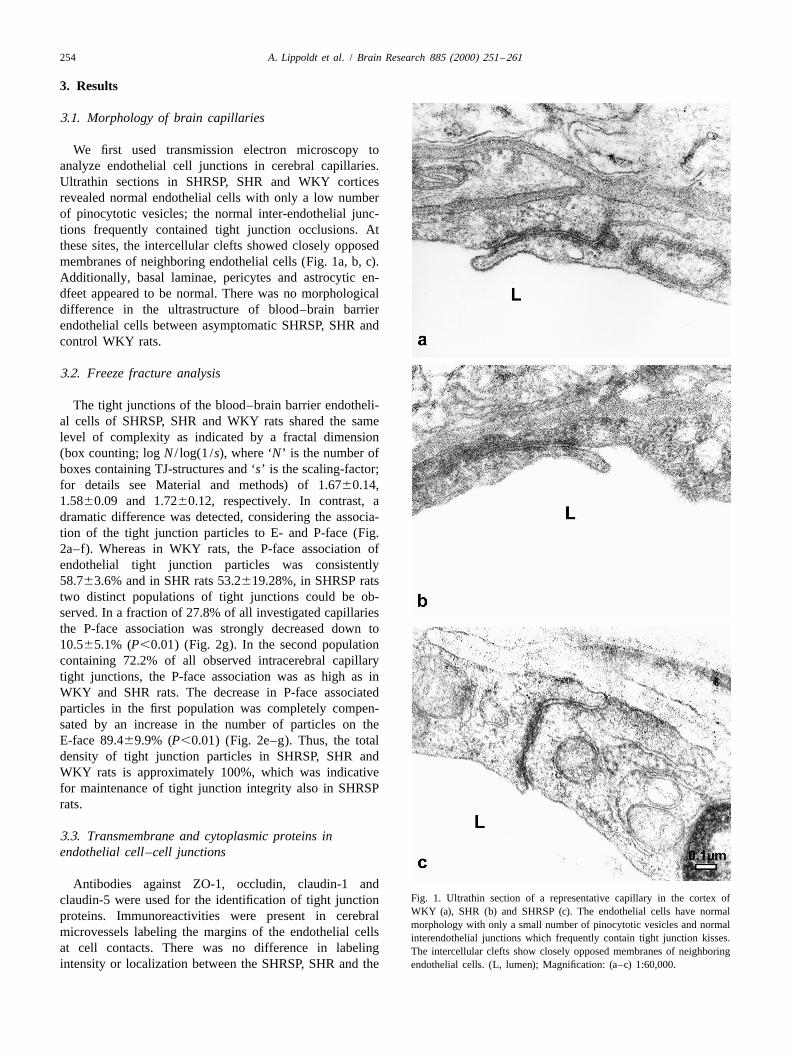
We first used transmission electron microscopy toanalyze endothelial cell junctions in cerebral capillaries.Ultrathin sections in SHRSP, SHR and WKY corticesrevealed normal endothelial cells with only a low numberof pinocytotic vesicles; the normal inter-endothelial junc-tions frequently contained tight junction occlusions. Atthese sites, the intercellular clefts showed closely opposedmembranes of neighboring endothelial cells (Fig. 1a, b, c).Additionally, basal laminae, pericytes and astrocytic en-dfeet appeared to be normal. There was no morphologicaldifference in the ultrastructure of blood–brain barrierendothelial cells between asymptomatic SHRSP, SHR andcontrol WKY rats.
3.2. Freeze fracture analysis
The tight junctions of the blood–brain barrier endotheli-al cells of SHRSP, SHR and WKY rats shared the samelevel of complexity as indicated by a fractal dimension(box counting; log N / log(1 /s), where ‘N’ is the number ofboxes containing TJ-structures and ‘s’ is the scaling-factor;for details see Material and methods) of 1.6760.14,1.5860.09 and 1.7260.12, respectively. In contrast, adramatic difference was detected, considering the associa-tion of the tight junction particles to E- and P-face (Fig.2a–f). Whereas in WKY rats, the P-face association ofendothelial tight junction particles was consistently58.763.6% and in SHR rats 53.2619.28%, in SHRSP ratstwo distinct populations of tight junctions could be ob-served. In a fraction of 27.8% of all investigated capillariesthe P-face association was strongly decreased down to10.565.1% (P,0.01) (Fig. 2g). In the second populationcontaining 72.2% of all observed intracerebral capillarytight junctions, the P-face association was as high as inWKY and SHR rats. The decrease in P-face associatedparticles in the first population was completely compen-sated by an increase in the number of particles on theE-face 89.469.9% (P,0.01) (Fig. 2e–g). Thus, the totaldensity of tight junction particles in SHRSP, SHR andWKY rats is approximately 100%, which was indicativefor maintenance of tight junction integrity also in SHRSPrats.
3.3. Transmembrane and cytoplasmic proteins inendothelial cell–cell junctions
Antibodies against ZO-1, occludin, claudin-1 andFig. 1. Ultrathin section of a representative capillary in the cortex ofclaudin-5 were used for the identification of tight junctionWKY (a), SHR (b) and SHRSP (c). The endothelial cells have normalproteins. Immunoreactivities were present in cerebralmorphology with only a small number of pinocytotic vesicles and normal
microvessels labeling the margins of the endothelial cells interendothelial junctions which frequently contain tight junction kisses.at cell contacts. There was no difference in labeling The intercellular clefts show closely opposed membranes of neighboringintensity or localization between the SHRSP, SHR and the endothelial cells. (L, lumen); Magnification: (a–c) 1:60,000.

A. Lippoldt et al. / Brain Research 885 (2000) 251 –261 255
Fig. 2. Freeze-fracture electronmicrographs of tight junction strands in capillaries of cerebral cortices. The figure shows E-face (a, c, e) and P-face (b, d, f)leaflets of WKY (a, b), SHR (c, d) and SHRSP (e, f) capillaries. The arrows in (f) point to particle aggregates only observed in SHRSP capillaries, whichmay be an indicator for tight junction dynamic. (g) The E-face association of tight junction particles was markedly increased in SHRSP capillaries to about89% (P,0.01) compared to WKY and SHR, whereas the P-face association was decreased to about 10% (P,0.01) (e and f). Magnification: 1:90,000.

256 A. Lippoldt et al. / Brain Research 885 (2000) 251 –261
four-fold more heavily labeled than the luminal membrane(Fig. 5a, c). In contrast, blood–brain barrier capillaries ofSHRSP rats consistently showed a sharp decrease in theabluminal / luminal gradient of Glut-1 density (Fig. 5b, c).While the cytoplasmic pool of anti-Glut-1 immunoreactivi-ty was identical to that in control rats, the density ofimmunogold particles at the abluminal membrane de-creased significantly (P,0.05) to 45615.9% in compari-son to controls. The density of immunogold particles at theluminal membrane increased to 30.9611.7% (P,0.05)(Fig. 5c). Thus the polarity of cerebrovascular endothelialcells as defined as the abluminal / luminal distribution ofthe Glut-1, had decreased from 4 to about 1.4 under thehypertensive conditions in SHRSP.
4. Discussion
Fig. 2. (continued) We tested the hypothesis that early changes in thecomposition of the junctional proteins may influenceendothelial cell function in the blood–brain barrier in
WKY brain microvessels (Fig. 3). ZO-1 (Fig. 3a–c) and SHRSP. We showed by freeze-fracture technique that theoccludin (Fig. 3d–f) shared the same distribution pattern morphology of the cell–cell contacts was altered. How-and intensity in SHRSP, SHR and WKY strains. Both ever, these changes in cell–cell contact in those youngantigens were exclusively found in the junctional regions asymptomatic rats did not lead to detectable changes inof blood–brain barrier endothelial cells. Claudin-5 im- permeability. Instead, they were associated with a loss ofmunoreactivity was found to share the same distribution endothelial cell polarity. Still, the immunoreactivities ofpattern as occludin at the junctional membranes in all tight junction components such as ZO-1, occludin, claudin-strains investigated (Fig. 3g–i). The claudin-1 immuno- 1 and claudin-5 were unaltered.reactivity was weaker as that of the other tight junction We used electron microscopy to analyze endothelialmolecules in the blood–brain barrier endothelial cells of all tight junctions in SHRSP. The freeze-fracture technique isthree rat strains (Fig. 3j–l). a powerful method to characterize tight junction structure
and complexity. The method has been widely used for3.4. Lanthanum nitrate permeability studies on blood–brain barrier development as well as for
tight junction regulation in epithelia. The common agree-Perfusion experiments using lanthanum nitrate as an ment is that the tight junction particles of partner strands
electron-dense tracer did not result in any staining of the are separated by the membrane cleavage used in thissubendothelial space and there was no difference between method [14,20]. Tight junctions of blood–brain barrierSHRSP and WKY rats. The distribution of tracer deposits endothelial cells show high protoplasmic face (P-face)in the intercellular space stopped precisely where tight association, whereas non-barrier blood vessels show a highjunctions were observed (Fig. 4a, b). Tracer deposits in external fracture face (E-face) association [28,44]. In anadjacent tissue were seen exclusively in peripheral organs intact blood–brain barrier, about 57% of the observedsuch as skeletal muscle (Fig. 4c), but not in the brain. particles are found in the P-face of the membrane whereasLanthanum nitrate deposits at the luminal surface of the about 44% of the protein particles belong to the E-facecapillaries were visible when the tracer was perfused [19]. It is now generally accepted that a switch of tighttogether with the fixative. junctional particles in the brain capillary endothelial cells
from the P- to the E-face correlates with an increase of the3.5. Endothelial cell polarity transendothelial permeability [39,44]. In contrast to nor-
motensive animals and SHR, we observed an increasedTo investigate endothelial cell polarity, we studied the E-face association in SHRSP. This predominant E-face
distribution of Glut-1 in the blood–brain barrier. We association resembled the one in blood–brain barrierperformed a quantitative evaluation of Glut-1 subcellular during rat brain development [19] or in peripheral blooddistribution across the luminal and abluminal surfaces of vessels [28]. Recently, many tight junction-associatedbrain capillary endothelial cells. In WKY, we observed the proteins have been identified, including the claudin super-typical asymmetric distribution of Glut-1 characteristic for family, occludin, and ZO-1 [6,23,25,26,34,40]. Occludinthe blood–brain barrier. The abluminal membrane was and claudins have functional relevance for intercellular

A. Lippoldt et al. / Brain Research 885 (2000) 251 –261 257
Fig. 3. Immunoreactivity of tight junction proteins in WKY (a, d, g, j), SHRSP (b, e, h, k) and SHR (c, f, i, l). Antibodies against ZO-1, occludin andclaudin-1 and claudin-5 were used. ZO-1 (a–c) and occludin (d–f) share the same distribution pattern and intensity in WKY, SHRSP and SHR strains. Bothantigens were exclusively found in the junctional regions of blood–brain barrier endothelial cells. Claudin-5 immunoreactivity was also observed at themargins of the endothelial cells with no difference between the rat strains (g–i). Claudin-1 immunoreactivity was weaker but also not different between thestrains (j–l). Scale bar56 mm.
adhesion and the paracellular barrier [11,25,34,40]. -5 and -11. Claudin-11/OSP was identified in oligoden-Claudin-1 and -5 were shown to induce the formation of drocytes [27], so that claudins-1 and -5, beside occludin,tight junctions when transfected to fibroblasts normally appear to be the most important structural components oflacking tight junctions [12,26]. Tight junctions formed blood–brain barrier tight junctions [21]. It has beenafter transfection with claudin-1 were associated largely hypothesized that the permeability-related quality ofwith the P-face, whereas tight junctions formed after blood–brain barrier tight junctions might essentially de-transfection with claudin-5 were associated with the E-face pend on the ratio of claudin-1 to claudin-5 [21]. In the[12,26]. SHRSP rat, we found a dramatic increase of the tight
In the brain, the only claudins to be found are claudin-1, junction particles associated with the E-face, thus indicat-

258 A. Lippoldt et al. / Brain Research 885 (2000) 251 –261
ing that the intracellular tight junction regulation may bealtered in genetic hypertension. Despite the structuralchanges, we did neither observe a changed claudin-1 andclaudin-5 expression, respectively, nor an increase inendothelial permeability. This is in contrast to observationsmade in transfection experiments where E-face associatedtight junctions are composed mainly of claudin-5 particlesand P-face-associated tight junctions of claudin-1 particles[12,26]. We hypothesize that under the investigatedpathological conditions not changes in gene expression butrather signalling events are responsible for the particle shiftto the other fracture face. Permeability studies usinghorseradish peroxidase or Evans blue, a marker for al-bumin permeability, have been done in older SHRSP afterthe onset of stroke [9,10,13,37]. In contrast, we studiedasymptomatic SHRSP at 13 weeks of age, which is beforethe onset of major changes in cerebral vessel morphologyand before the blood pressure maximum is reached.However, at this stage, there was no increased permeabilityto lanthanum nitrate and it seems reasonable to assume thatearly changes in endothelial permeability cannot be de-tected by lanthanum nitrate perfusion and we suggest thatelectron microscopically detectable changes in endothelialpermeability are a later phenomenon in the development ofstroke. This assumption is supported by the observationthat in experimental diabetic retinopathy, no increase inlanthanum permeability in retinal pigment epithelial cellshas been observed in early disease. This was despite oftight junctions that are characterized by an increased E-face association [3]. Obviously, ultrastructural changes intight junctions precede alterations in permeability. As thetight junctional alterations described in this study cannotbe explained by an increased capillary pressure, since ithas been found to be near normal in SHRSP rats [8], wecannot rule out signals from the blood to the capillary walloriginating from a changed metabolism under hypertensiveconditions. Thus a direct influence of blood pressure on theblood–brain barrier properties is highly unlikely. Alter-natively the observed changes in the endothelial cells ofthe blood–brain barrier could also be induced by theadjacent astrocytes. Astrocytes are generally believed to beimportant for the induction of the blood–brain barrier[16,32,33,44]. As well, astrocytes from SHRSP weredescribed to be involved in the induction of an impairedendothelial barrier in vitro [45]. The same phenomenonwas also observed in astrocytes from glial fibrillary acidicprotein (GFAP) knock out mice that were not able toinduce blood–brain barrier properties in vitro [31] as wellas in vivo [22].
Finally, we observed an altered endothelial cell polarity,Fig. 4. Lanthanum nitrate was used to detect permeability changes in as assessed by Glut-1 distribution. Glut-1 and its subcellu-cerebral capillaries of SHRSP. The distribution of the tracer deposits lar distribution is a specific marker for blood–brain barrierstopped precisely where the tight junctions were observed in brain endothelial cells. Glut-1 is asymmetrically distributed incerebral capillaries (a, b). (b) is a higher magnification of the tight
the cerebral microvasculature; the appearance of thisjunction area shown in (a) (box). In skeletal muscle used as a controlasymmetry is a specific parameter for the developingtissue, the tracer crossed the intercellular space between the endothelial
cells (c). Magnification: (a) 1:13,000; (b) 1:25,000; (c) 1:18,000. blood–brain barrier [1,7]. The asymmetric distribution

A. Lippoldt et al. / Brain Research 885 (2000) 251 –261 259
Fig. 5. Glut-1 immunoreactivity in cerebral capillaries of WKY (a) and SHRSP (b) demonstrated by immunogold labeling. In WKY rats we observed thetypical asymmetric distribution of Glut-1. The abluminal membrane was four-fold heavier labeled than the luminal membrane (a, c). In contrast,blood–brain barrier capillaries of SHRSP rats consistently showed a sharp decrease of the abluminal / luminal gradient of Glut-1-density (b, c). Whereas thecytoplasmic pool of anti-Glut-1 immunoreactivity was identical to that in the control rat, the density of immunogold particles at the abluminal membranedecreased significantly to 45% in comparison to the control (P,0.05), and the density of immunogold particles at the luminal membrane increased to30.8% (P,0.05) (c). Magnification (a) and (b): 1:90,000.
seems to be regulated by the fence function of the tight altered fracture face distribution of tight junction mole-junctions. It is not contradictory that we find a reduced cules may influence the blood–brain barrier maintenanceendothelial Glut-1-related polarity in all capillary profiles in genetic hypertension. We do not yet know if theinvestigated, and an increase of the E-face association of observed changes are induced by the developing hyperten-tight junction particles in only 27.8% of the tight junctions sion in these rats or if they are genetically determined. Theanalysed. A given replica cannot show the whole tight issue requires further investigation.junction of the appropriate cell. Therefore, 27.8% of In conclusion, we have demonstrated that in SHRSP ratsaltered tight junctions do not reflect 27.8% of endothelial the blood–brain barrier capillary endothelial cell tightcells with an altered fence function. One gap in the tight junctions show, prior to the manifestation of stroke, thejunction network which may only improbably be detected same alterations, namely an increase of the E-face associa-by freeze-fracturing may lead to a considerable change in tion, which previously were discussed to indicate angate and fence functions of the cells. increased permeability and/or decreased barrier function
Our data suggest that the tight junction structural under different conditions of injuries. The decrease of thechanges may lead to reduced abluminal Glut-1-transporter Glut-1-related endothelial polarity may be a consequencemolecule density and an increase in the luminal density. of this barrier alteration. Further studies have to clarifyThe data further suggest disturbed cytoskeletal link and which molecular mechanisms are responsible for the effectfence function in the junctional proteins. Moreover, the of hypertension on the immediate decrease of the fence

260 A. Lippoldt et al. / Brain Research 885 (2000) 251 –261
hypertensive rats studied with peroxidase as a tracer, Acta Pathol.function and the delayed decrease of the gate function inJap. 25 (1975) 565–574.blood–brain barrier endothelial cells in vivo.
[14] N. Hirokawa, The intramembrane structure of tight junctions. AnThe observed alterations point toward changes in endo- experimental analysis of the single-fibril and two-fibrils models
thelial cells induced by yet unknown influences that using the quick-freeze method, J. Ultrastr. Res. 80 (1982) 288–301.predispose these rats to stroke induced blood–brain barrier [15] K. Ikeda, Y. Nara, C. Matumoto, T. Mashimo, T. Tamada, M.
Sawamura, T. Nabika, Y. Yamori, The region responsible for strokebreak down rather than being the reason for stroke inon chromosome 4 in the stroke-prone spontaneously hypertensiveSHRSP.rat, Biochem. Biophys. Res. Commun. 229 (1996) 658–662.
[16] R.C. Janzer, M.C. Raff, Astrocytes induce blood–brain barrierproperties in endothelial cells, Nature 325 (1987) 253–257.
[17] B.B. Johansson, The blood–brain barrier in acute and chronicAcknowledgementshypertension, Adv. Exp. Med. Biol. 131 (1980) 211–226.
[18] U. Kniesel, A. Reichenbach, W. Risau, H. Wolburg, Quantification ofThis study was supported by a grant-in-aid from thetight junction complexity by means of fractal analysis, Tissue Cell
Deutsche Forschungsgemeinschaft to Andrea Lippoldt and 26 (1994) 901–912.Hermann Haller (DFG, Li 604/2-1) and from the Deutsche [19] U. Kniesel, W. Risau, H. Wolburg, Development of blood–brain
barrier tight junctions in the rat cortex, Dev. Brain Res. 96 (1996)Krebshilfe to Stefan Liebner and Hartwig Wolburg (10-229–240.¨1282-Wo I). We are grateful to Heike Thranhardt and
[20] N.J. Lane, T.J. Reese, B. Kachar, Structural domains of the tightHeike Michael for technical assistance and to Detlevjunctional intramembrane fibrils, Tissue Cell 24 (1992) 291–300.
Ganten and Friedrich C. Luft for critically reading the [21] S. Liebner, A. Fischmann, G. Rascher, F. Duffner, E.H. Grote, H.manuscript. Kalbacher, H. Wolburg, Claudin-1 and claudin-5 expression and
tight junction morphology are altered in blood vessels of humanglioblastoma multiforme, Acta Neuropathol. 100 (2000) 323–331.
[22] W. Liedtke, W. Edelman, P.L. Bieri, F.C. Chiu, N.J. Cowan, R.References Kucherlapati, C.S. Raine, GFAP is necessary for the integrity of
CNS white matter architecture and long-term maintenance of[1] S. Bolz, C.L. Farrell, K. Dietz, H. Wolburg, Subcellular distribution myelination, Neuron 17 (1996) 607–615.
of glucose transporter (GLUT-1) during development of the blood– [23] K.M. McCarthy, I.B. Skare, M.C. Stankewich, M. Furuse, S.brain barrier in rats, Cell Tiss. Res. 284 (1996) 355–365. Tsukita, R.A. Rogers, R.D. Lynch, E.E. Schneeberger, Occludin is a
[2] M.W. Brightman, I. Klatzo, Y. Olsson, T.S. Reese, The blood–brain functional component of the tight junction, J. Cell Sci. 109 (1996)barrier to proteins under normal and pathological conditions, J. 2287–2298.Neurol. Sci. 10 (1970) 215–239. [24] G. Mies, D. Hermann, U. Ganten, K.A. Hossmann, Hemodynamics
[3] R.B. Caldwell, S.M. Slapnick, B.J. McLaughlin, Lanthanum and and metabolism in stroke-prone spontaneously hypertensive ratsfreeze-fracture studies of retinal pigment epithelial cell junctions in before manifestation of brain infarcts, J. Cerebr. Blood Flow Metab.the streptozotocin diabetic rat, Curr. Eye Res. 14 (1985) 215–227. 19 (1999) 1238–1246.
[4] G.A. Donnan, A. Thrift, R.X. You, J.J. McNeill, Hypertension and [25] K. Morita, M. Furuse, K. Fujimoto, S. Tsukita, Claudin multigenestroke, J. Hypertens. 12 (1994) 865–869. family encoding four-transmembrane domain protein components of
[5] A.E. Doyle, G.A. Donnan, Stroke as a critical problem in hyperten- tight junction strands, Proc. Natl. Acad. Sci. USA 96 (1999) 511–sion, J. Cardiovasc. Pharmacol. 15 (Suppl. 1) (1990) S34–S37. 516.
[6] A.S. Fanning, B.J. Jameson, L.A. Jesaitis, J.M. Anderson, The tight [26] K. Morita, H. Sasaki, M. Furuse, S. Tsukita, Endothelial claudin:junction protein ZO-1 establishes a link between the transmembrane claudin-5 /TMVCF constitutes tight junction strands in endothelialprotein occludin and the actin cytoskeleton, J. Biol. Chem. 273 cells, J. Cell Biol. 147 (1999) 185–194.(1998) 29745–29753. [27] K. Morita, H. Sasaki, K. Fujimoto, M. Furuse, S. Tsukita, Claudin
[7] C.L. Farrell, W.M. Pardridge, Blood–brain barrier glucose transpor- 11/OSP-based tight junctions in myelinated sheaths of oligoden-ter is asymmetrically distributed on brain capillary endothelial drocytes and Sertoli cells in testis, J. Cell Biol. 145 (1999) 579–588.
¨lumenal and ablumenal plasma membranes: an electron microscopic [28] H. Muhleisen, H. Wolburg, E. Betz, Freeze-fracture analysis ofimmunogold study, Proc. Natl. Acad. Sci. USA 88 (1991) 779–783. endothelial cell membranes in rabbit carotid arteries subjected to
[8] K. Fredriksson, M. Ingvar, B.B. Johansson, Regional cerebral blood short-term atherogenic stimuli,Virch. Arch. B Cell Pathol. 56 (1989)flow in conscious stroke-prone spontaneously hypertensive rats, J. 413–417.Cerebr. Blood Flow Metab. 4 (1984) 103–106. [29] Y. Nishimura, A. Suzuki, Relaxant effects of vasodilator peptides on
[9] K. Fredriksson, R.N. Auer, H. Kalimo, C. Nordborg, Y. Olsson, B.B. isolated basilar arteries from stroke-prone spontaneously hyperten-Johansson, Cerebrovascular lesions in stroke-prone spontaneously sive rats, Clin. Exp. Pharmacol. Physiol. 24 (1997) 157–161.hypertensive rats, Acta Neuropathol. (Berl.) 68 (1985) 284–294. [30] W. Paschen, G. Mies, W. Bodsch, Y. Yamori, K.A. Hossmann,
[10] K. Fredriksson, H. Kalimo, C. Nordborg, Y. Olsson, B.B. Johansson, Regional cerebral blood flow, glucose metabolism, protein synthesis,Cyst formation and glial response in the brain lesions of stroke- serum protein extravasation, and content of biochemical substratesprone spontaneously hypertensive rats, Acta Neuropathol. (Berl.) 76 in stroke-prone spontaneously hypertensive rats, Stroke 16 (1985)(1988) 441–450. 841–845.
[11] M. Furuse, T. Hirase, M. Itoh, A. Nagafuchi, S. Yonemura, S. [31] M. Pekny, K. Stannes, C. Eliasson, C. Betsholtz, D. Janigro,Tsukita, S. Tsukita, Occludin: a novel integral membrane protein Impaired induction of blood–brain barrier properties in aorticlocalizing at tight junctions, J. Cell Biol. 123 (1993) 1777–1788. endothelial cells by astrocytes from GFAP-deficient mice, Glia 22
[12] M. Furuse, H. Sasaki, K. Fujimoto, S. Tsukita, A single gene (1998) 390–400.product, claudin-1 or -2, reconstitutes tight junction strands and [32] W. Risau, H. Wolburg, Development of the blood-brain barrier,recruits occludin in fibroblasts, J. Cell Biol. 143 (1998) 391–401. Trends Neurosci. 13 (1990) 174–178.
[13] F. Hazama, S. Amano, H. Haebara, K. Okamoto, Changes in [33] L.L. Rubin, D.E. Hall, S. Porter, K. Barbu, C. Cannon, H.C. Horner,vascular permeability in the brain of stroke-prone spontaneously M. Janatpour, C.W. Liaw, K. Manning, J. Morales, L.I. Tanner, K.J.
A. Lippoldt et al. / Brain Research 885 (2000) 251 –261 261
Tomaselli, F. Bard, A cell culture model of the blood–brain barrier, [43] A.W. Vorbrodt, A.S. Lossinsky, H.M. Wisniewski, R. Suzuki, T.J. Cell Biol. 115 (1991) 1725–1735. Yamaguchi, H. Masaoka, I. Klatzo, Ultrastructural observations on
[34] M. Saitou, K. Fujimoto, Y. Doi, M. Itoh, T. Fujimoto, M. Furuse, H. the transvascular route of protein removal in vasogenic brain edema,Takano, T. Noda, S. Tsukita, Occludin-deficient embryonic stem Acta Neuropathol. (Berl.) 66 (1985) 265–273.cells can differentiate into polarized epithelial cells bearing tight [44] H. Wolburg, J. Neuhaus, U. Kniesel, B. Krauss, E.-M. Schmid, M.
¨junctions, J. Cell Biol. 141 (1998) 397–408. Ocalan, C. Farrell, W. Risau, Modulation of tight junction structure[35] C.T. Stier Jr., N. Selig, H.D. Itskovitz, Enhanced vasodilatory in blood–brain barrier endothelial cells. Effect of tissue culture,
responses to bradykinin in stroke-prone spontaneously hypertensive second messengers and cocultured astrocytes, J. Cell Sci. 107rats, Eur. J. Pharmacol. 210 (1992) 217–219. (1994) 1347–1357.
[36] S. Strandgaard, O.B. Paulson, Pathophysiology of stroke, J. Car- [45] K. Yamagata, M. Tagami, Y. Nara, H. Fujino, A. Kubota, F.diovasc. Pharmacol. 15 (Suppl. 1) (1990) S38–S42. Numano, T. Kato, Y. Yamori, Faulty induction of blood–brain
[37] M. Tagami, Y. Nara, A. Kubota, H. Fujino, Y. Yamori, Ultra- barrier functions by astrocytes isolated from stroke-prone sponta-structural changes in cerebral pericytes and astrocytes of stroke- neously hypertensive rats, Clin. Exp. Pharmacol. Physiol. 24 (1997)prone spontaneously hypertensive rats, Stroke 21 (1990) 1064– 686–691.1071. [46] Y. Yamasaki, Y. Yamamoto, Y. Senga, M. Isogai, H. Shimizu, Y.
[38] M. Tagami, A. Kubota, T. Sunaga, H. Fujino, H. Maezawa, M. Yamori, Decreased cerebral metabolism in stroke-prone sponta-Kihara, Y. Nara, Y. Yamori, Increased transendothelial channel neously hypertensive rats (SHRSP) with stroke and its possibletransport of cerebral capillary endothelium in stroke-prone SHR, improvement by Solcoseryl, Clin. Exp. Hypertens. [A] 13 (1991)Stroke 14 (1983) 591–596. 1051–1057.
[39] S. Tsukita, M. Furuse, Occludin and claudins in tight junction [47] Y. Yamori, R. Horie, I. Akiguchi, M. Kihara, Y. Nara, W. Lovenberg,strands: leading or supporting players?, Trends Cell Biol. 9 (1999) Symptomatical classification in the development of stroke in stroke-268–273. prone spontaneously hypertensive rats, Jap. Circ. J. 46 (1981)
[40] C.M. Van Itallie, J.M. Anderson, Occludin confers adhesiveness 274–283.when expressed in fibroblasts, J. Cell Sci. 110 (1997) 1113–1121. [48] Y. Yamori, Implication of hypertensive rat models for primordial
[41] M. Volpe, G. Iaccarino, C. Vecchione, D. Rizzoni, R. Russo, S. nutritional prevention of cardiovascular diseases, Clin. Exp. Phar-Rubattu, G. Condorelli, U. Ganten, D. Ganten, B. Trimarco, K. macol. Physiol. 26 (1999) 568–572.Lindpaintner, Association and cosegregation of stroke with impaired [49] S.T. Yang, W.G. Mayhan, F.M. Faraci, D.D. Heistad, Endothelium-hypertensive rats, J. Clin. Invest. 98 (1996) 256–261. dependent responses of cerebral blood vessels during chronic
[42] A.W. Vorbrodt, Morphological evidence of the functional polariza- hypertension, Hypertension 17 (1991) 612–618.tion of brain microvascular endothelium, in: W.M. Pardrige (Ed.),The Blood–Brain Barrier, Raven Press Ltd, New York, 1993, pp.137–164.
Copyright © 2022 FDOKUMEN