Skin disease is prevented but nephritis is accelerated by multiple pregnancies in autoimmune MRL/LPR...
13
Skin disease is prevented but nephritis is accelerated by multiple pregnancies in autoimmune MRL/LPR mice G Kökény 1 , M Godó 1 , E Nagy 2 , M Kardos 3 , K Kotsch 4 , P Casalis 5 , C Bodor 1 , L Rosivall 1 , H-D Volk 4 , A C Zenclussen 4,† and P Hamar 1,†,* 1 Department of Pathophysiology, Semmelweis University, Budapest, Hungary; 2 Central Immunological Laboratory, Semmelweis University, Budapest, Hungary; 3 2nd Department of Pathology, Semmelweis University, Budapest, Hungary; 4 Institute of Medical Immunology, Medical University Berlin – Charité, Berlin, Germany; and 5 Department of Neurosurgery, Medical University Berlin – Charité, Berlin, Germany The role of pregnancy in the progression of systemic lupus erythematosus (SLE) is still poorly understood. We analysed the effect of repeated pregnancies in MRL/lpr mice, a murine model of SLE. Seven-week old female mice were used: multiparous mice underwent three consecutive pregnancies (M); age-matched virgin mice served as controls (V). Animals were harvested at 20 weeks of age. Skin lesions were characterized as hair loss and scabs in the dorsum of the neck. Virgin skins showed thickened dermis, fibrosis and mononuclear cell infiltrates, which were practically absent in M. This was accompanied by higher IFN- and lower IL-10 mRNA expression levels in V compared to M skin. Plasma IFN- protein levels were also upregulated in V versus M. However, survival and kidney function were dramatically reduced and accompanied by hypertension after multiple pregnancies. Kidney histology also showed markedly increased renal lesions in M. In contrast to plasma and skin levels, both IL-10 and IFN- mRNA were lower in the kidneys of V versus M mice. Concluding our findings, the pathomechanisms of lupus kidney and skin disease may be regulated differently at the organ level during pregnancy. Both IFN- and IL-10 may be important regulatory cytokines at the local level. Lupus (2007) 00, 1–13 Key words: cytokines; kidney; mouse; skin; systemic lupus erythematosus Lupus (2007) 00, 1–13 http://lup.sagepub.com PAPER Introduction Systemic lupus erythematosus (SLE) is an autoimmune disease characterized by pathogenic autoantibody overproduction 1 and a significant prevalence. 2 It is a complex disease affecting several organs, primarily the kidneys, skin and joints. The majority of SLE patients are female in fertile age. 3 Thus, understanding the role of pregnancy in SLE is of high relevance and would help to elaborate therapeutic strategies for women suffering from autoimmunity and who have still a desire to procreate. The MRL/MpJ-Fas lpr mouse strain (MRL/lpr) is a widely used model of SLE. 4 MRL/lpr mice sponta- neously develop a severe autoimmune disease, which shares most clinical signs and symptoms of human lupus. The lpr mutation affects the Fas gene, 5 resulting in a severe defect of apoptosis and proliferation of abnormal lymphocytes (CD4 , CD8 double negative (DN), B220 , CD3 T-lymphocytes 6 ) accompanied by abnormal lymphocyte function 7 and autoantibody pro- duction 8 leading to severe glomerulonephritis. 9 Skin pathology is characterized by immunoglobulin deposi- tion and cellular infiltration 10 and the lesions mimic those in human lupus. 11 Cytokines are one of the major regulators of SLE. Murine as well as human lupus is associated with a predominant proinflammatory cytokine response. In MRL/lpr mice elevated IFN- and significant reduc- tion in IL-4 and IL-10 cytokine production has been demonstrated. 12 Recent data indicate that human lupus nephritis is also associated with IFN- upregulation and IL-4, IL-10 downregulation. 13,14 IL-10 stimulates B-lymphocyte functions 15 and acts as an anti-inflammatory cytokine inhibiting cellular infiltration. 16 IL-10 overproduction has been demon- strated to be associated with autoantibody production in lupus patients 17 while IL-10 serum level has been correlated disease activity. 18 However, in the MRL/ lpr model, IL-10 had suppressive effects against T- lymphocyte driven autoimmunity. 19 © 2007 SAGE Publications 10.1177/0961203307079456 * Correspondence: Péter Hamar, Semmelweis University, Department of Pathophysiology, Nagyvárad tér 4., H-1089 Budapest, Hungary. E-mail: [email protected] Received 31 October 2006; accepted 23 March 2007
Transcript of Skin disease is prevented but nephritis is accelerated by multiple pregnancies in autoimmune MRL/LPR...

Skin disease is prevented but nephritis is accelerated by multiplepregnancies in autoimmune MRL/LPR mice
G Kökény1, M Godó1, E Nagy2, M Kardos3, K Kotsch4, P Casalis5, C Bodor1,L Rosivall1, H-D Volk4, A C Zenclussen4,† and P Hamar1,†,*
1Department of Pathophysiology, Semmelweis University, Budapest, Hungary; 2Central Immunological Laboratory,Semmelweis University, Budapest, Hungary; 32nd Department of Pathology, Semmelweis University, Budapest, Hungary; 4
Institute of Medical Immunology, Medical University Berlin – Charité, Berlin, Germany; and 5Department of Neurosurgery,Medical University Berlin – Charité, Berlin, Germany
The role of pregnancy in the progression of systemic lupus erythematosus (SLE) is still poorlyunderstood. We analysed the effect of repeated pregnancies in MRL/lpr mice, a murine model ofSLE. Seven-week old female mice were used: multiparous mice underwent three consecutivepregnancies (M); age-matched virgin mice served as controls (V). Animals were harvested at 20 weeks of age. Skin lesions were characterized as hair loss and scabs in the dorsum of the neck.Virgin skins showed thickened dermis, fibrosis and mononuclear cell infiltrates, which werepractically absent in M. This was accompanied by higher IFN-� and lower IL-10 mRNA expressionlevels in V compared to M skin. Plasma IFN-� protein levels were also upregulated in V versus M.However, survival and kidney function were dramatically reduced and accompanied by hypertensionafter multiple pregnancies. Kidney histology also showed markedly increased renal lesions in M. Incontrast to plasma and skin levels, both IL-10 and IFN-� mRNA were lower in the kidneys of Vversus M mice. Concluding our findings, the pathomechanisms of lupus kidney and skin disease maybe regulated differently at the organ level during pregnancy. Both IFN-� and IL-10 may be importantregulatory cytokines at the local level. Lupus (2007) 00, 1–13
Key words: cytokines; kidney; mouse; skin; systemic lupus erythematosus
Lupus (2007) 00, 1–13http://lup.sagepub.com
PAPER
Introduction
Systemic lupus erythematosus (SLE) is anautoimmune disease characterized by pathogenicautoantibody overproduction1 and a significantprevalence.2 It is a complex disease affecting severalorgans, primarily the kidneys, skin and joints. Themajority of SLE patients are female in fertile age.3
Thus, understanding the role of pregnancy in SLE is ofhigh relevance and would help to elaborate therapeuticstrategies for women suffering from autoimmunity andwho have still a desire to procreate.
The MRL/MpJ-Faslpr mouse strain (MRL/lpr) is awidely used model of SLE.4 MRL/lpr mice sponta-neously develop a severe autoimmune disease, whichshares most clinical signs and symptoms of humanlupus. The lpr mutation affects the Fas gene,5 resultingin a severe defect of apoptosis and proliferation of
abnormal lymphocytes (CD4�, CD8� double negative(DN), B220�, CD3� T-lymphocytes6) accompanied byabnormal lymphocyte function7 and autoantibody pro-duction8 leading to severe glomerulonephritis.9 Skinpathology is characterized by immunoglobulin deposi-tion and cellular infiltration10 and the lesions mimicthose in human lupus.11
Cytokines are one of the major regulators of SLE.Murine as well as human lupus is associated with apredominant proinflammatory cytokine response. InMRL/lpr mice elevated IFN-� and significant reduc-tion in IL-4 and IL-10 cytokine production has beendemonstrated.12 Recent data indicate that human lupusnephritis is also associated with IFN-� upregulationand IL-4, IL-10 downregulation.13,14
IL-10 stimulates B-lymphocyte functions15 and actsas an anti-inflammatory cytokine inhibiting cellularinfiltration.16 IL-10 overproduction has been demon-strated to be associated with autoantibody productionin lupus patients17 while IL-10 serum level has beencorrelated disease activity.18 However, in the MRL/lpr model, IL-10 had suppressive effects against T-lymphocyte driven autoimmunity.19
© 2007 SAGE Publications 10.1177/0961203307079456
*Correspondence: Péter Hamar, Semmelweis University, Department ofPathophysiology, Nagyvárad tér 4., H-1089 Budapest, Hungary. E-mail: [email protected] 31 October 2006; accepted 23 March 2007

Pregnancy induces a complex and only a partiallyunderstood change in cytokine production. Bothmurine and human pregnancies are proposed to beassociated with a shift towards anti-inflammatorycytokine predominance,20,21 and decreased proinflam-matory cytokine production.22,23 The influence ofpregnancy on systemic autoimmunity is controversial.Pregnancy may exacerbate human SLE according tosome reports.24,25 Some recent reports describe anadverse effect of gestation on disease progression,26,27
but not according to others.28,29 To the best of ourknowledge, there is no data on cytokine productionsduring MRL/lpr pregnancy.
We hypothesized that, due to an anti-inflammatorycytokine shift observed in pregnancy of normal mice,repeated pregnancies may delay the progression ofskin and kidney disease in MRL/lpr mice.
Materials and methods
Animals
MRL/lpr (MRL/MpJ-fasTnfsrlpr, MHC haplotype:H2:k) female mice (Jackson Laboratories, USA) hadaccess to standard rodent chow (Altromin, Germany)and water ad libitum. Mice were divided randomly intotwo groups at seven weeks of age. The multiparous(M) group females were mated three times consecu-tively with NMRI males (Charles River, Hungary) andweaned immediately after parturition to exclude thepossible immunmodulatory effects of lactation.30
NMRI males (MHC haplotype: H2:q) were used forallo-mating. Non-syngeneic matings were performedto model allogenic human pregnancies. Pregnancyduration was 21 days. Females had five days rest afterparturition, before the next mating. MRL/lpr femaleswere always paired with the same NMRI male (seeFigure 1 for study design). Mice of the second virgin
(V) group were housed apart, two virgin females werekept per cage to provide the same social environmentin both groups. The experiments were carried out infour parts. The first experiment (n � 12/group) was asurvival experiment; to determine time of harvest fortissue collection and to measure proteinuria.Molecular, histological and functional measurementswere performed in a second experiment (n � 6/group),which was terminated at 20 weeks of age. The thirdexperiment (n � 6/group) was carried out to increasethe number of samples for histological and molecularmeasurements. The fourth (n � 5/group) experimentwas carried out to reconfirm changes in skin pathologyand survival. All animal experiments were carried outaccording to the institutional regulations and theHungarian law on animal care and protection(1998/XVIII, 243/1998(XII.31)).
Renal functional studies
Twenty-four hour urine samples were collected fromall animals in diuresis cages (Tecniplast, Italy) at eight,12, 16, 20 weeks of age, right after parturition(n � 5–8/group). Blood samples were obtained byretro-orbital puncture and were anticoagulated withheparin. Blood urea nitrogen (mmol/L, n � 4–8/groupand preterminal uremic animals) levels were evaluatedin a Reflotron Plus laboratory machine (BoehringerMannheim, Germany). Proteinuria (mgprotein/24 hour) was measured photometrically usingthe Bradford method with Bio-Rad Protein Assay(Bio-Rad, USA) at 595 nm.
Blood pressure
To assess blood pressure at the time of harvest (V:n � 6, M: n � 4), a tail cuff blood pressure system(IITC Life Science, USA) was used. For optimal
Effect of pregnancy on MRL/LPR nephritisG Kökény et al.
2
Lupus
Figure 1 Study design and timeline of the experiments. Upper half: interventions on multiparous mice only; lower half: interventionson both multiparous and virgin mice.

results, the environmental temperature was set to 30°Cand the animals were habituated for the procedure threetimes before measurement. All values were determinedas mean value of three consecutive measurements.
Flow cytometry
Blood lymphocytes were used to evaluate the immunestate of the animals by analysing the percentage ofCD4�, CD8� CD3�, B220� (DN B220�) cells by flowcytometry (V: n � 9, M: n � 5). The following anti-mouse mAbs were purchased from BD PharMingen(Soft-Flow, Hungary): PE-conjugated anti-CD3-complex (17A2), FITC-conjugated anti-CD4 (GK 1,5),APC-conjugated anti-CD8 (53–6.72), PerCP-conjugatedanti-CD45/B220 (RA3-6B2), and anti-CD19�. Forbiotinylated antibodies, a streptavidin-APC complex(BD PharMingen) was used as second step reagent.Briefly, 30 �L peripheral blood sample was incubatedwith mAbs in the dark at room temperature for 20 minand then erythrocytes were lysed. Samples were washedin 2 mL PBS, then fixed in 350 �L cold 2%paraformaldehyde, resuspended in FACS buffer andanalysed using a BD FACSCalibur® flow cytometer(Becton Dickinson Immunocytometry, Mountain View,CA). Data were evaluated with CellQest software (BD).
ELISA of plasma INF-� and anti-dsDNAautoantibody levels
Plasma IFN-� levels were measured using a supersen-sitive IFN-� ELISA kit (Bender MedSystems, Austria)following the manufacturer’s instructions(n � 4/group).
The levels of anti-dsDNA antibodies in plasma sam-ples at specific time points (eight, 12, 16, 20 weeks ofage, n � 10/group) were evaluated by a modified anti-dsDNA ELISA31 using goat anti-mouse IgG (Sigma)as conjugate antibody.
Skin pathology
Evaluation of cutaneous lupus on the backside of theneck, the ears and the nose was carried out both macro-scopically (V: n � 20, M: n � 23) and microscopi-cally at the time of harvest. A macroscopic score wasestablished (range from 0 to 2): 0 meaning no signs ofcutan lesions, 1 meaning a mild scab without hair lossand 2 meaning severe lesions.
Histology
Kidney and neck cutaneous samples were fixed in 10%buffered formalin. Four �m paraffin sections were cutand stained with H&E, periodic-acid Schiff (PAS) or
Crossman’s tri-chrome. Samples were all evaluated ina blinded manner. Severity of skin disease (n � 9/group) was graded semiquantitatively (Table 1). Forkidney lesions (n � 9/group), the glomerulosclerosisindex and the tubular, interstitial and vascular damagescores were assessed on PAS stained sections asdescribed elsewhere.32 Perivascular cell accumulationwas determined semiquantitatively at 400� magnifica-tion by scoring the number of cell layers surrounding themedium-sized vessels as follows: 0) none; 1) �5 celllayers; 2) 5–10 cell layers; 3) 10 cell layers.Periglomerular infiltration was scored in a similar way,counting the infiltrating cell layers around the glomeruli:0) none; 1) 1–3 cell layers; 2) 4 or more cell layers.
Renal IgG and C3 deposition, and CD3� infiltration
Kidney samples (n � 4/group) were evaluated for IgGdeposition by direct immunofluorescence staining.Briefly, 8 �m cryostat sections were attached toSuperFrost slides at room temperature and fixed incold acetone. Air-dried slides were then washed inPBS, blocked with 20% normal goat serum thenstained with FITC-conjugated goat anti-mouse anti-body (Sigma) in 1 : 100 dilution and incubated in adark chamber at 37°C for 60 min. Samples werecoated using Fluorescent Mounting Medium (DAKO,Germany) and analysed in a fluorescent microscope(Leica DMR-HC, Leica, Germany) using ImageJ soft-ware (NIH, Bethesda, Maryland, USA).
Complement-3 deposition was evaluated in cryostatkidney sections (n � 4/group) using immunohisto-chemistry as described elsewhere.33 Glomerular stain-ing was evaluated in a blinded manner counting 30glomeruli per sample, with a semiquantitative scoresystem: 0) no staining, 1) weak staining, 2) moderatestaining in ~50% of glomerular tuft, 3) moderate stain-ing in 50% of glomerular tuft, 4) strong staining ofthe entire glomerulus.
CD3 infiltration (mouse monoclonal anti-CD3, 1 : 50,DAKO) was determined on paraffin sections(n � 8/group) with a commercial mouse-on-mouse kit(Innogenex, Biogenex, Germany). Antigen retrievalwas performed in citrtic buffer pH 6.0 for 20 min.
Effect of pregnancy on MRL/LPR nephritisG Kökény et al.
3
Lupus
Table 1 Scoring system used to evaluate the skin pathology inH&E stained paraffin sections
Score Acanthosisa Hyperkeratosisb Inflammationc Fibrosisd
0 None None None None1 Mild Mild Slight Slight2 Moderate Moderate Heavy Heavy3 Marked Marked — —
aThickening of the dermis; bincreased amount of keratin; cmononuclear cellinfiltrates; dincreased dermal cellularity and thickening.

Slides were blocked with 5% goat serum for 15 min,followed by the incubation with the primary antibodymixture overnight at 4°C. Streptavidin-conjugatedalkaline phosphatase (Biogenex) was applied for20 min. Samples were washed again and Fast Red sub-strate (DAKO) was added. Slides were counterstainedwith Mayer’s haemalaun and coated. Positive cellswere counted in 10 randomly selected fields at 400�magnification using a 10 � 10 points Zeiss eyepiece(Integration-plate II; Zeiss Co., Germany) andexpressed as CD3� cells per mm.2
Real-time RT-PCR
The RNA from kidney samples (n � 7–11/group) wasextracted with phenol-chlorophorm method usingTrizol (Gibco, Life Technologies, Germany). SkinmRNA was extracted using the Stratagene AbsolutelyRNA Miniprep kit (Stratagene, La Jolla, CA, USA).Reverse transcription and amplification reactions forIFN-�, IL-10, IL-4 and -actin were performed asdescribed elsewhere.34 All samples were normalized totheir -actin content. Expression levels were calcu-lated using the formula 2��Ct, where �Ct is the differ-ence of mean cytokine threshold cycle and mean-actin threshold cycle of triplicate measures. All reac-tions were performed on an ABI Prism 7700 SequenceDetection System (Perkin Elmer Applied Biosystems).Primer and probe sequences are available uponrequest.
Statistics
Results are shown as medians �75% quartiles (fornon-parametric data) or means � SD (for parametricdata). Data were analysed statistically using SPSS forWindows (SPSS Inc.). Kaplan–Meier and log-rank testwere made for survival analysis. TheKolmogorov–Smirnov (KS) test was performed to ana-lyze distribution normality. Mann–Whitney U-test wasapplied to analyse all non-parametric data (skinmacroscopy scores, skin and kidney histology scores,IgG and C3 depositions, proteinuria). For normallydistributed values of blood pressure, Student’s t-testwas employed.
Results
Dramatic improvement in skin pathology afterrepeated pregnancies
Pregnancy modifies the systemic cytokine expressionfavouring the production of anti-inflammatorycytokines,23 which could have a beneficial effect on
dermatitis. The autoimmune skin disease was dramati-cally ameliorated in multiparous mice. Only three outof 23 multiparous mice (13%) had macroscopic skinlesions (hair loss and mild scab formation) comparedto 15 out of 20 (75%) severe skin lesions of virginmice (Figure 2a–c, V: n � 20, M: n � 23, P � 0.0005,Mann–Whitney test). Lesions were found in the dorsalneck interscapular region and on the ears. None of theanimals lost due to terrminal renal failure during thefollow up period showed any macroscopic skin diseasein the multiparous group. Microscopically, lesions ofvirgin mice were characterized by ulceration, hyper-keratosis, thickening of the dermis, fibrosis andmononuclear cell infiltrates (Figure 2d,e, n � 9/group,P � 0.0078, Mann–Whitney test). Some virgin micehad extremely high amount of infiltrates in the skin(Figure 2g). Multiparous skins without macroscopicalteration showed normal histology (Figure 2f). Thefew multiparous animals having mild scabs showedonly moderate mononuclear infiltration and fibrosiswhile hyperkeratosis, ulceration and dermal thickeningwere completely absent (Figure 2h).
Survival and kidney function was dramaticallyreduced and accompanied by hypertension aftermultiple pregnancies
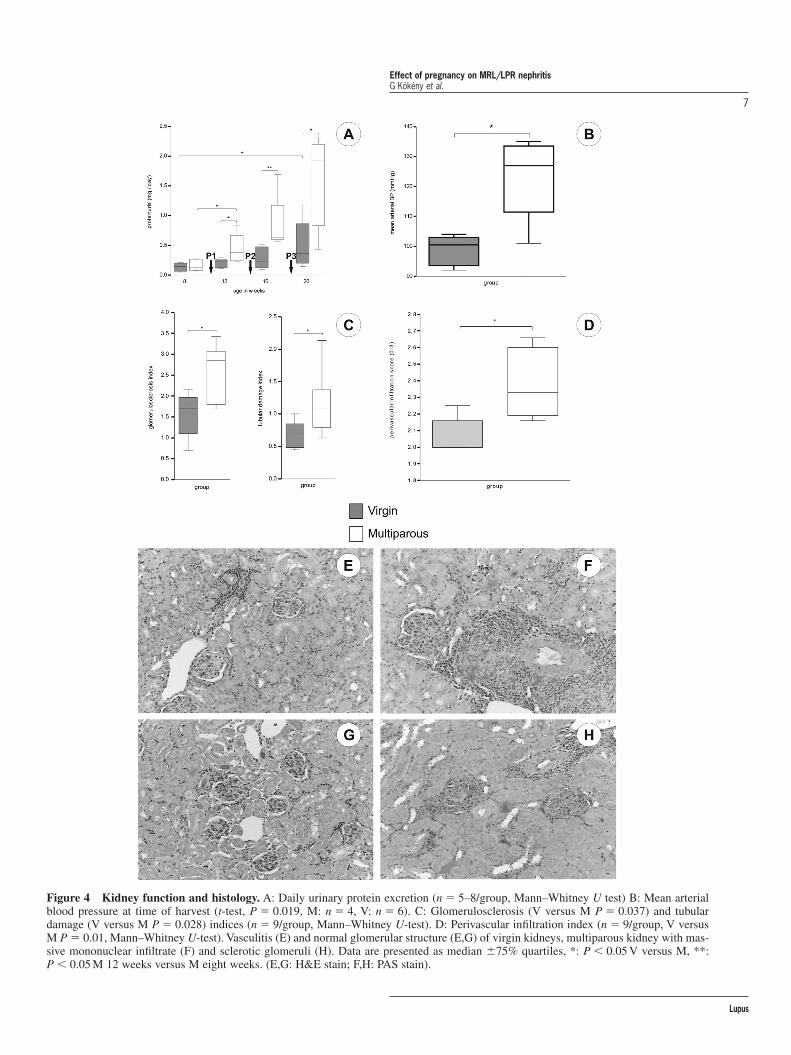
As in murine lupus IFN-� is thought to be a main con-tributor of the disease,12,35 we expected an improvementof renal pathology and survival in multiparous animalsdue to a continous shift towards anti-inflammatorycytokine production as a consequence of repeated preg-nancies.20 In contrast to the ameliorated autoimmuneskin disease, and contrary to our expectations, the lifespan of multiparous females was significantly shortenedto 20 weeks. Only 30% of M mice were alive at the ageof 20 weeks compared to 78% of V mice (Figure 3,P � 0.038, V versus M). In this first study, 60% of Manimals showed uremic symptoms (such as: subcuta-neous water retention, fuzzy hair, tremors and severelyreduced activity) preterminally, at 20 weeks of age, withblood urea levels exceeding 20 mmol/L. Proteinurialevels started to increase at 12 weeks of age (median0.22 � 0.07 mg/24 hour versus 0.38 � 0.2, V versus M,P � 0.029, n � 5–8/group) and reached 2.0 mg/day atweek 20 in multiparous animals (n � 5), while virginmice (n � 8) showed only mild increase in proteinuria(Figure 4a). Proteinuria of multiparous mice after thefirst pregnancy was significantly higher than beforepregnancy (P � 0.029, compared to eight weeks of age,n � 5–8/group). In addition, surviving multiparous ani-mals showed significantly worsened kidney function and70% of M mice died with uremic symptoms duringfollow-up. BUN levels closely paralleled proteinuria.
Effect of pregnancy on MRL/LPR nephritisG Kökény et al.
4
Lupus

Effect of pregnancy on MRL/LPR nephritisG Kökény et al.
5
Lupus
Figure 2 Macroscopy and histology of skin lesions. Macroscopic skin lesions were observed in 75% of virgin mice (A) compared to13% in multiparous mice (B), multiparous mice had lower macroscopic score (C, P � 0.001, V: n � 20, M: n � 23, Mann–Whitney U-test). Histological evaluation showed similar results (D, P � 0.0078, n � 9/group, Mann–Whitney–U-test). Lesions of virgin mice (E, G)showed thickened dermis (d) and mononuclear infiltrates (i) while multiparous animals showed no histological alteration (F) or had onlymild fibrosis and/or mild inflammation without hyperkeratosis or ulceration (H).

The severe kidney dysfunction was accompanied byelevated mean arterial blood pressure (Figure 4b) inmultiparous mice compared to virgin animals.
Histological evaluation of the kidneys showed thatrenal lesions were markedly increased in multiparousmice according to the functional data. Both glomeru-losclerosis and tubulointerstitial damage indices(Figure 4c) as well as perivascular infiltration (Figure4d) were more severe in the multiparous group (Figure4e–h). However, there was no difference in the extentof periglomerular infiltration (median score: V: 0.76versus M: 0.74, ns, n � 9/group) and vascular damageindices between the groups (median score: V: 1.49versus M: 1.71, ns, n � 9/group). The number ofCD3� infiltrating cells in the kidneys did not differbetween the two groups (median: V: 320 versus M:384 cells/mm2, ns, n � 8/group). Evaluation of IgG(Figure 5a,b) and C3 (Figure 5c,d) in cryostat kidneysections showed an increased deposition of both IgG(median fluorescence intensity: V: 31.1 versus M: 47.1,P � 0.02, n � 4/group) and C3 (median score: V: 2.04versus M: 2.76, P � 0.033, n � 4/group) in multi-parous mice.
Neither lymphoproliferation nor plasma anti-dsDNAlevels were affected by repeated pregnancies
We hypothesized that the systemic change in cytokinebalance during pregnancy can possibly affect lympho-proliferation, B-cell activity and/or autoantibodylevels, both systemic characteristics of SLE inMRL/lpr mice. Despite the fact that the proportion ofCD8� T-cells was consistently lower in blood samplesof multiparous mice (V: 6.48 � 0.94 versus M:
4.8 � 1.16, mean percent of total lymphocytes � SD),there was no difference in the percentage of CD4�
CD8� B220� DN cells (V: 67 � 9.3 versus M:62.1 � 18.3) or CD19� B-cells (V: 1.86 � 1.48 versusM: 1.43 � 1.15) between the groups. Anti-dsDNAantibody levels in the plasma of multiparous animalswere similar to those of virgin mice (median OD450 at12 weeks: V: 0.226 versus M: 0.232; at 20 weeks: V:0.317 versus M: 0.294, ns, n � 10/group). There wasno significant difference in spleen or lymph nodeenlargement between the groups (data not shown).
Marked and inverse changes in IFN-� production in the blood and the kidney
We observed significant diminished plasma IFN-�levels in multiparous mice when compared to virginanimals at the time of harvest (Figure 6a), supportingthe idea that pregnancy is accompanied by a dimin-ished systemic production of proinflammatorycytokines.23 To evaluate whether altered cytokine pro-duction at the organ level could contribute to thechanges seen in the kidney and skin, IFN-� and IL-10mRNA levels were measured in these organs, accom-panied by IL-4 mRNA measurements in kidneys.Interestingly, in the kidneys of multiparous mice,expression of IFN-�, IL-10 and IL-4 were significantlyhigher compared to virgins (Figure 6b,c). However inskin samples, we found that the expression of IL-10mRNA was higher, but IFN-� mRNA levels weremarkedly reduced after repeated pregnancies(Figure 7a,b). These data suggest that lupus may beregulated differently at the organ level duringpregnancy in this model.
Discussion
We report in the present study that skin disease dra-matically disappeared after multiple pregnancies com-pared to severe skin disease in virgin MRL/lpranimals, despite a more severe nephritis manifested asshortened survival and higher blood pressure. In thebackground of ameliorated skin pathology, weobserved decreased local IFN-� and elevated IL-10mRNA expression levels in the skin of multiparousmice accompanied by lower plasma IFN-� proteinlevels. Despite the reduction in skin and plasma IFN-�values, IFN-� as well as IL-10 mRNA levels were ele-vated in the kidneys of multiparous mice. Our findingssuggest that regulation of disease in MRL/lpr miceduring pregnancy may involve different mechanisms atthe organ level.
Effect of pregnancy on MRL/LPR nephritisG Kökény et al.
6
Lupus
Figure 3 Survival curves. Life span of multiparous mice (dottedline) was significantly shortened compared to virgin animals(P � 0.038, Kaplan–Meier, Log rank test, V versus M).
Effect of pregnancy on MRL/LPR nephritisG Kökény et al.
7
Lupus
Figure 4 Kidney function and histology. A: Daily urinary protein excretion (n � 5–8/group, Mann–Whitney U test) B: Mean arterialblood pressure at time of harvest (t-test, P � 0.019, M: n � 4, V: n � 6). C: Glomerulosclerosis (V versus M P � 0.037) and tubulardamage (V versus M P � 0.028) indices (n � 9/group, Mann–Whitney U-test). D: Perivascular infiltration index (n � 9/group, V versusM P � 0.01, Mann–Whitney U-test). Vasculitis (E) and normal glomerular structure (E,G) of virgin kidneys, multiparous kidney with mas-sive mononuclear infiltrate (F) and sclerotic glomeruli (H). Data are presented as median �75% quartiles, *: P � 0.05 V versus M, **:P � 0.05 M 12 weeks versus M eight weeks. (E,G: H&E stain; F,H: PAS stain).

The typical histological feature of MRL/lpr skinlesions is inflammatory cell infiltration.11 Althoughthere is IgG deposition at the dermo-epidermalborder,10 skin lupus is probably not a B-cell dependentdisease, as in complement receptor knockout MRL/lprmice ear necrosis was not influenced.36 However, sev-eral reports suggest that IFN-� plays an important rolein the development of skin lesions in SLE. The geneticdeficiency of IL-12 (a cytokine promoting IFN-� pro-duction) ameliorated skin disease of transgenicMRL/lpr mice.37 Moreover, mice with normal back-ground overexpressing IFN-� in the epidermis devel-oped inflammatory skin disease, similar to lupus.38
In our experiments, the severe lesions (10) found in75% of virgin mice were practically absent in the mul-tiparous group. In the background we detecteddecreased IFN-� mRNA in skin samples of multi-parous mice, supporting the central role of IFN-� in
the local regulation of skin lupus. On the other hand,IL-10 expression was enhanced, compared to virginmice, possibly due to the systemic cytokine shift gen-erally observed in pregnancy.22 Whether IL-10 and IL-4 are beneficial or deleterious in lupus is still a point ofdiscussion. IL-10 knock-out MRL/lpr mice had earlieronset of skin lesions, compared to wild type MRL/lprand recombinant IL-10 administration to MRL/lprmice ameliorated the disease.19 In our study, IL-10appeared to be important for the skin protection.
In both murine and in normal human pregnancy,there is a systemic cytokine profile change during ges-tation including increased production of IL-4 and IL-1020,22,23 accompanied by decreased IFN-�expression.39 However, in pregnant women with SLE,elevated IFN-�, and markedly increased IL-10 expres-sion was reported compared to healthy women.39 Thissystemic change in cytokine levels could explain the
Effect of pregnancy on MRL/LPR nephritisG Kökény et al.
8
Lupus
Figure 5 Representative photomicrographs of IgG and C3 deposition in the kidneys. A,B: Direct immunofluorescence of IgGdeposits, 400� magnification (A: virgin, B: multiparous). C,D: immunohistochemistry of C3, 400� magnification (C: virgin, D:multiparous).

Effect of pregnancy on MRL/LPR nephritisG Kökény et al.
9
Lupus
Figure 6 Cytokine expression in blood and kidneys. A: Plasma IFN-� levels were significantly lower in multiparous mice at the timeof harvest, compared to virgins (P � 0.028, n � 4/group, Mann–Whitney U-test). B–D: Renal cytokine mRNA expression analysis showedupregulation of both IFN-� (B, P � 0.005, V: n � 11, M: n � 9), IL-10 (C, P � 0.033, V: n � 11, M: n � 9) and IL-4 (D, P � 0.009, V:n � 8, M: n � 7) in multiparous animals. Data are presented as medians � 75% quartiles, *: P � 0.05, **: P � 0.01 V versus M,Mann–Whitney U-test.

clinical reports of worsened nephritis in pregnant SLEpatients.25,27 Our results further support these clinicalfindings, suggesting an adverse effect of pregnancy onlupus nephritis.
Lupus nephritis in the MRL/lpr mice and othermurine lupus models,12,40 as well as in humans, hasbeen strongly associated with IFN-� overproduc-tion.41,42 Decreased intrarenal or systemic IFN-� pro-duction40,43 dramatically ameliorated nephritis40 andimproved survival, whereas the induction of IFN-�production by IL-12 worsened the progression ofglomerulonephritis44 in MRL/lpr mice. Lupus patientswith diffuse proliferative nephritis showed increasedIFN-� expression levels both in blood and kidneybiopsy samples.41,45 In summary, IFN-� seems to be akey exacerbating cytokine in lupus nephritis. Thesedata support our observation that, in renal samples ofthe M group with more severe nephritis, IFN-� pro-duction was enhanced, despite lower serum IFN-�levels in these animals.
Surprisingly, not only IFN-� but IL-10 and IL-4were also augmented in multiparous kidneys.However, treatment of MRL/lpr mice with both anti-IL-446 and anti-IL-1047 antibodies ameliorated lupusnephritis. Moreover, genetic disruption of IL-10 accel-erated glomerulonephritis and shortened survival,19
thus supporting our hypothesis that high local IL-10
levels in the kidney may worsen nephritis leading toincreased glomerular IgG deposition, without signifi-cantly affecting the circulating levels of autoantibod-ies. In a recent study, Enghard et al. described apositive correlation between both IL-4 and IL-10 pro-duction and kidney damage of lupus prone NZB/W F1mice, supporting our results.48
In our experiment, a faster deterioration of kidneyfunction was manifested in elevated blood pressureand a shortened survival period. Although seven month(28 weeks) 50% survival in control MRL/lpr mice sug-gests an unusual mild disease, similar results with evenlonger (33 weeks) 50% survival had been observed byRobey et al.49 A putative factor contributing to longersurvival in our experiments might be the individuallyventilated cage (IVC) system wherein our animalswere housed. This protects them from pathogens – apossible exacerbating factor for the autoimmunepathology.
During pregnancy, renal blood flow increases by40–65% leading to elevated intraglomerular pressureand proteinuria. We found mild proteinuria in virginmice, significantly elevated in multiparous animalsright after the first pregnancy. Thus, hemodynamicchanges could have contributed to the high local IFN-� expression despite high IL-10 levels in the kidneysof multiparous mice.
Effect of pregnancy on MRL/LPR nephritisG Kökény et al.
10
Lupus
Figure 7 Cytokine expression in the skin. Cytokine mRNA expression analysis of skin samples showed significantly lower IFN-� (A,Mann–Whitney U, P � 0.0087, n � 6/group) levels and consistently higher IL-10 expression (B, P � 0.06, n � 6/group) in multiparousanimals compared to virgins. Data are shown as medians �75% quartiles, *: P � 0.05 V versus M.

Pre-eclampsia is an important issue in human SLEpregnancy, and it is hard to distinguish the clinicalsymptoms of renal flare and pre-eclampsia (PE).Although multiparous mice had hypertension, bloodpressure did not reach the levels of the murine PEmodel,50 and the glomerular lesions were not typicalfor murine PE.50 Furthermore, the timing of death inthe multiparous group was not associated with thirdtrimester of pregnancy, but death was always accompa-nied by symptoms of renal failure. These observationssuggests that pregnancy itself did not lead to pre-eclampsia in MRL/lpr mice. Our findings suggest animportant role of local immune regulation of lupusnephritis during pregnancy, independent of systemicchanges in the cytokine profile but possibly influencedby renal hemodynamic changes of pregnancy.
Hormonal changes during gestation are furtherissues that possibly affect the progression of SLEduring pregnancy. Estrogens and progesterone haveimmunomodulatory effects. Estrogens markedlyenhance IL-10 production in human Th cells,51 raisingthe possibility to exacerbate lupus nephritis duringpregnancy. Indeed, acceleration of glomerulonephritiswas reported in MRL/lpr mice after estrogen adminis-tration.52 Another study showed an opposite effect, inMRL/lpr mice.53 Elevation of serum estriol is oftenfound in female patients with SLE, and sometimes islinked to active disease.54 Progesterone favors a shiftto Th2 type immune response,55 thus it may worsenlupus nephritis. Thus, the role of sex-hormones inpregnancy associated changes of lupus disease activityis controversial. A possible background of this contro-versy could be the organ-dichotomy described in ourpresent report.
The design of the present study deserves some com-ment. Allogeneic matings (with NMRI males) wereperformed primarily for their relevance to humans.This raises the question of how fetal chimerism (‘allo-graft’ fetus) influences autoimmunity. As congenicMRL/lpr males have low testosterone level56 and lowfertility, the rate of matings can not be calculatedexactly, which does not allow the exact timing of par-turition required in our study. Therefore, we could notinclude a syngenic control group. Thus, the presentstudy is not conclusive regarding the influence of fetalmicrochimerism on the progression of autoimmunedisease. However, we have observed similar ameliora-tion of skin disease in our MRL/lpr breeder colony, buta shortened life span compared to non-breederfemales. This suggests that the observed alterations indisease activity are rather a consequence of pregnancyper se and not of the repeated allogenic exposure. Inconclusion, although the present study cannot dissectthe effects of repeated allo-exposures from the effectsof repeated pregnancies, the two cannot be separated in
the clinical situation either, as human pregnancies arealways allogenic.
In summary, skin disease of MRL/lpr mice is regu-lated differently from lupus nephritis during preg-nancy. In the present study, we first demonstrated adramatic improvement of skin pathology by repeatedpregnancies. However, mice undergoing multiple preg-nancies showed a shortened lifespan. Furthermore, ourdata suggest that skin and kidney diseases may havedifferent pathomechanisms during pregnancy inMRL/lpr mice with a divergent cytokine regulation.Revealing the local pathomechanisms contributing toorgan-specific lesions of SLE during or after preg-nancy may lead to a better understanding of the role ofpregnancy in SLE.
Acknowledgements
The authors would like to thank Éva Pállinger(Department of Biology, Semmelweis University,Budapest), Sarolta Adamkó (Department ofPathophysiology, Semmelweis University, Budapest)and Magdolna Pekár (Department of Pathology,Semmelweis University, Budapest) for their valuabletechnical assistance, and János Szebeni (Departmentof Pathophysiology, Semmelweis University,Budapest), and Paul Wafula (Reproductive immunol-ogy Group, Institute of Medical Immunology, ChariteMedical School, Berlin) for reviewing the manuscript,Anna Zambon Bertoja for her advice, André Sollwedeland Stefanie Ritschel (Reproductive immunologyGroup, Institute of Medical Immunology, ChariteMedical School, Berlin) for their RT-PCR methodol-ogy assistance. Funding sources: PH: OTKA T049022,ETT 225/2000, 432/2003, ACZ: UFF 2004-377.Further support was provided by the intergovernmen-tal collaboration project between Hungary andGermany by the Hungarian Ministry of Education(Project Nr: 6) and the German Academic ExchangeService (DAAD).
References
1 Klinman DM, Steinberg AD. Inquiry into murine and human lupus.Immunol Rev 1995; 144: 157–193.
2 Petri M. Epidemiology of systemic lupus erythematosus. Best. Pract.Res Clin Rheumatol 2002; 16: 847–858.
3 McCarty DJ, Manzi S, Medsger TA Jr, Ramsey-Goldman R, LaPorteRE, Kwoh CK. Incidence of systemic lupus erythematosus. Race andgender differences. Arthritis Rheum 1995; 38: 1260–1270.
4 Cohen PL, Eisenberg RA. Lpr and gld: single gene models of systemicautoimmunity and lymphoproliferative disease. Annu. Rev. Immunol1991; 9: 243–269.
5 Wu J, Zhou T, He J, Mountz JD. Autoimmune disease in mice due tointegration of an endogenous retrovirus in an apoptosis gene. J Exp. Med1993; 178: 461–468.
Effect of pregnancy on MRL/LPR nephritisG Kökény et al.
11
Lupus
6 Theofilopoulos AN, Balderas RS, Shawler DL, Lee S, Dixon FJ. Influ-ence of thymic genotype on the systemic lupus erythematosus-likedisease and T cell proliferation of MRL/Mp-lpr/lpr mice. J Exp. Med1981; 153: 1405–1414.
7 Wofsy D, Murphy ED, Roths JB, Dauphinee MJ, Kipper SB, Talal N.Deficient interleukin 2 activity in MRL/Mp and C57BL/6J mice bearingthe lpr gene. J Exp. Med 1981; 154: 1671–1680.
8 Pisetsky DS, McCarty GA, Peters DV. Mechanisms of autoantibody pro-duction in autoimmune MRL mice. J Exp. Med 1980; 152: 1302–1310.
9 Kolaja GJ, Fast PE. Renal lesions in MRL mice. Vet. Pathol 1982; 19:663–668.
10 Furukawa F, Tanaka H, Sekita K, Nakamura T, Horiguchi Y,Hamashima Y. Dermatopathological studies on skin lesions of MRLmice. Arch Dermatol Res 1984; 276: 186–194.
11 Kanauchi H, Furukawa F, Imamura S. Characterization of cutaneousinfiltrates in MRL/lpr mice monitored from onset to the full develop-ment of lupus erythematosus-like skin lesions. J Invest Dermatol 1991;96: 478–483.
12 Takahashi S, Fossati L, Iwamoto M et al. Imbalance towards Th1 pre-dominance is associated with acceleration of lupus-like autoimmunesyndrome in MRL mice. J Clin Invest 1996; 97: 1597–1604.
13 Gomez D, Correa PA, Gomez LM, Cadena J, Molina JF, Anaya JM.Th1/Th2 cytokines in patients with systemic lupus erythematosus: istumor necrosis factor alpha protective? Semin Arthritis Rheum 2004; 33:404–413.
14 Chan RW, Lai FM, Li EK et al. Imbalance of Th1/Th2 transcriptionfactors in patients with lupus nephritis. Rheumatology (Oxford) 2006;45: 951–957.
15 Rousset F, Garcia E, Defrance T et al. Interleukin 10 is a potent growthand differentiation factor for activated human B lymphocytes. Proc NatlAcad Sci USA 1992; 89: 1890–1893.
16 Huang XR, Kitching AR, Tipping PG, Holdsworth SR. Interleukin-10inhibits macrophage-induced glomerular injury. J Am Soc Nephrol 2000;11: 262–269.
17 Llorente L, Zou W, Levy Y et al. Role of interleukin 10 in the B lympho-cyte hyperactivity and autoantibody production of human systemiclupus erythematosus. J Exp Med 1995; 181: 839–844.
18 Park YB, Lee SK, Kim DS, Lee J, Lee CH, Song CH. Elevated inter-leukin-10 levels correlated with disease activity in systemic lupus ery-thematosus. Clin Exp Rheumatol 1998; 16: 283–288.
19 Yin Z, Bahtiyar G, Zhang N et al. IL-10 regulates murine lupus. JImmunol 2002; 169: 2148–2155.
20 Lin H, Mosmann TR, Guilbert L, Tuntipopipat S, Wegmann TG. Synthe-sis of T helper 2-type cytokines at the maternal-fetal interface. JImmunol 1993; 151: 4562–4573.
21 Wegmann TG, Lin H, Guilbert L, Mosmann TR. Bidirectional cytokineinteractions in the maternal-fetal relationship: is successful pregnancy aTH2 phenomenon? Immunol Today 1993; 14: 353–356.
22 Athanassakis I, Iconomidou B. Cytokine production in the serum andspleen of mice from day 6 to 14 of gestation:cytokines/placenta/spleen/serum. Dev. Immunol 1996; 4: 247–255.
23 Marzi M, Vigano A, Trabattoni D et al. Characterization of type 1 andtype 2 cytokine production profile in physiologic and pathologic humanpregnancy. Clin Exp Immunol 1996; 106: 127–133.
24 Mintz G, Niz J, Gutierrez G, Garcia-Alonso A, Karchmer S. Prospectivestudy of pregnancy in systemic lupus erythematosus. Results of a multi-disciplinary approach. J Rheumatol 1986; 13: 732–739.
25 Petri M. Prospective study of systemic lupus erythematosus pregnan-cies. Lupus 2004; 13: 688–689.
26 Georgiou PE, Politi EN, Katsimbri P, Sakka V, Drosos AA. Outcome oflupus pregnancy: a controlled study. Rheumatology (Oxford) 2000; 39:1014–1019.
27 Doria A, Ghirardello A, Iaccarino L et al. Pregnancy, cytokines, anddisease activity in systemic lupus erythematosus. Arthritis Rheum 2004;51: 989–995.
28 Lockshin MD. Pregnancy does not cause systemic lupus erythematosusto worsen. Arthritis Rheum 1989; 32: 665–670.
29 Molad Y, Borkowski T, Monselise A et al. Maternal and fetal outcome oflupus pregnancy: a prospective study of 29 pregnancies. Lupus 2005;14: 145–151.
30 Ratkay LG, Weinberg J, Waterfield JD. The effect of lactation in thepost-partum arthritis of MRL-lpr/fasmice. Rheumatology (Oxford) 2000;39: 646–651.
31 ter Borg EJ, Horst G, Hummel EJ, Limburg PC, Kallenberg CG.Measurement of increases in anti-double-stranded DNA antibody levelsas a predictor of disease exacerbation in systemic lupus erythematosus.A long-term, prospective study. Arthritis Rheum 1990; 33: 634–643.
32 Amann K, Rump LC, Simonaviciene A et al. Effects of low dosesympathetic inhibition on glomerulosclerosis and albuminuria insubtotally nephrectomized rats. J Am Soc Nephrol 2000; 11:1469–1478.
33 Girardi G, Berman J, Redecha P et al. Complement C5a receptors andneutrophils mediate fetal injury in the antiphospholipid syndrome. JClin Invest 2003; 112: 1644–1654.
34 Zenclussen AC, Gerlof K, Zenclussen ML et al. Abnormal T-cell reac-tivity against paternal antigens in spontaneous abortion: adoptive trans-fer of pregnancy-induced CD4�CD25� T regulatory cells preventsfetal rejection in a murine abortion model. Am J Pathol 2005; 166:811–822.
35 Csiszar A, Nagy G, Gergely P, Pozsonyi T, Pocsik E. Increasedinterferon-gamma (IFN-gamma), IL-10 and decreased IL-4 mRNAexpression in peripheral blood mononuclear cells (PBMC) from patientswith systemic lupus erythematosus (SLE). Clin Exp Immunol 2000; 122:464–470.
36 Boackle SA, Culhane KK, Brown JM et al. CR1/CR2 deficiency altersIgG3 autoantibody production and IgA glomerular deposition in theMRL/lpr model of SLE. Autoimmunity 2004; 37: 111–123.
37 Kikawada E, Lenda DM, Kelley VR. IL-12 deficiency in MRL-Fas(lpr)mice delays nephritis and intrarenal IFN-gamma expression, anddiminishes systemic pathology. J Immunol 2003; 170: 3915–3925.
38 Seery JP, Carroll JM, Cattell V, Watt FM. Antinuclear autoantibodiesand lupus nephritis in transgenic mice expressing interferon gamma inthe epidermis. J Exp Med 1997; 186: 1451–1459.
39 Munoz-Valle JF, Vazquez-Del Mercado M, Garcia-Iglesias T et al.T(H)1/T(H)2 cytokine profile, metalloprotease-9 activity and hormonalstatus in pregnant rheumatoid arthritis and systemic lupus erythematosuspatients. Clin Exp Immunol 2003; 131: 377–384.
40 Balomenos D, Rumold R, Theofilopoulos AN. Interferon-gamma isrequired for lupus-like disease and lymphoaccumulation in MRL-lprmice. J Clin Invest 1998; 101: 364–371.
41 Akahoshi M, Nakashima H, Tanaka Y et al. Th1/Th2 balance ofperipheral T helper cells in systemic lupus erythematosus. ArthritisRheum 1999; 42: 1644–1648.
42 Murray LJ, Lee R, Martens C. In vivo cytokine gene expression in T cellsubsets of the autoimmune MRL/Mp-lpr/lpr mouse. Eur J Immunol1990; 20: 163–170.
43 Peng SL, Moslehi J, Craft J. Roles of interferon-gamma and interleukin-4 in murine lupus. J Clin Invest 1997; 99: 1936–1946.
44 Schwarting A, Tesch G, Kinoshita K, Maron R, Weiner HL, Kelley VR.IL-12 drives IFN-gamma-dependent autoimmune kidney disease inMRL-Fas(lpr) mice. J Immunol 1999; 163: 6884–6891.
45 Calvani N, Richards HB, Tucci M, Pannarale G, Silvestris F. Up-regulation of IL-18 and predominance of a Th1 immune response isa hallmark of lupus nephritis. Clin Exp Immunol 2004; 138:171–178.
46 Nakajima A, Hirose S, Yagita H, Okumura K. Roles of IL-4 and IL-12in the development of lupus in NZB/W F1 mice. J Immunol 1997; 158:1466–1472.
47 Ishida H, Muchamuel T, Sakaguchi S, Andrade S, Menon S, Howard M.Continuous administration of anti-interleukin 10 antibodies delays onset of autoimmunity in NZB/W F1 mice. J Exp Med 1994; 179:305–310.
48 Enghard P, Langnickel D, Riemekasten G. T cell cytokine imbalancetowards production of IFN-gamma and IL-10 in NZB/W F1 lupus-pronemice is associated with autoantibody levels and nephritis. Scand JRheumatol 2006; 35: 209–216.
49 Robey IF, Peterson M, Horwitz MS et al. Terminal deoxynucleotidyl-transferase deficiency decreases autoimmune disease in diabetes-pronenonobese diabetic mice and lupus-prone MRL-Fas(lpr) mice. J Immunol2004; 172: 4624–4629.
50 Zenclussen AC, Fest S, Joachim R, Klapp BF, Arck PC. Introducing amouse model for pre-eclampsia: adoptive transfer of activated Th1 cellsleads to pre-eclampsia-like symptoms exclusively in pregnant mice. EurJ Immunol 2004; 34: 377–387.
51 Correale J, Arias M, Gilmore W. Steroid hormone regulation of cytokinesecretion by proteolipid protein-specific CD4� T cell clones isolated
Effect of pregnancy on MRL/LPR nephritisG Kökény et al.
12
Lupus

from multiple sclerosis patients and normal control subjects. J Immunol1998; 161: 3365–3374.
52 Carlsten H, Tarkowski A, Holmdahl R, Nilsson LA. Oestrogen is apotent disease accelerator in SLE-prone MRL lpr/lpr mice. Clin ExpImmunol 1990; 80: 467–473.
53 Apelgren LD, Bailey DL, Fouts RL et al. The effect of a selectiveestrogen receptor modulator on the progression of spontaneousautoimmune disease in MRL lpr/lpr mice. Cell Immunol 1996; 173:55–63.
54 Folomeev M, Dougados M, Beaune J et al. Plasma sex hormones andaromatase activity in tissues of patients with systemic lupus erythemato-sus. Lupus 1992; 1: 191–195.
55 Szekeres-Bartho J, Barakonyi A, Par G, Polgar B, Palkovics T, SzeredayL. Progesterone as an immunomodulatory molecule. Int Immunophar-macol 2001; 1: 1037–1048.
56 Sakic B, Gurunlian L, Denburg SD. Reduced aggressiveness and lowtestosterone levels in autoimmune MRL-lpr males. Physiol Behav 1998;63: 305–309.
Effect of pregnancy on MRL/LPR nephritisG Kökény et al.
13
Lupus