Simvastatin inhibits TGFβ1-induced fibronectin in human airway fibroblasts

Parker et al. Arthritis Research & Therapy 2013, 15:R163http://arthritis-research.com/content/15/5/R163
RESEARCH ARTICLE Open Access
Low oxygen tension increased fibronectinfragment induced catabolic activities - responseprevented with biomechanical signalsEleanor Parker1,3, Sandrine Vessillier2, Belinda Pingguan-Murphy3, Wan Abu Baker Wan Abas3, Dan L Bader1
and Tina T Chowdhury1*
Abstract
Introduction: The inherent low oxygen tension in normal cartilage has implications on inflammatory conditionsassociated with osteoarthritis (OA). Biomechanical signals will additionally contribute to changes in tissueremodelling and influence the inflammatory response. In this study, we investigated the combined effects ofoxygen tension and fibronectin fragment (FN-f) on the inflammatory response of chondrocytes subjected tobiomechanical signals.
Methods: Chondrocytes were cultured under free-swelling conditions at 1%, 5% and 21% oxygen tension orsubjected to dynamic compression in an ex vivo 3D/bioreactor model with 29 kDa FN-f, interleukin-1beta (IL-1β)and/or the nitric oxide synthase (NOS) inhibitor for 6 and 48 hours. Markers for catabolic activity (NO, PGE2), tissueremodelling (GAG, MMPs) and cytokines (IL-1β, IL-6 and TNFα) were quantified by biochemical assay. Aggrecan,collagen type II, iNOS and COX-2 gene expression were examined by real-time quantitative PCR. Two-way ANOVAand a post hoc Bonferroni-corrected t-test were used to analyse data.
Results: Both FN-fs and IL-1β increased NO, PGE2 and MMP production (all P < 0.001). FN-f was more active thanIL-1β with greater levels of NO observed at 5% than 1% or 21% oxygen tension (P < 0.001). Whilst FN-f reducedGAG synthesis at all oxygen tension, the effect of IL-1β was significant at 1% oxygen tension. In unstrainedconstructs, treatment with FN-f or IL-1β increased iNOS and COX-2 expression and reduced aggrecan and collagentype II (all P < 0.001). In unstrained constructs, FN-f was more effective than IL-1β at 5% oxygen tension andincreased production of NO, PGE2, MMP, IL-1β, IL-6 and TNFα. At 5% and 21% oxygen tension, co-stimulation withcompression and the NOS inhibitor abolished fragment or cytokine-induced catabolic activities and restoredanabolic response.
Conclusions: The present findings revealed that FN-fs are more potent than IL-1β in exerting catabolic effectsdependent on oxygen tension via iNOS and COX-2 upregulation. Stimulation with biomechanical signals abolishedcatabolic activities in an oxygen-independent manner and NOS inhibitors supported loading-induced recoveryresulting in reparative activities. Future investigations will utilize the ex vivo model as a tool to identify key targetsand therapeutics for OA treatments.
* Correspondence: [email protected] of Bioengineering, School of Engineering and Materials Science,Queen Mary University of London, Mile End Road, London E1 4NS, UKFull list of author information is available at the end of the article
© 2013 Parker et al.; licensee BioMed Central Ltd. This is an open access article distributed under the terms of the CreativeCommons Attribution License (http://creativecommons.org/licenses/by/2.0), which permits unrestricted use, distribution, andreproduction in any medium, provided the original work is properly cited.

Parker et al. Arthritis Research & Therapy 2013, 15:R163 Page 2 of 12http://arthritis-research.com/content/15/5/R163
IntroductionAnimal and in vitro studies have provided convincingevidence for a role of matrix degradation products inregulating cartilage homeostasis and driving osteoarthritis(OA) disease progression [1-3]. In chondrocytes, frag-ments derived from fibronectin initiate both catabolic andanabolic signalling cascades in a concentration-dependentmanner [3,4]. At low concentration, fragments augmentanabolic processes and facilitate reparative processes whenthe extracellular matrix is damaged. However, if fragmentlevels increase above a certain threshold, the pathwaysswitch from anabolic to catabolic and accelerate matrixdamage mediated by production of matrix metallopro-teinases (MMPs) and cytokines [2]. The importance offragment-induced damaging effects were highlighted inprevious clinical studies, which reported elevated levels offibronectin fragments (FN-fs) in osteoarthritic or rheuma-toid cartilage and OA synovial fluids [5-8]. The catabolicenvironment up-regulates tissue remodelling but the re-sponse will be influenced by mechanical factors whichinterfere with the pathways [9,10]. The mediators that ini-tiate the early phase of matrix damage are therefore com-plex and involve both mechanical and biological factors.In addition, the way in which biomechanical signalsmodulate fragment-induced mechanisms for repair and/ordegradation in early stage OA are unclear and require fur-ther investigation.Indeed, the amino-terminal FN-f has been shown to
have potent catabolic activities leading to enhancedlevels of nitric oxide (NO), prostaglandin E2 (PGE2) andMMPs in human or bovine cells cultured in 3D agarose,monolayer or explant models [1,3,11-13]. The signallingpathways involve the mitogen activated protein kinase(MAPK) and nuclear factor-kappa B (NFκB) cascadesmediated by stimulation of integrin receptors, leading tosuppression of proteoglycan synthesis and increasedproteoglycan depletion [14,15]. Furthermore, induciblenitric oxide synthase (iNOS) inhibitors have been shownto reduce the catabolic effect in cartilage explantstreated with FN-f and repair damaged tissue by facilitat-ing anabolic processes [12]. Recently, we showed thatintermittent compression applied in a dynamic mannerinhibits FN-f induced NO and PGE2 production and re-stores matrix synthesis in chondrocytes cultured in agar-ose constructs [16]. In this study, treatment with iNOSinhibitors and stimulation with mechanical signals wasshown to prevent FN-f-induced catabolic response. Inaddition, fibronectin concentrations were demonstratedto increase by cyclic impact load and alter matrix synthe-sis in cartilage explants [17]. Mechanical loading condi-tions that mimic injury and overloading may acceleratemild damage with an early rebuilding phase by increasingMMPs, matrix fragment levels and metabolic activity [18].However, the response will at least, in part, be dependent
on the type of mechanical loading regime, its duration andwhether loading was applied during the early or late stageof the disease process. It is, therefore, plausible thatphysiological mechanical signals compete with the cata-bolic pathways induced by the matrix fragments and con-tribute to early reparative signals.Furthermore, the oxygen tension of cartilage will influ-
ence the response of chondrocytes to inflammatoryfactors and biomechanical signals. In OA, the tissue ismore hypoxic than normal cartilage with pathophysio-logical levels less than 5% leading to increased productionof NO and PGE2 release in tissues involving the cartilageand meniscus [19-21]. The interactions of inflammatorymediators, such as interleukin-1β (IL-1β), with oxygentension has detrimental effects on matrix turnover, which,in turn, affects the ability of the cells to respond to mech-anical loading, possibly through the disruption of normalintegrin-based signals [19-21]. Given the potential inflam-matory effects of hypoxia on cell metabolism, it is highlylikely that oxygen tension will affect the response of chon-drocytes to both matrix fragments and mechanical stimuli.However, to date, no research groups have examined thecombined effect of fragments, oxygen tension and bio-mechanical signals in chondrocytes. The present study,therefore, investigated the effects of oxygen tension andFN-f on catabolic and anabolic activities in chondrocyte/agarose constructs subjected to dynamic compression andcompared the response to constructs treated with IL-1β.
MethodsChondrocyte isolation and culture in agarose constructsThis study involves bovine cells procured from a localabbatoir with authorization from the relevant meat inspec-tors (Dawn Cardington, Bedfordshire, UK). It does notinvolve humans, human tissue or experimentation on ani-mals. Cartilage explants were obtained from the metacar-palphalangeal joints of 18-month-old cattle and diced, aspreviously described [22,23]. The tissue was incubated onrollers for 1 h at 37°C in Dulbecco’s Modified Eagle’sMedium supplemented with 20% (v/v) foetal calf serum(DMEM+ 20% FCS), 2 μM L-glutamine, 5 μg/ml penicillin,5 μg/ml streptomycin, 20 mM Hepes buffer, and0.85 μM L-ascorbic acid in addition to 700 unit/ml pronase,and for a further 16 h at 37°C in medium supplementedwith 100 unit/ml collagenase type XI (All from Sigma-Aldrich Ltd, Gillingham, Dorset, UK). The chondrocytesuspension was washed and the cells counted using ahaemocytometer. Cell viability was calculated with thetrypan blue exclusion assay and resuspended in mediumat a concentration of 8 × 10⁶ cells/ml. The cell suspensionwas added to an equal volume of molten 6% (w/v) agarosetype VII in Earle’s Balanced Salt Solutions (EBSS) to yielda final cell concentration of 4 × 10⁶ cells/ml in 3% (w/v)agarose (Sigma-Aldrich Ltd, Gillingham, Dorset, UK). The
Parker et al. Arthritis Research & Therapy 2013, 15:R163 Page 3 of 12http://arthritis-research.com/content/15/5/R163
cell/agarose suspension was transferred into a sterile stain-less steel mould, containing holes, 5 mm in diameter and5 mm in height, and allowed to gel at 4°C for 10 minutes.Constructs were transferred into wells of a 24-well cultureplate and immediately subjected to ex vivo conditions.
Effects of oxygen tension and FN-f in chondrocyte/agarose constructsThe effects of 1, 5 and 21% oxygen tension were examinedin constructs cultured under free-swelling conditions in aglove-box style workspace integrated with the BiospherixLtd, Lacona, NY, USA incubator to ensure that the experi-mental conditions during set-up were uninterrupted(Figure 1). 1 ml DMEM+ 20% FCS was supplementedwith 0 or 1 μm of 29 kDa NH2-heparin-binding FN-f(generous gift from Prof Gene Homandberg) and/or1 mM of L-N-(1-iminoethyl)-ornithine (L-NIO). Thisagent inhibits all isoforms of the nitric oxide synthaseenzymes (Merck Chemicals, Nottingham, UK) for 48 hr.FN-fs were isolated from cathepsin D and thrombin di-gests of fibronectin from plasma adsorption, as previouslydescribed [24]. In addition, the FN-f-induced responsewas compared to constructs treated with 0 or 10 ng/mlIL-1β (Peprotech EC Ltd, London, UK) and/or L-NIO at1, 5 and 21% oxygen tension. The ex vivo conditions aresummarized in Figure 2.
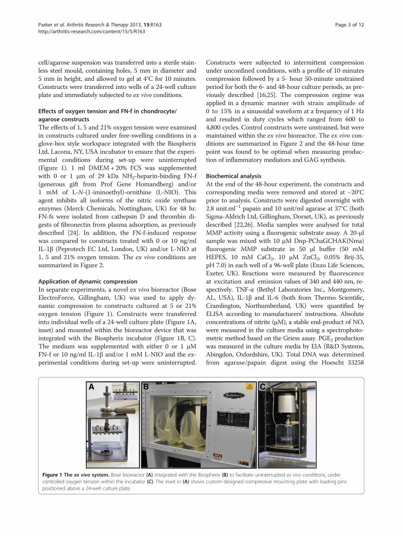
Application of dynamic compressionIn separate experiments, a novel ex vivo bioreactor (BoseElectroForce, Gillingham, UK) was used to apply dy-namic compression to constructs cultured at 5 or 21%oxygen tension (Figure 1). Constructs were transferredinto individual wells of a 24-well culture plate (Figure 1A,inset) and mounted within the bioreactor device that wasintegrated with the Biospherix incubator (Figure 1B, C).The medium was supplemented with either 0 or 1 μMFN-f or 10 ng/ml IL-1β and/or 1 mM L-NIO and the ex-perimental conditions during set-up were uninterrupted.
A B
Figure 1 The ex vivo system. Bose bioreactor (A) integrated with the Biocontrolled oxygen tension within the incubator (C). The inset in (A) showspositioned above a 24-well culture plate.
Constructs were subjected to intermittent compressionunder unconfined conditions, with a profile of 10 minutescompression followed by a 5- hour 50-minute unstrainedperiod for both the 6- and 48-hour culture periods, as pre-viously described [16,25]. The compression regime wasapplied in a dynamic manner with strain amplitude of0 to 15% in a sinusoidal waveform at a frequency of 1 Hzand resulted in duty cycles which ranged from 600 to4,800 cycles. Control constructs were unstrained, but weremaintained within the ex vivo bioreactor. The ex vivo con-ditions are summarized in Figure 2 and the 48-hour timepoint was found to be optimal when measuring produc-tion of inflammatory mediators and GAG synthesis.
Biochemical analysisAt the end of the 48-hour experiment, the constructs andcorresponding media were removed and stored at −20°Cprior to analysis. Constructs were digested overnight with2.8 unit.ml−1 papain and 10 unit/ml agarase at 37°C (bothSigma-Aldrich Ltd, Gillingham, Dorset, UK), as previouslydescribed [22,26]. Media samples were analysed for totalMMP activity using a fluorogenic substrate assay. A 20-μlsample was mixed with 10 μM Dnp-PChaGCHAK(Nma)fluorogenic MMP substrate in 50 μl buffer (50 mMHEPES, 10 mM CaCl2, 10 μM ZnCl2, 0.05% Brij-35,pH 7.0) in each well of a 96-well plate (Enzo Life Sciences,Exeter, UK). Reactions were measured by fluorescenceat excitation and emission values of 340 and 440 nm, re-spectively. TNF-α (Bethyl Laboratories Inc., Montgomery,AL, USA), IL-1β and IL-6 (both from Thermo Scientific,Cramlington, Northumberland, UK) were quantified byELISA according to manufacturers’ instructions. Absoluteconcentrations of nitrite (μM), a stable end-product of NO,were measured in the culture media using a spectrophoto-metric method based on the Griess assay. PGE2 productionwas measured in the culture media by EIA (R&D Systems,Abingdon, Oxfordshire, UK). Total DNA was determinedfrom agarase/papain digest using the Hoescht 33258
C
spherix (B) to facilitate uninterrupted ex vivo conditions, undercustom designed compressive mounting plate with loading pins

Gene expression Anabolic: aggrecan, col2A1. Catabolic:iNOS, COX2
(+/-) FN-f and/or L-NIO (+/-) IL-1 and/or L-NIO
Ex-vivo free-swelling
Ex-vivo bioreactor
1, 5 and 21% oxygen tension
5 or 21% oxygen tension Dynamic compression (15%, 1Hz)
(+/-) FN-f and/or L-NIO (+/-) IL-1 and/or L-NIO
6 hr 48 hr
NO/PGE2 release GAG/DNA synthesis
Anabolic: GAG/DNA Catabolic: NO, MMPs, PGE2
Pro-inflammatory cytokine release: IL-1 , IL-6, TNF
Chondrocyte/agarose constructs
48 hr
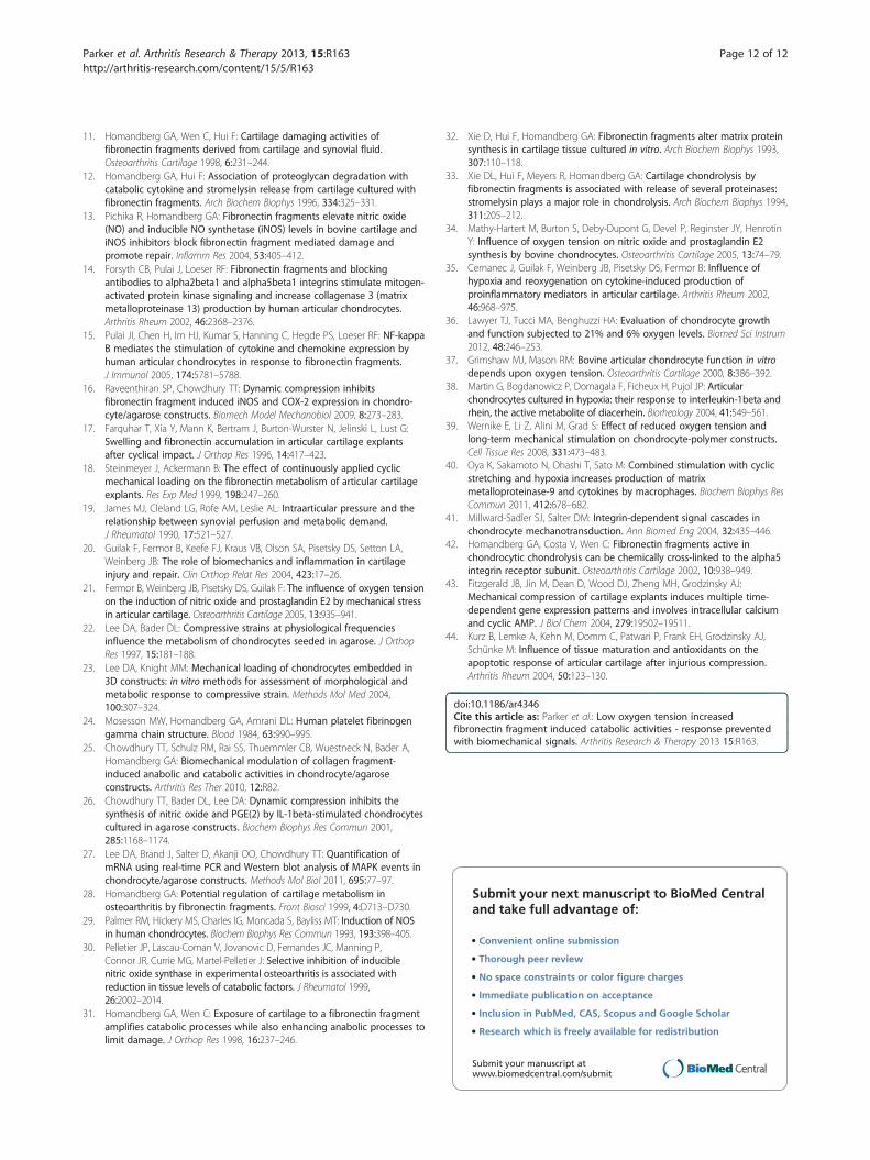
Figure 2 Summary of the ex vivo test conditions and types of analysis. Chondrocyte/agarose constructs were cultured ex vivo under free-swelling conditions (1, 5 and 21%) or subjected to dynamic compression (15%, 1 Hz) at 5 and 21% oxygen tension. At the end of the experiment,constructs and/or media samples were analysed by biochemical assay or RNA was extracted from constructs, reverse transcribed and geneexpression quantified by RT-qPCR.
Parker et al. Arthritis Research & Therapy 2013, 15:R163 Page 4 of 12http://arthritis-research.com/content/15/5/R163
method. GAG synthesis was measured in constructs andmedia samples by DMB assay. These methods have beenpreviously detailed [22,26].
RNA isolation, reverse transcription and real-timequantitative PCRAt the end of the six-hour bioreactor experiment, RNA wasisolated from chondrocytes cultured in agarose using theQIAquick Spin gel extraction and RNeasy kit (Qiagen,Crawley, West Sussex, UK), as previously described [27].The six-hour time point was found to be optimal whenexamining gene expression [25]. DNA-free DNase treatmentand removal reagents were used to eliminate any contamin-ating DNA from the RNA sample (Applied Biosystems,Warrington, UK). RNA was quantified on the NanodropND-1000 spectrophotometer (LabTech, Uckfield, EastSussex, UK), and reverse transcription was performed withEnhanced Avian RT First Strand cDNA synthesis kit, oligo(dT)23 primer, and a total of 200 ng of RNA (Sigma-Aldrich Ltd, Gillingham, Dorset, UK). Real-time quanti-tative polymerase chain (qPCR) assays coupled withMolecular Beacons were performed in 25-μl reactionmixtures containing 1 μl cDNA, 12.5 μl JumpStart TaqPCR Master Mix, primer pairs/probes with concentrationsdetailed in Table 1 and nuclease-free PCR-grade water to25 μl (Sigma Genosys, Cambridge, UK). Each sample wasrun in duplicate on the 96-well thermal system ofthe Mx3000P quantitative PCR instrument (Stratagene,Amsterdam, Netherlands). Thermocycling conditionscomprised an initial polymerase activation step at 95°C
for 10 minutes, followed by denaturation of 35 cycles at95°C for 30 seconds, annealing at 55°C for 1 minute, andextension at 72°C for 1 minute. The real-time PCR effi-ciencies (E) of amplification for each target were defined ac-cording to the relation, E = 10(−1/slope) and revealedefficiency values ranging from 1.96 to 2.05.Fluorescence data were collected during the annealing
stage of amplification, and data were analyzed on theMxPro qPCR software (version 3, Agilent TechnologiesInc, Wokingham, Berkshire, UK). Baselines and thresholdswere automatically set by the RG-3000 qPCR softwareand used after manual inspection. The cycle threshold(Ct) value for each duplicate reaction was expressed as themean value, and the results were exported into MicrosoftExcel for further analysis. Relative quantification of colla-gen type II, aggrecan, COX-2 and iNOS signals wereestimated by normalizing each target to the referencegene, GAPDH, and to the calibrator sample (untreated,unstrained condition) by a comparative Ct approach. Foreach sample, the ratio of target ΔCt and reference ΔCtwas calculated, as previously described [27].
StatisticsFor free-swelling studies, data represent the mean and SEMvalues of 12 replicates from three separate experiments. Forthe ex vivo/bioreactor experiments, biochemical and gene-expression data represent the mean and SEM valuesof up to 12 replicates from at least three separateexperiments. Statistical analysis was performed witha two-way analysis of variance (ANOVA) and the

Table 1 Beacon designer sequences and real-time reaction efficiencies of qPCR assays
Gene Accession number Sequences nM Efficiency
iNOS U14640 Probe:5′-FAM-CGCGATCCCTGCTTGGTGGCGAAGATGAGCGATCGCG- 200
DABCYL-3′ 200 2.01
Forward: 5′-GTAACAAAGGAGATAGAAACAACAGG-3′ 200 (± 0.08)
Reverse: 5′-CAGCTCCGGGCGTCAAAG-3′
Probe: 5′-FAM-
COX-2 AF031698 CGCGATCGTCAGAAATTCGGGTGTGGTACAGTTGATCGCG-DABCYL-3′ 200
Forward: 5′-CGAGGTGTATGTATGAGTGTAGG-3′ 300 1.99
Reverse: 5′-GTTGGGAGTGGGTTTCAGG-3′= 300 (± 0.04)
Probe: 5′-FAM- 200
Aggrecan U76615 CGCGATCCACTCAGCGAGTTGTCAGGTTCTGAGATCGCG-DABCYL-3′ 2.05
Forward: 5′-TGGTGTTTGTGACTCTGAGG-3′ 100 (± 0.07)
Reverse: 5′-GATGAAGTAGCAGGGGATGG-3′ 200
Collagen type II X02420 Probe: 5′-FAM-CGCGATGCGTCAGGTCAGGTCAGCCATATCGCG-
DABCYL-3′ 200
Forward: 5′-AAACCCGAACCCAGAACC-3′ 100 1.96
Reverse: 5′-AAGTCCGAACTGTGAGAGG-3′ 100 (± 0.08)
GAPDH U85042 Probe: 5′-HEX-CGCGATCCACCATCTTCCAGGAGCGAGATCCGATCGCG-
DABCYL-3′ 200 1.96
Forward: 5′-TTCAACGGCACAGTCAAGG-3′ 200 (± 0.01)
Reverse: 5′-TTCAACGGCACAGTCAAGG-3′ 200
Note: Real-time reaction efficiencies of qPCR assays with optimal primer and probe concentrations are shown. Probes contain FAM or HEX as a 5′- reporter dyeand DABCYL as 3′- quencher with the arm sequences underlined.
Parker et al. Arthritis Research & Therapy 2013, 15:R163 Page 5 of 12http://arthritis-research.com/content/15/5/R163
multiple post hoc Bonferroni-corrected t tests to com-pare differences between the various treatment groups,as indicated in the figure legend. For gene-expression data,ratio values were log transformed before analysis by atwo-way ANOVA and a post hoc Bonferroni-corrected ttest. In all cases, a level of 5% was considered statisticallysignificant (P <0.05).
ResultsFN-f maximally increased catabolic activities at 5%oxygen tensionThe ability of FN-f to influence the levels of NO, PGE2 andGAG synthesis in an oxygen-dependent manner werecompared to IL-1β in constructs cultured for 48 hours(Figure 3). At 1, 5 and 21% oxygen tension, NO productionwas enhanced with FN-f or IL-1β compared to untreatedcontrols (all P <0.001; Figure 3A). At 5% oxygen tension,FN-f increased NO production (45 μM) with values signifi-cantly greater than cytokine-treated (28 μM) constructs(P <0.001). At all oxygen tensions, co-incubation withL-NIO reduced fragment or cytokine-induced NO levelswith values returning to basal levels (<5 μM). PGE2 releasewas increased with either FN-f or IL-1β at 5 and 21%oxygen tension (P <0.001) but not at 1% oxygen tension(Figure 3B). Co-incubation with L-NIO partially reducedPGE2 levels in FN-f treated constructs cultured at 5 and
21% oxygen tension. Furthermore, FN-f reduced GAGsynthesis at 1, 5 and 21% oxygen tension (P <0.001;Figure 3C). In contrast, the cytokine decreased GAG syn-thesis at 1% oxygen tension (P <0.01; Figure 3F) but not at5 or 21% oxygen tension. At 1 or 5% oxygen tension, frag-ment or cytokine-induced inhibition on GAG synthesiswas reversed with L-NIO (both P <0.05).
Compression abolished FN-f induced catabolic effects at 5and 21% oxygen tensionSince the catabolic effects in response to FN-f weremaximal at 5% oxygen tension, further ex vivo studies ex-amined the effect of dynamic compression in constructscultured at 5 and 21% oxygen tension and compared theresponse to IL-1β. Figure 4 reveals that dynamic compres-sion inhibits NO release at 5 and 21% oxygen tension(P <0.001; Figure 4A). In unstrained constructs, FN-f en-hanced NO production (P <0.001) with values highest at5% oxygen tension when compared to the effects of IL-1β(P <0.001). The enhanced production of NO by fragment,cytokine or low oxygen tension was reduced with dy-namic compression (all P <0.001; Figure 4A). Stimula-tion with compression and the NOS inhibitor reducedNO levels further in fragment or cytokine treated con-structs (all P <0.001; Figure 4A).

0
1000
2000
3000
4000
5000
6000
7000
8000
PG
E2
rele
ase
(p
g/m
l)
***
*** ***
*** *** ***
*** ***
A
B
C
0
10
20
30
40
50
NO
rel
ease
(µ
M)
*** ***
*** ***
*** ***
*** ***
***
*** *** ***
*** *** ***
0
5
10
15
20
GA
G s
ynth
esis
(µ
g/µ
g D
NA
)
****
**
******
*
*
Untreated
FN-f
FN-f + L-NIO
IL-1
IL-1 + L-NIO
21% 5% 1%
21% 5% 1%
21% 5% 1%
Figure 3 Effect of oxygen tension and FN-f on catabolic and anabolic activities. Chondrocyte/agarose constructs were cultured underuninterrupted experimental conditions with 0 or 1 μM FN-f or 10 ng/ml IL-1β in the presence or absence of 1 mM L-NIO at 1, 5 and 21% oxygentension for 48 hours, where NO release (A), PGE2 production (B) and GAG synthesis (C). Error bars represent the mean and SEM of 12 values fromthree separate experiments.
Parker et al. Arthritis Research & Therapy 2013, 15:R163 Page 6 of 12http://arthritis-research.com/content/15/5/R163
At 5% oxygen tension, dynamic compression inhibitsPGE2 release in untreated constructs (P <0.001; Figure 4B).In unstrained constructs, the presence of the fragmentor cytokine enhanced PGE2 release with values broadly
similar and ranging from 7.6 to 8.0 pg/ml for constructscultured at 5 or 21% oxygen tension. Stimulation withdynamic compression and/or L-NIO reduced PGE2 levelsto basal values in an oxygen-independent manner (all

0
2000
4000
6000
8000
10000
PG
E2
rele
ease
(p
g/m
l)
Untreated FN-f FN-f + IL-1β IL-1β + L-NIO L-NIO
0
10
20
30
40
50
60
NO
rel
ease
(µ
M)
Untreated FN-f FN-f + IL-1β IL-1β + L-NIO L-NIO
A
B
C
D
Unstrained
Strained 21%
Unstrained
Strained 5%
***
******
***
***
*** ***
***
***
***
***
***
***
******
******
***
*** ***
*** ***
*** **
0
1
2
3
4
MM
P a
ctiv
ity
(AS
U)
Untreated FN-f FN-f + IL-1β IL-1β + L-NIO L-NIO
***
***
***
***
*** ***
***
******
****
** ***
*** **
*** ***
*** ***
*** ***
*** ***
0
5
10
15
20
25
30
35
GA
G s
ynth
esis
(μ
g/μg
DN
A)
Untreated FN-f FN-f + IL-1β IL-1β + L-NIO L-NIO
*****
*** *** ******
*** *** *** ***
*
**
*
*** ***
*** ***
Figure 4 Effect of oxygen tension, FN-f and dynamic compression on catabolic and anabolic activities. Chondrocyte/agarose constructswere cultured at 5 and 21% oxygen tension with 0 or 1 μM FN-f or 10 ng/ml IL-1β and/or 1 mM L-NIO for 48 hour, where NO release (A), PGE2production (B), MMP activity (C) and GAG synthesis (D). Error bars represent the mean and SEM of 8 to 12 replicates from three separateexperiments, where (*) indicates comparisons between the different treatment groups and (ψ) indicates comparisons in unstrained constructsbetween untreated and IL-1β at 5 or 21% oxygen tension. All other comparisons were not significant (not indicated).
Parker et al. Arthritis Research & Therapy 2013, 15:R163 Page 7 of 12http://arthritis-research.com/content/15/5/R163
P <0.001) with the magnitude in inhibition rangingbetween 77 and 87%.In untreated samples, total MMP activity was inhibited
with dynamic compression at 5% oxygen tension. Thefragment or cytokine increased MMP activity with max-imal values at 5% oxygen tension for constructs culturedwith FN-f when compared to IL-1β (P <0.001). At 5 or21% oxygen tension, stimulation with dynamic compres-sion or the NOS inhibitor abolished fragment or cyto-kine induced MMP activity (all P <0.001; Figure 4C). Inaddition, dynamic compression increased GAG synthesis(P <0.001; Figure 4D) with a greater magnitude in stimu-lation for constructs cultured at 21% oxygen tension(136%) when compared to 5% (79%). At 5 and 21% oxy-gen tension, the presence of the FN-f or IL-1β reducedGAG synthesis (all P <0.05; Figure 4D). This responsewas reversed by stimulation with dynamic compressionand L-NIO at 5 and 21% oxygen tension.
Compression inhibits FN-f induced gene expression at 5and 21% oxygen tensionThe effect of oxygen tension and FN-f on catabolic (iNOSand COX-2) and anabolic (aggrecan and type II collagen)gene expression is presented in Figure 5. Treatment withFN-f increased iNOS and COX-2 expression with a similarmagnitude to IL-1β (all P <0.001; Figure 5A, B, respect-ively). In unstrained constructs, the effect of hypoxia in-creased iNOS, aggrecan and collagen type II (all P <0.001),but not COX-2 expression. At 5 and 21% oxygen tension,stimulation with dynamic compression alone resulted in areduction of iNOS and COX-2 expression in constructscultured with FN-f or IL-1β (all P <0.001).Co-stimulation with compression and L-NIO marginally
reduced iNOS but not COX-2 expression further. Con-versely, fragment or cytokine treatment decreased aggre-can expression at 5 and 21% oxygen tension (both P<0.001; Figure 5C, D, respectively). In addition, the

C
D
Unstrained
Strained 21%
Unstrained
Strained 5%
A
B
0.1
1
10
100
Untreated FN-f FN-f + IL-1β IL-1β +
CO
X-2
gen
e ex
pres
sion
**
*** ***
**
************
0.1
1
10
Agg
reca
n ex
pres
sion
Agg
reca
n ge
ne e
xpre
ssio
n
Untreated FN-f FN-f + IL-1β IL-1β +
**
******
** ***
****
*
** ***
0.1
1
10
Typ
e II
colla
gen
gene
exp
ress
ion
Untreated FN-f FN-f + IL-1β IL-1β +
***
*****
****
****** * *
***
0.1
1
10
100
Untreated FN-f FN-f + IL-1β IL-1β +
iNO
S g
ene
expr
essi
on
***
**
******
*** ***
***
******
******
***
*** ***
*** ***
***
*** ***
*** ***
*** ***
L-NIO L-NIO L-NIO L-NIO
L-NIO L-NIO L-NIO L-NIO
Figure 5 Effect of oxygen tension, FN-f and dynamic compression on gene expression. Chondrocyte/agarose constructs were cultured at 5and 21% oxygen tension with 0 or 1 μM FN-f or 10 ng/ml IL-1β and/or 1 mM LNIO for six hours, where iNOS (A), COX-2 (B), aggrecan (C) andtype II collagen (D). Error bars represent the mean and SEM of eight replicates from three separate experiments, where (*) indicates comparisonsbetween the different treatment groups and (ψ) indicates comparisons in unstrained constructs between untreated and IL-1β at 5 or 21% oxygentension. All other comparisons were not significant (not indicated).
Parker et al. Arthritis Research & Therapy 2013, 15:R163 Page 8 of 12http://arthritis-research.com/content/15/5/R163
fragment, but not cytokine, reduced collagen type II ex-pression at 21% oxygen tension (P <0.001; Figure 5D). At5 and 21% oxygen tension, co-stimulation with compres-sion and the NOS inhibitor restored aggrecan and colla-gen type II expression in fragment or cytokine treatedconstructs cultured at 5 and 21% oxygen tension.
Compression inhibits FN-f induced cytokine production at5 and 21% oxygen tensionWe next examined the effect of FN-f, compression andoxygen tension on the production of pro-inflammatory cy-tokines and compared the response to constructs treatedwith exogenous IL-1β (Figure 6). In unstrained constructs,FN-f significantly increased IL-1β, IL-6 and TNFα produc-tion to a larger effect than the corresponding increasewith exogenous cytokine treatment (all P <0.001). At 5%
oxygen tension, the fragment or cytokine enhancedgreater production of IL-1β and TNFα than at 21% oxygentension. In contrast, the effects of fragment or cytokine onIL-6 production were broadly similar for constructs cul-tured at 5 and 21% oxygen tension. Cytokine productionwas reduced with dynamic compression and/or L-NIOin an oxygen-independent manner with the magnitudebroadly similar for all treatments.
DiscussionMatrix fragments are known to exert destructive effectsin OA pathophysiology and regulate tissue remodellingevents in chondrocytes [28]. In OA cartilage, exogenouscytokines increase iNOS expression and NO productionleading to proteoglycan degradation [29,30]. The cata-bolic environment attempts to assist with tissue

0
500
1000
1500
2000
2500
IL-6
rel
ease
(p
g/m
l)
Untreated FN-f FN-f + IL-1β IL-1β +
***
***
***
*** *** *** ***
********* **
***
*** ***
Unstrained
Strained 21%
Unstrained
Strained 5%
A
B
C
0
200
400
600
800
1000
1200
1400
TN
F r
elea
se
(pg/
ml)
Untreated FN-f FN-f + IL-1β IL-1β +
*
***
******
***
***
***
***
******
***
******
***
0
200
400
600
800
1000
IL-1
rel
ease
(p
g/m
l)
Untreated FN-f FN-f + IL-1β IL-1β +
***
***
***
***
*** ***
******
***
***
******
***
*** ***
***
*** ***
***
*** ***
***
***
L-NIO L-NIO
L-NIO L-NIO
L-NIO L-NIO
Figure 6 Effect of oxygen tension, FN-f and dynamic compressionon cytokine production. Chondrocyte/agarose constructs werecultured at 5 and 21% oxygen tension with 0 or 1 μM FN-f or 10 ng/mlIL-1β and/or 1 mM L-NIO for 48 hour where TNFα (A), IL-1β (B) and IL-6(C). Error bars represent the mean and SEM of eight replicates from threeseparate experiments, where (*) indicates comparisons between thedifferent treatment groups and (ψ) indicates comparisons in unstrainedconstructs between untreated and IL-1β at 5 or 21% oxygen tension.All other comparisons were not significant (not indicated).
Parker et al. Arthritis Research & Therapy 2013, 15:R163 Page 9 of 12http://arthritis-research.com/content/15/5/R163
remodelling but the response will be influenced bymechanical factors. The question arises as to whethermechanical loading could act to slow down tissue dam-age during the early phase of the disease process or ef-fectively contribute to OA progression. In addition, the
levels of oxygen tension in the injured joint will have asignificant impact on the metabolic processes thereby af-fecting the maintenance of fragment-induced pathway.The interactions among matrix fragments, oxygen tensionand mechanical loading are, therefore, complex, and thusmotivate the current investigation.At 21% oxygen tension, FN-fs ranging from 29 to
140 kDa have been observed to bind to the pericellularmatrix leading to NO, MMP and cytokine up-regulation,suppression of matrix synthesis and proteoglycan loss ina concentration-dependent manner [1,11,12,31-33]. Wereported that the 29 kDa NH2-heparin binding fragmentis highly active and increased NO and PGE2 production ina 3D/agarose model when compared with other matrixfragment types [16,25]. However, our previous studiestested commercially available fragments in contrast to thepresent work which generated FN-fs from cathepsin Dand thrombin digests isolated from human plasma fibro-nectin, as previously described [24]. Our data are in agree-ment with previous findings, demonstrating a greaterproduction of NO and PGE2 with FN-f when compared toIL-1β treatment and the response was reduced with NOSinhibitors. Interestingly, the fragment-induced catabolicresponse was greater at 5% when compared to 1 or 21%oxygen tension and resulted in an associated inhibition ofmatrix synthesis that was broadly similar at all oxygen ten-sions examined. However, we were unable to detect PGE2synthesis in constructs cultured with fragment or cytokineunder severe hypoxia (1%) conditions. Since prostaglandinsynthesis involves the oxygenation of arachidonic acid intointermediates that are oxidised by COX leading to PGE2synthesis, the oxygen concentration dependent effects willslow down production of inflammatory mediators. A simi-lar response was reported in bovine chondrocytes treatedwith IL-1β in suspension culture, with a more pronouncedincrease of NO and PGE2 production at 5% when com-pared to 1 or 21% oxygen tension [34]. The effect of FN-fsand oxygen tension has not been previously investigated inchondrocytes. This is the first study to show that thefragment-induced catabolic effects are oxygen-sensitive andamplified under hypoxic conditions. Furthermore, studieson the effect of oxygen tension on the inflammatory re-sponse in chondrocytes have resulted in conflicting out-comes. Normoxia (20%) conditions were observed toproduce greater levels of NO and PGE2 production in cyto-kine (IL-1α or TNFα) treated porcine explants when com-pared to severe hypoxia (1%) [35]. In contrast, moderatehypoxia (6%) reduced oxidative stress, stabilise hypoxia-inducible factor-1α (HIF-1α) expression and loweredMMP-9 levels in cytokine-induced chondrocytes comparedto normoxia (21%) [36]. In OA chondrocytes, HIF-1αover-expression is known to be detrimental to cartilagephysiology and its regulation by oxygen tension maypresent a potential therapeutic target for treating OA. In

iNOS COX-2
FN-fs are catabolic
Whilst NOS inhibitors reduced catabolic activities induced by FN-f, treatment with L-NIO alone did
not restore matrix synthesis at 5 and 21% oxygen tension
Aggrecan mRNA Collagen type II mRNA Matrix synthesis
(-)
(-) (-)
(+)
(-) Inhibitory effect
(+) Stimulatory effect
Catabolic activities amplified at low oxygen tension (5%)
NO, PGE2, MMPs, IL-6, TNF , IL-1
Biomechanical signals
FN-f and loading-induced response (%)
5% 21% NO - 77.9 (± 1.7) - 85.5 (± 1.1)
PGE2 - 77.5 (± 1.6) - 87.2 (± 0.2)
MMPs - 77.1 (± 0.5) - 81.9 (± 1.8)
IL-1 - 86.1 (± 0.3) - 80.1 (± 1.1)
IL-6 - 34.1 (± 1.2) - 29.5 (± 0.8)
TNF - 78.4 (± 0.1) - 72.7 (± 1.0)
GAG 97.2 (± 6.4) 116.6 (± 6.9)
(-) L-NIO
5% 21% - 75.3 (± 2.1) - 84.8 (± 3.3)
- 77.9 (± 0.7) - 93.1 (± 0.3)
- 75.6 (± 1.1) - 73.9 (± 1.6)
- 80.3 (± 0.7) - 69.4 (± 3.8)
- 17.4 (± 1.5) - 15.8 (± 0.5)
- 71.5 (± 1.2) - 66.7 (± 0.8)
146.1 (± 17.2) 98.3 (± 14.0)
(+) L-NIO
Biomechanical signals reduced catabolic activities in an oxygen-independent manner with values broadly similar (+/-) NOS inhibitor. In contrast, an additive effect was found for matrix
synthesis with L-NIO at 5% oxygen tension
5% 21% NO - 83.3 (± 0.5) - 65.5 (± 1.2)
PGE2 - 73.7 (± 1.0) - 59.2 (± 2.1)
MMPs - 60.1 (± 3.9) - 55.8 (± 2.5)
IL-1 - 48.1 (± 0.9) - 56.5 (± 1.7)
IL-6 - 23.5 (± 1.5) - 22.9 (± 0.7)
TNF - 63.8 (± 1.1) - 61.3 (± 0.8)
GAG 3.3 (± 6.3) -3.3 (± 7.2)
Effect of L-NIO (%)A
B
Figure 7 Summary of interactions between FN-f, oxygen tension and biomechanical signals. The presence of FN-f combined with lowoxygen tension amplifies catabolic activities and increased production of NO, PGE2, MMPs and cytokines. Catabolic activities were reduced withL-NIO. However, the response was abolished following stimulation with biomechanical signals alone leading to restoration of anabolic effects.The magnitude in the FN-f and loading-induced response was broadly similar for FN-f treated constructs cultured in the presence and absence ofL-NIO at 5 and 21% oxygen tension. Normalised values were expressed as a percentage change and either compared to unstrained constructstreated with FN-f and FN-f + L-NIO (A) or for unstrained and strained constructs treated with FN-f and FN-f + L-NIO (B), where n = 8 to 12; ± SEM.
Parker et al. Arthritis Research & Therapy 2013, 15:R163 Page 10 of 12http://arthritis-research.com/content/15/5/R163
addition, the cytokine-induced production of NO at 5%oxygen tension was mediated by factors involved in theMAPK and NFκβ pathways, highlighting additionaloxygen-sensitive mediators as potential targets for OAtherapy [34,37,38]. Taken together, these studies empha-sise the oxygen-dependency of the inflammatory responseand suggest that further studies should examine the inter-play of fragment and cytokine-induced pathways with oxy-gen tension.The effect of FN-fs and mechanical loading are sum-
marised in Figure 7. Whilst NOS inhibitors reduced FN-f induced catabolic activities in unstrained constructs,treatment with L-NIO alone did not restore matrix syn-thesis (Figure 7A). In contrast, the loading-induced re-sponse was found to be oxygen-independent, such thatstimulation with dynamic compression alone blockedfragment-induced catabolic effects at 5 or 21% oxygentension (Figure 7B). Furthermore, the beneficial responseassociated with anabolic activities was supported by en-hanced matrix synthesis and gene expression of aggrecanand collagen type II. This is the first report to show thecombined effects of oxygen tension and dynamic com-pression on the suppression of fragment-induced cata-bolic events in an ex vivo model. It is well recognisedthat mechanical loading combined with oxygen tensionwill influence the production of inflammatory mediators.
Indeed, in the absence of exogenous factors, mechanicalcompression increased NO production at 5 and 20%oxygen tension and the loading-induced catabolic re-sponse was reduced at 1% in porcine cartilage explants[21]. In contrast, long-term mechanical stimulation at 5%oxygen tension increased matrix synthesis and stabilisedchondrogenic gene expression when compared to nor-moxic conditions (21%) in a chondrocyte/polyurethanemodel [39]. Conversely, cyclic stretch was reported to in-crease IL-8 and TNFα production in macrophages and theresponse was independent of oxygen tension [40]. How-ever, it is not entirely known how biomechanical signalscombined with oxygen tension influence the inflammatorypathways induced by fragments or cytokines. Indeed, atnormoxic conditions, there is strong evidence which im-plicates the α5β1 integrin as a receptor for both mechan-ical loading and matrix fragments implicating overlappingpathways for these signals [41,42]. Integrin-mediatedmechanotransduction will contribute to chondroprotec-tive events resulting in an attempt by cells to stimulateanabolic processes locally and assist in tissue remodelling.However, conditions such as obesity or trauma that repre-sent excessive or injurious loading will increase catabolicactivities and accelerate matrix damage mediated by ab-normal integrin signals [43,44]. Integrins serve as recep-tors for several matrix proteins involving the collagens

Parker et al. Arthritis Research & Therapy 2013, 15:R163 Page 11 of 12http://arthritis-research.com/content/15/5/R163
and fibronectin. There may be shared ability amongmatrix fragments to disrupt integrin receptors at the cellsurface and enhance receptor internalisation, thereby pre-venting integrin cluster formation and adhesion withmatrix proteins. The disruption in normal interactionsand changes in oxygen tension will influence matrix turn-over, which, in turn, affects the ability of the tissue to re-spond to normal mechanical signals. It is not knownwhether oxygen tensions which exist in a diseased state(<5%) will aggravate mechanical and fragment-induced in-tegrin signals and this should be explored further.In free-swelling or unstrained constructs, the present
study demonstrates for the first time that exogenousFN-f combined with low oxygen tension amplifiesthe production of catabolic mediators in an oxygen-dependent manner. In unstrained constructs, the cata-bolic effects were reduced with NOS inhibitors andabolished following stimulation with biomechanical sig-nals alone. Both types of stimuli blocked the damagingeffects induced by matrix fragments and restored ana-bolic activities in an oxygen-independent manner. SinceFN-fs share the same receptors as mechanical loading,the effect of oxygen tension on integrin signals shouldbe explored to identify whether integrin agonists or frag-ment oxygen-sensitive antagonists could be developedwith ex vivo physio-biomimetic models. In addition, themechanical environment clearly influences both repara-tive and tissue remodelling events in vivo, therebyemphasising the need to dissect the effects induced byfragments, oxygen tension and biomechanical signals.This knowledge could lead to the development of noveltherapeutics for OA treatments.
ConclusionsThe 29 kDa NH2-FN-f is highly active and triggers themost damage ex vivo. The importance of fragment-induced damaging effects was highlighted in previousclinical studies which reported enhanced levels of FN-fsin human osteoarthritic cartilage and OA synovial fluids.Future therapeutics should, therefore, develop oxygen-sensitive molecular antagonists which are directed tospecific domains of fibronectin. This could either pre-vent the generation of matrix fragments through anti-protease therapy or neutralise the effects of fragmentsby targeting multiple integrins. In addition, developmentof ex vivo physio-biomimetic models may help to exam-ine the effect of an integrin agonist that maintainsnormal cell surface interactions with the extracellularmatrix. Since physiological biomechanical signals areanti-inflammatory, this approach will facilitate develop-ment of therapeutics which slow down inflammationand allow the protective effects of moderate exercise orphysiotherapy, to repair tissue and restore joint functionin vivo.
AbbreviationsDMEM: Dulbecco’s Modified Eagle’s Medium; FCS: Foetal calf serum;FN-f: Fibronectin fragment; HIF-1α: Hypoxia-inducible factor-1α;IL-1β: Interleukin-1β; iNOS: Inducible nitric oxide synthase;L-NIO: L-N-(1-iminoethyl)-ornithine; MAPK: Mitogen activated protein kinase;MMPs: Matrix metalloproteinases; NFκB: Nuclear factor-kappa B; NO: Nitricoxide; OA: Osteoarthritis; PGE2: Prostaglandin E2; TNF: Tumour necrosis factor.
Competing interestsThe authors declare they have no competing interests.
Authors’ contributionsEP, TC and SV carried out the ex vivo experiments, performed biochemical,gene expression and statistical analysis, and drafted the manuscript.SV developed the MMP assay and interpreted the data. EP, TC, BPM and DBparticipated in the design of the study and analysed the data. TC conceivedthe study, and EP, DB, BPM, SV and WAWAB participated in its design andcoordination and helped to draft the manuscript. All authors read andapproved the final manuscript.
AcknowledgementsWe thank Prof Gene Homandberg at University of North Dakota, USA for thegenerous provision of FN-fs for the proposed study. This work was supportedby project grants from the AO Foundation (S-09-83C) and UM High ImpactResearch Grant (UM.C/HIR/MOHE/ENG/44) from the Ministry of HigherEducation Malaysia.
Author details1Institute of Bioengineering, School of Engineering and Materials Science,Queen Mary University of London, Mile End Road, London E1 4NS, UK.2National Institute for Biological Standards and Control, BiotherapeuticsGroup, South Mimms, Potters Bar, Hertfordshire EN6 3QG, UK. 3Departmentof Biomedical Engineering, Faculty of Engineering, University of Malaya,50603, Kuala Lumpur, Malaysia.
Received: 10 May 2013 Accepted: 27 September 2013Published: 25 October 2013
References1. Homandberg GA, Meyers R, Xie DL: Fibronectin fragments cause
chondrolysis of bovine articular cartilage slices in culture. J Biol Chem1992, 267:3597–3604.
2. Homandberg GA, Hui F: High concentrations of fibronectin fragmentscause short-term catabolic effects in cartilage tissue while lowerconcentrations cause continuous anabolic effects. Arch Biochem Biophys1994, 311:213–218.
3. Homandberg GA, Hui F, Wen C, Purple C, Bewsey K, Koepp H, Huch K,Harris A: Fibronectin-fragment-induced cartilage chondrolysis isassociated with release of catabolic cytokines. Biochem J 1997,321:751–757.
4. Lorenzo P, Bayliss MT, Heinegård D: Altered patterns and synthesis ofextracellular matrix macromolecules in early osteoarthritis. Matrix Biol2004, 23:381–391.
5. Jones KL, Brown M, Ali SY, Brown RA: An immunohistochemical study offibronectin in human osteoarthritic and disease free articular cartilage.Ann Rheum Dis 1987, 46:809–815.
6. Lust G, Burton-Wurster N, Leipold H: Fibronectin as a marker for osteoarthritis.J Rheumatol 1987, 14:28–29.
7. Scott DL, Wainwright AC, Walton KW, Williamson N: Significance offibronectin in rheumatoid arthritis and osteoarthrosis. Ann Rheum Dis1981, 40:142–153.
8. Carnemolla B, Cutolo M, Castellani P, Balza E, Raffanti S, Zardi L:Characterization of synovial fluid fibronectin from patients withrheumatic inflammatory diseases and healthy subjects. Arthritis Rheum1984, 27:913–921.
9. Knobloch TJ, Madhavan S, Nam J, Agarwal S Jr, Agarwal S: Regulation ofchondrocytic gene expression by biomechanical signals. Crit Rev EukaryotGene Expr 2008, 18:139–150.
10. Loeser RF: Molecular mechanisms of cartilage destruction: mechanics,inflammatory mediators, and aging collide. Arthritis Rheum 2006,54:1357–1360.
Parker et al. Arthritis Research & Therapy 2013, 15:R163 Page 12 of 12http://arthritis-research.com/content/15/5/R163
11. Homandberg GA, Wen C, Hui F: Cartilage damaging activities offibronectin fragments derived from cartilage and synovial fluid.Osteoarthritis Cartilage 1998, 6:231–244.
12. Homandberg GA, Hui F: Association of proteoglycan degradation withcatabolic cytokine and stromelysin release from cartilage cultured withfibronectin fragments. Arch Biochem Biophys 1996, 334:325–331.
13. Pichika R, Homandberg GA: Fibronectin fragments elevate nitric oxide(NO) and inducible NO synthetase (iNOS) levels in bovine cartilage andiNOS inhibitors block fibronectin fragment mediated damage andpromote repair. Inflamm Res 2004, 53:405–412.
14. Forsyth CB, Pulai J, Loeser RF: Fibronectin fragments and blockingantibodies to alpha2beta1 and alpha5beta1 integrins stimulate mitogen-activated protein kinase signaling and increase collagenase 3 (matrixmetalloproteinase 13) production by human articular chondrocytes.Arthritis Rheum 2002, 46:2368–2376.
15. Pulai JI, Chen H, Im HJ, Kumar S, Hanning C, Hegde PS, Loeser RF: NF-kappaB mediates the stimulation of cytokine and chemokine expression byhuman articular chondrocytes in response to fibronectin fragments.J Immunol 2005, 174:5781–5788.
16. Raveenthiran SP, Chowdhury TT: Dynamic compression inhibitsfibronectin fragment induced iNOS and COX-2 expression in chondro-cyte/agarose constructs. Biomech Model Mechanobiol 2009, 8:273–283.
17. Farquhar T, Xia Y, Mann K, Bertram J, Burton-Wurster N, Jelinski L, Lust G:Swelling and fibronectin accumulation in articular cartilage explantsafter cyclical impact. J Orthop Res 1996, 14:417–423.
18. Steinmeyer J, Ackermann B: The effect of continuously applied cyclicmechanical loading on the fibronectin metabolism of articular cartilageexplants. Res Exp Med 1999, 198:247–260.
19. James MJ, Cleland LG, Rofe AM, Leslie AL: Intraarticular pressure and therelationship between synovial perfusion and metabolic demand.J Rheumatol 1990, 17:521–527.
20. Guilak F, Fermor B, Keefe FJ, Kraus VB, Olson SA, Pisetsky DS, Setton LA,Weinberg JB: The role of biomechanics and inflammation in cartilageinjury and repair. Clin Orthop Relat Res 2004, 423:17–26.
21. Fermor B, Weinberg JB, Pisetsky DS, Guilak F: The influence of oxygen tensionon the induction of nitric oxide and prostaglandin E2 by mechanical stressin articular cartilage. Osteoarthritis Cartilage 2005, 13:935–941.
22. Lee DA, Bader DL: Compressive strains at physiological frequenciesinfluence the metabolism of chondrocytes seeded in agarose. J OrthopRes 1997, 15:181–188.
23. Lee DA, Knight MM: Mechanical loading of chondrocytes embedded in3D constructs: in vitro methods for assessment of morphological andmetabolic response to compressive strain. Methods Mol Med 2004,100:307–324.
24. Mosesson MW, Homandberg GA, Amrani DL: Human platelet fibrinogengamma chain structure. Blood 1984, 63:990–995.
25. Chowdhury TT, Schulz RM, Rai SS, Thuemmler CB, Wuestneck N, Bader A,Homandberg GA: Biomechanical modulation of collagen fragment-induced anabolic and catabolic activities in chondrocyte/agaroseconstructs. Arthritis Res Ther 2010, 12:R82.
26. Chowdhury TT, Bader DL, Lee DA: Dynamic compression inhibits thesynthesis of nitric oxide and PGE(2) by IL-1beta-stimulated chondrocytescultured in agarose constructs. Biochem Biophys Res Commun 2001,285:1168–1174.
27. Lee DA, Brand J, Salter D, Akanji OO, Chowdhury TT: Quantification ofmRNA using real-time PCR and Western blot analysis of MAPK events inchondrocyte/agarose constructs. Methods Mol Biol 2011, 695:77–97.
28. Homandberg GA: Potential regulation of cartilage metabolism inosteoarthritis by fibronectin fragments. Front Biosci 1999, 4:D713–D730.
29. Palmer RM, Hickery MS, Charles IG, Moncada S, Bayliss MT: Induction of NOSin human chondrocytes. Biochem Biophys Res Commun 1993, 193:398–405.
30. Pelletier JP, Lascau-Coman V, Jovanovic D, Fernandes JC, Manning P,Connor JR, Currie MG, Martel-Pelletier J: Selective inhibition of induciblenitric oxide synthase in experimental osteoarthritis is associated withreduction in tissue levels of catabolic factors. J Rheumatol 1999,26:2002–2014.
31. Homandberg GA, Wen C: Exposure of cartilage to a fibronectin fragmentamplifies catabolic processes while also enhancing anabolic processes tolimit damage. J Orthop Res 1998, 16:237–246.
32. Xie D, Hui F, Homandberg GA: Fibronectin fragments alter matrix proteinsynthesis in cartilage tissue cultured in vitro. Arch Biochem Biophys 1993,307:110–118.
33. Xie DL, Hui F, Meyers R, Homandberg GA: Cartilage chondrolysis byfibronectin fragments is associated with release of several proteinases:stromelysin plays a major role in chondrolysis. Arch Biochem Biophys 1994,311:205–212.
34. Mathy-Hartert M, Burton S, Deby-Dupont G, Devel P, Reginster JY, HenrotinY: Influence of oxygen tension on nitric oxide and prostaglandin E2synthesis by bovine chondrocytes. Osteoarthritis Cartilage 2005, 13:74–79.
35. Cernanec J, Guilak F, Weinberg JB, Pisetsky DS, Fermor B: Influence ofhypoxia and reoxygenation on cytokine-induced production ofproinflammatory mediators in articular cartilage. Arthritis Rheum 2002,46:968–975.
36. Lawyer TJ, Tucci MA, Benghuzzi HA: Evaluation of chondrocyte growthand function subjected to 21% and 6% oxygen levels. Biomed Sci Instrum2012, 48:246–253.
37. Grimshaw MJ, Mason RM: Bovine articular chondrocyte function in vitrodepends upon oxygen tension. Osteoarthritis Cartilage 2000, 8:386–392.
38. Martin G, Bogdanowicz P, Domagala F, Ficheux H, Pujol JP: Articularchondrocytes cultured in hypoxia: their response to interleukin-1beta andrhein, the active metabolite of diacerhein. Biorheology 2004, 41:549–561.
39. Wernike E, Li Z, Alini M, Grad S: Effect of reduced oxygen tension andlong-term mechanical stimulation on chondrocyte-polymer constructs.Cell Tissue Res 2008, 331:473–483.
40. Oya K, Sakamoto N, Ohashi T, Sato M: Combined stimulation with cyclicstretching and hypoxia increases production of matrixmetalloproteinase-9 and cytokines by macrophages. Biochem Biophys ResCommun 2011, 412:678–682.
41. Millward-Sadler SJ, Salter DM: Integrin-dependent signal cascades inchondrocyte mechanotransduction. Ann Biomed Eng 2004, 32:435–446.
42. Homandberg GA, Costa V, Wen C: Fibronectin fragments active inchondrocytic chondrolysis can be chemically cross-linked to the alpha5integrin receptor subunit. Osteoarthritis Cartilage 2002, 10:938–949.
43. Fitzgerald JB, Jin M, Dean D, Wood DJ, Zheng MH, Grodzinsky AJ:Mechanical compression of cartilage explants induces multiple time-dependent gene expression patterns and involves intracellular calciumand cyclic AMP. J Biol Chem 2004, 279:19502–19511.
44. Kurz B, Lemke A, Kehn M, Domm C, Patwari P, Frank EH, Grodzinsky AJ,Schünke M: Influence of tissue maturation and antioxidants on theapoptotic response of articular cartilage after injurious compression.Arthritis Rheum 2004, 50:123–130.
doi:10.1186/ar4346Cite this article as: Parker et al.: Low oxygen tension increasedfibronectin fragment induced catabolic activities - response preventedwith biomechanical signals. Arthritis Research & Therapy 2013 15:R163.
Submit your next manuscript to BioMed Centraland take full advantage of:
• Convenient online submission
• Thorough peer review
• No space constraints or color figure charges
• Immediate publication on acceptance
• Inclusion in PubMed, CAS, Scopus and Google Scholar
• Research which is freely available for redistribution
Submit your manuscript at www.biomedcentral.com/submit
Copyright © 2022 FDOKUMEN