Role of material-driven fibronectin fibrillogenesis in cell differentiation
7
Role of material-driven fibronectin fibrillogenesis in cell differentiation Manuel Salmerón-Sánchez a, b, c, * , Patricia Rico a, b , David Moratal a , Ted T. Lee d, e , Jean E. Schwarzbauer f , Andrés J. García d, e, ** a Center for Biomaterials and Tissue Engineering, Universidad Politécnica de Valencia, Spain b CIBER de Bioingeniería, Biomateriales y Nanomedicina, Valencia, Spain c Regenerative Medicine Unit, Centro de Investigación Príncipe Felipe, Valencia, Spain d Woodruff School of Mechanical Engineering, Georgia Insitute of Technology, Atlanta, GA, USA e Petit Institute for Bioengineering and Bioscience, Georgia Insitute of Technology, Atlanta, GA, USA f Department of Molecular Biology, Princeton University, Princeton, NJ, USA article info Article history: Received 3 November 2010 Accepted 25 November 2010 Available online 24 December 2010 Keywords: Fibronectin Cell adhesion Integrin Interface abstract Fibronectin (FN) is a ubiquitous extracellular matrix protein (ECM) protein that is organized into fibrillar networks by cells through an integrin-mediated process that involves contractile forces. This assembly allows for the unfolding of the FN molecule, exposing cryptic domains that are not available in the native globular FN structure and activating intracellular signalling complexes. However, organization of FN into a physiological fibrillar network upon adsorption on a material surface has not been observed. Here we demonstrate cell-free, material-induced FN fibrillogenesis into a biological matrix with enhanced cellular activities. We found that simple FN adsorption onto poly(ethyl acrylate) surfaces, but not control poly- mers, triggered FN organization into a fibrillar network via interactions in the amino-terminal 70 kDa fragment, which is involved in the formation of cell-mediated FN fibrils. Moreover, the material-driven FN fibrils exhibited enhanced biological activities in terms of myogenic differentiation compared to individual FN molecules and even type I collagen. Our results demonstrate that molecular assembly of FN can take place at the material interface, giving rise to a physiological protein network similar to fibrillar matrices assembled by cells. This research identifies material surfaces that trigger the organization of extracellular matrix proteins into biological active fibrils and establishes a new paradigm to engineer ECM-mimetic biomaterials. Ó 2010 Elsevier Ltd. All rights reserved. 1. Introduction Fibronectin (FN) is a glycoprotein which forms dimers consisting of two subunits (w220 kDa) linked by a single disulfide bond near the carboxyl termini [1,2]. Each subunit contains three types of repeating modules (types I, II and III), which mediate interactions with other FN molecules, other ECM components, and cell surface receptors [3]. Cells primarily interact with FN via integrins, a family of transmembrane cell adhesion receptors [4]. Integrin-mediated adhesion is a complex process that involves integrin association with the actin cytoskeleton and clustering into supramolecular complexes that contain structural proteins (vinculin, talin, tensin, etc.) and signaling molecules [4,5]. Most cells assemble rich matrices via an integrin-dependent contractile process that incorporates FN molecules, either synthe- sized by cells or reorganized from their surroundings, into matrix fibrils [6,7]. The thickness of FN matrix fibrils ranges from 10 to 1000 nm in diameter and these fibrils consist of a few to hundred of FN molecules across. FN fibril assembly involves the 70 kDa amino- terminal domain of FN, through binding of I 1e5 either to III 1e2 or III 12e14 domains [8]. Significant efforts have focused on engineering materials that recapitulate characteristics of ECM, such as the presentation of cell adhesive motifs or protease degradable cross-links, in order to direct cellular responses [9,10]. However, materials-based approaches to reconstitute the network structure and bioactivity of FN fibrillar matrices have not been established. The use of denaturing or unfolding agents and applied forces to promote FN fibril assembly indicate that changes in the structure of FN are required to expose sites within the molecule to drive assembly into fibers [11e 16]. We hypothesized that adsorption of individual FN molecules onto particular surface chemistries would induce exposure of self- * Corresponding author. Tel.: þ34 96 3877275. ** Corresponding author. Tel.: þ1 404 894 9384. E-mail addresses: masalsan@fis.upv.es (M. Salmerón-Sánchez), andres.garcia@ me.gatech.edu (A.J. García). Contents lists available at ScienceDirect Biomaterials journal homepage: www.elsevier.com/locate/biomaterials 0142-9612/$ e see front matter Ó 2010 Elsevier Ltd. All rights reserved. doi:10.1016/j.biomaterials.2010.11.057 Biomaterials 32 (2011) 2099e2105
-
Upload
independent -
Category
Documents
-
view
4 -
download
0
Transcript of Role of material-driven fibronectin fibrillogenesis in cell differentiation
lable at ScienceDirect
Biomaterials 32 (2011) 2099e2105
Contents lists avai
Biomaterials
journal homepage: www.elsevier .com/locate/biomater ia ls
Role of material-driven fibronectin fibrillogenesis in cell differentiation
Manuel Salmerón-Sánchez a,b,c,*, Patricia Rico a,b, David Moratal a, Ted T. Lee d,e, Jean E. Schwarzbauer f,Andrés J. García d,e,**
aCenter for Biomaterials and Tissue Engineering, Universidad Politécnica de Valencia, SpainbCIBER de Bioingeniería, Biomateriales y Nanomedicina, Valencia, SpaincRegenerative Medicine Unit, Centro de Investigación Príncipe Felipe, Valencia, SpaindWoodruff School of Mechanical Engineering, Georgia Insitute of Technology, Atlanta, GA, USAe Petit Institute for Bioengineering and Bioscience, Georgia Insitute of Technology, Atlanta, GA, USAfDepartment of Molecular Biology, Princeton University, Princeton, NJ, USA
a r t i c l e i n f o
Article history:Received 3 November 2010Accepted 25 November 2010Available online 24 December 2010
Keywords:FibronectinCell adhesionIntegrinInterface
* Corresponding author. Tel.: þ34 96 3877275.** Corresponding author. Tel.: þ1 404 894 9384.
E-mail addresses: [email protected] (M. Salmerme.gatech.edu (A.J. García).
0142-9612/$ e see front matter � 2010 Elsevier Ltd.doi:10.1016/j.biomaterials.2010.11.057
a b s t r a c t
Fibronectin (FN) is a ubiquitous extracellular matrix protein (ECM) protein that is organized into fibrillarnetworks by cells through an integrin-mediated process that involves contractile forces. This assemblyallows for the unfolding of the FN molecule, exposing cryptic domains that are not available in the nativeglobular FN structure and activating intracellular signalling complexes. However, organization of FN intoa physiological fibrillar network upon adsorption on a material surface has not been observed. Here wedemonstrate cell-free, material-induced FN fibrillogenesis into a biological matrix with enhanced cellularactivities. We found that simple FN adsorption onto poly(ethyl acrylate) surfaces, but not control poly-mers, triggered FN organization into a fibrillar network via interactions in the amino-terminal 70 kDafragment, which is involved in the formation of cell-mediated FN fibrils. Moreover, the material-drivenFN fibrils exhibited enhanced biological activities in terms of myogenic differentiation compared toindividual FN molecules and even type I collagen. Our results demonstrate that molecular assembly of FNcan take place at the material interface, giving rise to a physiological protein network similar to fibrillarmatrices assembled by cells. This research identifies material surfaces that trigger the organization ofextracellular matrix proteins into biological active fibrils and establishes a new paradigm to engineerECM-mimetic biomaterials.
� 2010 Elsevier Ltd. All rights reserved.
1. Introduction
Fibronectin (FN) is a glycoproteinwhich forms dimers consistingof two subunits (w220 kDa) linked by a single disulfide bond nearthe carboxyl termini [1,2]. Each subunit contains three types ofrepeating modules (types I, II and III), which mediate interactionswith other FN molecules, other ECM components, and cell surfacereceptors [3]. Cells primarily interact with FN via integrins, a familyof transmembrane cell adhesion receptors [4]. Integrin-mediatedadhesion is a complex process that involves integrin associationwith the actin cytoskeleton and clustering into supramolecularcomplexes that contain structural proteins (vinculin, talin, tensin,etc.) and signaling molecules [4,5].
ón-Sánchez), andres.garcia@
All rights reserved.
Most cells assemble rich matrices via an integrin-dependentcontractile process that incorporates FN molecules, either synthe-sized by cells or reorganized from their surroundings, into matrixfibrils [6,7]. The thickness of FN matrix fibrils ranges from 10 to1000 nm in diameter and these fibrils consist of a few to hundred ofFN molecules across. FN fibril assembly involves the 70 kDa amino-terminal domain of FN, through binding of I1e5 either to III1e2 orIII12e14 domains [8].
Significant efforts have focused on engineering materials thatrecapitulate characteristics of ECM, such as the presentation of celladhesivemotifs or proteasedegradable cross-links, in order todirectcellular responses [9,10]. However, materials-based approaches toreconstitute the network structure and bioactivity of FN fibrillarmatrices have not been established. The use of denaturing orunfolding agents and applied forces to promote FN fibril assemblyindicate that changes in the structure of FN are required to exposesites within the molecule to drive assembly into fibers [11e16]. Wehypothesized that adsorption of individual FN molecules ontoparticular surface chemistries would induce exposure of self-
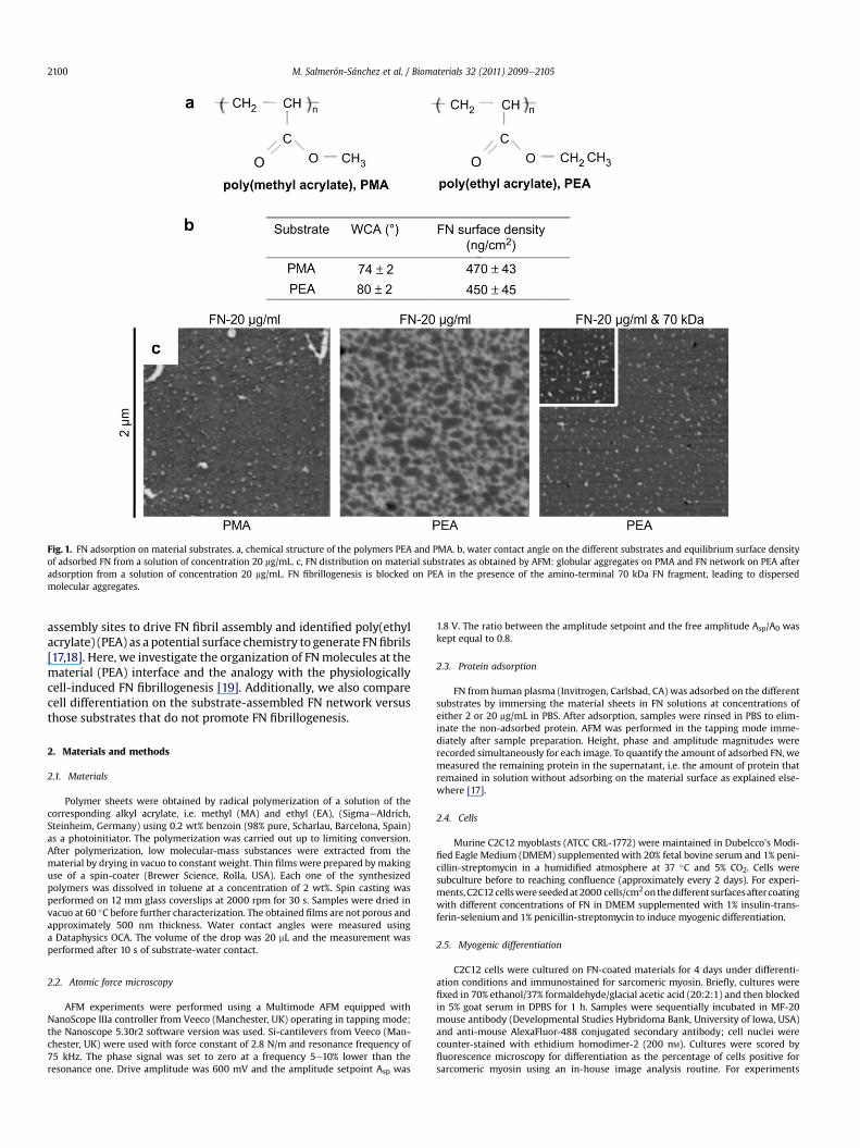
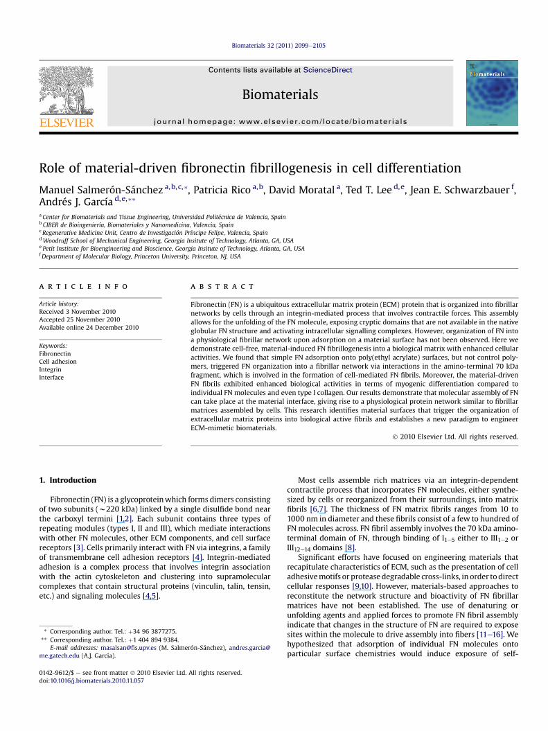
Fig. 1. FN adsorption on material substrates. a, chemical structure of the polymers PEA and PMA. b, water contact angle on the different substrates and equilibrium surface densityof adsorbed FN from a solution of concentration 20 mg/mL. c, FN distribution on material substrates as obtained by AFM: globular aggregates on PMA and FN network on PEA afteradsorption from a solution of concentration 20 mg/mL. FN fibrillogenesis is blocked on PEA in the presence of the amino-terminal 70 kDa FN fragment, leading to dispersedmolecular aggregates.
M. Salmerón-Sánchez et al. / Biomaterials 32 (2011) 2099e21052100
assembly sites to drive FN fibril assembly and identified poly(ethylacrylate) (PEA) as a potential surface chemistry to generate FNfibrils[17,18]. Here, we investigate the organization of FNmolecules at thematerial (PEA) interface and the analogy with the physiologicallycell-induced FN fibrillogenesis [19]. Additionally, we also comparecell differentiation on the substrate-assembled FN network versusthose substrates that do not promote FN fibrillogenesis.
2. Materials and methods
2.1. Materials
Polymer sheets were obtained by radical polymerization of a solution of thecorresponding alkyl acrylate, i.e. methyl (MA) and ethyl (EA), (SigmaeAldrich,Steinheim, Germany) using 0.2 wt% benzoin (98% pure, Scharlau, Barcelona, Spain)as a photoinitiator. The polymerization was carried out up to limiting conversion.After polymerization, low molecular-mass substances were extracted from thematerial by drying in vacuo to constant weight. Thin films were prepared by makinguse of a spin-coater (Brewer Science, Rolla, USA). Each one of the synthesizedpolymers was dissolved in toluene at a concentration of 2 wt%. Spin casting wasperformed on 12 mm glass coverslips at 2000 rpm for 30 s. Samples were dried invacuo at 60 �C before further characterization. The obtained films are not porous andapproximately 500 nm thickness. Water contact angles were measured usinga Dataphysics OCA. The volume of the drop was 20 mL and the measurement wasperformed after 10 s of substrate-water contact.
2.2. Atomic force microscopy
AFM experiments were performed using a Multimode AFM equipped withNanoScope IIIa controller from Veeco (Manchester, UK) operating in tapping mode;the Nanoscope 5.30r2 software version was used. Si-cantilevers from Veeco (Man-chester, UK) were used with force constant of 2.8 N/m and resonance frequency of75 kHz. The phase signal was set to zero at a frequency 5e10% lower than theresonance one. Drive amplitude was 600 mV and the amplitude setpoint Asp was
1.8 V. The ratio between the amplitude setpoint and the free amplitude Asp/A0 waskept equal to 0.8.
2.3. Protein adsorption
FN from human plasma (Invitrogen, Carlsbad, CA) was adsorbed on the differentsubstrates by immersing the material sheets in FN solutions at concentrations ofeither 2 or 20 mg/mL in PBS. After adsorption, samples were rinsed in PBS to elim-inate the non-adsorbed protein. AFM was performed in the tapping mode imme-diately after sample preparation. Height, phase and amplitude magnitudes wererecorded simultaneously for each image. To quantify the amount of adsorbed FN, wemeasured the remaining protein in the supernatant, i.e. the amount of protein thatremained in solution without adsorbing on the material surface as explained else-where [17].
2.4. Cells
Murine C2C12 myoblasts (ATCC CRL-1772) were maintained in Dubelcco’s Modi-fied Eagle Medium (DMEM) supplemented with 20% fetal bovine serum and 1% peni-cillin-streptomycin in a humidified atmosphere at 37 �C and 5% CO2. Cells weresubculture before to reaching confluence (approximately every 2 days). For experi-ments,C2C12cellswere seededat2000 cells/cm2onthedifferent surfacesaftercoatingwith different concentrations of FN in DMEM supplemented with 1% insulin-trans-ferin-selenium and 1% penicillin-streptomycin to induce myogenic differentiation.
2.5. Myogenic differentiation
C2C12 cells were cultured on FN-coated materials for 4 days under differenti-ation conditions and immunostained for sarcomeric myosin. Briefly, cultures werefixed in 70% ethanol/37% formaldehyde/glacial acetic acid (20:2:1) and then blockedin 5% goat serum in DPBS for 1 h. Samples were sequentially incubated in MF-20mouse antibody (Developmental Studies Hybridoma Bank, University of Iowa, USA)and anti-mouse AlexaFluor-488 conjugated secondary antibody; cell nuclei werecounter-stained with ethidium homodimer-2 (200 nM). Cultures were scored byfluorescence microscopy for differentiation as the percentage of cells positive forsarcomeric myosin using an in-house image analysis routine. For experiments
Fig. 2. Myogenic differentiation on the different substrates. a, fluorescence staining showing sarcomeric myosin-positive cells (green) and cell nuclei (red) on FN-coated surfaces(from solutions of concentration 2 and 20 mg/mL) and control collagen. b, myogenic differentiation as determined by the percentage of sarcomeric myosin-positive cells on thedifferent substrates after adsorbing FN from solutions of concentrations 2 and 20 mg/mL. c, cell density on each substrate.
M. Salmerón-Sánchez et al. / Biomaterials 32 (2011) 2099e2105 2101
including integrin blocking, HFN7.1 antibody (7.3 mg/mL final concentration) wasadded to culture media on day 1 in order to avoid altering initial adhesion. Forexperiments including contractility inhibitors, Y-27632 or blebbistatin was added toculture medium at different concentrations (10, 20 and 40 mM) after 2 h and main-tained for 4 days.
2.6. Integrin binding and FAK
Integrin binding to FN-coated materials was analyzed via immunostainingfollowing cross-linking of bound integrins to FN and extraction of cellularcomponents. Cells were seeded on FN-coated materials for 4 h. Cultures wererinsed in DPBS and incubated in ice-cold DTSSP (1.0 mM final concentration inDPBS þ 2 mM dextrose) for 30 min. Unreacted cross-linker was quenched with50 mM Tris in DPBS for 15 min and bulk cellular components were extracted in
0.1% SDS þ 350 mg/mL phenylmethylsulfonyl fluoride in DPBS. After blockingwith 5% FBS þ 0.1% Tween-20 for 1 h, bound integrins were immunostainedwith anti-a5 and AlexaFluor488-conjugated secondary antibody. For FAK assays,cells were detached and gently agitated in suspension under serum-freeconditions for 45 min to reduce background. Cells were then seeded on FN-coated materials serum-free for 2 h. Cells were lysed in RIPA buffer containingprotease inhibitors (1% Triton X-100, 1% sodium deoxycholate, 0.1% SDS, 150 mM
NaCl, 150 mM TriseHCl (pH 7.2), 350 mg/mL PMSF, 10 mg/mL leupeptin, and 10 mg/mL aprotonin) for 20 min at room temperature. Total protein was quantifiedusing micro-BCA (Pierce). Identical amounts of cell lysates were mixed insample buffer (50 mM TriseHCl pH 6.8, 100 mM DTT, 2% SDS, 10% glycerol, and0.1% bromophenol blue) and separated by SDSePAGE. After transferring tonitrocellulose membranes and blocking with Blotto (5% nonfat dry milk, 0.2%Tween-20) overnight at 4 �C, membranes were gently rocked in antibodies
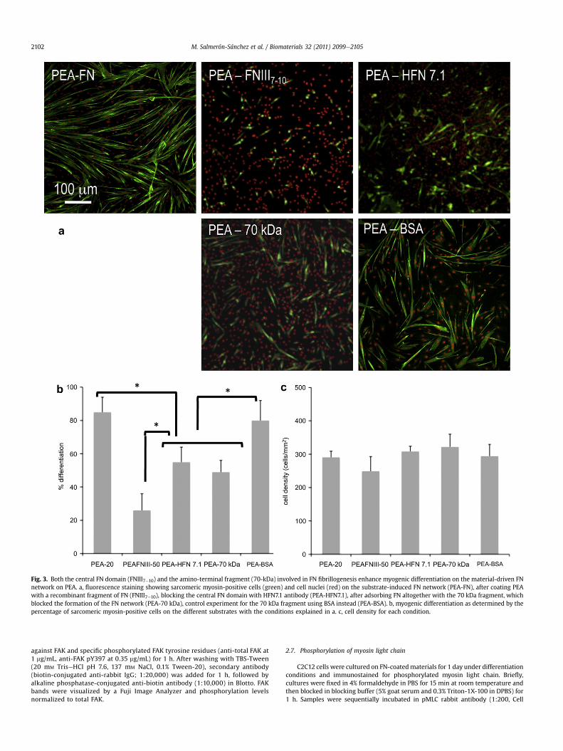
Fig. 3. Both the central FN domain (FNIII7e10) and the amino-terminal fragment (70-kDa) involved in FN fibrillogenesis enhance myogenic differentiation on the material-driven FNnetwork on PEA. a, fluorescence staining showing sarcomeric myosin-positive cells (green) and cell nuclei (red) on the substrate-induced FN network (PEA-FN), after coating PEAwith a recombinant fragment of FN (FNIII7e10), blocking the central FN domain with HFN7.1 antibody (PEA-HFN7.1), after adsorbing FN altogether with the 70 kDa fragment, whichblocked the formation of the FN network (PEA-70 kDa), control experiment for the 70 kDa fragment using BSA instead (PEA-BSA). b, myogenic differentiation as determined by thepercentage of sarcomeric myosin-positive cells on the different substrates with the conditions explained in a. c, cell density for each condition.
M. Salmerón-Sánchez et al. / Biomaterials 32 (2011) 2099e21052102
against FAK and specific phosphorylated FAK tyrosine residues (anti-total FAK at1 mg/mL, anti-FAK pY397 at 0.35 mg/mL) for 1 h. After washing with TBS-Tween(20 mM TriseHCl pH 7.6, 137 mM NaCl, 0.1% Tween-20), secondary antibody(biotin-conjugated anti-rabbit IgG; 1:20,000) was added for 1 h, followed byalkaline phosphatase-conjugated anti-biotin antibody (1:10,000) in Blotto. FAKbands were visualized by a Fuji Image Analyzer and phosphorylation levelsnormalized to total FAK.
2.7. Phosphorylation of myosin light chain
C2C12 cells were cultured on FN-coatedmaterials for 1 day under differentiationconditions and immunostained for phosphorylated myosin light chain. Briefly,cultures were fixed in 4% formaldehyde in PBS for 15 min at room temperature andthen blocked in blocking buffer (5% goat serum and 0.3% Triton-1X-100 in DPBS) for1 h. Samples were sequentially incubated in pMLC rabbit antibody (1:200, Cell

M. Salmerón-Sánchez et al. / Biomaterials 32 (2011) 2099e2105 2103
Signalling), and goat anti-rabbit AlexaFluor-488 conjugated secondary antibody(1:100); cell nuclei were counter-stained with Hoescht (200 nM). For experimentsincluding contractility inhibitors, Y-27632 or blebbistatin was added to culturemedium at different concentrations (10, 20 and 40 mM) after 2 h.
2.8. Statistics
All experiments were performed at least three times in triplicate unless other-wise noted. Data are reported as mean � standard error. Results were analyzed byone-way ANOVA using SYSTAT 8.0 (SPSS). If treatment level differences weredetermined to be significant, pair-wise comparisons were performed using a Tukeypost hoc test. A 95% confidence level was considered significant.
3. Results and discussion
3.1. Material-driven FN fibrillogenesis
We compared the organization of FN at the material interface ontwo similar chemistries: poly(ethyl acrylate) (PEA) and poly(methylacrylate) (PMA) which differ in one single carbon in the side chain(Fig.1a). Both surface chemistries show similar wettability and totalamount of adsorbed FN (Fig. 1b). However, the conformation anddistribution of the protein following passive adsorption onto thesesurfaces are completely different (Fig. 1c). Interconnected FN fibrilsare organized upon adsorption from a solution of concentration20 mg/mL on PEA (Fig. 1c) whereas only dispersed molecules arepresent on PMA (Fig.1c). FN fibril formation on PEA is dependent onthe FN solution concentration, as lower concentrations result indispersed adsorbed molecules (Supplementary Fig. S1). We nextexamined the requirements for the 70 kDa amino-terminal domainof FN in this material-driven fibrillogenesis. The 70 kDa amino-terminal regions are essential for cell-mediated FN assembly, andwithin this region, the I1e5 repeats confer FN binding activity [20].This domain is not accessible in the folded, compact structure of FNin solution and a conformational change of the molecule ismandatory for physiological matrix assembly to occur [6]. Strik-ingly, material-driven fibrillogenesis absolutely requires the 70 kDaamino-terminal region of FN. Addition of the 70 kDa fragmentcompletely blocks the organization of FN at the material interfaceand only discrete molecular aggregates can be observed (Fig. 1c)without any trace of the assembled FN network. The resultingadsorbed FN resembles the FN adsorbed onto the control PMApolymer. These results demonstrate that a particular polymerchemistry (PEA) drives assembly of adsorbed FN molecules into FNfibrils and this material-driven fibrillogenesis requires the 70 kDaamino-terminal domain of FN.
3.2. Cell differentiation on material-assembled FN
We hypothesized that material-driven FN fibrils have enhancedbiological activity compared to adsorbed FN molecules because thefibrillar structure recapitulates the native structure of FN matrices.We evaluated the biological activity of the material-driven FNnetworks by examining the myogenic differentiation process [21].Sarcomeric myosin expression and cell bipolar alignment andfusion into myotubes, markers of myogenesis, were significantlyhigher on the substrate-induced FN network (Fig. 2, PEA-20) ascompared to the same substrate coated with a lower density of FNthat does not lead to the fibrillar organization of the protein at thematerial interface (Fig. 2, PEA-2), and PMA polymer coated with thesame density of FN but lacking any fibrillar organization (Fig. 2,PMA-20). Surprisingly, myogenic differentiation was considerablymore robust on the PEA-assembled FNmatrix than on collagen typeI, which represents that standard substrate for myogenic differen-tiation (Fig. 2b) [22]. It is important to emphasize that thesedifferences in myogenic differentiation are not due to differences inthe number of cells on the substrates (Fig. 2c).
To further evaluate the biological activity of the material-drivenFN fibrils, we used different blocking antibodies and FN fragmentsto assess the importance of different parts of the FN molecule onthe myogenic differentiation process (Fig. 3). Addition of theadhesion-blocking HFN7.1 antibody inhibited differentiation onPEA to levels found for those substrates on which FN is not orga-nized into a network (Fig. 3, PEA-HFN7.1). The HFN7.1 antibodybinds to the flexible linker between the 9th and 10th type III repeatsof FN where the integrin-binding domain is located, demonstratingthat the integrin-binding domain of FN is essential for myogenicdifferentiation on FN fibrils. Moreover, HFN7.1 is specific for humanFN and does not cross-react with mouse FN, indicating that thehuman FN adsorbed onto the substrate prior cell seeding providedthe dominant signal for differentiation. We next examined whethera recombinant fragment of FN spanning the 7the10th type IIIrepeats (FNIII7e10) that encompass the integrin-binding domainrecapitulates the material-dependent differences in myogenicdifferentiation [23]. In contrast to complete FN, adsorption ofFNIII7e10 onto PEA yielded minimal levels of myogenic differenti-ation (Fig. 3, PEA-FNIII7e10). FNIII7e10 does not contain domainsinvolved in FNeFN interactions (I1e5 and III1e2 or III12e14 domains),demonstrating that both integrin-binding domains and domainsinvolved in FNeFN interactions are required for the enhancedmyogenic differentiation on PEA-driven FN matrices.
Consistent with our observations for FN network formation,addition of the 70 kDa amino-terminal fragment during theadsorption process of FN blocked the differentiation of myoblastson PEA (Fig. 3, PEA-70 kDa). This result demonstrates the impor-tance of FNeFN interactions and the fibrillar structure of theprotein on the cell differentiation process e only when the FNnetwork is assembled on PEA via interactions involving the 70 kDaamino-terminal fragment does the differentiation process occurs.As a control, when albumin (a protein with similar molecularweight to the 70 kDa fragment) is added at the same concentrationduring the network assembly process of FN at the material inter-face, the formation of the network is not disturbed and myoblastdifferentiation levels remain as in the original material-assembledFN network (Fig. 3, PEA-BSA). Importantly, the addition of the70 kDa FN fragment once the FN network is already assembled onthe material surface has no effect on subsequent myoblast differ-entiation (Supplementary Fig. S2).
3.3. Cell contractility on material-assembled FN
To gain insights into the mechanisms controlling the enhancedcellular responses to material-driven FN networks, we examinedbindingofa5b1 integrin to the adsorbed FN since this receptorprovidesthe primary adhesion mechanism in this cell model [22]. Immunoflu-orescence staining following crosslinking of bound integrins to FN andextractionof cellular components revealednosignificantdifferences inintegrin binding among FN on different surfaces, demonstrating thatthe substrate-assembled FN network does not alter integrin binding(Supplementary Fig. S3). Similarly, no differences in FAK phosphory-lation, a critical signal related to integrin-binding and myoblastdifferentiation, were observed between FN molecules on PEA andcontrol PMA substrates (Supplementary Fig. S3) [24,25].
Contractile forces generated within the cell regulate several func-tions, including muscle cell contraction and differentiation [26].Contractility results from dynamic interactions between actin fila-ments andmyosin,which are regulated via phosphorylation ofmyosinlight chain (MLC) [27]. Rho GTPases control the formation of stressfibers and focal adhesion assembly by modulating MLC phosphoryla-tion and generating actin-myosin contractility. Myoblast differentia-tion is characterized by the synthesis of the proteins of the contractileapparatus and the fusion of single myoblasts into multinucleated

Fig. 4. The FN network upregulates differentiation by enhancing cell contractility. a, fluorescence staining showing sarcomeric myosin-positive cells (green) and cell nuclei (red) onthe substrate-induced FN network (PEA-control) and the reference polymer (PMA-control). The effect of adding Y-27632 at different concentrations in the culture medium is shown.b. myogenic differentiation as determined by the percentage of sarcomeric myosin-positive cells on the different substrates (PEA, PMA) in the presence of contractility inhibitors atdifferent concentrations (Y-27632, blebbistatin) in the culture medium.
M. Salmerón-Sánchez et al. / Biomaterials 32 (2011) 2099e21052104
myotubes [28]. Immunofluorescence staining for phosphorylatedMLCrevealed the presence of well-defined fibers, coincident with the actincytoskeleton, on cells cultured on the FN network (PEA) but not oneither PMA or collagen (Supplementary Fig. S4). We used pharmaco-logical inhibitors that impair contractility to examine the contributionsof contractility on myogenic differentiation on the substrate-inducedFN network. Y-27632 is a specific inhibitor of Rho-kinase, and bleb-bistatin is a specific inhibitor of myosin II activity [29,30]. Staining forphosphorylatedMLCwas reduced in a dose-dependentmanner in the
presence of these inhibitors on the different substrates(Supplementary Fig. S4). More importantly, myoblast differentiationwas drastically arrested by these contractile inhibitors only on thesubstrate-induced FN network (Fig. 4). Differentiation levels dimin-ished from 85% to 40% on PEA, but were not altered on PMA, whichremains approximately equal to 40% independently of the presence ofeither Y-27632 or blebbistatin (Supplementary Fig. S5, S6). Theseresults reveal the importance of the material-assembled FN networkonmyoblast differentiation and show that actin-myosin contractility is
M. Salmerón-Sánchez et al. / Biomaterials 32 (2011) 2099e2105 2105
activated by the physiological material-assembled FN fibrils to drivedifferentiation.
4. Conclusions
We have identified a synthetic material e poly(ethyl acrylate) ethat drives cell-free, physiological organization of FN into fibrillarnetworks upon adsorption. This material promotes the biomimeticassembly of FN in absence of cells. This research facilitates thepreparation of new tools to advance in the investigation of thebiological process of FN fibrillogenesis and establishes a newstrategy for engineering of biomimetic materials.
Acknowledgements
This work was supported by NSF DMR-0909002 and MAT2009-14440-C02-01. MSS was supported by the Spanish Governmentthrough PR2009-0351 to stay in Atlanta (Georgia Institute ofTechnology) for a sabbatical during 2010.
Appendix
Figures with essential color discrimination. Figs. 2e4 in this articleare difficult to interpret in black andwhite. The full color images can befound in the online version, at doi:10.1016/j.biomaterials.2010.11.057.
Appendix. Supplementary material
Supplementary data related to this article can be found online atdoi:10.1016/j.biomaterials.2010.11.057.
References
[1] Erickson HP, McDonagh J. FN molecule visualized in electron microscopy:a long, thin, flexible strand. J Cell Biol 1981;91:673e8.
[2] Erickson HP, Carell NA. FN in extended and compact conformations. Electronmicroscopy and sedimentation analysis. J Biol Chem 1983;258:14539e44.
[3] Pankov R, Yamada KM. FN at a glance. J Cell Sci 2002;115:3861e3.[4] Hynes RO. Integrins: bidirectional, allosteric signaling machines. Cell
2002;110:673e87.[5] García AJ. Get a grip: integrins in cell-biomaterial interactions. Biomaterials
2005;26:7525e9.[6] Mao Y, Schwarzbauer JE. FN fibrillogenesis, a cell-mediated matrix assembly
process. Matrix Biol 2005;24:389e99.[7] Altankov G, Grinnell F, Groth T. Studies of the biocompatibility of materials:
fibroblast reorganization of substratum-bound FN on surfaces varying inwettaability. J Biomed Mater Res 1996;30:385e91.
[8] Geiger B, Bershadsky A, Pankov R, Yamada KM. Transmembrane extracellularmatrix e cytoskeleton crosstalk. Nat Rev Mol Cell Bio 2001;2:793e805.
[9] Lutolf MP, Gilbert PM, Blau HM. Designing materials to direct stem-cell fate.Nature 2009;462:433e41.
[10] Petrie TA, Raynor JE, Dumbauld DW, Lee TT, Jagtap S, Templeman KL, et al.Multivalent integrin-specific ligands enhance tissue healing and biomaterialintegration. Sci Transl Med 2010;2:45ra60.
[11] Mosher DF, Johnson RB. In vitro formation of disulfide-bonded FN multimers.J Biol Chem 1983;258:6595e601.
[12] Williams EC, Janmey PA, Johnson RB, Mosher DF. FN effect of disulfide bondreduction on its physical and functional properties. J Biol Chem1983;258:5911e4.
[13] Morla A, Zhang Z, Rouslahti E. Super FN is a distinct form of FN. Nature1994;367:193e6.
[14] Baneyx G, Vogel V. Self-assembly of FN into fibrillar networks underneathdipalmitoyl phosphatidylcholine monolayers: role of lipid matrix and tensileforces. Proc Natl Acad Sci USA 1999;96:12518e23.
[15] Ulmer J, Geiger B, Spatz JP. Force-induced FN fibrillogenesis. Soft Matter2008;4:1998e2007.
[16] Kaiser P, Spatz JP. Differential adhesion of fibroblasts and neuroblastoma cellson size- and geometry-controlled nanofibrils of the extracellular matriz. SoftMatter 2010;6:113e9.
[17] Rico P, Rodríguez Hernández JC, Moratal D, Altankov G, Monleón Pradas M,Salmerón-Sánchez M. Substrate-induced assembly of fibronectin intonetworks: influence of surface chemistry and effect on osteoblast adhesion.Tissue Eng Part A 2009;15:3271e81.
[18] Gugutkov D, González-García C, Rodríguez Hernández JC, Altankov G,Salmerón-Sánchez M. Biological activity of the substrate-induced FNnetwork: insight into the third dimension through electrospun fibers.Langmuir 2009;25:10893e900.
[19] Aguirre KM, McCormick RJ, Schwarzbauer JE. FN self-association is mediatedby complementary sites within the amino-terminal one-third of the molecule.J Biol Chem 1994;269:27863e8.
[20] Schwarzbauer JE. Identification of FN sequences required for assembly ofa fibrillar matrix. J Cell Biol 1991;113:1463e73.
[21] Sabourin LA, Rudnicki MA. The molecular regulation of myogenesis. ClinGenet 2000;57:16e25.
[22] García AJ, Vega MD, Boettiger D. Modulation of cell proliferation and differ-entiation through substrate-dependent changes in FN conformation. Mol BiolCell 1999;10:785e98.
[23] Petrie TA, Raynor JE, Reyes CD, Burns KL, Collard D, García AJ. The effect ofintegrin-specific bioactive coatings on tissue healing and implant osteointe-gration. Biomaterials 2008;29:2849e57.
[24] Michael KE, Dumbauld DW, Burns KL, Hanks SK, García AJ. FAK modulates celladhesion strengthening via integrin activation. Mol Biol Cell 2009;20:2508e19.
[25] Quach NL, Biressi S, Reichardt LF, Keller C, Rando TA. Focal adhesion kinasesignaling regulates the expression of caveolin 3 and b1 integrin, genesessential for normal myoblast fusion. Mol Biol Cell 2009;20:3422e35.
[26] Griffin MA, Sen S, Sweeney HL, Discher DE. Adhesion-contractile balance inmyocite differentiation. J Cell Sci 2004;117:5855e63.
[27] Kaibuchi K, Kuroda S, Amano M. Regulation of the cytoskeleton and celladhesion by the Rho family GTPases in mammalian cells. Annu Rev Biochem1999;68:459e86.
[28] Andres V, Walsh K. Myogenin expression, cell cycle withdrawal, and pheno-typic differentiation are temporally separable events that precede cell fusionupon myogenesis. J Cell Biol 1996;132:657e66.
[29] Narumiya S, Ishizaki T, Uehata M. Use and properties of ROCK-specificinhibitor Y-27632. Methods Enzymol 2000;325:273e84.
[30] Kovács M, Tóth J, Hetényi C, Málnási-Csizmadia A, Sellers JR. Mechanism ofblebbistatin inhibition of myosin II. J Biol Chem 2004;279:35557e63.