Antibiotic resistant bacteria/genes dissemination in lacustrine ...
Upload
khangminh22Category
view
0download
0
Cellular differentiation and antibiotic
production by Streptomyces nodosus
immobilised in alginate capsules
A thesis submitted as a requirement for the degree of
Doctor of Philosophy
by
Marie Antoinette Tanya Pereira
Master of Applied Science
School of Natural Sciences
University of Western Sydney
March 2007

With all my love to Dad, Mum and Ro

Acknowledgements
Supervisors, Dr Jo-Anne Chuck, Dr Gary Dennis and Prof. Phil Moore, I wish to
thank and at the commencement of my acknowledgements to mention,
Thankyou for sharing your wealth of knowledge, guiding me through my study;
through ups and downs, you have directed me to a new horizon!
Researcher I am, but would I be so without the support of you Technical Staff, Susie,
Sandy, Aneel, Paul, Wayne, Bert, Kieran, Antonetta, Herbert, Greg and Mal?
Ever willing to do the necessary, provide consumables or fix a computer problem!
Particular admiration I have for you Ms Jacqueline Timms, a School Administrative
Officer of not just high calibre but also commendable people skills have you,
Thank you for your efficiency, thank you for you!
Opportunity to enhance my teaching skills you provided me, Dr Margaret Seto-Poon,
thank you for your example of remarkable dedication and for the opportunity to
work with you.
Manager of the Microscopy Unit at Macquarie University, Ms Debra Birch, the
world of Electron Microscopy you have introduced me to, with experience and
exceptional skill you have trained me, for this I am indebted to you!
Young and radiant with intelligence, you shared your skill, especially in microtomy,
Ms Nicole Vella, for this and your friendship, I wish to say thank you!
Colleagues, Dr Clare Gordon-Thomson for your diamond knife, Dr Susan Chambers,
Dr Narsimha Reddy, Dr Alan McCutcheon and Dr Sundar Rao for your
friendship and for your interest in my study.
Evenings watching the Broncos we�ve spent, laughter and tears we�ve shared,
Blossom/Gertrude/Sunshine says, �Thank you dear Olive for the warmth and
safety of your home but most of all for the warmth of your most generous heart!�
Security staff, Stephen, Michael, Christine, Jimmy, Graham, Ron, Daniel, Anita, Ian,
Greg and Tony,
Not unnoticed were your acts to always make sure of my safety and to go that extra
mile, thank you for all that you have done for me!
Office mates and uni mates, thank you for the experiences we�ve shared and for
those priceless corridor chats, thank you Sashi, Nick, Catherine, Brigitte, Alka,
Stef, Lis, Poonam, Adam, Meagan, Laurel, Shyama and Thil.
Direction and counsel you offer me, with gentleness and strength you guide me,
picked me up during my weakest moment, for this, I thank you Fr Al.
Overflowing is my gratitude to you my darling Paul, thank you for standing by me
and with me, thank you for the big and small things that you do but most of all
thank you for your immeasurable love�.�you are the best�!
Support and strength that is bound neither by time nor space, that crosses oceans,
seas and continents, thank you Dad, Mum and Ro for your unfailing love and
prayers.
Under your mantle of protection You have kept me, taken care of my every need,
thank you dear Lord for being with me every step of the way.
Sincere gratitude to the University of Western Sydney, in particular to the School of
Natural Sciences and the UWS Priority Postgraduate Research Scholarship for
making this work possible.

Publications in Support of this Thesis
1. Pereira, T., Millar, T. J. & Chuck, J. (2005). Viability analysis of alginate
encapsulated micro-organisms using fluorescent stains. J Microencapsul 22,
787�792.
2. Pereira, T., Nikodinovic, J., Nakazono, C., Barrow, K. D. & Chuck, J.
Sporulation and antibiotic production by planktonic and immobilised
Streptomyces nodosus cultured in liquid environments. Microbiology,
submitted for publication.

Statement of Authentication
The work presented in this thesis is to the best of my knowledge and belief original
except as acknowledged in the text. I hereby declare that I have not submitted this
material, either in full or in part, for a degree at this or any other institution.
�����������������
(Tanya Pereira)

i
Table of Contents
Page
List of Tables ii
List of Figures and Illustrations iii
List of Abbreviations ix
Abstract xi
Chapter 1 Introduction 1
Chapter 2 Materials and Methods 40
Chapter 3 Optimisation of capsule production and the kinetics
of amphotericin B release in vitro 53
Chapter 4 Immobilisation of Streptomyces nodosus in alginate,
monitoring of viability, morphology and antibiotic
production during fermentation 79
Chapter 5 Cross-feeding studies using immobilised S. nodosus
cultured in conditioned media from Streptomyces spp. 111
Chapter 6 Conclusions and Future Directions 144
References 149

ii
List of Tables
Table 3.1. Results of three independent experiments measuring diameter of
capsules prepared with 1 %, 2 %, 3 % and 4 % (w/v) alginate
concentrations using manual extrusion (29-gauge needle) and semi-
automated extrusion (peristaltic pump) falling from a height of 6 cm
into calcium chloride solution.
Table 3.2. Selection of a suitable internal standard.
Table 4.1. The precision of BacLight viability stains in quantifying viability of
E. coli.
Table 5.1. Effect of culturing immobilised log phase (LP) and stationary phase
(SP) cultures of S. nodosus in conditioned media on the development
of a co-existing planktonic population.

iii
List of Figures and Illustrations
Fig. 1.1. Chemical structure of griseofulvin.
Fig. 1.2. Chemical structures of some azoles.
Fig. 1.3. Chemical structure of 5-FC.
Fig. 1.4. Chemical structures of some allylamines and thiocarbamates.
Fig. 1.5. Chemical structures of some echinocandins.
Fig. 1.6. Chemical structure of nystatin.
Fig. 1.7. Chemical structure of AmB.
Fig. 1.8. Fingerprint UV-Vis absorption spectrum of AmB showing the 4
characteristic UV absorptions at 346, 364, 382 and 405 nm.
Fig. 1.9. Chemical structure of AmA.
Fig. 1.10. Model of an AmB/sterol channel showing (a) top view and (b) lateral
view.
Fig. 1.11. Modular structure of AmB PKS.
Fig. 1.12. Life cycle of the model Streptomyces species,
Streptomyces coelicolor, when cultured on solid media.
Fig. 1.13. Chemical structures of quorum sensing molecules produced by
different Streptomyces spp.
Fig. 3.1. Experimental set-up for the preparation of alginate capsules by (a) the
manual method and (b) the semi-automated method.
Fig. 3.2. Representative stereomicroscope image revealing surface morphology
of capsules made from droplets of a 2 % (w/v) alginate solution
falling into calcium chloride solution (100 mM) from a height of
(a) 6 cm and (b) 12 cm.

iv
Fig. 3.3. Representative HPLC elution profiles of AmB in (a) water, (b) YMG
and (c) conditioned medium from S. nodosus MA�
hyg1 with a
mobile phase of ACN:0.1 % (v/v) TFA (60:40 v/v) monitored at 408,
386 and 366 nm.
Fig. 3.4. Absorption spectra of compounds detected at 408 nm with retention
times of (a) 4.1 and (b) 3.5 min from samples of AmB dissolved in
conditioned medium from S. nodosus MA�
hyg1.
Fig. 3.5. Representative HPLC elution profiles of an AmB standard in (a)
methanol and (b) conditioned medium from S. nodosus MA�
hyg1
with a mobile phase of MeOH/0.1 % (v/v) TFA (85:15 v/v) monitored
at 408, 386, 366, 550 and 220 nm.
Fig. 3.6. Standard curve for quantification of AmB using external calibration.
Fig. 3.7. Representative chromatogram of biphenyl using a mobile phase of
MeOH/0.1 % (v/v) TFA (85:15 v/v) monitored at 408, 386, 366, 550
and 220 nm.
Fig. 3.8. Zone of inhibition around capsules after 20 h incubation (28 °C) of
(a) 0.5 g and (b) 1 g AmB capsules placed on a lawn culture of
Rhodotorula sp. cultured on malt extract agar.
Fig. 3.9. Release profiles of AmB encapsulated in alginate capsules and
incubated in Milli-Q water, YMG and conditioned media from
S. nodosus MA�
hyg1 over 48 h at 28 °C.
Fig. 3.10. Absorption spectrum of T. fusca porphyrin at 400 nm.
Fig. 4.1. Growth kinetics of S. nodosus cultured in YMG, R5 and GA media
incubated at 28 °C for 7 d.

v
Fig. 4.2. UV-Vis spectra of AmB produced in culture fluids derived from free-
dwelling S. nodosus grown in YMG, R5 and GA media after 7 d of
fermentation at 28 °C.
Fig. 4.3. Growth and AmB production by free-dwelling S. nodosus cultured in
YMG at 28 °C at 120 rpm for 7 d.
Fig. 4.4. Light microscopy images of typical capsules containing S. nodosus
mycelia (48 h culture) produced using (a) 1 % alginate and (b) 2 %
alginate.
Fig. 4.5. Effect of alginate concentration on the production of AmB.
Fig. 4.6. Fluorescence staining of encapsulated E. coli under ×400
magnification (a) 100 % live, FITC filter and (b) 100 % isopropanol-
killed, Texas Red filter.
Fig. 4.7. Fluorescence staining of encapsulated S. nodosus spores under ×400
magnification (a) S. nodosus spores, 100 % live, FITC filter and
(b) S. nodosus spores, 100 % isopropanol-killed, Texas Red filter.
Fig. 4.8. Fluorescence staining of encapsulated S. nodosus mycelia under ×400
magnification (a) S. nodosus mycelia, 100 % live, FITC filter and
(b) S. nodosus mycelia, 100 % isopropanol-killed, Texas Red filter.
Fig. 4.9. Fluorescence staining of immobilised S. nodosus under ×400
magnification on d 0.
Fig. 4.10. Fluorescence staining of immobilised S. nodosus under ×400
magnification on d 7.
Fig. 4.11. AmB production by immobilised S. nodosus log phase mycelia,
stationary phase mycelia and spores cultured in YMG.

vi
Fig. 4.12. Viability and morphology of immobilised S. nodosus mycelia (48 h)
when cultured in YMG in shake flasks on (a) d 0, (b) d 1, (c) d 2,
(d) d 4, (e) d 7, (f) d 14, (g) d 21 and (h) d 30 using CLSM with a
515 nm interference emission filter and a long pass 565 nm emission
filter at ×1000 magnification.
Fig. 4.13. Viability and morphology of planktonic population co-existing with
the immobilised S. nodosus mycelia during fermentation in YMG
cultured in shake flasks on (a) d 4, (b) d 7, (c) d 14 and (d) d 30 using
CLSM at ×1000 magnification.
Fig. 4.14. SEM of wild-type S. nodosus cultured on solid YMG agar for 48 h.
Fig. 4.15. SEM of wild-type S. nodosus cultured in liquid YMG medium for
48 h.
Fig. 4.16. SEM of wild-type S. nodosus mycelia associated with the surface of
an alginate capsule when cultured in YMG for 48 h.
Fig. 4.17. SEM of wild-type S. nodosus mycelia embedded within the alginate
capsule cultured in YMG for 48 h.
Fig. 4.18. Model of Streptomyces at a solid�air or solid�liquid interface.
Fig. 5.1. Immobilised S. nodosus log phase mycelia cultured in microtitre
plates.
Fig. 5.2. Viability and morphology of immobilised (a) S. nodosus spores and
(b) S. nodosus log phase mycelia (48 h) on d 2 cultured in YMG in
microtitre plates, using CLSM at ×1000 magnification.

vii
Fig. 5.3. Viability and morphology of immobilised S. nodosus mycelia (48 h)
when cultured in conditioned media from S. nodosus on (a) d 2,
(b) d 4, (c) d 7, (d) d 14, (e) d 21 and (f) d 30 using CLSM at ×1000
magnification.
Fig. 5.4. SEM of encapsulated wild-type S. nodosus mycelia (48 h) cultured in
(a) YMG for 2 d, (b) YMG for 4 d, (c) conditioned medium from
S. nodosus for 2 d and (d) conditioned medium from S. nodosus for
4 d.
Fig. 5.5. TEM of S. nodosus hyphal forms associated with solid�liquid
interfaces cultured in unconditioned YMG medium for (a�c) 2 d and
(d�f) for 4 d.
Fig. 5.6. TEM of S. nodosus hyphal forms associated with solid�liquid
interfaces cultured in conditioned media from S. nodosus for (a�d) 2 d
and (e�h) for 4 d.
Fig. 5.7. CLSM images of planktonic population stained with BacLight
viability stain.
Fig. 5.8. Viability and morphology of immobilised S. nodosus mycelia (48 h)
when cultured in conditioned media from mutant S. nodosus
MA�
hyg1 on (a) d 2, (b) d 4, (c) d 7, (d) d 14, (e) d 21 and (f) d 30
using CLSM at ×1000 magnification.
Fig. 5.9. Growth of free-dwelling S. virginiae, S. griseus, S. natalensis,
S. nodosus MA�
hyg1 or S. coelicolor cultured in YMG at 28 °C at
60 rpm for 7 d.

viii
Fig. 5.10. Viability and morphology of immobilised S. nodosus mycelia (48 h)
when cultured in conditioned medium from S. coelicolor on (a) d 2,
(b) d 5, (c) d 7, (d) d 14, (e) d 21 and (f) d 30 using CLSM at ×1000
magnification.
Fig. 5.11. Viability and morphology of immobilised S. nodosus mycelia (48 h)
when cultured in conditioned medium from S. griseus on (a) d 2, (b) d
5, (c) d 7, (d) d 14, (e) d 21 and (f) d 30 using CLSM at ×1000
magnification.
Fig. 5.12. Viability and morphology of immobilised S. nodosus mycelia (48 h)
when cultured in conditioned medium from S. natalensis on (a) d 2,
(b) d 5, (c) d 7, (d) d 14, (e) d 21 and (f) d 30 using CLSM at ×1000
magnification.
Fig. 5.13. Viability and morphology of immobilised S. nodosus mycelia (48 h)
when cultured in conditioned medium from S. virginiae on (a) d 2, (b)
d 5, (c) d 7, (d) d 14, (e) d 21 and (f) d 30 using CLSM at ×1000
magnification.
Fig. 5.14. SEM of encapsulated wild-type S. nodosus mycelia (48 h) cultured for
2 d in conditioned media from (a) S. coelicolor, (b) S. griseus,
(c) S. natalensis and (d) S. virginiae.
Fig. 5.15. SEM of encapsulated wild-type S. nodosus mycelia (48 h) cultured for
22 d in conditioned media from (a) S. coelicolor, (b) S. griseus,
(c) S. natalensis and (d) S. virginiae.
Fig. 6.1. Hypothesised functions of aerial hyphae that are destined for different
functions in soil.

ix
List of Abbreviations
5-FC 5-Fluorocytosine
ACN Acetonitrile
ACP Acyl Carrier Protein
AIDS Acquired Immune Deficiency Syndrome
AmA Amphotericin A
AmB Amphotericin B
AT Acyl Transferase
CFU Colony Forming Units
CLSM Confocal Laser Scanning Microscopy/Microscope
DAD Diode-Array Detector
DH Dehydratase
DMSO Dimethyl Sulfoxide
ER Enoyl Reductase
FAS Fatty Acid Synthase
FITC Fluorescein Isothiocyanate
G Guluronic Acid
GA Glycerol Asparagine
HPLC High Performance Liquid Chromatography
KR Ketoreductase
KS Ketoacyl Synthase
LB Luria-Bertani
M Mannuronic Acid
MeOH Methanol
MG Alternating Mannuronic and Guluronic Acid

x
OD Optical Density
ORF Open Reading Frames
PCD Programmed Cell Death
PI Propidium Iodide
PIPES Piperazine-N,N'-bis(2-ethanesulfonic acid)
PKS Polyketide Synthase
PLL Poly-L-Lysine
SEM Scanning Electron Microscopy/Microscope
SPE Solid-Phase Extraction
TE Thioesterase
TEM Transmission Electron Microscopy/Microscope
TFA Trifluoroacetic Acid
UV Ultraviolet
Vis Visible (reference to detection)
YMG Yeast extract-Malt extract-Glucose

xi
Abstract
Encapsulation is a novel technique that involves the entrapment of materials such as
cells, enzymes or chemicals within a semi-permeable matrix and is being explored as
a drug delivery system. This project investigated the encapsulation of Streptomyces
nodosus in alginate to assess whether this organism can produce the antifungal drug
amphotericin B from within the matrix. New methods were developed to immobilise
S. nodosus mycelia and spores in alginate capsules, assess bacterial viability and
detect ng mL�1
quantities of amphotericin B in culture fluids. When capsules were
cultured and cell proliferation was encouraged, organisms formed protrusions on the
surface of the capsules. Differentiated branched hyphae that never progressed to
sporogenic hyphae were observed on the surface of these structures. Viability was
maintained for up to 30 days and low levels of amphotericin B were produced. The
emergence of a co-existing free-dwelling population was also observed. Culturing
immobilised organisms using conditioned media from an amphotericin deficient S.
nodosus strain, augmented the development of the free-dwelling population resulting
in the detection of amphotericin B in the culture fluid and full differentiation to
sporogenic hyphae. This is the first report of sporulation of S. nodosus in liquid
environments and demonstrates that immobilised S. nodosus can produce antibiotics.
The sporulation of free-dwelling organisms was also induced using conditioned
media and manipulation of quorum size, indicating a solid surface is not required for
sporulation. Conditioned media from other Streptomyces spp. induced variable
responses including sporulation, pigment formation and antibiotic production,
possibly demonstrating communication between species and/or alteration in
nutritional status. This new model for the life cycle of S. nodosus will permit the
study of developmental pathways, antibiotic production, microbial community
structure and inter-species and intra-species signalling.

Chapter 1
Introduction

2
1.1 Human fungal infections and their treatment
The total number of fungal species has been estimated to be about 1.5 million with
about 70,000 species having been identified or described to date. Of these, about 200
species are known to be harmful to humans (Ellis, 2002). Fungi can cause allergic
reactions, toxic reactions or mycoses (fungal infections). Mycoses are the most
serious compared with allergic or toxic fungal reactions and are the most difficult to
diagnose and treat (Barrett, 2002). They include a spectrum of diseases ranging from
minor superficial skin and mucous membrane infections such as dermatophytoses
and anchomycoses to life-threatening invasive mycoses, such as candidiasis and
aspergillosis (Georgopapadakou, 1998).
Since the 1980s there has been a significant increase in the incidence of systemic
fungal infections primarily caused by the opportunistic species (Pfaller & Diekema,
2004). This increase in fungal infections corresponds to the increase in the size of
the immunocompromised population in society (Steinbach & Perfect, 2003). This
includes patients undergoing intensive chemotherapy for cancer, allogeneic
hematopoietic stem cell and solid organ transplant recipients, patients on
immunosuppressive therapy, acquired immune deficiency syndrome (AIDS) patients,
premature birth and advanced age (Nucci & Marr, 2005). Other factors that have
contributed to an increase in mycoses include poor nutrition, treatment with broad-
spectrum antibacterial drugs and invasive procedures such as surgery and implanted
surgical devices (DiDomenico, 1999). Consequently, research on opportunistic
fungal pathogens and antifungals have become increasingly important in current
medical microbiology, though they previously received relatively less attention due

3
to the focus on bacterial infections and therapy (Hazen, 1995; Odds et al., 2003; de
Pauw, 2000).
The recent escalated increase in fatal mycoses has sparked a new wave of intensive
research in effective antifungal agents. The world market for antifungals is
expanding at a rate of 20 % per annum (Gupte et al., 2002). Selective toxicity is
difficult to achieve as it is difficult to choose intracellular targets whose inhibition
would not be detrimental to the host cell (DiDomenico, 1999). The ideal antifungal
drug would exhibit excellent fungicidal activity, have a broad antifungal spectrum,
would not be susceptible to fungal resistance, have the ability to overcome multi-
drug resistance and have minimal toxicity (Borowski, 2000). Current clinically
approved antifungals can be broadly classified into six categories, i) griseofulvin,
ii) the azole derivatives, iii) the fluoropyrimidines, iv) the allylamines and
thiocarbamates, v) the echinocandins and vi) the polyenes (François et al., 2005).
Griseofulvin, isolated from Penicillium griseofulvum, was the first antifungal
compound and this marked the beginning of antifungal drug discovery (Fig. 1.1)
(Oxford et al., 1939). It was introduced as an antifungal in the treatment of human
dermatophytosis in 1958 (Develoux, 2001; Sheehan et al., 1999) and remained the
principal treatment until the introduction of imidazoles and allylamines (François et
al., 2005). Griseofulvin is known to be fungistatic but its mode of action is still
unknown. Several hypotheses have been proposed, the most accepted being through
the interference of the fungal microtubule assembly (Develoux, 2001; Odds et al.
2003).

4
O
O
H3CO
Cl
OCH3 O OCH3
H3C
Fig. 1.1. Chemical structure of griseofulvin.
The azole class of antifungals were discovered in 1944 but were clinically introduced
in the late 1960s and approved as treatment for systemic mycoses in 1981 (Sheehan
et al., 1999). Currently there is an increase in development activity for this class of
antifungal compounds (Maertens, 2004; Kauffman, 2006). Azoles are synthetic
antifungals consisting of a five-membered azole ring with two (imidazole) or three
nitrogens (triazole). They are active against most yeasts and filamentous fungi and
their mode of action is by inhibiting the synthesis of ergosterol (the fungal membrane
sterol) through an interaction with the cytochrome P450-dependent enzyme
lanosterol 14-�-demethylase which is necessary for the conversion of lanosterol to
ergosterol (Bossche et al., 1986; Maertens, 2004). The azoles currently in clinical
use for the treatment of systemic mycoses are ketoconazole, itraconazole and
fluconazole (Fig. 1.2). Although azoles are not usually associated with severe side
effects, in some rare cases, fatal hepatotoxicity has been reported. They may also
cause endocrine-associated side effects like decreased release of testosterone and
glucocorticoids. This results in gynecomastia and adrenal insufficiency, respectively
(Georgopapadakou & Walsh, 1996). Other drawbacks of azoles are that they are
fungistatic rather than fungicidal, with some fungi (e.g. Candida albicans,
Cryptococcus neoformans, Aspergillus fumigatus) developing resistance towards
some azoles (Sheehan et al., 1999; Coker & Harris, 1991). The new addition to this
class of antifungals, namely voriconazole, is the first antifungal since the polyene
antifungal, amphotericin B (AmB), to be approved as first-line therapy for invasive

5
aspergillosis (Knapp & Flynn, 2005). However, voriconazole interacts with many
drugs, including immunosuppressants, which could be detrimental for patients on
these medications or those receiving multiple medications (Carrillo-Muñoz et al.,
2004).
N NH3C
O
OO
O
N
N
Cl
Cl
N
N
N
F
F
N
HO
N
N
Ketoconazole Fluconazole
N N
OO
O
N
N
N
Cl
Cl
N
N
N
O
H3C
CH3
N
N
N
F
F
N N
F
OH
CH3
Itraconazole Voriconazole
Fig. 1.2. Chemical structures of some azoles.
The fluoropyrimidine class of antifungals, which includes 5-fluorocytosine (5-FC)
(Fig. 1.3), was initially developed in 1957 for the treatment of cancer. However, in
1968 it was approved for the treatment of candidiasis and cryptococcosis (Francis
&Walsh, 1992). It is an oral fluorinated pyrimidine that inhibits fungal pyrimidine
metabolism, DNA, RNA and protein synthesis. Its disadvantages include a limited
spectrum of activity, a weak therapeutic activity and hence, 5-FC is generally used in
conjunction with AmB, for the treatment of cryptococcal meningitis and

6
disseminated candidiasis (Andriole, 1999; Francis &Walsh, 1992; Georgopapadakou
& Walsh, 1996).
N
NH
O
NH2
F
Fig. 1.3. Chemical structure of 5-FC.
Allylamines and thiocarbamates are structurally related synthetic antifungals
introduced in the 1980s (Sheehan et al., 1999). They are reversible, non-competitive
inhibitors of squalene epoxidase, an enzyme which acts in conjunction with squalene
cyclase to convert squalene to lanosterol (Ryder, 1988). Hence, the conversion of
lanosterol to ergosterol is inhibited, which results in the accumulation of squalene
and the depletion of ergosterol, thereby affecting membrane structure and function
(Georgopapadakou & Bertasso, 1992). Naftifine and terbinafine (allylamines) and
tolnaftate (thiocarbamate) are the currently approved antimycotics (Fig. 1.4). The
main disadvantage of allylamines and thiocarbamates is that their clinical use is
limited to topical fungal infections (skin and nail fungal infections) caused by
dermatophytes such as Trichophyton mentagrophytes and Microsporum canis
(François et al., 2005; Georgopapadakou, 1998).
N
CH3
N
CH3
CH3
CH3
CH3
N
S
O
CH3
H3C
Naftifine Terbinafine Tolnaftate
Fig. 1.4. Chemical structures of some allylamines and thiocarbamates.

7
The echinocandins are cyclic lipopeptides which were discovered by random
screening in the late 1970s (Georgopapadakou, 1998). Echinocandins are relatively
well tolerated clinically, compared with other antifungal agents, as they target the
fungal cell wall which has no counterpart in the mammalian cell (Knapp & Flynn,
2005). The mode of action of echinocandins is through inhibition of the enzyme �
-
1,3-D-glucan synthase which is required for the polymerisation of �
-1,3-D-glucan, a
major component of the fungal cell wall (Stone et al., 2002). Anidulafungin is the
most recently approved drug of this class for the treatment of oesophageal
candidiasis, with caspofungin in clinical use since 2001 for the treatment of
aspergillosis and candidiasis (Fig. 1.5) (Torre & Reboli, 2007; Kartsonis et al., 2003,
François et al., 2005). The main disadvantages of this class of drugs are the lack of
activity against cryptococci, Fusarium spp. and Scedosporium spp., and although
only been used clinically for a few years, resistant Candida spp. have already been
reported (François et al., 2005; Carrillo-Muñoz et al., 2004).
H3C
NH
N
NH
NH
HN
HN
N
O
CH3 CH3
OHNH
H2N
O
O
O
O
O
OOH
CH3
HO
HO
OH
H2N
HO
OH
HO
NH
N
NH
NH
HN
HN
N
O
OHHO
O
O
O
O
O
OOH
CH3
HO
HO
OH
H3C
HO
OH
HO
H3C
O
H3C
Caspofungin Anidulafungin
Fig. 1.5. Chemical structures of some echinocandins.

8
Polyene antibiotics are produced by gram positive soil bacteria Streptomyces spp.
and were discovered in the early 1950s (Martín, 1977). Polyenes are fungicidal and
have the broadest activity spectrum of all clinically useful antifungals known to date
(Georgopapadakou & Walsh, 1996). Structurally, they are characterised by a large
macrocyclic ring of carbon atoms closed by lactonisation with a series of 4�7
conjugated double bonds and generally possess a sugar moiety (Hamilton-Miller,
1973; Martín, 1977). They interact with ergosterol in fungal cytoplasmic membranes
causing damage to the targeted cells, resulting in cell death (Gottlieb & Shaw, 1970;
Bolard, 1986). AmB, nystatin (Fig. 1.6), and pimaricin are typical examples of
antifungal polyenes. Although AmB is associated with severe side effects, it is still
considered the �gold standard� in the treatment of severe systemic mycoses
(Georgopapadakou & Walsh, 1994; Brajtburg et al., 1990; Gallis et al., 1990).
O
O OH OH OH O
OH
CH3
H3C
H3C
HO
COOH
OH
OO
NH2OH
CH3
OH
OH
OH
Fig. 1.6. Chemical structure of nystatin.
As outlined above although significant progress has been achieved in the
development of antifungals since the isolation of griseofulvin, none of the current
systemic antifungals completely satisfy medical requirements. They fail either in
their spectrum of potency, safety or pharmacokinetic properties (Borowski, 2000;
Georgopapadakou, 1998). Hence, there is an urgent need to develop novel
antifungals or delivery systems for these drugs, with ideal pharmacological and

9
pharmacokinetic properties, including a broad fungicidal spectrum of action and
fewer dose-limiting side effects. The principle aim of this project was to investigate
a novel slow release system using immobilised micro-organisms as a delivery system
for AmB. This drug was chosen as it is by far the most potent antimycotic and
resistance to AmB is rare (Hoeprich, 1978; Hartsel & Bolard, 1996; Younsi et al.,
2000; Ellis, 2002).
1.2 Amphotericin B
1.2.1 Discovery and production
AmB is one of the 200 compounds that belong to the polyene family of antifungals
(Omura & Tanaka, 1984). It is naturally produced by the soil actinomycete
Streptomyces nodosus ATCC 14899 (Trejo & Bennett, 1963). Actinomycetes are a
well known class of filamentous bacteria and they produce more than 70 % of the
known antibiotics (Challis & Hopwood, 2003; Lechevalier & Lechevalier, 1967;
Ensign, 1978). S. nodosus was isolated from soil collected along the Orinoco River
in Tembladora, Venezuela, by W. Gold and antifungal activity was quantified at the
Squibb laboratories (Gold et al., 1956). In 1959, the first patent for AmB production
was granted and in 1962, the organism was officially characterised as S. nodosus
according to the International Code of Nomenclature of Bacteria and Viruses, with
the full name Streptomyces nodosus Trejo (Trejo & Bennette, 1963). AmB is
produced on an industrial scale by large-scale fermentation of S. nodosus. Total
chemical synthesis is possible, but this is not the preferred method of production due
to high costs and low yields associated with the known chemical synthetic pathway
(Nicolaou et al., 1988).

10
1.2.2 Chemical structure and properties
O
O OH OH
OH
OH OH O
OH
CH3
H3C
H3C
HO
COOH
OH
OO
NH2OH
CH3
OH
Fig. 1.7. Chemical structure of AmB.
AmB is a macrolide, with a non-aromatic 38-membered lactone ring with seven
conjugated double bonds, making it a heptaene (Fig. 1.7) (Martín, 1977). The
conjugated double bonds are responsible for the characteristic deep-yellow to orange
colour of the compound. Further, the conjugated double bond region of AmB is an
ultraviolet (UV) chromophore which can be used as a fingerprint for this class of
molecules. The characteristic UV absorption spectrum of polyenes enables
classification of the polyenes into trienes, tetraenes, pentaenes, hexaenes or
heptaenes. The UV-visible (UV-Vis) spectrum of AmB has four peaks at
wavelengths of 346, 364, 382 and 405 nm (Fig. 1.8). It is an amphipathic molecule
as it posses both hydrophobic (polyene) and hydrophilic (polyol) groups. This
characteristic structure explains some of its peculiar detergent-like properties
(Hamilton-Miller, 1973).

11
Fig. 1.8. Fingerprint UV-Vis absorption spectrum of AmB showing the 4
characteristic UV absorptions at 346, 364, 382 and 405 nm.
Amphotericin A (AmA) (Fig. 1.9), a highly toxic homolog of AmB, is co-produced
during fermentation and must be removed from the crude fermentation product to
achieve an acceptable therapeutic index (Dutcher, 1968). The structure of AmA is
similar to AmB except there is a reduced double bond between carbons 28 and 29.
The compound is thought to be synthesised by the same polyketide synthase complex
(PKS) as AmB with the structural difference due to the skipping of an enoyl
reductase step (Caffrey et al., 2001).
O
O OH OH
OH
OH OH O
OH
CH3
H3C
H3C
HO
COOH
OH
OO
NH2OH
CH3
OH
Fig. 1.9. Chemical structure of AmA.

12
Despite there being 10 hydroxyl groups and a carboxyl group in the molecular
structure of AmB, it has extremely poor water solubility of about 1 mg L�1 at pH 6�7
(Yu et al., 1998). Poor water solubility is a common feature of polyenes (Hamilton-
Miller, 1973). Due to its amphipathic nature, AmB forms aggregates in water at
concentrations higher than 1.0 µM (critical aggregation concentration) (Aramwit et
al., 2000). Thus, in an aqueous system, AmB may be present as free molecules,
water soluble aggregates or water insoluble aggregates. These forms of AmB play a
significant role in determining solubility and the level of toxicity (Legrand et al.,
1992). The free molecules are the preferred state because aggregation appears to
decrease the antifungal activity and increase the toxicity (Legrand et al., 1992; Yu et
al., 1998; Aramwit et al., 2000).
AmB may form salts in either basic environments, through reaction with the carboxyl
group, or in acidic environments, by reaction with the amine group (Gallis et al.,
1990). These salts show improved water solubility but they exhibit reduced
antimycotic activity compared with the parent compound (Lemke et al., 2005). The
solubility of AmB in organic solvents is higher than for water but it is still low.
Typical solubility in dimethylformamide (DMF) is 2�4 mg mL�1 and in dimethyl
sulfoxide (DMSO) is 30�40 mg mL�1 (Windholz et al., 1983).
Like most polyenes, AmB is sensitive to both light and heat. The most damage is
caused by exposure to light between the wavelengths of 380�410 nm (Hamilton-
Miller, 1973; Martín, 1977). Other degradation conditions include moisture,
exposure to atmospheric oxygen and to polyvalent metals (Worthen et al., 2001).
AmB is thus routinely stored in amber bottles at 4 °C to prevent degradation.

13
1.2.3 Pharmacokinetics, mode of action and toxicity
AmB is the treatment of choice for severe blastomycosis, coccidioidomycosis
(pulmonary, meningeal and disseminated), paracoccidioidomycosis, histoplasmosis,
fusariosis, candidosis, cryptococcal meningitis, aspergillosis and mucormycosis
(Andriole, 1999). In addition, it also used as second-line therapy for protozoan
parasitic infections such as visceral and cutaneous leishmaniases (Sereno et al., 2000;
Hartsel & Bolard, 1996).
Due to its low water solubility, AmB is poorly absorbed when administered orally.
To aid in absorption, AmB is usually administered intravenously as a micellar
dispersion of AmB solubilised in deoxycholate (a product called Fungizone®)
(Dutcher, 1968). This mode of administration requires hospitalisation because it is
delivered by slow intravenous infusion at a dosage of ~ 0.6 mg kg�1 at the rate of
0.1 mg mL�1 over 2�6 h (Gallis et al., 1990; Dupont, 2002). Studies by Eriksson and
colleagues revealed that slower continuous infusion of AmB into patients over a
period of 24 h reduced side effects compared with those who received the same dose
during a 4 h period (Eriksson et al., 2001).
The antifungal property of AmB comes partly from the amphoteric nature of the
polyene. Its mode of action is by the preferential attachment to the fungal sterol,
ergosterol, forming transmembrane pores (Hamilton-Miller, 1973). Ergosterol is
also the major component of cell membranes of the Leishmania spp. protozoans
(Hartsel & Bolard, 1996). Baginski and colleagues proposed a model where 6�8
AmB molecules and cholesterol self assemble to form a membrane channel with an
opening measuring about 10 Å (Fig.1.10a). The shape of the channel resembles a

14
barrel with inter-locking and twisted AmB molecules (Fig.1.10b), with the hydroxyl
groups forming the surface of the pore (Baginski et al., 1997). It has been suggested
that the polyene chain interacts through Van der Waals forces with the sterol
molecule and the phospholipids in the cell membrane. These channels that are held
open by hydrogen bonding interactions create membrane disruptions in fungi
allowing cellular contents to leak out. The uncontrolled loss of ions (Na+ and K+)
and cellular contents leads to a change in osmolarity, which causes a fungistatic
action or ultimately destroys the cell (Teerlink et al., 1980; Rogers et al., 1997). In
addition, AmB may cause oxidative damage which may be responsible for some of
its fungicidal properties (Bossche et al., 1994).
Fig. 1.10. Model of an AmB/sterol channel showing (a) top view - AmB carbon
atoms (green), oxygen atoms (red), nitrogen atoms (blue), hydrogen atoms (white),
cholesterol molecules (magenta) and (b) lateral view - AmB (green), cholesterol
(magenta), water molecules (red and white triangles) (Baginski et al., 1997).

15
While AmB is extremely effective at disrupting fungal membranes, it also self
assembles to a lesser extent, with the human sterol, cholesterol (Hartsel & Bolard,
1996). In human membranes, cholesterol forms rafts and caveoli which are
microdomains in the outer plasma membrane. AmB binds to these sites which
results in destruction of the host cell. This is considered to be a cause of the severe
nephrotoxicity in patients undergoing treatment with AmB. Other side effects in
humans include: anaphylaxis, phlebitis, anorexia, chills, high fever, nausea and/or
skin discoloration or irritation. This array of side effects and unacceptable toxicity
coupled with long therapeutic regimes questions its usefulness in all but the most
life-threatening systemic fungal infections (Medoff et al., 1983; Caffrey et al., 2001).
1.2.4 Novel Formulations
Formulations utilising vehicles other than deoxycholate may improve dose-limiting
toxicity and improve pharmacokinetics of AmB (Brajtburg et al., 1990). These
formulations include lipid preparations, nanosuspensions, cochleates, polymeric
micelles and microemulsions (Charvalos et al., 2006; Fukui et al., 2003; Esposito et
al., 2003; Lincopan et al., 2003; Falk et al., 1999; Kayser et al., 2003). The three
approved lipid formulations of AmB are liposome AmB (Ambiosome®) and AmB
lipid complexes (Abelcet® and Amphocil®). All three formulations have shown less
nephrotoxicity than conventional AmB, of which Ambiosome® is associated with the
least toxicity while retaining its antifungal activity. Higher doses are required for
these products, typically between 3�5 mg kg�1 for the lipid formulations, whereas
conventional AmB is usually administered at 0.6 mg kg�1 (Dupont, 2002). Some
side effects like infusion related adverse effects, allergic reactions and
cardiopulmonary toxicity have been associated with patients treated with lipid based

16
formulations of AmB (Hann & Prentice, 2001; Dupont, 2002). Further, lipid
formulations are expensive (up to 74 times more expensive than Fungizone) and their
cost-effectiveness is questionable. Their long-term toxicity effects are also unknown
(Mullen et al., 1997; Andriole, 1999).
Other novel formulations are the encapsulation of AmB within biodegradable
polymeric carriers. These carriers are nanospheres of poly(�-caprolactone), a
hydrophobic polyester. Though this product was less toxic than conventional AmB
its toxicity was higher than some liposome formulations (Espuelas et al., 1997). A
recent formulation of AmB in poly(ethyleneglycol)-block-poly(✁-caprolactone-co-
trimethylenecarbonate) succeeded in reducing toxicity of AmB but this was achieved
at the expense of decreased efficacy (Vandermeulen et al., 2006).
1.3 Streptomyces - the amphotericin producing organism
1.3.1 Biosynthesis of Polyketides
AmB is a polyketide, a term coined by Collie that originally referred to natural
compounds with multiple carbonyl and/or hydroxyl groups each separated by a
methylene group (Collie, 1893). Now polyketides also include aromatic, polyether
and macrolactone compounds (Hutchinson et al., 1992; Carreras & Santi, 1998).
The biosynthesis of polyketides involves utilisation of building units such as acetate,
propionate or butyrate units with each unit contributing two carbon atoms, of which
the ✂
-carbon always carries the keto group (Cane et al., 1998; Hopwood & Sherman,
1990). During the process, some of the keto groups are reduced or removed but most
are retained, thus contributing to the name �polyketide� (Hopwood & Sherman,
1990).

17
There are about 10,000 known polyketides, some of which are clinically useful
compounds for the treatment of disease (Carreras & Santi, 1998). About 70 %
polyketides are produced by actinomycetes (primarily Streptomyces spp.) with other
important sources being myxobacteria, filamentous fungi and plants (Aparicio et al.,
2000; Pfeifer & Khosla, 2001; Pfeifer et al., 2001). Thus as a class, polyketides are
one of the richest sources of pharmaceuticals and generate significant revenues.
They include antibiotics such as erythromycin, tetracycline, anti-tumour agents such
as doxorubicin, immunosuppressants such as FK506, rapamycin, anti-parasitic agents
such as avermectin, nemadectin, antifungal agents such as AmB, griseofulvin,
cardiovascular agents such as lovastatin, compactin and veterinary products such as
monensin, tylosin (Carreras & Santi, 1998). The discovery of PKS genes has been a
major breakthrough for biotechnology as these structurally complex compounds may
be modified by genetic manipulations of biochemical pathways to produce novel
analogs (Cane et al., 1998).
Polyketides are synthesised from acyl-CoA precursors by the sequential activity of a
large number of enzymes and carrier proteins collectively known as PKSs. These are
similar in structure, architecture and biosynthesis to fatty acid synthases (FASs)
(Pfeifer & Khosla, 2001; Liou & Khosla, 2003). PKSs and FASs differ in the starter
and extender units and the modification of �-keto groups. FASs typically employ
acetyl-CoA (starter unit) and malonyl-CoA as (extender unit) whereas PKSs employ
acetyl or propionyl-CoA starter units and malonyl-, methylmalonyl- or occasionally
ethylmalonyl-CoA as extender units. FASs typically modify the �-keto group
introduced after each condensation by a cycle of keto-reduction, dehydration and
enoyl reduction. This reductive step is shortened or may be even absent in most or

18
all of the condensation steps catalysed by the PKSs. This explains the greater
structural diversity and functional variety of polyketides over fatty acids (Hopwood
& Sherman, 1990; Leadlay, 1997).
PKSs can be classified into 3 different categories based on the architecture of the
proteins carrying the enzymatic activities. Type I PKS (Modular or Non-Iterative
PKS), consists of a number of multifunctional proteins in the form of units
(modules). It has a different active site for each enzyme-catalysed reaction involved
in chain elongation and functional group processing. Modular PKS use Acyl Carrier
Protein (ACP) to activate acyl-CoA substrates and to channel the growing polyketide
intermediates which are covalently attached to the protein (Yadav et al., 2003). Type
II PKS (Iterative PKS) uses smaller proteins containing a single active site, which
may catalyse more than one reaction. Iterative PKSs are involved in the biosynthesis
of aromatic polyketides - eg. tetracenomycin C. Type II PKSs also depend on ACP
for the activation of acyl-CoA. Type III PKS (Chalcone Synthase) are homodimeric
enzymes that are iteratively acting condensing enzymes. Their single active site
performs decarboxylation and condensation directly on acyl-CoA substrates and
hence are ACP independent (Shen, 2003; Lal et al., 2000; Austin & Noel, 2003).
AmB biosynthesis involves a type I PKS (McNamara et al., 1998; Caffrey et al.,
2001) (Fig. 1.11). In type I PKSs, synthesis begins at the loading protein or module
which delivers a carbon unit to the first functional elongation module. Each module
contains several domains with different enzymatic activities. Three core domains
however are obligatory; these being Ketoacyl Synthase (KS), ACP and Acyl
Transferase (AT) domains. KS catalyses the condensation of the next carboxylic

19
acid involved in chain elongation whereby the ACP domain provides a flexible thiol-
containing arm (provided by the post-translational modification of pantetheinylation)
onto which biosynthetic intermediates are appended. AT domains select the extender
unit (acetate or propionate) and transfers the building blocks from one acyl-CoA
precursor to the ACP. In addition to these core domains there may be one to three
optional reductive domains - the Ketoreductase (KR), Dehydratase (DH) and Enoyl
Reductase (ER) domains. These are involved in determining the reduced state of the
incorporated extender unit. Finally the thioesterase (TE) cleaves the growing
polyketide chain from the PKS upon completion of synthesis. The cyclisation of
some polyketide chains is due to the activities of TE. After the synthesis the
polyketide can undergo hydroxylation, glycosylation, methylation, and/or acylation
which maybe important for biological activity (Caffrey et al., 2001; Liou & Khosla,
2003).
In type I PKSs, the template of instruction is determined by the order of active
domains within the modules (Carreras & Santi, 1998). Thus if the domains within
the modules are modified or re-arranged, it results in a modified polyketide.
Modular PKSs have thus captured the imagination of biochemists and chemical
engineers because of the potential to produce new structures with novel properties.
In addition PKSs also lend themselves to combinatorial biosynthesis, where
swapping of homologous genes can make an additional array of structural analogs
(Hutchinson, 1998; Hopwood 1997).

20
S
HO
O
S
O
HO
HO HO
HO
S
O
HO
HO
S
HO
HO
S
HO
HO
S
O O O
HO
HO
S
HO
HO
S
O O
HO
HO
S
O
HO
HO
S
HO
O
HO
HO
HO
S
HO
O
HO
HO
HO
HO
S
O
HO
HO
HO
HO
HO
HO
S
O
O
HO
HO
HO
HO
HO
HO
O
S
O
HO
HO
HO
HO
HO
HO
O
HO
S
O
HO
HO
HO
HO
HO
HO
O
HO
S
O
HO
HO
HO
HO
HO
HO
O
HO
S
O
HO
HO
HO
HO
HO
HO
HO
O
HO
HO
S
O
HO
KS AT ACP KS AT ACP
KR KR
KS AT ACP
DH
KS AT ACP KS AT
DH KR DH KR
ACP KS AT ACP KS AT ACP KS AT ACP KS AT ACP
DH KR DH KR DH KRDH KR
ER
KS AT ACP
DH KR
KS AT ACP
KR
KS AT ACP
KR
KS AT ACP
KR
KS AT ACP
KR
KS AT ACP
KR
KS AT
DH KR
ACP KS AT ACP KS AT ACP
DH KR
ER
KS AT ACP
DH KR
TE
AmphA AmphB AmphC
AmphI AmphJ AmphK
Amphotericin B
DH KR
Fig. 1.11. Modular structure of AmB PKS. AmphA, B, C, I, J and K are the six PKS
proteins indicated along with their domain structures involved in the biosynthesis of
AmB. ER domain in red represents partially active ER5 possibly responsible for
AmA synthesis. KR and DH domains in blue represent inactive domains (adapted
from Caffrey et al., 2001).

21
1.3.2 Amphotericin biosynthetic gene cluster
PKS genes
Amphotericin PKS is encoded by 6 genes - amphA, amphB, amphC, amphI, amphJ,
amphK (Caffrey et al., 2001) (Fig. 1.11). The amphA gene encodes the loading
module (AmphA) with the domain structure KSS-AT-DH-ACP. The KS has a serine
substituent in-place of a cysteine in the active site of the domain rendering it inactive
for condensation. AmphA is postulated to function as a separate loading protein
recruiting substrates for elongation by AmphB. Our group has made a S. nodosus
mutant (S. nodosus MA�
hyg1) whereby a kanamycin resistance cassette has been
introduced into amphA. This mutant does not make AmA or AmB and shows normal
growth and morphology in both liquid and solid media (Nikodinovic, 2004).
AmphB is responsible for the first 2 elongation steps with AT domains being
methylmalonate specific and lacking DH domains. AmphC is one of the largest PKS
proteins (10,910 amino acids) and responsible for elongation steps 3 to 8 in the
polyketide chain formation. Module 5 has a reduction loop containing an ER domain
which is thought to be responsible for the synthesis of AmA (Fig. 1.11) (Caffrey et
al., 2001). During processing of the polyketide chain a small number of molecules
are thought to by-pass this activity resulting in AmA rather than AmB synthesis.
AmphI and amphJ encodes hexamodular proteins and are responsible for elongation
steps 9 to 14 and 15 to 17 respectively. The final elongation step is undertaken by a
single modular protein (AmphK). It also contains the chain terminating TE activity
(Caffrey et al., 2001).

22
Other non-PKS open reading frames (ORF) associated with the amphotericin gene
cluster
Two genes for cytochrome P450s - amphL and amphN are responsible for post-
polyketide modifications during the biosynthesis of AmB, adding a hydroxyl group
at C8 and a carboxyl group at C41 of the final AmB structure. The gene cluster also
includes enzymes for the biosynthesis and transfer of the sugar moieties of AmB
(Caffrey et al., 2001). Based on sequence homology there are also two genes
postulated to encode transporter proteins, AmphG and AmphH associated with the
cluster. These proteins are most likely involved with the release of the antibiotic
from the organism (Caffrey et al., 2001).
Analogous to other antibiotic biosynthetic gene clusters, S. nodosus also possess
pathway-specific antibiotic regulatory genes that control the expression of the
structural genes (Carmody et al., 2004; Aparicio et al., 2003). S. nodosus contains 4
regulatory genes flanking the left of the antibiotic gene cluster, namely amphRI, RII,
RIII and RIV genes and one regulatory gene amphRVI on the right side of the cluster
(Carmody et al., 2004). Although the genes involved in the biosynthesis on AmB
have been identified, they have not been correlated with antibiotic synthesis or
cellular differentiation.
1.4 Cellular differentiation, cell signalling and regulation of secondary
metabolism
1.4.1 Life cycle of Streptomyces
The life cycle of Streptomyces spp. are known to be complex when compared with
the life cycle of unicellular prokaryotes (Hardisson & Manzanal, 1976; Kieser et al.,

23
2000; Miguélez et al., 1997; Elliot et al., 1998; Manteca et al., 2006). The life cycle
involves a progression from spores through different mycelial forms and finally back
to spore formation (Fig. 1.12) (Kieser et al., 2000). The biochemical pathways of
Streptomyces cellular differentiation are still being investigated because these have
major implications for the understanding of the physiology of the organism in soil
ecology as well as biotechnological interest given that they make a range of
pharmacologically important products linked to differentiation.
Fig. 1.12. Life cycle of the model Streptomyces species, Streptomyces coelicolor,
when cultured on solid media. Development begins with the emergence of one or
two germ tubes from a spore, which develops into substrate mycelia to establish the
colony. The bld gene cascade initiates the development of erect aerial hyphae which
progress to septation via expression of the whiH & whiI genes and finally progresses
to sporulation by the action of the whiD, whiE and sigF regulatory loci (adapted from
Kieser et al., 2000).

24
Streptomyces have different life cycles depending on whether it is cultured at a solid�
air interface or in liquid environments. In both solid and liquid environments,
development starts with the germination of a uninucleate spore by the formation of a
germ tube (Kieser et al., 2000). Growth occurs by cell wall extension at the tip
(apical extension) elongating to form hyphae which occurs simultaneously with the
replication of the chromosome (Miguélez et al., 1992; Chater, 1993). The germ tube
is aseptate with septation occurring with progressive cell growth and development of
a hyphal filament (Kieser et al., 2000). The growth pattern on solid media as well as
the characteristic growth by hyphal tip elongation is similar to fungal growth (Allan
& Prosser, 1987; Chater, 1989; Miguélez et al., 1992; Flärdh, 2003).
The hyphal filaments that develop from spores are termed vegetative hyphae which,
by repeated branching, eventually develop into a network or mat-like mass referred
to as substrate or vegetative mycelia (Fig. 1.12) (Erikson, 1949; Ensign, 1978;
Kendrick & Ensign, 1983; Chater, 1989; Miguélez et al., 1992; Wezel et al., 2000).
Vegetative hyphae/substrate hyphae are responsible for establishing the bacterial
colony and are observed in both solid and liquid environments. As indicated by the
names, the principal function of this mycelium is the establishment of the biomass
and the procurement of nutrients which occurs by the mycelium growing both on the
surface of the substrate and penetrating into the substrate (Miguélez et al., 1992;
García, 1995). The filamentous morphology forms an intricate network which
enables maximum utilisation of substrate nutrients and superior colonisation of soil
particles compared with other non-motile bacteria (Miguélez et al., 2000).
Structurally, vegetative mycelia are compartmentalised by infrequent single-walled
septae containing multiple copies of the genome. Thus vegetative hyphae are

25
multinucleate hyphae (Kwak et al., 2001; Manteca et al., 2005a). The vegetative
septae are permeable, allowing translocation of nutrients and they are generally not
sites of cell separation. The segments towards the centre of the hyphae are
considered as storage sites with accumulation of glycogen, lipids and other storage
compounds (Chater, 1993). Substrate hyphae are hydrophilic and have a smooth
ultrastructure (Claessen et al., 2006).
When cultured on solid media, after the development of substrate hyphae, erect
hyphal branches called aerial hyphae develop (Fig. 1.12) (Erikson, 1949). They are
called aerial hyphae because they leave the surface of the biomass and grow in the
air. Aerial mycelium is also referred to as secondary mycelium as it is formed from
substrate mycelium (Ensign, 1978). The growth appearance of aerial hyphae on
solid media gives the hairy appearance to the bacterial colony (Erikson, 1949;
Kelemen & Buttner, 1998). The primary function of aerial hyphae is reproductive,
as they eventually develop to form spores at their distal ends, a site for maximal
dispersal. Concurrent with the development of aerial hyphae is the death of a large
proportion of the substrate hyphae that undergo lysis to provide nutrients for the
developing aerial hyphae (Wildermuth, 1970; Chater, 1993). This �cannibalism� is a
phenomenon observed in other sporulating organisms where part of the community
produces a killing factor and thus develops by feeding or scavenging on another part
of the community (González-Pastor et al., 2003).
The formation of aerial hyphae requires a change in osmotic pressure and a sense of
direction as they grow away from the surface or plane of growth of substrate
mycelium (Chater, 1993). The solubilisation of macromolecules such as glycogen is
26
thought to provide the required change in osmotic pressure and the directionality is
thought to be provided by spore-associated proteins (Saps) which enable the hyphae
to break the surface tension of the aqueous environment and grow into the air
(Willey et al., 1991; Kelemen & Buttner, 1998). Aerial hyphae are generally thicker
than substrate hyphae and are hydrophobic to avoid desiccation which is likely to
occur as these hyphae are exposed to air (Hardisson & Manzanal, 1976; Ensign,
1978; Chater, 1993; Fernández & Sánchez, 2002). Hydrophobicity of aerial hyphae
is attributed to the presence of hydrophobins known as rodlins. The rodlins are
hydrophobic proteins which form a thin fibrous sheath, the rodlet layer, around aerial
mycelia (Claessen et al., 2006). It is interesting that the rodlet layer is similar to the
layer of hydrophobins present in the aerial structures in fungi (Chater, 1993; Wösten,
2001; Claessen et al., 2006).
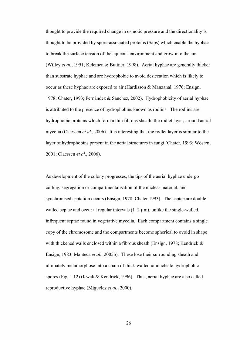
As development of the colony progresses, the tips of the aerial hyphae undergo
coiling, segregation or compartmentalisation of the nuclear material, and
synchronised septation occurs (Ensign, 1978; Chater 1993). The septae are double-
walled septae and occur at regular intervals (1�2 µm), unlike the single-walled,
infrequent septae found in vegetative mycelia. Each compartment contains a single
copy of the chromosome and the compartments become spherical to ovoid in shape
with thickened walls enclosed within a fibrous sheath (Ensign, 1978; Kendrick &
Ensign, 1983; Manteca et al., 2005b). These lose their surrounding sheath and
ultimately metamorphose into a chain of thick-walled uninucleate hydrophobic
spores (Fig. 1.12) (Kwak & Kendrick, 1996). Thus, aerial hyphae are also called
reproductive hyphae (Miguélez et al., 2000).

27
Streptomyces spores are classified as arthospores or exospores due to the asexual
mode of formation (Cross, 1970). Arthospores are different to bacterial endospores
in their sporogenesis process as well as their properties (Hardisson & Manzanal,
1976). Arthospores can remain viable in a dormant state for several years,
germinating when conditions are favourable (Ensign, 1978). Ultrastructural studies
on spores have revealed the presence of rodlins and chaplins. Their position on the
tip of the hyphae together with their resistance to desiccation, lytic enzymes and
osmotic extremes, aid efficient mass spore disposal and propagation of the species
(Kendrick & Ensign, 1983). Although the primary method of species propagation is
via spores, another method of dispersal is through dispersal of mycelial fragments.
By definition therefore, in liquid environments, substrate hyphae cannot differentiate
into �aerial hyphae� nor have equivalent erect hyphae emerging from mycelial
masses been previously reported. Instead substrate hyphae in liquid environments
are observed as aggregates and progress into a death phase and in some instances
progress into sporulation phase (Miguélez et al., 1993). For example, sporulation in
liquid media, under conditions of nutrient depletion, has been reported in S. griseus.
(Kendrick & Ensign, 1983). Here, spores are formed from sporogenic hyphae,
characterised by the presence of double-walled septae, similar to aerial or
reproductive hyphae septae. These submerged spores are not identical to spores
formed from aerial hyphae. Submerged spores are sensitive to lysozyme treatment
whereas spores formed from aerial hyphae are lysozyme resistant. Further,
submerged spores are formed from sporogenic hyphae that are not known to possess
a sheath, a feature characteristic of sporogenic aerial hyphae (Kendrick & Ensign,
1983; Miguélez et al., 1992). In Streptomyces spp., in addition to aerial spores and

28
submerged spores, ectopic spores produced directly from substrate hyphae when
cultured on solid media has been reported in a mutant S. griseus NP4. These ectopic
spores are said to be indistinguishable to that of spores formed for aerial hyphae.
Thus, there is a notion that all hyphae have the potential to sporulate (Ohnishi et al.,
2002).
The regulation of morphogenesis and sporogenesis at the biochemical level has been
extensively studied in S. coelicolor through analysis of aerial hyphae deficient
mutants, bld mutants (termed �bald�) and spore or spore pigment deficient whi
mutants (termed �white�) (Kwak & Kendrick, 1996; Kelemen & Buttner, 1998).
This lead to the identification of range of gene expression control elements which
include DNA binding proteins, sigma factors, unusual tRNAs, transcription factors
and some primary metabolism genes. This suggests that bld gene cascade responds
to nutrient availability, community stress and quorum size. It has been postulated the
bld cascade initiates a second pathway (termed �sky pathway�) which regulates
further aerial growth and differentiation (Claessen et al., 2006). This includes the
onset of chaplin synthesis. Mutants unable to sporulate had given rise to
identification of the whi regulatory loci (whiA, B, G, H, I, J and whiD, E and sigF).
whiG being one of the most important as it encodes a sigma factor of RNA
polymerase, that specifically initiates the development pathway leading to
sporulation from aerial hyphae and the whiE gene cluster is responsible for the grey
spore pigment (Chater, 1989; Kieser et al., 2000; Chater, 2001).

29
1.4.2 Cell death in Streptomyces
The process of cell death in Streptomyces antibioticus has been well examined
(Miguélez et al., 1999; Manteca et al., 2005a; Manteca et al., 2006). Cell death or
apoptosis in Streptomyces occurs in a highly organised manner involving the
progressive disorganisation of internal constituents. This is referred to as
programmed cell death (PCD) and is contrary to the previous notion that hyphal
death was predominantly due to random autolysis (Miguélez et al., 1999; Erikson,
1949). PCD starts with the disorganisation of the nucleoid and degradation of the
ribosomes making the cytoplasm less electron-dense. The cytoplasmic membrane
permeability is also altered, retracting from the wall and dissociating into small
vesicles (Manteca et al., 2006; Miguélez et al., 1999). The aberrant hyphal structure
composed of collapsed but intact cell wall structures eventually degrades. Two death
rounds have been observed, the first corresponds with the development of aerial
mycelia and the second coincides with sporulation. The second death round affects
only the basal part of the sporulating hyphae. It has also been established that the
dead hyphae provide mechanical stability for the aerial hyphae as well as aid in the
translocation of nutrients and solvents (Miguélez et al., 1999). A very early death
round has been recently reported in young S. antibioticus cultures (10�18 h) that
resulted in hyphae with alternate live and dead segments, described as variegated
(Manteca et al., 2005a). It is known that Streptomyces possesses apoptotic ATPases
similar to eukaryotic apoptotic signals. However, the exact function of these signals
is yet to be investigated (Koonin & Aravind, 2002; Manteca et al., 2005a).

30
1.4.3 Secondary Metabolism in Streptomyces
Apart from the complex and intriguing developmental cycle, Streptomyces spp. are
of particular interest to microbiologists due to their ability to produce secondary
metabolites, many of which are therapeutic compounds (Vining, 1990; Demain,
1999). Secondary metabolism is commonly observed in organisms that lack an
immune system, e.g. as bacteria, algae, corals, suggesting a defence function
(Maplestone et al., 1992). In Streptomyces spp., antibiotic production or secondary
metabolism on solid cultures is said to coincide with or slightly precede the
formation of aerial hyphae, while in liquid cultures, antibiotic production begins
during the stationary phase of growth (Kieser et al., 2000; Bibb, 2005).
In filamentous organisms, including Streptomyces spp., growth is differentiated into
the trophophase and the idiophase (Martin & Demain, 1980). During the
trophophase, the organisms are in the logarithmic growth phase with rapid synthesis
of proteins and nucleic acids and the production of secondary metabolites has not yet
commenced. The idiophase is when the growth rate has decreased (stationary phase)
and secondary metabolism has commenced. The idiophase is thus commonly
referred to as the �production phase� and the secondary metabolites as �idiolites�
(Walker, 1974). Secondary metabolites, such as antibiotics are not essential for the
growth of the organism and hence have a complex regulation system involving a
number of factors, a key factor being the presence of a critical concentration of
chemical signalling molecules (Horinouchi, 2007).

31
1.4.4 Communication and cell signalling
The coordination of antibiotic production and cellular differentiation during the life
cycle of Streptomyces spp. is thought to be controlled by signalling molecules
(quorum sensing molecules), the synthesis of which is a response to the community
size, nutritional status and environmental conditions (Horinouchi & Beppu, 1992).
With the identification of signalling molecules, the conventional notion of bacteria
functioning as individual single-celled unicellular organisms has been revolutionised
to the modern theory of �bacterial multicellularity�. This theory describes bacteria to
possess sophisticated communication systems and engage in cooperative community
behaviour similar to that observed in multicellular organisms (Swift et al., 1994;
Dunny & Leonard, 1997; Shapiro, 1998; Miller & Bassler, 2001; Winzer et al.,
2002). Signalling molecules are called quorum sensing molecules, a term coined by
Fuqua and colleagues, as they are released when the bacterial population reaches a
critical density or quorum size, and the external concentration of these molecules
proportionally increase with increase in bacterial population (Fuqua et al., 1994;
Waters & Bassler, 2005). They are also referred to as autoinducers, or
autoregulatory factors, or bacterial pheromones as they are similar to hormones or
pheromones of higher organisms. They are able to regulate gene expression and
initiate specific physiological processes such as sporulation, biofilm formation and
virulence factor secretion at extremely low concentrations in the order of 10�9 M
(Sato et al., 1989; Li et al., 1992; Fuqua et al., 2001; Lee et al., 2005; Basseler &
Losick, 2006; Zhu et al., 2002).
In Streptomyces spp., the growth status of the community is communicated via these
low molecular weight molecules which, together with the nutritional status of the

32
environment, trigger a cascade of events including growth augmentation, cellular
differentiation and antibiotic production (Horinouchi & Beppu, 1992; Kaiser &
Losick, 1993; Barabas et al., 1994). The growth pattern of closely associated
developing hyphae of Streptomyces spp. is conducive to an elaborate communication
system (Barabas et al., 1994).
Quorum sensing molecules interact with cellular receptors mediating a response.
They can be species specific or interspecies specific and can be broadly categorised
into three classes, the acyl homoserine lactones, used by gram-negative bacteria, the
oligopeptides produced by gram-positive bacteria, and AI-2, also known as the
universal signalling molecule as it is used by both gram-positive and gram-negative
bacteria (March & Bentley, 2004; Lowery et al., 2005).
The �-butyrolactones, also referred to as the Streptomyces hormones, since about
60 % of Streptomyces species have been estimated to employ �-butyrolactones for
the regulation of antibiotic synthesis and/or also for the control of differentiation
(Wang & Vining, 2003; Lee et al., 2005; Takano, 2006). The butyrolactones
accumulate in culture media and act as quorum molecules by releasing their
corresponding �-butyrolactones proteins from operator sites, thereby activating gene
expression (Flocher et al., 2001). A-factor (Fig. 1.13) [2-(6�-methylheptanoyl)-3R-
hydroxymethyl-4-butanolide] was one of the first quorum sensing molecules to be
discovered in the culture fluid of Streptomyces griseus. A-factor was essential for
the production of streptomycin and for the development of aerial mycelia (Khokhlov
et al., 1973). Studies by Yamada et al. reported the presence of a mixture of
autoregulatory factors that influenced production of virginiamycin (staphylomycin)

33
in Streptomyces virginiae, collectively called virginiae butanolides (VBs) (Fig. 1.13)
(Yamada et al., 1987). An autoinducer, the PI factor (2,3-diamino-2,3-bis
(hydroxymethyl)-1,4-butanediol) (Fig. 1.13) produced by Streptomyces natalensis
for the synthesis of the antibiotic pimaricin was shown to be structurally novel.
Interestingly, that synthesis of the antibiotic could be initiated by a �-butyrolactone
not produced by the host, indicating that cross communication and integration of
signals from other organisms may be important (Recio et al., 2004). Quorum
sensing molecules produced by S. coelicolor are butanolides (SCB1, SCB2, SCB3)
(Fig. 1.13) and stimualte actinorhodin production (Takano, 2006).
A-Factor
OH
O
OO
*
PI Factor
H2N
NH2HO
HO
OH
OH
SCB1
OH
O
OOH
*
**
VB-A
OH
O
OOH
*
**
H
SCB2
OH
O
OOH
*
**
VB-B
OH
O
OOH
*
**
H
SCB3
OH
O
OOH
*
**
VB-C
OH
O
OOH
*
**
H
Fig. 1.13. Chemical structures of quorum sensing molecules produced by different
Streptomyces spp. (* denotes stereocentres)
These signal transduction pathways appear to be complex, diverse and are yet to be
delineated (Chater & Horinouchi, 2003; Takano, 2006). Since quorum sensing
depends on the concentration of autoinducers, a bacterial population at low cell

34
density can be induced into an artificial sense of high cell density by addition of
synthetic autoregulatory factors, or can be introduced into conditioned media,
causing the activation of the respective genes (Lowery et al., 2005). Thus
manipulation of quorum size may enable further understanding into the signalling
cascade and hence the biosynthetic pathways.
1.5 Encapsulation
As a drug delivery system, encapsulated cells capable of producing bioactive
compounds offers a therapeutic option for long-term local and systemic drug
administration (Orive et al., 2003a). Encapsulation is a process that involves the
physical entrapment of biomaterials such as cells, enzymes or chemicals within a
semi-permeable capsule (Chang & Prakash, 1998). For organisms like Streptomyces,
this system would also permit investigation into the communication and regulation
effecting secondary metabolism and/or morphogenesis.
1.5.1 History of encapsulation as a delivery system
The concept of �artificial cells� or immobilised biomaterials for immunoprotection
was introduced by T.M.S. Chang (Chang, 1964). The semi-permeable matrix or
membrane acts as a dual barrier, allowing diffusion of small molecules such as
nutrients, gases, products and waste materials, but preventing the penetration of
larger substances (e.g. cells of the immune system) through the capsule. The large
surface area (e.g. 10 mL of capsules of diameter 20 µm have a total surface area of
~ 2 m2) of the capsules enables rapid diffusion of substrates and products (Chang &
Prakash, 1998). The advantage of encapsulated cells is the double protective
function that may allow allogenic or xenogenic cells to be transplanted into the host

35
while the immunoprotective and biocompatible nature of the capsule prevents an
immunological response (Li, 1998). Thus bioactive materials receive nutrients from
the host and are protected from the immune cells of the host, while the host is
protected from the entrapped contents and receives any beneficial products secreted
by the entrapped material. Further advantages is that implantation of these capsules
could be via a simple injection rather than a surgical operation and/or eliminating the
need of administrating chronic immunosuppressants and long-term hospitalisation
for systemic delivery.
The applications of this technology are potentially enormous and in the past few
decades increased research has been undertaken in the field of medicine, employing
encapsulated cells for use as drug delivery systems or in the making of artificial
organs. In the field of drug delivery, encapsulation is seen as a possible method of
introducing cells that would work as �living factories�, secreting therapeutic
molecules at the right time, in a reproducible manner, to a specific target and at the
required concentration (Orive et al., 2003a). An important feature for drug delivery
system is the porosity/permeability of the capsule, as this will affect kinetics of drug
release. A wide range of parameters can control the permeability of the
microcapsule, the primary one being the matrix for immobilising the contents.
Natural or synthetic polymers may be employed but the most frequently utilised are
hydrocolloid gels such as alginate and carrageenan (Gerbsch & Buchholz, 1995;
Pillai & Panchagnula, 2001; Orive et al., 2006).

36
1.5.2 Polymers employed for encapsulation
Though other polymers can be used, alginate, a natural polysaccharide derived from
brown algae, is commonly used for encapsulation (Ertesvåg & Valla, 1998).
Alginate is considered to be an ideal matrix for the immobilisation of living cells as
capsules are formed by a mild process that can be accomplished in a single step,
alginate is cheap and readily available and it is known to be biodegradable (Klöck et
al., 1997; Remuñán-Lopez & Bodmeier, 1997; Fundueanu et al., 1999). It is an
anionic polysaccharide composed of regions of �-L-guluronic acid (G) and ✁
-D-
mannuronic acid (M) interspersed with regions of mixed sequences (MG) (Draget et
al., 1997; Ertesvåg & Valla, 1998; Simpson et al., 2003). Capsules are produced by
preparing a suspension of the biomaterial in an aqueous solution of alginate and the
suspension is then extruded through a narrow orifice, the droplets harden upon
contact with a solution containing multivalent cations such as Ca2+. Capsules form
instantaneously as a result of ionic cross-linking, and thus the biomaterial is
entrapped within a 3D lattice. This enables the viability and integrity of the cells to
be maintained.
These properties of alginate have permitted encapsulation of islets of Langerhans,
hepatocytes and parathyroid cells for therapeutical applications (Chang & Prakash,
1998). Mice implanted with encapsulated pancreatic islets for insulin secretion, have
shown bioreactor viability for 12✂21 months and functionality resulting in decreased
blood glucose levels and improved the diabetic condition of the affected mice (Sun &
O�Shea, 1985). Such results in animal models have allowed allotransplantations of
encapsulated cells in humans to be successfully performed. Patients suffering from
hypoparathyroidism and diabetes have received encapsulated parathyroid cells and

37
pancreatic islets respectively (Hasse et al., 1997; Calafiore et al., 2006).
Nonimmunosuppressed patients with type 1 diabetes with an initial mean blood
glucose level of 275 ± 98 mg dl�1 have shown a reduced level of 167 mg dl�1 after 1
year of receiving encapsulated donor islets cells (Calafiore et al., 2006).
1.5.3 Encapsulation of Micro-organisms
Encapsulation of micro-organisms in polymers such as alginate is an emerging area
of applied microbiology. This technology has many advantages compared with free-
living organisms, especially when placed in harsh environments. Some of the
applications in applied microbiology include, in agriculture where encapsulated
bacteria are used as biofertilisers and biopesticides, as the immobilised bacteria are
protected from high temperature and acidity of some soils (Russo et al., 2001; Elçin,
1995; Kuek et al., 1992). In the food industry, capsules containing probiotic
organisms maintain viability as the immobilised organisms are protected from the
highly acidic conditions of the gut (Chandramouli et al., 2004; Krasaekoopt et al.,
2003). In fermentation, encapsulated micro-organisms have been shown to increase
product yield and have simplified down stream processing (Najafpour et al., 2004).
In wastewater treatment, the capsules containing bacteria have been easily retrieved
for reuse (Torres et al., 1998).
Antibiotic production from micro-organisms is usually by the extraction of culture
fluid produced by batch cultures of free-dwelling or planktonic organisms but some
studies have shown that they can also be produced by the continuous/semi-
continuous method using immobilised cells. There are a few reports on the
production of antibiotics from encapsulated Streptomyces spp. (e.g. chlortetracycline,

38
oxytetracycline, erythromycin, cephamycin) (Teruel et al., 1997; Yang & Yueh,
2001; Bandyopadhyay et al., 1993; Devi & Sridhar, 2000). Adinarayana et al. have
reported a 62 % increase in production of neomycin when using immobilised
Streptomyces marinensis (Adinarayana et al., 2004).
These examples indicate some of the varied applications of encapsulated micro-
organisms. However, there are very few reports on the use of encapsulated bacteria
for therapeutical applications. Chang and Prakash have reported the use of
encapsulated E. coli DH5 cells containing Klebsiella aerogenes urease genes for urea
degradation. Studies on ureamic rats showed significant reduction in urea levels
when treated with orally administered capsules containing non-pathogenic E. coli
DH5 cells. It was also shown that the encapsulated cells remained inside the
microcapsules, passed down the intestine and were excreted in the faeces causing no
adverse effects to the host (Prakash & Chang, 1996).
Aim and objective of project
The aim of this project was to demonstrate the production of the antifungal drug
AmB from alginate encapsulated Streptomyces nodosus in vitro. It was envisaged
that S. nodosus would be a suitable test organism because other encapsulated
Streptomyces spp. have produced antibiotics, it is non-pathogenic, pyrogen-free
(endotoxin-free) and it does not grow at 37 °C. It was not known whether S. nodosus
could be immobilised in a biocompatible polymer such as alginate and produce AmB
from within the capsules. Although many technical and biological issues would have
to be overcome before being a viable drug delivery mechanism, this project would
give insight into whether prokaryotes are able to survive and produce diffusible

39
compounds from alginate capsules; a result of interest to other microbiological
applications where organisms at solid-liquid interfaces. If successful such
bioreactors could potentially provide slow, localised delivery of low doses of the
drug. This novel delivery may mitigate some of the side effects of the drug prevalent
with systemic administration.
This project required developing methods for encapsulating AmB and S. nodosus and
studying the kinetics of AmB release from capsules. To analyse antibiotic release
from capsules, a method to detect low concentrations of AmB in biological matrices
was also developed. The key to the success of bioreactors using Streptomyces spp. is
an understanding of the regulation of secondary product formation. As described,
the organism has a complex growth cycle linked to antibiotic production and for this
reason monitoring morphology and physiology of encapsulated organisms during
in vitro assessment was essential. This necessitated the development of an assay to
monitor both the viability and morphology of encapsulated organisms during
culturing. Using electron microscopy and confocal microscopy, the community
structure, progression of cellular development and antibiotic production were
analysed.

Chapter 2
Materials and Methods

41
2.1 Reagents and Media Components
All reagents were analytical grade. Ethanol, acetonitrile (ACN), trifluoroacetic acid
(TFA), sodium hydroxide, glucose and glycerol were purchased from Univar
(Seattle, USA). Media components such as agar, malt extract, tryptone, �Lablemco�
beef extract and N-Z-amine A (enzymatic digest of casein) were purchased from
Oxoid (Hampshire, England) and yeast extract was from Difco laboratories (Kansas
City, USA). Tomato paste and oatmeal were of food grade and were purchased from
Woolworths (Sydney, Australia). AmB standard, sodium alginate (medium
viscosity), calcium chloride and piperazine-N, N�-bis(2-ethanesulfonic acid) (PIPES)
were purchased from Sigma (St Louis, MO, USA). Methanol (MeOH) used in the
chromatography experiments was HPLC grade and purchased from LabScan
Analytical Sciences (Lomb Scientific, Sydney, Australia). Pioloform resin was from
Structure Probe, Inc. (West Chester, USA) and paraformaldehyde (extra pure) was
purchased from Merck (Darmstadt, Germany). Glutaraldehyde (50 % solution),
osmium tetroxide and LR white resin (medium grade) were purchased from
ProSciTech (Queensland, Australia).
2.2 Micro-organisms
The bacteria used in this study were E. coli XL1-Blue (Stratagene, La Jolla, USA),
Streptomyces nodosus ATCC 14899 (Rockville, MA, USA), Streptomyces nodosus
MA✁
hyg1 (Nikodinovic, 2004), Streptomyces natalensis (NRRL B-5314),
Streptomyces coelicolor (NRRL B-2812), Streptomyces virginiae (NRRL ISP-5094),
Streptomyces griseus (NRRL B-2165) and Rhodotorula sp. X6 (UWS Hawkesbury
culture collection). The NRRL strains were kindly provided by Dr David P. Labeda,
Curator of the Actinobacterial Culture Collection USDA, Agricultural Research

42
Service, National Centre for Agricultural Utilisation Research, Microbial Genomics
and Bioprocessing Research, ARS Culture Collection, Peoria, Illinois, USA.
2.3 Maintenance of Bacterial Cultures
Bennett's broth (yeast extract, 1 g L�1; �Lablemco� beef extract, 1 g L�1; N-Z-amine
A (enzymatic digest of casein), 2 g L�1; glucose, 10 g L�1) was used to resuscitate the
lyophilised Streptomyces cultures. The ampoules containing stock cultures were
wiped with alcohol, scored with a file in the middle and snapped open. The broken
end was flamed and the plug of lyophilised bacteria was introduced into sterile
Bennett's broth (2.5 mL) and incubated at 20 °C until uniformly resuspended.
Aliquots (200 µL) were plated on yeast extract-malt extract-glucose (YMG) (yeast
extract, 4 g L�1; malt extract, 10 g L�1; glucose, 4 g L�1; agar, 18 g L�1; pH 7.2). The
plates were incubated at 28 °C until a homogenous layer of spores developed (~ 7 d).
Streptomyces spore suspensions were prepared essentially by the method of Kieser
(Kieser et al, 2000). Autoclaved Milli-Q water (9 mL) was added to a freshly grown
well-sporulated plate culture and a sterile cotton swab used to gently suspend the
spores. The crude suspension was transferred into a McCartney bottle, vortexed
(1 min) and filtered through a syringe barrel packed with glass wool by gravity flow
into a sterile centrifuge tube (15 mL). The filtrate was centrifuged at 1,000 × g for
10 min at 20 °C and the supernatant was immediately discarded. The pellet of spores
was vortexed (10 s) in Milli-Q water (50 �L) to disperse the spores, resuspended in
20 % (v/v) sterile glycerol (1 mL) and stored at �20 °C. For long-term storage,
aliquots of the spore suspensions (0.5 mL) were stored at �80 °C.

43
Enumeration of spore suspensions was carried out by counting the spores using a
Neubauer� haemocytometer under a light microscope or by direct culturing of
serially diluted stock preparations on YMG agar. The plates were incubated for 7 d
at 28 °C and the number of colony forming units (CFU) was counted. Spore
suspensions typically had a titre of ~ 3 × 10⁹ spores mL�1 and no loss of viability for
up to 1 year.
Cultures of S. nodosus, S. virginiae and S. griseus were routinely maintained on
YMG agar while S. coelicolor and S. natalensis were maintained on tomato oatmeal
sporulation medium (tomato paste, 20 g L�1; oatmeal, 25 g L�1; agar, 25 g L�1).
Streptomyces spore suspensions (100 µL) were spread through the entire surface of
the agar with a sterile cotton swab to achieve confluent cultures. This plating
technique was necessary as Streptomyces colonies spread only a limited distance
from the point of inoculation (Kieser et al., 2000). Plates were incubated at 28 °C
until well-sporulated and stored at 4 °C.
Stock cultures of E. coli XL1-Blue (Stratagene) for establishment of viability assays
were resuscitated by culturing on Luria-Bertani (LB) agar (tryptone, 10 g L�1; yeast
extract, 5 g L�1; NaCl, 5 g L�1; glucose, 1g L�1; agar, 15 g L�1) and the plates were
incubated at 37 °C for 14 h. A single isolated colony was inoculated into LB broth
(50 mL) in a 250 mL Erlenmeyer flask and incubated at 37 °C with constant shaking
at 160 rpm (Gallenkamp, Loughborough, United Kingdom) until the required growth
phase was achieved (calculated by measuring OD600). Cultures were maintained on
LB agar plates, subcultured every three to four weeks and stored at 4 °C. For long-

44
term storage of E. coli XL1-Blue, sterile glycerol was added to an overnight culture
in the ratio 1:4 (v/v) and stored at �80 °C.
2.4 Bacterial Growth Conditions
For routine fermentation experiments in liquid environments, Streptomyces were
cultured in YMG liquid medium (250 mL). Pilot growth experiments were
conducted using Glycerol Asparagine (GA) medium (L-asparagine, 1 g L�1; glycerol,
10 g L�1 ; K2HPO4, 1 g L�1; pH 7.2) or R5 [sucrose, 103 g L�1; K2SO4, 0.25 g L�1;
MgCl2.6H2O, 10.12 g L�1; glucose, 10 g L�1; casaminoacids, 0.1 g L�1; trace element
solution (ZnCl2, 40 mg L�1; FeCl3.6H2O, 200 mg L�1; CuCl2.2H2O, 10 mg L�1;
MnCl2.4H2O, 10 mg L�1; Na2B4O7.10H2O, 10 mg L�1; (NH4)6Mo7O24.4H2O,
10 mg L�1), 2 mL L�1; yeast extract, 5 g L�1 ; TES buffer, 5.73 g L�1]. Just prior to
use, KH2PO4 (0.5 %), 1 mL, CaCl2.2H2O (5M), 0.4 mL, L-proline (20 %), 1.5 mL
and NaOH (1N), 0.7 mL were added per 100 mL of R5 medium (Kieser et al., 2000).
Spores (~ 3 × 10⁶ spores) (Section 2.3) were used as inoculum and cultured in glass
baffled flasks (1 L) to provide adequate aeration and promote dispersed growth.
Flasks were incubated at 28 °C with constant shaking at 160 rpm (TLM-530
Thermoline, Australia) until the required growth phase was attained.
Growth of Streptomyces cultures was determined by measuring the dry weight of
biomass. Aliquots (5 mL) of Streptomyces culture fluid were drawn at 24 h intervals
for a period of 7 d. The culture fluid was filtered under vacuum onto a pre-dried and
pre-weighed mixed cellulose ester Millipore filter membrane (0.22 µm pore size)
(Billerica, MA, USA). The filter paper containing the biomass was placed in a pre-
dried and pre-weighed aluminium pan, placed in a glass Petri dish and dried at 70 °C

45
until constant weight was achieved. The membrane was allowed to cool in a
desiccator (S.E.M., South Australia, Australia) before weighing using a four decimal
place balance (Sartorius, Germany). The analysis was performed in triplicate and
average values were calculated for each sample.
Conditioned media was prepared by growing flasks of S. nodosus wild-type,
S. nodosus MA�
hyg1, S. natalensis, S. coelicolor, S. virginiae and S. griseus in
baffled flasks of YMG liquid medium (50 mL) at 28 °C at 160 rpm until the required
growth phase. The culture fluid was centrifuged at 3,000 × g for 30 min at 20 °C and
the supernatant was pre-filtered under vacuum using a mixed cellulose ester
Millipore filter (0.8 µm followed by 0.45 µm pore size) with final filtration into an
Erlenmeyer flask (250 mL) using a Millex filter unit with Durapore membrane (33
mm, 0.22 µm). The resultant filtrate was used as conditioned media of the respective
organism.
2.5 Immobilisation of micro-organisms
Calcium alginate capsules were prepared by extrusion using a simple one-step
process. Sodium alginate solutions (2 % w/v) were prepared in distilled water and
were homogenised using a magnetic stirrer for 3 h at 20 °C, sterilised by autoclaving
and cooled to 4 °C. S. nodosus was cultured in liquid YMG (50 mL) (Section 2.4).
The mycelia were harvested by centrifugation (1,800 × g, 10 min, 20 °C) and the cell
pellet resuspended in 0.9 % (w/v) NaCl (5 mL) by careful pipetting. Saline
suspensions of spores or mycelia were added to the 2 % (w/v) alginate solution
(45 mL) and inverted several times until the suspension appeared visually uniform.
For the preparation of the cationic solution for cross-linking the alginate to produce

46
capsules, calcium chloride dihydrate (100 mM) was prepared in distilled water,
autoclaved and cooled to 4 °C. Capsules were prepared by either of 2 methods:
manually using a syringe or by semi-automated extrusion using a peristaltic pump
(Alitea, Stockholm, Sweden). Syringe needles of 29-gauge and 19-gauge were used
for manual preparation of capsules, and 316 stainless steel tubing with an outside
diameter of 1.59 mm and an internal diameter of 1.02 mm was used with the
peristaltic pump. The alginate solution was either manually extruded through a
syringe or dropped through a steel tubing of the pump, into an ice-cold solution of
CaCl2 (100 mM) from a constant height (6 cm or 12 cm). The peristaltic pump
produced droplets at a constant rate of 100 min�1. Capsules formed at the instant the
alginate solution droplet fell into the calcium chloride solution due to the rapid cross-
linking of the alginate molecules with the calcium cations. The capsules were left
undisturbed in the calcium chloride solution for 30 min at 20 °C, rinsed twice with
saline and individual capsules were examined for viability and morphology. All
procedures were carried out under aseptic conditions.
A similar procedure was used for the immobilisation of E. coli cultures. An E. coli
culture (1 mL) derived from an overnight culture was used as inoculum into LB and
incubated at 37 °C with constant shaking at 160 rpm (Gallenkamp, Loughborough,
United Kingdom). Once reaching an OD600 of 0.4 (~ 3 h), the culture (2 × 10 mL)
was harvested by centrifugation (1,800 × g, 10 min, 20 °C). E. coli (Section 4.2.1)
was suspended in 0.9 % (w/v) NaCl (1 mL), added to a defined concentration of
alginate (9 mL) and capsules were prepared as described above.

47
2.6 Fermentation experiments using immobilised S. nodosus
Capsules (10 g) containing micro-organisms were transferred to YMG or conditioned
media (50 mL) in an Erlenmeyer flask (250 mL) and incubated at 28 °C for up to
30 d. The flasks were shaken constantly but at the slower rate of 60 rpm (TLM-530
Thermoline, Australia) to minimise damage to the capsules caused by collision. At
defined intervals, the media were analysed for AmB using high performance liquid
chromatography (HPLC) and/or UV-Vis spectrophotometry (Section 2.7) and the
capsules were assessed for viability and morphology (Section 2.8).
2.7 Quantification of AmB production
Preparation of standards
Stock solutions of AmB were prepared at a concentration of 1000 µg mL�1 by
dissolving commercially available AmB derived from S. nodosus (Sigma) in HPLC
grade methanol. Working solutions of standards (50 µg mL�1 to 0.1 µg mL�1) were
prepared by diluting appropriate volumes of the stock solution with methanol (HPLC
grade). Standards were prepared fresh on the day of experimentation. Calibration
curves were used for quantification of the AmB in the culture fluids.
UV-Vis
Initial detection of AmB in culture broth was performed by UV-Vis
spectrophotometry. DMSO (1 mL) was added to an equal volume of a culture of
S. nodosus, incubated at room temperature (5 min) and the mixture was centrifuged
(1,800 × g, 10 min, 20 °C). The resultant supernatant was diluted 1:9 (v/v) with
methanol. The absorption spectrum was recorded from 250�500 nm using a Lambda
EZ210 spectrophotometer. The instrument parameters were a scan speed of

48
800 nm min�1, sampled at 0.5 nm intervals, a slit width of 2 nm and a path length of
10 mm.
HPLC System and Conditions
Liquid chromatography experiments were performed on a HP1100 system (Agilent
Technologies, Palo Alto, CA) equipped with a G1310A isocratic pumping system, a
G1313A autosampler, a G1316A column thermostat and a G1315A diode-array
detector (DAD). The column was a 5 �m C-18 reverse-phase LiChrospher® 100
RP-18 (250 mm by 4 mm, LiChroCART; Merck, Darmstadt, Germany). The mobile
phase was either solvent system 1 - ACN (solvent A) and aqueous 0.1 % TFA
(solvent B) (60:40, v/v) with a flow rate of 1 mL min�1 at 25 °C and a run time of 7
min, or solvent system 2 - MeOH (solvent A) and 0.1 % TFA (solvent B) (85:15,
v/v) with a flow rate of 1 mL min�1 at 25 °C and a run time of 13 min. The DAD
recorded signals at 408, 386 and 366 nm which are characteristic for AmB. Full UV-
Vis spectra were also recorded for each peak detected during a run, and these spectra
were used to confirm that the analyte was AmB or to assign the compound class for
some other organic molecules detected during this analysis. Detection was also
monitored at 550 nm and 220 nm to detect other compounds (e.g. porphyrins). The
chromatographic system was controlled by the Chemstation software.
For routine testing of AmB from fermentation experiments, culture fluids were
clarified by centrifugation (1,800 × g, 10 min, 20 °C) and were filtered (0.22 µm
Millex filter) before analysis. Samples were directly injected (20 µL) into the HP
1100 system. The detection limit of this assay was found to be 100 ng mL�1.

49
Solid-phase extraction
To increase the sensitivity of the assay, AmB was pre-concentrated from culture
fluids using solid-phase extraction (SPE). Oasis HLB cartridge (sorbent weight of 60
mg, particle size 30 µm, pore size 80 Å and syringe barrel size 3cc, Waters, Milford
Massachusetts USA) were used with a Supelco solid-phase 12 port extraction
vacuum system (Visiprep). The cartridge was conditioned with methanol (3 mL),
equilibrated with water (2 mL) and the filter-sterilised sample (10�50 mL) was
introduced into the cartridge under vacuum (1.5 psig). AmB was eluted with
methanol (1 mL). The eluent was evaporated to dryness, resuspended in methanol
(1 mL), spiked with the internal standard solution (10 µg mL�1, biphenyl) and
injected (20 µL) into the HPLC system. Using this assay, an increased sensitivity
was achieved with a detection limit of 6 ng mL�1.
2.8 Viability Assay
A staining solution containing SYTO 9 (0.5 µL mL�1) and propidium iodide (PI)
(0.5 µL mL�1) (BacLight bacterial viability kit L-13152, Molecular Probes, Eugene,
USA) was prepared. The green fluorescent dye, SYTO 9, stains DNA in all cells
while the red fluorescent dye, PI, penetrates those cells with damaged membranes
and, hence, stains only dead cells. In dead or non-viable cells, where both stains
penetrate, quenching of the SYTO 9 fluorescence occurs, thus only PI is detected.
The staining solution (1 mL) was added to ~ 15 capsules and incubated in the dark
for 20 min at 20 °C. After removal of the staining solution, capsules were suspended
in water (1 mL) and gently agitated for 10 s. Single capsules were immediately
removed, placed on a glass slide and a cover-slip used to press the material into a
thin smear.

50
2.9 Confocal Laser Scanning Microscopy
For viability and morphology assessment by confocal laser scanning microscopy
(CLSM), stained samples (Section 2.8) were examined using a Fluoview 300 laser
scanning confocal system fitted with an Olympus IX70 inverted microscope
(Hamburg, Germany) under a ×100 oil immersion objective (NA=1.35). SYTO 9
fluorescence was detected with an Argon laser (488 nm laser excitation) with a
515 nm interference emission filter, PI fluorescence was detected with a HeNe green
laser (543 nm laser excitation) and a long pass 565 nm emission filters. Sequential
dual channel scanning was used to display green and red fluorescence. For triple
channel imaging, a transmitted light photomultiplier tube was used in conjunction
with Normaski optics. Three-dimensional confocal images were created by
combining stacks of individual in-focus fluorescence microscopy images (Z-stack).
Quantitative viability assessments were determined using analysis performed on a
Windows� computer using the public domain ImageJ program (developed at the
U.S. National Institutes of Health and available on the internet at
http://rsb.info.nih.gov/ij/).
2.10 Scanning and Transmission Electron Microscopy
Capsules selected for scanning electron microscopy (SEM) and transmission electron
microscopy (TEM) were fixed in 4 % (w/v) paraformaldehyde and 3 % (w/v)
glutaraldehyde in 0.1 M PIPES buffer, pH 7.2 at room temperature for 4 h. Capsules
were washed three times in PIPES buffer and post-fixed in 1 % (w/v) osmium
tetroxide in the same buffer at room temperature for 1 h. The samples were rinsed
three times in distilled water and dehydrated through a graded series of ethanol 50 %,

51
70 %, 80 %, 90 %, 95 % and 2 × 100 % (v/v) for 15 min per solution at room
temperature.
After dehydration, the samples for SEM were dried to the critical point (Emitech
K850 CPD Kent, England) with liquid carbon dioxide, mounted on a metal stub with
a carbon tab and sputter coated (Emitech K550) with ~ 20 nm of high purity gold.
Observations were made on a JEOL scanning electron microscope (JSM 6480LA) at
an acceleration voltage of 5 or 15 kV.
The samples for TEM, after dehydration in ethanol, were infiltrated with a 50 %
(v/v) solution of resin in ethanol (LR White resin, medium grade, Proscitech, C023)
for 1 h. This was followed by infiltration with 100 % resin for a further 1 h.
Capsules were then added to a fresh aliquot of 100 % resin and incubated overnight
at 4 °C before being embedded in resin in gelatine capsules and polymerised at 60 °C
for 20 h. Ultrathin sections (60 nm) were prepared using an ultramicrotome
(Reichert Ultracut S) with a glass knife or a diamond knife (Ge-Fe-Ri, Italy),
mounted on pioloform (0.2 % w/v) coated copper grids (300 mesh). The sections
were treated in a humid chamber with saturated aqueous uranyl acetate (35 min),
rinsed with water (30 s), stained with Reynolds� lead citrate (5 min) (Reynolds,
1963) and rinsed in water (5 × 30 s each). The grids were dried and examined using
a Philips CM-10 TEM (Eindhoven, Netherlands) at an acceleration voltage of 100
kV. Images were captured using a Kodak electron microscope 4489 film (Eastman
Kodak, USA). The films were processed and negatives were developed using D19
Kodak developer and Hypam fixer (Eastman Kodak, USA). The negatives were
scanned on a Microteck ScanMaker 5 scanner (PROSCAN, Australia) equipped with

52
Scan Wizard software (version 3.24, Microteck International). Image J (National
Institute of Health, USA) or Adobe Photoshop (versions 5.0 & 9.0, Adobe Systems
Inc., USA) was used to analyse images and add scale bars.

Chapter 3
Optimisation of capsule production and
the kinetics of amphotericin B release in vitro

54
3.1 Introduction
Prior to embarking on the study of immobilisation of micro-organisms and their
antibiotic production, it was essential to determine whether AmB can diffuse from
alginate capsules. Of the factors that affect drug release and bacterial physiology in
immobilised systems, matrix composition and permeability are most important. In
this study, alginate was selected as the polymer for capsule production because it is
widely accepted as a suitable polymer for cell immobilisation (Orive et al., 2006).
The concentration of alginate will also affect the diffusion of the encapsulated
material (Blandino et al., 1999). In addition, capsules may also be coated with layers
of other polymers such as poly-L-lysine (PLL) or chitosan to alter diffusion kinetics
(Ertesvåg & Valla, 1998). The size and shape of the capsules is also important as the
diffusion rate is also dependent on the surface area to volume ratio of the beads.
Other parameters such as mechanical properties (e.g. resistance to rupture, elasticity,
pore size distribution, surface properties and morphology) determine ease of
handling, biocompatibility in vivo and the uniformity and consistency of the outputs
from the immobilised system (Torre et al., 2002; Orive et al., 2003b). The rate of
diffusion from capsules is also dependent on the concentration and solubility of the
drug (Torre et al., 2002).
The primary aim of the work constituting this chapter was to establish that AmB
could be encapsulated in alginate and released from the capsules into the surrounding
environment. The concentration of alginate, size and shape of capsules were
optimised to produce uniformly smooth and spherical capsules. In addition, the low
water solubility of AmB and the complexity of the biological matrix (unconditioned

55
and conditioned media) from where it had to be measured made quantification of the
drug difficult and this necessitated the development of a new analytical assay for the
drug. In this work, a new highly sensitive HPLC method with a pre-concentration
step using SPE was developed that was tailored to detect nanogram quantities of
AmB in bacterial culture fluids. Using this assay, the kinetics of AmB release from
alginate capsules was determined.
3.2 Materials and methods
3.2.1 Capsule production
Alginate solutions at concentrations of 1 %, 2 %, 3 % and 4 % (w/v) were prepared
in distilled water. Each solution was homogenised using a magnetic stirrer for 3 h at
20 °C, sterilised by autoclaving and cooled to 4 °C. Calcium chloride dihydrate
(100 mM) was prepared in distilled water, autoclaved and cooled to 4 °C. Capsules
were prepared by either of two methods - manual extrusion and semi-automated
extrusion (Section 2.5). Samples of the capsules (n=5) were examined by light
microscopy to measure size and morphology.
3.2.2 Examination of capsule morphology and hardness
Capsules prepared from different alginate concentrations or dropped from different
heights were transferred from saline onto a microscope slide and the surface moisture
gently wicked away with an absorbent tissue. Capsules were examined under a
Leica macroscope (Wild M420, Wetzlar, Germany) fitted with a CCD video camera.
Micrographs were taken immediately to minimise dehydration, and the images used
to assess surface morphology. The external diameter was measured using the image
processing software Image J after calibration using an image of an Olympus

56
micrometer (Tokyo, Japan). Five capsules were measured for each experiment and
the average diameter calculated. After the specified hardening times of 5, 15, 30 and
45 min in calcium chloride solution, individual capsules were manually crushed
between a slide and a cover slip to assess relative capsule hardness.
3.2.3 AmB encapsulation
AmB (4 mg) was dissolved in HPLC grade methanol (5 mL) and agitated in an
ultrasonic bath for 1 min. This was added to a sterilised and cooled alginate solution
(2 % w/v, 200 mL) and ultrasonicated (10 min, 40 kHz) to obtain a uniform solution
with final concentration ~ 20 µg mL�1
. Using a peristaltic pump, capsules were
prepared by dropping the solution into calcium chloride solution (100 mM) from a
height of 6 cm, producing capsules at a constant rate of 100 capsules min�1
.
Capsules were left to harden in calcium chloride solution for 30 min at 20 °C.
3.2.4 Diffusion studies of encapsulated AmB
Release of AmB from alginate capsules (Section 3.2.3) was studied using a bioassay
and also by chromatography. For the bioassay, the yeast Rhodotorula sp. X6 was
used as the test organism. A lawn culture derived from an overnight liquid culture
(100 µL) of Rhodotorula sp. was plated on malt extract agar (Oxoid, Hampshire,
England). AmB capsules (0.5 g or 1 g wet weight of capsules) were rinsed in saline,
transferred to the Petri plate and incubated at 28 °C for 20 h. All plates were
examined for a zone of inhibition.
For HPLC analysis, capsules (10 g, Section 3.2.3) were rinsed and transferred to
Schott bottles (250 mL) containing Milli-Q water, YMG or conditioned medium

57
from S. nodosus MA�
hyg1 (100 mL) (Section 2.4). The bottles were incubated at
28 °C with constant shaking at 60 rpm. At defined time points, the solution (2 × 50
mL) from a bottle was removed, pre-concentrated by SPE and analysed for AmB by
HPLC (Section 2.7).
3.3 Results
3.3.1 Optimisation of capsule production
Three parameters: concentration of alginate, the distance of needle from the calcium
chloride solution and the hardening time were optimised to produce capsules of
acceptable size, shape and hardness.
Fig. 3.1. Experimental set-up for the preparation of alginate capsules by (a) the
manual method and (b) the semi-automated method.
In the first series of experiments, the alginate concentration was varied from 1�4 %
(w/v) and dropped from a height of 6 cm into CaCl2 solution by manual extrusion
using a 29-gauge needle (Fig. 3.1a). Microscopic examination of the capsules using

58
a stereomicroscope revealed smooth and spherical capsules of diameter ranging
between 1.1�1.3 ± 0.05 mm (n=5). This result was independent of the concentration
of alginate used (Table 3.1).
Although acceptable capsules were formed for alginate concentrations of 1 %, 2 %,
3 % and 4 % (w/v) when prepared from a height of 6 cm, the transfer of solution into
the syringe and extrusion became increasingly more difficult at alginate viscosities of
3 % and 4 % (w/v). By contrast, capsules formed from the 1 % (w/v) alginate
solution were easily damaged. Thus a 2 % (w/v) alginate solution was used for the
encapsulation of AmB.
Although manual extrusion successfully produced capsules with uniform size and
shape within a single batch, there were significant variations in size (~ 300 µm) from
batch to batch or experiment to experiment (Table 3.1). Hence, the peristaltic pump
was trialled to reduce the inconsistencies in flow rate and height variation inherent in
manual extrusion (Fig. 3.1b). This method also had the advantage of large scale
production of capsules since multiple refilling of syringe barrels would not be
required.

59
Table 3.1. Results of three independent experiments measuring diameter of capsules prepared with 1 %, 2 %, 3 % a
concentrations using manual extrusion (29-gauge needle) and semi-automated extrusion (peristaltic pump) falling from
calcium chloride solution. Values represent average ± standard deviation, n=5.
1 % (w/v) alginate 2 % (w/v) alginate 3 % (w/v) alginate 4
Capsule Diameter (mm) Capsule Diameter (mm) Capsule Diameter (mm)
29-gauge
needle
Peristaltic
pump
29-gauge
needle
Peristaltic
pump
29-gauge
needle
Peristaltic
pump
Expt 1 1.12 ± 0.06 2.85 ± 0.03 1.14 ± 0.05 2.86 ± 0.03 1.18 ± 0.05 2.89 ± 0.02
Expt 2 1.14 ± 0.05 2.87 ± 0.03 1.26 ± 0.05 2.86 ± 0.02 1.07 ± 0.05 2.87 ± 0.03
Expt 3 1.19 ± 0.05 2.86 ± 0.02 1.04 ± 0.05 2.87 ± 0.01 1.29 ± 0.05 2.88 ± 0.02

60
In the next set of experiments, a peristaltic pump was used and the distance of the
needle from the calcium chloride solution was varied (6 cm and 12 cm) with the
concentration of alginate kept constant (2 % w/v). Figure 3.2a shows a
representative capsule formed using a height of 6 cm. The capsules had smooth
surfaces and were spherical with a uniform diameter (2.86 ± 0.02 mm, n=5).
However, the capsules formed from a height of 12 cm had rough surfaces and were
slightly oval in shape (Fig. 3.2b). Hence, a height of 6 cm was used in all further
experiments.
Fig. 3.2. Representative stereomicroscope image revealing surface morphology of
capsules made from droplets of a 2 % (w/v) alginate solution falling into calcium
chloride solution (100 mM) from a height of (a) 6 cm and (b) 12 cm.
The third parameter examined was the hardening time in calcium chloride solution.
Capsules were produced using a 2 % (w/v) alginate solution falling from a height of
6 cm into calcium chloride solution and assessed for hardness after 5, 15, 30 and 45
min. Capsules cured in calcium chloride solution for 5 and 15 min showed less
resistance to damage by manual handling and crushed significantly more easily than
the capsules that were incubated for 30 or 45 min. This indicated that a minimum of

61
30 min was required for optimum cross-linking of the gel and that this should
provide the necessary durability for fermentation experiments that required agitation.
Thus, the optimum parameters for the production of highly mono-disperse spherical
alginate capsules were an alginate concentration of 2 % (w/v), with droplets falling
into calcium chloride solution from a height of 6 cm and a hardening time of 30 min.
These conditions were used in all subsequent experiments.
3.3.2 Development of an assay to detect AmB in nanogram quantities in aqueous
samples
In order to demonstrate diffusion of AmB from capsules, a sensitive assay for AmB
needed to be developed. In our laboratory, the analysis of a saturated solution of
AmB in Milli-Q water using HPLC showed that AmB had a solubility of
1.23 µg mL�1
which is low relative to levels achieved in culture. This necessitated
the development of a highly sensitive analytical method to measure AmB at ng mL�1
levels in solution if diffusion of AmB from capsules into water was to be detected.
This was achieved by a pre-concentration step using SPE. Prior to developing the
methodology for SPE, a suitable solvent system for the separation of AmB from
other organic compounds in conditioned culture medium was first established.
To develop a HPLC method, the first step was the selection of a suitable mobile
phase system. A mobile phase of ACN:0.1 % (v/v) TFA (60:40 v/v) resulted in
AmB eluting at retention time of 4.1 min when analysed from a sample of either a
saturated solution of AmB in water (Fig. 3.3a) or a saturated solution of AmB in
YMG (Fig. 3.3b). The eluting compound was confirmed to be AmB by analysis of

62
the spectrum of the compound. However, AmB could not be detected in the
saturated solution of AmB in conditioned medium derived from S. nodosus
MA�
hyg1 (Fig. 3.3c). A compound eluting at the identical retention time (4.1 min)
but with a different UV-Vis absorption spectrum was detected (Fig. 3.4a). This
compound had absorption maxima at wavelengths at which AmB elution was
monitored (408, 386 and 366 nm). The absorption spectrum of this elution peak with
a retention time of 4.1 min was also similar to that of the elution peak at 3.5 min
(Fig. 3.4b).
As AmB and this new compound co-eluted, a new mobile phase using methanol
(MeOH)/ water was developed to separate the compounds. With other
chromatographic conditions kept constant, a mobile phase of 85:15 (v/v)
MeOH/0.1 % (v/v) TFA gave the best selectivity for AmB but at the cost of a longer
analysis time (retention time of 9.3 min, Fig. 3.5a) than the ACN mobile phase. The
peak that previously co-eluted with AmB in the conditioned medium had a retention
time of 5.0 min using the MeOH mobile phase while AmB had a retention time of
9.2 min (Fig. 3.5b). This peak in addition to being monitored at 408, 386 and 366
nm, could be monitored at 550 nm, a wavelength at which AmB does not have an
absorbance. Consequently, this solvent system of 85:15 (v/v) MeOH/0.1 % (v/v)
TFA was used for the kinetic release experiments which involved analysis of AmB in
water, YMG and conditioned media.

63
Fig. 3.3. Representative HPLC elution profiles of AmB in (a) water, (b) YMG and
(c) conditioned medium from S. nodosus MA�
hyg1 with a mobile phase of
ACN:0.1 % (v/v) TFA (60:40 v/v) monitored at 408, 386 and 366 nm.

64
Fig. 3.4. Absorption spectra of compounds detected at 408 nm with retention times
of (a) 4.1 and (b) 3.5 min from samples of AmB dissolved in conditioned medium
from S. nodosus MA�hyg1 (HPLC elution profile Fig. 3.3c).

65
Fig. 3.5. Representative HPLC elution profiles of an AmB standard in (a) methanol
and (b) conditioned medium from S. nodosus MA�
hyg1 with a mobile phase of
MeOH/0.1 % (v/v) TFA (85:15 v/v) monitored at 408, 386, 366, 550 and 220 nm.
The peak at 9.3 min is AmB.

66
Having developed the solvent system, the next step was to optimise a SPE procedure.
Two SPE cartridges were trialled, a Sep-pak C-18 cartridge (Waters, Massachusetts,
USA) and an Oasis HLB extraction cartridge (Waters, Massachusetts, USA).
Solutions of known concentrations of AmB in Milli-Q water (50 mL) were prepared
in volumetric flasks. The SPE cartridges were conditioned with methanol (3 mL),
equilibrated with water (2 mL) and the sample (50 mL) was loaded onto the cartridge
under vacuum (1.5 psig) using the Supelco SPE manifold vacuum system (Section
2.7). AmB was eluted with MeOH (1 mL) and injected (20 µL) into the HPLC
system (Section 2.7).
After extraction and chromatography, peak areas were determined and a standard
curve was used to calculate the concentrations of each sample. The recovery of
AmB for each concentration (performed in duplicate) was calculated using the
formula given below.
100)/(
)/( �mLgaddedAmBofAmount
mLgmeasuredAmBofAmount ✁✁
Using a standard curve (Fig. 3.6), the extraction efficiency (recovery) was calculated,
which resulted in a recovery of 89.1 % of 500 ng mL�1
AmB by the C18 cartridge
and 90.7 % of 450 ng mL�1
AmB by the HLB cartridge. Analysis of solutions with a
concentration of 20 ng mL�1
of AmB were expected to be close to limits of
quantitation and gave recoveries of 125 % and 140 % by the C18 and HLB cartridges
respectively. The errors in the analysis could have been reduced by increasing the
volume of the sample passed through the SPE cartridge but 50 mL was the practical
limit for these fermentation studies. It was also anticipated that the concentrations of
AmB present in the kinetic experiments would be between the aqueous solubilities of

67
1000 ng mL�1
to a lower limit of 20 ng mL�1
. The results were similar for both solid
supports, so the more cost-effective HLB cartridge was used for all further
experiments that required the pre-concentration step prior to HPLC analysis.
y = 131.84x - 24.906
R2 = 0.9999
0
500
1000
1500
2000
2500
3000
0 5 10 15 20 25
[AmB] µg mL-1
Area
(mA
U)
Fig. 3.6. Standard curve for quantification of AmB using external calibration.
Although external standardisation gave good results for analysis of AmB in samples
of media, the results were not consistent using SPE. This was due to variation in the
recovery volume during extraction of the AmB from the cartridges. Internal
standardisation was examined as a solution to this problem.
A range of internal standards were trialled (Table 3.2) in the solvent system. The
internal standards were added (5 µg mL�1
) to each sample prior to injection.
Biphenyl was selected as it had a retention time of 6.7 min (Fig. 3.7) which is
between the retention time of AmB and the other compounds observed in
conditioned medium. Using internal standardisation improved the reproducibility of

68
the SPE method. This new assay for detection of AmB in bacterial culture fluids had
a lower limit of quantitation of 6 ng mL�1
, and was linear between 20�200 ng mL�1
.
Table 3.2. Selection of a suitable internal standard.
Name of Compound Retention Time (min)
4-Nitrophenol 3.4
4-Chlorophenol 2.8
Caffeine 5.5
Benzil 11.6
Acetanilide 2.9
Benzophenone 10.7
1-Naphthol 3.1
1-Bromonapthalene 5.5
Naphthalin-2,7diol 2.8
Biphenyl 6.9
Fig. 3.7. Representative chromatogram of biphenyl using a mobile phase of
MeOH/0.1 % (v/v) TFA (85:15 v/v) monitored at 408, 386, 366, 550 and 220 nm.
The peak at 6.7 min represents biphenyl.

69
3.3.3 Analyses of AmB release from alginate capsules
A bioassay was used to test the qualitative release of encapsulated AmB from
capsules using a lawn culture of the yeast Rhodotorula sp. as an indicator organism.
AmB capsules (0.5 g or 1 g wet weight of capsules containing 10 and 20 µg mL�1
AmB respectively) were placed on the centre of a Petri plate and a zone of inhibition
measuring 2.5 mm developed around the capsules (Fig 3.8a&b) after 20 h incubation
at 28 °C. This zone of inhibition corresponded to results obtained for a disc diffusion
assay containing 5 µg AmB. There was no difference in the diameter of the zone of
diffusion for the plate with 1 g of AmB capsules when compared with the plate with
0.5 g of AmB capsules. Control experiments performed using empty alginate
capsules or alginate with MeOH showed no zone of inhibition.
Fig. 3.8. Zone of inhibition around capsules after 20 h incubation (28 °C) of (a) 0.5 g
and (b) 1 g AmB capsules placed on a lawn culture of Rhodotorula sp. cultured on
malt extract agar.
Kinetic release studies of the drug from capsules were conducted in a variety of
liquid environments by measuring the AmB that diffused from alginate capsules into
liquid over time. The release kinetics of AmB encapsulated in 2 % (w/v) alginate
into water over a period of 48 h when incubated at 28 °C is depicted in Figure 3.9.
There was an initial burst release of AmB with 30 % of the maximum concentration
(a) (b)

70
in liquid observed within the first hour and 60 % within the first 3 h. After 9 h, 90 %
of the maximum concentration was released. There was a decrease in the release of
AmB (~ 20 %) into the liquid after 48 h.
0
50
100
150
200
250
300
350
400
0 10 20 30 40 50
Time (h)
[Am
B]
ng
mL-1
Conditioned Media
YMG
Water
Fig. 3.9. Release profiles of AmB encapsulated in alginate capsules and incubated in
Milli-Q water, YMG and conditioned media from S. nodosus MA�
hyg1 over 48 h at
28 °C. Error bars represent maximum deviation of two samples.
To ensure that the measured AmB diffused from within the capsules and was not
merely that associated with the surface of the capsule, this experiment was repeated
with replacement of the liquid after a defined time period. Capsules were incubated
at 28 °C in Milli-Q water for 1 h or 20 h and the water was removed and analysed for
AmB (Section 2.7). The capsules were immediately re-introduced into fresh aliquots
of Milli-Q water, incubated for a further 1 or 20 h and the solutions removed for
analyses. The concentration of AmB in the solution for capsules incubated for 1 h
after the first extraction was 60.2 ng mL�1
and the amount measured after second
extraction was 61.7 ng mL�1
. The amount of AmB in the solution after the first
extraction for capsules incubated for 20 h was 160.2 ng mL�1
and after second

71
extraction was 170.6 ng mL�1
. These consistent results demonstrated that entrapped
AmB diffused through the alginate capsule and was not only that which was
associated with the surface of the capsule.
The release experiments were also tested using liquid media (YMG and conditioned
medium from S. nodosus MA�
hyg1) in place of Milli-Q water as these were the
aqueous environments where encapsulated organisms would be cultured. This
resulted in release curves similar to that obtained for the release study in water
(Fig. 3.9). The initial release burst was observed releasing 95 % of the maximum
concentration released within the first 9 h. Again, a slight decrease of about 15 %
was observed at 48 h in both experiments. The amount released at all time points
was the highest for conditioned medium, followed by YMG and water.
Favre et al. showed that for diffusion of compounds from hydrogels, the diffusion
coefficient of the compound, D, could be calculated from the release kinetic data
using:
M/M✁ = 6/r (Dt/✂)1/2
where M is the concentration at time, t, M✁ is the concentration at infinite time, and r
is the radius of the particles (Favre et al., 2001). This analysis gives values of the
diffusion coefficients of 8.4 × 10�12
m2
s�1
in water, 8.6 × 10�12
m2 s
�1 in YMG and
7.2 × 10�12
m2
s�1
in conditioned medium. The diffusion coefficients for AmB in
water and YMG are about the same, but the diffusion coefficient for AmB into
conditioned medium was slightly less than that for the other liquids. This could be

72
due to other components in conditioned media or because the viscosity of
conditioned media is slightly higher than either water or YMG.
3.4 Discussion
The delivery of drugs from capsules required a series of optimisation experiments.
These experiments resulted in a procedure for the preparation of uniform spherical
capsules: 1�3 % (w/v) alginate falling from a height of 6 cm into calcium chloride
solution, with a hardening time of 30 min. The size, smoothness and mechanical
stability of capsules are factors that impact on biocompatibility (Orive et al., 2006).
Furthermore physical dimensions (eg. diameter, surface area) of capsule influence
rates of diffusion of the immobilised material (Uludag et al., 2000). The preferred
size of capsules used in transplantation experiments are in the order of 200�450 µm
for optimal biocompatibility and functionality. These microcapsules are produced
using an encapsulator, an automated system that allows of stringent control of several
parameters enabling mass production of spherical microcapsules. Since the capsules
prepared for this project were not for the purpose of implantation, acceptable
capsules of much larger size (1�3 mm) were produced via manual extrusion through
syringes or by extrusion using a peristaltic pump. However, the procedure was
optimised for the reproducible production of smooth and uniformly spherical
capsules to reduce variability in the release kinetic study and future studies with
micro-organisms, where irregular size and surfaces can influence bacterial
physiology and viability and hence longevity of the bioreactor.
The composition of alginate, in particular, the ratio of guluronic to mannuronic acids
is a key factor that determines the mechanical stability of the capsules. It is known

73
that the composition and purity of each acid affects the size and integrity of the
capsules (Kendall et al., 2004). Researchers have reported that capsules formed from
alginates with high guluronic content possess superior mechanical strength and
maintain their integrity for a longer duration compared with capsules formed from
alginates with high mannuronic content (Draget et al., 1997; Simpson et al., 2003).
The alginate used in this work had a G:M ratio of 1.56:1 (data provided by
manufacturer) and has been successfully employed for similar work by other authors
(Kurachi et al., 2005). A 30 min hardening time was observed to be optimal; this
period of hardening has been used for encapsulation of other bacteria in alginate
(Walker et al., 2004). For the purpose of a slow release system, this hardening time
has also shown to be ideal as demonstrated by Dong and colleagues (Dong et al.,
2006). They studied the effect of hardening time on the release of ciprofloxacin
hydrochloride from alginate capsules which showed that the longer the curing or
cross-linking time, the slower was the rate of diffusion. This is due to a higher
degree of cross-linking in the matrix which results in reduced diffusion of the drug.
Both the manual and semi-automated method produced capsules of acceptable size
and shape. However, the semi-automated method had distinct advantages over the
manual method. The number of capsules that can be produced is a major restricting
factor in the manual method as the syringe is limited by its capacity. Also, there
were significant variations (300 µm) in the capsule diameters between batches and
experiments. This was presumably due to inconsistent position of the needle after
the syringe was refilled or a new syringe used. These problems were overcome by
using the peristaltic pump, which also had the advantage of producing large number

74
of capsules with uniform size and shape. Further, it is a simple, time effective
method where results were reproducible.
Capsule permeability was important for optimisation of the release of antibiotics
from the immobilised micro-organisms. Capsule permeability is governed by two
primary factors, the thermodynamic parameter, (equilibrium coefficient) and the
kinetic parameter (diffusion coefficient) (Orive et al., 2004). Capsule permeability
will dictate the viability of the entrapped cells and the functionality of the system as
it mediates the exchange of substances (e.g. nutrients, metabolites and waste
products) through the membrane.
The diffusion of AmB through the alginate capsules was successfully demonstrated
using both the bioassay and the chromatography assay. The qualitative bioassay
revealed that the encapsulated AmB diffused from the alginate capsule in a bioactive
form into the solid medium as it prevented the growth of Rhodotorula sp., an
organism known to be susceptible to AmB. This indicated that the encapsulation
procedure had no inhibitory effect on the functionality of the drug. There was no
significant difference in the zone of inhibition between 0.5 g and 1 g wet weight of
capsules (~10 and 20 µg of AmB) possibly due to the complex diffusion
characteristics and low solubility of the drug in the medium.
The kinetics of the release of encapsulated AmB in water, YMG and conditioned
media is highly complex, due to the low water solubility of AmB and the
heterogeneous porosity of the alginate capsule. The initial loading concentration of
AmB (~ 200 µg) was above its solubility in water (1.23 µg mL�1
). In such systems

75
where the initial loading concentration is above the solubility limit of the drug, the
geometry of the network of micro-channels is an important parameter as it dictates
the diffusion kinetics of the drug (Gurny et al., 1982). In these systems, the
dissolution and diffusion of the drug through the water filled micro-channels must
precede the release of the drug into the medium. Thus, AmB encapsulated within the
alginate capsules must first dissolve to give a saturated solution in the water within
the pores of the capsule and then diffuse from the capsule into the surrounding
medium. This explains the result of the maximum AmB released from the capsules
(0.25 µg mL�1
) into water being less than the saturated concentration of AmB in
water (1.23 µg mL�1
). After the drug is depleted below its solubility limit in water,
the diffusion kinetics in such a system could be based on simple Fickian diffusion
(Gurny et al., 1982).
Some industrial strains of S. nodosus have been reported to produce the compound at
significantly higher concentrations of 3�4 g L�1
in aqueous environments (Liu, 1984).
This suggests that the organism could produce a surfactant, a solubilising chemical or
delivery packaging system for the chemical to make it more water-soluble. The
possibility existed that the conditioned medium derived from S. nodosus MA�hyg1
which lacked AmB may contain such components. Hence, the release profiles of
encapsulated AmB were examined in conditioned medium derived from the mutant
S. nodosus MA�hyg1 and compared with water and YMG (Fig. 3.9). The
characteristic initial burst release similar to that observed in water was also seen in
both YMG and conditioned media. The maximum release in YMG was higher than
in water (0.3 µg mL�1
). The maximum concentration of AmB released in
conditioned medium was 0.45 µg mL�1
. Although this was more than the maximum

76
concentration released from the capsules into water and YMG, it was still less than
the saturation concentration of AmB in water. These differences were not significant
enough to account for the observed high AmB concentrations achieved in industrial
production. This suggests that the organism may package AmB for release (e.g.
association with fatty acids).
During the kinetic release study, the concentration of AmB present after 48 h
appeared to have decreased when compared to that released at 24 h. The HPLC data
revealed break-down products with AmB spectra thus indicating that the decrease
was possibly due to the break-down of AmB in the surrounding medium and not due
to a decrease in the release from the capsules. AmB is known to degrade when
exposed to light and air (Hamilton-Miller, 1973; Martín, 1977). The incubation of
the capsules may have caused degradation of some of the AmB that was released into
the surrounding medium.
To carry out the abovementioned experiments, a sensitive, new assay was developed.
Most of the current analytical methods have been developed for the detection of
AmB in human samples with high lipid and protein contents. In 1977, Nilsson-Ehle
and colleagues were the first to employ an analytical method using HPLC for clinical
applications to quantify AmB in human serum and cerebrospinal fluid (Nilsson-Ehle,
1977). Prior to this, AmB was quantified by microbiological assays which are
comparatively less sensitive, less accurate and more time consuming assays than
HPLC methods. Based on the method of Nilsson-Ehle, several researchers have
developed modified HPLC methods (Warnock et al., 1982, Golas et al., 1983,
Mayhew et al., 1983). Most of these methods involved the precipitation of proteins,

77
followed by direct injection of the supernatant onto the column. A method that
involved pre-concentration of the sample using a �Bond-Elut� phenyl extraction
column prior to HPLC analysis with a lower limit of quantitation of 10 ng mL�1
was
developed (Bach, 1984). Other researchers have developed HPLC methods
employing either liquid�liquid extraction or solid-phase extraction. However,
sample pre-concentration using SPE was the simplest method for quantification of
AmB (Eldem & Arican-Cellat, 2001).
The method developed in this work was tailored for the detection of AmB in
bacterial culture fluids. This method proved to be highly sensitive and accurate with
a lower limit of quantitation of 6 ng mL�1
when compared to the other HPLC assays
for AmB (10 ng mL�1
in human samples, Eldem & Arican-Cellat, 2001). Utilization
of a diode array detector was very important for confirming production of AmB
because monitoring at a single wavelength (Eldem & Arican-Cellat, 2001) would
have lead to incorrect results being obtained from analysis of the culture fluids.
The peak that co-eluted with AmB in the solvent system of ACN:0.1 % (v/v) TFA
was most likely to be a porphyrin as it had the characteristic spectrum, especially the
peak at 406 nm (Soret band) and two low intensity peaks in the visible region at 536
and 574 nm (Fig. 3.4). The spectral profile is similar to the porphyrin produced by
the actinomycetes Thermobifida fusca and Streptomyces viridosporus (Mason et al.,
2001) (Fig. 3.10). Porphyrins are cyclic tetrapyrole by-products of heme
biosynthesis pathway and are excreted into the culture medium during fermentation
(Musilek, 1962, Bo�ek et al., 2005, Mason et al., 2001). The porphyrin was

78
successfully separated from AmB with MeOH/0.1 % (v/v) TFA (85:15 v/v) as the
mobile phase.
Fig. 3.10. Absorption spectrum of T. fusca porphyrin at 400 nm (adapted from
Mason et al., 2001).
This chapter has demonstrated that AmB can be released in a functional form from
alginate capsules exposed to water, YMG and conditioned medium from
Streptomyces. In addition the parameters for optimal capsule production were
determined and an accurate HPLC assay to detect AmB was development. All these
outcomes were necessary prior to embarking on S. nodosus encapsulation and
antibiotic production studies.

Chapter 4
Immobilisation of Streptomyces nodosus in
alginate, monitoring of viability, morphology
and antibiotic production during fermentation

80
4.1 Introduction
This chapter examines the fermentation of immobilised S. nodosus in culture media
investigating morphological development, physiology and antibiotic production. In
order to do this it was necessary to develop a viability assay for immobilised bacteria
and study their physiology and antibiotic production during fermentation. This
chapter details the immobilisation of S. nodosus in alginate, the development of a
viability assay for immobilised bacteria and describes a model to study growth,
development and differentiation of Streptomyces at the solid�liquid interface.
Typically, secondary metabolism, such as antibiotic production, starts during the
stationary phase (Bu�lock, 1961; Vining, 1992). Hence, the rate of production of
AmB will be dependent on the growth phase of S. nodosus. Prior to studies using
immobilised bacteria, preliminary analysis of growth and antibiotic production were
conducted on planktonic or free-dwelling S. nodosus. This was to identify biomasses
with different antibiotic production rates, as the growth phase of the encapsulated
hyphae could affect release rates from immobilised S. nodosus. For example it is not
known whether encapsulation and subsequent culturing will allow for PKS gene
expression. The effect of altering the alginate concentration on the production and
release of AmB from immobilised organisms at different growth phases was
examined. In addition, cell viability of the immobilised organisms was also
monitored.
Bacterial viability of an immobilised culture is typically assessed by colony counts
on agar plates after dissolving the capsule (Doria-Serrano et al. 2001; Lamas et al.
2001; Lian et al. 2003). This technique is relatively costly, time-consuming, delays

81
the availability of results and it is inaccurate for filamentous organisms like
Streptomyces. This necessitated the development of an alternate methodology for
viability assessment of encapsulated micro-organisms. The successful use of
SYTO 9 and PI as viability stains for free-dwelling Streptomyces (Fernández &
Sánchez, 2001) and the use of similar fluorescent stains to test the viability of
encapsulated mammalian cells (Kim et al. 2003; Cui et al. 2004) prompted this
technique to be investigated to assess the viability and morphology of micro-
organisms encapsulated in alginate. This new rapid method using fluorescent stains
to assess microbial viability within alginate capsules allows viability assessment of
slow growing or filamentous organisms and in addition the morphology of the
organism can be monitored over time.
The bacterial community structure at the solid�liquid interface represented by the
capsular surface and aqueous culture fluid was also examined using SEM. This
environment is akin to biofilms exposed to bulk flow and, when encouraged to
proliferate, specialisation of community members was observed with limited but
spatially distinct differentiation. Antibiotic production was also observed.
4.2 Materials and methods
4.2.1 Encapsulation of micro-organisms
E. coli was grown to an OD600 of 0.4 in LB at 37 °C (~ 3 h) with constant shaking
and the culture (2 × 10 mL) harvested by centrifugation (1800 × g, 10 min, 20 °C).
After washing with saline, one cell pellet was resuspended in saline (10 mL) while
the second pellet was suspended in 70 % (v/v) isopropanol (10 mL) to generate a
killed bacterial sample. Cells were harvested, washed (2 × 10 mL, 1800 × g, 10 min,

82
20 °C) and resuspended in saline. Live and dead cell preparations were mixed in
fixed ratios (total volume=1 mL) before encapsulation.
E. coli preparations, spores (3 × 10⁸ CFU) and mycelia of S. nodosus were suspended
in saline (1 mL) and added to 1 %, 2 %, 3 %, 4 % (w/v) alginate (9 mL). Micro-
organisms were immobilised as described in Section 2.5.
4.2.2 Fermentation experiments using capsules containing S. nodosus
Capsules (~ 500) containing micro-organisms were transferred to YMG and the
fermentation conditions were as described in Section 2.6. Capsules were removed
every 24 h and viability assessed as described below.
4.2.3 Viability assay
Samples were prepared by crushing a capsule between a slide and a cover slip
(Section 2.8). Alternatively, capsules were coated with an embedding polymer,
Cryomatrix (ThermoShandon, Pittsburg, PA 15275, USA) and either frozen using the
freezing plate (�40 °C) in the cryostat (Shandon, Pittsburg, PA 15275, USA) or by
immersing in liquid nitrogen. The capsules were sectioned (10 µm) at �20 °C using
the cryostat. Fluorescence was detected using an epifluorescent microscope (Leica
DBMRE, Heidelberg, Germany) equipped with Texas Red and Fluorescein
Isothiocyanate (FITC) filter optics or by CLSM (Section 2.9).
4.2.4 HPLC assay for AmB production
AmB production by free-dwelling and immobilised S. nodosus was monitored using
HPLC. Culture fluids from these samples were filtered and directly analysed by

83
HPLC with no pre-concentration step prior to analysis. This procedure was used
because the concentrations of AmB were above 100 ng mL�1
and the SPE method
was not required. ACN/water was the mobile phase with all other conditions as
described in the methods chapter (Section 2.7).
4.3 Results
4.3.1 Growth and AmB production by free-dwelling S. nodosus
Preliminary studies of the effect of culture media on the growth and AmB production
of S. nodosus were conducted to ensure that the culture media employed in
immobilised studies supported growth and antibiotic production. Growth and
production of AmB from free-dwelling S. nodosus cultured in YMG, R5 and GA
media in shake flasks were evaluated (Fig. 4.1 & 4.2). In these initial studies, the
kinetics of growth were assessed throughout the fermentation, however, AmB
production was assessed only at d 7 by spectroscopic methods.
0
1
2
3
4
5
6
7
8
0 1 2 3 4 5 6 7
Time (days)
Bio
ma
ss
(mg
dry
weig
ht
mL-1) R5
YMG
GA
Fig. 4.1. Growth kinetics of S. nodosus cultured in YMG, R5 and GA media
incubated at 28 °C for 7 d.

84
Fig. 4.2. UV-Vis spectra of AmB produced in culture fluids derived from free-
dwelling S. nodosus grown in YMG, R5 and GA media after 7 d of fermentation at
28 °C.
The growth of S. nodosus in batch fermentation in shake flasks resembled that of
most antibiotic producing Streptomyces (Martin & Demain, 1980). There was an
initial period of accelerated growth, the trophophase or the growth phase (0�48 h),
flowed by the idiophase or the production phase, during which growth rate decreased
(48�72 h of incubation). The organisms cultured in R5 medium produced the largest
biomass (7.6 mg mL�1
), followed by YMG (2.7 mg mL�1
), with GA producing a very
low biomass yield (0.3 mg mL�1
).
As the aim was to assess production of AmB from immobilised S. nodosus, the
production of AmB was more important than the growth rate. The production of
AmB in culture fluids at d 7 of fermentation (idiophase) was assessed by UV-Vis
spectrophotometry (Fig. 4.2). The AmB yield in culture fluid was the highest for the

85
organisms cultured in YMG. The organisms cultured in the R5 produced less AmB
although produced the highest biomass and the S. nodosus culture grown in GA
produced the least amount of AmB and had the lowest biomass yield. Thus, YMG
was selected as the medium for growth in all further experiments.
Having selected the culture medium, it was now necessary to further investigate the
kinetics of AmB production in relation to growth rate when S. nodosus is cultured in
YMG. This was to determine the effect, if any, on mycelia at different growth
phases and/or stage of induction of antibiotic synthesis for final fermentation
experiments using immobilised organisms. Hence, in these experiments, S. nodosus
was cultured in YMG and assessed for growth and AmB yield determined by HPLC
throughout the 5 d fermentation. Fig. 4.3 shows the AmB production in batch culture
during growth, with the onset of AmB synthesis at d 2, when the organisms were in
the trophophase/log phase. The maximum production of AmB (14 µg mL�1
) was
observed at d 3 when the organisms were in the idiophase/early stationary phase. A
decrease in antibiotic concentration in culture fluids was subsequently observed.
0
2
4
6
8
10
12
14
16
0 1 2 3 4 5
Time (days)
[Am
B]
(µg
mL-1
)
0
0.5
1
1.5
2
2.5
3
3.5
Bio
ma
ss
(mg
dry
wt m
L -1)
AmB
Biomass
Fig. 4.3. Growth and AmB production by free-dwelling S. nodosus cultured in YMG
at 28 °C at 120 rpm for 7 d. Error bars represent maximum deviation of 2 samples
for AmB and 3 samples for biomass.

86
4.3.2 Immobilisation of S. nodosus
Although the concentration of alginate had been optimised for capsule production
(Section 3.3.1), it was necessary to trial these experiments again for the
immobilisation of S. nodosus as the addition of biomass could cause variations in
parameters such as viscosity or surface tension which could result in unacceptable
capsule size and morphology. In these experiments, S. nodosus mycelia (48 h
culture) were immobilised in 1 %, 2 %, 3 % and 4 % (w/v) alginate solutions by
manual extrusion and semi-automated extrusion. Capsules made from the 1 % (w/v)
alginate appeared deformed with �tails� (Fig. 4.4a); whereas, capsules formed from
the 2 % (Fig. 4.4 b), 3 % and 4 % (w/v) alginate had acceptable capsule morphology
being uniformly smooth and spherical. Similar to �empty� alginate capsules (i.e. no
biomass or drugs), size variation was observed for different gauge needles but not
when different alginate concentrations were used. The capsules produced by manual
extrusion using 29- and 19-gauge needles measured 1.2 ± 0.05 mm and
2.1 ± 0.05 mm (n=5) in diameter respectively and the capsules produced by semi-
automated extrusion using a needle with an internal diameter of 1.02 mm measured
3.1 ± 0.05 mm (n=5) in diameter.
Fig. 4.4. Light microscopy images of typical capsules containing S. nodosus mycelia
(48 h culture) produced using (a) 1 % alginate and (b) 2 % alginate.

87
4.3.3 Effect of alginate concentration on AmB production from immobilised
cultures of S. nodosus
S. nodosus (log phase, 118 mg dry wt and stationary phase, 121 mg dry wt) were
immobilised in 3 different concentrations of alginate (2 %, 3 % and 4 %) to assess if
this has any effect on the physiology of the immobilised bacteria. After
encapsulation, capsules were incubated in YMG with agitation for 3 d and the culture
fluid analysed for AmB by HPLC. As depicted in Figure 4.5, AmB production was
observed using both log (trophophase) and stationary phase (idiophase) immobilised
biomass and from capsules made from 2 %, 3 % and 4 % alginate. The stationary
phase cultures produced approximately 40 % more AmB than the log cultures for all
three concentrations of alginate. The organisms immobilised in 2 % alginate (both
log and stationary) produced about 45 % more AmB than those immobilised in 3 %
and 4 % alginate solutions. All cultures produced less AmB than that produced by a
planktonic population at d 3 (14 µg mL�1
).
0.0
1.0
2.0
3.0
4.0
5.0
6.0
7.0
8.0
2% 3% 4%
[Alginate] (w/v)
[Am
B]
µg
mL-1
Log phase
Stationary phase
Fig. 4.5. Effect of alginate concentration on the production of AmB. AmB
concentration in culture fluids were measured by HPLC after incubation of
immobilised S. nodosus log and stationary phase cultures for 3 d incubated at 28 °C
in YMG. Error bars represent maximum deviation of 2 samples.

88
4.3.4 Development of a viability assay for encapsulated micro-organisms
A method needed to be developed to assess the viability of organisms in the capsules.
Initially, the assay was developed using the model unicellular organism E. coli. The
organism was immobilised in 2 % and 3 % (w/v) alginate as described in Section 2.5.
Capsules were prepared by manual extrusion using 19- and 29-gauge needles. Cell
viability of the immobilised E. coli was evaluated immediately after encapsulation
using the fluorescent stains SYTO 9 and PI. The capsules were stained, washed and
smears were easily prepared for microscopic examination within 30 min. Figure
4.6(a) and (b) shows a typical field using line band-pass filters of 515 nm (green) and
610�675 nm (red) emission of capsules containing live and killed log phase E. coli.
Microscopic observations using the green filter enabled clear imaging of live cells
under ×400 magnification (Fig. 4.6a), whereas the same field examined using the red
filter showed few or no dead cells. Using capsules containing isopropanol-killed
E. coli, similar imaging revealed one-to-two cells using the green filter, whereas the
red filter detected dead cells in similar numbers to the log phase untreated sample
(Fig. 4.6b). To validate the test for viability determinations, fixed ratios of the live
and isopropanol-killed cells were mixed, encapsulated, stained and analysed. The
number of cells in a field was counted using both filters. Table 4.1 shows the
percentage of live cells in capsules containing known ratios of live and dead cells. A
high degree of correlation between the expected and observed ratio of live to dead
cells was achieved using this technique.

89
Fig. 4.6. Fluorescence staining of encapsulated E. coli under ×400 magnification
(a) 100 % live, FITC filter and (b) 100 % isopropanol-killed, Texas Red filter.
Table 4.1. The precision of BacLight viability stains in quantifying viability of
E. coli. Log phase E. coli was mixed with isopropanol-killed E. coli in fixed ratios.
After staining, organisms were counted in five random fields from two capsules
using FITC and Texas Red filters and converted to a percentage. The experiment
was done on two sizes of capsules. Results represented as mean ± SD.
Theoretical percentage of
encapsulated live cells
Number of live
bacteria/100 cells
(diameter of capsule
1.21 ± 0.02 mm, n=5)
Number of live
bacteria/100 cells
(diameter of capsule
2.16 ± 0.03mm, n=5)
100 96.8 ± 1.5 97.4 ± 1.1
75 72.4 ± 2.7 73.6 ± 3.0
50 47.6 ± 3.4 47.4 ± 2.3
25 22.4 ± 2.4 24.6 ± 3.4
0 0.4 ± 0.5 0.8 ± 0.8
Both the size of the capsule and the concentration of the alginate potentially could
affect the penetration of the dyes into the capsule. Therefore, experiments were done
on two sizes of capsules (~1.2 and 2.2 mm) and on capsules made using 2 % and 3 %
(w/v) alginate. The results of the viability assays from the different sized capsules

90
made with 2 % (w/v) alginate were indistinguishable (Table 4.1). Similarly, when
the alginate concentration was increased to 3 % (w/v) sensitivity of the assay was not
affected (results not shown).
This viability assay was then tested using encapsulated S. nodosus spores and
mycelia and revealed information about the viability and morphology of the micro-
organism. Encapsulated spores (Section 2.5) were not visualised using the red filter
and were only seen with the green filter, suggesting viable spores (Fig. 4.7a).
Isopropanol-killed spore preparations stained red and did not germinate on solid
agar, suggesting that the assay was applicable for spore viability testing (Fig. 4.7b).
Similarly, live and isopropanol-killed mycelial preparations were encapsulated and
visualised using the FITC and Texas Red filters revealing green and red mycelia with
the characteristic morphology of hyphae as depicted in Fig. 4.8a&b.
Fig. 4.7. Fluorescence staining of encapsulated S. nodosus spores under ×400
magnification (a) S. nodosus spores, 100 % live, FITC filter and (b) S. nodosus
spores, 100 % isopropanol-killed, Texas Red filter.

91
Fig. 4.8. Fluorescence staining of encapsulated S. nodosus mycelia under ×400
magnification (a) S. nodosus mycelia, 100 % live, FITC filter and (b) S. nodosus
mycelia, 100 % isopropanol-killed, Texas Red filter.
An attempt was made to assess the distribution of viable organisms within the
capsule because it is possible that different micro-environments could exist altering
growth rate and viability. Analysis of cryostat sections containing 100 % live E. coli
cells showed uniform distribution across the bead; however, only 5 % viability was
observed. Presumably the sectioning procedure affected viability as similar results
were observed when 100 % live Streptomyces cultures were examined.
4.3.5 Viability, development and antibiotic production from immobilised
S. nodosus cultured in YMG medium
When capsules containing S. nodosus spores and mycelia (48 and 120 h) were
cultured in media, monitoring the germination of spores was possible with the
development of mycelia visible after 24 h incubation. Mycelial masses developed
which commonly had non-viable cells within the centre (Fig. 4.9 & 4.10). Based on
visual qualitative assessment, ~ 80�90 % of mycelia appeared viable after 7 d.
Although accurate quantification of viable mycelia was difficult using conventional

92
fluorescence microscopy, the morphology and progression of cell death was easily
monitored.
AmB production from encapsulated cultures was monitored using HPLC (Fig. 4.11).
There was a time difference for the onset of AmB production in the different
immobilised cultures. The immobilised stationary phase cultures, being in the
idiophase, at the point of encapsulation, were able to produce AmB at d 2
(2.8 µg mL�1
). This was earlier than the encapsulated log phase culture which
produced AmB at d 3 (3.2 µg mL�1
) and was followed by encapsulated spores that
produced AmB on d 4 (2.3 µg mL�1
). The maximum production of AmB was at d 4
(5.5 µg mL�1
) for the encapsulated log phase and at d 3 (7.5 µg mL�1
) for the
stationary phase cultures after which the production decreased to 4.0 µg mL�1
in the
log phase cultures and 3.0 µg mL�1
in the stationary phase cultures. However, the
production from the culture using encapsulated spores showed a continuous increase
up to d 7 (5.3 µg mL�1
). All cultures produced less AmB than a planktonic culture of
S. nodosus (14 µg mL�1
). In all cultures, in addition to the immobilised population,
there was a co-existing planktonic population that was observed from d 2 of the
fermentation.

93
Fig. 4.9. Fluorescence staining of immobilised S. nodosus under ×400 magnification
on d 0. (a) Spores of S. nodosus, FITC filter; (b) Spores of S. nodosus, Texas Red
filter; (c) S. nodosus log phase mycelia, FITC filter; (d) S. nodosus log phase
mycelia, Texas Red filter; (e) S. nodosus stationary phase mycelia, FITC filter and
(f) S. nodosus stationary phase mycelia, Texas Red filter.

94
Fig. 4.10. Fluorescence staining of immobilised S. nodosus under ×400
magnification on d 7. (a) S. nodosus spores, FITC filter, (b) S. nodosus spores, Texas
Red filter, (c) S. nodosus log phase culture, FITC filter, (d) S. nodosus log phase
culture, Texas Red filter, (e) S. nodosus stationary phase culture, FITC filter and
(f) S. nodosus stationary phase culture, Texas Red filter.

95
0
1
2
3
4
5
6
7
8
0 1 2 3 4 5 6 7
Time (days)
[Am
B]
µg
mL-1
Spores
Log phase
Stationary
phase
Fig. 4.11. AmB production by immobilised S. nodosus log phase mycelia, stationary
phase mycelia and spores cultured in YMG. Error bars represent maximum
deviation of two samples.
Capsules containing S. nodosus (48 h, log phase) were incubated in YMG in shake
flasks were assessed for viability using Image J (Section 2.9) and morphology by
CLSM using fluorescent stains for up to 30 d (Fig. 4.12). Figure 4.12a shows
sheared mycelia (48 h) encapsulated at d 0 (Fig. 4.12a, 98 % viable) increasing in
biomass after 1 d fermentation in YMG (Fig. 4.12b). After 7 d of culturing, 60 %
(n=5) of the biomass was viable (Fig. 4.12e). After 30 d of fermentation, 85 % of the
existing population was still viable (n=5) though total biomass appeared to be lower
(Fig. 4.12h). At all times, hyphae appeared to have infrequent branching and no
sporulation was observed by both fluorescence and differential interference contrast
microscopy. Similar results were obtained when spores were encapsulated revealing
germination after 24 h and biomass that remained as hyphae for up to 30 d with no
indication of spore formation.

96
Fig. 4.12. Viability and morphology of immobilised S. nodosus mycelia (48 h) when
cultured in YMG in shake flasks on (a) d 0, (b) d 1, (c) d 2, (d) d 4, (e) d 7, (f) d 14,
(g) d 21 and (h) d 30 using CLSM with a 515 nm interference emission filter and a
long pass 565 nm emission filter at ×1000 magnification. Bars represent 10 µm.

97
In all experiments, a co-existing planktonic population was visible after 2 d. The
planktonic organisms remained as mycelia masses showing less than 5 % viability
after 30 d fermentation (Fig. 4.13).
Fig. 4.13. Viability and morphology of planktonic population co-existing with the
immobilised S. nodosus mycelia during fermentation in YMG cultured in shake
flasks on (a) d 4, (b) d 7, (c) d 14 and (d) d 30 using CLSM at ×1000 magnification.
Bars represent 10 µm.
4.3.7 Examination of morphology and community structure of immobilised
S. nodosus cultured in YMG by Scanning Electron Microscopy
SEM was used to visualise community structure of immobilised S. nodosus during
fermentation in liquid YMG. To have representative morphologies for substrate and
aerial hyphae from S. nodosus, the organism was cultured on YMG agar (Fig. 4.14)
and in liquid YMG medium (Fig. 4.15) and examined by SEM.

98
Control experiments of hyphal forms at a solid�air interface showed uniform
confluent growth on the surface of the agar (Fig. 4.14a�c). Hyphae appeared erect
(growing away from the surface) (Fig. 4.14d�e). This morphology is characteristic
of aerial hyphae which are known to emerge from substrate hyphae and progress to
develop into reproductive or sporogenic hyphae to eventually form spores. Substrate
hyphae were not visualised. Control experiments of mycelia cultured in liquid
environments showed irregular branching with no defined orientation (Fig. 4.15a�c).
Hyphae were longer and less branched, the characteristic morphology of substrate
hyphae (Fig. 4.15d�e).
Similar analyses were undertaken for immobilised S. nodosus spores which had been
cultured in YMG for 2 d. The cultures were examined both on the surface on the
capsules as well as within the capsule. Interestingly, unlike the growth on solid
YMG agar, the surface of the capsules showed regular protrusions of mycelial
masses measuring approximately 100 µm in diameter emerging from the capsules
(Fig. 4.16a�c). At higher magnification, hyphae on the surface of these projections
were erect and branching consistent with the morphology of aerial hyphae as
observed on the solid YMG agar (Fig. 4.16d�f). In contrast, the organisms
embedded within the capsule had a different morphology (Fig. 4.17). Mycelia
appeared to grow in a single plane (Fig. 4.17a�f), similar to substrate or vegetative
hyphae that were observed in organisms cultured in liquid YMG medium (Fig.
4.15d�f). Some hyphae within the capsule showed the early features of hyphal
degeneration typically associated with physiological hyphal death (Fig. 4.17f). The
hyphae associated with the surface of the capsule showed no sign of hyphal
degeneration.

99
Fig. 4.14. SEM of wild-type S. nodosus cultured on solid YMG agar for 48 h.
Images are taken at magnifications of (a) ×150, (b) ×450, (c) ×900, (d) ×4,000,
(e) ×10,000 and (f) ×15,000 showing typical growth pattern on a solid surface and
morphology of aerial hyphae. Bars represent: (a) 100 µm, (b) 50 µm, (c) 20 µm,
(d) 5 µm, (e & f) 1 µm

100
Fig. 4.15. SEM of wild-type S. nodosus cultured in liquid YMG medium for 48 h.
Images are taken at magnifications of (a) ×900, (b) ×2,000, (c) ×4,000, (d) ×5,000,
(e) ×10,000 and (f) ×15,000 showing typical growth pattern in liquid medium and
morphology of substrate hyphae. Bars represent: (a) 20 µm, (b) 10 µm,
(c & d) 5 µm, (d) (e & f) 1 µm

101
Fig. 4.16. SEM of wild-type S. nodosus mycelia associated with the surface of an
alginate capsule when cultured in YMG for 48 h. Morphology of mycelia protruding
into media at magnifications of (a) ×33, (b) ×150, (c) ×900, (d) ×4,000, (e) ×10,000
and (f) ×20,000. Bars represent: (a) 500 µm, (b) 100 µm, (c) 20 µm, (d) 5 µm,
(e & f) 1 µm.

102
Fig. 4.17. SEM of wild-type S. nodosus mycelia embedded within the alginate
capsule cultured in YMG for 48 h. Morphology of mycelia at magnifications of
(a) ×150, (b) ×450, (c) ×900 (d) ×4,000, (e) ×10,000 and (f) ×20,000. Bars
represent: (a) 100 µm, (b) 50 µm, (c) 20 µm, (d) 5 µm, (e & f) 1 µm. Arrows
indicate hyphae showing early stages of physiological hyphal death.

103
4.4 Discussion
Growth and antibiotic production of free-dwelling S. nodosus was investigated in
three different types of media - complex YMG medium and chemically defined
glycerol-asparagine and R5 media. The nutritional environment is known to alter
physiology of the organism and dramatically affects antibiotic production (Vining,
1990). Correlating antibiotic production with a defined growth phase is difficult in
filamentous organisms as there is no true differentiation between the phases (Martin
& Demain, 1980). Trophophase however is considered to be equivalent to the log
phase and the idiophase is considered as the late log to early stationary phase (Okami
& Hotta, 1988). When cultured in R5 and YMG, the characteristic growth phases of
an initial rapid growth phase (trophophase), followed by a phase of reduced growth
rate (idiophase) was clearly observed. Trophophase was typically between 0�48 h of
incubation when cultured in YMG and R5. However, organisms cultured in GA had
comparatively very poor biomass yields and there was no clear differentiation of
growth phases. This is similar to other antibiotic fermentations where this typical
trophophase-idiophase growth curve was observed in complex media that supports
rapid growth, but the two phases overlap when cultured in a defined media
supporting slow growth (Frøyshov, 1974; Lur�e et al., 1975; Malik & Vining, 1970;
Pirt & Righelato, 1967).
Primary metabolism results from catabolic and anabolic reactions eventuating in an
increase in biomass, whereas secondary metabolism results in the biosynthesis of
metabolites that have no further apparent requirement in metabolism (Hodgson,
2000). In Streptomyces, the onset of morphological differentiation is linked with
physiological differentiation or the activation of secondary metabolism (Bibb, 2005).

104
An interesting and characteristic feature observed in most secondary metabolism, is
that these metabolites, normally produced in the idiophase, are produced at low
specific growth rates of the producing cultures. Secondary metabolism appears
therefore to be governed primarily by the overall regulatory controls which operate at
specific growth rates as well as specific regulatory effects on individual pathways.
The factors responsible for the activation of secondary metabolism are still unclear,
however one of the factors may be the depletion of a nutritional growth-limiting
component (Martin & Demain, 1980). The chemically defined GA medium, while
maintaining slow growth, might have also been deficient in such a critical factor
which could have influenced antibiotic synthesis. However, preliminary UV-Vis
analysis of culture fluids showed that the lowest amount of AmB was produced by
the organisms cultured in GA.
Organisms cultured in complex YMG produced high biomass and the highest yield
of AmB compared with organisms cultured in GA and R5. In addition to superior
antibiotic yields, YMG medium is also known to be highly suitable for maintenance,
determination of morphology, assessment of spore colour and colour of soluble
pigments for numerous Streptomyces (Pridham et al., 1956) and hence was selected
as the medium of choice for all further fermentation experiments.
As hypothesised, the growth phase of the immobilised bacteria and the different
alginate concentrations resulted in variation in the AmB yields. On d 2 of
fermentation, a co-existing planktonic population presumably from outgrowth of the
immobilised culture was evident. Hence, although AmB was detected in all
immobilised cultures and at all concentrations of alginate tested, this was the result

105
from two populations of bacteria - an immobilised population as well as a planktonic
population. Hence, it is not known whether the AmB measured was produced from
the immobilised culture. Other studies using encapsulated Streptomyces as
bioreactors for antibiotic production have reported varying yields without addressing
the contribution from the planktonic population (Yang & Yueh, 2001;
Bandyopadhyay et al., 1993). Prior to addressing this issue of the development of a
planktonic population, it was necessary to first establish a viability assay for the
immobilised organisms.
The viability of the immobilised organism is important to determine the longevity of
the bioreactor. Furthermore, the viability assay also gives information about the
morphology of the organism. The viability assay employing fluorescent nucleic
acids stains was successfully tested on organisms immobilised in 2 %, 3 % and 4 %
(w/v) alginate capsules, as 3 % (w/v) alginate is the upper limit of most
microencapsulation experiments (Li, 1998). The assay was also tested on 3 sizes of
capsules which showed this had no affect in sensitivity, indicating that there was no
diffusion limitation when capsules up to 3.1 mm in diameter were assayed with this
method.
The viability assay was also tested for spore viability and appeared to be accurate.
This observation is consistent with previous studies which have indicated that these
dyes can be used for endospore viability testing in Bacillus (Laflamme et al. 2004).
The assay was thus suitable to assess morphology and viability of encapsulated
S. nodosus.

106
Using the assay, viability and morphology of encapsulated spores and trophophase
and idiophase cultures of S. nodosus were monitored during fermentation. Typically,
there appeared non-viable cells in the centre of viable mycelia. This viability pattern
is analogous to other studies with free-dwelling actinomycetes (Fernández &
Sánchez, 2001). However, it may be dependent on culture conditions, for example
Saccharopolyspora erythraea grown in glucose-limited medium showed non-viable
hyphae close to the centroid of the mycelium; however, non-viable cells were found
randomly throughout the mycelium when cultured with nitrate-limited medium
(Stocks & Thomas, 2001). Analogous to imaging carried out with encapsulated
mammalian cells, the use of this assay in conjunction with CLSM proved to be more
beneficial compared with conventional fluorescence microscopy as it enabled
accurate viability analyses as CLSM circumvented the problem of background
fluorescence (Rokstad et al., 2001).
The fermentation experiments conducted in shake flasks demonstrated that the
immobilised bacteria were viable for up to 30 d. Although, the viability appeared to
be 85 % of the total biomass on d 30, this was not a true indication of the number of
viable cells as there was a decrease in the biomass of the population. In experiments
involving Streptomyces spp., substrate hyphae lyse and the products of lysis are
cannibalised by the aerial hyphae (Hodgson, 2000). Hence, although there were only
15 % cells that appeared non-viable, this does not account for the cells that had
completely lost their nucleic acid contents, as they would not have been detected by
this assay.

107
CLSM of the immobilised and planktonic population revealed differences in
morphology as well as viability. The immobilised culture appeared to have long
hyphae while the planktonic population consisted of fragmented mycelia with the
exception of the population analysed on d 7, which were comparable to the
immobilised culture. In addition, when analysed on d 30 of fermentation, there was a
marked difference in the viability of the planktonic population as there was a
significant increase in the population of non-viable cells (95 %) when compared with
the immobilised population, again highlighting the difference in physiology (15 %).
The physiology of S. nodosus appeared to be altered by the co-existence of the
immobilised population and the free-dwelling population as the production of AmB
was reduced in all cultures when compared to that produced by a planktonic
population. The increase in total biomass of the encapsulated population was not
determined however the free dwelling population achieved cell densities consistent
with cultures not containing encapsulated organisms.
SEM revealed that when immobilised S. nodosus was given the opportunity to
proliferate in YMG medium, the encapsulated organism formed a community
structure with apparent specialisation. S. nodosus did not grow as a uniform
structure on the surface of the capsules but rather with protrusions forming channels.
Such channels have been reported for other organisms at solid�liquid interfaces and
are thought to facilitate flow and increase the surface area of the biofilm (Pasmore &
Costerton, 2003). The formation of erect hyphae on the surface of these projections
away from the solid support is analogous to the emergence of aerial hyphae into the
air on solid media. In many organisms, including fungi, this can require the
production of surface active compounds to facilitate augmentation of surface tension

108
at the interface. Indeed extracellular addition of SapB, a extracellular hydrophobic
protein, has restored the formation of aerial hyphae in bld mutants of Streptomyces
coelicolor or Streptomyces tendae that are otherwise incapable of aerial hyphae
formation (Wösten & Willey, 2000).
In addition, it appears that substrate hyphae left the surface of the matrix forming the
core of the projections and allowing for erect hyphae to form only on the most
exposed regions. This is in contrast with the accepted models for community
structure for the life cycle of Streptomyces in which only aerial hyphae leave the
surface (Kieser et al., 2000). The different community structure at the solid�liquid
interface may reflect the environmental parameters and shear forces that the
organisms are exposed to. The substrate hyphae could be providing the mechanical
support to allow aerial hyphae formation at a site where maximal dispersion of
spores is possible, a site where bulk flow of the medium would highest. This type of
multi-cellular cooperativity with prokaryotes would be dependent on intercellular
communication and is also found in organisms such as Bacillus subtilis that produce
fruiting bodies for spore dispersal (Branda et al., 2001).
On solid media, it has been postulated that aerial hyphae emerge from substrate
mycelial masses not only to aid dispersion of spores but also to explore new
environments for growth opportunities (Yeo & Chater, 2005). These reconnoitring
hyphae are consistent with the hyphal forms seen on the surface of the protrusions
associated with the surface of the capsules (Fig. 4.18). It is not known whether their
formation is to scout for new environments or to increase the surface area of the

109
biomass exposed to the liquid environment for nutrient uptake and translocation to
the rest of the community.
Fig. 4.18. Model of Streptomyces at a solid�air or solid�liquid interface. Vegetative
or substrate hyphae associated with a solid surface show infrequent branching and
septation. Hyphae emerging from this biomass (termed �aerial hyphae� at a solid�air
interface) have infrequent septa and an indeterminate fate. These hyphae can
develop into branched reproductive/sporogenic hyphae with frequent double walled
septa or continue growing forming erect hyphae with no commitment to sporulation
(reconnoitring hyphae).
In this chapter, S. nodosus was successfully immobilised in alginate capsules and the
growth, development and antibiotic production was examined. A model was
developed to study Streptomyces differentiation at the solid�liquid interface and the
viability assay using fluorescent stains was developed. SEM revealed the appearance
of hyphae with morphology similar to aerial hyphae and the presence of a specialised
community structure of S. nodosus at the solid�liquid interface that had previously
not been reported. This model can be adapted for the study of inter-species
communication and for easy monitoring of morphological and physiological changes
in a defined liquid environment. The viability assay is an easily performed rapid

110
method for viability and morphology analysis of immobilised micro-organisms and
can be adapted for the emerging technology of bioencapsulation that require periodic
analysis of different capsules.

Chapter 5
Cross-feeding studies using immobilised
S. nodosus cultured in conditioned media
from Streptomyces spp.

112
5.1 Introduction
Although there have been several reports of encapsulated Streptomyces producing
antibiotics, the contribution to production from free-dwelling organisms derived
from outgrowth from capsules is difficult to assess (Yang & Yueh, 2001;
Bhattacharyya & Sen, 2002; Bandyopadhyay et al., 1993, Teruel et al., 1997). This
makes it difficult to ascertain whether AmB production is from the immobilised
micro-organisms, free-dwelling organisms or both. As antibiotic production is not
dependent on cell growth, it was hypothesised that the exposure of micro-organisms
within capsules to media, which should contain the correct quorum sensing
molecules and nutritional environment, should arrest the planktonic population
observed in experiments in Chapter 4, but still allow antibiotic production and/or
cellular differentiation.
In this chapter, conditioned media from S. nodosus was assessed for the potential to
arrest extensive cell proliferation and the formation of a planktonic population while
still allowing antibiotic production when encapsulated S. nodosus is cultured. In
addition to the nutrient depletion of the conditioned media, it may also contain
S. nodosus specific quorum or cell signalling molecules to coordinate the bacterial
community�s response to the conditioned media. The availably of a mutant of
S. nodosus impaired in AmA and AmB synthesis allowed for S. nodosus conditioned
media to be available lacking AmA and AmB. The appearance of AmA and/or AmB
in culture fluid after fermentation of capsules with immobilised wild-type organisms
and no planktonic population would be definitive evidence that antibiotic production
was from the immobilised wild-type organisms.

113
In addition to antibiotic production, community structure, differentiation and
viability was also analysed during fermentation in the culture fluid, revealing a fully
differentiated life cycle. This model of cellular differentiation was investigated
resulting in the identification of the conditions to promote sporulation of S. nodosus
in a liquid environment, something not previously reported in the literature for this
industrially important organism.
This chapter also reports the effect of culturing immobilised S. nodosus in
conditioned media from other Streptomyces spp. known to produce diffusible,
chemical signalling molecules or quorum sensing molecules. Novel physiological
changes in the immobilised S. nodosus population were observed.
5.2 Methods
5.2.1 Fermentation experiments using capsules containing S. nodosus
Spores (3 × 10⁸ CFU) and mycelia of S. nodosus were suspended in saline and added
to 2 % (w/v) alginate. Micro-organisms were immobilised as described in Section
2.5. Individual capsules containing S. nodosus spores and log phase cultures were
cultured in microtitre plates containing saline, YMG or conditioned media from S.
nodosus. Alternately, capsules containing immobilised wild-type S. nodosus were
cultured in YMG or conditioned media produced by S. coelicolor, S. griseus, S.
natalensis or S. virginiae (50 mL) for 28 °C at 60 rpm for 30 d. The fermentation
conditions were as described in Section 2.6. Capsules were removed every 24 h and
viability assessed using CLSM as described in Section 2.9.

114
5.2.2 HPLC assay for AmB production
AmB production by free-dwelling (planktonic population) and immobilised
S. nodosus incubated in conditioned media from Streptomyces spp. was monitored by
the HPLC. Culture fluid from these samples were filtered and directly analysed by
HPLC with a pre-concentration step using SPE prior to analysis. MeOH/water was
the mobile phase with all other conditions as described in the general methods
Chapter (Section 2.7).
5.3 Results
5.3.1 Culturing of encapsulated S. nodosus in conditioned media produced from
S. nodosus
To develop an environment which will arrest the development of free-dwelling
S. nodosus when immobilised S. nodosus is cultured, a set of pilot studies were
carried out using conditioned media from S. nodosus after 12 h increments of growth.
Individual capsules containing S. nodosus log phase mycelia (48 h) were placed in
microtitre plate wells and assessed for the development of a planktonic population.
In these experiments, immobilised S. nodosus mycelia were incubated at 28 °C at
60 rpm in saline, YMG and filter-sterilised conditioned media produced by culturing
S. nodosus for 12, 24, 36, 48 and 60 h (Section 2.4). The capsules were examined for
durability, analysed for cell viability and morphology using the viability assay.
The cultures were examined on d 2 using an inverted light microscope (Zeiss,
Germany) to assess the development of a planktonic population and the morphology
of capsules. Immobilised log phase S. nodosus (48 h culture) incubated in saline
revealed that the integrity of the capsule was maintained with mycelia remaining

115
within the capsules and no evidence of either a planktonic population or the
development of a population on the external surface on the capsule (Fig 5.1a).
Immobilised cells cultured in YMG showed the development of a population
completely surrounding the alginate capsule as well as mycelial pellets in the
surrounding medium (Fig 5.1b). Capsules cultured in 12, 24 and 36 h conditioned
media revealed the development of a capsule surface population, seen as an opaque
layer of dense mycelia. Mycelial pellets were also observed indicating the
development of a planktonic population in the surrounding culture media, similar to
that observed when capsules were incubated in YMG (Fig. 5.1c�e). Capsules
cultured in 48 h conditioned medium had mycelia observed as a halo protruding to
the exterior of the capsule with the edge of the capsule visible. In these cultures, no
development of mycelial masses in the surrounding culture medium was observed
(Fig. 5.1f). A similar result was obtained when capsules were incubated in the 60 h
conditioned medium (Fig. 5.1g). The microtitre plates were further incubated up to
d 5. Interestingly, capsules incubated in the 48 h and 60 h conditioned media
developed a grey colouration (Table 5.1). Further, there was no development of a
planktonic population in the surrounding media. The experiment was repeated with
immobilised stationary phase cultures and the result was identical to the experiments
suing encapsulated log phase cultures (Table 5.1).

116
Fig. 5.1. Immobilised S. nodosus log phase mycelia cultured in microtitre plates. Capsules were incubated in (a)
conditioned media produced from a (c) 12 h culture, (d) 24 h culture, (e) 36 h culture, (f) 48 h culture and (g) 60 h c
incubated for 2 d at 28 °C at 60 rpm.

117
Table 5.1. Effect of culturing immobilised log phase (LP) and stationary phase (SP)
cultures of S. nodosus in conditioned media on the development of a co-existing
planktonic population.
Planktonic Population
Grey pigmentation Conditioned Media
(hours)
LP SP LP SP
0 (YMG) + + � �
12 + + � �
24 + + � �
36 + + + +
48 � � + +
60 � � + +
Using image analysis (Image J), quantification of viability was also possible.
Viability analysis of immobilised spores (Fig. 5.2a) and mycelia (Fig. 5.2b) cultured
in YMG revealed that 86 % of the population were non-viable on d 2.
Fig. 5.2. Viability and morphology of immobilised (a) S. nodosus spores and
(b) S. nodosus log phase mycelia (48 h) on d 2 cultured in YMG in microtitre plates,
using CLSM at ×1000 magnification. Bars represent 10 µm.

118
Thus, as viability was a concern when capsules were cultured in microtitre plates and
to have sufficient culture medium to assess for antibiotic production, the experiment
was repeated by culturing the capsules in conditioned medium produced by growth
of S. nodosus for 48 h (50 mL) in shake flasks. Though not quantified, when
immobilised cultures were incubated in conditioned media generated after 12�36 h of
growth of the organism, the rate of the emergence of the free-dwelling population
was reduced. Similar to the microtitre plate experiments, culture medium after 48 h
growth of the organism was required to suppress a free-dwelling population from the
capsules and was used in subsequent experiments. Capsules were assessed for
viability and morphology by CSLM using fluorescent stains (Fig. 5.3) and SEM
(Fig. 5.4) and tested for AmB production by concentrating the samples using SPE
and quantifying by HPLC.
Analogous to immobilised bacteria incubated in YMG (Fig 4.12), immobilised
mycelia incubated in conditioned medium from S. nodosus appeared to grow within
the capsules for up to at least 2 d (Fig. 5.3a) when compared to the amount of
biomass in capsules at d 0 (Fig. 4.12). The extent of growth was also comparable to
that observed in capsules exposed to unconditioned medium (Fig 4.12c). Using
CLSM the hyphae were indistinguishable between the two samples (Fig 5.3a and
4.2c), however a few hyphae in capsules incubated in conditioned medium from the
S. nodosus appeared to be compartmentalised revealing segregated nucleoids
(Fig. 5.3a insert). On assessment of viability, 60 % of hyphae were non-viable when
cultured in the conditioned medium compared with 5 % for those in capsules
exposed to YMG.

119
After 4 d exposure to 48 h conditioned medium, both visual and microscopic
assessment of capsules revealed differences in the immobilised organisms.
Compared with capsules cultured in YMG, those exposed to conditioned media from
S. nodosus developed a grey coloration consistent with that produced by spores on
solid media. Using CLSM in conjunction with DIC imaging, this was confirmed
with the appearance of chains of spores (Fig. 5.3b). In most cultures that sporulated,
hyphae were not visible, indicating degradation of nucleic acid (Fig. 5.3b). There
was a significant increase in the number of spores on d 7 (Fig. 5.3c) with a decrease
from d 14 onwards (Fig. 5.3d). By contrast, viable mycelia were observed on d 7
(Fig. 5.3c), with an increase in mycelia on d 14 (Fig. 5.3d). A few spores that stained
red were observed on d 14, suggesting non-viable spores. There was an increase in
the number of non-viable spores for capsules sampled on d 21 (Fig. 5.3e) and d 30
(Fig. 5.3f) of fermentation. Although, there was an increase in the number of non-
viable spores and a decrease in viable mycelia, viable spores and fragmented mycelia
were present up to 30 d of fermentation of immobilised S. nodosus incubated in
conditioned media from S. nodosus (Fig. 5.3f).

120
Fig. 5.3. Viability and morphology of immobilised S. nodosus mycelia (48 h) when
cultured in conditioned media from S. nodosus on (a) d 2, (b) d 4, (c) d 7, (d) d 14,
(e) d 21 and (f) d 30 using CLSM at ×1000 magnification. Bars represent 10 µm (a,
d�f) and 5 µm (b, c).

121
Samples were examined by SEM on d 2 and 4 of fermentation of capsules incubated
in both unconditioned YMG medium and conditioned medium from the S. nodosus.
On d 2, SEM micrographs revealed that capsules incubated in YMG had hyphae with
smooth and rigid surfaces indicating viable hyphae (Fig. 5.4a), while immobilised
hyphae associated with capsules incubated in conditioned medium from S. nodosus
were shrunk or had aberrant morphology, suggestive of dead hyphae (Fig. 5.4c).
This was consistent with the viability results obtained by CLSM.
Spores were not visible for samples incubated in both YMG and conditioned media
from S. nodosus by SEM on d 4 of fermentation (Fig. 5.4b&d). However, regular
constrictions similar to that expected for sporogenic or reproductive hyphae were
observed in immobilised hyphae incubated in conditioned medium from S. nodosus.
In addition, completely collapsed hyphae were also observed (Fig. 5.4d). No
constrictions were observed in immobilised hyphae incubated in YMG. Hyphae
appeared similar to those observed on d 2 of fermentation with a few hyphae
showing signs of hyphal death (Fig. 5.4b). Control samples from a well-sporulated
culture of S. nodosus grown on solid YMG agar also showed no chains of spores
when examined by SEM indicating that the spore chains were lost through sample
fixation and processing.

122
Fig. 5.4. SEM of encapsulated wild-type S. nodosus mycelia (48 h) cultured in (a) YMG for 2 d, (b) YMG for 4 d,
S. nodosus for 2 d and (d) conditioned medium from S. nodosus for 4 d. Bars represent 1 µm.

123
TEM of capsular-associated hyphae was used to assess cell size, septa frequency and
structure, as well as degree of branching in cultures incubated in YMG and
conditioned media from S. nodosus. In samples incubated in YMG, hyphae (0.5 µm
diameter) with regular septation at 3�4 µm intervals were observed (Fig. 5.5). Both
branched (Fig. 5.5a) and unbranched hyphae (Fig. 5.5b) were observed. Septa (15�
25 nm thickness) showed no evidence of the characteristic double wall structure
expected for sporogenic hyphae (Fig. 5.5d&e). Hyphae showed a decrease or loss of
nuclear material was observed on d 4 (Fig. 5.5f) when compared to d 2 (Fig. 5.5c) as
indicated by decreased fluorescence. Infrequent branching was noted with no basal
septation at the base of hyphal branch points (Fig. 5.5a). In contrast, TEM images of
sporulating immobilised cultures, incubated in conditioned medium from S. nodosus,
showed a variety of hyphal morphologies (Fig. 5.6). Long narrow cells (4 × 0.4 µm)
with thick septa (25 nm) and with extracellular polymers associated with cell walls
were observed (Fig. 5.6 a&b), which is significantly different to the cells derived
from cultures fermented in non-conditioned medium. In some hyphae large
inclusions were observed after 2 d of incubation (Fig. 5.6c&d). Basal septation was
observed at some branch points with the intracellular space almost entirely consisting
of inclusions (Fig. 5.6d). After 4 d, the hyphae were thicker with more frequent
septation resulting in reduced cell length (0.8 × 0.6 µm) (Fig. 5.6e). The septa
displayed the double walled morphology of sporulation septa, indicative of
reproductive hyphae (Fig. 5.6f&g). These hyphae were enveloped by a sheath that is
characteristic of sporogenic hyphae or immature spores. Structures resembling
mature spores with rounded morphology and the lack of a sheath for attachment to
neighbouring cells were also evident (Fig. 5.6h).
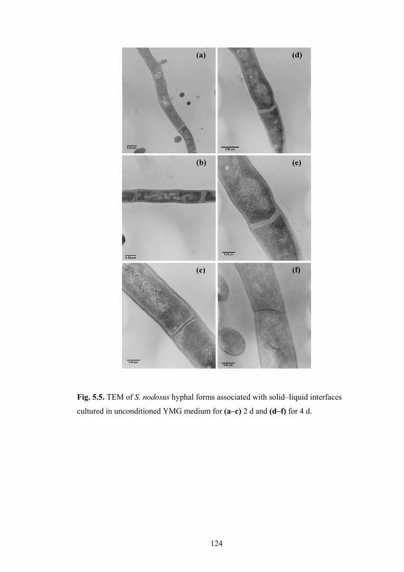
124
Fig. 5.5. TEM of S. nodosus hyphal forms associated with solid�liquid interfaces
cultured in unconditioned YMG medium for (a�c) 2 d and (d�f) for 4 d.

125
Fig. 5.6. TEM of S. nodosus hyphal forms associated with solid�liquid interfaces
cultured in conditioned media from S. nodosus for (a�d) 2 d and (e�h) for 4 d.

126
5.3.2 Induction of sporulation in planktonic cultures of S. nodosus
To determine whether sporulation was dependent on a solid surface, a reduced
population of the planktonic wild-type organism (6 % v/v inoculum, 48 h culture)
was returned to filter-sterilised 48 h conditioned medium and cultured for up to 9 d.
This quantity of biomass was chosen because it was equivalent to the biomass
returned as immobilised cells in the sporulated solid�liquid interface cultures.
CLSM revealed extensive sporulation (Fig. 5.7b) whereas control experiments with
unperturbed planktonic populations cultured in YMG for 9 d showed no evidence of
sporulation (Fig. 5.7a).
Fig. 5.7. CLSM images of planktonic population stained with BacLight viability
stain. (a) Planktonic population from the culture of encapsulated S. nodosus grown in
YMG after 9 d showing presence of hyphae and absence of spores and
(b) unencapsulated wild-type S. nodosus cultured in conditioned medium from wild-
type S. nodosus after 9 d showing presence of spores chains. Bars represent 10 �m.
(b) (a)

127
5.3.3 Assessment of antibiotic production from immobilised S. nodosus
To assess the production of AmB from immobilised S. nodosus, conditioned medium
produced from the mutant S. nodosus MA�
hyg1 was employed. This mutant is
incapable of AmB production as it is genetically disrupted in the loading module of
the cluster (Nikodinovic, 2004). Conditioned medium was produced from a log
phase (48 h) culture of mutant S. nodosus MA�
hyg1. Capsules containing S.
nodosus were incubated in this medium and monitored for viability, morphology as
well as antibiotic production.
CLSM of capsules incubated in conditioned medium produced from the mutant
S. nodosus MA�
hyg1 revealed a viability of 45 % on d 2, with progression to
sporulation on d 4 (Fig. 5.8 a&b). This result was similar to immobilised S. nodosus
cultured in conditioned medium from wild-type S. nodosus including the
development of mycelia with defined nucleoid regions compartmentalising into
segments as well as the development of a grey coloration.
On assessment of antibiotic production, a significant decrease was observed
compared with the amount of AmB produced by immobilised capsules cultured in
unconditioned YMG. AmB production after 4 d fermentation of capsules in 48 h
conditioned medium from the mutant S. nodosus MA�
hyg1 was assessed by pre-
concentration using SPE and quantified using HPLC to be 500 ng mL�1
.

128
Fig. 5.8. Viability and morphology of immobilised S. nodosus mycelia (48 h) when
cultured in conditioned media from mutant S. nodosus MA�hyg1 on (a) d 2, (b) d 4,
(c) d 7, (d) d 14, (e) d 21 and (f) d 30 using CLSM at ×1000 magnification. Bars
represent 10 µm (a, f) and 5 µm (b�e).

129
5.3.4 Culturing of immobilised S. nodosus in conditioned media from other
Streptomyces spp.
To examine the effect of conditioned media from other Streptomyces spp. on
S. nodosus at the solid�liquid interface, culture fluid from log phase cells needed to
be generated. Therefore, the growth of S. coelicolor, S. griseus, S. natalensis or
S. virginiae was studied in YMG by measuring change in biomass in order to identify
the log phase (Fig. 5.9). All Streptomyces spp. grew in YMG, with S. coelicolor
producing the least biomass. All cultures entered log phase at or by d 1 and were in
log phase at d 2 of fermentation, with cultures in stationary phase by d 4.
0
1
2
3
4
5
6
7
0 1 2 3 4 5 6 7
Time (days)
Bio
ma
ss
(mg
mL-1
)
S. virginiae
S. griseus
S. natalensis
MA�
hyg1
S. coelicolor
Fig. 5.9. Growth of free-dwelling S. virginiae, S. griseus, S. natalensis, S. nodosus
MA✁
hyg1 or S. coelicolor cultured in YMG at 28 °C at 60 rpm for 7 d. Error bars
represent maximum deviation of 3 samples.
Capsules containing wild-type S. nodosus were incubated in conditioned media
produced from S. coelicolor, S. griseus, S. natalensis or S. virginiae (48 h, log phase)
(Section 2.4). All immobilised cultures were incubated for a period of 30 d at 28 °C
with constant shaking at 60 rpm. Capsules were removed periodically and assessed

130
for viability and morphology (Section 2.8). Immobilised cells revealed differences in
morphology and physiology in the different conditioned media. Immobilised
S. nodosus incubated in S. coelicolor (Fig. 5.10) and S. griseus (Fig. 5.11)
conditioned media had morphology consistent with vegetative hyphae with no
indication of spore formation when incubated up to d 30. There was no significant
variation in pigmentation in the immobilised cultures incubated in conditioned media
from S. coelicolor, analogous to cultures incubated in YMG. However, cultures
incubated in conditioned medium from S. griseus developed a deep orange coloration
in the culture fluid but no pigmentation was associated with the capsules. Further,
hyphae with segregated nucleoids were observed on d 7 but did not progress into
spores when cultured up to d 30. Both samples showed fragmented mycelia with a
significant decrease in biomass, consistent with hyphal degeneration as observed in
ageing cultures.
By contrast, immobilised cultures incubated in conditioned medium from S.
natalensis (Fig. 5.12) and S. virginiae (Fig. 5.13) progressed to sporulation,
analogous to experiments with conditioned medium from the mutant S. nodosus
MA�
hyg1. However, the onset of sporulation was delayed, occurring on d 21 for
both S. natalensis (Fig. 5.12e) and S. virginiae (Fig. 5.13e) conditioned media,
compared to conditioned medium from S. nodosus where sporulation was observed
as early as d 4. Although not quantified, the number of spores from immobilised
cultures incubated in S. natalensis was comparable to the number of spores in
capsules incubated in conditioned medium from the S. nodosus, however a
significantly lower number of spores were observed in capsules incubated in

131
conditioned medium from S. virginiae. In addition to spores, fragmented mycelia
were also observed in these samples.
Fig. 5.10. Viability and morphology of immobilised S. nodosus mycelia (48 h) when
cultured in conditioned medium from S. coelicolor on (a) d 2, (b) d 5, (c) d 7, (d) d
14, (e) d 21 and (f) d 30 using CLSM at ×1000 magnification. Bars represent 10 µm.

132
Fig. 5.11. Viability and morphology of immobilised S. nodosus mycelia (48 h) when
cultured in conditioned medium from S. griseus on (a) d 2, (b) d 5, (c) d 7, (d) d 14,
(e) d 21 and (f) d 30 using CLSM at ×1000 magnification. Bars represent 10 µm.

133
Fig. 5.12. Viability and morphology of immobilised S. nodosus mycelia (48 h) when
cultured in conditioned medium from S. natalensis on (a) d 2, (b) d 5, (c) d 7, (d) d
14, (e) d 21 and (f) d 30 using CLSM at ×1000 magnification. Bars represent 10 µm.

134
Fig. 5.13. Viability and morphology of immobilised S. nodosus mycelia (48 h) when
cultured in conditioned medium from S. virginiae on (a) d 2, (b) d 5, (c) d 7, (d) d
14, (e) d 21 and (f) d 30 using CLSM at ×1000 magnification. Bars represent 10 µm.

135
Samples were also examined using SEM on d 2 (Fig. 5.14) and d 22 (Fig. 5.15) of
fermentation. Fibrous connections extending between hyphae were observed after 2
d of fermentation in samples incubated in conditioned media from all other
Streptomyces spp. (Fig. 5.14a�d) other than S. nodosus. These structures were not
observed in capsules incubated in unconditioned YMG medium (Fig. 5.4a). The
hyphae associated with capsules incubated in these conditioned media showed
disfigured and collapsed hyphae.
On d 22, similar to SEM micrographs of sporulating cultures in previous
experiments, samples incubated in conditioned media from S. natalensis and
S. virginiae showed no spores, but rather the presence of thickened hyphae with
constrictions, presumably sporogenic hyphae (Fig. 5.15c&d). These thickened
hyphae were absent in cultures incubated in conditioned media from S. coelicolor
and S. griseus (Fig. 5.15a&b).
Culture fluids at the end of the fermentation of these cross-feeding studies were
concentrated by SPE, and the resultant extract was examined for AmB production by
HPLC. AmB was not detected in culture fluids of capsules incubated in conditioned
media from S. griseus and S. virginiae, however trace amounts were detected in
culture fluids of capsules incubated in S. natalensis (400 ng mL�1
) and S. coelicolor
(300 ng mL�1
).

136
Fig. 5.14. SEM of encapsulated wild-type S. nodosus mycelia (48 h) cultured for 2 d in conditioned media from
(c) S. natalensis and (d) S. virginiae. Bars represent 1 µm.

137
Fig. 5.15. SEM of encapsulated wild-type S. nodosus mycelia (48 h) cultured for 22 d in conditioned media from
(c) S. natalensis and (d) S. virginiae. Bars represent 1 µm

138
Discussion
The biochemical pathways of Streptomyces cellular differentiation are still not fully
elucidated, but they are very important because they have major implications for the
understanding of the physiology of the organism in soil ecology as well as for
biotechnology given that they make a range of pharmacologically important
products. Aerial hyphae-deficient mutants have been used to identify a range of
genetic products encoded by bld genes, which together with other pathways assess
environmental parameters, transduce and mitigate the transition to aerial hyphae
(Chater & Horinouchi, 2003). Separate pathways have been postulated to detect
aerial growth and control further developmental progression such as sporulation
(Claessen et al., 2006; Chater, 2001).
The nutritional and environmental triggers for hyphae transition and/or natural
product production are not known. Streptomyces generally do not complete a full
life cycle when cultured in liquid media. Some species are only known to sporulate
at a solid�air interface (Kieser et al., 2000; Chater, 2001). Likewise, some
antibiotics are only produced in significant yield from solid supports presumably due
to the link between differentiation and production (Kieser et al., 2000). As liquid
environments can be tightly controlled, research using organisms that do sporulate in
liquid environments, such as S. griseus, have been used to accelerate Streptomyces
developmental research (Kendrick & Ensign, 1983; Chater, 2001). The liquid�air
interface model for Streptomyces differentiation using glass beads also highlighted
the advantages of a liquid phase to analyse cellular differentiation. This system,
using a liquid environment, is especially advantageous as it can be easily monitored

139
when the addition of nutrients, growth factors and labelled substrates is involved
(Nguyen et al., 2005).
Encapsulated S. nodosus may now be used as a model of cellular differentiation for a
species previously not known to undergo a complete life cycle in a liquid
environment. This system has the advantages of being able to be controlled and
allowing for detailed analyses of the environment and natural product formation.
The trigger for sporulation in S. nodosus, however, must be more complex than the
provision of a surface for colonisation as sporulation was induced in a planktonic
culture when a reduced biomass was returned to conditioned media. This culture
was in the late-log phase and thus growth arrested due to the availability of nutrients
or presence of waste and/or extra-cellular signalling compounds. The lack of
sporulation observed in encapsulated organisms co-cultivated with a rapidly growing
free-dwelling population for up to 30 d indicates that the resulting environment was
different to that provided by the conditioned culture fluid produced by the free-
dwelling cells. This may be due to the biphasic culture competing for nutrients
resulting in secretion of different signalling molecules and/or nutritional
environment. This is supported by studies on the phenotypic changes of organisms
in biofilms compared to planktonic organisms revealing the magnitude of difference
can be as large as those seen between different species (Pasmore & Costerton, 2003).
When exposed to conditioned medium from S. nodosus, the encapsulated organisms
grew within the capsules but the extent of growth was such that a free-dwelling
population was not established. The viability of the cells before sporulation was less
than those exposed to unconditioned medium, a phenomenon consistent with that

140
expected for aerial hyphae transition where rounds of substrate hyphae cell death are
required (Miguélez et al., 1999; Manteca et al., 2005a). After 4 d, sporulation was
evident throughout the capsules; however the capsular location has not been fully
established as there are difficulties in determining whether spores were found within
the matrix or at a solid�liquid interface along the pore channels within the capsules.
Though not optimised, AmB production from encapsulated organisms was
diminished, whether a free-dwelling population was present or not. Reports with
immobilised Streptomyces for optimised antibiotic production have been variable
(El-Naggar et al., 2003; Yang & Yueh, 2001; Teruel et al., 1997; Bandyopadhyay et
al., 1993; Bhattacharyya & Sen, 2002). The biphasic cultures produced around 5 %
of the AmB yield compared to cultures without immobilised organisms in YMG
(14 µg mL�1
), again highlighting the difference in the physiology of the system. It is
not known whether this is due to the culturing conditions especially oxygen
limitations or other nutrient and waste diffusion issues. The production, albeit low,
from organisms in conditioned medium was encouraging for future studies
manipulating the system for alteration of the rate of differentiation and antibiotic
production.
Studies using immobilised S. nodosus in conditioned media from other Streptomyces
spp. resulted in a variety of physiological responses and presumably indicate the
occurrence of interspecies communication in S. nodosus. S. natalensis produces
pimaricin, a structurally related antifungal compound to AmB. PI factor, a chemical
known to influence pimaricin production, was identified in the conditioned medium
from S. natalensis (Recio et al., 2004). Chemical signalling molecules, such as the
PI factor, present in conditioned media of S. natalensis or S. virginiae, which is

141
known to produce a variety of quorum sensing molecules, collectively known as
virginiae butanolides, and/or the nutritional environment induced sporulation in
S. nodosus and the immobilised organisms underwent a full life cycle similar to that
observed when incubated in conditioned medium from S. nodosus. However this
progression to spores was not observed when cultures were incubated in conditioned
media from S. coelicolor or S. griseus.
Interestingly, the cultures incubated in conditioned medium from S. griseus although
did not sporulate and remained as mycelia up to d 30, did develop a deep orange
coloration which was not observed in other cultures. Autoregulatory factors, such as
IM-2 has been shown to induce a blue pigment in Streptomyces sp. FRI-5 and A-
factor, the factor produced by wild-type S. griseus has been shown to trigger
production of a diffusible yellow pigment in S. griseus (Sato et al., 1989; Horinouchi
& Beppu, 1993; Ohnishi et al., 2004). Whether, A-factor also influences pigment
secretion in S. nodosus is not known.
In addition, cultures incubated in conditioned media from S. coelicolor or
S. natalensis produced AmB. However no AmB was detected in cultures incubated
in conditioned media from S. virginiae or S. griseus. �Quorum quenching� is a
phenomenon where substances interfere with quorum sensing molecules and prevent
bacterial communication and hence respective responses (Zhang, 2003; Waters &
Bassler, 2005). These results indicate that although secondary metabolism is
commonly linked with morphological differentiation, it is possible that these two
pathways may occur independently of each other as the cultures that sporulated were
not necessarily the cultures that showed the presence of AmB and vice versa. It

142
should be also noted that the end point of cellular differentiation of this organism,
which is the production of spores, are an entity not able to produce antibiotics.
When immobilised S. nodosus were cultured in conditioned media from S.
coelicolor, S. griseus, S. natalensis or S. virginiae, a reduced planktonic population
was present indicating that the antibiotics or other secondary metabolites if released
from these organisms did not have a bacteriocidal or bacteriostatic effect on S.
nodosus.
A significant drawback of using conditioned media is that in addition to the presence
of signalling molecules, it also contains fermentation products, metabolites, proteins
or toxins that may elicit a stress response in the bacterium. Stress responses could
include the organisms going into the sporulation phase or the production of
antibiotics. Hence, it is necessary to conduct further studies to conclude that the
responses observed when immobilised S. nodosus was incubated in conditioned
media from other Streptomyces spp. are due to the presence of chemical signalling
molecules and not due the presence of another component present in the conditioned
media.
Attempts to modulate the effect of the conditioned media on encapsulated S. nodosus
by dilution with YMG were not undertaken. This was due to the emerging literature
on the ability of organisms to degrade quorum sensing or signalling molecules during
fermentation meaning that the concentration of these molecules reflects more than
just the community size at a particular time point (Dunn & Stabb, 2007). The effect
on the reintroduction of nutrients from the unconditioned media is of interest

143
however with the overlaying effect on quorum sensing molecule concentrations
would give results which are difficult to interpret.
Using this model for cellular differentiation, the signalling processes in this
industrially important organism were investigated, and this work will continue in
other projects. Once delineated, manipulation of these pathways in S. nodosus and
other important actinomycetes may lead to higher production yields or altered
production processes, especially for organisms where secondary metabolites are only
produced in significant yield with solid surface cultivation. It should be pointed out
that for some biological applications, only the wild-type organism maybe required to
produce conditioned media for culturing of encapsulated organisms.

Chapter 6
Conclusions and Future Directions

145
This work successfully developed methods to immobilise S. nodosus in alginate
capsules and examined cellular differentiation at the solid�liquid interface. To assess
bacterial viability in capsules, a new method was developed using fluorescent stains
and microscopy. This method was applicable for unicellular and filamentous
bacteria and while an indication that the method could be used for spore viability this
requires further investigation. The method will also be of interest to applications
where organisms exist in a solid matrix such as the food and agricultural industries.
Encapsulation of S. nodosus in alginate with exposure to appropriate media
supported growth and maintained viability for over 30 days. When allowed to
proliferate, an organised community structure developed similar to that observed in a
biofilm exposed to bulk flow (Branda et al., 2001). The observation of
�reconnoitring hyphae� which are morphologically similar to aerial hyphae on solid
media but not destined to sporulate, has been suggested as a new function for hyphae
emerging from mycelial masses (Yeo & Chater, 2005). Whether these hyphae are
scouting for new environments for colonisation, or to expose nutrient scavenging
hyphae to environments where nutrients are at maximum availability (in this case the
bulk flow of culture fluid) needs to be investigated. The presence of reconnoitring
aerial hyphae in natural habitats has been hypothesised (Yeo & Chater, 2005). This
was based on evidence that hyphae may span soil particles through an air interface
with the purpose of colonising new environments. This means that the organism has
an additional mechanism for dispersal, that is, through cell elongation rather that
from hyphal fragmentation or spore distribution. As Streptomyces are important in
the biodegradation and recycling of organic matter, studies identifying the existence

146
and function of reconnoitring hyphae will be important to the ecology of the
environment
Fig. 6.1. Hypothesised functions of aerial hyphae that are destined for different
functions in soil (Yeo & Chater, 2005).
In addition, the identification of a model where the commitment to sporulation has
been decoupled from pathways inducing hyphae to emerge from substrate mycelial
masses, would allow biochemical and genetic analysis of these two pathways.
Previously these have had to been examined through mutant analysis (Aparicio,
2006). In Streptomyces these developmental pathways are complex and still poorly
characterised (Takano, 2006). This could be undertaken using microarrays or other
comparative genomic or proteomic approaches.
This project also determined conditions that permit S. nodosus to undergo a complete
life cycle through to sporulation in a liquid environment, something not previously
reported. These conditions were identified using a novel solid�liquid interface
model with restricted cellular proliferation. The trigger for sporulation was the

147
reduction of quorum size at log phase during batch culture, and was not dependent on
the availability a solid surface. Further examination of this model is required for the
elucidation of the biochemical pathways and signalling processes involved in cellular
differentiation and antibiotic production. In addition, whether this model for
sporulation is suitable for other Streptomyces spp. and other industrially important
micro-organisms needs to be investigated. This may be very significant for
Streptomyces spp. which only produce antibiotic and/or sporulate on solid supports
which is problematic for industrial applications.
For biotechnological applications, immobilised Streptomyces or other prokaryotes
may permit rapid screening and identification of quorum sensing molecules and
nutrient dependence on antibiotic production. These parameters/compounds alone or
in combination could potentially allow exogenous control of antibiotic production,
affecting yields and fermentation times (Horinouchi & Beppu, 1994; Kieser et al.,
2000). Whether these molecules are derived from the antibiotic producer or other
species is also worth considering given the difference in physiology of immobilised S
.nodosus when exposed to conditioned media from other species.
The use of conditioned medium from the amphA mutant S. nodosus MA�
hyg1
which lacks the ability to synthesise amphotericins permitted successful
demonstration of the production of AmB from the immobilised S. nodosus. The
production of an antibiotic from immobilised cultures without the co-existence of a
planktonic/free-dwelling population has not been previously reported. For in vivo
implantation of bioreactors, minimal cell escape would be required, so the ability of
S. nodosus to produce AmB without high cellular proliferation rates is encouraging.

148
Whether these bioreactors could be implanted and function in vivo is unknown and
would require initial testing in animal models, including animal models with induced
fungal infections. If unsuccessful, future research exploring the use of other bacteria
to express the antibiotic gene cluster to heterologously produce AmB could be
pursued. It is worthwhile noting that polyketides have been successfully produced
from genetically modified Streptomyces lividans, S. coelicolor and even E. coli
(Hutchinson & Fujii, 1995).
In conclusion, this project has made advances towards realisation of the concept of
using immobilised S. nodosus as a drug delivery system for AmB. If successful,
such a system could have a significant impact in antifungal therapy. However, the
outcomes from developing this system have implications for a range of other
technologies involving both artificially immobilised organisms. These include
encapsulation for probiotic protection, inoculum development for fermentation and
organisms for waste water treatment. It could also permit an increased understanding
of the ecological role of S. nodosus in its natural environment.

References

150
1. Adinarayana, K., Srinivasulu, B., Bapi Raju, K. V. V. S. N. & Ellaiah, P.
(2004). Continuous neomycin production by immobilized cells of Streptomyces
marinensis NUV-5 in an airlift bioreactor. Process Biochem 39, 1407�1414.
2. Allan, E. J. & Prosser, J. I. (1987). Colony growth of Streptomyces coelicolor
A3(2) under conditions of continuous nutrient supply. FEMS Microbiol Lett 43,
139�142.
3. Andriole, V. T. (1999). Current and future antifungal therapy: new targets for
antifungal agents. J Antimicrob Chemother 44, 151�162.
4. Aparicio, J. F., Caffery, P., Gil, J. A. & Zotchev, S. B. (2003). Polyene
antibiotic biosynthesis gene clusters. Appl Microbiol Biotechnol 61, 179�188.
5. Aparicio, J. F. (2006). Generating novel polyene antifungal drugs. Chem Biol
12, 509�510.
6. Aparicio, J. F., Fouces, R., Mendes, M. V., Olivera, N. & Martín, J. F.
(2000). A complex multienzyme system encoded by five polyketide synthase
genes is involved in the biosynthesis of the 26-membered polyene macrolide
pimaricin in Streptomyces natalensis. Chem Biol 7, 895�905.
7. Aramwit, P., Yu, B. G., Lavasanifar, A., Samuel, J. & Kwon, G. S. (2000).
The effect of serum albumin on the aggregation state and toxicity of
amphotericin. J Pharm Sci 89, 1589�1593.
8. Austin, M. B. & Noel, J. P. (2003). The chalcone synthase superfamily of type
III polyketide synthases. Nat Prod Rep 20, 79�110.
9. Bach, P. R. (1984). Quantitative extraction of amphotericin B from serum and
its determination by high-pressure liquid chromatography. Antimicrob Agents
Chemother 26, 314�317.

151
10. Baginski, M., Resat, H. & McCammon, J. A. (1997). Molecular properties of
amphotericin B membrane channel: a molecular dynamics simulation. Mol
Pharmacol 52, 560�570.
11. Bandyopadhyay, A., Das, A. K. & Mandal, S. K. (1993). Erythromycin
production by Streptomyces erythreus entrapped in calcium alginate beads.
Biotechnol Lett 15, 1003�1106.
12. Barabas, G., Penyige, A. & Hirano, T. (1994). Hormone-like factors
influencing differentiation of Streptomyces cultures. FEMS Microbiol Rev 14,
75�82.
13. Barrett, B. (2002). From natural products to clinically useful antifungals.
Biochim Biophys Acta 1587, 224�233.
14. Bassler, B. L. & Losick, R. (2006). Bacterially speaking. Cell 125, 237�246.
15. Bhattacharyya, B. K. & Sen, S. K. (2002). Ester antibiotic accumulation by
Streptomyces hygroscopicus. Microbiologica 25, 477�484.
16. Bibb, M. J. (2005). Regulation of secondary metabolism in streptomycetes. Curr
Opin Microbiol 8, 208�215.
17. Blandino, A., Macías, M. & Cantero, D (1999). Formation of calcium alginate
gel capsules: influence of sodium alginate and CaCl2 concentration on gelation
kinetics. J Biosci Bioeng 88, 686�689.
18. Bolard, J. (1986). How do the polyene macrolide antibiotics affect the cellular
membrane properties? Biochim Biophys Acta 864, 257�304.
19. Borowski, E. (2000). Novel approaches in the rational design of antifungal
agents of low toxicity. Farmaco 55, 206�208.

152
20. Bossche, H. V., Bellens, D., Cools, W., Gorrens, J., Marichal, P., Willemsens,
V. G., De Coster, R., Beerens, D., Haelterman, C. & other authors (1986).
Cytochrome p-450: Target for itraconazole. Drug Dev Res 8, 287�298.
21. Bossche, H. V., Marichal, P. & Odds, F. C. (1994). Molecular mechanisms of
drug resistance in fungi. Trends Microbiol 2, 393�400.
22. Bo�ek, P., Hutta, M. & Hrivnáková, B. (2005). Rapid analysis of porphyrins at
low ng/l and µg/l levels in human urine by a gradient liquid chromatography
method using octadecylsilica monolithic columns. J Chromatogr A 1084, 24�32.
23. Brajtburg, J., Powderly, W. G., Kobayashi, G. S. & Medoff, G. (1990).
Amphotericin B: current understanding of mechanisms of action. Antimicrob
Agents Chemother 34, 183�188.
24. Branda, S. S., González-Pastor, J. E., Ben-Yehuda, S., Losick, R. & Kolter,
R. (2001). Fruiting body formation by Bacillus subtilis. Proc Natl Acad Sci USA
98, 11621�11626.
25. Bu�Lock, J. D. (1961). Intermediary metabolism and antibiotic synthesis. Adv
Appl Microbiol 3, 293�342.
26. Caffrey, P., Lynch, S., Flood, E., Finnan, S. & Oliynyk, M. (2001).
Amphotericin biosynthesis in Streptomyces nodosus: deductions from analysis of
polyketide synthase and late genes. Chem Biol 8, 713�723.
27. Calafiore, R., Basta, G., Luca, G., Lemmi, A., Montanucci, M. P., Calabrese,
G., Racanicchi, L., Mancuso, F. & Brunetti, P. (2006). Microencapsulated
pancreatic islet allografts into nonimmunosuppressed patients with type 1
diabetes. Diabetes Care 29, 137�138.

153
28. Cane, D. E, Walsh, C. T. & Khosla, C. (1998). Harnessing the biosynthetic
code: combinations, permutations, and mutations. Science 282, 63�68.
29. Carmody, M., Byrne, B., Murphy, B., Breen, C., Lynch, S., Flood, E.,
Finnan, S. & Caffrey, P. (2004). Analysis and manipulation of amphotericin
biosynthetic genes by means of modified phage KC515 transduction techniques,
Gene 343, 107�115.
30. Carreras, C. W. & Santi, D. V. (1998). Engineering of modular polyketide
synthases to produce novel polyketides. Curr Opin Biotechnol 9, 403�411.
31. Carrillo-Muñoz, A. J., Quindós, G. & Lopez-Ribot, J. L. (2004). Current
developments in anti-fungal agents. Curr Med Chem 3, 297�323.
32. Challis, G. L. & Hopwood, D. A. (2003). Synergy and contingency as driving
forces for the evolution of multiple secondary metabolite production by
Streptomyces species. Proc Natl Acad Sci U S A 100, 14555�14561.
33. Chandramoulia, V., Kailasapathya, K, Peiris, P. & Jones, M. (2004). An
improved method of microencapsulation and its evaluation to protect
Lactobacillus spp. in simulated gastric conditions. J Microbiol Methods 56, 27�
35.
34. Chang, T. M. (1964). Semipermeable microcapsules. Science 146, 524�525.
35. Chang, T. M. S. & Prakash, S. (1998). Therapeutic uses of microencapsulated
genetically engineered cells. Mol Med Today 4, 221�227.
36. Charvalos, E., Tzatzarakis, M. N., Bambeke, F. V., Tulkens, P. M.,
Tsatsakis, A. M., Tzanakakis, G. N. & Mingeot-Leclercq, M. (2006). Water-
soluble amphotericin B�polyvinylpyrrolidone complexes with maintained

154
antifungal activity against Candida spp. and Aspergillus spp. and reduced
haemolytic and cytotoxic effects. J Antimicrob Chemother 57, 236�244.
37. Chater, K. F. & Horinouchi, S. (2003). Signalling early developmental events
in two highly diverged Streptomyces species. Mol Microbiol 48, 9�15.
38. Chater, K. F. (1989). Multilevel regulation of Streptomyces differentiation.
Trends Genet 5, 372�377.
39. Chater, K. F. (1993). Genetics of differentiation in Streptomyces. Annu Rev
Microbiol 47, 685�713.
40. Chater, K.F. & Horinouchi, S. (2003). Signalling and early developmental
events in two highly diverged Streptomyces species. Mol Microbiol 48, 9�15.
41. Chater, K.F. (2001). Regulation of sporulation in Streptomyces coelicolor
A3(2): a checkpoint multiplex? Curr Opin Microbiol 4, 667�673.
42. Claessen, D., de Jong, W., Dijkhuizen, L. & Wosten, H. A. B. (2006).
Regulation of Streptomyces development: reach for the sky. Trends Microbiol
14, 313�319.
43. Coker, R. J. & Harris, J. R. (1991). Failure of fluconazole treatment in
cryptococcal meningitis despite adequate CSF levels. J Infect 23, 101�103.
44. Collie, N. (1893). The formation of orcinol and other condensation products
from dehydracetic acid. J Chem Soc 1893, 122�126.
45. Cross, T. (1970). Symposium on bacterial spores: 8. the diversity of bacterial
spores. J Appl Bacteriol 33, 95�102.
46. Cui, W., Barr, G., Faucher, K. M., Sun, X.-L., Safley, S. A., Weber, C. J. &
Chaikof, E. L. (2004). A membrane-mimetic barrier for islet encapsulation.
Transplant Proc 36, 1206�1208.

155
47. Demain, A. L. (1999). Pharmaceutically active secondary metabolites of
microorganisms. Appl Microbiol Biotechnol 52, 455�463.
48. de Pauw, B. (2000). Is there a need for new antifungal agents? Clin Microbiol
Infect 6, 23�28.
49. Develoux, M. (2001). Griseofulvin. Ann Dermatol Venereol 128, 1317�1325.
50. Devi, S. & Sridhar, P. (2000). Production of cephamycin C in repeated batch
operations from immobilized Streptomyces clavuligerus. Process Biochem 36,
225�231.
51. DiDomenico, B. (1999). Novel antifungal drugs. Curr Opin Microbiol 2, 509�
515.
52. Dong, Z., Wang, Q. & Du, Y. (2006). Alginate/gelatin blend films and their
properties for drug controlled release. J Memb Sci 280, 37�44.
53. Doria-Serrano, M. C., Ruiz-Treviño, F. A., Rios-Arciga, C., Hernández-
Esparza, M. & Santiago, P. (2001). Physical characteristics of poly(vinyl
alcohol) and calcium alginate hydrogels for the immobilization of activated
sludge. Biomacromolecules 2, 568�574.
54. Draget, K. I., Skjåk-Bræk, G. & Smidsrød, O. (1997). Alginate based new
materials. Int J Biol Macromol 21, 47�55.
55. Dunn, A. K. & Stabb, E. V. (2007). Beyond quorum sensing: the complexities
of prokaryotic parliamentary procedures. Anal Bioanal Chem 387, 391�398.
56. Dunny, G. M. & Leonard, B. A. B. (1997). Cell-cell communication in gram-
positive bacteria. Annu Rev Microbiol 51, 527�564.
57. Dupont, B. (2002). Overview of the lipid formulations of amphotericin B. J
Antimicrob Chemother 49, 31�36.

156
58. Dutcher, J. D. (1968). The discovery and development of amphotericin B. Dis
Chest 54, 296�298.
59. Elçin, Y. M. (1995). Bacillus sphaericus 2362-calcium alginate microcapsules
for mosquito control. Enzyme Microb Technol 17, 587�591.
60. Eldem, T. & Arican-Cellat, N. (2001). Determination of amphotericin B in
human plasma using solid-phase extraction and high-performance liquid
chromatography. J Pharm Biomed Anal 25, 53�64.
61. Elliot, M., Damji, F., Passantino, R., Chater, K. & Leskiw, B. (1998). The
bldD gene of Streptomyces coelicolor A3(2): a regulatory gene involved in
morphogenesis and antibiotic production. J Bacteriol 180, 1549�1555.
62. Ellis, D. (2002). Amphotericin B: spectrum and resistance. J Antimicrob
Chemother 49, 7�10.
63. El-Naggar, M. Y., Hassan, M. A., Said, W. Y. & El-Aassar, S. A. (2003).
Effect of support materials on antibiotic MSW2000 production by immobilized
Streptomyces violatus. J Gen Appl Microbiol 49, 235�243.
64. Ensign, J. C. (1978). Formation, properties and germination of actinomycete
spores. Annu Rev Microbiol 32, 185�219.
65. Erikson, D. (1949). The morphology, cytology and taxonomy of the
actinomycetes. Annu Rev Microbiol 3, 23�54.
66. Eriksson, U., Seifert, B. & Schaffner, A. (2001). Comparison of effects of
amphotericin B deoxycholate infused over 4 or 24 hours: randomised controlled
trial. BMJ 322, 1�6.
67. Ertesvåg, H. & Valla, S. (1998). Biosynthesis and applications of alginates.
Polym Degrad Stab 59, 85�91.

157
68. Esposito, E., Bortolotti, F., Menegatti, E. & Cortesi, R. (2003). Amphiphilic
association systems for amphotericin B delivery. Int J Pharm 260, 249�260.
69. Espuelas, M. S., Legrand, P., Irache, J. M., Gamazo, C., Orecchioni, A. M.,
Devissaguet, J. P. & Ygartua, P. (1997). Poly(�-caprolacton) nanospheres as an
alternative way to reduce amphotericin B toxicity. Int J Parmaceut 158, 19�27.
70. Falk, R., Domb, A. J. & Polacheck, I. (1999). A novel injectable water-soluble
amphotericin B-arabinogalactan conjugate. Antimicrob Agents Chemother 43,
1975�1981.
71. Favre E., Leonard, M., Laurent, A. & Dellacherie, E. (2001). Diffusion of
polyethyleneglycols in calcium alginate hydrogels. Colloids Surf A Physicochem
Eng Asp 194, 197�206.
72. Fernández, M. & Sánchez, J. (2001). Viability staining and terminal
deoxyribonucleotide transferase-mediated dUTP nick end labelling of the
mycelium in submerged cultures of Streptomyces antibioticus ETH7451. J
Microbiol Methods 47, 293�298.
73. Fernández, M. & Sánchez, J. (2002). Nuclease activities and cell death
processes associated with the development of surface cultures of Streptomyces
antibioticus ETH 7451. Microbiology 148, 405�412.
74. Flärdh, K. (2003). Growth polarity and cell division in Streptomyces. Curr Opin
Microbiol 6, 564�571.
75. Folcher, M., Gaillard, H., Nguyen, L. T., Nguyen, K. T., Lacroix, P., Bamas-
Jacques, N., Rinkel, M. & Thompson, C. J. (2001). Pleiotropic functions of a
Streptomyces pristinaespiralis autoregulator receptor in development, antibiotic

158
biosynthesis, and expression of a superoxide dismutase. J Biol Chem 276,
44297�44306.
76. Francis, P. & Walsh, T. J. (1992). Evolving role of flucytosine in
immunocompromised patients: new insights into safety, pharmacokinetics and
antifungal therapy. Clin Infect Dis 15, 1003�1018.
77. François, I. E. J. A., Aerts, A. M., Cammue B. P. A. & Thevissen, K. (2005).
Currently used antimycotics: spectrum, mode of action and resistance
occurrence. Curr Drug Targets 6, 895�907.
78. Frøyshov, Ø. (1974). Bacitracin biosynthesis by three complementary fractions
from Bacillus licheniformis. FEBS Lett 44, 75�78.
79. Fukui, H., Koike, T., Saheki, A., Sonoke, S. & Seki, J. (2003). A novel
delivery system for amphotericin B with lipid nano-sphere (LNS®
). Int J Pharm
265, 37�45.
80. Fundueanu, G., Nastruzzi, C., Carpov, A., Desbrieres, J. & Rinaudo, M.
(1999). Physico-chemical characterization of Ca-alginate microparticles
produced with different methods. Biomaterials 20, 1427�1435.
81. Fuqua, C., Parsek, M. R. & Greenberg, E. P. (2001). Regulation of gene
expression by cell-to-cell communication: acyl-homoserine lactone quorum
sensing. Annu Rev Genet 35, 439�468.
82. Fuqua, W. C., Winans, S. C. & Greenberg, E. P. (1994). Quorum sensing in
bacteria: the LuxR-LuxI family of cell density-responsive transcriptional
regulators. J Bacteriol 176, 269�275.
83. Gallis, H. A., Drew, R. H. & Pickard, W. W. (1990). Amphotericin B: 30 years
of clinical experience. Rev Infect Dis 12, 308�329.

159
84. García, M. (1995). A membrane-like structure envelops substrate mycelium
during colony development in Streptomyces. FEMS Microbiol Lett 131, 107�
111.
85. Georgopapadakou, N. H. & Bertasso, A. (1992). Effects of squalene epoxidase
inhibitors on Candida albicans. Antimicrob Agents Chemother 36, 1779�1781.
86. Georgopapadakou, N. H. & Walsh, T. J. (1994). Human mycoses: drugs and
targets for emerging pathogens. Science 264, 371�373.
87. Georgopapadakou, N. H. & Walsh, T. J. (1996). Antifungal agents:
chemotherapeutic targets and immunologic strategies. Antimicrob Agents
Chemother 40, 279�291.
88. Georgopapadakou, N. H. (1998). Antifungals: mechanism of action and
resistance, established and novel drugs. Curr Opin Microbiol 1, 547�557.
89. Gerbsch, N. & Buchholz, R. (1995). New processes and actual trends in
biotechnology. FEMS Microbiol Rev 16, 259�269.
90. Golas, C. L., Prober, C. G., MacLeod S. M. & Soldin, S. J. (1983).
Measurement of amphotericin b in serum or plasma by high-performance liquid
chromatography. J Chromatogr B Biomed Sci Appl 278, 387�395.
91. Gold, W., Stout, H. A., Pagano, J. F. & Donovick, R. (1956). Amphotericins A
and B, antifungal antibiotics produced by a Streptomycete. I. In vitro studies.
Antibiot Annu 1955�1956, 579�586.
92. González-Pastor, J. E., Hobbs, E. C. & Losick, R. (2003). Cannibalism by
sporulating bacteria. Science 301, 510�513.

160
93. Gottlieb, D. & Shaw, P. D. (1970). Mechanism of action of antifungal
antibiotics. Annu Rev Phytopathol 8, 371�402.
94. Gupte, M. Kulkarni, P. &. Ganguli B. N. (2002). Antifungal antibiotics. Appl
Microbiol Biotechnol 58, 46�57.
95. Gurny, R., Doelker, E. & Peppas, N. A. (1982). Modelling of sustained release
of water-soluble drugs from porous, hydrophobic polymers. Biomaterials 3, 27�
32.
96. Hamilton-Miller, J. M. T. (1973). Chemistry and biology of the polyene
macrolide antibiotics. Bacteriol Rev 37, 166�196.
97. Hann, I. M. & Prentice, H. G. (2001). Lipid-based amphotericin B: a review of
the last 10 years of use. Int J Antimicrob Agents 17, 161�169.
98. Hardisson, C. & Manzanal, M. B. (1976). Ultrastructural studies of sporulation
in Streptomyces. J Bacteriol 127, 1443�1454.
99. Hartsel, S. & Bolard, J. (1996). Amphotericin B: new life for an old drug.
Trends Pharmacol Sci 17, 445�449.
100. Hasse, C., Klöck, G., Schlosser, A., Zimmermann, U. & Rothmund,
M. (1997). Parathyroid allotransplantation without immunosuppression. Lancet
North Am Ed 350, 1296�1297.
101. Hazen, K. C. (1995). New and emerging yeast pathogens. Clin Microbiol
Rev 8, 462�478.
102. Hodgson, D. A. (2000). Primary metabolism and its control in streptomycetes:
a most unusual group of bacteria. Adv Microb Physiol 42, 47�238.

161
103. Hoeprich, P. D. (1978). Chemotherapy of systemic fungal diseases. Annu Rev
Pharmacol Toxicol 18, 205�231.
104. Hopwood, D. A. & Sherman, D. H. (1990). Molecular genetics of polyketides
and its comparison to fatty acid biosynthesis. Annu Rev Genet 24, 37�62.
105. Hopwood, D. A. (1997). Genetic contributions to understanding polyketide
synthases. Chem Rev 97, 2465�2497.
106. Horinouchi, S. & Beppu, T. (1992). Autoregulatory factors and
communication in actinomycetes. Annu Rev Microbiol 46, 377�398.
107. Horinouchi, S. & Beppu, T. (1993). A-factor and streptomycin biosynthesis in
Streptomyces griseus. Antonie Van Leeuwenhoek 64, 177�186.
108. Horinouchi, S. & Beppu, T. (1994). A-factor as a microbial hormone that
controls cellular differentiation and secondary metabolism in Streptomyces
griseus. Mol Microbiol 12, 859�864.
109. Horinouchi, S. (2007). Mining and polishing of the treasure trove in the
bacterial genus Streptomyces. Biosci Biotechnol Biochem 71, 283�299.
110. Hutchinson, C. R. & Fujii, I. (1995). Polyketide synthase gene manipulation:
a structure-function approach in engineering novel antibiotics. Annu Rev
Microbiol 49, 201�238.
111. Hutchinson, C. R. (1998). Combinatorial biosynthesis for new drug discovery.
Curr Opin Microbiol 1, 319�329.
112. Hutchinson, C. R., Decker, H., Guilfoile, P., Shen, B., Summers, R., Wendt-
Pienkowski, E. & Wessel, B. (1992). Secondary-metabolite biosynthesis and

162
metabolism, pp. 3�10. Edited by R. J. Petroski, S. P. McCormick. Plenum Press,
New York.
113. Kaiser, D. & Losick, R. (1993). How and why bacteria talk to each other? Cell
73, 873�885.
114. Kartsonis, N. A., Nielsen, J. & Douglas, C. M. (2003). Caspofungin: the first
in a new class of antifungal agents. Drug Resist Updat 6, 197�218.
115. Kauffman, C. A. (2006). Clinical efficacy of new antifungal agents. Curr Opin
Microbiol 9, 483�488.
116. Kayser, O., Olbrich, C., Yardley, V., Kiderlen, A. F. & Croft, S. L. (2003).
Formulation of amphotericin B as nanosuspension for oral administration. Int J
Pharm 254, 73�75.
117. Kelemen, G. H. & Buttner, M. J. (1998). Initiation of aerial mycelium
formation in Streptomyces. Curr Opin Microbiol 1, 656�662.
118. Kendall, W. F., Darrabie, M. D., El-Shewy, H. M. & Opara, E. C. (2004).
Effect of alginate composition and purity on alginate microspheres. J
Microencapsul 21, 821�828.
119. Kendrick, K. E. & Ensign, J. C. (1983). Sporulation of Streptomyces griseus
in submerged cultures. J Bacteriol 155, 357�366.
120. Khokhlov, A. S., Anisova, L. N., Tovarova, I. I., Kleiner, E. M., Kovalenko,
I. V., Krasilnikova, O. I., Kornitskaya, E. Y. & Pliner, S. A. (1973). Effect of
A-factor on the growth of asporogenous mutants of Streptomyces griseus, not
producing this factor. Z Allg Mikrobiol 13, 647�655.

163
121. Kieser, T., Bibb, M. J., Buttner, M. J., Chater, K. F. & Hopwood, D. A.
(2000). Practical Streptomyces Genetics. The John Innes Foundation, Norwich,
England.
122. Kim, T.-K, Sharma, B., Williams, C. G., Ruffner, M. A., Malik, A.,
McFarland E. G. & Elisseeff, J. H. (2003). Experimental model for cartilage
tissue engineering to regenerate the zonal organization of articular cartilage.
Osteoarthritis Cartilage 11, 653�664.
123. Klöck, G., Pfefferrmann, A., Ryser, C., Gröhn, P., Kuttler, B., Hahn, H. &
Zimmermann, U. (1997). Biocompatibility of mannuronic acid-rich alginates.
Biomaterials 18, 707�713.
124. Knapp, K. M. & Flynn, P. M. (2005). Newer treatments for fungal infections.
J Support Oncol 3, 290�298.
125. Koonin, E. V. & Aravind, L. (2002). Origin and evolution of eukaryotic
apoptosis: the bacterial connection. Cell Death Differ 9, 394�404.
126. Krasaekoopt, W., Bhandari, B. & Deeth, H. (2003). Evaluation of
encapsulation techniques of probiotics for yoghurt. Int Dairy J 13, 3�13.
127. Kuek, C., Tommerup, I. C. & Malajczuk, N. (1992). Hydrogel bead inocula
for the production of ectomycorrhizal eucalypts for plantations. Mycol Res 96,
237�277.
128. Kurachi, M., Nakashima, T., Miyajima, C., Iwamoto, Y., Muramatsu, T.,
Yamaguchi, K. & Oda, T. (2005). Comparison of the activities of various
alginates to induce TNF-� secretion in RAW264.7 cells. J Infect Chemother 11,
199�203.

164
129. Kwak, J. & Kendrick, K. E. (1996). Bald mutants of Streptomyces griseus
that prematurely undergo the key events of sporulation. J Bacteriol 178, 4643�
4650.
130. Kwak, J., Dharmatilake, A. J., Jiang, H. & Kendrick, K. E. (2001).
Differential regulation of ftsZ transcription during septation of Streptomyces
griseus. J Bacteriol 183, 5092�5101.
131. Laflamme, C., Lavigne, S., Ho, J. & Duchaine, C. (2004). Assessment of
bacterial endospore viability with fluorescent dyes. J Appl Microbiol 96, 684�
692.
132. Lal, R., Kumari, R., Kaur, H., Khanna, R., Dhingra, N. & Tuteja, D.
(2000). Regulation and manipulation of the gene clusters encoding type-I PKSs.
Tibtech 18, 264�274.
133. Lamas, M. C., Bregni, C., D�Aquino, M., Degrossi, J. & Firenstein, R.
(2001). Calcium alginate microspheres of Bacillus subtilis. Drug Dev Ind Pharm
27, 825�829.
134. Leadlay, P. (1997). Combinatorial approaches to polyketide biosynthesis. Curr
Opin Chem Biol 1, 162�168.
135. Lechevalier, H. A. & Lechevalier, M. P. (1967). Biology of actinomycetes.
Annu Rev Microbiol 21, 71�100.
136. Lee, K., Lee, C. Choi, S., Park, H., Kitani, S., Nihira, T. & Hwang, Y.
(2005). Cloning and in vivo functional analysis by disruption of a gene encoding
the �-butyrolactone autoregulator receptor from Streptomyces natalensis. Arch
Microbiol 184, 249�257.

165
137. Legrand, P., Romero, E. A., Cohen, B. E. & Bolard, J. (1992). Effects of
aggregation and solvent on the toxicity of amphotericin B to human erythrocytes.
Antimicrob Agents Chemother 36, 2518�2522.
138. Lemke, A., Kiderlen, A. F. & Kayser, O. (2005). Amphotericin B. Appl
Microbiol Biotechnol 68, 151�162.
139. Li, R. H. (1998). Materials for immunoisolated cell transplantation. Adv Drug
Deliv Rev 33, 87�109.
140. Li, W., Nihira, T., Sakuda, S., Nishida, T. & Tamada, Y. (1992). New
inducing factors for virginiamycin production from Streptomyces antibioticus. J
Ferment Bioeng 74, 214�217.
141. Lian, W., Hsiao, H. & Chou, C. (2003). Viability of microencapsulated
bifidobacteria in simulated gastric juice and bile solution. Int J Food Microbiol
86, 293�301.
142. Lincopan, N., Mamizuka, E. M. & Carmona-Ribeiro, A. M. (2003). In vivo
activity of a novel amphotericin B formulation with synthetic cationic bilayer
fragments. J Antimicrob Chemother 52, 412�418.
143. Liou, G. F. & Khosla, C. (2003). Building-block selectivity of polyketide
synthases. Curr Opin Chem Biol 7, 279�284.
144. Liu, Y. (1984). Biosynthesis of amphotericins, candicin and nystatin by means
of mutation. Proc Natl Sci Counc Repub China B 8, 182�186.
145. Lowery, C. A., McKenzie, K. M., Qi, L., Meijler, M. M. & Janda, K. D.
(2005). Quorum sensing in Vibrio harveyi: probing the specificity of the LuxP
binding site. Bioorg Med Chem Lett 15, 2395�2398.

166
146. Lur'e, L. M., Verkhovtseva, T. P. & Levitov, M. M. (1975). Biosynthesis of
penicillin and the biphasal development of a P. chrysogenum culture. Antibiotiki
20, 291�295.
147. Maertens, J. A. (2004). History of the development of azole derivatives. Clin
Microbiol Infect 10, 1�10.
148. Malik, V. S. & Vining, L. C. (1970). Metabolism of chloramphenicol by the
producing organism. Can J Microbiol 16,173�179.
149. Manteca, A., Fernández, M. & Sánchez, J. (2005a). A death round affecting
a young compartmentalized mycelium precedes aerial mycelium dismantling in
confluent surface cultures of Streptomyces antibioticus. Microbiology 151,
3689�3697.
150. Manteca, A., Fernández, M. & Sánchez, J. (2005b). Mycelium development
in Streptomyces antibioticus ATCC11891 occurs in an orderly pattern which
determines multiphase growth curves. BMC Microbiol 5, 51.
151. Manteca, A., Fernández, M. & Sánchez, J. (2006). Cytological and
biochemical evidence for an early cell dismantling event in surface cultures of
Streptomyces antibioticus. Res Microbiol 157, 143�152.
152. Maplestone, R. A., Stone M. J. & Williams, D. H. (1992). The evolutionary
role of secondary metabolites-a review. Gene 115, 151�157.
153. March, J. C. & Bentley, W. E. (2004). Quorum sensing and bacterial cross-
talk in biotechnology. Curr Opin Biotechnol 15, 495�502.
154. Martín, J. (1977). Biosynthesis of polyene macrolide antibiotics. Annu Rev
Microbiol 31, 13�38.

167
155. Martin, J. F. & Demain, A. L. (1980). Control of antibiotic biosynthesis.
Microbiol Rev 44, 230�251.
156. Mason, M. G., Ball, A. S., Reeder, B. J., Silkstone, G., Nicholls, P. &
Wilson, M. T. (2001). Extracellular heme peroxidases in actinomycetes: a case
of mistaken identity. Appl Environ Microbiol 67, 4512�4519.
157. Mayhew, J. W., Fiore, C., Murray, T. & Barza, M. (1983). An internally-
standardized assay for amphotericin B in tissues and plasma. J Chromatogr B
Biomed Sci Appl 274, 271�279.
158. McNamara, C. M., Box, S., Crawforth, J. M., Hickman, B. S., Norwood, T.
J. & Rawlings, B. J. (1998). Biosynthesis of amphotericin B. J Chem Soc Perkin
Trans I 1, 83�87.
159. Medoff, G., Brajtburg, J. & Kobayashi, G. S. (1983). Antifungal agents
useful in therapy of systemic fungal infections. Ann Rev Pharmacol Toxicol 23,
303�330.
160. Miguélez, E. M., Hardisson, C. & Manzanal, M. B. (1999). Hyphal death
during colony development in Streptomyces antibioticus: morphological
evidence for the existence of a process of cell deletion in a multicellular
prokaryote. J Cell Biol 145, 515�525.
161. Miguélez, E. M., Hardisson, C. & Manzanal, M. B. (2000). Streptomycetes:
a new model to study cell death. Int Microbiol 3, 153�158.
162. Miguélez, E. M., Martín, C., Hardisson, C. & Manzanal, M. B. (1993).
Synchronous germination of Streptomyces antibioticus spores: tool for the
analysis of hyphal growth in liquid cultures. FEMS Microbiol Lett 109, 123�130.

168
163. Miguélez, E. M., Martín, C., Manzanal, M. B. & Hardisson, C. (1992).
Growth and morphogenesis in Streptomyces. FEMS Microbiol Lett 100, 351�
360.
164. Miguélez, E. M., Rueda, B., Hardisson, C. & Manzanal, M. B. (1997).
Colony development in Streptomyces carpinensis: a streptomycete with substrate
mycelium spores. FEMS Microbiol Lett 109, 103�107.
165. Miller, M. B. & Bassler, B. L. (2001). Quorum sensing in bacteria. Annu Rev
Microbiol 55, 165�199.
166. Mullen, A. B., Carter, K. C. & Baillie, A. J. (1997). Comparison of the
efficacies of various formulations of amphotericin B against murine visceral
leishmaniasis. Antimicrob Agents Chemother 41, 2089�2092.
167. Musilek, V. (1962). Occurrence of a porphyrin pigment in streptomycetes.
Science 137, 674.
168. Najafpour, G., Younesi, H. & Ismail, K. S. K. (2004). Ethanol fermentation
in an immobilized cell reactor using Saccharomyces cerevisiae. Bioresour
Technol 92, 251�260.
169. Nguyen, L. D., Kalachova, L., Novotna, J., Holub, M., Kofronova, O.,
Benada, O., Thompson, C. J. & Weiser, J. (2005). Cultivation system using
glass beads immersed in liquid medium facilitates studies of Streptomyces
differentiation. App Environ Microbiol 71, 2848�2852.
170. Nicolaou, K. C., Daines, R. A., Ogawa, Y. & Chakraborty, T. K. (1988).
Total synthesis of amphotericin B. 3. the final stages. J Am Chem Soc 110, 4696�
4705.

169
171. Nikodinovic, J. (2004). Molecular genetics of amphotericin biosynthesis in
Streptomyces nodosus. PhD Thesis, University of New South Wales, Sydney,
Australia.
172. Nilsson-Ehle, I., Yoshikawa, T. T., Edwards, J. E., Schotz, M. C. & Guze,
L. B. (1977). Quantitation of amphotericin B with use of high-pressure liquid
chromatography. J Infect Dis 135, 414�422.
173. Nucci, M. & Marr, K. A. (2005). Emerging fungal diseases. Clin Infect Dis
41, 521�526.
174. Odds, F. C., Brown, A. J. P. & Gow, N. A. R. (2003). Antifungal agents:
mechanisms of action. Trends Microbiol 11, 272�279.
175. Ohnishi, Y., Furusho, Y., Higashi, T., Chun, H. K., Furihata, K., Sakuda,
S. & Horinouchi, S. (2004). Structures of grixazone A and B, A-factor-
dependent yellow pigments produced under phosphate depletion by
Streptomyces griseus. J Antibiot (Tokyo) 57, 218�223.
176. Ohnishi, Y., Seo, J & Horinouchi, S. (2002). Deprogrammed sporulation in
Streptomyces. FEMS Microbiol Lett 216, 1�7.
177. Okami, Y. & Hotta, K. (1988). Search and discovery of new antibiotics. In:
Goodfellow M, Williams ST, Modarski M (eds) Actinomycetes in
biotechnology. pp 33�67. Academic Press, New York.
178. Omura, S. & Tanaka, H. (1984). Production, structure and antifungal activity
of polyene macrolides. In macrolide antibiotics: chemistry, biology and practice,
pp. 351�396. Academic Press, Inc, New York.

170
179. Orive G, Tam, S. K, Pedraz J. L. & Hallé J. (2006). Biocompatibility of
alginate-poly-L-lysine microcapsules for cell therapy. Biomaterials 27, 3691�
3700.
180. Orive, G., Hernández, R. M., Gascón, A. R., Calafiore, R., Chang, T. M. S.,
de Vos, P., Hortelano, G., Hunkeler, D., Lacík, I & Pedraz, J. L. (2004).
History, challenges and perspectives of cell microencapsulation. Trends
Biotechnol 22, 87�92.
181. Orive, G., Hernández, R. M., Gascón, A. R., Domínguez-Gil, A. & Pedraz,
J. L. (2003a). Drug delivery in biotechnology: present and future. Curr Opin
Biotechnol 14, 659�664.
182. Orive, G., Hernández, R. M., Gascón, A. R., Igartua, M. & Pedraz, J. L.
(2003b). Development and optimisation of alginate-PMCG-alginate
microcapsules for cell immobilisation. Int J Pharm 259, 57�68.
183. Oxford, A. E., Raistrick, H. & Simonart, P. (1939). Griseofulvin,
C17H17O6Cl, a metabolic product of Penicillium griseofulvum dierckx. Biochem J
33, 240�248.
184. Pasmore, M. & Costerton, J. W. (2003). Biofilms, bacterial signalling, and
their ties to marine biology. J Ind Microbiol Biotechnol 30, 407�413.
185. Pfaller, M. A. & Diekema, D. J. (2004). Rare and emerging opportunistic
fungal pathogens: concern for resistance beyond Candida albicans and
Aspergillus fumigatus. J Clin Microbiol 42, 4419�4431.
186. Pfeifer, B. A. & Khosla, C. (2001). Biosynthesis of polyketides in
heterologous hosts. Microbiol Mol Biol Rev 65, 106�118.

171
187. Pfeifer, B. A., Admiraal, S. J., Gramajo, H., Cane, D. E. & Khosla, C.
(2001). Biosynthesis of complex polyketides in a metabolically engineered strain
of E. coli. Science 291, 1790�1792.
188. Pillai, O. & Panchagnula, R. (2001). Polymers in drug delivery. Curr Opin
Chem Biol 5, 447�451.
189. Pirt, S. J. & Righelato, R. C. (1967). Effect of growth rate on the synthesis of
penicillin by Penicillium chrysogenum in batch and chemostat cultures. Appl
Environ Microbiol 15, 1284�1290.
190. Prakash, S. & Chang, T. M. (1996). Microencapsulated genetically
engineered live E. coli DH5 cells administered orally to maintain normal plasma
urea level in uremic rats. Nat Med 2, 883�887.
191. Pridham, T. G., Anderson, P., Foley, C., Lindenfelser, L. A, Hesseltine, C.
W. & Benedict, R. G. (1956-1957). A selection of media for maintenance and
taxonomic study of Streptomyces. Antibiot Annu 1956-1957, 947�953.
192. Recio, E., Colinas, Á., Rumbero, Á., Aparicio, J. F. & Martín, J.F. (2004).
PI factor, a novel type of quorum-sensing inducer elicits pimaricin production in
Streptomyces natalensis. J Biol Chem 279, 41586�41593.
193. Remuñán-López, C. & Bodmeier, R. (1997). Mechanical, water uptake and
permeability properties of crosslinked chitosan glutamate and alginate films. J
Control Release 44, 215�225.
194. Rogers, B. N., Selsted, M. E. & Rychnovsky, S. D. (1997). Synthesis and
biological evaluation of non-polyene analogs of amphotericin B. Bioorgan Med
Chem Lett 7, 3177�3182.

172
195. Rokstad, A. M., Kulseng, B, Strand, B.L., Skjak-Braek, G. & Espevik, T.
(2001). Transplantation of alginate microcapsules with proliferating cells in
mice: capsular overgrowth and survival of encapsulated cells of mice and human
origin. Ann N Y Acad Sci 944, 216�225.
196. Russo, A., Basaglia, M., Tola, E. & Casella, S. (2001). Survival, root
colonisation and biocontrol capacities of Pseudomonas fluorescens F113 LacZY
in dry alginate microbeads. J Ind Microbiol Biotechnol 27, 337�342.
197. Ryder, N. S. (1988). Mechanism of action and biochemical selectivity of
allylamine and antimycotic agents. Ann N Y Acad Sci 544, 208�220.
198. Sato, K., Nihira, T., Sakuda, S., Yanagimoto, M. & Yamada, Y. (1989).
Isolation and structure of a new butyrolactone autoregulator from Streptomyces
sp. FRI-5. J Ferment Bioeng 68, 170�173.
199. Sereno, D., Holzmuller, P. & Lemesre, J. L. (2000). Efficacy of second line
drugs on antimonyl-resistant amastigotes of Leishmania infantum. Acta Trop 74,
25�31.
200. Shapiro, J. A. (1998). Thinking about bacterial populations as multicellular
organisms. Annu Rev Microbiol 52, 81�104.
201. Sheehan, D. J. Hitchcock, C. A. & Sibley, C. M. (1999). Current and
emerging azole antifungal agents. Clin Microbiol Rev 12, 40�47.
202. Shen, B. (2003). Polyketide biosynthesis beyond the type I, II and III
polyketide synthase paradigms. Curr Opin Chem Biol 7, 285�295.
203. Simpson, N. E., Grant, S. C., Blackband S. J. & Constantinidis, I. (2003).
NMR properties of alginate microbeads. Biomaterials 24, 4941�4948.

173
204. Steinbach, W. J. & Perfect, J. R. (2003). Newer antifungal therapy for
emerging fungal pathogens. Int J Infect Dis 7, 5�20.
205. Stocks, S. M. & Thomas, C. R. (2001). Viability, strength and fragmentation
of Saccharopolyspora erythraea in submerged fermentation, Biotechnol Bioeng
75, 702�709.
206. Stone, E. A., Fung, H. B. & Kirschenbaum, H. L. (2002). Caspofungin: an
echinocandin antifungal agent. Clin Ther 24, 351�377.
207. Sun, A. M. & O�Shea, G. M. (1985). Microencapsulation of living cells � a
long-term delivery system. J Control Release 2, 137�141.
208. Swift, S., Bainton, N. J. & Winson, M. K. (1994). Gram-negative bacterial
communication by N-acyl homoserine lactones: a universal language? Trends
Microbiol 2, 193�198.
209. Takano, E. (2006). �-butyrolactones: Streptomyces signalling molecules
regulating antibiotic production and differentiation. Curr Opin Microbiol 9, 1�8.
210. Teerlink, T., De Kruijff, B. & Demel, R. A. (1980). The action of pimaricin,
etruscomycin and amphotericin B on liposomes with varying sterol content.
Biochim Biophys Acta 599, 484�492.
211. Teruel, M. L. A., Gontier, E., Bienaime, C., Saucedo, J. E. N. & Barbotin,
J.-N. (1997). Response surface analysis of chlortetracycline and tetracycline
production with K-carrageenan immobilized Streptomyces aureofaciens. Enzyme
Microb Technol 21, 314�320.
212. Torre, M. L., Faustini, M., Norberti, R., Maggi, L., Maffeo, G., Conte, U. &
Vigo, D. (2002). Barium alginate cell-delivery systems: correlation between
technological parameters. Int J Pharm 242, 389�391.

174
213. Torre, P. & Reboli, A. C. (2007). Anidulafungin: a new echinocandin for
candidal infections. Expert Rev Anti Infect Ther 5, 45�52.
214. Torres, L. G., Sánchez-de-la-Vega, A., Beltrán, N. A. & Jiménez, B. E.
(1998). Production and characterization of a Ca-alginate biocatalyst for removal
of phenol and chlorophenols from wastewaters. Process Biochem 33, 625�634.
215. Trejo, W. H. & Bennett, R. E. (1963). Streptomyces nodosus sp. n., the
amphotericin-producing organism. J Bacteriol 85, 436�439.
216. Uludag, H., De Vos, P. & Tresco, P. A. (2000). Technology of mammalian
cell encapsulation. Adv Drug Deliv Rev 42, 29�64.
217. Vandermeulen, G., Rouxhet, L., Arien, A., Brewster, M. E. & Préat, V.
(2006). Encapsulation of amphotericin B in poly(ethyleneglycol)-block-poly(�-caprolactone-co-trimethylenecarbonate) polymeric micelles. Int J Pharm 309,
234�240.
218. Vining, L. C. (1990). Functions of secondary metabolites. Annu Rev Microbiol
44, 395�427.
219. Vining, L. C. (1992). Secondary metabolism, inventive evolution and
biochemical diversity- a review. Gene 115, 135�140.
220. Walker, J. B. (1974). Biosynthesis of the monoguanidinated inositol moiety of
bluensomycin, a possible evolutionary precursor of streptomycin. J Biol Chem
249, 2397�2404.
221. Walker, R., Rossall, S. & Asher, M. J. C. (2004). Comparison of application
methods to prolong the survival of potential biocontrol bacteria on stored sugar-
beet seed. Appl Microbiol 97, 293�305.

175
222. Wang, L. & Vining, L. C. (2003). Control of growth, secondary metabolism
and sporulation in Streptomyces venezuelae ISP5230 by jadW1, a member of the
afsA family of �
-butyrolactone regulatory genes. Microbiology 149, 1991�2004.
223. Warnock, D. W., Richardson, M. D. & Turner, A. (1982). High performance
liquid chromatographic (HPLC) and other non-biological methods for
quantitation of antifungal drugs. J Antimicrob Chemother 10, 467�478.
224. Waters, C. M. & Bassler, B. L. (2005). Quorum sensing: Cell-to-cell
communication in bacteria. Annu Rev Cell Dev Biol 21, 319�346.
225. Wezel, G. P., Meulen, J., Kawamoto, S., Luiten, R. G. M., Koerten, H. K.
& Kraal, B. (2000). ssgA is essential for sporulation of Streptomyces coelicolor
A3(2) and affects hyphal development by stimulating septum formation J
Bacteriol 182, 5653�5662.
226. Wildermuth, H. (1970). Development and organization of the aerial mycelium
in Streptomyces coelicolor. J Gen Microbiol 60, 43�50.
227. Willey, J., Santamaria, R, Guijarro, J., Geistlich, M. & Losick, R. (1991).
Extracellular complementation of a developmental mutation implicates a small
sporulation protein in aerial mycelium formation by S. coelicolor. Cell 65, 641�
650.
228. Windholz, M., Budavari, S., Blumetti, R. F., Otterbein, E. S. (1983).
Amphothericin B. In: The Merck index. Merck, New York, pp. 85.
229. Winzer, K., Hardie, K. R. & Williams, P. (2002). Bacterial cell-to-cell
communication: sorry, can�t talk now - gone to lunch! Curr Opin Microbiol 5,
216�222.

176
230. Worthen, D. R., Jay, M. & Bummer, P. M. (2001). Methods for the recovery
and purification of polyene antifungals. Drug Dev Ind Pharm 27, 277�286.
231. Wösten, H A. B. & Willey, J. M. (2000). Surface-active proteins enable
microbial aerial hyphae to grow into the air. Microbiology 146, 767�773.
232. Wösten, H. A. B. (2001). Hydrophobins: multipurpose proteins. Annu Rev
Microbiol 55, 625�646.
233. Yadav, G., Gohkale, R. S. & Mohanty, D. (2003). Computational approach
for prediction of domain organization and substrate specificity of modular
polyketide synthases. J Mol Biol 328, 335�363.
234. Yamada, Y., Sugamura, K., Kondo, K., Yanagimoto, M. & Okada, H.
(1987). The structure of inducing factors for virginiamycin production in
Streptomyces virginiae. J Antibiot (Tokyo) 40, 496�504.
235. Yang, S. & Yueh, C. (2001). Oxytetracyclin production by immobilised
Streptomyces rimosus. J Microbiol Immunol Infect 34, 235�242.
236. Yeo, M. & Chater, K. (2005). The interplay of glycogen metabolism and
differentiation provides an insight into the developmental biology of
Streptomyces coelicolor. Microbiol 151, 855�861.
237. Younsi, M., Ramanandraibe, E., Bonaly, R., Donner, M. & Coulon, J.
(2000). Amphotericin B resistance and membrane fluidity in Kluyveromyces
lactis strains. Antimicrob Agents Chemother 44, 1911�1916.
238. Yu, B. G., Okano, T., Kataoka, K., Sardari, S. & Kwona, G. S. (1998). In
vitro dissociation of antifungal efficacy and toxicity for amphotericin B-loaded
poly(ethylene oxide)-block-poly(�-benzyl-L-aspartate) micelles. J Control
Release 56, 285�291.

177
239. Zhang, L. (2003). Quorum quenching and proactive host defense. Trends Plant
Sci 8, 238�244.
240. Zhu, H., Thuruthyil, S. J. & Willcox, M. D. P. (2002). Determination of
quorum-sensing signal molecules and virulence factors of Pseudomonas
aeruginosa isolates from contact lens-induced microbial keratitis. J Med
Microbiol 51, 1063�1070.
Copyright © 2022 FDOKUMEN