Expression of Bacillus subtilis ABCF antibiotic
16
6174–6189 Nucleic Acids Research, 2022, Vol. 50, No. 11 Published online 14 June 2022 https://doi.org/10.1093/nar/gkac497 Expression of Bacillus subtilis ABCF antibiotic resistance factor VmlR is regulated by RNA polymerase pausing, transcription attenuation, translation attenuation and (p)ppGpp Hiraku Takada 1,2,3,* , Zachary F. Mandell 4 , Helen Yakhnin 4 , Anastasiya Glazyrina 3 , Shinobu Chiba 1 , Tatsuaki Kurata 2 , Kelvin J.Y. Wu 5 , Ben I.C. Tresco 5 , Andrew G. Myers 5 , Gemma C. Aktinson 2 , Paul Babitzke 4,* and Vasili Hauryliuk 2,3,6,* 1 Faculty of Life Sciences, Kyoto Sangyo University and Institute for Protein Dynamics, Kamigamo, Motoyama, Kita-ku, Kyoto 603-8555, Japan, 2 Department of Experimental Medical Science, Lund University, 221 00 Lund, Sweden, 3 Department of Molecular Biology, Ume˚ a University, Building 6K, 6L University Hospital Area, 90187 Ume˚ a, Sweden, 4 Department of Biochemistry and Molecular Biology, Center for RNA Molecular Biology, Pennsylvania State University, University Park, PA, USA, 5 Department of Chemistry and Chemical Biology, Harvard University, Cambridge, MA, USA and 6 University of Tartu, Instituteof Technology, 50411, Tartu, Estonia Received March 25, 2022; Revised May 22, 2022; Editorial Decision May 25, 2022; Accepted May 26, 2022 ABSTRACT Since antibiotic resistance is often associated with a fitness cost, bacteria employ multi-layered regu- latory mechanisms to ensure that expression of re- sistance factors is restricted to times of antibiotic challenge. In Bacillus subtilis, the chromosomally- encoded ABCF ATPase VmlR confers resistance to pleuromutilin, lincosamide and type A streptogramin translation inhibitors. Here we show that vmlR ex- pression is regulated by translation attenuation and transcription attenuation mechanisms. Antibiotic- induced ribosome stalling during translation of an upstream open reading frame in the vmlR leader region prevents formation of an anti-antiterminator structure, leading to the formation of an antitermi- nator structure that prevents intrinsic termination. Thus, transcription in the presence of antibiotic in- duces vmlR expression. We also show that NusG- dependent RNA polymerase pausing in the vmlR leader prevents leaky expression in the absence of antibiotic. Furthermore, we demonstrate that induc- tion of VmlR expression by compromised protein synthesis does not require the ability of VmlR to res- cue the translational defect, as exemplified by con- stitutive induction of VmlR by ribosome assembly defects. Rather, the specificity of induction is deter- mined by the antibiotic’s ability to stall the ribosome on the regulatory open reading frame located within the vmlR leader. Finally, we demonstrate the involve- ment of (p)ppGpp-mediated signalling in antibiotic- induced VmlR expression. INTRODUCTION Expression of genetic information involves coordinated reg- ulation of transcription, translation and post-translational control of protein concentration and activity. The gen- eral transcription elongation factors NusA and NusG are global regulators of transcription in bacteria (1). In Bacillus subtilis, NusA and NusG stimulate transcription termina- tion at weak intrinsic terminators containing hairpin mis- matches and/or distal U tract interruptions (2). In addi- tion, NusG-dependent RNA polymerase (RNAP) pausing occurs genome-wide via NusG interaction with conserved TTNTTT motifs in the nontemplate DNA strand within the paused transcription bubble (3–6). In addition to interact- ing with RNAP, Escherichia coli NusG interacts with ribo- somal protein S10 of the lead ribosome, which physically couples transcription and translation in the so-called ex- pressome complex (7–10); similar expressome architecture was observed in Mycoplasma pneumoniae (11). However, di- rect physical coupling of RNAP and the ribosome though NusG is not universal in bacteria, e.g. in B. subtilis, RNAP outpaces the lead ribosome through runaway transcription (12). * To whom correspondence should be addressed. Tel: +81 80 7800 6466; Email: [email protected] Correspondence may also be addressed to Paul Babitzke. Tel: +1 814 865 0002; Email: [email protected] Correspondence may also be addressed to Vasili Hauryliuk. Email: [email protected] C The Author(s) 2022. Published by Oxford University Press on behalf of Nucleic Acids Research. This is an Open Access article distributed under the terms of the Creative Commons Attribution License (http://creativecommons.org/licenses/by/4.0/), which permits unrestricted reuse, distribution, and reproduction in any medium, provided the original work is properly cited. Downloaded from https://academic.oup.com/nar/article/50/11/6174/6607916 by guest on 07 July 2022
-
Upload
khangminh22 -
Category
Documents
-
view
4 -
download
0
Transcript of Expression of Bacillus subtilis ABCF antibiotic
6174–6189 Nucleic Acids Research, 2022, Vol. 50, No. 11 Published online 14 June 2022https://doi.org/10.1093/nar/gkac497
Expression of Bacillus subtilis ABCF antibioticresistance factor VmlR is regulated by RNApolymerase pausing, transcription attenuation,translation attenuation and (p)ppGppHiraku Takada1,2,3,*, Zachary F. Mandell4, Helen Yakhnin4, Anastasiya Glazyrina3,Shinobu Chiba 1, Tatsuaki Kurata2, Kelvin J.Y. Wu5, Ben I.C. Tresco5, Andrew G. Myers5,Gemma C. Aktinson2, Paul Babitzke4,* and Vasili Hauryliuk 2,3,6,*
1Faculty of Life Sciences, Kyoto Sangyo University and Institute for Protein Dynamics, Kamigamo, Motoyama,Kita-ku, Kyoto 603-8555, Japan, 2Department of Experimental Medical Science, Lund University, 221 00 Lund,Sweden, 3Department of Molecular Biology, Umea University, Building 6K, 6L University Hospital Area, 90187 Umea,Sweden, 4Department of Biochemistry and Molecular Biology, Center for RNA Molecular Biology, Pennsylvania StateUniversity, University Park, PA, USA, 5Department of Chemistry and Chemical Biology, Harvard University,Cambridge, MA, USA and 6University of Tartu, Institute of Technology, 50411, Tartu, Estonia
Received March 25, 2022; Revised May 22, 2022; Editorial Decision May 25, 2022; Accepted May 26, 2022
ABSTRACT
Since antibiotic resistance is often associated witha fitness cost, bacteria employ multi-layered regu-latory mechanisms to ensure that expression of re-sistance factors is restricted to times of antibioticchallenge. In Bacillus subtilis, the chromosomally-encoded ABCF ATPase VmlR confers resistance topleuromutilin, lincosamide and type A streptogramintranslation inhibitors. Here we show that vmlR ex-pression is regulated by translation attenuation andtranscription attenuation mechanisms. Antibiotic-induced ribosome stalling during translation of anupstream open reading frame in the vmlR leaderregion prevents formation of an anti-antiterminatorstructure, leading to the formation of an antitermi-nator structure that prevents intrinsic termination.Thus, transcription in the presence of antibiotic in-duces vmlR expression. We also show that NusG-dependent RNA polymerase pausing in the vmlRleader prevents leaky expression in the absence ofantibiotic. Furthermore, we demonstrate that induc-tion of VmlR expression by compromised proteinsynthesis does not require the ability of VmlR to res-cue the translational defect, as exemplified by con-stitutive induction of VmlR by ribosome assemblydefects. Rather, the specificity of induction is deter-
mined by the antibiotic’s ability to stall the ribosomeon the regulatory open reading frame located withinthe vmlR leader. Finally, we demonstrate the involve-ment of (p)ppGpp-mediated signalling in antibiotic-induced VmlR expression.
INTRODUCTION
Expression of genetic information involves coordinated reg-ulation of transcription, translation and post-translationalcontrol of protein concentration and activity. The gen-eral transcription elongation factors NusA and NusG areglobal regulators of transcription in bacteria (1). In Bacillussubtilis, NusA and NusG stimulate transcription termina-tion at weak intrinsic terminators containing hairpin mis-matches and/or distal U tract interruptions (2). In addi-tion, NusG-dependent RNA polymerase (RNAP) pausingoccurs genome-wide via NusG interaction with conservedTTNTTT motifs in the nontemplate DNA strand within thepaused transcription bubble (3–6). In addition to interact-ing with RNAP, Escherichia coli NusG interacts with ribo-somal protein S10 of the lead ribosome, which physicallycouples transcription and translation in the so-called ex-pressome complex (7–10); similar expressome architecturewas observed in Mycoplasma pneumoniae (11). However, di-rect physical coupling of RNAP and the ribosome thoughNusG is not universal in bacteria, e.g. in B. subtilis, RNAPoutpaces the lead ribosome through runaway transcription(12).
*To whom correspondence should be addressed. Tel: +81 80 7800 6466; Email: [email protected] may also be addressed to Paul Babitzke. Tel: +1 814 865 0002; Email: [email protected] may also be addressed to Vasili Hauryliuk. Email: [email protected]
C© The Author(s) 2022. Published by Oxford University Press on behalf of Nucleic Acids Research.This is an Open Access article distributed under the terms of the Creative Commons Attribution License (http://creativecommons.org/licenses/by/4.0/), whichpermits unrestricted reuse, distribution, and reproduction in any medium, provided the original work is properly cited.
Dow
nloaded from https://academ
ic.oup.com/nar/article/50/11/6174/6607916 by guest on 07 July 2022
Nucleic Acids Research, 2022, Vol. 50, No. 11 6175
Secondary messenger nucleotides guanosinetetraphosphate (ppGpp) and pentaphosphate(pppGpp)––collectively referred to as (p)ppGpp––wereinitially discovered in E. coli as the key regulators ofthe transcriptional response to amino acid starvation(13). Decades of research have established (p)ppGpp asa near-universal pleotropic bacterial messenger, whichregulates metabolism by targeting multiple key metabolicenzymes, and gene expression by regulating transcription,translation and ribosome assembly (14–18). By coordinat-ing transcription and translation, (p)ppGpp plays a majorrole in adaptive responses to metabolic limitations (19),and it is possible that (p)ppGpp-mediated coordinationof transcription and translation is equally important forbacterial adaptation to other stresses, such as antibioticchallenges. While the key role of (p)ppGpp in antibiotictolerance and resistance is well recognised (20), the possiblerole of this alarmone in inducing expression of antibioticresistance determinants has not been reported.
Antibiotics are small molecules that specifically inhibitessential cellular processes, such as protein synthesis. Theribosome is targeted by numerous antibiotic classes includ-ing pleuromutilins, lincosamides, streptogramins, phenicolsand macrolides, and, conversely, bacteria employ an arrayof resistance mechanisms dedicated to protecting the ribo-some from antibiotics (21,22). Expression of dedicated an-tibiotic resistance factors often involves tight regulation,since their constitutive production can be associated witha significant fitness cost (23). This dichotomy presents aregulatory challenge: while expression of the resistance fac-tor needs to be increased upon antibiotic challenge, effi-cient expression of genetic information under these condi-tions is compromised by the drug. To induce the expres-sion of resistance determinants in response to antibioticchallenges, bacteria often employ translation attenuationmechanisms that rely on the formation of alternative struc-tures in the nascent RNA (24). Translational stalling onthe nascent mRNA regulates transcription to induce the ex-pression of several B. subtilis resistance factors: (i) the 23SrRNA methylase RlmAII which confers resistance to themacrolide tylosin (25), (ii) the multidrug resistance determi-nant BmrC/BmrD (26), a heterodimeric ATP-binding cas-sette (ABC) transporter (27) and (iii) the pleuromutilin, lin-cosamide and type A streptogramin (PLSA) resistance fac-tor VmlR (28).
VmlR belongs to a family of ABCF ATPase antibiotic re-sistance (ARE) factors that have attracted significant atten-tion in recent years (29–32). After being mis-annotated asefflux pumps for decades, these proteins are now recognisedto operate on the ribosome by displacing antibiotics thattarget the peptidyltransferase center (PTC) and the nascentpolypeptide exit tunnel (33–37). Horizontally transferredplasmid-encoded ARE ABCFs are exemplified by staphy-lococcal Vga (38) and PoxtA (39), as well as enterococ-cal OptrA (40). The rapid spread of these resistance fac-tors is alarming, especially in the case of PoxtA and Op-trA, which mediate resistance against last resort oxazolidi-none antibiotics, such as linezolid (41). Some ARE ABCFsare chromosomally-encoded, with the best studied repre-sentatives found in Firmicutes, such as Enterococcus fae-calis LsaA (37,42), Listeria monocytogenes VgaL/Lmo0919
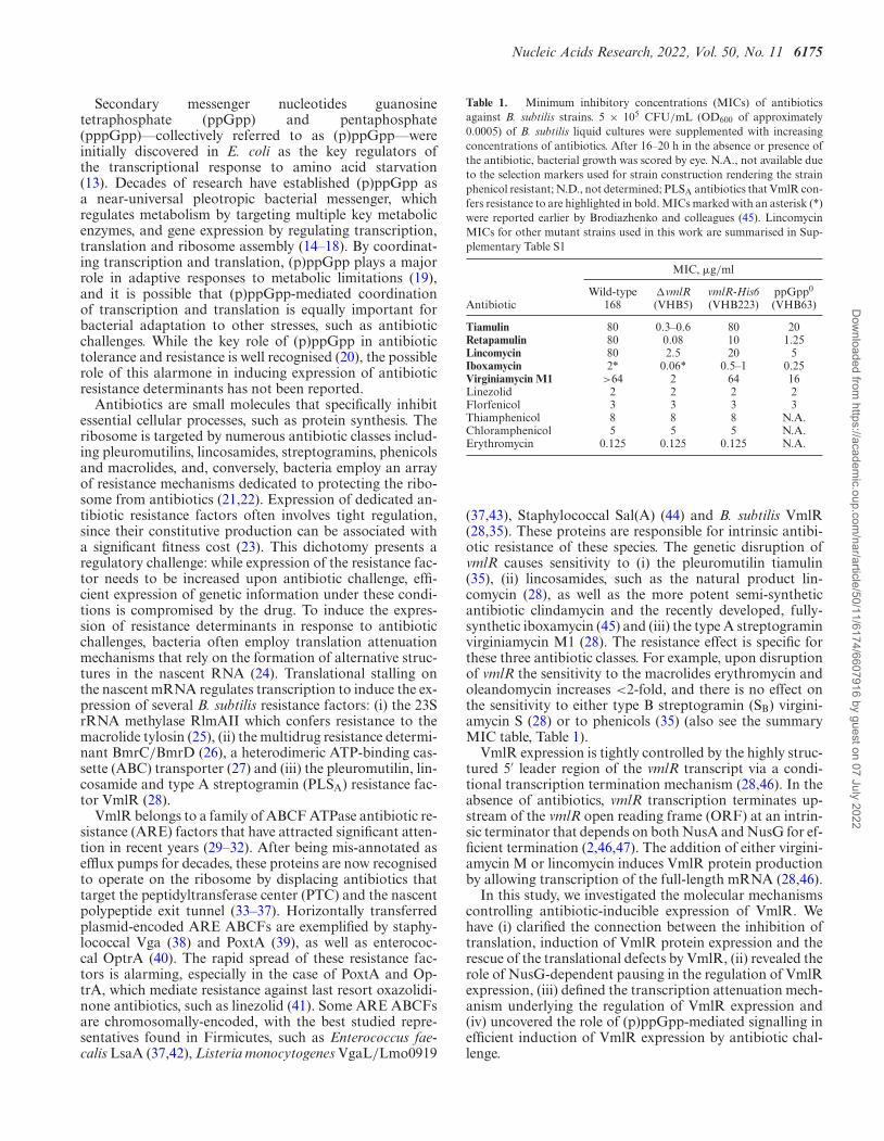
Table 1. Minimum inhibitory concentrations (MICs) of antibioticsagainst B. subtilis strains. 5 × 10⁵ CFU/mL (OD600 of approximately0.0005) of B. subtilis liquid cultures were supplemented with increasingconcentrations of antibiotics. After 16–20 h in the absence or presence ofthe antibiotic, bacterial growth was scored by eye. N.A., not available dueto the selection markers used for strain construction rendering the strainphenicol resistant; N.D., not determined; PLSA antibiotics that VmlR con-fers resistance to are highlighted in bold. MICs marked with an asterisk (*)were reported earlier by Brodiazhenko and colleagues (45). LincomycinMICs for other mutant strains used in this work are summarised in Sup-plementary Table S1
MIC, �g/ml
AntibioticWild-type
168�vmlR(VHB5)
vmlR-His6(VHB223)
ppGpp0
(VHB63)
Tiamulin 80 0.3–0.6 80 20Retapamulin 80 0.08 10 1.25Lincomycin 80 2.5 20 5Iboxamycin 2* 0.06* 0.5–1 0.25Virginiamycin M1 >64 2 64 16Linezolid 2 2 2 2Florfenicol 3 3 3 3Thiamphenicol 8 8 8 N.A.Chloramphenicol 5 5 5 N.A.Erythromycin 0.125 0.125 0.125 N.A.
(37,43), Staphylococcal Sal(A) (44) and B. subtilis VmlR(28,35). These proteins are responsible for intrinsic antibi-otic resistance of these species. The genetic disruption ofvmlR causes sensitivity to (i) the pleuromutilin tiamulin(35), (ii) lincosamides, such as the natural product lin-comycin (28), as well as the more potent semi-syntheticantibiotic clindamycin and the recently developed, fully-synthetic iboxamycin (45) and (iii) the type A streptograminvirginiamycin M1 (28). The resistance effect is specific forthese three antibiotic classes. For example, upon disruptionof vmlR the sensitivity to the macrolides erythromycin andoleandomycin increases <2-fold, and there is no effect onthe sensitivity to either type B streptogramin (SB) virgini-amycin S (28) or to phenicols (35) (also see the summaryMIC table, Table 1).
VmlR expression is tightly controlled by the highly struc-tured 5′ leader region of the vmlR transcript via a condi-tional transcription termination mechanism (28,46). In theabsence of antibiotics, vmlR transcription terminates up-stream of the vmlR open reading frame (ORF) at an intrin-sic terminator that depends on both NusA and NusG for ef-ficient termination (2,46,47). The addition of either virgini-amycin M or lincomycin induces VmlR protein productionby allowing transcription of the full-length mRNA (28,46).
In this study, we investigated the molecular mechanismscontrolling antibiotic-inducible expression of VmlR. Wehave (i) clarified the connection between the inhibition oftranslation, induction of VmlR protein expression and therescue of the translational defects by VmlR, (ii) revealed therole of NusG-dependent pausing in the regulation of VmlRexpression, (iii) defined the transcription attenuation mech-anism underlying the regulation of VmlR expression and(iv) uncovered the role of (p)ppGpp-mediated signalling inefficient induction of VmlR expression by antibiotic chal-lenge.
Dow
nloaded from https://academ
ic.oup.com/nar/article/50/11/6174/6607916 by guest on 07 July 2022
6176 Nucleic Acids Research, 2022, Vol. 50, No. 11
MATERIALS AND METHODS
Bacterial strains, plasmids and oligonucleotides
Strains (and information regarding their sensitivity to lin-comycin), plasmids, oligonucleotides, and synthetic DNAsequences used in this study are provided in SupplementaryTable S1.
Synthesis of iboxamycin
Iboxamycin was synthesised as described previously (48).
Sequence and structure analyses
To retrieve homologous regions of the B. subtilis 5′ leaderregion, blastn (https://blast.ncbi.nlm.nih.gov/) was carriedout using the NCBI representative genome database, usingthe upstream intergenic region of vmlR as the query. Se-quences were visualised and aligned with Aliview v. 1.26(49) and MAFFT v7.453 with the L-INS-i strategy (50).Secondary structures of the vmlR 5′ leader region were pre-dicted using RNA Fold (51) and Mfold (52).
Growth assays
B. subtilis strains were pre-grown on LB plates overnight at30◦C. Individual colonies were re-streaked on an LB plate,pre-grown at 37◦C for approximately 4 h, and fresh colonieswere used to inoculate 200 ml of LB (OD600 adjusted to0.1) and growth was continued at 37◦C. OD600 was moni-tored at half-hour intervals. For western blotting analysis,cells were collected at various times by centrifugation (8000rpm, 5 min). After removing the supernatant, cell pelletswere frozen by liquid nitrogen and stored at –80◦C.
Antibiotic sensitivity testing
Experiments were performed as described previously (35).Briefly, B. subtilis cells were pre-grown on LB plates supple-mented with 1 mM IPTG if needed overnight at 30◦C. Indi-vidual colonies were re-streaked on an LB plate, pre-grownat 37◦C for approximately 4 h, and fresh colonies were usedto inoculate filtered LB medium in the presence and absenceof 1 mM IPTG, and OD600 adjusted to 0.01. The cultureswere seeded on a 100-well honeycomb plate (Oy GrowthCurves AB Ltd, Helsinki, Finland), and plates were incu-bated in a Bioscreen C (Labsystems, Helsinki, Finland) at37◦C with continuous shaking. The indicated concentrationof antibiotics was added from the beginning or added after90 min of growth (OD600 ≈ 0.1).
The minimum inhibitory concentrations (MIC) were cal-culated based on guidelines from the European Committeeon Antimicrobial Susceptibility Testing (EUCAST) (http://www.eucast.org/ast of bacteria/mic determination). B.subtilis strains were grown in LB medium supplementedwith increasing concentrations of antibiotics after inocu-lation with 5 × 10⁵ CFU/ml (OD600 of ∼0.0005). After16–20 h at 37◦C without shaking, the presence or absenceof bacterial growth was scored by eye.
Antibiotics treatment and sampling for western blotting anal-ysis
B. subtilis strains were pre-grown on LB plates overnight at30◦C. Individual colonies were streaked on an LB plate, in-cubated at 37◦C for ∼4 h, and fresh colonies were used to in-oculate LB culture (OD600 adjusted to 0.02) that were grownat 37◦C. At OD600 of ≈0.25, the indicated concentration ofantibiotics was added. After the indicated time, treated cellswere collected by centrifugation (8000 rpm, 5 min). Afterremoving the supernatant, cell pellets were frozen by liquidnitrogen and stored at –80◦C.
Preparation of B. subtilis 30S ribosomal subunits
B. subtilis 70S ribosomes were purified as described pre-viously (53), diluted with low-magnesium HEPES:Polymixbuffer (1 mM MgOAc) and incubated on ice for 30 min topromote the dissociation of subunits. The sample was thenresolved on a 10–40% sucrose gradient in overlay buffer (60mM NH4Cl, 1 mM Mg(OAc)2, 0.25 mM EDTA, 3 mM �-mercaptoethanol, 20 mM Tris–HCl (pH 7.5) in a zonal ro-tor (Ti 15, Beckman, 18 h at 21 000 rpm). The peak con-taining pure 30S ribosomal subunits was pelleted by cen-trifugation (48 h at 35 000 rpm), and the final ribosomalpreparation was dissolved in HEPES:Polymix buffer with 5mM Mg(OAc)2.
Sucrose gradient fractionation before and after cold stress
The experiments were performed as described previously(53) with minor modifications. B. subtilis strains were pre-grown on LB plates overnight at 30◦C. Individual colonieswere re-streaked on LB plate, grown at 37◦C for ∼4 h, andfresh colonies were used to inoculate 200 mL LB culturesthat were grown at 37◦C. At OD600 of 0.2, the cultures weretransferred to 20◦C, grown for an additional 60 min, andthen samples were collected by centrifugation. After remov-ing the supernatant, cell pellets were frozen by liquid nitro-gen and stored at -80◦C. Cell pellets were dissolved in 0.5mL of HEPES:Polymix buffer (5 mM Mg(OAc)2) supple-mented with 1 mM PMSF, lysed using a FastPrep homoge-nizer (MP Biomedicals) by four 20 s pulses at a speed of 4.0m/s with chilling on ice for 3 min between each cycle), andclarified by centrifugation. These samples were also used forwestern blotting analysis as described below. Ten A260 unitsof each extract were loaded onto 10–35% (w/v) sucrose den-sity gradients in Polymix buffer, 5 mM Mg(OAc)2. Gradi-ents were resolved by centrifugation at 36 000 rpm for 3 hat 4◦C in a SW41 rotor (Beckman). Sucrose gradients wereboth poured and fractionated using a Biocomp GradientStation (BioComp Instruments).
Western blotting analysis
Frozen cell pellets were treated as described above for su-crose gradient fractionation. 0.2 A260 unit of each cell ex-tract was boiled with 2× SDS loading buffer (100 mM Tris–HCl pH 6.8, 4% SDS (w/v) 0.02% Bromophenol blue, 20%glycerol (w/v) 4% � -mercaptoethanol) were resolved by10% or 12% SDS PAGE and transferred to a nitrocellulosemembrane (Trans-Blot Turbo Midi Nitrocellulose Transfer
Dow
nloaded from https://academ
ic.oup.com/nar/article/50/11/6174/6607916 by guest on 07 July 2022
Nucleic Acids Research, 2022, Vol. 50, No. 11 6177
Pack, Bio-Rad, 0.2 �m pore size) with the use of a Trans-Blot Turbo Transfer Starter System (Bio-Rad) (10 min,2.5 A, 25 V). C-terminally His6-tagged VmlR and BmrDwere detected using anti-His6 primary antibodies (Wako,010-21861; 1:10 000 dilution) combined with anti-mouse-HRP secondary antibodies (Rockland; 610-103-040; 1:10000 dilution). 50S ribosomal proteins L3 and L11 were de-tected using anti-L3 and anti-L11 primary antibodies (1:20000 dilution, both provided by Fujio Kawamura), com-bined with goat anti-rabbit IgG-HRP secondary antibod-ies (Sigma-Aldrich, A0545; 1:10 000 dilution). The hiber-nation Promoting Factor (HPF) was detected using a poly-clonal anti-HPF antibody (1:10 000 dilution, provided byFujio Kawamura (54)) combined with goat anti-rabbit IgG-HRP secondary antibodies (Sigma-Aldrich, A0545; 1:10000 dilution). ECL detection was performed using Western-Bright™ Quantum (K-12042-D10, Advansta) western blot-ting substrate and an ImageQuant LAS 4000 (GE Health-care) imaging system.
Functional genomics analyses of NusA-stimulated termina-tion and NusG-dependent pause sites
We previously performed native elongating transcript se-quencing followed by RNase I digestion (RNET-seq) underwild-type (PLBS730, +IPTG), NusA depletion (PLBS730,–IPTG), and �nusG (PLBS731, +IPTG) conditions (3,55).The RNET-seq data are available in the National Cen-ter for Biotechnology Information Sequence Read Archive(BioProject ID numbers PRJNA603835) and GEO (acces-sion number GSE186285). Term-seq was previously per-formed under wild-type, NusA depletion, nusG deletion,and combined NusA depletion / nusG deletion conditions(47), and the data are available in GEO (accession numbersGSE67058 and GSE154522).
In vitro transcription
We designed a gBlock gene fragment that containedmodifications of the natural vmlR promoter region, re-sulting in consensus –35 and extended –10 promoterelements, as well as ideal spacing between the –10 elementand the +1 nucleotide. The final promoter sequence wasTTGACATGAATTTAAAGGTATGTTATAATGTTTGTA, where bold nucleotides highlight the –35 and extended-10 elements, and the +1 nucleotide. We also inserted twoadenosine residues five nucleotides downstream of +1 toyield a 14-nt G-less cassette. The G-less cassette was fol-lowed by the vmlR leader region and the first 18 codons ofthe vmlR coding sequence. After the gBlock was amplifiedby PCR using primers vmlR-amp-For and vmlR-amp-Rev,the PCR product was cloned into the EcoRI and HindIIIsites of pTZ19R (Thermo Fisher Scientific), resulting inplasmid pZM72. The NusG-dependent pause motif in thevmlR leader (3) was subsequently mutated from TTATTTto TTACAA via the QuikChange protocol (Agilent)using pZM72 as template and primers vmlR-mut-For andvmlR-mut-Rev, resulting in plasmid pZM73. Templatesfor in vitro transcription containing the WT or mutantNusG-dependent pause motif were PCR-amplified usingpZM72 and pZM73 as templates and the primer pairvmlR-amp-For and vmlR-amp-Rev, respectively.
Analysis of RNAP pausing and termination was per-formed as described previously with modifications (2).Halted elongation complexes containing a 14-nt transcriptwere formed for 5 min at 37◦C by combining equal vol-umes of 2x template (50–200 nM) with 2× halted elonga-tion complex master mix containing 80 �M ATP and CTP,2 �M UTP, 100 �g/ml bovine serum albumin, 150 �g/ml(0.38 �M) B. subtilis RNAP holoenzyme, 0.76 �M �A, 2�Ci of [�-32P]UTP and 2× transcription buffer (1× = 40mM Tris–HCl, pH 8.0, 5 mM MgCl2, 5% trehalose, 0.1mM EDTA and 4 mM dithiothreitol). RNAP and �A wereadded from a 10× stock solution containing 0.75 mg/mlRNAP and 0.175 mg/ml �A in enzyme dilution buffer (20mM Tris–HCl, pH 8.0, 40 mM KCl, 1 mM dithiothre-itol and 50% glycerol). A 4× solution containing either 0 or4 �M of NusG and/or NusA in 1x transcription buffer wereadded and the resulting solution was incubated for 5 min at23◦C. A 4× extension master mix containing 80 �M KCl,600 �M of each NTP, 400 �M rifampicin, in 1× transcrip-tion buffer was added. For each pausing assay, the reactionwas incubated at 23◦C, with aliquots removed and addedto 2× stop/gel loading solution (40 mM Tris-base, 20 mMNa2EDTA, 0.2% sodium dodecyl sulfate, 0.05% bromophe-nol blue, and 0.05% xylene cyanol in formamide) at thespecified time points. A 30 min time point was included forall pausing assays, which we considered to be our termina-tion condition. RNA bands were separated on standard 5%sequencing polyacrylamide gels, which were subsequentlyimaged on a Typhoon 8600 Phosphorimager (GE Health-care Life Sciences). Each in vitro transcription experimentwas repeated at least twice, with representative gels shown.Termination efficiencies and pausing half-lives were quan-tified as described previously (56).
Toeprint assay
Toeprint assays followed a published procedure (25). DNAtemplates for in vitro transcription were generated by PCRusing plasmids pYH389 (WT) and pYH399 (SD mu-tant) and primer pairs T7-WT-For/PCR-Rev and T7-mut-For/PCR-Rev, respectively. WT and SD mutant vmlR RNAwere synthesized using the RNAMaxx kit (Agilent Tech-nologies). RNA containing from +1 to +165 relative to thevmlR transcription start site was gel purified. The toeprintprimer (vmlR-Toeprint) complementary to +79 to +101 ofvmlR was 5′-end labelled using T4 polynucleotide kinase(New England Biolabs) and [� -32P] ATP (7000 Ci/mmol)(Perkin Elmer). Each toeprint reaction mixture (10 �l) con-tained 2 �l of hybridization mixture (1 �l of 1 �M labelledprimer and 1 �l of 1 �M RNA), 2 �l of 17 �M B. sub-tilis 30S ribosomal subunits or dilution buffer, 6 �l of re-action mixture (1× reverse transcriptase buffer, 10 �M Es-cherichia coli fMet-tRNAi
fMet, 2 �g of yeast RNA, 375 �Meach deoxynucleoside triphosphate (dNTP), 10 mM dithio-threitol and 0.1 mg/ml bovine serum albumin). Hybridiza-tion mixtures were incubated for 3 min at 80◦C followed byslow cooling for 10 min at room temperature. Prior to addi-tion, 30S ribosomal subunits were activated by incubationfor 15 min at 37◦C. After addition, 30S ribosomal subunitmixtures were incubated for 10 min at 37◦C. 1 �l AMV (1U) was added, and incubation was continued for 15 min at
Dow
nloaded from https://academ
ic.oup.com/nar/article/50/11/6174/6607916 by guest on 07 July 2022
6178 Nucleic Acids Research, 2022, Vol. 50, No. 11
BA
C
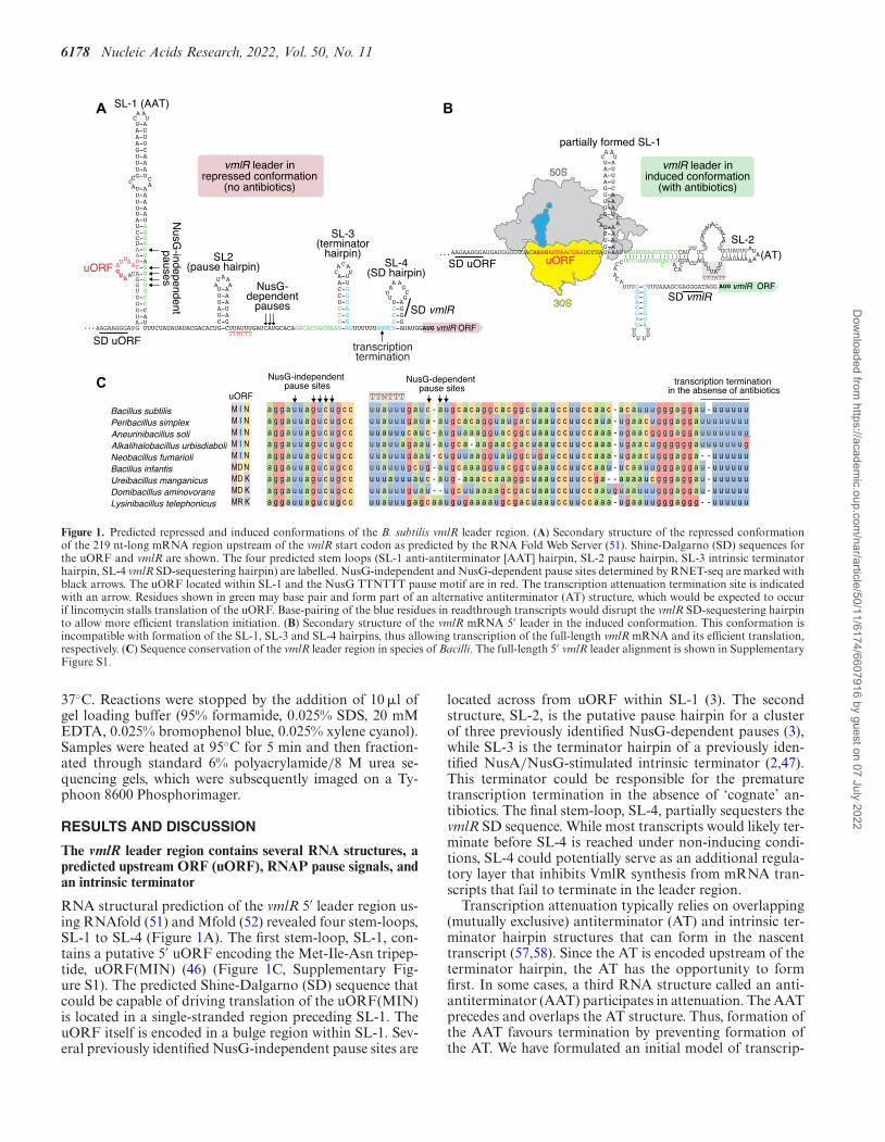
Figure 1. Predicted repressed and induced conformations of the B. subtilis vmlR leader region. (A) Secondary structure of the repressed conformationof the 219 nt-long mRNA region upstream of the vmlR start codon as predicted by the RNA Fold Web Server (51). Shine-Dalgarno (SD) sequences forthe uORF and vmlR are shown. The four predicted stem loops (SL-1 anti-antiterminator [AAT] hairpin, SL-2 pause hairpin, SL-3 intrinsic terminatorhairpin, SL-4 vmlR SD-sequestering hairpin) are labelled. NusG-independent and NusG-dependent pause sites determined by RNET-seq are marked withblack arrows. The uORF located within SL-1 and the NusG TTNTTT pause motif are in red. The transcription attenuation termination site is indicatedwith an arrow. Residues shown in green may base pair and form part of an alternative antiterminator (AT) structure, which would be expected to occurif lincomycin stalls translation of the uORF. Base-pairing of the blue residues in readthrough transcripts would disrupt the vmlR SD-sequestering hairpinto allow more efficient translation initiation. (B) Secondary structure of the vmlR mRNA 5′ leader in the induced conformation. This conformation isincompatible with formation of the SL-1, SL-3 and SL-4 hairpins, thus allowing transcription of the full-length vmlR mRNA and its efficient translation,respectively. (C) Sequence conservation of the vmlR leader region in species of Bacilli. The full-length 5′ vmlR leader alignment is shown in SupplementaryFigure S1.
37◦C. Reactions were stopped by the addition of 10 �l ofgel loading buffer (95% formamide, 0.025% SDS, 20 mMEDTA, 0.025% bromophenol blue, 0.025% xylene cyanol).Samples were heated at 95◦C for 5 min and then fraction-ated through standard 6% polyacrylamide/8 M urea se-quencing gels, which were subsequently imaged on a Ty-phoon 8600 Phosphorimager.
RESULTS AND DISCUSSION
The vmlR leader region contains several RNA structures, apredicted upstream ORF (uORF), RNAP pause signals, andan intrinsic terminator
RNA structural prediction of the vmlR 5′ leader region us-ing RNAfold (51) and Mfold (52) revealed four stem-loops,SL-1 to SL-4 (Figure 1A). The first stem-loop, SL-1, con-tains a putative 5′ uORF encoding the Met-Ile-Asn tripep-tide, uORF(MIN) (46) (Figure 1C, Supplementary Fig-ure S1). The predicted Shine-Dalgarno (SD) sequence thatcould be capable of driving translation of the uORF(MIN)is located in a single-stranded region preceding SL-1. TheuORF itself is encoded in a bulge region within SL-1. Sev-eral previously identified NusG-independent pause sites are
located across from uORF within SL-1 (3). The secondstructure, SL-2, is the putative pause hairpin for a clusterof three previously identified NusG-dependent pauses (3),while SL-3 is the terminator hairpin of a previously iden-tified NusA/NusG-stimulated intrinsic terminator (2,47).This terminator could be responsible for the prematuretranscription termination in the absence of ‘cognate’ an-tibiotics. The final stem-loop, SL-4, partially sequesters thevmlR SD sequence. While most transcripts would likely ter-minate before SL-4 is reached under non-inducing condi-tions, SL-4 could potentially serve as an additional regula-tory layer that inhibits VmlR synthesis from mRNA tran-scripts that fail to terminate in the leader region.
Transcription attenuation typically relies on overlapping(mutually exclusive) antiterminator (AT) and intrinsic ter-minator hairpin structures that can form in the nascenttranscript (57,58). Since the AT is encoded upstream of theterminator hairpin, the AT has the opportunity to formfirst. In some cases, a third RNA structure called an anti-antiterminator (AAT) participates in attenuation. The AATprecedes and overlaps the AT structure. Thus, formation ofthe AAT favours termination by preventing formation ofthe AT. We have formulated an initial model of transcrip-
Dow
nloaded from https://academ
ic.oup.com/nar/article/50/11/6174/6607916 by guest on 07 July 2022
Nucleic Acids Research, 2022, Vol. 50, No. 11 6179
0 0.02
0 0.04
0.1
0.2
0.4
1 2 4 10 20 40
T0 T30 (min)
lincomycin (μg/ml)
VmlRHis6 (64 kDa)
L3 (23 kDa)wt
T30 (min) erythromycin (μg/ml)linezolid
0 0 0.02
50.
050.
10.
250.
51 0.
0250.
050.
10.
250.
51
T0
VmlRHis6 L3
wt
D0 0.
001
0 0.00
20.
0040.
010.
020.
040.1
0.2
0.4 1 2
T0 T30 (min)
repatamulin(μg/ml)
VmlRHis6
L11wt
E
induction window
C T0
0 5 20 60
T30
(ng/ml)Iboxamycin
wt
B
1 μg/ml 2 μg/ml 4 μg/ml 10 μg/ml 20 μg/ml 40 μg/ml
no antibiotic
0.01
2
468
0.1
2
468
1
129630
20,40
10
1
24
00-40 μg/ml
OD
600,
a.u
.
Time, h
wt
+ lincomycinA
VmlRHis6 (64 kDa)
L11 (15 kDa)
(min)
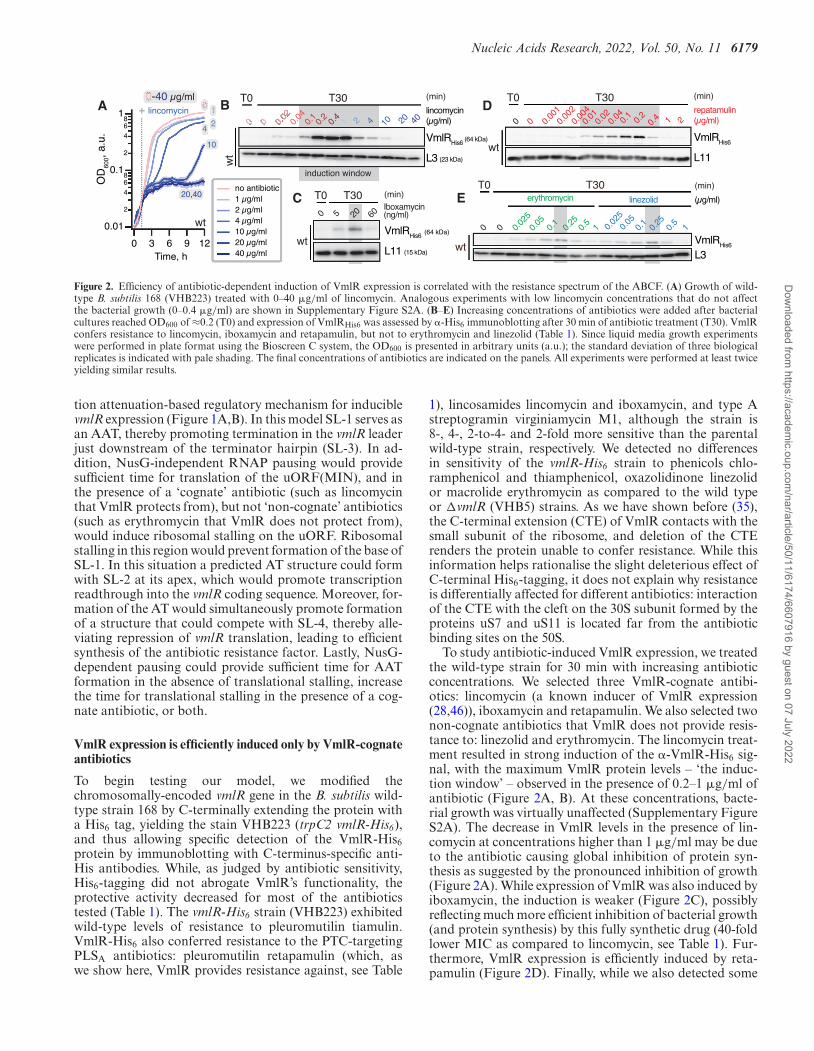
Figure 2. Efficiency of antibiotic-dependent induction of VmlR expression is correlated with the resistance spectrum of the ABCF. (A) Growth of wild-type B. subtilis 168 (VHB223) treated with 0–40 �g/ml of lincomycin. Analogous experiments with low lincomycin concentrations that do not affectthe bacterial growth (0–0.4 �g/ml) are shown in Supplementary Figure S2A. (B–E) Increasing concentrations of antibiotics were added after bacterialcultures reached OD600 of ≈0.2 (T0) and expression of VmlRHis6 was assessed by �-His6 immunoblotting after 30 min of antibiotic treatment (T30). VmlRconfers resistance to lincomycin, iboxamycin and retapamulin, but not to erythromycin and linezolid (Table 1). Since liquid media growth experimentswere performed in plate format using the Bioscreen C system, the OD600 is presented in arbitrary units (a.u.); the standard deviation of three biologicalreplicates is indicated with pale shading. The final concentrations of antibiotics are indicated on the panels. All experiments were performed at least twiceyielding similar results.
tion attenuation-based regulatory mechanism for induciblevmlR expression (Figure 1A,B). In this model SL-1 serves asan AAT, thereby promoting termination in the vmlR leaderjust downstream of the terminator hairpin (SL-3). In ad-dition, NusG-independent RNAP pausing would providesufficient time for translation of the uORF(MIN), and inthe presence of a ‘cognate’ antibiotic (such as lincomycinthat VmlR protects from), but not ‘non-cognate’ antibiotics(such as erythromycin that VmlR does not protect from),would induce ribosomal stalling on the uORF. Ribosomalstalling in this region would prevent formation of the base ofSL-1. In this situation a predicted AT structure could formwith SL-2 at its apex, which would promote transcriptionreadthrough into the vmlR coding sequence. Moreover, for-mation of the AT would simultaneously promote formationof a structure that could compete with SL-4, thereby alle-viating repression of vmlR translation, leading to efficientsynthesis of the antibiotic resistance factor. Lastly, NusG-dependent pausing could provide sufficient time for AATformation in the absence of translational stalling, increasethe time for translational stalling in the presence of a cog-nate antibiotic, or both.
VmlR expression is efficiently induced only by VmlR-cognateantibiotics
To begin testing our model, we modified thechromosomally-encoded vmlR gene in the B. subtilis wild-type strain 168 by C-terminally extending the protein witha His6 tag, yielding the stain VHB223 (trpC2 vmlR-His6),and thus allowing specific detection of the VmlR-His6protein by immunoblotting with C-terminus-specific anti-His antibodies. While, as judged by antibiotic sensitivity,His6-tagging did not abrogate VmlR’s functionality, theprotective activity decreased for most of the antibioticstested (Table 1). The vmlR-His6 strain (VHB223) exhibitedwild-type levels of resistance to pleuromutilin tiamulin.VmlR-His6 also conferred resistance to the PTC-targetingPLSA antibiotics: pleuromutilin retapamulin (which, aswe show here, VmlR provides resistance against, see Table
1), lincosamides lincomycin and iboxamycin, and type Astreptogramin virginiamycin M1, although the strain is8-, 4-, 2-to-4- and 2-fold more sensitive than the parentalwild-type strain, respectively. We detected no differencesin sensitivity of the vmlR-His6 strain to phenicols chlo-ramphenicol and thiamphenicol, oxazolidinone linezolidor macrolide erythromycin as compared to the wild typeor �vmlR (VHB5) strains. As we have shown before (35),the C-terminal extension (CTE) of VmlR contacts with thesmall subunit of the ribosome, and deletion of the CTErenders the protein unable to confer resistance. While thisinformation helps rationalise the slight deleterious effect ofC-terminal His6-tagging, it does not explain why resistanceis differentially affected for different antibiotics: interactionof the CTE with the cleft on the 30S subunit formed by theproteins uS7 and uS11 is located far from the antibioticbinding sites on the 50S.
To study antibiotic-induced VmlR expression, we treatedthe wild-type strain for 30 min with increasing antibioticconcentrations. We selected three VmlR-cognate antibi-otics: lincomycin (a known inducer of VmlR expression(28,46)), iboxamycin and retapamulin. We also selected twonon-cognate antibiotics that VmlR does not provide resis-tance to: linezolid and erythromycin. The lincomycin treat-ment resulted in strong induction of the �-VmlR-His6 sig-nal, with the maximum VmlR protein levels – ‘the induc-tion window’ – observed in the presence of 0.2–1 �g/ml ofantibiotic (Figure 2A, B). At these concentrations, bacte-rial growth was virtually unaffected (Supplementary FigureS2A). The decrease in VmlR levels in the presence of lin-comycin at concentrations higher than 1 �g/ml may be dueto the antibiotic causing global inhibition of protein syn-thesis as suggested by the pronounced inhibition of growth(Figure 2A). While expression of VmlR was also induced byiboxamycin, the induction is weaker (Figure 2C), possiblyreflecting much more efficient inhibition of bacterial growth(and protein synthesis) by this fully synthetic drug (40-foldlower MIC as compared to lincomycin, see Table 1). Fur-thermore, VmlR expression is efficiently induced by reta-pamulin (Figure 2D). Finally, while we also detected some
Dow
nloaded from https://academ
ic.oup.com/nar/article/50/11/6174/6607916 by guest on 07 July 2022
6180 Nucleic Acids Research, 2022, Vol. 50, No. 11
induction of VmlR expression in the presence of the non-cognate antibiotics erythromycin and linezolid, the induc-tion is substantially weaker than in the case of the cognateantibiotics (Figure 2E).
VmlR is induced by defects in translation and induction doesnot depend on VmlR activity
A recent study of Staphylococcal antibiotic resistance factorVga(A) suggested that the resistance profile of this ABCFfactor is the key determinant of which antibiotics induce itsexpression (59). To establish whether the same holds truefor VmlR, we examined the possible connection between theability of VmlR to counteract the translation defect causedby the antibiotic by providing resistance and the ability ofthe translational defect to induce VmlR expression.
The lack of efficient VmlR induction by erythromycinand linezolid could be due to the regulatory elements con-trolling VmlR expression failing to sense and respond totranslational defects caused by non-cognate antibiotics. Thetwo tested VmlR-inducing PLSA antibiotics retapamulinand lincomycin are expected to stall the ribosome at initi-ation. Initiation-specific stalling was directly shown for re-tapamulin (60). Biochemical and structural studies demon-strated that lincosamides lincomycin (61,62) and iboxam-ycin (63) interfere with efficient accommodation of the A-site tRNA, and, therefore, in the cellular context are ex-pected to compromise the efficient progression from initi-ation to polypeptide elongation. On the other hand, line-zolid and erythromycin target elongation; efficient stallingby these antibiotics is context-specific and relies on the in-teractions between the antibiotic, the nascent polypeptidechain, and the ribosome peptide exit tunnel (64–70). If thepredicted three-codon uORF(MIN) (46) plays a role in con-trolling VmlR expression, it is possible that lincosamidesand pleuromutilins would cause the ribosome to stall lead-ing to VmlR induction, whereas ribosomes would fail tostall in the presence of oxazolidinones or macrolides, andtherefore would not induce VmlR expression. Another ex-planation for inefficient VmlR induction when the cells arechallenged with antibiotics that VmlR does not confer re-sistance to is that while inhibition of ribosomal function isnecessary and sufficient to induce VmlR expression, due tothe lack of protection from these non-cognate antibiotics,the net increase in VmlR expression is negligible because theprotein synthesis is effectively abrogated. Thus, the width ofthe antibiotic concentration window in which VmlR expres-sion is induced, but global protein production is not yet sup-pressed, reflects the difference in antibiotic sensitivity priorto VmlR induction and post-induction. This induction win-dow is expected to be relatively wide for cognate antibioticsand narrow for non-cognate antibiotics. Finally, one wouldexpect the window to be narrower in the case of more potentantibiotic variants, such as iboxamycin (compare Figure 2Band Figure 2C).
To test this induction window hypothesis, we constructeda vmlRE129Q (VHB628) strain in which the first ATPasecassette of VmlR is rendered inactive by substituting thecatalytic glutamic acid residue of the Walker B motif forglutamine. This substitution rendered the ABCF resistancefactor non-functional, resulting in the vmlRE129Q strain be-
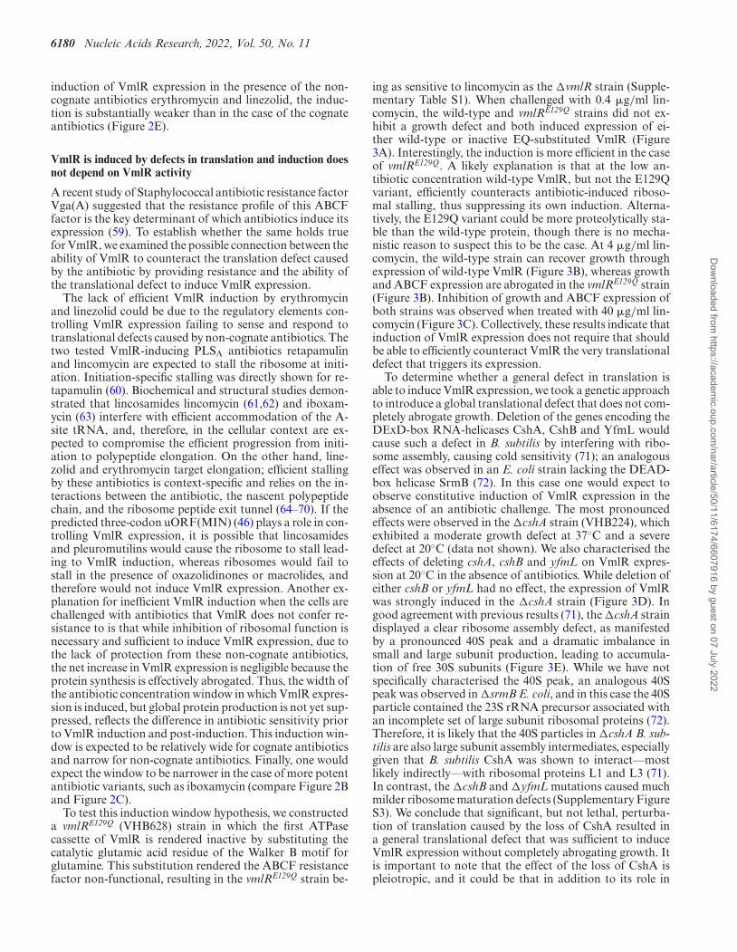
ing as sensitive to lincomycin as the �vmlR strain (Supple-mentary Table S1). When challenged with 0.4 �g/ml lin-comycin, the wild-type and vmlRE129Q strains did not ex-hibit a growth defect and both induced expression of ei-ther wild-type or inactive EQ-substituted VmlR (Figure3A). Interestingly, the induction is more efficient in the caseof vmlRE129Q. A likely explanation is that at the low an-tibiotic concentration wild-type VmlR, but not the E129Qvariant, efficiently counteracts antibiotic-induced riboso-mal stalling, thus suppressing its own induction. Alterna-tively, the E129Q variant could be more proteolytically sta-ble than the wild-type protein, though there is no mecha-nistic reason to suspect this to be the case. At 4 �g/ml lin-comycin, the wild-type strain can recover growth throughexpression of wild-type VmlR (Figure 3B), whereas growthand ABCF expression are abrogated in the vmlRE129Q strain(Figure 3B). Inhibition of growth and ABCF expression ofboth strains was observed when treated with 40 �g/ml lin-comycin (Figure 3C). Collectively, these results indicate thatinduction of VmlR expression does not require that shouldbe able to efficiently counteract VmlR the very translationaldefect that triggers its expression.
To determine whether a general defect in translation isable to induce VmlR expression, we took a genetic approachto introduce a global translational defect that does not com-pletely abrogate growth. Deletion of the genes encoding theDExD-box RNA-helicases CshA, CshB and YfmL wouldcause such a defect in B. subtilis by interfering with ribo-some assembly, causing cold sensitivity (71); an analogouseffect was observed in an E. coli strain lacking the DEAD-box helicase SrmB (72). In this case one would expect toobserve constitutive induction of VmlR expression in theabsence of an antibiotic challenge. The most pronouncedeffects were observed in the �cshA strain (VHB224), whichexhibited a moderate growth defect at 37◦C and a severedefect at 20◦C (data not shown). We also characterised theeffects of deleting cshA, cshB and yfmL on VmlR expres-sion at 20◦C in the absence of antibiotics. While deletion ofeither cshB or yfmL had no effect, the expression of VmlRwas strongly induced in the �cshA strain (Figure 3D). Ingood agreement with previous results (71), the �cshA straindisplayed a clear ribosome assembly defect, as manifestedby a pronounced 40S peak and a dramatic imbalance insmall and large subunit production, leading to accumula-tion of free 30S subunits (Figure 3E). While we have notspecifically characterised the 40S peak, an analogous 40Speak was observed in �srmB E. coli, and in this case the 40Sparticle contained the 23S rRNA precursor associated withan incomplete set of large subunit ribosomal proteins (72).Therefore, it is likely that the 40S particles in �cshA B. sub-tilis are also large subunit assembly intermediates, especiallygiven that B. subtilis CshA was shown to interact––mostlikely indirectly––with ribosomal proteins L1 and L3 (71).In contrast, the �cshB and �yfmL mutations caused muchmilder ribosome maturation defects (Supplementary FigureS3). We conclude that significant, but not lethal, perturba-tion of translation caused by the loss of CshA resulted ina general translational defect that was sufficient to induceVmlR expression without completely abrogating growth. Itis important to note that the effect of the loss of CshA ispleiotropic, and it could be that in addition to its role in
Dow
nloaded from https://academ
ic.oup.com/nar/article/50/11/6174/6607916 by guest on 07 July 2022
Nucleic Acids Research, 2022, Vol. 50, No. 11 6181
VmlRHis6
L340
0 30 60 90 120
wt
0 30 60 90 120
E129Q
(min)
VmlRHis6 L34
μg/mllincomycin
0 30 60 90 120
wt E129Q (min)
150
0 30 60 90 120
150
μg/mllincomycin
E129Q
4
68
1
2
150
1209060300
4
68
1
2
120
9060300
wt
+ 4 μg/ml lincomycin
+ 40 μg/ml lincomycin
Time, min
Time, min
B
C
wt E
10 - 35 % sucrose gradient
T0 (OD600 = 0.18) T60 (OD600 = 0.43)
polysomes
70S
50S40
S
30S
A26
0, a
.u.
top,
10%
35%
∆cshA
E129Q
A0
VmlRHis6
L3
15 30 45 60
wt
0 15 30 45 60
E129Q (min)
0.4μg/ml
lincomycin456
1
2
604530150
Time, min
OD
600
+ 0.4 μg/ml lincomycin
wt
E129Q
DT0 T60
wt
T0 T60
∆cshA
T0 T60
∆cshB
T0 T60
∆yfmL
VmlRHis6
L3
Time after downshift to 20˚C (min)
MIC (μg/ml)2.5 20
MIC (μg/ml)2.5 20
MIC (μg/ml)2.5 20
OD
600
OD
600
Figure 3. VmlR is induced by defects in translation and induction does not depend on VmlR activity. (A–C) Lincomycin was added at a final concentra-tion of 0.4 �g/ml after wild-type (VHB627) or the ATPase-deficient E129Q mutant VmlR (VHB628) strains reached OD600 of 0.2–0.3. (A) Permissiveconcentration (0.4 �g/ml) for both strains; the two growth curves are identical and overlay each other. (B) Lincomycin was added at a final concentrationof 4 �g/ml, which is a subinhibitory concentration for the wild-type strain and an inhibitory concentration for the vmlRE129Q strain. (C) Lincomycin wasadded at a final concentration of 40 �g/ml, which is an inhibitory concentration for both strains. Efficient induction of VmlR expression was conditionalon bacterial growth, but not on VmlR functionality since the E129Q variant does not confer resistance to lincomycin. VmlRHis6 expression was detectedby �-His6 immunoblotting. (D) The effect of RNA helicase mutations on induction of VmlR expression. Wild-type 168 (VHB223) and isogenic �cshA(VHB224), �cshB (VHB225) and �yfmL (VHB226) strains were grown to an OD600 of ≈0.2 (T0) at which time the cultures were transferred to 20◦C,and then grown for an additional 60 min (T60). VmlRHis6 expression was detected by �-His6 immunoblotting. (E) Polysome profile of the �cshA strainbefore and after temperature downshift from 37◦C to 20◦C. Analogous experiments with wild-type, �cshB and �yfmL strains are shown in SupplementaryFigure S3. All experiments were performed at least twice, yielding similar results.
rRNA processing, the DEAD-box helicase might operateon the highly structured vmlR 5′ leader region.
Collectively, our results suggest that induction of VmlRexpression is not strictly coupled to the protective functionof the ABCF itself, but is instead mediated by the regula-tory elements controlling the expression of the vmlR ORF.Therefore, we next dissected the roles of the individual reg-ulatory elements located in the vmlR leader region.
NusG-dependent pausing prevents leaky VmlR expression inthe absence of antibiotic and potentiates VmlR inductionupon lincomycin challenge
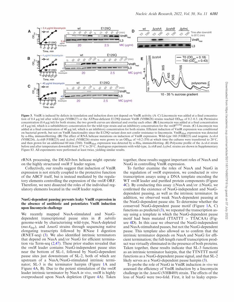
We recently mapped NusA-stimulated and NusG-dependent transcriptional pause sites in B. subtilisgenome-wide by characterising wild-type, NusA-depleted(nusAdep), and �nusG strains through sequencing nativeelongating transcripts followed by RNase I digestion(RNET-seq) (3). We also identified intrinsic terminatorsthat depend on NusA and/or NusG for efficient termina-tion via Term-seq (2,47). These prior studies revealed thatthe vmlR leader contains NusG-independent pause sitesnear the bottom of SL-1, followed by NusG-dependentpause sites just downstream of SL-2, both of which areupstream of a NusA/NusG-stimulated intrinsic termi-nator; SL-3 is the terminator hairpin (Figure 1A andFigure 4A, B). Due to the potent stimulation of the vmlRleader intrinsic terminator by NusA in vivo, vmlR is highlyoverproduced upon NusA depletion (Figure 4A). Taken
together, these results suggest important roles of NusA andNusG in controlling VmlR expression.
To further examine the roles of NusA and NusG inthe regulation of vmlR expression, we conducted in vitrotranscription assays using a DNA template encoding theWT vmlR leader and purified protein components (Figure4C). By conducting this assay ±NusA and/or ±NusG, weconfirmed the existence of NusG-independent and NusG-dependent pausing, as well as the intrinsic terminator. Inaddition, we observed weak NusA-dependent pausing atthe NusG-dependent pause site. To determine whether theconserved NusG-dependent pause motif (Figure 1A, C)functions as predicted (3), we repeated the transcription as-say using a template in which the NusG-dependent pausemotif had been mutated (TTATTT > TTACAA) (Fig-ure 4D). In this case we observed the NusG-independentand NusA-stimultated pauses, but not the NusG-dependentpause. This template also allowed us to confirm that theintrinsic terminator depends on NusA and NusG for effi-cient termination; the full-length runoff transcription prod-uct was virtually eliminated in the presence of both proteins.Taken together, these results indicate that SL-3 functionsas an intrinsic terminator hairpin, that the TTNTTT motiffunctions as a NusG-dependent pause signal, and that SL-2likely serves as a NusG-dependent pause hairpin (3).
To probe the role of NusG in VmlR induction in vivo weassessed the efficiency of VmlR induction by a lincomycinchallenge in the �nusG (VHB489) strain. The effects of theloss of NusG were two-fold. First, it led to leaky expres-
Dow
nloaded from https://academ
ic.oup.com/nar/article/50/11/6174/6607916 by guest on 07 July 2022
6182 Nucleic Acids Research, 2022, Vol. 50, No. 11
30s
60s
90s
120s
45s
30’
30s
60s
90s
120s
45s
30’
30s
60s
90s
120s
45s
30’
30s
60s
90s
120s
45s
30’
30s
60s
90s
120s
45s
30’
30s
60s
90s
120s
45s
30’
30s
60s
90s
120s
45s
30’
30s
60s
90s
120s
45s
30’
- NusA- NusG
+ NusA- NusG
- NusA+ NusG
+ NusA+ NusG
- NusA- NusG
+ NusA- NusG
- NusA+ NusG
+ NusA+ NusG
runoff transcription
wild-type vmlR leader(TTATTT)
mutated vmlR leader(TTATTT>TTACAA)
NusG-dependentpause site
NusG-independentpause site
73 86 96 92 45 87 62 96 %T (30’)
NusA-stimulated
intrinsic terminatorNusG-stimulated
C D
A wt Term-seq wt
nusAdep RNA-seq
wt ΔnusG
nusAdep RNET-seq ΔnusG
NusG-independent pauses
NusG-dependent pauses
wt
ΔnusG
NusG-independent pauses
NusG-dependent pauses
BvmlR ORF
E
VmlRHis6
L11∆nusG
0 0.02
0 0.04
0.1
0.2
0.4
1 2 4 10 20 40
T0 T30 (min)
lincomycin (μg/ml)
F
VmlRHis6
L11
wt
T0 T30 ∆nusG
T0 T30
T0 T30
T0 T30
SL-2* TTNTTT*
SL-2*: GCUAUUU>AAAAUUUTTNTTT*: UUAUUU>UUACAA
Time (min) after the addition of 0.4 μg/ml lincomycin
MIC (μg/ml)20 20 20 20
product
Figure 4. NusG controls lincomycin-dependent induction of VmlR expression. (A, B) Term-seq and RNA-seq (3,47) as well as RNET-seq (3) in wild-type(PLBS730 grown with IPTG), nusAdep (PLBS730 grown without IPTG), and �nusG (PLBS731 grown with IPTG) strains as indicated; ectopic expressionof nusA is controlled by an IPTG-inducible Phy-spankpromoter. (A) The intrinsic terminator in the vmlR leader region requires both NusA and NusG forefficient termination. The NusG-dependent and NusG-independent pauses are marked. The arrow at the top indicates the direction of transcription. (B)Zoomed in magnification showing the NusG-dependent and NusG-independent pauses. (C, D) In vitro reconstitution of NusG-dependent RNAP pausingand NusA/NusG-stimulated termination that controls VmlR expression. In vitro transcription termination assays using DNA templates with wild-type(TTATTT, C) or mutant (TTACAA, (D) NusG pause motifs were performed in the absence or presence of NusA, NusG, or both. Mutations in red are inthe consensus NusG-dependent pause motif. Percent termination is shown below each of the 30 min chase reaction lanes. (E) VmlR expression is inducedby sub-MIC concentrations of lincomycin and the basal expression of VmlR is repressed by NusG. NusG-deficient (�nusG) strain encoding His6-taggedVmlR (VHB489) was treated with increasing concentrations of lincomycin for 30 min (T30) and VmlRHis6 was detected by �-His6 immunoblotting. (F)NusG recognizes the TTNTTT motif in the vmlR leader region to repress VmlR expression. Wild-type (VHB223), �nusG (VHB489), SL-2* (VHB612) andTTNTTT* (VHB611) strains were treated with 0.4 �g/ml lincomycin for 30 min (T30) and levels of VmlRHis6 were assessed by �-His6 immunoblotting.Mutations of SL-2* and TTNTTT* are highlighted in red.
sion of VmlR in the absence of the antibiotic and, second,while the antibiotic stimulates expression, the effect is muchweaker compared to the wild-type strain (Figure 4E, com-pare to Figure 2B). Notably, this level of VmlR expression isstill sufficient to confer wild-type levels of lincomycin resis-tance, with both wild-type and the �nusG strain having thesame MIC of 20 �g/ml. Since the NusG-dependent pausesite is located between SL-1 and before the 3′ portion of theAT structure, pausing at this position probably promotesformation of SL-1 (the AAT structure) in the absence of in-ducing antibiotic. Thus, an increase in AT formation causedby the loss of pausing would explain the leaky expression
observed in the absence of NusG. The weaker inductioncan be explained by the loss of NusG-dependent pausing,which would reduce the time for lincomycin-induced ribo-some stalling to occur at the uORF(MIN). In this case, theAAT would form more frequently leading to increased ter-mination.
To further explore the role of NusG-dependent paus-ing in vivo, we constructed two strains predicted tointerfere with pausing. One strain (VHB612) con-tained mutations (SL-2*) that abrogate the formationof SL-2 (GCUAUUU > AAAAUUU), while the otherstrain (VHB611, TTNTTT*) contained the same mu-
Dow
nloaded from https://academ
ic.oup.com/nar/article/50/11/6174/6607916 by guest on 07 July 2022
Nucleic Acids Research, 2022, Vol. 50, No. 11 6183
tations in the NusG pause motif that we tested in vitro(UUAUUU > UUACAA). SL-2* (VHB612) displayednear-wild-type behaviour upon a lincomycin challenge,both in terms of VmlR expression and lincomycin sensi-tivity as judged by the MIC (Figure 4F), suggesting thatSL-2 is not crucial for NusG-dependent pausing or thatthis mutation also reduces the ability of the AT to preventtermination, resulting in no net change in expression.However, the TTNTTT* strain phenocopied �nusG withVmlR expression being leaky in the absence of antibioticand a lower level of antibiotic-dependent induction (Figure4F). However, this level of induction is sufficient to providewild-type levels of resistance as judged by the MIC.
SL-1 is essential for lincomycin-dependent induction of VmlRexpression and SL-3 is essential for repression of VmlR ex-pression in the absence of antibiotics
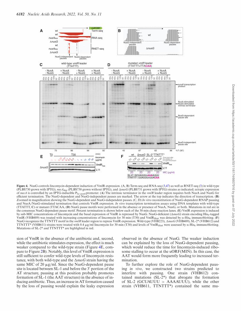
Next, we either mutationally disrupted (*) or deleted (�)stem-loops 1, 3 and 4 and assessed the lincomycin-inducedexpression of VmlR-His6 by immunoblotting as well as thelincomycin sensitivity of the mutant strains (Figure 5A).Deletion of the uORF-containing SL-1 resulted in com-plete loss of VmlR expression, both in the presence and ab-sence of lincomycin, and sensitised the strain to lincomycin,with the MIC corresponding to that of the �vmlR strain(2.5 �g/ml). This result strongly supports the importanceof the AT, because in the absence of SL-1 there is no wayto interfere with formation of the downstream termina-tor hairpin (Figure 1A). As predicted by our model, bothdeletion and destabilization (SL-3*, CCUUCC > UUU-UUU) of the terminator hairpin (SL-3) resulted in consti-tutive, lincomycin-insensitive overexpression of VmlR andhigh levels of lincomycin resistance exceeding that of thewild-type strain (MIC 20–40 �g/ml versus 20 �g/ml). Asexpected, once intrinsic termination is eliminated, expres-sion of VmlR is no longer dependent on SL-1, since the SL-3* �SL-1 (VHB583) strain containing disruptions of bothelements displays the same VmlR expression phenotypes asthe single SL-3* (VHB572) mutant strain. This result is con-sistent with SL-1 containing a portion of the AT, which is re-quired to prevent terminator hairpin formation. Mutationaldisruption of SL-4 (SL-4*, UCCCU > UGGGU) resultedin a moderate defect of VmlR inducible expression and acorresponding increase in lincomycin sensitivity (MIC 5–10 �g/ml), and did not affect the ‘leaky’ expression in theabsence of the antibiotic. The latter result is to be expectedsince in the absence of ribosomal stalling, RNAP naturallyterminates upstream of SL-4 (Figure 1A). The near-wild-type functionality of inducible VmlR expression is consis-tent with the SL-4 sequence not directly participating in theformation of the AT element but rather forming a hairpinwhat would potentially indirectly promote the formation ofthe AT element (Figure 1B).
Translation initiation of the uORF is essential for antibiotic-dependent induction of VmlR expression
Given the crucial role of the uORF-containing SL-1 incontrol of VmlR expression, we next examined the spe-cific role of the uORF in controlling VmlR expression.
Since the VmlR-cognate antibiotics retapamulin and lin-comycin stall initiating ribosomes, we performed toeprint-ing experiments to identify the position of the start codonof the predicted uORF. For this analysis we used B. subtilis30S ribosomal subunits supplemented with aminoacylatedand formylated E. coli initiator tRNA, fMet-tRNAi
fMet,and either wild-type vmlR leader RNA or the same regionwith mutations in the predicted Shine-Dalgarno (SD) re-gion (SD uORF*, AGAAGGG > AAAAAAA) (Figure5B). A strong toeprint signal was observed at a position16 nucleotides downstream of the A in the uORF initiationcodon, confirming that this is an authentic initiation codon.Furthermore, the toeprint signal was not observed on themutant mRNA SD uORF*, thus confirming the predictedlocation of the SD sequence.
Next, to determine whether translation of the uORF isrequired for antibiotic-dependent induction of VmlR ex-pression, we constructed B. subtilis strains containing themutant SD region (SD uORF*, VHB573) or a mutantuORF start codon (uORF*, AUGAUUAAC > CUGAU-UAAC, VHB604). We assessed the effects of both muta-tions on VmlR-His6 expression levels following a 30-minlincomycin challenge and the lincomycin sensitivity of themutant strains (Figure 5C). Disruption of the SD sequenceand mutation of the start codon virtually eliminated expres-sion of VmlR and phenocopied the �vmlR strain in termsof lincomycin sensitivity.
Finally, we probed the role of the uORF coding se-quence identity in lincomycin-mediated induction of VmlRexpression. We have mutated the wild-type B. subtilisuORF(MIN) to encode the three sequence variants that arefound in other Bacilli species: MDN, MDK and MRK (Fig-ure 1C). Both in terms of VmlR induction levels upon lin-comycin challenge and the achieved resistance levels (MIC),uORF(MDN) and uORF(MDK) are at least as functional(or even more efficient) as the wild-type B. subtilis MINvariant (Figure 5D). The uORF(MRK) variant is less ef-ficient, with the mutant strain being moderately sensitive tolincomycin (MIC 10 �g/ml, a 2-fold decrease) due to signif-icantly compromised efficiency of VmlRHis6 induction (Fig-ure 5D).
Collectively, our results suggest that the regulatory mech-anism controlling VmlR expression is analogous (althoughprobably not homologous) to that described for Strepto-coccal MsrD macrolide ABCF resistance factor (73). Simi-lar to VmlR, induction of MsrD requires antibiotic-specific(macrolide-specific elongation arrest in this case) ribosomalstalling on the uORF to counter constitutive intrinsic tran-scription termination. Unlike the case with VmlR, a NusG-dependent pause site was not identified in the msrD mRNA5′ region (73).
The lack of (p)ppGpp-mediated signalling reduces inducibleVmlR-dependent antibiotic resistance
Since incomplete, but significant inhibition of translation isthe key to induction of VmlR expression, we reasoned that,similarly to VmlR induction in the translationally compro-mised �cshA background, VmlR might be induced in thestationary phase when bacteria experience nutrient limi-tation and accumulate (p)ppGpp (74). This is due to the
Dow
nloaded from https://academ
ic.oup.com/nar/article/50/11/6174/6607916 by guest on 07 July 2022
6184 Nucleic Acids Research, 2022, Vol. 50, No. 11
T0 T30
T0 T30
T0 T30
wt uORF*
SD uORF*: AGAAGGG>AAAAAAAuORF*: AUGAUUAAC>CUGAUUAAC
VmlRHis6
L11
MIC (μg/ml)20 2.5 2.5
CSD uORF*
AT0 T30
T0 T30
T0 T30
T0 T30
T0 T30
T0 T30
VmlRHis6
L11
wt ∆SL-1 ∆SL-3 SL-3* ∆SL-1 SL-4* SL-3*
SL-3*: CCUUCC>UUUUUU SL-4*: UCCCU>UGGGUMIC (μg/ml)20 2.5 20-40 5-10 20-40 20-40
Time (min) after the addition of 0.4 μg/ml lincomycin
A C G U + + + + +
toeprint
wt wtSD uORF*
5’-AACAAGAAGGGAUGAUGGGUGGACAAUGAUUAACUAAUCUUA-3’SD uORF
FL
+16 toeprint
B
uORF(MIN)
AUG
UAA
SD uORF*: AGAAGGG>AAAAAAA
AUUAAC
+16
fMet-tRNAiMet
30S
DT0 T30
T0 T30
T0 T30
T0 T30
MIC (μg/ml)20
uORF(MIN) MDN MDK MRK
20-40 40 10
L11
VmlRHis6
Time (min) after the addition of 0.4 μg/ml lincomycin
Time (min) after the addition of 0.4 μg/ml lincomycin
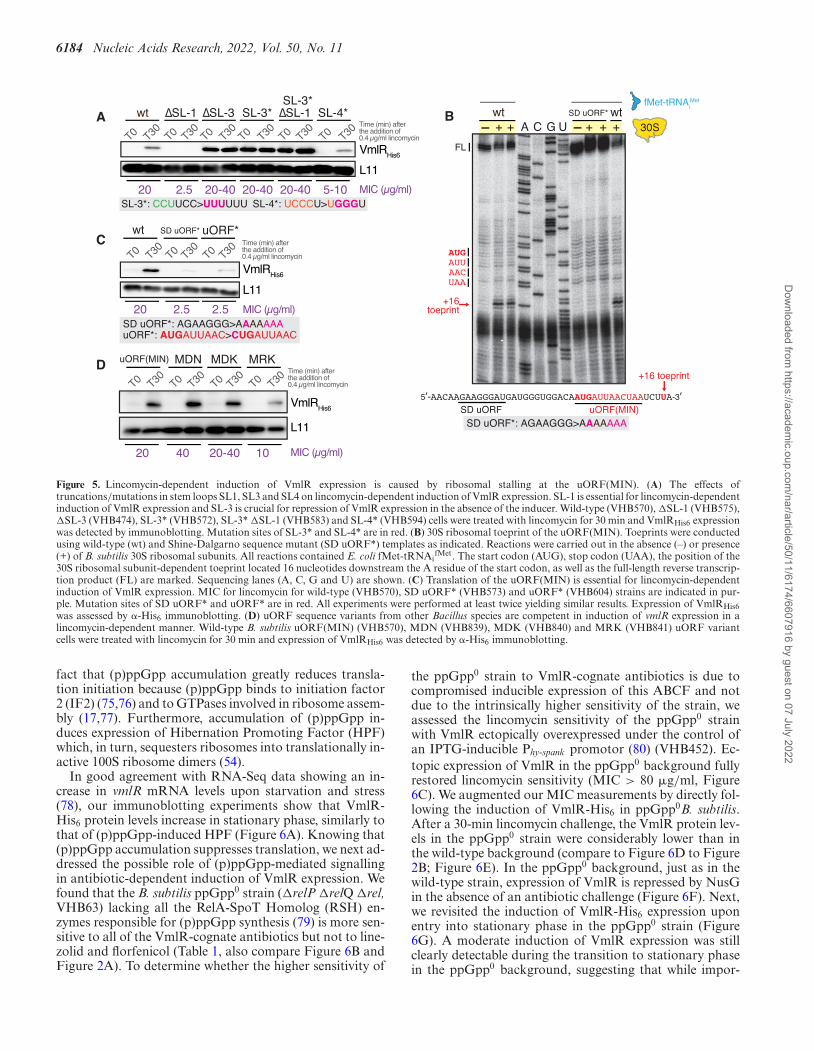
Figure 5. Lincomycin-dependent induction of VmlR expression is caused by ribosomal stalling at the uORF(MIN). (A) The effects oftruncations/mutations in stem loops SL1, SL3 and SL4 on lincomycin-dependent induction of VmlR expression. SL-1 is essential for lincomycin-dependentinduction of VmlR expression and SL-3 is crucial for repression of VmlR expression in the absence of the inducer. Wild-type (VHB570), �SL-1 (VHB575),�SL-3 (VHB474), SL-3* (VHB572), SL-3* �SL-1 (VHB583) and SL-4* (VHB594) cells were treated with lincomycin for 30 min and VmlRHis6 expressionwas detected by immunoblotting. Mutation sites of SL-3* and SL-4* are in red. (B) 30S ribosomal toeprint of the uORF(MIN). Toeprints were conductedusing wild-type (wt) and Shine-Dalgarno sequence mutant (SD uORF*) templates as indicated. Reactions were carried out in the absence (–) or presence(+) of B. subtilis 30S ribosomal subunits. All reactions contained E. coli fMet-tRNAi
fMet. The start codon (AUG), stop codon (UAA), the position of the30S ribosomal subunit-dependent toeprint located 16 nucleotides downstream the A residue of the start codon, as well as the full-length reverse transcrip-tion product (FL) are marked. Sequencing lanes (A, C, G and U) are shown. (C) Translation of the uORF(MIN) is essential for lincomycin-dependentinduction of VmlR expression. MIC for lincomycin for wild-type (VHB570), SD uORF* (VHB573) and uORF* (VHB604) strains are indicated in pur-ple. Mutation sites of SD uORF* and uORF* are in red. All experiments were performed at least twice yielding similar results. Expression of VmlRHis6was assessed by �-His6 immunoblotting. (D) uORF sequence variants from other Bacillus species are competent in induction of vmlR expression in alincomycin-dependent manner. Wild-type B. subtilis uORF(MIN) (VHB570), MDN (VHB839), MDK (VHB840) and MRK (VHB841) uORF variantcells were treated with lincomycin for 30 min and expression of VmlRHis6 was detected by �-His6 immunoblotting.
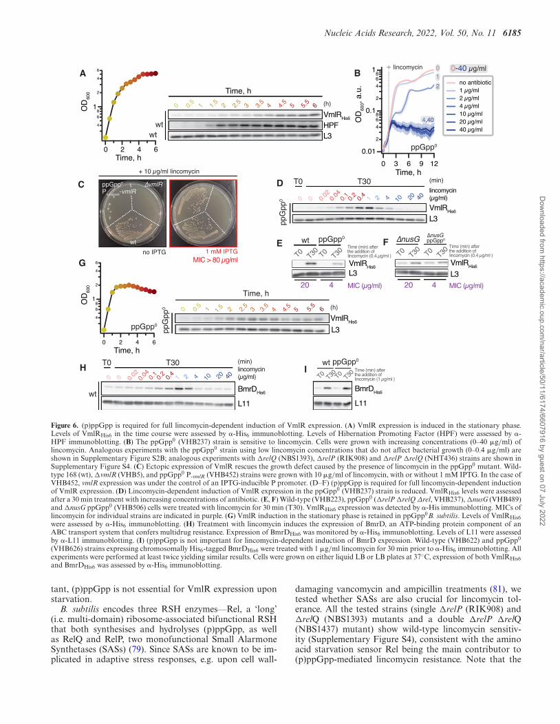
fact that (p)ppGpp accumulation greatly reduces transla-tion initiation because (p)ppGpp binds to initiation factor2 (IF2) (75,76) and to GTPases involved in ribosome assem-bly (17,77). Furthermore, accumulation of (p)ppGpp in-duces expression of Hibernation Promoting Factor (HPF)which, in turn, sequesters ribosomes into translationally in-active 100S ribosome dimers (54).
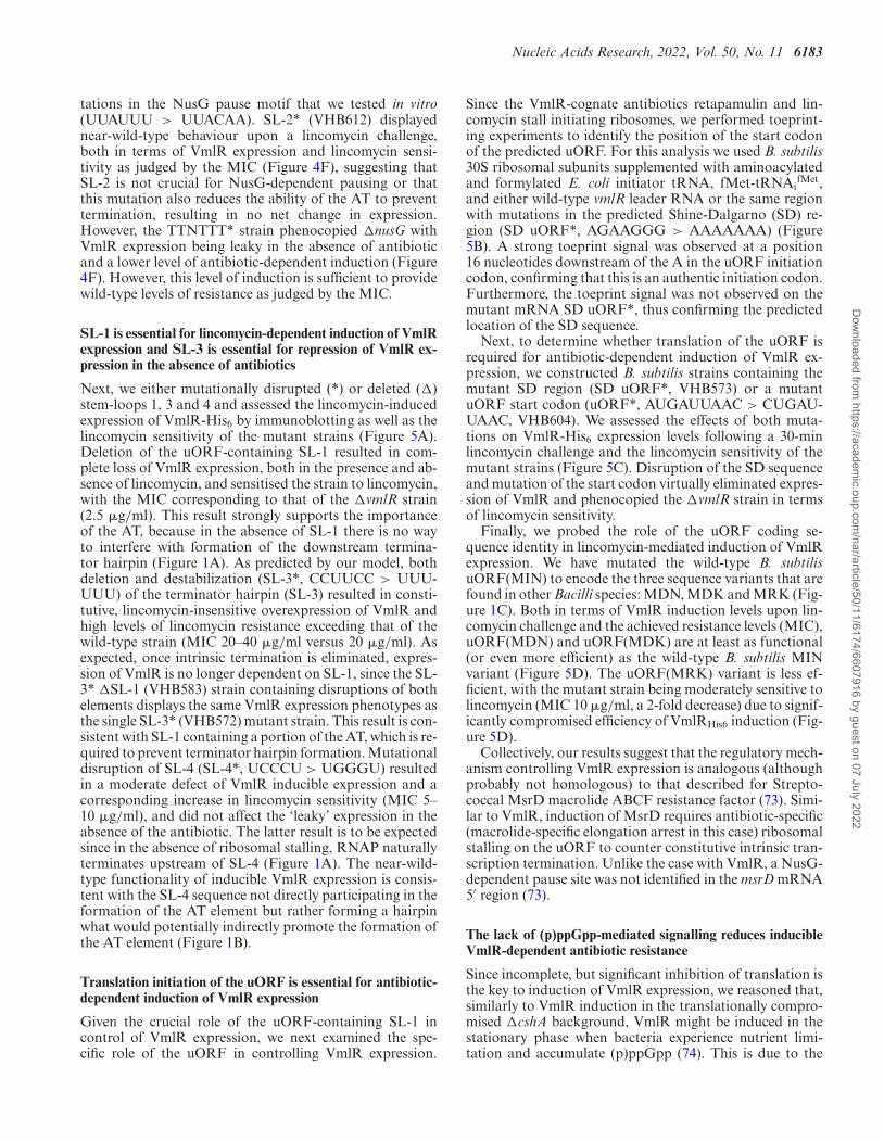
In good agreement with RNA-Seq data showing an in-crease in vmlR mRNA levels upon starvation and stress(78), our immunoblotting experiments show that VmlR-His6 protein levels increase in stationary phase, similarly tothat of (p)ppGpp-induced HPF (Figure 6A). Knowing that(p)ppGpp accumulation suppresses translation, we next ad-dressed the possible role of (p)ppGpp-mediated signallingin antibiotic-dependent induction of VmlR expression. Wefound that the B. subtilis ppGpp0 strain (�relP �relQ �rel,VHB63) lacking all the RelA-SpoT Homolog (RSH) en-zymes responsible for (p)ppGpp synthesis (79) is more sen-sitive to all of the VmlR-cognate antibiotics but not to line-zolid and florfenicol (Table 1, also compare Figure 6B andFigure 2A). To determine whether the higher sensitivity of
the ppGpp0 strain to VmlR-cognate antibiotics is due tocompromised inducible expression of this ABCF and notdue to the intrinsically higher sensitivity of the strain, weassessed the lincomycin sensitivity of the ppGpp0 strainwith VmlR ectopically overexpressed under the control ofan IPTG-inducible Phy-spank promotor (80) (VHB452). Ec-topic expression of VmlR in the ppGpp0 background fullyrestored lincomycin sensitivity (MIC > 80 �g/ml, Figure6C). We augmented our MIC measurements by directly fol-lowing the induction of VmlR-His6 in ppGpp0B. subtilis.After a 30-min lincomycin challenge, the VmlR protein lev-els in the ppGpp0 strain were considerably lower than inthe wild-type background (compare to Figure 6D to Figure2B; Figure 6E). In the ppGpp0 background, just as in thewild-type strain, expression of VmlR is repressed by NusGin the absence of an antibiotic challenge (Figure 6F). Next,we revisited the induction of VmlR-His6 expression uponentry into stationary phase in the ppGpp0 strain (Figure6G). A moderate induction of VmlR expression was stillclearly detectable during the transition to stationary phasein the ppGpp0 background, suggesting that while impor-
Dow
nloaded from https://academ
ic.oup.com/nar/article/50/11/6174/6607916 by guest on 07 July 2022
Nucleic Acids Research, 2022, Vol. 50, No. 11 6185
0 0.02
0 0.04
0.1
0.2
0.4
1 2 4 20 40
T0 T30 (min)
lincomycin (μg/ml)
VmlRHis6 L3
10
ppG
pp0
D+ 10 μg/ml lincomycin
no IPTG 1 mM IPTG
∆vmlR
H
ppGpp0
wt
Phy-spank-vmlR
I
C
0 0.020 0.
04 0.1
0.2
0.4
1 2 4 10 20 40
T0 T30 (min) lincomycin (μg/ml)
BmrDHis6
L11wt
BmrDHis6
L11
T0 T30
wt
T0 T30
ppGpp0
Time (min) after the addition of lincomycin (1 μg/ml )
0 1.5
2.5
3.5
4.5
5.5
1 2 3 4 5 6 (h)
VmlRHis6 HPFL3
wt
Time, h
0.5
A
Time, h
Time, h
4
68
1
2
4
6
6420
wt
MIC > 80 μg/mlG
6 (h)
VmlRHis6
L3
5 5.5
0 1
Time, h
0.5
ppG
pp0 2 32.
53.
51.
5 4 4.5
4
68
1
2
4
6
6420
ppGpp0
OD
600
OD
600
E
VmlRHis6
L3
T0T30
wt
T0T30
ppGpp0
T0T30
∆nusG
T0T30
∆nusG ppGpp0
MIC (μg/ml)4 20 4 20
Time (min) after the addition of lincomycin (0.4 μg/ml )
F
VmlRHis6 L3
MIC (μg/ml)
Time (min) after the addition of lincomycin (0.4 μg/ml )
0.01
2
468
0.1
2
468
1
129630
1 μg/ml 2 μg/ml 4 μg/ml 10 μg/ml 20 μg/ml 40 μg/ml
no antibiotic
Time, h
0-40 μg/ml10
2
4,40
+ lincomycin
ppGpp0
OD
600,
a.u
.
B
Figure 6. (p)ppGpp is required for full lincomycin-dependent induction of VmlR expression. (A) VmlR expression is induced in the stationary phase.Levels of VmlRHis6 in the time course were assessed by �-His6 immunoblotting. Levels of Hibernation Promoting Factor (HPF) were assessed by �-HPF immunoblotting. (B) The ppGpp0 (VHB237) strain is sensitive to lincomycin. Cells were grown with increasing concentrations (0–40 �g/ml) oflincomycin. Analogous experiments with the ppGpp0 strain using low lincomycin concentrations that do not affect bacterial growth (0–0.4 �g/ml) areshown in Supplementary Figure S2B; analogous experiments with �relQ (NBS1393), �relP (RIK908) and �relP �relQ (NHT436) strains are shown inSupplementary Figure S4. (C) Ectopic expression of VmlR rescues the growth defect caused by the presence of lincomycin in the ppGpp0 mutant. Wild-type 168 (wt), �vmlR (VHB5), and ppGpp0 P-vmlR (VHB452) strains were grown with 10 �g/ml of lincomycin, with or without 1 mM IPTG. In the case ofVHB452, vmlR expression was under the control of an IPTG-inducible P promoter. (D–F) (p)ppGpp is required for full lincomycin-dependent inductionof VmlR expression. (D) Lincomycin-dependent induction of VmlR expression in the ppGpp0 (VHB237) strain is reduced. VmlRHis6 levels were assessedafter a 30 min treatment with increasing concentrations of antibiotic. (E, F) Wild-type (VHB223), ppGpp0 (�relP �relQ Δrel, VHB237), �nusG (VHB489)and �nusG ppGpp0 (VHB506) cells were treated with lincomycin for 30 min (T30). VmlRHis6 expression was detected by �-His immunoblotting. MICs oflincomycin for individual strains are indicated in purple. (G) VmlR induction in the stationary phase is retained in ppGpp0B. subtilis. Levels of VmlRHis6were assessed by �-His6 immunoblotting. (H) Treatment with lincomycin induces the expression of BmrD, an ATP-binding protein component of anABC transport system that confers multidrug resistance. Expression of BmrDHis6 was monitored by �-His6 immunoblotting. Levels of L11 were assessedby �-L11 immunoblotting. (I) (p)ppGpp is not important for lincomycin-dependent induction of BmrD expression. Wild-type (VHB622) and ppGpp0
(VHB626) strains expressing chromosomally His6-tagged BmrDHis6 were treated with 1 �g/ml lincomycin for 30 min prior to �-His6 immunoblotting. Allexperiments were performed at least twice yielding similar results. Cells were grown on either liquid LB or LB plates at 37◦C, expression of both VmlRHis6and BmrDHis6 was assessed by �-His6 immunoblotting.
tant, (p)ppGpp is not essential for VmlR expression uponstarvation.
B. subtilis encodes three RSH enzymes––Rel, a ‘long’(i.e. multi-domain) ribosome-associated bifunctional RSHthat both synthesises and hydrolyses (p)ppGpp, as wellas RelQ and RelP, two monofunctional Small AlarmoneSynthetases (SASs) (79). Since SASs are known to be im-plicated in adaptive stress responses, e.g. upon cell wall-
damaging vancomycin and ampicillin treatments (81), wetested whether SASs are also crucial for lincomycin tol-erance. All the tested strains (single �relP (RIK908) and�relQ (NBS1393) mutants and a double �relP �relQ(NBS1437) mutant) show wild-type lincomycin sensitiv-ity (Supplementary Figure S4), consistent with the aminoacid starvation sensor Rel being the main contributor to(p)ppGpp-mediated lincomycin resistance. Note that the
Dow
nloaded from https://academ
ic.oup.com/nar/article/50/11/6174/6607916 by guest on 07 July 2022
6186 Nucleic Acids Research, 2022, Vol. 50, No. 11
�rel strain is highly unstable: in the absence of (p)ppGpphydrolysis that is carried out by Rel, the alarmone producedby the SAS enzymes is not degraded. This results in strongselective pressure yielding suppressor mutants that inac-tivate the expression of catalytically-competent RelQ andRelP to convert the strain into ppGpp0 (82). Therefore, wecould not reliably assess lincomycin resistance of the �relstrain.
Finally, to test whether the observed (p)ppGpp effectsare specific for VmlR induction, we characterised the ef-fects of lincomycin on expression of the heterodimeric mul-tidrug exporter BmrC/BmrD in wild-type and ppGpp0
backgrounds. Similar to VmlR, expression of this antibi-otic resistance determinant is controlled through antibiotic-specific (induced by lincomycin, chloramphenicol and ery-thromycin, but not kanamycin and gentamycin) tran-scription attenuation via ribosome stalling on a uORF(26). First, we followed the expression of chromosomally-encoded His6-tagged BmrD as a function of lincomycinconcentration (Figure 6H). The optimal induction was at1 �g/ml, which is in good agreement with a previousstudy where it was reported that maximal induction ofGFP expression driven by the native bmrC promotor wasat 2 �g/ml (26). We then assessed the expression levelsof BmrD-His6 in the absence or presence of 1 �g/ml lin-comycin, both in wild-type and the ppGpp0 strains (Figure6I). In stark contrast to VmlR, we observed equally efficientBmrD induction in the ppGpp0 and wild type backgrounds.
Collectively, our results suggest that (p)ppGpp-mediatedsignalling finetunes inducible VmlR expression upon antibi-otic challenge.
CONCLUSIONS AND PERSPECTIVE
In this study we have dissected a multilayered regulatorymechanism that controls the expression of B. subtilis an-tibiotic resistance factor VmlR. The efficient induction ofVmlR upon a ‘cognate’ antibiotic challenge relies on NusG-dependent RNAP pausing, antibiotic-dependent ribosomestalling during translation of an uORF (translation attenu-ation), transcription attenuation and signaling via the alar-mone nucleotide (p)ppGpp.
To our knowledge, VmlR presents the second example ofNusG playing an essential role in controlling the inducibleexpression of an antibiotic resistance determinant, with thefirst being the B. subtilis 23S rRNA methylase TlrB that me-diates resistance to macrolide tylosin (25). A dedicated sys-tematic search for NusG TTNTTT pause motifs may un-cover the potential involvement of NusG in other mech-anisms of inducible antibiotic resistance. Both TlrB andVmlR induction mechanisms rely on antibiotic-specific ri-bosomal stalling on a uORF. However, they differ in thatthe induction of TlrB relies on ribosomal arrest by tylosinat translation elongation step at an RYR motif (25), whilethe induction of VmlR relies on ribosomal arrest duringtranslation initiation. In recent years it has become evi-dent that many protein synthesis inhibitors act context-dependently––i.e. they specifically stall ribosomes trans-lating certain amino acid motifs––and that the molecu-lar mechanisms of context-specific ribosomal stalling shapethe amino acid sequences of the regulatory uORF ele-
ments that control the expression of resistance determinants(65,66,68,83,84). Therefore, inducible expression of antibi-otic resistance determinants driven by antibiotic-inducedribosomal stalling on regulatory uORFs could potentiallybe defeated by new novel antibiotic variants with alteredstalling preferences. By failing to stall the ribosome on theregulatory uORF, these antibiotics would comprise cellu-lar protein synthesis without triggering cellular antibioticresistance countermeasures. As we show here, when chal-lenged by a novel fully synthetic lincosamide iboxamycin,B. subtilis expresses VmlR at a significantly lower level com-pared with when it is challenged by the natural compoundlincomycin. This understanding opens possibilities for fur-ther antibiotic design. Finally, as we show here, due to thecrucial role of (p)ppGpp in control of VmlR induction, theantibiotic sensitivity pattern of ppGpp0B. subtilis pheno-copies that of antibiotic-sensitive �vmlR strain (Table 1).This finding further contributes to the appeal of (p)ppGpp-mediated signalling as a drug development target for the at-tenuation of bacterial pathogens (85).
DATA AVAILABILITY
The RNET-seq data (3) used in the study are available inthe NCBI SRA (BioProject ID PRJNA603835) and GEO(GSE186285), Term-seq (47) data are available in GEO(GSE67058 and GSE 154522).
SUPPLEMENTARY DATA
Supplementary Data are available at NAR Online.
ACKNOWLEDGEMENTS
We are grateful to Protein Expertise Platform (PEP) atUmea University and Mikael Lindberg for plasmid con-struction, Fujio Kawamura for sharing anti-L3 and anti-L11 primary antibodies as well as polyclonal anti-HPF an-tobody, and Jorgen Johansson for fruitful discussions onDEAD-box RNA-helicases, ribosome assembly defects andregulation of gene expression.
FUNDING
National Institutes of Health [GM098399 to P.B.]; SwedishResearch Council [2018-00956 within the RIBOTARGETconsortium under the framework of JPIAMR, projectgrants 2017-03783 and 2021-01146 to V.H., project grant2019-01085 to G.C.A.]; Knut and Alice Wallenberg Foun-dation [2020.0037 to G.C.A.]; Ragnar Soderberg Foun-dation [M23/14 to V.H.]; European Regional Develop-ment Fund through the Centre of Excellence for Molecu-lar Cell Technology (to V.H.); Estonian Science Founda-tion [PRG335 to V.H.]; postdoctoral grant from the UmeaaCentre for Microbial Research, UCMR (to H.T.); JST, ACTX, Japan [JP1159335 to H.T.]; MEXT, JSPS Grant-in-Aidfor Scientific Research [grants 20H05926 and 21K06053 toS.C.]; Institute for Fermentation, Osaka [grant G-2021-2-063 to S.C.]. Funding for open access charge: Swedish Re-search Council [2021-01146 to V.H.]
Dow
nloaded from https://academ
ic.oup.com/nar/article/50/11/6174/6607916 by guest on 07 July 2022
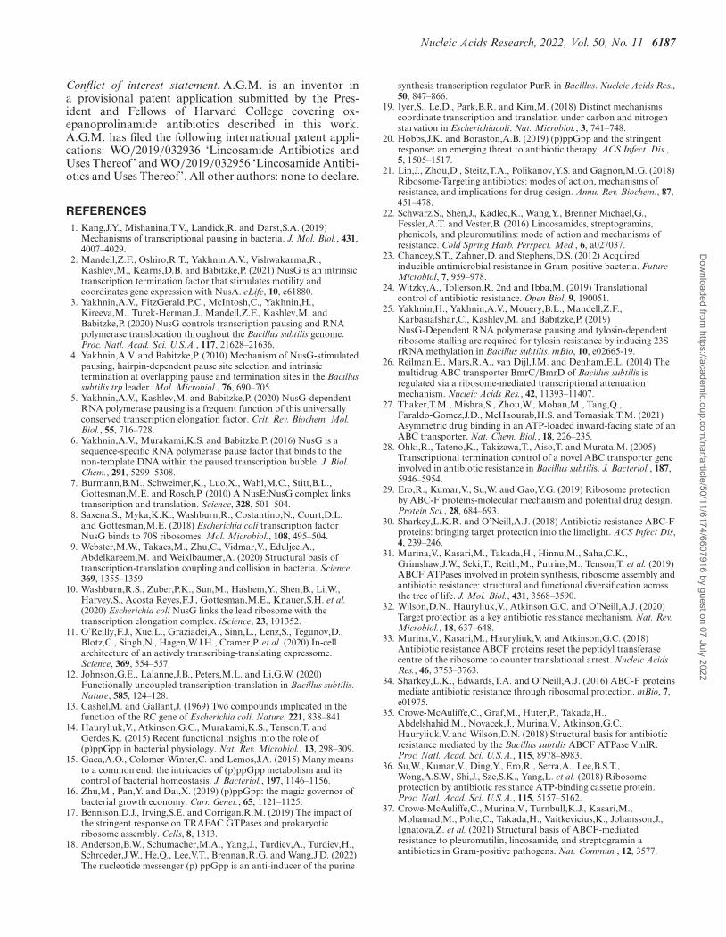
Nucleic Acids Research, 2022, Vol. 50, No. 11 6187
Conflict of interest statement. A.G.M. is an inventor ina provisional patent application submitted by the Pres-ident and Fellows of Harvard College covering ox-epanoprolinamide antibiotics described in this work.A.G.M. has filed the following international patent appli-cations: WO/2019/032936 ‘Lincosamide Antibiotics andUses Thereof’ and WO/2019/032956 ‘Lincosamide Antibi-otics and Uses Thereof’. All other authors: none to declare.
REFERENCES1. Kang,J.Y., Mishanina,T.V., Landick,R. and Darst,S.A. (2019)
Mechanisms of transcriptional pausing in bacteria. J. Mol. Biol., 431,4007–4029.
2. Mandell,Z.F., Oshiro,R.T., Yakhnin,A.V., Vishwakarma,R.,Kashlev,M., Kearns,D.B. and Babitzke,P. (2021) NusG is an intrinsictranscription termination factor that stimulates motility andcoordinates gene expression with NusA. eLife, 10, e61880.
3. Yakhnin,A.V., FitzGerald,P.C., McIntosh,C., Yakhnin,H.,Kireeva,M., Turek-Herman,J., Mandell,Z.F., Kashlev,M. andBabitzke,P. (2020) NusG controls transcription pausing and RNApolymerase translocation throughout the Bacillus subtilis genome.Proc. Natl. Acad. Sci. U.S.A., 117, 21628–21636.
4. Yakhnin,A.V. and Babitzke,P. (2010) Mechanism of NusG-stimulatedpausing, hairpin-dependent pause site selection and intrinsictermination at overlapping pause and termination sites in the Bacillussubtilis trp leader. Mol. Microbiol., 76, 690–705.
5. Yakhnin,A.V., Kashlev,M. and Babitzke,P. (2020) NusG-dependentRNA polymerase pausing is a frequent function of this universallyconserved transcription elongation factor. Crit. Rev. Biochem. Mol.Biol., 55, 716–728.
6. Yakhnin,A.V., Murakami,K.S. and Babitzke,P. (2016) NusG is asequence-specific RNA polymerase pause factor that binds to thenon-template DNA within the paused transcription bubble. J. Biol.Chem., 291, 5299–5308.
7. Burmann,B.M., Schweimer,K., Luo,X., Wahl,M.C., Stitt,B.L.,Gottesman,M.E. and Rosch,P. (2010) A NusE:NusG complex linkstranscription and translation. Science, 328, 501–504.
8. Saxena,S., Myka,K.K., Washburn,R., Costantino,N., Court,D.L.and Gottesman,M.E. (2018) Escherichia coli transcription factorNusG binds to 70S ribosomes. Mol. Microbiol., 108, 495–504.
9. Webster,M.W., Takacs,M., Zhu,C., Vidmar,V., Eduljee,A.,Abdelkareem,M. and Weixlbaumer,A. (2020) Structural basis oftranscription-translation coupling and collision in bacteria. Science,369, 1355–1359.
10. Washburn,R.S., Zuber,P.K., Sun,M., Hashem,Y., Shen,B., Li,W.,Harvey,S., Acosta Reyes,F.J., Gottesman,M.E., Knauer,S.H. et al.(2020) Escherichia coli NusG links the lead ribosome with thetranscription elongation complex. iScience, 23, 101352.
11. O’Reilly,F.J., Xue,L., Graziadei,A., Sinn,L., Lenz,S., Tegunov,D.,Blotz,C., Singh,N., Hagen,W.J.H., Cramer,P. et al. (2020) In-cellarchitecture of an actively transcribing-translating expressome.Science, 369, 554–557.
12. Johnson,G.E., Lalanne,J.B., Peters,M.L. and Li,G.W. (2020)Functionally uncoupled transcription-translation in Bacillus subtilis.Nature, 585, 124–128.
13. Cashel,M. and Gallant,J. (1969) Two compounds implicated in thefunction of the RC gene of Escherichia coli. Nature, 221, 838–841.
14. Hauryliuk,V., Atkinson,G.C., Murakami,K.S., Tenson,T. andGerdes,K. (2015) Recent functional insights into the role of(p)ppGpp in bacterial physiology. Nat. Rev. Microbiol., 13, 298–309.
15. Gaca,A.O., Colomer-Winter,C. and Lemos,J.A. (2015) Many meansto a common end: the intricacies of (p)ppGpp metabolism and itscontrol of bacterial homeostasis. J. Bacteriol., 197, 1146–1156.
16. Zhu,M., Pan,Y. and Dai,X. (2019) (p)ppGpp: the magic governor ofbacterial growth economy. Curr. Genet., 65, 1121–1125.
17. Bennison,D.J., Irving,S.E. and Corrigan,R.M. (2019) The impact ofthe stringent response on TRAFAC GTPases and prokaryoticribosome assembly. Cells, 8, 1313.
18. Anderson,B.W., Schumacher,M.A., Yang,J., Turdiev,A., Turdiev,H.,Schroeder,J.W., He,Q., Lee,V.T., Brennan,R.G. and Wang,J.D. (2022)The nucleotide messenger (p) ppGpp is an anti-inducer of the purine
synthesis transcription regulator PurR in Bacillus. Nucleic Acids Res.,50, 847–866.
19. Iyer,S., Le,D., Park,B.R. and Kim,M. (2018) Distinct mechanismscoordinate transcription and translation under carbon and nitrogenstarvation in Escherichiacoli. Nat. Microbiol., 3, 741–748.
20. Hobbs,J.K. and Boraston,A.B. (2019) (p)ppGpp and the stringentresponse: an emerging threat to antibiotic therapy. ACS Infect. Dis.,5, 1505–1517.
21. Lin,J., Zhou,D., Steitz,T.A., Polikanov,Y.S. and Gagnon,M.G. (2018)Ribosome-Targeting antibiotics: modes of action, mechanisms ofresistance, and implications for drug design. Annu. Rev. Biochem., 87,451–478.
22. Schwarz,S., Shen,J., Kadlec,K., Wang,Y., Brenner Michael,G.,Fessler,A.T. and Vester,B. (2016) Lincosamides, streptogramins,phenicols, and pleuromutilins: mode of action and mechanisms ofresistance. Cold Spring Harb. Perspect. Med., 6, a027037.
23. Chancey,S.T., Zahner,D. and Stephens,D.S. (2012) Acquiredinducible antimicrobial resistance in Gram-positive bacteria. FutureMicrobiol, 7, 959–978.
24. Witzky,A., Tollerson,R. 2nd and Ibba,M. (2019) Translationalcontrol of antibiotic resistance. Open Biol, 9, 190051.
25. Yakhnin,H., Yakhnin,A.V., Mouery,B.L., Mandell,Z.F.,Karbasiafshar,C., Kashlev,M. and Babitzke,P. (2019)NusG-Dependent RNA polymerase pausing and tylosin-dependentribosome stalling are required for tylosin resistance by inducing 23SrRNA methylation in Bacillus subtilis. mBio, 10, e02665-19.
26. Reilman,E., Mars,R.A., van Dijl,J.M. and Denham,E.L. (2014) Themultidrug ABC transporter BmrC/BmrD of Bacillus subtilis isregulated via a ribosome-mediated transcriptional attenuationmechanism. Nucleic Acids Res., 42, 11393–11407.
27. Thaker,T.M., Mishra,S., Zhou,W., Mohan,M., Tang,Q.,Faraldo-Gomez,J.D., McHaourab,H.S. and Tomasiak,T.M. (2021)Asymmetric drug binding in an ATP-loaded inward-facing state of anABC transporter. Nat. Chem. Biol., 18, 226–235.
28. Ohki,R., Tateno,K., Takizawa,T., Aiso,T. and Murata,M. (2005)Transcriptional termination control of a novel ABC transporter geneinvolved in antibiotic resistance in Bacillus subtilis. J. Bacteriol., 187,5946–5954.
29. Ero,R., Kumar,V., Su,W. and Gao,Y.G. (2019) Ribosome protectionby ABC-F proteins-molecular mechanism and potential drug design.Protein Sci., 28, 684–693.
30. Sharkey,L.K.R. and O’Neill,A.J. (2018) Antibiotic resistance ABC-Fproteins: bringing target protection into the limelight. ACS Infect Dis,4, 239–246.
31. Murina,V., Kasari,M., Takada,H., Hinnu,M., Saha,C.K.,Grimshaw,J.W., Seki,T., Reith,M., Putrins,M., Tenson,T. et al. (2019)ABCF ATPases involved in protein synthesis, ribosome assembly andantibiotic resistance: structural and functional diversification acrossthe tree of life. J. Mol. Biol., 431, 3568–3590.
32. Wilson,D.N., Hauryliuk,V., Atkinson,G.C. and O’Neill,A.J. (2020)Target protection as a key antibiotic resistance mechanism. Nat. Rev.Microbiol., 18, 637–648.
33. Murina,V., Kasari,M., Hauryliuk,V. and Atkinson,G.C. (2018)Antibiotic resistance ABCF proteins reset the peptidyl transferasecentre of the ribosome to counter translational arrest. Nucleic AcidsRes., 46, 3753–3763.
34. Sharkey,L.K., Edwards,T.A. and O’Neill,A.J. (2016) ABC-F proteinsmediate antibiotic resistance through ribosomal protection. mBio, 7,e01975.
35. Crowe-McAuliffe,C., Graf,M., Huter,P., Takada,H.,Abdelshahid,M., Novacek,J., Murina,V., Atkinson,G.C.,Hauryliuk,V. and Wilson,D.N. (2018) Structural basis for antibioticresistance mediated by the Bacillus subtilis ABCF ATPase VmlR.Proc. Natl. Acad. Sci. U.S.A., 115, 8978–8983.
36. Su,W., Kumar,V., Ding,Y., Ero,R., Serra,A., Lee,B.S.T.,Wong,A.S.W., Shi,J., Sze,S.K., Yang,L. et al. (2018) Ribosomeprotection by antibiotic resistance ATP-binding cassette protein.Proc. Natl. Acad. Sci. U.S.A., 115, 5157–5162.
37. Crowe-McAuliffe,C., Murina,V., Turnbull,K.J., Kasari,M.,Mohamad,M., Polte,C., Takada,H., Vaitkevicius,K., Johansson,J.,Ignatova,Z. et al. (2021) Structural basis of ABCF-mediatedresistance to pleuromutilin, lincosamide, and streptogramin aantibiotics in Gram-positive pathogens. Nat. Commun., 12, 3577.
Dow
nloaded from https://academ
ic.oup.com/nar/article/50/11/6174/6607916 by guest on 07 July 2022
6188 Nucleic Acids Research, 2022, Vol. 50, No. 11
38. Allignet,J., Loncle,V. and el Sohl,N. (1992) Sequence of astaphylococcal plasmid gene, vga, encoding a putative ATP-bindingprotein involved in resistance to virginiamycin A-like antibiotics.Gene, 117, 45–51.
39. Antonelli,A., D’Andrea,M.M., Brenciani,A., Galeotti,C.L.,Morroni,G., Pollini,S., Varaldo,P.E. and Rossolini,G.M. (2018)Characterization of poxtA, a novelphenicol-oxazolidinone-tetracycline resistance gene from an MRSAof clinical origin. J. Antimicrob. Chemother., 73, 1763–1769.
40. Wang,Y., Lv,Y., Cai,J., Schwarz,S., Cui,L., Hu,Z., Zhang,R., Li,J.,Zhao,Q., He,T. et al. (2015) A novel gene, optrA, that conferstransferable resistance to oxazolidinones and phenicols and itspresence in Enterococcus faecalis and Enterococcus faecium of humanand animal origin. J. Antimicrob. Chemother., 70, 2182–2190.
41. Sadowy,E. (2018) Linezolid resistance genes and genetic elementsenhancing their dissemination in enterococci and streptococci.Plasmid, 99, 89–98.
42. Singh,K.V., Weinstock,G.M. and Murray,B.E. (2002) AnEnterococcusf aecalis ABC homologue (Lsa) is required for theresistance of this species to clindamycin and quinupristin-dalfopristin.Antimicrob. Agents Chemother., 46, 1845–1850.
43. Chesneau,O., Ligeret,H., Hosan-Aghaie,N., Morvan,A. andDassa,E. (2005) Molecular analysis of resistance to streptogramin acompounds conferred by the vga proteins of staphylococci.Antimicrob. Agents Chemother., 49, 973–980.
44. Mohamad,M., Nicholson,D., Saha,C.K., Hauryliuk,V.,Edwards,T.A., Atkinson,G.C., Ranson,N.A. and O’Neill,A.J. (2022)Sal-type ABC-F proteins: intrinsic and common mediators ofpleuromutilin resistance by target protection in staphylococci. NucleicAcids Res., 50, 2128–2142.
45. Brodiazhenko,T., Turnbull,K.J., Wu,K.J.Y., Takada,H.,Tresco,B.I.C., Tenson,T., Myers,A.G. and Hauryliuk,V. (2022)Synthetic oxepanoprolinamide iboxamycin is highly active againsthuman pathogen Listeria monocytogenes. JAC Antimicrob. Resist.,https://doi.org/10.1093/jacamr/dlac061.
46. Dar,D., Shamir,M., Mellin,J.R., Koutero,M., Stern-Ginossar,N.,Cossart,P. and Sorek,R. (2016) Term-seq reveals abundantribo-regulation of antibiotics resistance in bacteria. Science, 352,aad9822.
47. Mondal,S., Yakhnin,A.V., Sebastian,A., Albert,I. and Babitzke,P.(2016) NusA-dependent transcription termination preventsmisregulation of global gene expression. Nat. Microbiol., 1, 15007.
48. Mason,J.D., Terwilliger,D.W., Pote,A.R. and Myers,A.G. (2021)Practical gram-scale synthesis of iboxamycin, a potent antibioticcandidate. J. Am. Chem. Soc., 143, 11019–11025.
49. Larsson,A. (2014) AliView: a fast and lightweight alignment viewerand editor for large datasets. Bioinformatics, 30, 3276–3278.
50. Katoh,K. and Standley,D.M. (2013) MAFFT multiple sequencealignment software version 7: improvements in performance andusability. Mol. Biol. Evol., 30, 772–780.
51. Gruber,A.R., Lorenz,R., Bernhart,S.H., Neubock,R. andHofacker,I.L. (2008) The Vienna RNA websuite. Nucleic Acids Res.,36, W70–W74.
52. Zuker,M. (2003) Mfold web server for nucleic acid folding andhybridization prediction. Nucleic Acids Res., 31, 3406–3415.
53. Takada,H., Roghanian,M., Murina,V., Dzhygyr,I., Murayama,R.,Akanuma,G., Atkinson,G.C., Garcia-Pino,A. and Hauryliuk,V.(2020) The C-Terminal RRM/ACT domain is crucial for fine-tuningthe activation of ‘Long’ RelA-SpoT homolog enzymes by ribosomalcomplexes. Front. Microbiol, 11, 277.
54. Tagami,K., Nanamiya,H., Kazo,Y., Maehashi,M., Suzuki,S.,Namba,E., Hoshiya,M., Hanai,R., Tozawa,Y., Morimoto,T. et al.(2012) Expression of a small (p)ppGpp synthetase, YwaC, in the(p)ppGpp(0) mutant of Bacillus subtilis triggers YvyD-dependentdimerization of ribosome. MicrobiologyOpen, 1, 115–134.
55. Jayasinghe,O.T., Mandell,Z.F., Yakhnin,A.V., Kashlev,M. andBabitzke,P. (2022) Transcriptome-wide effects of NusA on RNApolymerase pausing in Bacillus subtilis. J. Bacteriol., 204, e0053421.
56. Yakhnin,A.V. and Babitzke,P. (2002) NusA-stimulated RNApolymerase pausing and termination participates in the Bacillussubtilis trp operon attenuation mechanism in vitro. Proc. Natl. Acad.Sci. U.S.A., 99, 11067–11072.
57. Gollnick,P. and Babitzke,P. (2002) Transcription attenuation.Biochim. Biophys. Acta, 1577, 240–250.
58. Santangelo,T.J. and Artsimovitch,I. (2011) Termination andantitermination: RNA polymerase runs a stop sign. Nat. Rev.Microbiol., 9, 319–329.
59. Vimberg,V., Cavanagh,J.P., Novotna,M., Lenart,J., Nguyen ThiNgoc,B., Vesela,J., Pain,M., Koberska,M. and Balikova Novotna,G.(2020) Ribosome-mediated attenuation of vga(A) expression isshaped by the antibiotic resistance specificity of Vga(a) proteinvariants. Antimicrob. Agents Chemother., 64, e00666-20.
60. Meydan,S., Marks,J., Klepacki,D., Sharma,V., Baranov,P.V.,Firth,A.E., Margus,T., Kefi,A., Vazquez-Laslop,N. and Mankin,A.S.(2019) Retapamulin-Assisted ribosome profiling reveals thealternative bacterial proteome. Mol. Cell, 74, 481–493.
61. Dunkle,J.A., Xiong,L., Mankin,A.S. and Cate,J.H. (2010) Structuresof the Escherichia coli ribosome with antibiotics bound near thepeptidyl transferase center explain spectra of drug action. Proc. Natl.Acad. Sci. U.S.A., 107, 17152–17157.
62. Tu,D., Blaha,G., Moore,P.B. and Steitz,T.A. (2005) Structures ofMLSBK antibiotics bound to mutated large ribosomal subunitsprovide a structural explanation for resistance. Cell, 121, 257–270.
63. Mitcheltree,M.J., Pisipati,A., Syroegin,E.A., Silvestre,K.J.,Klepacki,D., Mason,J.D., Terwilliger,D.W., Testolin,G., Pote,A.R.,Wu,K.J.Y. et al. (2021) A synthetic antibiotic class overcomingbacterial multidrug resistance. Nature, 599, 507–512.
64. Kannan,K., Kanabar,P., Schryer,D., Florin,T., Oh,E., Bahroos,N.,Tenson,T., Weissman,J.S. and Mankin,A.S. (2014) The general modeof translation inhibition by macrolide antibiotics. Proc. Natl. Acad.Sci. U.S.A., 111, 15958–15963.
65. Marks,J., Kannan,K., Roncase,E.J., Klepacki,D., Kefi,A., Orelle,C.,Vazquez-Laslop,N. and Mankin,A.S. (2016) Context-specificinhibition of translation by ribosomal antibiotics targeting thepeptidyl transferase center. Proc. Natl. Acad. Sci. U.S.A., 113,12150–12155.
66. Beckert,B., Leroy,E.C., Sothiselvam,S., Bock,L.V., Svetlov,M.S.,Graf,M., Arenz,S., Abdelshahid,M., Seip,B., Grubmuller,H. et al.(2021) Structural and mechanistic basis for translation inhibition bymacrolide and ketolide antibiotics. Nat. Commun., 12, 4466.
67. Choi,J., Marks,J., Zhang,J., Chen,D.H., Wang,J., Vazquez-Laslop,N.,Mankin,A.S. and Puglisi,J.D. (2020) Dynamics of the context-specifictranslation arrest by chloramphenicol and linezolid. Nat. Chem. Biol.,16, 310–317.
68. Kannan,K., Vazquez-Laslop,N. and Mankin,A.S. (2012) Selectiveprotein synthesis by ribosomes with a drug-obstructed exit tunnel.Cell, 151, 508–520.
69. Davis,A.R., Gohara,D.W. and Yap,M.N. (2014) Sequence selectivityof macrolide-induced translational attenuation. Proc. Natl. Acad. Sci.U.S.A., 111, 15379–15384.
70. Seip,B., Sacheau,G., Dupuy,D. and Innis,C.A. (2018) Ribosomalstalling landscapes revealed by high-throughput inverse toeprinting ofmRNA libraries. Life Sci. Alliance, 1, e201800148.
71. Lehnik-Habrink,M., Rempeters,L., Kovacs,A.T., Wrede,C.,Baierlein,C., Krebber,H., Kuipers,O.P. and Stulke,J. (2013)DEAD-Box RNA helicases in Bacillus subtilis have multiple functionsand act independently from each other. J. Bacteriol., 195, 534–544.
72. Charollais,J., Pflieger,D., Vinh,J., Dreyfus,M. and Iost,I. (2003) TheDEAD-box RNA helicase SrmB is involved in the assembly of 50Sribosomal subunits in Escherichia coli. Mol. Microbiol., 48,1253–1265.
73. Fostier,C.R., Ousalem,F., Leroy,E.C., Ngo,S., Soufari,H., Innis,C.A.,Hashem,Y. and Boel,G. (2021) Regulation and mechanistic basis ofmacrolide resistance by the ABC-F ATPase MsrD. bioRxiv doi:https://doi.org/10.1101/2021.11.29.470318, 29 November 2021,preprint: not peer reviewed.
74. Varik,V., Oliveira,S.R.A., Hauryliuk,V. and Tenson,T. (2017)HPLC-based quantification of bacterial housekeeping nucleotidesand alarmone messengers ppGpp and pppGpp. Sci. Rep., 7, 11022.
75. Diez,S., Ryu,J., Caban,K., Gonzalez,R.L. Jr and Dworkin,J. (2020)The alarmones (p)ppGpp directly regulate translation initiationduring entry into quiescence. Proc. Natl. Acad. Sci. U.S.A., 117,15565–15572.
76. Vinogradova,D.S., Zegarra,V., Maksimova,E., Nakamoto,J.A.,Kasatsky,P., Paleskava,A., Konevega,A.L. and Milon,P. (2020) Howthe initiating ribosome copes with ppGpp to translate mRNAs. PLoSBiol., 18, e3000593.
Dow
nloaded from https://academ
ic.oup.com/nar/article/50/11/6174/6607916 by guest on 07 July 2022
Nucleic Acids Research, 2022, Vol. 50, No. 11 6189
77. Corrigan,R.M., Bellows,L.E., Wood,A. and Grundling,A. (2016)ppGpp negatively impacts ribosome assembly affecting growth andantimicrobial tolerance in Gram-positive bacteria. Proc. Natl. Acad.Sci. U.S.A., 113, E1710–E1719.
78. Nicolas,P., Mader,U., Dervyn,E., Rochat,T., Leduc,A.,Pigeonneau,N., Bidnenko,E., Marchadier,E., Hoebeke,M.,Aymerich,S. et al. (2012) Condition-dependent transcriptome revealshigh-level regulatory architecture in Bacillus subtilis. Science, 335,1103–1106.
79. Nanamiya,H., Kasai,K., Nozawa,A., Yun,C.S., Narisawa,T.,Murakami,K., Natori,Y., Kawamura,F. and Tozawa,Y. (2008)Identification and functional analysis of novel (p)ppGpp synthetasegenes in Bacillus subtilis. Mol. Microbiol., 67, 291–304.
80. Britton,R.A., Eichenberger,P., Gonzalez-Pastor,J.E., Fawcett,P.,Monson,R., Losick,R. and Grossman,A.D. (2002) Genome-wideanalysis of the stationary-phase sigma factor (sigma-H) regulon ofBacillus subtilis. J. Bacteriol., 184, 4881–4890.
81. Geiger,T., Kastle,B., Gratani,F.L., Goerke,C. and Wolz,C. (2014) Twosmall (p)ppGpp synthases in Staphylococcus aureus mediate toleranceagainst cell envelope stress conditions. J. Bacteriol., 196, 894–902.
82. Srivatsan,A., Han,Y., Peng,J., Tehranchi,A.K., Gibbs,R., Wang,J.D.and Chen,R. (2008) High-precision, whole-genome sequencing oflaboratory strains facilitates genetic studies. PLoS Genet., 4,e1000139.
83. Syroegin,E.A., Flemmich,L., Klepacki,D., Vazquez-Laslop,N.,Micura,R. and Polikanov,Y.S. (2022) Structural basis for thecontext-specific action of the classic peptidyl transferase inhibitorchloramphenicol. Nat. Struct. Mol. Biol., 29, 152–161.
84. Tsai,K., Stojkovic,V., Lee,D.J., Young,I.D., Szal,T., Klepacki,D.,Vazquez-Laslop,N., Mankin,A.S., Fraser,J.S. and Fujimori,D.G.(2022) Structural basis for context-specific inhibition of translation byoxazolidinone antibiotics. Nat. Struct. Mol. Biol., 29, 162–171.
85. Kushwaha,G.S., Oyeyemi,B.F. and Bhavesh,N.S. (2019) Stringentresponse protein as a potential target to intervene persistent bacterialinfection. Biochimie, 165, 67–75.
Dow
nloaded from https://academ
ic.oup.com/nar/article/50/11/6174/6607916 by guest on 07 July 2022