A Bacillus subtilis Secreted Protein with a Role in Endospore Coat Assembly and Function

JOURNAL OF BACTERIOLOGY, Mar. 2008, p. 1812–1821 Vol. 190, No. 50021-9193/08/$08.00�0 doi:10.1128/JB.01394-07Copyright © 2008, American Society for Microbiology. All Rights Reserved.
Localization and Interactions of Teichoic Acid Synthetic Enzymes inBacillus subtilis�†
Alex Formstone,1 Rut Carballido-Lopez,1,2 Philippe Noirot,2Jeffery Errington,1,3* and Dirk-Jan Scheffers1,4
Sir William Dunn School of Pathology, University of Oxford, South Parks Road, Oxford OX1 3RE, United Kingdom1;Genetique Microbienne, Institut National de la Recherche Agronomique, 78352 Jouy-en-Josas Cedex, France2;
Institute for Cell and Molecular Biosciences, Faculty of Medical Sciences, Newcastle University,Framlington Place, Newcastle NE2 4HH, United Kingdom3; and Molecular Microbiology,
Institute for Molecular Cell Biology, VU University Amsterdam, De Boelelaan 1085,1081 HV Amsterdam, The Netherlands4
Received 28 August 2007/Accepted 11 December 2007
The thick wall of gram-positive bacteria is a polymer meshwork composed predominantly of peptidoglycan(PG) and teichoic acids, both of which have a critical function in maintenance of the structural integrity andthe shape of the cell. In Bacillus subtilis 168 the major teichoic acid is covalently coupled to PG and is knownas wall teichoic acid (WTA). Recently, PG insertion/degradation over the lateral wall has been shown to occurin a helical pattern. However, the spatial organization of WTA assembly and its relationship with cell shapeand PG assembly are largely unknown. We have characterized the localization of green fluorescent proteinfusions to proteins involved in several steps of WTA synthesis in B. subtilis: TagB, -F, -G, -H, and -O. All of theselocalized similarly to the inner side of the cytoplasmic membrane, in a pattern strikingly similar to thatdisplayed by probes of nascent PG. Helix-like localization patterns are often attributable to the morphogeniccytoskeletal proteins of the MreB family. However, localization of the Tag proteins did not appear to besubstantially affected by single disruption of any of the three MreB homologues of B. subtilis. Bacterial andyeast two-hybrid experiments revealed a complex network of interactions involving TagA, -B, -E, -F, -G, -H, and-O and the cell shape determinants MreC and MreD (encoded by the mreBCD operon and presumably involvedin the spatial organization of PG synthesis). Taken together, our results suggest that, in B. subtilis at least, thesynthesis and export of WTA precursors are mediated by a large multienzyme complex that may be associatedwith the PG-synthesizing machinery.
In most bacteria, the rigid cell wall (CW) is responsible forproviding shape and structural integrity to the cell. The thickCW of gram-positive bacteria is a multilayered structure com-posed predominantly of peptidoglycan (PG) (also calledmurein) and anionic polymers, particularly teichoic acids (TA)(for recent reviews, see references 5, 47, and 54). The highlycross-linked PG polymer (poly-N-acetylglucosamine and N-acetylmuramic acid) network is an essential determinant of cellshape and is responsible for protection from the cellular turgorpressure. Many roles for TA have been proposed, includingcell shape maintenance (61, 68), resistance to antimicrobialpeptides (1, 35, 36), biofilm formation (27), acid tolerance (9)and, efficient release of secreted proteins into the culture me-dium (49).
TA are either covalently bound to the PG (wall TA [WTA])or anchored to the cytoplasmic membrane (lipo-TA). In thegram-positive model organism Bacillus subtilis, WTA is presentin quantities roughly equal to those of PG and constitutes themajor class of anionic polymers (26). The type of WTA poly-
mer varies between strains. In B. subtilis 168, the major WTAconsists of a poly-glycerol-phosphate [poly-(Gro-P)] chain of45 to 60 subunits and a “PG linkage unit” of N-acetylglu-cosamine-�-(1–4)-N-acetylmannosamine (GlcNAc-ManNAc).WTA is covalently linked to the PG through a phosphodiesterbond between the anomeric carbon of GlcNAc in the PGlinkage unit and the 6-hydroxyl of MurNAc in the PG chain.
In B. subtilis 168 the genes responsible for WTA synthesisare tagABDEFGHO and mnaA (Fig. 1). According to proposedmodels, WTA biosynthesis occurs predominantly at the cyto-plasmic side of the membrane (5). Initially, TagO is thought tocouple GlcNAc to the membrane-embedded lipid undecapre-nyl-pyrophosphate. The next step in the pathway, thought to becatalyzed by TagA, is the addition of ManNAc, which is madefrom the epimerization of GlcNAc by MnaA (59, 60), to pro-duce the lipid-linked GlcNAc-ManNAc disaccharide. Recentin vitro biochemical studies from the Brown laboratory haveestablished that TagB then functions as the “Tag primase,”which couples the first glycerol-phosphate to ManNAc (7),followed by TagF acting as the “Tag polymerase,” which addsthe additional glycerol-phosphates extending the poly-(Gro-P)chain (56). TagD activates glycerol-phosphate through linkageto CTP, making CDP-glycerol, the substrate for both TagB andTagF (50), and TagE glucosylates glycerol-phosphate subunitsin the poly-(Gro-P) chain (51). Finally, TagG and TagH arethought to constitute a two-component ABC-type transporterthat mediates the translocation of the TA polymer across the
* Corresponding author. Mailing address: Institute for Cell and Mo-lecular Biosciences, Faculty of Medical Sciences, Newcastle University,Framlington Place, Newcastle NE2 4HH, United Kingdom. Phone:(44) 191 222 8126. Fax: (44) 191 222 7424. E-mail: [email protected].
† Supplemental material for this article may be found at http://jb.asm.org/.
� Published ahead of print on 21 December 2007.
1812
on April 22, 2016 by guest
http://jb.asm.org/
Dow
nloaded from

membrane. TagH constitutes the nucleotide binding domain(NBD), and TagG is the membrane-spanning domain and pre-sumed translocase component of the ABC-type transporterTagGH (40). After translocation, the WTA polymer is co-valently attached to the PG through an unknown mechanism(40). As all of these synthetic reactions are believed to takeplace at the membrane in an ordered fashion (5), it could beenvisaged that these proteins form a complex in which thelipid-linked WTA precursor molecule is passed through thechain of biosynthetic steps, but evidence for such a complexhas not yet been provided.
Until recently it was thought that WTA was an indispensablecomponent of the CW, as mutations in any of the tag genes,except tagE, were lethal in B. subtilis (see, e.g., references 6, 40,and 59). However, careful analysis has revealed that in factWTA is dispensable for growth in B. subtilis and Staphylococ-cus aureus, provided that the first committed step catalyzed byTagO is deleted (20, 21). The cause for the lethality of down-stream mutations probably lies in the sequestration of metab-olites such as undecaprenyl-pyrophosphate, which is also re-quired as a lipid linker for the translocation of PG precursors(20). Nevertheless, loss of WTA in B. subtilis is associated withloss of rod shape and swelling, nonuniform thickening of thePG layer, aberrant placement of the division septum, and re-duced growth rate, indicating that WTA plays an importantrole in maintaining the shape and structural integrity of thebacterial cell (20).
A number of shape mutants (namely, Rod mutants) thatshow a rounded phenotype in B. subtilis have been identified.Two of the early rod genes to be identified were mapped to taggenes. rodC1 was a temperature sensitive mutation in tagF, androdD was a mutation in tagE (29). The other two classes of Rodmutations were mapped to rodA and mreD (rodB), which are
both believed to play a role in PG synthesis (28, 67). mreBmorphogenes have come to the fore since the discovery thatthey encode actin homologues. B. subtilis contains three MreBisoforms (MreB, Mbl, and MreBH) that assemble into helicalfilaments around the cell periphery (19, 32, 65) and colocalizein what appears to be a single helical structure (11, 18). Use offluorescent derivatives of the antibiotics vancomycin and ramo-planin, which label newly incorporated PG precursors, hasrevealed an underlying helical pattern of PG synthesis in B.subtilis (15, 63). Whether this pattern is influenced by theMreB cytoskeleton is at present controversial. Nevertheless, ithas been suggested that the MreB isoforms are involved in thepositioning of PG synthases known as penicillin-binding pro-teins (PBPs) (24), a PG hydrolase (LytE) (11), and/or othermembrane-associated cell morphogenesis proteins, such asMreC and MreD (22, 23, 38, 41, 66). Conversely, it has beensuggested that MreB requires MreC/D for proper localizationin B subtilis (17). MreC and MreD (encoded by the mreBCDoperon) are essential membrane proteins that also localize in ahelix-like pattern and that are believed to function in the samemorphogenetic pathway as MreBs. MreC has been recentlyshown to interact with high-molecular-weight PBPs in B. sub-tilis (66) and in Escherichia coli (38) by bacterial two-hybrid(B2H) and yeast two-hybrid (Y2H) experiments, respectively,and in Caulobacter crescentus by affinity chromatography (22).Altogether, it is currently believed that MreBCD form an es-sential membrane-bound complex involved in the spatial orga-nization of the PG synthesis machinery along the cylindricalCW (5, 54).
Several studies have attempted to determine the localizationof WTA on the surface of B. subtilis cells (12, 52, 57, 58). It hasbeen suggested that they localize at division septa and either inpatches or uniformly along the cell cylinder, but none of these
FIG. 1. Overview of WTA biosynthesis. The biosynthetic steps in the formation of WTA precursors are shown. Arrows indicate catalytic steps,and the enzymes responsible are shown. The precursor is assembled at the cytoplasmic membrane prior to transport by TagGH and linkage to thePG. The question mark indicates an unknown enzymatic reaction.
VOL. 190, 2008 TEICHOIC ACID-SYNTHESIZING COMPLEXES IN B. SUBTILIS 1813
on April 22, 2016 by guest
http://jb.asm.org/
Dow
nloaded from

studies were conclusive. One recent study addressed the loca-tion of the “Tag primase” TagB and showed that it localizes tothe cytoplasmic membrane in a disperse pattern (7). Since aconsiderable body of recent work has suggested that CW bio-genesis may occur in a helical manner governed by the MreB(actin-like) proteins, we decided to make a more systematicstudy of Tag protein localization and of its relationship withthe MreB cytoskeleton. Imaging of functional green fluores-cent protein (GFP) fusions to TagO, TagB, TagF, TagG, andTagH revealed a similar set of membrane-associated localiza-tion patterns. Fluorescence was present at the division sitesand in a pattern consistent with a helix along the sidewall. ForTagG and TagH at least, it was reduced at mature cell poles.We then addressed the protein-protein interactions of Tagproteins by using B2H and Y2H assays (30, 34). Extensiveinteractions between the WTA biosynthetic proteins as well aswith the putative translocase TagGH were revealed. Interac-tions between several of the Tag proteins and the shape de-terminants MreC and MreD were also detected. Our findingssuggest that the synthesis and translocation of WTA precursorsare mediated by a large multienzyme complex (consisting of atleast TagO, TagA, TagB, TagF, TagG, and TagH) that local-
izes at sites of the cytoplasmic membrane similar to thosewhere insertion of nascent PG takes place, possibly throughinteractions with the bacterial cytoskeleton proteins MreCand MreD.
MATERIALS AND METHODS
General methods. The strains and plasmids used in this study are listed inTable 1 (for a full list of plasmids used in the two-hybrid studies, see Table S2 inthe supplemental material). B. subtilis cells were made competent for transfor-mation with DNA either by the method of Kunst and Rapoport (39) or by themethod of Anagnostopoulos and Spizizen (2) as modified by Jenkinson (31).DNA manipulations and E. coli DH5� or XL1-Blue transformations were car-ried out using standard methods (53). Solid medium used for growing B. subtiliswas nutrient agar (Oxoid), and liquid medium was either casein hydrolysate(CH) medium (62) or S medium (33) supplemented with 1% (vol/vol) CH (S�
medium) and with xylose (0.5%). Where SMM was added, the above media weremade at 2� concentration and diluted 50:50 with a 2� SMM solution (1 Msucrose, 33.7 mM maleic acid, 40 mM MgCl2, pH 7.0). Antibiotics were used atthe following concentrations: kanamycin, 5 �g/ml; chloramphenicol, 5 �g/ml;erythromycin, 1 �g/ml; lincomycin, 25 �g/ml; and/or spectinomycin, 50 �g/ml.Media used for growing E. coli strains were 2� TY (53) and nutrient agarsupplemented with ampicillin (100 �g/ml) or kanamycin (5 �g/ml), as required.
Construction of GFP fusions. Primers and restriction endonucleases used arelisted in Table S1 in the supplemental material. For the gfp-tagG and gfp-tagHfusions, approximately one-third of each of the promoter-proximal parts of the
TABLE 1. Bacterial strains and plasmids used in this study
Strain or plasmid Relevant characteristicsa Source, reference, or constructionb
B. subtilis strains168 trpC2 Laboratory collectionEB247 tagF1 (rodC1), S644F ts mutant 562501 trpC2 �(mbl::pSG4503, lacZ lacI bla erm Pspac-�mbl�) 102505 trpC2 �(mbl::spc) 322535 trpC2 �(mreBH::spc) 112580J trpC2 �(amyE::spc Pxyl-gfp-tagB) pSG4537J3168 (Spc)3110 trpC2 tagH::pSG5050 (cat Pxyl-gfp-tagH1–648) pSG50503168 (Cm)3506 trpC2 �(mbl::spc) tagG::pSG5322 (cat Pxyl-gfp-tagG1–346) 250533512 (Spc)3512 trpC2 tagG::pSG5322 (cat Pxyl-gfp-tagG1–346) pSG53223168 (Cm)3530 trpC2 �(mbl::spc) tagH::pSG5050 (cat Pxyl-gfp-tagH1–648) 250533110 (Spc)3725 trpC2 �neo3427 �mreB 253741 trpC2 �neo3427 �mreB tagG::pSG5322 (cat Pxyl-gfp-tagG1–346) 372533512 (Neo)3742 trpC2 �neo3427 �mreB tagH::pSG5050 (cat Pxyl-gfp-tagH1–648) 372533110 (Neo)3746 trpC2 �(mreBH::spc) tagG::pSG5322 (cat Pxyl-gfp-tagG1–346) 253533512 (Spc)3747 trpC2 �(mreBH::spc) tagH::pSG5050 (cat Pxyl-gfp-tagH1–648) 253533110 (Spc)3798 trpC2 tagB::pSG5879 (tagB-gfp cat) pSG58793168 (Cm)3799 trpC2 tagF::pSG5880 (tagF-gfp cat) pSG58803168 (Cm)3800 trpC2 tagO::pSG5881 (tagO-gfp cat) pSG58813168 (Cm)
E. coli strainsDH5� F endA1 hsdR17 supE44 thi-1 -recA1 gyrA96 relA1 �(lacZYA-argF)U169
�80 dlacZ �M15GIBCO-BRL
BTH101 F glnV44 recA1 endA gyrA96 thi-1 hsdR17 spoT1 rfbD1 cya-854 34XL1-Blue recA1 endA1 gyrA96 thi-1 hsdR17 supE44 relA1 lac �F� proAB lacIqZ�M15
Tn10 (Tetr) Stratagene Ltd.
PlasmidspSG1151 bla cat gfp 43pSG1729 bla amyE::spc Pxyl-gfp 43pSG4537J bla amyE::spc Pxyl-gfp-tagB This workpSG4902 bla cat Pxyl-gfp 69pSG5050 bla cat Pxyl-gfp-tagH1–648 This workpSG5322 bla cat Pxyl-gfp-tagG1–346 This workpSG5879 bla cat tagB-gfp This workpSG5880 bla cat tagF-gfp This workpSG5881 bla cat tagO-gfp This work
a gfp, F64L S65T variant of GFP gene (13); �mbl� denotes 5�–3� gene truncation.b X3Y indicates that strain Y was transformed with DNA from source X, with the selected marker in parentheses: Cm, chloramphenicol; Sp, spectinomycin; Neo,
neomycin.
1814 FORMSTONE ET AL. J. BACTERIOL.
on April 22, 2016 by guest
http://jb.asm.org/
Dow
nloaded from

tagG and tagH genes was amplified by PCR and cloned into pSG4902 (69).Transformation of the resulting plasmids (Table 1) into B. subtilis, with selectionfor chloramphenicol resistance, resulted in strains that carried a gfp fusion totagG or tagH at the chromosomal locus as the only copy of the gene of interestand under the control of the Pxyl promoter. Correct integration at the chromo-somal locus was confirmed by PCR.
For tagB-gfp, tagF-gfp, and tagO-gfp fusions, approximately one-third of the3�-proximal parts of each of the tagB, tagF, and tagO genes was amplified by PCRand cloned into pSG1151 (43), generating plasmids pSG5879, pSG5880, andpSG5881, respectively. The plasmids were checked by sequencing and trans-formed into B. subtilis 168, with selection for chloramphenicol resistance toproduce strains carrying a gfp fusion to tagB (strain 3798), tagF (strain 3799), andtagO (strain 3800) at the chromosomal locus under control of the native pro-moter.
For the gfp-tagB and gfp-tagF fusions, the full-length coding regions of tagB andtagF were PCR amplified and cloned into plasmid pSG1729 (43) to generateplasmids pSG4535J and pSG4537J. The resulting inserts were sequenced, andthe plasmids pSG4535J and pSG4537J were transformed into wild-type B. subtilis168 cells, with selection for spectinomycin resistance, to give strains 2575J and2580J, which contain both the natural copy of tagF and tagB and a second,xylose-dependent copy of tagF or tagB, respectively, fused to gfp at the ectopicamyE locus.
To test the functionality of the GFP-TagF fusion, B. subtilis strain EB247 (56),harboring the tagF1 (rodC1) temperature-sensitive mutation, was transformed byplasmid pSG4535J to produce strain 2577J, which contains the tagF1 mutation atthe chromosomal locus and the xylose-inducible gfp-tagF fusion at amyE. Dis-ruption of amyE was confirmed for all pSG1729 derivates using a starch plateassay (14), and the correct integration of the inserts at the amyE locus wasconfirmed by PCR.
To disrupt the native mbl gene in the strains containing the GFP fusions,chromosomal DNA of strain 2505 [�(mbl::spc)] (32) was used to transformstrains 3110 and 3512, to produce strains 3530 and 3506, respectively. For mreBdisruptions, chromosomal DNA of strain 3725 (�neo3427 �mreB) (25) was usedto transform strains 3110 and 3512, to produce strains 3742 and 3741, respec-tively. For mreBH disruptions, chromosomal DNA of strain 2535 [�(mreBH::spc)](11) was used to transform strains 3110 and 3512, to produce strains 3747 and 3746,respectively.
Microscopy. Microscopy was performed essentially as described previously(55). Image acquisition was done as described previously (42) using Metamorphv 6.0 software (Universal Imaging Corp., Downingtown, PA). Images from asingle focal plane were deconvolved using the “No Neighbors” algorithm fromthe Metamorph software package. Overlays of micrographs were assembledusing Metamorph before exporting the images to Adobe Photoshop v 6.0.
B2H plasmid construction and assay. The method used was that of Karimovaet al. (34), adapted as described by Daniel et al. (16). The coding sequence ofeach gene was amplified by PCR from the wild-type strain 168 genomic DNAusing the primers listed in Table S1 in the supplemental material and insertedinto pKT25, pUT18C, and pUT18. For a full list of plasmids used in this study,see Table S2 in the supplemental material.
Y2H plasmid construction and assay. Coding sequences of each gene wereamplified by PCR from the wild-type strain 168 genomic DNA using the primerslisted in Table S1 in the supplemental material and cloned as translationalfusions with the Gal4 DNA-binding (BD) and Gal4 activation (AD) domains inthe recipient vectors pGBDU-C1 (bait, Ura�) and pGAD-C1 (prey, Leu�),respectively (30). Two-hybrid bait and prey constructions were introduced intothe Saccharomyces cerevisiae haploid strains PJ69-4a and PJ69-4�, respectively,by gap repair (44). The two-hybrid assays were performed according to a previ-ously described mating strategy (48). Interaction phenotypes were scored byreplica plating the diploids onto plates selecting for the expression of the HIS3and ADE2 interaction reporters. Control matings with empty pGBDU andpGAD vectors were used to detect self-activation and as negative controls forinteraction.
RESULTS
Localization of the putative WTA transporter TagGH in ahelix-like pattern. Lipid-linked WTA precursors are thoughtto be transported across the membrane by a dedicated ABC-type transporter, TagGH. TagG and TagH are predicted tohave 6 transmembrane spans and 1 transmembrane span, re-spectively, with both N termini in the cytosol (TMHMM v 2.0;
http://www.cbs.dtu.dk/services/TMHMM/) (37). To examinethe sites of WTA export, i.e., the sites of nascent WTA inser-tion, N-terminal gfp fusions to tagG and tagH were constructed.The gfp-tagG and gfp-tagH fusions were integrated into thechromosome at the tagGH locus, replacing the respective wild-type copy of the gene. This generated two strains containingeither gfp-tagG (strain 3512) or gfp-tagH (strain 3110) underthe control of the xylose-inducible promoter Pxyl. Both strainsdisplayed xylose-dependent growth (not shown), indicatingthat expression of gfp-tagG and gfp-tagH could replace that ofthe essential tagG and tagH genes, respectively. In the presenceof inducer, these strains showed no morphological defects,further indicating that the GFP fusions were functional. GFP-TagG and GFP-TagH were both expressed as full-length fu-sion proteins, as shown by Western blotting using a specificanti-GFP antibody (data not shown), and showed similar lo-calization patterns (Fig. 2A and C). They localized to ongoingdivision sites, and a weak, uneven fluorescence signal was de-tected over the cell cylinder. Strikingly, little fluorescence wasdetected in old cell poles, and, consistent with this, a gapbetween the fluorescence signal at the division site and that inthe cylinder emerged as cells completed division (Fig. 2A andC). Out-of-focus light in the GFP fluorescence images wasreduced by two-dimensional (2D) deconvolution (see Materi-als and Methods), which was applied to single images of cellsexpressing either GFP-TagG or GFP-TagH. Interestingly, inboth the raw and the deconvolved images of GFP-TagG (Fig.2A and B, respectively) or GFP-TagH (Fig. 2C and D), thecylindrical staining was reminiscent of the helix-like patternsseen in B. subtilis cells stained with fluorescent probes fornascent PG (15, 63) or expressing a GFP fusion to the CWhydrolase LytE (11), which were shown to be dependent on theMreB isoforms Mbl and MreBH, respectively, and of the he-lical configuration displayed at the membrane by GFP fusionsto MreC and MreD (41). To test if the helix-like distribution ofTagG and/or TagH (and thus WTA export) was also influ-enced by the actin-like cytoskeleton, the GFP-TagG and GFP-TagH fusions were examined in mreB, mreBH, and mbl singlemutant strains. These strains were grown under conditions(CH medium supplemented with SMM) previously shown toallow these mutants to grow with near-wild-type morphology(11, 25). Under these conditions, the localization pattern ofeither GFP-TagG or GFP-TagH in any of the three mreB-likesingle mutants (Fig. 2E to J) was not significantly differentfrom that displayed in wild-type cells (Fig. 2B and D). mreCand mreD are essential under normal growth conditions, butviability can also be rescued by high concentrations of magne-sium (41). However, mreC and mreD mutants are unstable anddisplay severe shape defects under these conditions (41), un-like the mreB-like mutants, in which the rod shape is restoredby Mg2� (11, 25). Thus, a possible effect of MreC and/or MreDon the positioning of the WTA transporter TagGH could notbe tested. Indeed, in a �mreC or a �mreD background, anychange in the localization patterns of TagGH could be attrib-uted either to the lack of MreCD proteins or to a secondaryeffect resulting from the shape defects.
Localization of the WTA biosynthetic enzymes TagO, TagB,and TagF to the membrane. The first step of WTA biosynthesisis thought to be catalyzed by TagO (Fig. 1), which is predictedto have 11 transmembrane spans with its C terminus located in
VOL. 190, 2008 TEICHOIC ACID-SYNTHESIZING COMPLEXES IN B. SUBTILIS 1815
on April 22, 2016 by guest
http://jb.asm.org/
Dow
nloaded from

the cytosol (TMHMM v 2.0) (37). To examine the subcellularlocalization of TagO, a gfp fusion was made to the C terminusof the gene. The fusion was integrated into the chromosome atthe tagO locus, under control of the native tagO promoter(strain 3800). The TagO-GFP fusion, which was the only copyof TagO present in the cells, was judged to be functional, ascells displayed a wild-type morphology whereas cells with tagOdisrupted become spherical (20). TagO-GFP localized to the
membrane, in a nonuniform pattern along the cylindrical re-gion, and also to the division sites (Fig. 3A). Deconvolution(Fig. 3D) revealed that this pattern was also consistent with ahelix-like organization, similar to that displayed by the TagG-and TagH-GFP fusions (Fig. 2B and D, respectively). Again,the fluorescence intensity appeared to be reduced at older cellpoles (Fig. 3A), although this effect was less clear than for theTagGH proteins.
FIG. 2. Localization of the ABC transporter TagGH. Fluorescence images of cells expressing GFP-TagG (A and B), GFP-TagH (C and D) ina wild type background or in the absence of Mbl (E and H), MreB (F and I), or MreBH (G and J) are shown. Cells were grown to mid-exponentialstage at 30°C in CH medium (or CH supplemented with SMM for the mutants [see text]) with 5 mM K2HPO4/KH2PO4 (pH 7.5) and 0.5% xyloseand imaged on wet agarose slides. Panels B and D show 2D deconvolutions of panels A and C, respectively. Panels E to J show 2D deconvolutionsof raw images that are not included in the figure. In panels A and C, white arrowheads represent ongoing division sites and white arrows indicateregions of reduced fluorescence between the division septum and the lateral CW. White arrowheads in B and D indicate the absence of TagGHfrom polar regions. Bar, 5 �m.
FIG. 3. Localization of TagO, TagB, and TagF. Fluorescence images of cells expressing TagO-GFP (A), TagB-GFP (B), and TagF-GFP (C) areshown. Panels D to F show 2D deconvolutions of the images in panels A to C, respectively. Cells were grown to mid-exponential stage at 30°C inCH medium with 5 mM K2HPO4/KH2PO4 (pH 7.5) and imaged on wet agarose slides. Expression of the fusion proteins was under control of thenative promoters. Bar, 5 �m.
1816 FORMSTONE ET AL. J. BACTERIOL.
on April 22, 2016 by guest
http://jb.asm.org/
Dow
nloaded from

TagB, the “Tag primase,” was previously reported to localizeto the cell periphery and septa using a TagB-GFP fusioncloned at an ectopic locus, under control of an inducible pro-moter and in the presence of the wild-type tagB copy (7). Totest whether a similar localization of TagB was observed at itsendogenous levels of expression, we integrated a tagB-gfp fu-sion in place of wild-type tagB at the chromosomal locus (strain3798). Cells expressing the tagB-gfp fusion as the only copy ofthe essential tagB gene in the cell were viable and displayednormal growth and morphology, indicating that the fusion pro-tein was functional (data not shown). Expression of TagB-GFPfrom the native tagB promoter produced a very weak fluores-cence signal, the majority of which was associated with thedivision sites in either raw or deconvolved images (Fig. 3B and,E, respectively). Here deconvolution underscored the weak-ness of the signal and did not improve the resolution as muchas for the other GFP fusions described. A fluorescence signalat the membrane along the cell cylinder was occasionally seen,but it was not as strong and/or uniform as that previouslyreported for the (overexpressed) inducible TagB-GFP fusion(7). Thus, at endogenous expression levels and under thegrowth conditions tested, TagB mainly localizes to divisionsites, with perhaps only a few molecules present in the sidewall.
The polymerization of the poly-(Gro-P) chain, catalyzed bythe “Tag polymerase” TagF, is a key step in WTA biosynthesis(56). To establish the sites of WTA polymerization in B. subtilisand their relationship with the sites of WTA nucleation (i.e.,TagB localization) and of WTA export (i.e., TagGH localiza-tion), N- and C-terminal GFP fusions were constructed fortagF. The tagF-gfp fusion was integrated into the chromosomeat the tagF locus, placing it under control of the native pro-moter (strain 3799). This fusion was judged to be functional, asit could replace the essential tagF gene in the cell without anymorphological defects (not shown). Despite the fact that TagFhas no predicted membrane-spanning sequences, the TagF-GFP fusion (Fig. 3C) displayed a pattern of localization similarto those of TagO-GFP (Fig. 3A), GFP-TagG (Fig. 2A), andGFP-TagH (Fig. 2C). Again, deconvolution revealed a patternthat could be consistent with a helix-like organization along thesidewall (Fig. 3F), although the possible helicity of TagF wasnot as striking as that for the membrane proteins TagG andTagH (see above). As for the TagO-GFP fusion, a possiblereduction in fluorescence at old cell poles was not especiallyevident. The N-terminal gfp-tagF fusion was integrated at anectopic locus (amyE) under the control of Pxyl, in addition tothe wild-type copy of tagF (strain 2575J). This fusion wasjudged to be functional, as it could complement the tempera-ture-sensitive rodC1 (tagF1) mutation (strain 2577J; data notshown). The inducible GFP-TagF fusion gave a very brightsignal that localized uniformly around the cell periphery (notshown), in a pattern similar to that described by Bhavsar et al.for the inducible TagB-GFP fusion (7). Thus, both TagB andTagF localize nonuniformly along the cell cylinder at theirendogenous levels but are more uniformly distributed aroundthe membrane when overexpressed.
Interaction network of the WTA biosynthetic machinery.The similarity between the localization patterns of TagO,TagF, TagG, and TagH, which act at different steps of theWTA biosynthetic pathway (see Fig. 1), suggested commonsites of action for all these WTA-synthesizing enzymes and
prompted us to address the possibility that WTA might besynthesized by a membrane-associated multienzyme complex.Potential binary interactions between all the Tag proteins in-volved in WTA synthesis (TagABDEFGHO) (Fig. 1) weretested in vivo using both the Y2H (30) and the B2H (34)systems.
For the Y2H assay, fusions of the Gal4 BD (“bait”) and theGal4 AD (“prey”) to full-length copies of the Tag proteinswere constructed. Bait and prey fusions were combined inyeast diploid cells, and their ability to interact was tested (Fig.4A). TagB expressed as bait (BD-TagB) exhibited a self-acti-vation phenotype (a in Fig. 4A) and so was uninformative inthese assays. A strong interaction was detected between thetwo components of the putative ABC-type transporter, TagGand TagH, in the orientation BD-TagH/AD-TagG althoughnot in the reverse orientation of the AD and BD fusions. Thislack of reciprocal interaction, which might be due to confor-mational changes in the proteins fused to the AD and BDdomains, is not unusual in Y2H assays (for instance, see Table2 in reference 64). The “Tag primase” TagB interacted withthe “Tag polymerase” TagF. Self-interactions were detectedwith TagD and TagF, suggesting that these proteins dimerizeor oligomerize in vivo. Tests such as that of AD-TagD/BD-TagO in Fig. 4A, in which only one of the two independentmatings reporting an interaction showed a positive result andwhere the interaction was not seen in the reciprocal experi-ment, were disregarded.
To further investigate the interactions between the WTAproteins, the B2H system was used. In the B2H system, the T25and the T18 fragments of the catalytic domain of Bordetellapertussis adenylate cyclase are fused to full-length copies of theTag proteins. One potential advantage of this system is that itdoes not require the interaction partners to be associated withthe DNA for the interaction to be reported and is thereforemore suited for membrane proteins (34) (see Discussion). Asshown in Fig. 4C, TagA, TagB, TagH, and TagF all showedstrong self-interactions. TagD also showed self-interaction, al-beit with a slightly weaker readout. TagO, TagA, TagB, TagF,and TagH all gave positive interaction signals with each other,and these interactions were reciprocal, with the exception ofTagO, for which only the T25-TagO fusion gave a positiveresult. In previous observations with the FtsW protein of E. coli(34) it was suggested that T18 fusions to proteins that contain10 or more transmembrane spans (FtsW and TagO contain 10and 11, respectively) cannot insert correctly into the cytoplas-mic membrane. TagH also showed a strong and reciprocal inter-action with TagG. TagE, the last enzyme acting on the WTAprecursor prior to export, showed interactions only when fused toT18. TagE interacted with TagA and TagH, as well as TagB andTagO, but for the latter two only when TagE was fused to theC-terminal end of T18. The lack of reciprocity means that theTagE results should be interpreted with caution.
In B. subtilis, MreC and MreD proteins localize in a helix-like pattern (41), and MreC was recently shown to interact withseveral high-molecular-weight PBPs, including PbpA andPbpH (66), suggesting that MreCD may help to position thePG machinery (22–24, 41, 66). To address whether they mightalso be involved in positioning the WTA biosynthetic machin-ery, the Y2H and B2H systems were used to test for interac-tions between MreCD and the Tag proteins. In the Y2H assay,
VOL. 190, 2008 TEICHOIC ACID-SYNTHESIZING COMPLEXES IN B. SUBTILIS 1817
on April 22, 2016 by guest
http://jb.asm.org/
Dow
nloaded from

interactions between MreD and TagH and TagO were de-tected (Fig. 4B). In the B2H, interactions between both MreCand MreD and TagH, TagO, TagA, TagB, and TagF weredetected (Fig. 4D). The MreB proteins of B. subtilis do notseem to be functional in standard two-hybrid experiments (ourunpublished results) and were therefore excluded from the testmatrices.
DISCUSSION
Subcellular sites of synthesis and export of WTA. We havecharacterized the localization of functional GFP fusions toTagB, TagF, TagG, TagH, and TagO. All of the fusionsshowed localization at division sites and, to a greater or lesserextent, along the lateral sides of the cells. In general, thefluorescence intensity for fusions under native control was rel-atively low, making imaging difficult, with TagB being weakestand TagGH strongest. Since TagGH comprises the putativeABC-type transporter for WTA, the GFP fusions to TagGH
probably identify the sites of export of WTA precursors. BothTagG and TagH localized to new septa in dividing cells andunevenly, in a helix-like pattern (see below), along the lengthof the cell (Fig. 2A to D). The fluorescence signal at the poleswas reduced in separating cells (Fig. 2A and C) and was es-sentially absent from poles after cell separation (Fig. 2B andD). We note that the helix-like pattern is best observed forGFP fusions to proteins containing at least one transmem-brane span. The virtual absence of TagGH in the newly com-pleted cell pole suggests that the export and hence the incor-poration of WTA into the crosswalls occurs during division(septum formation) and is absent after cell separation andformation of the cell poles. This may provide the molecularbasis for the observation that turnover of WTA at the cell poleis significantly slower than that in the cell cylinder, as detectedusing both phage binding assays (3, 4, 12) and concanavalin Abinding (46). If WTA and PG synthesis are coupled, this is alsoconsistent with the observation that the turnover of PG is veryslow at cell poles (12, 57). Interestingly, the fusions to the other
FIG. 4. Two-hybrid interaction assays. (A and B) Y2H interactions. Matrices of Y2H interactions occurring between TagABDFGHO (A) andbetween TagABDFGHO and MreCD (B) are shown. The indicated proteins were expressed as baits (BD, Gal4 BD fusion) and/or as preys (AD,Gal4 AD fusion). Pairs of independent diploid yeast cells (1 and 2) expressing the various BD/AD combinations were arrayed as indicated andsubjected to selection for expression of the HIS3 interaction reporter as described in Materials and Methods. The empty bait and prey vectors (BDand AD) were used to detect self-activation and as negative controls for interaction (). The BD-TagB fusion protein exhibited a strongself-activation phenotype (a). Positive interactions are boxed or circled in red. (C and D) B2H interactions. Matrices of bacterial two-hybridinteractions occurring between TagABDEFGHO (C) and crosses of TagABDEFGHO with MreC and MreD (D) are shown. C refers to a fusionto the C terminus of T18 in the high-copy pUT18C vector. Other fusions are made to the C terminus of T25 (low-copy pKT25 vector) or to theN terminus of T18 (high-copy pUT18 vector). Transformations were carried out and plates incubated at 30°C for a maximum of 36 h.
1818 FORMSTONE ET AL. J. BACTERIOL.
on April 22, 2016 by guest
http://jb.asm.org/
Dow
nloaded from
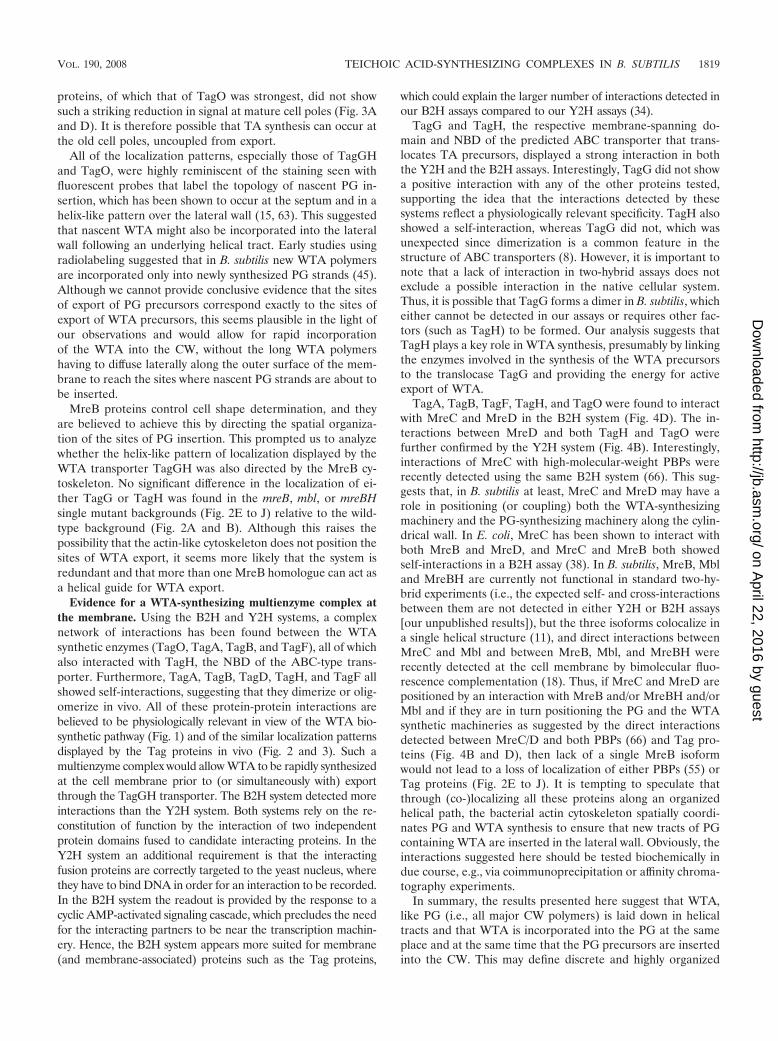
proteins, of which that of TagO was strongest, did not showsuch a striking reduction in signal at mature cell poles (Fig. 3Aand D). It is therefore possible that TA synthesis can occur atthe old cell poles, uncoupled from export.
All of the localization patterns, especially those of TagGHand TagO, were highly reminiscent of the staining seen withfluorescent probes that label the topology of nascent PG in-sertion, which has been shown to occur at the septum and in ahelix-like pattern over the lateral wall (15, 63). This suggestedthat nascent WTA might also be incorporated into the lateralwall following an underlying helical tract. Early studies usingradiolabeling suggested that in B. subtilis new WTA polymersare incorporated only into newly synthesized PG strands (45).Although we cannot provide conclusive evidence that the sitesof export of PG precursors correspond exactly to the sites ofexport of WTA precursors, this seems plausible in the light ofour observations and would allow for rapid incorporationof the WTA into the CW, without the long WTA polymershaving to diffuse laterally along the outer surface of the mem-brane to reach the sites where nascent PG strands are about tobe inserted.
MreB proteins control cell shape determination, and theyare believed to achieve this by directing the spatial organiza-tion of the sites of PG insertion. This prompted us to analyzewhether the helix-like pattern of localization displayed by theWTA transporter TagGH was also directed by the MreB cy-toskeleton. No significant difference in the localization of ei-ther TagG or TagH was found in the mreB, mbl, or mreBHsingle mutant backgrounds (Fig. 2E to J) relative to the wild-type background (Fig. 2A and B). Although this raises thepossibility that the actin-like cytoskeleton does not position thesites of WTA export, it seems more likely that the system isredundant and that more than one MreB homologue can act asa helical guide for WTA export.
Evidence for a WTA-synthesizing multienzyme complex atthe membrane. Using the B2H and Y2H systems, a complexnetwork of interactions has been found between the WTAsynthetic enzymes (TagO, TagA, TagB, and TagF), all of whichalso interacted with TagH, the NBD of the ABC-type trans-porter. Furthermore, TagA, TagB, TagD, TagH, and TagF allshowed self-interactions, suggesting that they dimerize or olig-omerize in vivo. All of these protein-protein interactions arebelieved to be physiologically relevant in view of the WTA bio-synthetic pathway (Fig. 1) and of the similar localization patternsdisplayed by the Tag proteins in vivo (Fig. 2 and 3). Such amultienzyme complex would allow WTA to be rapidly synthesizedat the cell membrane prior to (or simultaneously with) exportthrough the TagGH transporter. The B2H system detected moreinteractions than the Y2H system. Both systems rely on the re-constitution of function by the interaction of two independentprotein domains fused to candidate interacting proteins. In theY2H system an additional requirement is that the interactingfusion proteins are correctly targeted to the yeast nucleus, wherethey have to bind DNA in order for an interaction to be recorded.In the B2H system the readout is provided by the response to acyclic AMP-activated signaling cascade, which precludes the needfor the interacting partners to be near the transcription machin-ery. Hence, the B2H system appears more suited for membrane(and membrane-associated) proteins such as the Tag proteins,
which could explain the larger number of interactions detected inour B2H assays compared to our Y2H assays (34).
TagG and TagH, the respective membrane-spanning do-main and NBD of the predicted ABC transporter that trans-locates TA precursors, displayed a strong interaction in boththe Y2H and the B2H assays. Interestingly, TagG did not showa positive interaction with any of the other proteins tested,supporting the idea that the interactions detected by thesesystems reflect a physiologically relevant specificity. TagH alsoshowed a self-interaction, whereas TagG did not, which wasunexpected since dimerization is a common feature in thestructure of ABC transporters (8). However, it is important tonote that a lack of interaction in two-hybrid assays does notexclude a possible interaction in the native cellular system.Thus, it is possible that TagG forms a dimer in B. subtilis, whicheither cannot be detected in our assays or requires other fac-tors (such as TagH) to be formed. Our analysis suggests thatTagH plays a key role in WTA synthesis, presumably by linkingthe enzymes involved in the synthesis of the WTA precursorsto the translocase TagG and providing the energy for activeexport of WTA.
TagA, TagB, TagF, TagH, and TagO were found to interactwith MreC and MreD in the B2H system (Fig. 4D). The in-teractions between MreD and both TagH and TagO werefurther confirmed by the Y2H system (Fig. 4B). Interestingly,interactions of MreC with high-molecular-weight PBPs wererecently detected using the same B2H system (66). This sug-gests that, in B. subtilis at least, MreC and MreD may have arole in positioning (or coupling) both the WTA-synthesizingmachinery and the PG-synthesizing machinery along the cylin-drical wall. In E. coli, MreC has been shown to interact withboth MreB and MreD, and MreC and MreB both showedself-interactions in a B2H assay (38). In B. subtilis, MreB, Mbland MreBH are currently not functional in standard two-hy-brid experiments (i.e., the expected self- and cross-interactionsbetween them are not detected in either Y2H or B2H assays[our unpublished results]), but the three isoforms colocalize ina single helical structure (11), and direct interactions betweenMreC and Mbl and between MreB, Mbl, and MreBH wererecently detected at the cell membrane by bimolecular fluo-rescence complementation (18). Thus, if MreC and MreD arepositioned by an interaction with MreB and/or MreBH and/orMbl and if they are in turn positioning the PG and the WTAsynthetic machineries as suggested by the direct interactionsdetected between MreC/D and both PBPs (66) and Tag pro-teins (Fig. 4B and D), then lack of a single MreB isoformwould not lead to a loss of localization of either PBPs (55) orTag proteins (Fig. 2E to J). It is tempting to speculate thatthrough (co-)localizing all these proteins along an organizedhelical path, the bacterial actin cytoskeleton spatially coordi-nates PG and WTA synthesis to ensure that new tracts of PGcontaining WTA are inserted in the lateral wall. Obviously, theinteractions suggested here should be tested biochemically indue course, e.g., via coimmunoprecipitation or affinity chroma-tography experiments.
In summary, the results presented here suggest that WTA,like PG (i.e., all major CW polymers) is laid down in helicaltracts and that WTA is incorporated into the PG at the sameplace and at the same time that the PG precursors are insertedinto the CW. This may define discrete and highly organized
VOL. 190, 2008 TEICHOIC ACID-SYNTHESIZING COMPLEXES IN B. SUBTILIS 1819
on April 22, 2016 by guest
http://jb.asm.org/
Dow
nloaded from

sites on the membrane where PG and WTA coupled synthesistakes place, allowing the balanced composition and structureof the CW that is necessary to ensure controlled CW growth inrod-shaped bacteria.
ACKNOWLEDGMENTS
We thank Eric Brown for providing B. subtilis strain EB247.R.C.-L. was funded by a long-term fellowship from the Human
Frontier Science Programme Organization (HFSPO) during part ofthis work. R.C-L. and P.N. are supported by the Institut National de laRecherche Agronomique (INRA). Work in the Errington lab wassupported by a grant from the HFSPO. D.-J.S. was funded by a VENIfellowship from the Research Council for Earth and Life Sciencesfrom The Netherlands Organization for Scientific Research (NWO).
REFERENCES
1. Abachin, E., C. Poyart, E. Pellegrini, E. Milohanic, F. Fiedler, P. Berche, andP. Trieu-Cuot. 2002. Formation of D-alanyl-lipoteichoic acid is required foradhesion and virulence of Listeria monocytogenes. Mol. Microbiol. 43:1–14.
2. Anagnostopoulos, C., and J. Spizizen. 1961. Requirements for transforma-tion in Bacillus subtilis. J. Bacteriol. 81:741–746.
3. Anderson, A. J., R. S. Green, A. J. Sturman, and A. R. Archibald. 1978. Cellwall assembly in Bacillus subtilis: location of wall material incorporatedduring pulsed release of phosphate limitation, its accessibility to bacterio-phages and concanavalin A, and its susceptibility to turnover. J. Bacteriol.136:886–899.
4. Archibald, A. R., and H. E. Coapes. 1976. Bacteriophage SP50 as a markerfor cell wall growth in Bacillus subtilis. J. Bacteriol. 125:1195–1206.
5. Bhavsar, A. P., and E. D. Brown. 2006. Cell wall assembly in Bacillus subtilis:how spirals and spaces challenge paradigms. Mol. Microbiol. 60:1077–1090.
6. Bhavsar, A. P., L. K. Erdman, J. W. Schertzer, and E. D. Brown. 2004.Teichoic acid is an essential polymer in Bacillus subtilis that is functionallydistinct from teichuronic acid. J. Bacteriol. 186:7865–7873.
7. Bhavsar, A. P., R. Truant, and E. D. Brown. 2005. The TagB protein inBacillus subtilis 168 is an intracellular peripheral membrane protein that canincorporate glycerol phosphate onto a membrane-bound acceptor in vitro.J. Biol. Chem. 280:36691–36700.
8. Biemans-Oldehinkel, E., M. K. Doeven, and B. Poolman. 2006. ABC trans-porter architecture and regulatory roles of accessory domains. FEBS Lett.580:1023–1035.
9. Boyd, D. A., D. G. Cvitkovitch, A. S. Bleiweis, M. Y. Kiriukhin, D. V.Debabov, F. C. Neuhaus, and I. R. Hamilton. 2000. Defects in D-alanyl-lipoteichoic acid synthesis in Streptococcus mutans results in acid sensitivity.J. Bacteriol. 182:6055–6065.
10. Carballido-Lopez, R., and J. Errington. 2003. The bacterial cytoskeleton. Invivo dynamics of the actin-like protein Mbl of Bacillus subtilis. Dev. Cell4:19–28.
11. Carballido-Lopez, R., A. Formstone, Y. Li, S. D. Ehrlich, P. Noirot, and J.Errington. 2006. Actin homolog MreBH governs cell morphogenesis bylocalization of the cell wall hydrolase LytE. Dev. Cell 11:399–409.
12. Clarke-Sturman, A. J., A. R. Archibald, I. C. Hancock, C. R. Harwood, T.Merad, and J. A. Hobot. 1989. Cell wall assembly in Bacillus subtilis: partialconservation of polar wall material and the effect of growth conditions on thepattern of incorporation of new material at the polar caps. J. Gen. Microbiol.135:657–665.
13. Cormack, B. P., R. H. Valdivia, and S. Falkow. 1996. FACS-optimizedmutants of the green fluorescent protein (GFP). Gene 173:33–38.
14. Cutting, S. M., and P. B. Vander Horn. 1990. Genetic analysis, p. 27–74. InC. R. Harwood and S. M. Cutting (ed.), Molecular bological methods forBacillus. John Wiley and Sons Ltd., Chichester, United Kingdom.
15. Daniel, R. A., and J. Errington. 2003. Control of cell morphogenesis inbacteria: two distinct ways to make a rod-shaped cell. Cell 113:767–776.
16. Daniel, R. A., M. F. Noirot-Gros, P. Noirot, and J. Errington. 2006. Multipleinteractions between the transmembrane division proteins of Bacillus subtilisand the role of FtsL instability in divisome assembly. J. Bacteriol. 188:7396–7404.
17. Defeu Soufo, H. J., and P. L. Graumann. 2005. Bacillus subtilis actin-likeprotein MreB influences the positioning of the replication machinery andrequires membrane proteins MreC/D and other actin-like proteins forproper localization. BMC Cell Biol. 6:10.
18. Defeu Soufo, H. J., and P. L. Graumann. 2006. Dynamic localization andinteraction with other Bacillus subtilis actin-like proteins are important forthe function of MreB. Mol. Microbiol. 62:1340–1356.
19. Defeu Soufo, H. J., and P. L. Graumann. 2004. Dynamic movement ofactin-like proteins within bacterial cells. EMBO Rep. 5:789–794.
20. D’Elia, M. A., K. E. Millar, T. J. Beveridge, and E. D. Brown. 2006. Wallteichoic acid polymers are dispensable for cell viability in Bacillus subtilis. J.Bacteriol. 188:8313–8316.
21. D’Elia, M. A., M. P. Pereira, Y. S. Chung, W. Zhao, A. Chau, T. J. Kenney,M. C. Sulavik, T. A. Black, and E. D. Brown. 2006. Lesions in teichoic acidbiosynthesis in Staphylococcus aureus lead to a lethal gain of function in theotherwise dispensable pathway. J. Bacteriol. 188:4183–4189.
22. Divakaruni, A. V., R. R. Loo, Y. Xie, J. A. Loo, and J. W. Gober. 2005. Thecell-shape protein MreC interacts with extracytoplasmic proteins includingcell wall assembly complexes in Caulobacter crescentus. Proc. Natl. Acad. Sci.USA 102:18602–18607.
23. Dye, N. A., Z. Pincus, J. A. Theriot, L. Shapiro, and Z. Gitai. 2005. Twoindependent spiral structures control cell shape in Caulobacter. Proc. Natl.Acad. Sci. USA 102:18608–18613.
24. Figge, R. M., A. V. Divakaruni, and J. W. Gober. 2004. MreB, the cellshape-determining bacterial actin homologue, co-ordinates cell wall mor-phogenesis in Caulobacter crescentus. Mol. Microbiol. 51:1321–1332.
25. Formstone, A., and J. Errington. 2005. A magnesium-dependent mreB nullmutant: implications for the role of mreB in Bacillus subtilis. Mol. Microbiol.55:1646–1657.
26. Foster, S. J., and D. L. Popham. 2002. Structure and synthesis of cell wall,spore cortex, teichoic acids, S-layers, and capsules, p. 21–41. In L. Sonen-shein, R. Losick, and J. A. Hoch (ed.), Bacillus subtilis and its closest rela-tives: from genes to cells. American Society for Microbiology, Washington,DC.
27. Gross, M., S. E. Cramton, F. Gotz, and A. Peschel. 2001. Key role of teichoicacid net charge in Staphylococcus aureus colonization of artificial surfaces.Infect. Immun. 69:3423–3426.
28. Henriques, A. O., P. Glaser, P. J. Piggot, and C. P. Moran, Jr. 1998. Controlof cell shape and elongation by the rodA gene in Bacillus subtilis. Mol.Microbiol. 28:235–247.
29. Honeyman, A. L., and G. C. Stewart. 1988. Identification of the proteinencoded by rodC, a cell-division gene from Bacillus subtilis. Mol. Microbiol.2:735–741.
30. James, P., J. Halladay, and E. A. Craig. 1996. Genomic libraries and a hoststrain designed for highly efficient two-hybrid selection in yeast. Genetics144:1425–1436.
31. Jenkinson, H. F. 1983. Altered arrangement of proteins in the spore coat ofa germination mutant of Bacillus subtilis. J. Gen. Microbiol. 129:1945–1958.
32. Jones, L. J. F., R. Carballido-Lopez, and J. Errington. 2001. Control of cellshape in bacteria: helical, actin-like filaments in Bacillus subtilis. Cell 104:913–922.
33. Karamata, D., and J. D. Gross. 1970. Isolation and genetic analysis oftemperature-sensitive mutants of Bacillus subtilis defective in DNA synthesis.Mol. Gen. Genet. 108:277–287.
34. Karimova, G., N. Dautin, and D. Ladant. 2005. Interaction network amongEscherichia coli membrane proteins involved in cell division as revealed bybacterial two-hybrid analysis. J. Bacteriol. 187:2233–2243.
35. Kovacs, M., A. Halfmann, I. Fedtke, M. Heintz, A. Peschel, W. Vollmer, R.Hakenbeck, and R. Bruckner. 2006. A functional dlt operon, encoding pro-teins required for incorporation of D-alanine in teichoic acids in gram-positive bacteria, confers resistance to cationic antimicrobial peptides inStreptococcus pneumoniae. J. Bacteriol. 188:5797–5805.
36. Kristian, S. A., V. Datta, C. Weidenmaier, R. Kansal, I. Fedtke, A. Peschel,R. L. Gallo, and V. Nizet. 2005. D-Alanylation of teichoic acids promotesgroup A Streptococcus antimicrobial peptide resistance, neutrophil survival,and epithelial cell invasion. J. Bacteriol. 187:6719–6725.
37. Krogh, A., B. Larsson, G. von Heijne, and E. L. Sonnhammer. 2001. Pre-dicting transmembrane protein topology with a hidden Markov model: ap-plication to complete genomes. J. Mol. Biol. 305:567–580.
38. Kruse, T., J. Bork-Jensen, and K. Gerdes. 2005. The morphogenetic Mre-BCD proteins of Escherichia coli form an essential membrane-bound com-plex. Mol. Microbiol. 55:78–89.
39. Kunst, F., and G. Rapoport. 1995. Salt stress is an environmental signalaffecting degradative enzyme synthesis in Bacillus subtilis. J. Bacteriol. 177:2403–2407.
40. Lazarevic, V., and D. Karamata. 1995. The tagGH operon of Bacillus subtilis168 encodes a two-component ABC transporter involved in the metabolismof two wall teichoic acids Mol. Microbiol. 16:345–355.
41. Leaver, M., and J. Errington. 2005. Roles for MreC and MreD proteins inhelical growth of the cylindrical cell wall in Bacillus subtilis. Mol. Microbiol.57:1196–1209.
42. Lewis, P. J., and J. Errington. 1997. Direct evidence for active segregation oforiC regions of the Bacillus subtilis chromosome and co-localization with theSpo0J partitioning protein. Mol. Microbiol. 25:945–954.
43. Lewis, P. J., and A. L. Marston. 1999. GFP vectors for controlled expressionand dual labelling of protein fusions in Bacillus subtilis. Gene 227:101–109.
44. Ma, H., S. Kunes, P. J. Schatz, and D. Botstein. 1987. Plasmid constructionby homologous recombination in yeast. Gene 58:201–216.
45. Mauck, J., and L. Glaser. 1972. On the mode of in vivo assembly of the cellwall of Bacillus subtilis. J. Biol. Chem. 247:1180–1187.
46. Mobley, H. L. T., A. L. Koch, R. J. Doyle, and U. N. Streips. 1984. Insertionand fate of the cell wall in Bacillus subtilis. J. Bacteriol. 158:169–179.
47. Neuhaus, F. C., and J. Baddiley. 2003. A continuum of anionic charge:
1820 FORMSTONE ET AL. J. BACTERIOL.
on April 22, 2016 by guest
http://jb.asm.org/
Dow
nloaded from

structures and functions of D-alanyl-teichoic acids in gram-positive bacteria.Microbiol. Mol. Biol. Rev. 67:686–723.
48. Noirot-Gros, M. F., E. Dervyn, L. J. Wu, P. Mervelet, J. Errington, S. D.Ehrlich, and P. Noirot. 2002. An expanded view of bacterial DNA replica-tion. Proc. Natl. Acad. Sci. USA 99:8342–8347.
49. Nouaille, S., J. Commissaire, J. J. Gratadoux, P. Ravn, A. Bolotin, A. Gruss,Y. Le Loir, and P. Langella. 2004. Influence of lipoteichoic acid D-alanylationon protein secretion in Lactococcus lactis as revealed by random mutagen-esis. Appl. Environ. Microbiol. 70:1600–1607.
50. Park, Y. S., T. D. Sweitzer, J. E. Dixon, and C. Kent. 1993. Expression,purification, and characterization of CTP:glycerol-3-phosphate cytidylyl-transferase from Bacillus subtilis. J. Biol. Chem. 268:16648–16654.
51. Pooley, H. M., F. X. Abellan, and D. Karamata. 1991. A conditional-lethalmutant of Bacillus subtilis 168 with a thermosensitive glycerol-3-phosphatecytidylyltransferase, an enzyme specific for the synthesis of the major wallteichoic acid. J. Gen. Microbiol. 137:921–928.
52. Pooley, H. M., J. M. Schlaeppi, and D. Karamata. 1978. Localised insertionof new cell wall in Bacillus subtilis. Nature 274:264–266.
53. Sambrook, J., E. F. Fritsch, and T. Maniatis. 1989. Molecular cloning: alaboratory manual, 2nd ed. Cold Spring Harbor Laboratory Press, ColdSpring Harbor, NY.
54. Scheffers, D.-J. 2007. The cell wall of Bacillus subtilis, p. 331–373. In P. L.Graumann (ed.), Bacillus: cellular and molecular biology. Horizon ScientificPress, Norwich, United Kingdom.
55. Scheffers, D.-J., L. J. F. Jones, and J. Errington. 2004. Several distinctlocalization patterns for penicillin-binding proteins in Bacillus subtilis. Mol.Microbiol. 51:749–764.
56. Schertzer, J. W., and E. D. Brown. 2003. Purified, recombinant TagF proteinfrom Bacillus subtilis 168 catalyzes the polymerization of glycerol phosphateonto a membrane acceptor in vitro. J. Biol. Chem. 278:18002–18007.
57. Schlaeppi, J.-M., and D. Karamata. 1982. Cosegregation of cell wall andDNA in Bacillus subtilis. J. Bacteriol. 152:1231–1240.
58. Schlaeppi, J.-M., O. Schaefer, and D. Karamata. 1985. Cell wall and DNAcosegregation in Bacillus subtilis studied by electron microscope autoradiog-raphy. J. Bacteriol. 164:130–135.
59. Soldo, B., V. Lazarevic, and D. Karamata. 2002. tagO is involved in the
synthesis of all anionic cell-wall polymers in Bacillus subtilis 168. Microbiol-ogy 148:2079–2087.
60. Soldo, B., V. Lazarevic, H. M. Pooley, and D. Karamata. 2002. Character-ization of a Bacillus subtilis thermosensitive teichoic acid-deficient mutant:gene mnaA (yvyH) encodes the UDP-N-acetylglucosamine 2-epimerase. J.Bacteriol. 184:4316–4320.
61. Steen, A., G. Buist, K. J. Leenhouts, M. El Khattabi, F. Grijpstra, A. L.Zomer, G. Venema, O. P. Kuipers, and J. Kok. 2003. Cell wall attachment ofa widely distributed peptidoglycan binding domain is hindered by cell wallconstituents. J. Biol. Chem. 278:23874–23881.
62. Sterlini, J. M., and J. Mandelstam. 1969. Committment to sporulation inBacillus subtilis and its relationship to the development of actinomycin re-sistance. Biochem. J. 113:29–37.
63. Tiyanont, K., T. Doan, M. B. Lazarus, X. Fang, D. Z. Rudner, and S. Walker.2006. Imaging peptidoglycan biosynthesis in Bacillus subtilis with fluorescentantibiotics. Proc. Natl. Acad. Sci. USA 103:11033–11038.
64. Uetz, P., L. Giot, G. Cagney, T. A. Mansfield, R. S. Judson, J. R. Knight, D.Lockshon, V. Narayan, M. Srinivasan, P. Pochart, A. Qureshi-Emili, Y. Li,B. Godwin, D. Conover, T. Kalbfleisch, G. Vijayadamodar, M. Yang, M.Johnston, S. Fields, and J. M. Rothberg. 2000. A comprehensive analysis ofprotein-protein interactions in Saccharomyces cerevisiae. Nature 403:623–627.
65. van den Ent, F., L. A. Amos, and J. Lowe. 2001. Prokaryotic origin of theactin cytoskeleton. Nature 413:39–44.
66. van den Ent, F., M. Leaver, F. Bendezu, J. Errington, P. de Boer, and J.Lowe. 2006. Dimeric structure of the cell shape protein MreC and its func-tional implications. Mol. Microbiol. 62:1631–1642.
67. Varley, A. W., and G. C. Stewart. 1992. The divIVB region of the Bacillussubtilis chromosome encodes homologs of Escherichia coli septum placement(MinCD) and cell shape (MreBCD) determinants. J. Bacteriol. 174:6729–6742.
68. Wecke, J., M. Perego, and W. Fischer. 1996. D-Alanine deprivation of Ba-cillus subtilis teichoic acids is without effect on cell growth and morphologybut affects the autolytic activity. Microb. Drug Resist. 2:123–129.
69. Wu, L. J., and J. Errington. 2003. RacA and the Soj-Spo0J system combineto effect polar chromosome segregation in sporulating Bacillus subtilis. Mol.Microbiol. 49:1463–1475.
VOL. 190, 2008 TEICHOIC ACID-SYNTHESIZING COMPLEXES IN B. SUBTILIS 1821
on April 22, 2016 by guest
http://jb.asm.org/
Dow
nloaded from
Copyright © 2022 FDOKUMEN