Bacillus subtilis spores as vaccine adjuvants: further insights into the mechanisms of action
Upload
sorbonne-frCategory
view
4download
0
BioMed CentralBMC Microbiology
BMC Microbiology 2001, 1 :15Research articleMtnK, methylthioribose kinase, is a starvation-induced protein in Bacillus subtilisAgnieszka Sekowska*1, Laurence Mulard2, Susanne Krogh3, Jane KS Tse1
and Antoine Danchin1,4
Address: 1HKU-Pasteur Research Centre, Dexter HC Man Building, Sassoon Road, Pokfulam, Hong Kong, China, 2Chimie Organique, Institut
Pasteur, 28 rue du Docteur Roux, Paris, 75724, France, 3Department of Genetics, Smurfit Institute, Trinity College, Dublin, 2, Ireland and 4Génétique des Génomes Bactériens, Institut Pasteur, 28 rue du Docteur Roux, Paris, 75724, France
E-mail: Agnieszka Sekowska* - [email protected]; Laurence Mulard - [email protected]; Susanne Krogh - [email protected]; Jane KS Tse - [email protected]; Antoine Danchin - [email protected]
*Corresponding author
AbstractBackground: Methylthioadenosine, the main by-product of spermidine synthesis, is degraded inBacillus subtilis as adenine and methylthioribose. The latter is an excellent sulfur source and theprecursor of quorum-sensing signalling molecules. Nothing was known about methylthioriboserecycling in this organism.
Results: Using trifluoromethylthioribose as a toxic analog to select for resistant mutants, wedemonstrate that methylthioribose is first phosphorylated by MtnK, methylthioribose kinase, theproduct of gene mtnK (formerly ykrT), expressed as an operon with mtnS (formerly ykrS) in anabundant transcript with a S-box leader sequence. Although participating in methylthioriboserecycling, the function of mtnS remained elusive. We also show that MtnK synthesis is boostedunder starvation condition, in the following decreasing order: carbon-, sulfur- and nitrogen-starvation. We finally show that this enzyme is part of the family Pfam 01633 (choline kinases) whichbelongs to a large cluster of orthologs comprizing antibiotic aminoglycoside kinases and proteinserine/threonine kinases.
Conclusions: The first step of methylthioribose recycling is phosphoryltaion by MTR kinase,coded by the mtnK (formerly ykrT) gene. Analysis of the neighbourhood of mtnK demonstrates thatgenes located in its immediate vicinity (now named mtnUVWXYZ, formerly ykrUVWXYZ) are alsorequired for methylthioribose recycling.
BackgroundStarvation for essential metabolites results in the expres-
sion of many proteins, often of still unknown function,some of which related to quorum-sensing. In the course
of our study of sulfur metabolism in bacteria we have
witnessed that expression of many genes was induced
when cells were deprived of sulfur [1]. In particular,
upon entry into the stationary growth phase (which is of-
ten the consequence of starvation in one of the major cellmetabolite supplies: carbon, nitrogen or phosphorus),
we observed that polyamine biosynthesis was much af-
fected, in parallel with the expression of S-adenosylme-
Published: 8 August 2001
BMC Microbiology 2001, 1:15
Received: 27 June 2001Accepted: 8 August 2001
This article is available from: http://www.biomedcentral.com/1471-2180/1/15
© 2001 Sekowska et al; licensee BioMed Central Ltd. Verbatim copying and redistribution of this article are permitted in any medium for any non-commercial purpose, provided this notice is preserved along with the article's original URL. For commercial use, contact [email protected]

BMC Microbiology 2001, 1:15 http://www.biomedcentral.com/1471-2180/1/15
thionine decarboxylase [2]. This prompted us to
investigate the fate of the products of this important en-
zyme.
Aminopropyl transfer, during polyamine metabolism,
yields methylthioadenosine, which is split into adenine
and methylthioribose by the mtnA gene (yrrU) [3]. This
molecule is excreted in Escherichia coli[4], and degrad-
ed by oxidation steps in Klebsiella pneumoniae[5]. In
Eukarya, its fate is not known except for parasites [6] and
in part in plants [7] and mammals [8], and, because it ap-
pears to differ in different organisms, pharmaceutical
companies have endeavoured to use analogues as drugs
[9]. Nothing is known in other organisms. We have es-
tablished that MTR is an excellent sulfur source in Bacil-
lus subtilis[3]. Scanning the genome sequence however
did not reveal obvious similarities with known pathways.
Using a toxic analogue of MTR and transposon mutagen-
esis we screened for resistant mutants. This led us to
identify the ykrT gene as a major bottleneck in MTR me-
tabolism, and to identify it as the methylthioribose ki-
nase gene (now named mtnK). Further study of the gene
uncovered an unusual expression pattern under starva-
tion conditions, discussed in the present article.
ResultsTransposon mutagenesisMethylthioribose (MTR) can be used by B. subtilis as the
sole sulfur source. However, the way by which the sulfurmoiety is recycled remains unknown. To elucidate this
pathway, we used an analogue of MTR, trifluoromethyl-
thioribose (3F-MTR), which has been assayed as a possi-
ble drug lead for killing pathogens [10]. After transposon
mutagenesis (see Materials and Methods), eleven 3F-
MTR resistant clones were obtained (located in genes
ykrW, ykrY and ykrT). Among them, one mutant carried
the transposon insertion in the putative promoter region
of the ykrT gene (strain BSHP7035). This insertion was
situated 73 bp upstream of translational start point and
was comprised within the regulatory S-box structure
[11]. Analysis of the remaining ten mutants will be the
object of a separate publication (see Conclusion). Inacti-
vation of gene ykrT resulted in complete loss of MTR uti-
lization, although some revertant clones able to grow
very poorly on MTR appeared after few days incubation.
In silico analysis of the protein sequence revealed that it
contained a sequence (PFAM CD motif 01633) typical of
choline/ethanolamine kinases (Fig. 1 and [http://
www.sanger.ac.uk/cgi-bin/Pfam/getacc?PF01633] ).
We therefore explored whether as in K. pneumoniae, the
first step of MTR catabolism would be phosphorylation
of the ribose moiety.
MtnK (YkrT) is a methylthioribose kinaseBefore investigating the involvement of YkrT in MTR
metabolism we reanalysed its sequence, as predicted in
SubtiList [http://genolist.pasteur.fr/SubtiList/] . Theregion preceding the putative ATG start site revealed the
presence of three ATGs in a row. The putative ribosome
binding site GGAGGT, should be located at least 5 nt and
preferably 7–12 nt before the start site [12,13]. We there-
fore propose that the start of the protein is the third ATG
in this sequence, and not the first one:
tttacggccacatattaattaattacataattGGAGGTt atg atg ATG
gga gtc aca
Sequence comparison with proteins present in bacterial
genome databases [14] revealed, in addition to many
genes annotated as choline/ethanolamine kinases with
weak similarities (Fig. 1), nine proteins which displayed
high similarity with YkrT: in K. pneumoniae (a protein
annotated as a MTR kinase in the unpublished entry: En-
trez Protein AAG43573), in Bacillus stearothermophil-
us, in two similar sequences of Bacillus anthracis, in
Treponema denticola, in Sinorhizobium meliloti, in Me-
sorhizobium loti and in Arabidopsis thaliana (Fig. 2).
The similarity with the sequence of B. stearothermophil-
us further substantiated our identification of the third
ATG codon as the translation start site in B. subtilis.
Because it is well known that in silico function identifica-tion can be very misleading [15], we looked for a bio-
chemical identification of the activity. Using protocols
derived from the work of Riscoe and co-workers [16], we
set up a cell-free biochemical assay, comparing the wild
type strain, a disrupted conditional mutant (BFS1850),
and this same mutant grown in the present of IPTG (see
also below), using radioactive ATP as the phosphate do-
nor. As shown in Fig. 3, we found activity only in the wild
type. Radioactive MTR-1-P was detected in the wild type
when adding exogenous MTR, but not in its absence, nor
in the mtnK of the enzyme for MTR has been mutant.
The KM approximately evaluated to be ca 60 µM (Fig. 4).
We also found that in the preparations containing 5%
glycerol, the wild type contained glycerol-1-P, whereas
the mutant did not, showing that MtnK can phosphor-
ylate glycerol (data not shown).
mtnK and mtnS form an operonSequence analysis of the chromosome region of mtnK
shows that this gene is situated between two predicted
transcriptional terminators together with the adjacent
ykrS gene. The ykrS gene is separated by only 7 bp from
mtnK. This suggests that both genes could be co-tran-
scribed. The transposon insertion yielding 3F-MTR re-
sistance was identified in the promoter region of themtnK ykrS genes. This could possibly abolish the expres-

BMC Microbiology 2001, 1:15 http://www.biomedcentral.com/1471-2180/1/15
sion of both genes, as we were unable to tentatively iden-
tify in silico any putative promoter for ykrS gene alone.
To explore this question, we used strain BFS1850
(ykrT::lacZ) which allows the expression of the down-
stream gene under the control of the IPTG inducible
Pspac promoter. The BFS1850 strain was assayed for its
ability to use MTR as the sole sulfur source in the pres-
ence or in the absence of IPTG. In both cases this strain
failed to grow on MTR. Furthermore, expressing the
ykrS gene from Pspac did not change the outcome of the
biochemical experiment with inactivated mtnK, showing
that ykrS does not directly participate in the MTR kinase
activity (Fig. 3). As a further exploration of the ykrS gene
role, we constructed a strain deleted of ykrS alone
(BSHP7010). This strain was also unable to grow with
MTR as the sulfur source. These results show that both
mtnK and ykrS are implicated in the MTR recycling
pathway (we propose therefore to rename ykrS mtnS),
but that MtnK alone is involved in the phosphorylation
step of the substrate.
Subsequently, the RNA synthesis in this region was ana-lysed by RT-PCR in cells growing exponentially in mini-
mal medium. This experiment confirmed that mtnK and
mtnS were transcribed together (data not shown). Be-
cause we found that disruptants of the upstream ykrU
(mtnU) gene (an other adjacent gene of yet unidentified
function – hydrolase/nitrilase-like), were unable to grow
with MTR as the sulfur source, we further investigated by
RT-PCR whether mtnK was co-transcribed with the
mtnU gene. These experiments showed that there was no
co-transcription, in line with the observation that mtnU
is separated from mtnK by a transcriptional terminator
(data not shown).
Expression of the mtnK geneUsing a mtnK::lacZ fusion (strain BFS1850) we studied
the behaviour of the gene in a variety of environmental
conditions. In the standard growth conditions with sul-
fate as sulfur source, the expression of mtnK was high,
and very stable during growth, decreasing from 1700 U/
mg of protein in the exponential growth phase to 1200
U/mg of protein in the stationary growth phase. Al-
though the mtnK mtnS operon is a member of the S-box
regulon, no significant factor of regulation by methio-
nine availability could be observed under our growthconditions, when sulfate was replaced by methionine as
sole sulfur source (30% variation, see Table 1). Under
Figure 1Comparison of MtnK with the first eight proteins listed in the choline kinase family Pfam 01633. Identical residues in the family01633 are indicated by a "=" sign, while conserved residues are indicated by a "+" sign (amino acid families are: ASTPG, MLIV,HKR, WYF, DNEQ, and C). When these same residues are also conserved in MtnK they are colored: red for identical resi-dues, blue for conserved residues.

BMC Microbiology 2001, 1:15 http://www.biomedcentral.com/1471-2180/1/15
Figure 2Alignment of the eight putative MtnK protein present in the genome libraries with B. subtilis MtnK together and with the Pfam016533 consensus of choline kinases (CHLK_CONS) obtained after Blast search of the WWWDDL at the NCBI. Codes as inFigure 1. BACSU: B. subtilis; BACST: B. stearothermophilus; BACA: B. anthracis; TREDE: Treponema denticola; KLEPN: Klebsiellapneumoniae; MESLO: Mesorhizobium loti; SINME: Sinorhizobium meliloti; ARATH: Arabidopsis thaliana. The sequences wereextracted from the databases presented in [14].
BMC Microbiology 2001, 1:15 http://www.biomedcentral.com/1471-2180/1/15
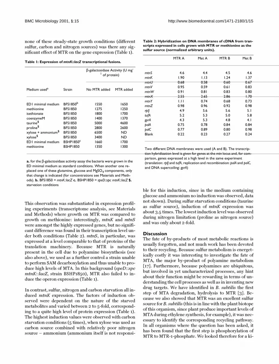
none of these steady-state growth conditions (different
sulfur, carbon and nitrogen sources) was there any sig-
nificant effect of MTR on the gene expression (Table 1).
This observation was substantiated in expression profil-
ing experiments (transcriptome analysis, see Materials
and Methods) where growth on MTR was compared to
growth on methionine: interestingly, mtnK and mtnS
were amongst the highly expressed genes, but no signifi-
cant difference was found in their transcription level un-
der both conditions (Table 2). mtnS, in particular, was
expressed at a level comparable to that of proteins of the
translation machinery. Because MTR is naturally
present in the cell due to polyamine biosynthesis (see
also above), we used as a further control a strain unable
to perform SAM decarboxylation and thus unable to pro-
duce high levels of MTA. In this background (speD::spc
mtnK::lacZ, strain BSHP1850), MTR also failed to in-
duce the operon expression (Table 1).
In contrast, sulfur, nitrogen and carbon starvation all in-
duced mtnK expression. The factors of induction ob-
served were dependent on the nature of the starved
metabolites and varied between 2 to 5-fold, correspond-
ing to a quite high level of protein expression (Table 1).
The highest induction values were observed with carbon
starvation conditions (5 times), when xylose was used as
carbon source combined with relatively poor nitrogensource – ammonium (ammonium itself is not responsi-
ble for this induction, since in the medium containing
glucose and ammonium no induction was observed, data
not shown). During sulfur starvation conditions (taurine
as sulfur source), induction of mtnK expression wasabout 3.5 times. The lowest induction level was observed
during nitrogen limitation (proline as nitrogen source)
and was only about 2-fold.
DiscussionThe fate of by-products of most metabolic reactions is
usually forgotten, and not much work has been devoted
to their recycling. Because sulfur metabolism is energet-
ically costly it was interesting to investigate the fate of
MTA, the major by-product of polyamine metabolism
[17]. Furthermore, because polyamines are ubiquitous,
but involved in yet uncharacterized processes, any hint
about their function might be rewarding in terms of un-
derstanding the cell processes as well as in inventing new
drug targets. We have identified in B. subtilis the first
step of MTA degradation, hydrolysis to MTR [3]. Be-
cause we also showed that MTR was an excellent sulfur
source for B. subtilis (this is in line with the plant biotope
of this organism, since plant produce important levels of
MTA during ethylene synthesis, for example), it was nec-
essary to identify the corresponding recycling pathway.
In all organisms where the question has been asked, it
has been found that the first step is phosphorylation of
MTR to MTR-1-phosphate. We looked therefore for a ki-
Table 1: Expression of mtnK::lacZ transcriptional fusions.
β-galactosidase Activity (U mg-
1 of protein)
Medium useda Strain No MTR added MTR added
ED1 minimal medium BFS1850b 1550 1650methionine BFS1850 1275 1250isethionate BFS1850 1800 1700coenzymeM BFS1850 1400 1370taurine$ BFS1850 5000 4600proline$ BFS1850 2800 2600xylose + ammonium$ BFS1850 6500 NDxylose$ BFS1850 3400 NDED1 minimal medium BSHP1850c 1660 1700methionine BSHP1850 1350 1300
a. for the β-galactosidase activity assay the bacteria were grown in the ED minimal medium as standard conditions. When another one re-placed one of these glutamine, glucose and MgSO4 components, only that change is indicated (for concentrations see Materials and Meth-ods). b. BFS1850 = mtnK::lacZ c. BSHP1850 = speD::spc mtnK::lacZ $. starvation conditions
Table 2: Hybridization on DNA membranes of cDNA from tran-scripts expressed in cells grown with MTR or methionine as the sulfur source (normalized arbitrary units).
MTR A Met A MTR B Met B
mtnS 4.6 4.4 4.5 4.6mtnK 1.90 1.13 1.24 1.37mtnU 0.68 0.58 0.60 0.67mtnV 0.95 0.59 0.61 0.83mtnW 0.91 0.81 0.83 0.80mtnX 2.55 2.65 2.86 1.70mtnY 1.11 0.74 0.68 0.73mtnZ 0.98 0.96 0.92 0.98rplJ 4.9 5.6 5.6 5.1tufA 5.2 5.3 5.0 5.8gyrA 4.3 5.3 4.8 4.1polA 0.75 0.78 0.84 0.84polC 0.77 0.89 0.80 0.98Blank 0.22 0.23 0.27 0.24
Two different DNA membranes were used (A and B). The transcrip-tion hybridization level is given for genes at the mtn locus and, for com-parison, genes expressed at a high level in the same experiment (translation: rplJ and tufA, replication and recombination: polA and polC, and DNA supercoiling: gyrA)

BMC Microbiology 2001, 1:15 http://www.biomedcentral.com/1471-2180/1/15
nase that would fit the missing step. Gene ykrT was a
possible candidate, since it codes for a protein of the
choline/ethanolamine kinase family, while it belongs to
a S-box regulated transcription unit [11]. Two types of ge-
netic experiment substantiated this hypothesis: the
ykrT::lacZ fusion constructed during the B. subtilis
functional analysis program [18] failed to grow on MTR
as a sulfur source, and more importantly, selecting for
3F-MTR resistance after transposon mutagenesis yield-
ed an insertion in the leader mRNA of the ykrTS operon.
Our study further demonstrated that both genes of this
operon (which does not comprise the neighbour mtnU
gene) are involved in MTR metabolism. We further
showed biochemically that YkrT is indeed a MTR kinase,
and named it accordingly MtnK. Using both expression
profiling experiments and transcriptional fusions we
demonstrated that both genes are expressed at a fairly
high level. This contrasts with the observations pub-lished by Henkin and co-workers who found a much low-
er expression of these genes [11]. Furthermore, we did
not find significant differences in the expression factor of
mtnK and in its repression factor by methionine in con-
trast to the data presented in the same Henkin et al. arti-
cle. We could not find any straightforward explanation
for this discrepancy. However, because starvation condi-
tions appear to play an important role in mtnK expres-
sion it may be that subtle differences in growth
conditions result in a drastic alteration of the expression
pattern of the mtnKmtnS operon. Indeed, we have ob-
served, using transcriptome experiments, that some
gene expression is extremely sensitive to environmental
conditions [19]. In addition, it should be remarked that
our lacZ fusions have been constructed in situ, while they
are located in a different strain at the distant SP β phage
locus in the chromosome in Henkin et al. study [11]. Fi-
nally, our study shows no sulfur effect under steady-state
conditions, while starvation for carbon, nitrogen or sul-
fur results in a strong enhancement of mtnKS synthesis.This may account for the difference in our experiments,
Figure 3Autoradiograph showing the outcome of the MTR kinaseassay. The assay was carried out for 90 min at 37°C (seeMaterials and Methods). Lane 1 corresponds to [•32P]ATP asstandard; lanes 2, 4 and 6: no MTR was added for the reac-tions with cell-free extracts of wild type, BFS1850 andBFS1850 grown with IPTG, respectively; lanes 3, 5 and 7:reaction performed in the presence of MTR and cell-freeextracts of wild type, BFS1850 and BFS1850 grown withIPTG, respectively.
Figure 4Autoradiograph of the MTR kinase assay with increasing con-centrations of substrate. The assay was carried out for 90min at 37°C with cell-free extracts of wild type (see Materi-als and Methods). Lane 1 corresponds to [32P]ATP as stand-ard; lanes 2: no MTR added; lanes 3, 4, 5, 6 and 7: reactionperformed with increasing concentrations of MTR (20, 40,80, 160 and 320 µM, respectively). Saturation is observed at160 µM.

BMC Microbiology 2001, 1:15 http://www.biomedcentral.com/1471-2180/1/15
while showing that these genes belong to the starvation
condition regulon.
That MtnK belongs to the choline kinase family Pfam01633 is unexpected and interesting [20]. This is consist-
ent with the involvement of a catalytic site binding the N-
methyl or S-methyl group as important for recognition of
the substrate, and not with a straightforward evolution
from the kinases which phosphorylate ribose derivatives.
This family is a member of a much wider Cluster of Or-
thologous Genes (COG0510) group, which belongs to a
group of many other kinases, including carbohydrate an-
tibiotic resistance gene (COG 3570) and serine/threo-
nine protein kinases (COG 0661, COG 3178) [21]. Most
conserved residues are charged residues, suggesting that
they may be important in the catalytic activity of the en-
zyme. It would be of interest to explore further the phyl-
ogeny of these enzymes, basing alignments on gap and
insertions rather than simply on similar or identical ami-
noacids [15,22].
Finally, the function of the mtnS gene remains elusive:
while it seems to be necessary for the use of MTR as a sul-
fur source (in the context of the B. subtilis genes as they
are grouped together) it does not seem to be necessary
for the first step of MTR recycling. Further work is need-
ed to delineate its function. MtnK is induced at a fairly
high level under starvation conditions (with MtnS ex-
pressed at a very high level). This is consistent with ad-aptation of the cell to famine conditions, where
byproducts need to be scavenged from the cytoplasm and
from the environment, rather than lost. This may also be
related to quorum-sensing signalling, which is known to
occur under similar conditions. It would therefore be in-
teresting to explore the relationship between the use of
such by-products, cell density and quorum sensing.
ConclusionMtnK performs the first step in MTR recycling. Consist-
ent with the fact that MTR is a by-product of an anabolic
pathway, MtnK expression is enhanced in starvation
conditions. Isolation of 3F-MTR resistant mutants also
yielded several other types of inserts failing to grow on
MTR, thus demonstrating that 3F-MTR is phosphorylat-
ed and that downstream derivatives of this molecule are
toxic to the cell. In particular, ykrW and ykrY insertion
mutants were resistant to this toxic metabolite. These
genes all belong to a neighbouring S-box regulated oper-
on, and, despite the fact that they are quite dissimilar to
the genes involved in K. pneumoniae MTR metabolism,
this indicates that they are needed for recycling the sul-
fur moiety of this molecule, probably with similar chem-
ical steps enacted by enzymes recruited from another
group of proteins [23]. Work in progress is characteriz-ing the corresponding activities and regulations.
Materials and MethodsBacterial strains and plasmids, and growth mediaE. coli and B. subtilis strains as well as plasmids used in
this work are listed in Table 3. Despite the fact that thereare no regulations yet in this domain in China, all exper-
iments were performed in accordance with the European
regulation requirements concerning the contained use of
Genetically Modified Organisms of Group-I (French
agreement N° 2735). E. coli and B. subtilis were grown in
Luria-Bertani (LB) medium [24] and in ED minimal me-
dium: K2HPO4, 8 mM; KH2PO4, 4,4 mM; glucose, 27
mM; Na3-citrate, 0.3 mM; L-glutamine, 15 mM; L-tryp-
tophan, 0.244 mM; ferric citrate, 33.5 µM; MgSO4, 2
mM; MgCl2, 0.61 mM; CaCl2, 49.5 µM; FeCl3, 49.9 µM;
MnCl2, 5.05 µM; ZnCl2, 12.4 µM; CuCl2, 2.52 µM; CoCl2,
2.5 µM; Na2MoO4, 2.48 µM. For tests for growth with
various sulfur sources, a sulfur-free medium was used.
This medium was as described above but MgSO4 was re-
placed by MgCl2 at the same concentration (2 mM); the
sulfur source tested were added at the followings concen-
trations: taurine, 5 mM; isethionate and coenzyme M, 2
mM; methionine, 1 mM; MTR, 0.2 mM. For nitrogen
starvation, glutamine was replaced by proline (15 mM)
and for carbon starvation, glucose was replaced by xylose
(0.5%) and glutamine by ammonium (15 mM). For as-
saying growth on plates, either the MgSO4containing
medium or the sulfur-free basal medium was used. In the
latter case, 10 µl of the sulfur source under investigation
was applicated onto paper discs (MTR, 200 mM stocksolution and methionine, 100 mM stock solution) depos-
ited at the center of the plate, after bacteria had been uni-
formly spread at the surface of the plate, and growth was
measured around the disk. When necessary IPTG was in-
cluded at 1 mM concentration. LB and ED plates were
prepared by addition of 17 g/liter Bacto agar or Agar No-
ble (Difco) respectively to the medium. When included,
antibiotics were added to the following concentrations:
ampicillin, 100 mg/liter; spectinomycin, 100 mg/liter;
erythromycin plus lincomycin, 1 mg/liter and 25 mg/lit-
er. Bacteria were grown at 37°C. The optical density (OD)
of bacterial cultures was measured at 600 nm. MTR was
prepared from MTA (Sigma, D5011) by acid hydrolysis as
described by Schlenk [25]. 3-fluoromethythiorybose
(3F-MTR, 5-thio-5-S-trifluoromethyl-D-ribose) was
synthesised accordingly to [26].
TransformationStandard procedures were used to transform E. coli[27]
and transformants were selected on LB plates containing
ampicillin or ampicillin plus spectinomycin. B. subtilis
cells were transformed with plasmid DNA following the
two-step protocol described previously [28]. Transform-
ants were selected on LB plates containing erythromycin
plus lincomycin or spectinomycin or spectinomycin pluserythromycin plus lincomycin.

BMC Microbiology 2001, 1:15 http://www.biomedcentral.com/1471-2180/1/15
Molecular genetics proceduresPlasmid DNA was prepared from E. coli by standard pro-
cedures [27]. B. subtilis chromosomal DNA was purified
as described by Saunders [29]. Restriction enzymes and
T4 DNA ligase were used as specified by manufacturers.
DNA fragments used for cloning experiments were pre-pared by PCR using PfuTurbo DNA polymerase (Strata-
gene). Amplified fragments were purified by QIAquick
PCR Purification Kit (Qiagen). DNA fragments were pu-
rified from a gel using Spin-X columns from Corning
Costar by subsequent centrifugation and precipitation.
To construct the ykrS deletion strain, a SmaI restricted
spectinomycin resistance cassette [30] was used. Two
DNA fragments, one upstream from the ykrS gene (nu-
cleotides -490 to +35 relative to the translational start
point of ykrS) and the second one downstream from the
ykrS gene (nucleotides -9 to +452 relative to the ykrS
stop codon) were amplified by PCR using primers intro-
ducing, for the first one, aBamHI cloning site at the 5'
end and a SmaI cloning site at the 3' end of the fragment,
and for the second one, a SmaI cloning site at the 5' end
and an EcoRI site at the 3' end of the fragment. PCR
products and the spectinomycin cassette were ligated
and inserted into theBamHI and EcoRI sites of pUC19
(Roche) producing plasmid pHPP7010. Prior to trans-
formation, this plasmid was linearised at its unique ScaIsite. Complete deletion of the gene was obtained by a
double cross-over event, giving strain BSHP7010.
The DNA downstream from the mtnK gene (nucleotides
+295 to +604 relative to the translation start point) was
amplified by PCR using primers introducing an EcoRI
cloning site at the 5' end and a BamHI cloning site at the
3' end of the fragment, then inserted into the EcoRI and
BamHI sites of plasmid pMutin4mcs [31] producing
plasmid pDU1850. Plasmids disrupting the mtnU gene
was obtained by PCR amplification of downstream re-
gions of mtnU gene (+105 to +346) as described for
pDU1850, producing the plasmids pDU1851. Both plas-
mids were introduced into the chromosome by a single
cross-over event, giving strains BFS1850 and BFS1851,
respectively.
Transposon mutagenesisTransposon bank was constructed by introduction of
mini-Tn10 delivery vector pIC333 [32] into B. subtilis
168 strain as described previously [33]. Several thou-
sands independent clones were pooled together and 5
samples of chromosomal DNA were prepared for further
use. To obtain 3F-MTR resistant clones, B. subtilis 168
was transformed with chromosomal DNA containingpreviously prepared transposon banks and clones were
selected on LB plates containing spectinomycin. Then,
using velvets replicas, clones were transferred onto min-
imal medium plates containing 3F-MTR at 100 µM con-
centration and allowed to grow for 24 hrs. The single
transposon insertion event was confirmed by back-cross
in 168 strain and check for 3F-MTR resistance. To deter-
mine the location of the transposon insertion, chromo-
somal DNA was prepared, followed by subsequent
digestion with HindIII, self ligation in E. coli XL1-Blue
strain and plasmid sequencing. The primers used for se-
quencing of transposon insertions were the followings:
Tn10 left: 5'GGCCGATTCATTAATGCAGGG3' and Tn10
right: 5'CGATATTCACGGTTTACCCAC3'.
RNA isolation and transcriptome analysisTotal RNA was obtained from cells growing on ED1 of 0.5
using "High Pure RNA Isolation Kit" from Roche (for
RT-PCR minimal medium to an OD600experiment) or as
described in [19] for transcriptome analysis. RT-PCR ex-
periments were performed using RT-PCR System
(Promega) as specified by the manufacturer. For cDNA
synthesis in macro-array study, CDS-specific primers
(cDNA Labelling primers – optimized forB. subtilis, Sig-
ma-GenoSys Biotechnologies, Inc.) and two quantities oftotal RNA (1 and 10 µg) were used. Hybridization probes
Table 3: Bacterial strains and plasmids used or created in this study.
Strain or plasmid Genotype or description Source or reference
StrainsEscherichia coliXL1-Blue K12 supE44 hsdR17 recA1
endA1 gyrA46 thiLaboratory collection
relA1 lac - F' [proAB + lacI q lacZ ∆ M15 Tn10(tetR)]
Bacillus subtilis168 trpC2 [36]BSIP7004 trpC2 speD::spc [2]BFS1850 trpC2mtnK::lacZ This worka
BFS1851 trpC2mtnU::lacZ This worka
BSHP1850 trpC2speD::spc mtnT::lacZ This workBSHP7010 trpC2 mtnS::spc This workBSHP7035 trpC2pmtnK::Tn10 This workPlasmidspUC19 cloning vector, AmpR [37]pIC333 mini-Tn10 delivery vector,
SpcR, EryR[32]
pMutin4mcs cloning vector, ErmR [31]pDU1850 pMutin4mcs::mtnK This workpDU1851 pMutin4mcs::mtnU This workpHPP7010 pUC19mtnS::spc b This work
a. This strain has been constructed during the frame of the EC project for the functional characterization of the genome of B. subtilis in Europe b. spc is the spectinomycin resistance gene from Staphylococcus aureus

BMC Microbiology 2001, 1:15 http://www.biomedcentral.com/1471-2180/1/15
were synthesized as described in [19]. Approximately
60–75% incorporation of labeled nucleotides was
achieved in these conditions. Panorama™ B. subtilis
gene arrays (Sigma-GenoSys Biotechnologies, Inc.) wereused and the transcript levels corresponding to the genes
analyzed in the present study were kept for further study.
Since in the present study we did not require a thorough
statistical analysis of the transcriptome data (performed
in [19]), each experiment was averaged and the average
was used as a standard for the analysis, since there was
no obvious difference between the MTR and methionine
growth conditions. Data presented here are normalized
for each of these conditions.
Preparation of cell-free extractsB. subtilis was grown to middle-exponential growth
phase ∼ 0.7–0.8). The organisms were harvested by cen-
trifugation (14,000 rpm, 4°C, 3 min), and the (OD600 cell
pellets were resuspended in a solution containing 150
mM glycine (pH 9) and 1 mM β-mercaptoethanol (β-
ME). The bacterial cells were disrupted by sonication (4
min, maximum amplitude) and cellular debris was re-
moved by centrifugation (14,000 rpm, 4°C, 10 min).
Enzyme assaysB. subtilis cells containing lacZ fusions were assayed for
β-galactosidase activity as described previously [34].
Specific activity was expressed in Units per mg protein.
The Unit used is equivalent to 0.28 nmols min-1 at 28°C.Protein concentration was determined by Bradford's
method using a protein assay Kit (Bio-Rad Laborato-
ries). At least two independent cultures were monitored.
MTR kinase was assayed as described by [16] with mi-
nors modifications (glycerol was present as a stabilizer in
the first experiments, while it was absent in the final ones
because we found that MtnK phosphorylated this mole-
cule). Briefly, the reaction mixture of 100 µl contained
150 mM glycine (pH 9), 1 mM MgCl2, 1 mM β-ME, 40 µM
MTR, 0.15 µg/ml crude extract (final) and 1 mM [γ-32P]
ATP. [γ-32P] ATP (10 Ci/mmol, Dupont-New England
Nuclear) was diluted with nonradioactive ATP to yield a
10 mM stock solution. Specific activity of the stock ATP
mixture was 20 µCi/µmol (0.2 µCi/µl). The reactions
were carried out at 37°C for 90 min. After this period, re-
actions were terminated by sitting tubes on ice. [γ-32P]
MTR-P was separated from [γ32P] ATP, 32 PPi and 32Pi
on PEI-Cellulose F plates Merck as described by [35]. 1 µl
samples were loaded on the plate and separated with 1 M
LiCl. Samples were allowed to resolve until the solvent
front was about 1 cm below the edge of plate (approxi-
mately 40 min). The plate was dried, and the radioactiv-
ity was detected by autoradiography using Biomax-MR
Kodak film.
List of Abbreviationsbp: base pairs; β-ME, β-mercaptoethanol; CDS: coding
sequence; IPTG: isopropyl β-D-thiogalactopyranoside;
MTA: methylthioadenosine; MTR: methylthioribose;3F-MTR: trifluoromethylthioribose; nt, nucleotides
AcknowledgementsWe wish to thank Catherine Guerreiro (Unité de Chimie Organique) for her contribution to part of the chemical synthesis of 3FMTR. The whole MTR degradation pathway was presented as a poster at the Area of Excel-lence workshop organized in Hong Kong University on December 22–23, 2000.
References1. Coppée JY, Auger S, Turlin E, Sekowska A, Le Caer JP, Labas V, Vag-
ner V, Danchin A, Martin-Verstraete I: Sulfur-limitation-regulat-ed proteins in Bacillus subtilis : a two-dimensional gelelectrophoresis study. Microbiology 2001, 147:1631-1640
2. Sekowska A, Coppée JY, Le Caer JP, Martin-Verstraete I, Danchin A:S-adenosylmethionine decarboxylase of Bacillus subtilis isclosely related to archaebacterial counterparts. Mol. Microbiol.2000, 36:1135-1147
3. Sekowska A, Danchin A: Identification of yrrU as the methylth-ioadenosine nucleosidase gene in Bacillus subtilis. DNA Res.1999, 6:255-264
4. Schroeder HR, Barnes CJ, Bohinski RC, Mallette MF: Biological pro-duction of 5-methylthioribose. Can. J. Microbiol. 1973, 19:1347-1354
5. Heilbronn J, Wilson J, Berger BJ: Tyrosine aminotransferase cat-alyzes the final step of methionine recycling in Klebsiellapneumoniae. J. Bacteriol. 1999, 181:1739-1747
6. Riscoe MK, Ferro AJ, Fitchen JH: Analogs of 5-methylthioribose,a novel class of antiprotozoal agents. Antimicrob. Agents Chemoth-er. 1988, 32:1904-1906
7. Guranowski AB, Chiang PK, Cantoni GL: 5'-Methylthioadenosinenucleosidase. Purification and characterization of the en-zyme from Lupinus luteus seeds. Eur. J. Biochem. 1981, 114:293-299
8. Backlund PS Jr, Chang CP, Smith RA: Identification of 2-keto-4-methylthiobutyrate as an intermediate compound in me-thionine synthesis from 5'-methylthioadenosine. J. Biol. Chem.1982, 257:4196-4202
9. Bacchi CJ, Goldberg B, Rattendi D, Gorrell TE, Spiess AJ, Sufrin JR:Metabolic effects of a methylthioadenosine phosphorylasesubstrate analog on African trypanosomes. Biochem. Pharmacol.1999, 57:89-96
10. Gianotti AJ, Tower PA, Sheley JH, Conte PA, Spiro C, Ferro AJ, Fitch-en JH, Riscoe MK: Selective killing of Klebsiella pneumoniae by5-trifluoromethylthioribose. Chemotherapeutic exploita-tion of the enzyme 5-methylthioribose kinase. J. Biol. Chem.1990, 265:831-837
11. Grundy FJ, Henkin TM: The S box regulon: a new global tran-scription termination control system for methionine andcysteine biosynthesis genes in gram-positive bacteria. Mol.Microbiol. 1998, 30:737-749
12. Dalboge H, Carlsen S, Jensen EB, Christensen T, Dahl HH: Expres-sion of recombinant growth hormone in Escherichia coli : ef-fect of the region between the Shine-Dalgarno sequence andthe ATG initiation codon. DNA 1988, 7:399-405
13. Wang G, Liu N, Yang K: High-level expression of prochymosinin Escherichia coli : effect of the secondary structure of theribosome binding site. Protein Expr. Purif. 1995, 6:284-290
14. Kyrpides N: GOLD. [http://wit.integratedgenomics.com/GOLD/] 15. Sekowska A, Danchin A, Risler JL: Phylogeny of related functions:
the case of polyamine biosynthetic enzymes. Microbiology 2000,146:1815-1828
16. Cornell KA, Winter RW, Tower PA, Riscoe MK: Affinity purifica-tion of 5-methylthioribose kinase and 5-methylthioadenos-ine/S-adenosylhomocysteine nucleosidase from Klebsiellapneumoniae. Biochem. J. 1996, 317:285-290
17. Sekowska A, Kung HF, Danchin A: Sulfur metabolism in Es-cherichia coli and related bacteria: facts and fiction. J MolMicrobiol Biotechnol 2000, 2:145-177

BMC Microbiology 2001, 1:15 http://www.biomedcentral.com/1471-2180/1/15
18. Harwood CR, Wipat A: Sequencing and functional analysis ofthe genome of Bacillus subtilis strain 168. FEBS Lett. 1996,389:84-87
19. Sekowska A, Robin S, Daudin J-J, Hénaut A, Danchin A: Extractingbiological information from DNA arrays: an unexpected linkbetween arginine and methionine metabolism in Bacillussubtilis. Genome Biol 2001, 2:1-19
20. Bateman A, Birney E, Durbin R, Eddy SR, Finn RD, Sonnhammer EL:Pfam 3.1: 1313 multiple alignments and profile HMMs matchthe majority of proteins. Nucleic Acids Res . 1999, 27:260-262
21. Tatusov RL, Natale DA, Garkavtsev IV, Tatusova TA, ShankavaramUT, Rao BS, Kiryutin B, Galperin MY, Fedorova ND, Koonin EV: TheCOG database: new developments in phylogenetic classifica-tion of proteins from complete genomes. Nucleic Acids Res.2001, 29:22-28
22. Gupta RS: Protein phylogenies and signature sequences: A re-appraisal of evolutionary relationships among archaebacte-ria, eubacteria, and eukaryotes. Microbiol Mol Biol Rev 1998,62:1435-1491
23. Danchin A: Homeotopic transformation and the origin oftranslation. Prog. Biophys. Mol. Biol. 1989, 54:81-86
24. Bertani G: Studies in lysogenesis I. The mode of phage libera-tion by lysogenic Escherichia coli. J Bacteriol 1951, 62:293-300
25. Schlenk F, Zydek-Cwick CR, Dainko JL: 5'-Methylthioadenosineand related compounds as precursors of S-adenosylmethio-nine in yeast. Biochim. Biophys. Acta 1973, 320:357-362
26. Bouchu M, Large S, Steng M, Langlois B, Praly J-P: An unprecedent-ed access to trifluoromethylthiosugar derivatives from thio-cyanate precursors upon treatment withtrifluoromethylsilane. Carbohydr. Res. 1998, 314:37-45
27. Sambrook J, Fritsch E, Maniatis T: Molecular cloning: A laborato-ry manual. Cold Spring Harbor Laboratory, Cold Spring Harbor,N.Y. 1989
28. Kunst F, Rapoport G: Salt stress in an environmental signal af-fecting degradative enzyme synthesis in Bacillus subtilis. JBacteriol 1995, 177:2403-2407
29. Saunders C, Schmidt B, Morot M, Thompson L, Guyer M: Use ofchromosomal integration in the establishment and expres-sion of blaZ, a Staphylococcus aureus β-lactamase gene, inBacillus subtilis. J Bacteriol 1984, 157:718-726
30. Murphy E: Nucleotide sequence of a spectinomycin adenyl-transferase AAD(9) determinant from Staphylococcus au-reus and its relationship to AAD(3')(9). Mol. Gen. Genet. 1985,200:33-39
31. Vagner V, Dervyn E, Ehrlich SD: A vector for systematic gene in-activation in Bacillus subtilis. Microbiology 1998, 144:3097-3104
32. Steinmetz M, Richter R: Easy cloning of mini-Tn10 insertionsfrom the Bacillus subtilis chromosome. J Bacteriol 1994,176:1761-1763
33. Dartois V, Djavakhishvili T, Hoch JA: KapB is a lipoprotein re-quired for KinB signal transduction and activation of thephosphorelay to sporulation in Bacillus subtilis. Mol. Microbiol.1997, 26:1097-1108
34. Msadek T, Kunst F, Henner D, Klier A, Rapoport G, Dedonder R:Signal transduction pathway controlling synthesis of a classof degradative enzymes in Bacillus subtilis : expression of theregulatory genes and analysis of mutations in degS anddegU. J Bacteriol 1990, 172:824-834
35. Tyagi RK, Azrad A, Degani H, Salomo Y: Stimulation of fructose1,6-bisphosphate production in melanoma cells by alpha-melanocyte-stimulating hormone 31P/13C-NMR and 32P-la-beling studies. Eur. J. Biochem. 1998, 258:68-77
36. Spizizen J: Transformation of biochemically deficient strains ofBacillus subtilis by deoxyribonucleate. Proc. Natl. Acad. Sci. USA1958, 44:1072-1078
37. Yanisch-Perron C, Vieira J, Messing J: Impoved M13 phage cloningvectores and host strains: nucleotide sequences of theM13mp18 and pUC19 vectors. Gene 1985, 33:103-119
Publish with BioMed Central and every scientist can read your work free of charge
"BioMedcentral will be the most significant development for disseminating the results of biomedical research in our lifetime."
Paul Nurse, Director-General, Imperial Cancer Research Fund
Publish with BMC and your research papers will be:
available free of charge to the entire biomedical community
peer reviewed and published immediately upon acceptance
cited in PubMed and archived on PubMed Central
yours - you keep the copyright
[email protected] your manuscript here:http://www.biomedcentral.com/manuscript/
BioMedcentral.com
Copyright © 2022 FDOKUMEN