A study biological control of Aspergillus flavus using Psudomonas fluorescens and Bacillus...
8
-
Upload
independent -
Category
Documents
-
view
0 -
download
0
Transcript of A study biological control of Aspergillus flavus using Psudomonas fluorescens and Bacillus...


ISSN:2322-0015VOLUME 2 | ISSUE 6| Nov.- Dec, 2014
INTERNATIONAL RESEARCH JOURNAL OF
SCIENCE &ENGINEERING SCIENCE &ENGINEERING An International Peer Reviewed Open Access Online Journal
Int. Res. J. of Sci & Engg.2014; 2(6) Nov- Dec., 2014
Editor-in- ChiefDr. Arvind Chavhan
STATUTORY WARNINGArticle, data, figures, scientific content and its interpretation and authenticity reported by author (s) and published in IRJSE are the exclusive views of author(s). The editorial board, IRJSE is not responsible for any controversies arising out of them. In case of any plagiarism found, author(s) will have to face its consequences.
All enquiries and Manuscript should be directed to
All the material in this journal is copyright and may not be reproduced except with the return permission of the publisher.© Copyright International Research Journal of Science & Engineering
The Editor-in-Chief, IRJSEG-3, Dattamandir, Vinayak NagarNagpur Road, Amravati -444603India
Contact : +91-942 077 5527+91-997 055 9438
Email : [email protected] : www.irjse.in
IRJSE
IJLSCI
Typewriter
Kumar Rajesh | Singh Ved Pal |Sharma Anuradha
IJLSCI
Typewriter
IJLSCI
Typewriter
IJLSCI
Typewriter

© 2014| All right reserved 213
Int. Res. J. of Science & Engineering, 2014; Vol. 2 (6): 213-218 ISSN: 2322-0015
A study biological control of Aspergillus flavus using Psudomonas
fluorescens and Bacillus subtilis
Kumar Rajesh1*, Singh Ved Pal1 and Sharma Anuradha2
1Department of Botany, Hindu College, University of Delhi, 110007. Delhi.
2Applied microbiology and Biotechnoloogy laboratory, Department of Botany, University of Delhi. Delhi 110007.
*Corresponding Authors email ID [email protected]
Manuscript Details ABSTRACT
Received : 07.05.2014
Accepted : 12.08.2014
Revised Received :27.10.2014
Accepted: 11.11.2014
Published : 18.11.2014
ISSN: 2322-0015
Editor: Dr. Arvind Chavhan
Cite this article as:
Kumar Rajesh, Singh Ved Pal and
Sharma Anuradha. A study biological
control of Aspergillus flavus using
Psudomonas fluorescens and Bacillus
subtilis, Int. Res. J. of Sci. & Engg., 2014;
2 (6): 213-218.
Copyright: © Author(s), This is an
open access article under the terms of
the Creative Commons Attribution
Non-Commercial No Derivs License,
which permits use and distribution in
any medium, provided the original
work is properly cited, the use is non-
commercial and no modifications or
adaptations are made.
Aspergillus flavus is a cosmopolitan fungus that uses to grow almost every
type of environmental and nutritional conditions. It produces a group of
toxins called aflatoxins. These aflatoxins are the primary cause of liver
cancer and immunosuppressant in the peoples that consume the aflatoxins
rich diet. Hepatocellular carcinoma (HCC) is one of the most common
cancers worldwide. The majority of the cases are in south East Asia and
Africa. Prospective epidemiological studies have shown a multiplicative
interaction between the hepatic virus B (HBV) and aflatoxins in term of
HCC risk. In the present study we are trying to isolate microbes from the
soil that can potentially be used for the biocontrol of Aspergillus flavus
toxigenic strain. A microbial library was formed and each microbe was
tested under various varied nutritional changes. We have taken soil
samples from the various soil locations including the outer skirts of
Himalaya river Yamuna, some garden soil and soil from extreme
environmental conditions. Using serial dilution and agar plating methodes
we tried to isolate the microbial flora present in the soil and these flora
were tested against the A. flavus if that isolated microbe have some
potential to control the A. flavus. That microbe is identified using the
16srRNA gene sequence analysis..
Keywords: A. flavus, HBV, Aflatoxin, HCC, 16srRNA, Phylogenetic analysis.
INTRODUCTION
A. flavus is a filamentous fungus which produces carcinogenic secondary
metabolites called Aflatoxins B1 and B2 (Wogan, 1999). When young, the
conidia of A. flavus appear yellow green in color. As the fungus ages the
spores turn a darker green. A. flavus can also be pathogenic on several
plant and animal species, including humans and domestic animals
(Hedayati et al., 2007). The fungus can infect seeds of corn, peanuts,
cotton, and nut trees. The fungus can often be seen sporulating on injured
seeds such as in maize kernels (Abbas et al., 2006; Cotty 1989; Bayman et
OPEN ACCESS
RESEARCH ARTICLE

Kumar et al., 2014
214 www.irjse.in
al., 2002). Most foods are susceptible to aflatoxigenic
fungi (Palmgren and Hayes, 1987) at some stage of
production, processing, transportation, and storage.
Often, only a few kernels will be visibly infected.
(Dorner, 2004; Dorner, 2008; Palmgren and Hayes
1987).
The outbreak of aflatoxicosis (famous as Turkey “X”
disease) in England in 1960 caused the death of a large
population of livestock (Blount, 1961; Vander Zijden et
al, 1962) and led to the discovery of aflatoxin in
groundnut meal contaminated by A. flavus (Hesseltine,
1979). Subsequently, aflatoxins were found in other
feeds, especially maize (Shotwell, 1977; Chakrabarty,
1981) and cottonseed meal (Lillehoj et al., 1979;
Sharma et al., 1994).
Growth of the fungus on foods leads to contamination
with aflatoxin. A. flavus is also the second leading cause
of aspergillosis in humans. Patients infected with A.
flavus often have reduced or compromised immune
systems.
MATERIALS AND METHODS
Microorganisms
The microorganisms used in the present study were
Aspergillus flavus, A. flavus (CMI 102566) toxigenic
strain produces aflatoxins, whereas A. flavus (CMI
93803) non-toxigenic strain does not produce
aflatoxins. Both these strains of A. flavus were provided
by Prof. J.E. Smith, of Applied Microbiology Division,
University of Strathclyde, Glasgow, U.K.
Two fungi Trichoderma reesei and Talaromyces flavus
were isolated from soil of Garhwal, Himalaya and two
bacteria Bacillus licheniformis and Bacillus cereus, isolated
from garden soil. Bacterial strain which was used for
this study is Rugerria sp. (Arora M, et al 2012).
Furthermore microorganisms were isolated from soil
sample collected from Hindu College, Yamuna river
water, Hot stream water, Delhi University garden, Road
side plant rhizospheric soil.
Media Used in the study
Commercially available media were used Potato
Dextrose Agar Medium (PDA), Nutrient Agar Medium
(NA) and Czapek Dox Agar Medium (CDA).
Sterilization
The parameters for sterilization with an autoclave are
121oC at 15 psi for 15 minutes.
Serial Dilution of Samples
Samples collected from various places were subjected
to serial dilution, which involves repeatedly mixing of 1
gm/1ml of sample with 9 ml of sterilized saline, and
further dilutions were made accordingly.
Preparation of Pure Culture
With the help of streaking we obtained the pure
cultures of the isolated microbes.
Duel Culture assay
Duel Culture Assay (Huang and Huang, 1976) was
performed in sterilized petri plates containing
autoclaved and solidified PDA and CDA medium. Each
of the known organisms i.e., Trichoderma viridae and
Talaromyces flavus, Rugerris sp. and the unknown
isolated organisms were also point inoculated at a
distance of approximately 1 cm distance along with
Aspergillus flavus toxigenic strain. Presence of any
inhibition zone in the Petriplates was observed after
every 24 hr.
Weight loss study
Growth inhibition by bacteria was also be recorded by
weight loss method. A. flavus was grown in liquid
medium and kept in incubator shaker at 30o C and
rotation of 200 rpm after 24h of fungal growth 20ml of
24 hr grown bacterial suspension was added. This
culture media were kept in incubator shaker for 7 days
at 30oC. After then, the dry weight of mycelia was
noted.
Molecular identification of isolated bacteria using
16S rRNA gene sequences analysis
DNA was isolated from the bacterial pure culture. Its
quality was evaluated on 1.2% Agarose Gel, a single
band of high-molecular weight DNA has been observed.
Fragment of 16S rDNA gene was amplified by PCR from
the above isolated DNA. A single discrete PCR amplicon
band of 1500 bp was observed when resolved on
Agarose Gel. The PCR amplicon was purified to remove
contaminants. Forward and reverse DNA sequencing
reaction of PCR amplicon was carried out with 8F and
1492R primers using BDT v3.1 Cycle sequencing kit on
ABI 3730xl Genetic Analyzer. Consensus sequence of
1331bp 16S rDNA gene was generated from forward
and reverse sequence data using aligner software.
The 16S rDNA gene sequence was used to carry out
BLAST with the nrdatabase of NCBI genbank database.
Based on maximum identity score first ten sequences
were selected and aligned using multiple alignment
software program Clustal W. Distance matrix was
generated using RDP database and the phylogenetic
tree was constructed using MEGA 5.
A study biological control of Aspergillus flavus using Psudomonas fluorescens and Bacillus subtilis
Int. Res. J. of Science & Engineering, 2014; Volume 2, No. 6, November –December, 2014. 215
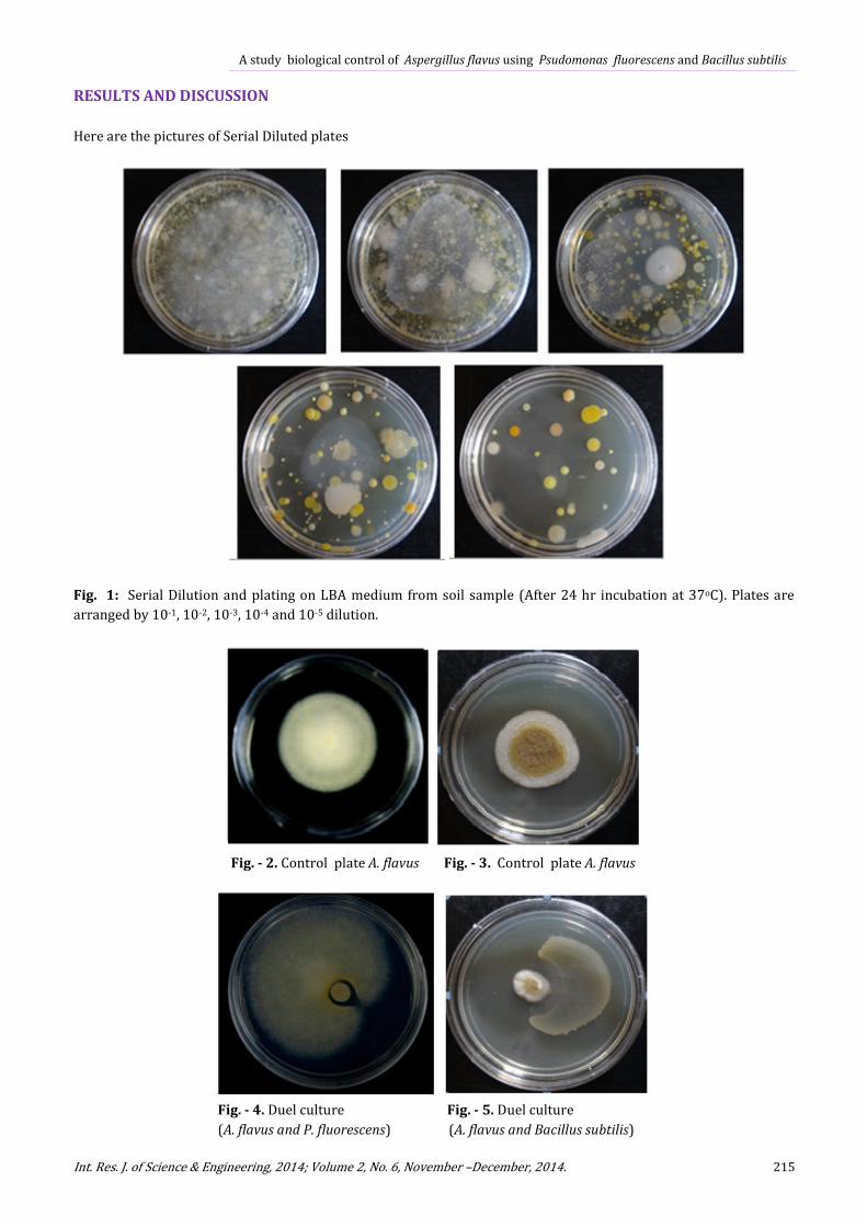
RESULTS AND DISCUSSION
Here are the pictures of Serial Diluted plates
Fig. 1: Serial Dilution and plating on LBA medium from soil sample (After 24 hr incubation at 37oC). Plates are
arranged by 10-1, 10-2, 10-3, 10-4 and 10-5 dilution.
Fig. - 2. Control plate A. flavus Fig. - 3. Control plate A. flavus
Fig. - 4. Duel culture Fig. - 5. Duel culture
(A. flavus and P. fluorescens) (A. flavus and Bacillus subtilis)

Kumar et al., 2014
216 www.irjse.in
Table 1: Colony radius of Duel Culture and Control on CDA and PDA medium after incubation for 48 hrs. at 30oC. All of
the experiments were done 3 times and their mean value was taken.
Serial
No.
Microbe Used Control Diameter of A.
flavus (cm.)
Diameter in Duel (cm.) Percentage of
inhibition
1. P. fluorescens 2.1 0.7 66.66%
2. Bacillus subtilis) 1.5 0.3 83%
Table 2: Comparative chart of dry weight of Duel Culture and Control organism grown on LB for 7 days at 30oC. All of the
experiments were done 3 times and their mean value was taken.
Serial
No.
Organism Used Control Weight A. flavus
(gm.)
Dual culture Weight (gm.) % Weight loss
1. P. fluorescens 1.85 0.56 69.72
2. Bacillus subtilis 3.80 1.08 72.68
Percentage inhibition of A. flavus
Data were obtained for the percentage inhibition of
radial growth [100 x (R1 - R2)/R1], where R1 = radial
growth of the pathogen in control and R2 = radial
growth of the pathogen in dual culture with antagonist
(Mohsin T, et al 2010). Results are means of three
replicates.
Weight Loss of A. flavus
Weight loss study was also done with cell extract of
pseudomonas fluorescens and Bacillus subtilis.
Morphological Identification:
Normal morphological identification was done for the
bacterium. P. fluorescens appears pale yellow in young
and reddish yellow in old while grown in NA medium. It
is a rod shaped Gram (-)ve bacteria. Other Bacillus sp.
were appeared off-white on LB medium.
16s rRNA sequencing and identification:
One antagonist organism which named as HGA was
found to be closely related to Pseudomonas fluorescens
and other three are believed to be Bacillus sp. based on
the basis of nucleotide homology and phylogenetic
analysis.
Gel Image of 16SrDNA amplicon (Sample: HGA)
Lane 1 2
Fig. 6: Agarose Gel Image (Lane 1:16S rDNA amplicon
band and Lane 2: DNA marker)
Analysis of Phylogenetic tree:
The culture, which was labeled as Pseudomonas
fluorescens HGA was found to be closely related (99%
similarity) to Pseudomonas fluorescens strain R15
based on nucleotide homology and phylogenetic
analysis.
Information about other close homologs for the microbe is
shown in the Alignment View table.

A study biological control of Aspergillus flavus using Psudomonas fluorescens and Bacillus subtilis
Int. Res. J. of Science & Engineering, 2014; Volume 2, No. 6, November –December, 2014. 217
Fig. 7: Phylogenetic Tree (Neighbor Joining) Pseudomonas fluorescens strain R15
Fig. 8: Phylogenetic Tree (Neighbor Joining) Bacillus subtilis
Kumar et al., 2014
218 www.irjse.in
The two microbes were isolated from the soil and
identified as Pseudomonas fluorescens and Bacillus
subtilis. Both the microbe showed significant inhibition
and weight loss of the A. flavus toxigenic strain. These
two microbes can be utilized as bio-control agents.
REFERENCES
1. Abbas HK, Zablotowicz RM, Bruns HA, Abel CA.
Biocontrol of aflatoxin in corn by inoculation with non-
aflatoxigenic Aspergillus flavus isolates. Biocontrol Sci.
Technol 2006 .16(5):437-449.
2. Arora M, Anil AC, Delany J, Rajarajan N, Emami K,
Mesbahi E. Carbohydrate degrading bacteria closely
associated with Tetraselmis indica: influence on algal cell
growth, Aquatic Biology, 2012. 15: 61–71. doi:
10.3354/ab00402
3. Bayman P, Baker JL, Mahoney NE. Aspergillus on tree
nuts: incidence and associations. Mycopathologia. 2002.
155: 161–169.
4. Blount WP. Turkey “X” disease. Turkeys 1961. 9(2): 52,
55-58, 61,77.
5. Chakrabarty AB. Detoxification of aflatoxin in Corn. J.
Food Prot 1981. 44: 173–176.
6. Cotty PJ. Virulence and cultural characteristics of two
Aspergillus flavus strains pathogenic on cotton.
Phytopathology, 1989 79(7):808-814.
7. Dorner JW. Management and prevention of mycotoxins
in peanuts. Food Additives and Contaminants. Toxin
Review. 2008. 25(2):203-208.
8. Dorner JW. Biological control of aflatoxin contamination
of crops. Journal of Toxin Rev.2004 23(2and3):425-450.
9. Huang HC, Hoes JA. Penetration and infection of
Sclerotinia sclerotiorum by Coniothyrium minitans.
Canadian Journal of Botany, 1976, 54(5-6): 406-410.
10. Hedayati MT, Pasqualotto AC, Warn PA, Bowyer P,
Denning DW. Aspergillus flavus: human pathogen,
allergen, and mycotoxin producer. Microbiology 2007
(153): 1677–1692.
11. Hesseltine CW. Introduction, definition and history of
mycotoxins of importance top animal production.
Interactions of mycotoxins in animal production. Natl.
Acad. Sci. Washington D.C.1979. (USA) 3–18.
12. Lillehoj EB, Logoida AB, Maisch WF. The fate of aflatoxin
in naturally contaminated corn during the ethanol
fermentation., Can. J. Microbiol., 1979 25: 911-914.
13. Mohsin Tariq, Sumera Yasmin, Hafeez Fauzia Y. Biological
control of potato black scurf by rhizosphere associated
bacteria Braz. J. Microbiol. 2010 vol.41.2
14. Palmgren MS, Hayes AW. A flatoxins in food, Palle
Kroghs, Academic Press, London1987, 65-96.
15. Sharma RS, Trivedi KR., Wadodkar UR, Murthy TN,
Punjarath JS. Aflatoxin B1 content in deoiled cakes, cattle
feeds and damaged grains during different seasons in
India. J. Food Sci. Tech. 1994. 31: 3, 244-246.
16. Shotwell OL. A flatoxin in corn. J. Am. Oil. Chem. Soc. .
1977. 54: 216A- 224A.
17. Vander Zijden ASM, Koelensmid WAAB, Bolding J, Barett
CB, Ord OW, Philip J. solation in crystalline form of a
toxin responsible for Turkey X disease. Nature (London)
.1962. 195: 1060–1062.
18. Wogan GN. A flatoxin as a human carcinogen. Hepatology
1999.30 (2): 573-575.
© 2014| Published by IRJSE