Endoscopic and pathologic changes of the upper gastrointestinal tract in Crohn's disease
10.1128/IAI.70.12.6567-6575.2002.
2002, 70(12):6567. DOI:Infect. Immun. Sutton, David Bruckner and Jonathan BraunBo Wei, Tiffany Huang, Harnisha Dalwadi, Christopher L. and T-Cell SuperantigenCrohn's Disease-Associated I2 Sequence
Encodes thePseudomonas fluorescens
http://iai.asm.org/content/70/12/6567Updated information and services can be found at:
These include:
REFERENCEShttp://iai.asm.org/content/70/12/6567#ref-list-1at:
This article cites 50 articles, 16 of which can be accessed free
CONTENT ALERTS more»articles cite this article),
Receive: RSS Feeds, eTOCs, free email alerts (when new
http://journals.asm.org/site/misc/reprints.xhtmlInformation about commercial reprint orders: http://journals.asm.org/site/subscriptions/To subscribe to to another ASM Journal go to:
on March 31, 2014 by guest
http://iai.asm.org/
Dow
nloaded from
on March 31, 2014 by guest
http://iai.asm.org/
Dow
nloaded from
INFECTION AND IMMUNITY, Dec. 2002, p. 6567–6575 Vol. 70, No. 120019-9567/02/$04.00�0 DOI: 10.1128/IAI.70.12.6567–6575.2002Copyright © 2002, American Society for Microbiology. All Rights Reserved.
Pseudomonas fluorescens Encodes the Crohn’s Disease-Associated I2Sequence and T-Cell Superantigen
Bo Wei,1 Tiffany Huang,2 Harnisha Dalwadi,1 Christopher L. Sutton,3 David Bruckner,1 andJonathan Braun1,2*
Departments of Pathology and Laboratory Medicine,1 Molecular Biology Institute,2 University of California, Los Angeles,California 90095, and Santarus, Inc., San Diego, California 921303
Received 1 May 2002/Returned for modification 2 July 2002/Accepted 21 August 2002
Commensal bacteria have emerged as an important disease factor in human Crohn’s disease (CD) andmurine inflammatory bowel disease (IBD) models. We recently isolated I2, a novel gene segment of microbialorigin that is associated with human CD and that encodes a T-cell superantigen. To identify the I2 microor-ganism, BLAST analysis was used to identify a microbial homologue, PA2885, a novel open reading frame(ORF) in the Pseudomonas aeruginosa genome. PCR and Southern analysis identified Pseudomonas fluorescensas the originating species of I2, with homologues detectable in 3 of 13 other Pseudomonas species. Genomiccloning disclosed a locus containing the full-length I2 gene (pfiT) and three other orthologous genes, includinga homologue of the pbrA/pvdS iron response gene. CD4� T-cell responses to recombinant proteins were potentfor I2 and pfiT, but modest for PA2885. pfiT has several features of a virulence factor: association with aniron-response locus, restricted species distribution, and T-cell superantigen bioactivity. These findings suggestroles for pfiT and P. fluorescens in the pathogenesis of Crohn’s disease.
Human inflammatory bowel disease (IBD) represents a setof a chronic, relapsing, and remitting intestinal inflammatorydisorders involving T-cell-mediated mucosal and mural de-struction, with polygenic disease susceptibility (4, 13, 38). Al-though the etiology of these diseases remains uncertain, sev-eral lines of evidence implicate commensal bacteria as animportant pathogenic element in clinical disease, particularlyin Crohn’s disease (CD). Patients with CD are sensitive toalteration of fecal flow, and some investigators have reportedevidence of a clinical responsiveness to antibiotic therapy (21,24, 34). In both pharmacologic and genetic animal models ofIBD, development of disease is strictly dependent on the pres-ence of enteric microflora (19, 29, 35–37, 40).
The identity of colitigenic bacterial species remains uncer-tain. In humans, a variety of bacterial and viral species havebeen implicated in CD, mainly on the basis of seroreactivity (3,5, 8, 28, 44). However, CD patients typically express elevatedlevels of antibodies to many bacterial proteins, perhaps due tomucosal disruption and local immunologic challenge by diverseintestinal microflora. Mycobacterium has received attention asa candidate human pathogen, based on serologic evidence, butthe presence of mycobacterial DNA in lesions is controversial(9, 12, 31, 32, 43, 47). Bacteroides vulgatus and Helicobacterhepaticus have been evaluated with various rodent IBD mod-els, but other intestinal commensal organisms yet undefinedare likely to account for the major colitigenic species (14, 25,35, 36).
Our laboratory recently introduced subtractive cloning as afresh approach to identify candidate organisms in human CD(10, 45). A novel microbial gene, I2, was isolated from lesional
versus adjacent uninvolved colon tissues of a patient with CD.In a population-based study (45), the I2 gene was selectivelydetected in lesional CD colonic tissue, although it was alsoprevalent in both healthy and CD ileum. In that study, anti-bodies reactive with the recombinantly expressed protein alsowere detected in CD patients. The recombinant I2 protein alsowas found to be a murine CD4 T-cell superantigen (11). Theselines of evidence suggest that the I2 protein and its originatingbacterium might play roles in the pathogenesis of CD. Thepresent study addresses the identity of the originating bacte-rium and the genomic locus bearing the I2 gene segment.
MATERIALS AND METHODS
Bacterial strains. The Escherichia coli XL-1 Blue strain (Stratagene, La Jolla,Calif.) and TOP10 strain (Invitrogen, Carlsbad, Calif.) were used for all cloningand recombinant expression experiments. Thirteen clinical isolates of Pseudo-monas spp. were obtained from the University of California—Los Angeles(UCLA) Clinical Laboratories (Table 1). In addition to UCLA 268, Pseudomo-nas fluorescens strains were obtained from the American Type Culture Collection(ATCC 13525; an environmental isolate from a water tower), and clinical isolateswere obtained from the University of Illinois Chicago Medical Center (8-1, 8-3,8-4, 8-5, 19-5, 19-8, 22-23, 25-39, 25-54, 25-55, 25-60, S40628-2). The latter werea generous gift from P. Schreckenberger. The isolates were from sputum, urine,cerebrospinal fluid, and foot wound sources. All Pseudomonas strains weregrown on Trypticase soy agar (TSA) plates at 26°C. The identities of the UCLAand ATCC isolates were assessed and coded by biochemical criteria with an API20NE analyzer (0147555 and 0147514 for the UCLA and ATCC isolates, corre-sponding to “excellent” and “good” identifications for P. fluorescens, respectively).
DNA manipulation. The pCR2.1 TA cloning vector (Invitrogen, Carlsbad,Calif.) was used to clone PCR products according to the manufacturer’s instruc-tions. A His-tagged protein expression vector, pQE30 (Qiagen, Valencia, Calif.),was used to express protein in E. coli. Expression plasmids were constructed byinserting cloned fragments into the BamHI and SacI sites of the vector. Allrestriction enzymes used in this study were purchased from New England Biolabs(Beverly, Mass.).
Sequence analysis. Plasmid clones were sequenced by dideoxy chain termina-tion, assembled with the University of Wisconsin Genetics Computer Group(GCG) programs and the BLAST program (version 20.11, 20 January 2000), aswell as the National Center for Biotechnology Information nonredundant data-base (2). Amino acid sequence alignment was performed with the ClustalW
* Corresponding author. Mailing address: Department of Pathologyand Lab Medicine, UCLA School of Medicine, CHS 13-222, LosAngeles, CA 90095-1732. Phone: (310) 794-7953. Fax: (310) 825-5674.E-mail: [email protected].
6567
on March 31, 2014 by guest
http://iai.asm.org/
Dow
nloaded from

program at the EMBL Outstation European Bioinformatics Institute and withthe GenDoc program (www.psc.edu/biomed/genedoc).
Cloning full-length pfiT and its adjacent genes. Genome-walking (rapid am-plification of cDNA ends [RACE] cloning) was employed to clone full-lengthpfiT gene from P. fluorescens genome (Universal GenomeWalker kit; Clontech,Palo Alto, Calif.). Reverse and forward GSP oligonucleotide pairs were designedto obtain upstream and downstream flanking sequences of the primary I2 genefragment, respectively (Table 2). Genomic DNA of P. fluorescens was amplifiedon a GeneAmp PCR system 9700 (PE Applied Biosystem) in 50 �l with 1 �g ofgenomic DNA, 0.5 U of Taq polymerase, 2 mM deoxynucleoside triphosphates(dNTP), and 1 �M primers. Primary PCR was done with 7 cycles of 94°C for 2 sand 70°C for 3 min, followed by 32 cycles of 94°C for 2 s, 65°C for 3 min, and then65°C for 4 min. Secondary PCR was performed with 5 cycles of 94°C for 2 s and70°C for 3 min, followed by 20 cycles of 94°C for 2 s, 65°C for 3 min, and 65°Cfor 4 min. The prevalence of the I2 gene segment of pfiT and a downstreamsegment in 13 representative Pseudomonas strains was detected by PCR (Table1). The PCR was performed for 5 min at 95°C, with 30 cycles of 95°C for 60 s,65°C for 60 s, 72°C for 60 s, and 72°C for 5 min.
Southern blot analysis. Genomic DNA samples were restricted with EcoRI,electrophoresed on a 0.8% agarose gel, denatured in a mixture of 0.5 M Tris-Cland 1.5 M NaOH, neutralized, and transferred onto Hybond N� membrane(Amersham) by capillary blotting. Membranes were equilibrated in Rapid-Hybbuffer (Amersham) and then hybridized at 65°C for 6 h in the same buffer with32P-labeled primary I2 segment or pfiT downstream fragment (nucleotides 1108to 2630) prepared from the P. fluorescens genome by I2-5�/I2-3� and pfiT-DF/pfiT-DR primer pairs, respectively (Table 2). Membranes were washed with 2�standard saline citrate (SSC) in 0.1% sodium dodecyl sulfate (SDS) at roomtemperature, then washed with 0.1� SSC containing 0.1% SDS at 60°C. Auto-radiography was carried out at room temperature with Hyper Film (Amersham).
Expression and purification of recombinant proteins. Recombinant proteinswere constructed for the P. aeruginosa homologue (PA2885), pfiT, and theoriginal I2 gene product into pQE-30 (Table 2). E. coli XL-1 Blue transformantswere induced (1 mM isopropyl-�-D-thiogalactopyranoside [IPTG]), and six-His-tagged proteins were purified with a HisTrap column (Amersham) under dena-tured conditions according to the manufacturer. According to SDS-polyacryl-amide gel electrophoresis (PAGE) and gel densitometry, the proteinpreparations were of similar purities (�85%).
CD4� T-cell proliferation. Erythrocyte-depleted splenocytes, used as antigen-presenting cells (APCs), were cultured with 1 or 5 �g of antigen per ml at 5 �106 cells per ml for 12 to 16 h at 37°C and then washed and irradiated at 3,000rads (567.7 cGy/min for 5.5 min) prior to T-cell coculture. Splenic CD4� T cells(�85% pure by flow cytometry) were isolated by depletion with nylon wool andanti-B220 and anti-CD8 magnetic beads. CD4� T cells (4 � 105) were incubatedin triplicate with 4 � 105 antigen-pulsed APCs in wells of a 96-well flat-bottomtissue culture plate at 37°C in 5% CO2 humidified air. After 48 h of incubation,0.5 �Ci of [3H]thymidine was added to each culture for the last 18 h of theincubation period, and then the cells were collected with a Micro 96 harvester(Skatron Instruments, Tranby, Norway). Proliferation was assessed with a Beta-plate liquid scintillation counter (Wallac, Gaithersburg, Md.).
Detection of P. fluorescens in fecal samples. Fresh fecal samples from patientswith CD were sequentially accrued from the population used in our recent study(10). DNA was isolated with a commercial kit (Stratagene, La Jolla, Calif.), and0.1 �g was assayed for I2, pfiT, and Omp-W sequences by our previously reportedsemiquantitative PCR method (10). Doping experiments indicated that the limitof detection for both assays was �2,000 molecules per g of fecal sample. Samplesfrom 30 patients with satisfactory quality were subjected to analysis. In parallel,serial dilutions of fecal samples were cultured on TSA plates at 26°C for con-ventional microbiologic detection of P. fluorescens. Doping with UCLA 268indicated a limit of detection of 104 CFU/g of sample.
Nucleotide sequence accession number. The nucleotide sequence data for pfiTand the flanking genomic region in P. fluorescens are available under GenBankaccession no. AF173683.
RESULTS
I2 homologues are present in a limited number of species inthe family Pseudomonadaceae. A BLAST search for close ho-mologues of the I2 sequence revealed PA2885, an open read-
TABLE 1. List of bacterial strains used in this study
rRNA group and species
Identification by:
I2 Southernblotting I2 PCR
Group I (Fluorescens group)Pseudomonas aeruginosa � �Pseudomonas putida � �Pseudomonas fluorescens � �
Group I (Alcaligenes group)Pseudomonas pseudoalcaligenes � �Pseudomonas alcaligenes � �
Group I (Stutzeri group)Pseudomonas mendocina � �Pseudomonas stutzeri � �
Group II (Pseudomallei group)Ralstonia picketti � �
Group IV (Diminuta group)Bevundimonas diminuta � �Bevundimonas vesicularis � �
Group V (Unknown nucleic acid homology)Shewanella putrefaciens � �Sphingomonas paucimobilis � �Flavimonas oryzihabitans � �
TABLE 2. Oligonucleotide primers used in this study
Primer Sequence (5�33�) Purpose in this study
5�-PA2885 ATTGGATCCATGCTGGAGCTGGTGGCTACCGGACAGCT PA2885 protein expression3�-PA2885 TATGAGCTCTCAGGCGTTCTTGATCACCA PA2885 protein expressionpfiT-5� ATTGGATCCATGCGCACCATGGTCGACAGT PFTR protein expressionpfiT-3� TATGAGCTCCTCAGTCCGACTTCAGCACCATCAAC PFTR protein expressionI2-BamHI ATTGGATCCGATCTGGCCAGCGCCGTGGGCATCCA I2 protein expressionI2-SacI TATGAGCTCTCAGATCTGCTCATACACGTCA I2 protein expressionI2-5� TCTGCTCATACACGTCACG I2 PCR detectionI2-3� CCGTGGGCATCCAGTCCG I2 PCR detectionpfiT-DF TGACCCTTGAGGAGTTGGCCGAAGA pfiT downstream regionpfiT-DR AATCCAGCGCCGACATATGTGGGTAA pfiT downstream region5�-pfiT-GSP1 ATCATCGCGGTGTTGTAATGGATGGTTTCCTCCAT pfiT upstream cloning5�-pfiT-GSP2 ATCTCATCCTTGCTCTTGAAGTGATGAAAGATGCT pfiT upstream cloning3�-pfiT-GSP1 TGATCCGCTGCGAGTTGCAGTCGATCATG pfiT downstream cloning3�-pfiT-GSP2 AGTGGCGAGGCCATGGCGGTGCTGGTCTACGAAT pfiT downstream cloning3�-pfiT-GSP-A AGCATCTTTCATCACTTCAAGAGCAAGGATGAGAT pfiT downstream cloning3�-pfiT-GSP-B ATGGAGGAAACCATCCATTACAACACCGCGATGAT pfiT downstream cloning
6568 WEI ET AL. INFECT. IMMUN.
on March 31, 2014 by guest
http://iai.asm.org/
Dow
nloaded from

ing frame (ORF) of the recently reported P. aeruginosa ge-nome (42). I2 and PA2885 shared 84% nucleotide identity andamino acid sequence identity and similarity of 86 and 93%,respectively. This finding indicated that the I2 gene might haveoriginated from a member of the family Pseudomonadaceae. Apanel of 13 Pseudomonas species were examined for the oc-currence of the I2 gene segment (Table 1). In Fig. 1, Southernanalysis revealed four species positive for the gene (Pseudo-monas aeruginosa, Pseudomonas pseudoalcaligenes, P. fluore-scens, and Shewanella putrefasciens), among which P. fluore-scens gave the strongest hybridization signal. ChromosomalDNA samples were further analyzed for the I2 gene segmentwith the I2-5� and I2-3� primers (Table 2). Among the 13Pseudomonas species tested, an I2 PCR product was only de-tected in P. fluorescens (Fig. 2). Sequencing revealed that thesequence of the I2 product from this P. fluorescens strain wasnearly identical (1 nucleotide difference) to the index I2 se-quence. These findings indicated that the I2 gene segmentoriginated from P. fluorescens.
Cloning and characterization of the full-length pfiT ORFfrom P. fluorescens. Genome-walking PCR with the I2 se-quence was employed to clone the full-length I2 gene andflanking genomic region from P. fluorescens (Fig. 3). A novel207-amino-acid ORF, pfiT, was identified, which contained the
I2 sequence between residues 49 and 148. Compared to the I2sequence isolated from human CD tissue, this segment had99% (296 of 300 positions) nucleotide identity and 98 and 99%amino acid identity and similarity, respectively. pfiT was ho-mologous to PA2885 of P. aeruginosa, with 77% nucleotideidentity and 78 and 92% amino acid identity and similarity,respectively. These ORFs in P. fluorescens and P. aeruginosawere similar in length, differing by a 9-amino-acid segment atthe N terminus.
In order to further confirm the presence of pfiT in P. fluo-rescens, PCR (with I2-5� and I2-3� amplimers) was performedon genomic DNA from 14 P. fluorescens isolates (1 environ-mental isolate and 13 clinical isolates; see Materials and Meth-ods). Among these isolates, all were positive (data not shown).For comparison to our clinical isolate, a second pfiT gene wascloned by PCR from a standard P. fluorescens strain (ATCC13525). The pfiT genes in ATCC and UCLA strains shared89% nucleotide identity and 94 and 99% amino acid identityand similarity, respectively (Fig. 3). These findings reflect mod-est molecular divergence of P. fluorescens strains at this locus.
The pfiT locus includes an iron-response gene and a genet-ically unstable element. RACE cloning of pfiT flanking se-quence disclosed a previously undefined locus in P. fluorescens.As shown in Fig. 4, P. fluorescens and P. aeruginosa were
FIG. 1. Southern analysis of I2 gene homologues in members of the family Pseudomonadaceae. Genomic DNA (3 �g) from 13 clinical isolatesof Pseudomonadaceae were digested with EcoRI, separated on 0.8% agarose gel, and transferred to nitrocellulose membranes. The membraneswere hybridized with 32P-labeled I2 gene segment at moderate stringency, washed at high stringency, and visualized by exposure to Hyper Film.
VOL. 70, 2002 ROLES OF pfiT AND P. FLUORESCENS IN CROHN�S DISEASE 6569
on March 31, 2014 by guest
http://iai.asm.org/
Dow
nloaded from

homologous for several colinear ORFs. Cloning of the regionupstream of pfiT reached a hypothetical ORF that was homol-ogous to PA2886, a novel ORF in the same genomic positionrelative to PA2885 in P. aeruginosa.
Cloning of the region downstream of pfiT disclosed twonotable elements. First, we identified pbrA, a previously re-ported iron-regulated transcription factor in P. fluorescens(41). In P. aeruginosa, this genomic position is occupied bypvdS, a homologous iron starvation sigma factor and is also
homologous to the pfrI gene in P. putida (26, 49). A compar-ison of pbrA from UCLA 268 to other genes in this familydemonstrates strong conservation at the peptide level, with theexception of a divergent C-terminal region (Fig. 5).
Second, we identified a novel 1.5-kb downstream region(nucleotide positions 1108 to 2630) between pfiT and pbrA.This region encoded three putative proteins (PFX1, PFX2, andPFX3) without database homology (Fig. 4). The G�C contentof this downstream region (56%) was different from those of
FIG. 2. P. fluorescens contains the I2 sequence genomic DNA samples from 13 clinical strains of Pseudomonas were analyzed by PCR for theI2 gene segment by using I2-specific primers. The expected product (298 bp) was detected only in P. fluorescens. I2 “mimic” DNA (a 600-bp“stuffer” segment flanked with 5� and 3� I2 primer sequences) was used as the positive control.
FIG. 3. Sequence comparison of PfiT and PA2885. The predicted amino acid sequences of I2, PfiT, and PA2885 were aligned by using theClustalW multiple-sequence alignment program and displayed by using the GenDoc program. The two PfiT sequences were derived from P.fluorescens ATCC 13525 and UCLA 268, respectively. The darker-shaded areas in each column indicate identical residues among four sequencesthat are listed underneath as capitalized letters. Lighter-shaded areas indicate the identical residues in three sequences shown as lowercasecharacters. I2 and pfiT (UCLA) share 99% nucleotide identity and 98 and 99% amino acid identity and similarity, respectively. The ATCC andUCLA pfiT genes share 89% nucleotide identity and 94 and 99% amino acid identity and similarity, respectively. The pfiT (UCLA) and PA2885genes share 77% nucleotide identity and 78 and 92% amino acid identity and similarity, respectively.
6570 WEI ET AL. INFECT. IMMUN.
on March 31, 2014 by guest
http://iai.asm.org/
Dow
nloaded from

the adjacent regions (hypothetical protein, 68%; pfiT, 62%;pbrA, 58%). Notably, five 17-bp direct repeats (AGATCAAGATCACAAGC) were found within pfx3. This sequence in-cludes a core hexamer (AGATCA), repeated 11 times in thedirect repeat region spanning nucleotide positions 2473 to2565. These features suggested that the novel region was cre-ated by deletion mutagenesis.
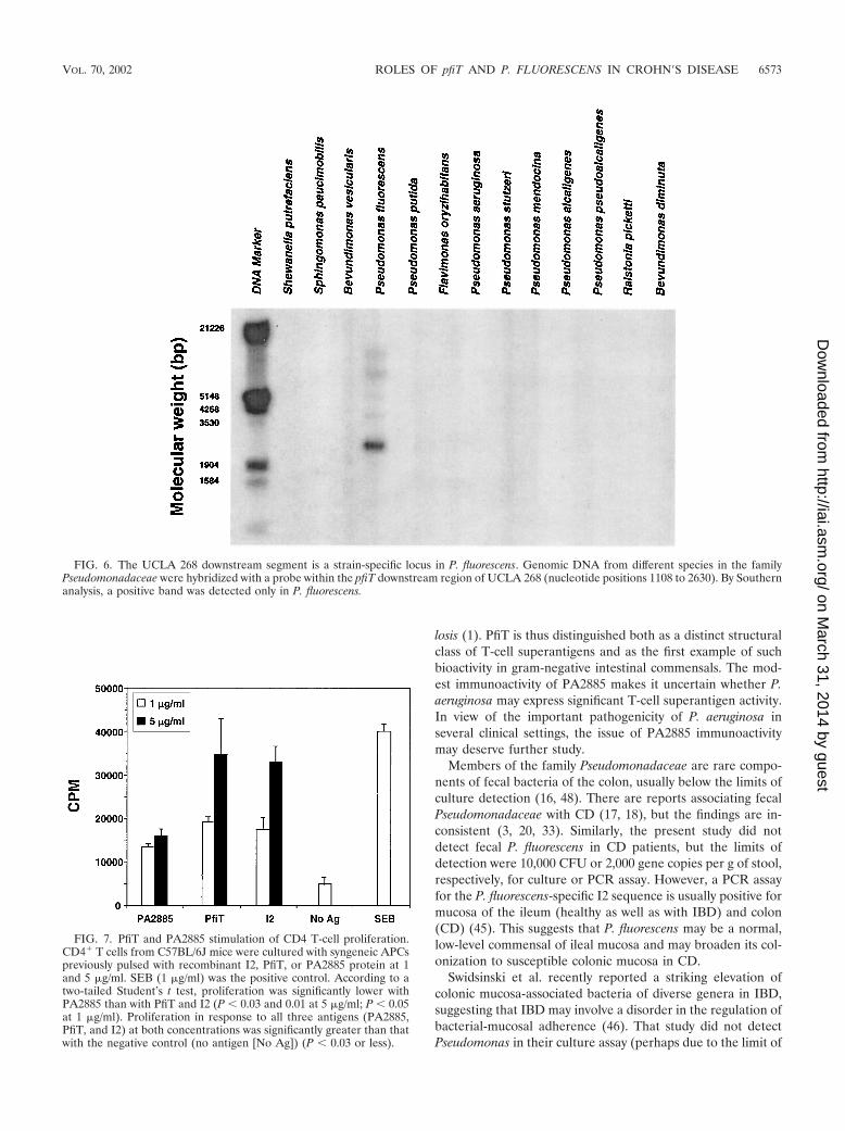
The presence of the novel pfiT downstream region was eval-uated with a DNA probe (nucleotide positions 1108 to 2630) inother Pseudomonas species. This region was detected only in P.fluorescens by Southern analysis (Fig. 6). With the pfiT-DF andpfiT-DR primers, a PCR product was detected in UCLA 268,but not in ATCC 13525 or other P. fluorescens clinical isolates(data not shown). These findings indicate that this region ispolymorphic among different P. fluorescens isolates.
In the P. aeruginosa genome, the orthologous region be-tween PA2885 and pvdS/PA2426) was much larger (�515 kb).This region encoded 459 putative ORFs, with diverse func-tional categories of these genes, including transcriptional reg-ulators, two-component regulatory systems, small moleculetransport, and energy metabolism. Fifty-two percent of thehypothetical proteins in this region lacked definable biologicalfunction. In comparison to this region in P. aeruginosa, thefeatures of the pfiT downstream region in the UCLA 268 strainof P. fluorescens (short length, replacement with a novel genesegment, and featured repeat sequence) suggest that it repre-sents a recombination-mediated deletional process (39).
CD4� T-cell activation by PfiT protein homologues. The I2protein activates murine T cells, displaying features most con-sistent with a T-cell superantigen (11). We were thereforeinterested in evaluating whether the full-length parent protein(PfiT) or its P. aeruginosa homologue (PA2885) was also im-munostimulatory. In the previous report, recombinant proteinswere expressed as glutathione S-transferase (GST) fusion pro-teins. In this study, a His-tagged protein expression vector wasused to express I2, PfiT, and PA2885, and the recombinantproteins were purified by nickel chromatography. The proteinswere preincubated with APCs and tested for their ability tostimulate CD4� T-cell proliferation (Fig. 7). Both I2 and PfiTstrongly stimulated proliferation, at levels similar to each otherand to that of the superantigen control (SEB). In contrast,PA2885 stimulated a weaker response. This difference was notdue detectable differences in protein purity. First, the proteinswere of similar purities by SDS-PAGE criteria. Second, exper-iments combining I2 (1 or 5 �g/ml) with PA2885 (0.5, 1, and2.5 �g/ml) revealed the same or additive proliferation com-pared to experiments with I2 alone. Thus, there did not appearto be an inhibitor in the PA2885 preparation for I2-inducedCD4� T-cell stimulation (data not shown). These results indi-cated that PfiT induces CD4� T-cell proliferation and that thecentral third of the PfiT protein (I2) is the functional domainfor this bioactivity. Sequence polymorphisms between PfiT andPA2885 may be structurally important for CD4� T-cell stim-ulation.
FIG. 4. Comparison of the homologue genomic regions in P. aeruginosa and P. fluorescens The P. fluorescens was cloned by genome-walkingPCR. The top of the figure lists predicted restriction enzyme sites and nucleotide numbering beginning at the 5� end of the cloned region. Thehomologous region in P. aeruginosa was identified by BLAST analysis of the recently reported genome of this microorganism (42); the numberingcorresponds to the genomic nucleotide position. Sequence analysis revealed significant sequence homology between the two genomes for each ofthe colinear ORFs. A direct repeat (hexamer element, 11 repeats) within PFX3 of P. fluorescens is also indicated (nucleotide positions 2473 to2565).
VOL. 70, 2002 ROLES OF pfiT AND P. FLUORESCENS IN CROHN�S DISEASE 6571
on March 31, 2014 by guest
http://iai.asm.org/
Dow
nloaded from

Absence of detectable P. fluorescens in CD fecal samples.Two assays were used to assess the presence of P. fluorescens infresh fecal samples sequentially accrued from 30 CD patients.First, samples were assessed for Pseudomonas colony forma-tion by conventional microbiologic culture. However, nogrowth was detected (limit of detection, 10,000 colonies per gof fecal material). Second, samples were assessed for P. fluo-rescens DNA by PCR for the species-specific I2 and pfiT se-quences. However, no sequence was detected in any of the 30specimens (limit of detection, copy number of 2,000/g of fecalmaterial).
DISCUSSION
I2 is a novel microbial gene recently associated with colonlesions in CD and encodes a T-cell superantigen (11, 45). Thepresent study demonstrates that I2 is derived from pfiT, anewly identified gene of P. fluorescens. The pfiT homologuesare restricted mainly to the RNA group 1 family of Pseudo-monadaceae, and the genomic region harboring this gene in P.fluorescens has features of a virulence locus. Immunologicanalysis indicates that the I2 segment of the PfiT protein fullyaccounts for its T-cell superantigen activity. The discussionaddresses the phylogeny of pfiT, the nature of its genomiclocus, and its potential immunologic role in CD pathogenesis.
I2, PfiT, and P. fluorescens. Several lines of evidence indicatethe I2 is derived from the pfiT gene of P. fluorescens. Southernand PCR analyses of 13 bacterial species in the family Pseudo-monadaceae demonstrated that the I2 sequence was present inP. fluorescens alone. Molecular cloning of this genomic regionfrom a clinical isolate of P. fluorescens disclosed a new gene,
pfiT, which was detected in all of the 14 P. fluorescens isolatesavailable for examination. The physiologic role of PfiT and itsP. aeruginosa homologue (PA2885) is unknown. On structuralgrounds, they are predicted to be a tetR-like component oftranscription complexes (42, 45).
Analysis of the genomic region surrounding pfiT in P. fluo-rescens revealed overall homology with the corresponding re-gion of P. aeruginosa with respect to both the organization andcomplement of predicted ORFs. One of these adjacent geneswas pbrA, an iron-responsive gene of P. fluorescens, with ho-mologues pvdS and pfrI in P. aeruginosa and Pseudomonasputida, respectively (26, 41, 49). PbrA protein upregulates cer-tain siderophores and exotoxins facilitating the iron depriva-tion response, and in some systems, it is an important virulencefactor (50).
I2 was recently shown in murine assays to be a new structuralclass of T-cell superantigen, activating CD4� T cells particu-larly by using the T-cell receptor (TCR) V�5 gene (11). In thisstudy, PfiT was also found to activate CD4� T cells, with apotency comparable to that of the I2 protein. This indicatesthat the central region of PfiT (the I2 sequence) fully accountsfor the immunologic activity of the protein. Compared to PfiT,PA2885 was only modestly stimulatory, suggesting that PA2885may differ in sequences affecting the avidity of major histocom-patibility complex (MHC) or TCR binding.
T-cell superantigens are an important virulence trait of cer-tain pathogenic bacteria, due to direct and indirect tissue dam-age mediated by the cognately activated T-cell population (27,30). Among species known to express T-cell superantigens,gram-negative bacteria are notably absent, with the exceptionof an apparent T-cell superantigen in Yersinia pseudotubercu-
FIG. 5. Alignment analysis of pbrA, pvdS, and pfrI proteins The predicted pbrA sequence isolated from UCLA 268 (pbrA-v) was aligned andcompared to those of the genes pbrA of P. fluorescens, pvdS of P. aerugonosas, and pfrI of P. putida. Alignment was performed with ClustalW, andthe results are displayed by the GenDoc program, as described in the legend to Fig. 4. The variable region at the extreme C terminus of theseiron-responsive genes is indicated.
6572 WEI ET AL. INFECT. IMMUN.
on March 31, 2014 by guest
http://iai.asm.org/
Dow
nloaded from
losis (1). PfiT is thus distinguished both as a distinct structuralclass of T-cell superantigens and as the first example of suchbioactivity in gram-negative intestinal commensals. The mod-est immunoactivity of PA2885 makes it uncertain whether P.aeruginosa may express significant T-cell superantigen activity.In view of the important pathogenicity of P. aeruginosa inseveral clinical settings, the issue of PA2885 immunoactivitymay deserve further study.
Members of the family Pseudomonadaceae are rare compo-nents of fecal bacteria of the colon, usually below the limits ofculture detection (16, 48). There are reports associating fecalPseudomonadaceae with CD (17, 18), but the findings are in-consistent (3, 20, 33). Similarly, the present study did notdetect fecal P. fluorescens in CD patients, but the limits ofdetection were 10,000 CFU or 2,000 gene copies per g of stool,respectively, for culture or PCR assay. However, a PCR assayfor the P. fluorescens-specific I2 sequence is usually positive formucosa of the ileum (healthy as well as with IBD) and colon(CD) (45). This suggests that P. fluorescens may be a normal,low-level commensal of ileal mucosa and may broaden its col-onization to susceptible colonic mucosa in CD.
Swidsinski et al. recently reported a striking elevation ofcolonic mucosa-associated bacteria of diverse genera in IBD,suggesting that IBD may involve a disorder in the regulation ofbacterial-mucosal adherence (46). That study did not detectPseudomonas in their culture assay (perhaps due to the limit of
FIG. 6. The UCLA 268 downstream segment is a strain-specific locus in P. fluorescens. Genomic DNA from different species in the familyPseudomonadaceae were hybridized with a probe within the pfiT downstream region of UCLA 268 (nucleotide positions 1108 to 2630). By Southernanalysis, a positive band was detected only in P. fluorescens.
FIG. 7. PfiT and PA2885 stimulation of CD4 T-cell proliferation.CD4� T cells from C57BL/6J mice were cultured with syngeneic APCspreviously pulsed with recombinant I2, PfiT, or PA2885 protein at 1and 5 �g/ml. SEB (1 �g/ml) was the positive control. According to atwo-tailed Student’s t test, proliferation was significantly lower withPA2885 than with PfiT and I2 (P 0.03 and 0.01 at 5 �g/ml; P 0.05at 1 �g/ml). Proliferation in response to all three antigens (PA2885,PfiT, and I2) at both concentrations was significantly greater than thatwith the negative control (no antigen [No Ag]) (P 0.03 or less).
VOL. 70, 2002 ROLES OF pfiT AND P. FLUORESCENS IN CROHN�S DISEASE 6573
on March 31, 2014 by guest
http://iai.asm.org/
Dow
nloaded from

detection) and observed the disordered adherence found inboth ulcerative colitis and CD. However, it points to the ideathat disordered mucosal adherence in CD may be a predispos-ing factor for the association of mucosal P. fluorescens in CD.Colonization by P. aeruginosa and its pathogenicity of P. aerugi-nosa are opportunistic, with dependence on local host factorsand microbial traits modifying host-bacterial interaction (15,23, 42). In mouse experimental and genetic models, diversemodes of immunologic or structural disturbance of the intes-tinal mucosa promote a CD-like disease (6, 7). While P. fluo-rescens rarely involves opportunistic infection (22), the disor-dered mucosal environment in genetically susceptibleindividuals may suit P. fluorescens opportunism and throughexpression of the PfiT superantigen may contribute a role inCD mucosal damage.
ACKNOWLEDGMENTS
We are grateful for the advice of Paul Colonna and Jeffrey H. Millerand the able and cordial technical assistance of Kathleen Lechowitzcand Kevin Ward. We appreciate the gift of P. fluorescens isolates fromPaul C. Schrenckenberger (Director of Clinical Microbiology, Univer-sity of Illinois Chicago Medical Center). We thank Dominica Salvatorefor assistance with preparation of the manuscript.
This work was supported by NIH DK46763 (J.B.), the JonssonComprehensive Cancer Center (J.B.), and the Crohn’s and ColitisFoundation of America (W.B.).
REFERENCES
1. Abe, J., M. Onimaru, S. Matsumoto, S. Noma, K. Baba, Y. Ito, T. Kohsaka,and T. Takeda. 1997. Clinical role for a superantigen in Yersinia pseudotu-berculosis infection. J. Clin. Investig. 99:1823–1830.
2. Altschul, S. F., W. Gish, W. Miller, E. W. Meyers, and D. J. Lippman. 1990.Basic local alignment search tool. J. Mol. Biol. 215:403–410.
3. Auer, I. O., A. Roder, F. Wensinck, J. P. van de Merwe, and H. Schmidt.1983. Selected bacterial antibodies in Crohn’s disease and ulcerative colitis.Scand. J. Gastroenterol. 18:217–223.
4. Bhan, A. K., E. Mizoguchi, R. N. Smith, and A. Mizoguchi. 1999. Colitis intransgenic and knockout animals as models of human inflammatory boweldisease. Immunol. Rev. 169:195–207.
5. Blaser, M. J., R. A. Miller, J. Lacher, and J. W. Singleton. 1984. Patients withactive Crohn’s disease have elevated serum antibodies to antigens of sevenenteric bacterial pathogens. Gastroenterology 87:888–894.
6. Blumberg, R. S., L. J. Saubermann, and W. Strober. 1999. Animal models ofmucosal inflammation and their relation to human inflammatory bowel dis-ease. Curr. Opin. Immunol. 11:648–656.
7. Bregenholt, S., D. Delbro, and M. H. Claesson. 1997. T-cell transfer andcytokine TCR gene deletion models in the study of inflammatory boweldisease. APMIS 105:655–662.
8. Cohavy, O., D. Bruckner, L. K. Gordon, R. Misra, B. Wei, M. E. Eggena,S. R. Targan, and J. Braun. 2000. Colonic bacteria express an ulcerativecolitis pANCA-related protein epitope. Infect. Immun. 68:1542–1548.
9. Cohavy, O., G. Harth, M. Horwitz, M. Eggena, C. Landers, C. Sutton, S. R.Targan, and J. Braun. 1999. Identification of a novel mycobacterial histoneH1 homologue (HupB) as an antigenic target of pANCA monoclonal anti-body and serum immunoglobulin A from patients with Crohn’s disease.Infect. Immun. 67:6510–6517.
10. Dalwadi, H., B. Wei, and J. Braun. 2000. Defining new pathogens andnon-culturable infectious agents: the case of inflammatory bowel disease.Curr. Opin. Gastroenterol. 16:56–59.
11. Dalwadi, H., B. Wei, M. Kronenberg, C. L. Sutton, and J. Braun. 2001. TheCrohn’s disease-associated bacterial protein I2 is a novel enteric T cellsuperantigen. Immunity 15:149–158.
12. Del Prete, R., M. Quaranta, A. Lippolis, V. Giannuzzi, A. Mosca, E. Jirillo,and G. Miragliotta. 1998. Detection of Mycobacterium paratuberculosis instool samples of patients with inflammatory bowel disease by IS900-basedPCR and colorimetric detection of amplified DNA. J. Microbiol. Methods33:105–114.
13. De Winter, H., H. Cheroutre, and M. Kronenberg. 1999. Mucosal immunityand inflammation. II. The yin and yang of T cells in intestinal inflammation:pathogenic and protective roles in a mouse colitis model. Am. J. Physiol.276:G1317–G1321.
14. Dieleman, L. A., A. Arends, S. L. Tonkonogy, M. S. Goerres, D. W. Craft, W.Grenther, R. K. Sellon, E. Balish, and R. B. Sartor. 2000. Helicobacter
hepaticus does not induce or potentiate colitis in interleukin-10-deficientmice. Infect. Immun. 68:5107–5113.
15. Epelman, S., T. F. Bruno, G. G. Neely, D. E. Woods, and C. H. Mody. 2000.Pseudomonas aeruginosa exoenzyme S induces transcriptional expression ofproinflammatory cytokines and chemokines. Infect. Immun. 68:4811–4814.
16. Finegold, S. M., V. L. Sutter, P. T. Sugihara, H. A. Elder, S. M. Lehmann,and R. L. Phillips. 1977. Fecal microbial flora in Seventh Day Adventistpopulations and control subjects. Am. J. Clin. Nutr. 1:1781–1792.
17. Foreman, N. K., W. C. Wang, E. J. Cullen, G. L. Stidham, T. A. Pearson, andJ. L. Shenep. 1991. Endotoxic shock after transfusion of contaminated redblood cells in a child with sickle cell disease. Pediatr. Infect. Dis. J. 10:624–626.
18. Franzetti, F., M. Cernuschi, R. Esposito, and M. Moroni. 1992. Pseudomo-nas infections in patients with AIDS and AIDS-related complex. J. Intern.Med. 231:437–443.
19. Garcia-Lafuente, A., M. Antolin, F. Guarner, E. Crespo, A. Salas, P.Forcada, and J. Malagelada. 1998. Derangement of mucosal barrier functionby bacteria colonizing the rat colonic mucosa. Eur. J. Clin. Investig. 28:1019–1026.
20. Graham, D. Y., H. H. Yoshimura, and M. K. Estes. 1983. DNA hybridizationstudies of the association of Pseudomonas maltophilia with inflammatorybowel diseases. J. Lab. Clin. Med. 101:940–954.
21. Gui, G. P. H., P. R. S. Thomas, M. L. V. Tizard, J. Lake, J. D. Sanderson, andJ. Hermon-Taylor. 1997. Two-year-outcomes analysis of Crohn’s diseasetreated with rifabutin and macrolide antibiotics. J. Antimicrob. Chemother.39:393–400.
22. Hsueh, P.-R., L.-J. Teng, H.-J. Pan, Y.-C. Chen, C.-C. Sun, S.-W. Ho, andK.-T. Luh. 1998. Outbreak of Pseudomonas fluorescens bacteremia amongoncology patients. J. Clin. Microbiol. 36:2914–2917.
23. Ichikawa, J. K., A. Norris, M. G. Bangera, G. K. Geiss, A. B. Wout, R. E.Bumgarner, and S. Lory. 2000. Interaction of Pseudomonas aeruginosa withepithelial cells: identification of differentially regulated genes by expressionmicroarray analysis of human cDNAs. Proc. Natl. Acad. Sci. USA 97:9659–9664.
24. Janowitz, H. D., E. C. Croen, and D. B. Sachar. 1998. The role of the fecalstream in Crohn’s disease: an historical and analytic review. Inflamm. BowelDis. 4:29–39.
25. Kullberg, M. C., J. M. Ward, P. L. Gorelick, P. Caspar, S. Hieny, A. Cheever,D. Jankovic, and A. Sher. 1998. Helicobacter hepaticus triggers colitis inspecific-pathogen-free interleukin-10 (IL-10)-deficient mice through an IL-12- and gamma interferon-dependent mechanism. Infect. Immun. 66:5157–5166.
26. Leoni, L., N. Orsi, V. de Lorenzo, and P. Visca. 2000. Functional analysis ofPvdS, an iron starvation sigma factor of Pseudomonas aeruginosa. J. Bacte-riol. 182:1481–1491.
27. Li, H., A. Llera, E. L. Malchiodi, and R. A. Mariuzza. 1999. The structuralbasis of T cell activation by superantigens. Annu. Rev. Immunol. 17:435–466.
28. Liu, Y., H. J. Van Kruiningen, A. B. West, R. W. Cartun, A. Cortot, and J.-F.Colombel. 1995. Immunocytochemical evidence of Listeria, Escherichia coli,and Streptococcus antigens in Crohn’s disease. Gastroenterology 108:1396–1404.
29. Madsen, K. L., J. S. Doyle, M. M. Tavernini, L. D. Jewell, R. P. Rennie, andR. N. Fedorak. 2000. Antibiotic therapy attenuates colitis in interleukin 10gene-deficient mice. Gastroenterology 118:1094–1105.
30. Marrack, P., and J. Kappler. 1994. Subversion of the immune system bypathogens. Cell 76:323–332.
31. Moss, M. T., E. P. Green, M. L. Tizard, Z. P. Malik, and J. Hermon-Taylor.1991. Specific detection of Mycobacterium paratuberculosis by DNA hybridi-sation with a fragment of the insertion element IS900. Gut 32:395–398.
32. Naser, S. A., K. Hulten, I. Shafran, D. Y. Graham, and F. A. El Zaatari. 2000.Specific seroreactivity of Crohn’s disease patients against p35 and p36 anti-gens of M. avium subsp. paratuberculosis. Vet. Microbiol. 77:497–504.
33. Parent, K., and P. Mitchell. 1978. Cell wall-defective variants of Pseudomo-nas-like (group Va) bacteria in Crohn’s disease. Gastroenterology 75:368–372.
34. Prantera, C., F. Zannoni, M. L. Scribano, E. Berto, A. Andreoli, A. Kohn,and C. Luzi. 1996. An antibiotic regimen for the treatment of active Crohn’sdisease: a randomized, controlled clinical trial of metronidazole plus cipro-floxacin. Am. J. Gastroenterol. 91:328–332.
35. Rath, H. C., H. H. Herfarth, J. S. Ikeda, W. B. Grenther, T. E. Hamm, Jr.,E. Balish, J. D. Taurog, R. E. Hammer, K. H. Wilson, and R. B. Sartor. 1996.Normal luminal bacteria, especially Bacteroides species, mediate chroniccolitis, gastritis, and arthritis in HLA-B27/human beta2 microglobulin trans-genic rats. J. Clin. Investig. 98:945–953.
36. Rath, H. C., M. Schultz, R. Freitag, L. A. Dieleman, F. Li, H.-J. Linde, J.Scholmerich, and R. B. Sartor. 2001. Different subsets of enteric bacteriainduce and perpetuate experimental colitis in rats and mice. Infect. Immun.69:2277–2285.
37. Saparov, A., L. A. Kraus, Y. Cong, J. Marwill, X. Y. Xu, C. O. Elson, andC. T. Weaver. 1999. Memory/effector T cells in TCR transgenic mice developvia recognition of enteric antigens by a second, endogenous TCR. Int. Im-munol. 11:1253–1264.
6574 WEI ET AL. INFECT. IMMUN.
on March 31, 2014 by guest
http://iai.asm.org/
Dow
nloaded from
38. Sartor, R. B. 1997. Pathogenesis and immune mechanisms of chronic inflam-matory bowel diseases. Am. J. Gastroenterol. 92:5S–11S.
39. Scott, J. R., and G. G. Churchward. 1995. Conjugative transposition. Annu.Rev. Microbiol. 49:367–397.
40. Sellon, R. K., S. Tonkonogy, M. Schultz, L. A. Dieleman, W. Grenther, E.Balish, D. M. Rennick, and R. B. Sartor. 1998. Resident enteric bacteria arenecessary for development of spontaneous colitis and immune system acti-vation in interleukin-10-deficient mice. Infect. Immun. 66:5224–5231.
41. Sexton, R., P. R. Gill, D. N. Dowling, and F. O’Gara. 1996. Transcriptionalregulation of the iron-responsive sigma factor gene pbrA. Mol. Gen. Genet.250:50–58.
42. Stover, C. K., X. Q. Pham, A. L. Erwin, S. D. Mizoguchi, P. Warrener, M. J.Hickey, F. S. Brinkman, W. O. Hufnagle, D. J. Kowalik, M. Lagrou, R. L.Garber, L. Goltry, E. Tolentino, S. Westbrock-Wadman, Y. Yuan, L. L.Brody, S. N. Coulter, K. R. Folger, A. Kas, K. Larbig, R. Lim, K. Smith, D.Spencer, G. K. Wong, Z. Wu, and I. T. Paulsen. 2000. Complete genomesequence of Pseudomonas aeruginosa PA01, an opportunistic pathogen. Na-ture 406:959–964.
43. Suenaga, K., Y. Yokoyama, I. Nishimori, S. Sano, M. Morita, K. Okazaki,and S. Onishi. 1999. Serum antibodies to Mycobacterium paratuberculosis inpatients with Crohn’s disease. Dig. Dis. Sci. 44:1202–1207.
44. Sutton, C., H.-Y. Yang, J. I. Rotter, S. R. Targan, and J. Braun. 2000.Familial expression of anti-Saccharomyces cerevisiae mannan antibodies
(ASCA) in affected and unaffected relatives of Crohn’s disease patients. Gut46:58–63.
45. Sutton, C. L., J. Kim, A. Yamane, H. Dalwadi, B. Wei, C. Landers, S. R.Targan, and J. Braun. 2000. Identification of a novel bacterial sequenceassociated with Crohn’s disease. Gastroenterology 119:23–28.
46. Swidsinski, A., A. Ladhoff, A. Pernthaler, S. Swidsinski, V. Loening-Baucke,M. Ortner, J. Weber, U. Hoffmann, S. Schreiber, M. Dietel, and H. Lochs.2002. Mucosal flora in inflammatory bowel disease. Gastroenterology 122:44–54.
47. Van Kruiningen, H. J. 1999. Lack of support for a common etiology inJohne’s disease of animals and Crohn’s disease in humans. Inflamm. Bowel.Dis. 5:183–191.
48. Wilson, K. H., J. S. Ikeda, and R. B. Blitchington. 1997. Phylogenetic place-ment of community members of human colonic biota. Clin. Infect. Dis.25(Suppl. 2):S114–S116.
49. Wilson, M. J., B. J. McMorran, and I. L. Lamont. 2001. Analysis of pro-moters recognized by PvdS, an extracytoplasmic-function sigma factor pro-tein from Pseudomonas aeruginosa. J. Bacteriol. 183:2151–2155.
50. Xiong, Y. Q., M. L. Vasil, Z. Johnson, U. A. Ochsner, and A. S. Bayer. 2000.The oxygen- and iron-dependent sigma factor pvdS of Pseudomonas aerugi-nosa is an important virulence factor in experimental infective endocarditis.J. Infect. Dis. 181:1020–1026.
Editor: J. D. Clements
VOL. 70, 2002 ROLES OF pfiT AND P. FLUORESCENS IN CROHN�S DISEASE 6575
on March 31, 2014 by guest
http://iai.asm.org/
Dow
nloaded from
Copyright © 2022 FDOKUMEN