NMR structure of protein YvyC from Bacillus subtilis reveals unexpected structural similarity...
9
NMR Structure of Protein YvyC from Bacillus subtilis Reveals Unexpected Structural Similarity between Two PFAM Families Alexander Eletsky 1,4 , Dinesh K. Sukumaran 1,4 , Rong Xiao 2,4 , Tom Acton 2,4 , Burkhard Rost 3,4 , Gaetano T. Montelione 2,4 , and Thomas Szyperski 1,4,* 1 Department of Chemistry, State University of New York at Buffalo, Buffalo, New York 14260 2 Center of Advanced Biotechnology and Medicine and Department of Molecular Biology and Biochemistry, Rutgers University, Piscataway, New Jersey 08854 3 Department of Biochemistry and Molecular Biophysics, Columbia University, New York, New York 10032 4 Northeast Structural Genomics Consortium Keywords Structural genomics; GFT NMR; flagella; YvyC; chaperone Introduction 109-residue protein YvyC (MW=13 kDa) from B. subtilis (gi|580862, SwissProt/TrEMBL ID YVYC_BACSU, accession number P39737) was selected as a target of the Protein Structure Initiative 2 and assigned to the Northeast Structural Genomics consortium (NESG; http://www.nesg.org) for structure determination (NESG Target ID SR482). YvyC belongs to the Pfam1 protein family FlaG (PF03646) and is required for proper assembly of bacterial flagella. The FlaG family contains 215 members (for sequence alignment of the FlaG family seeds see Fig. S1 in the Supporting Information). The NMR structure of YvyC presented here is the first atomic resolution structure available for this family. Self-assembly of bacterial flagella has been studied extensively in recent decades and comprehensive reviews are available.2 - 4 The gene encoding YvyC is a part of the fliD operon comprising the genes yvyC, fliD, fliS and fliT.5 This operon is located immediately downstream of the gene hag (also called fliC) encoding flagellin – the major protein required for assembly of the flagellar filaments. During filament elongation, flagellin monomers are exported through the central channel of a flagellum and oligomerize at its distal end.2 , 4 FliD (also named ‘hook-associated protein 2’, or HAP26) forms a pentameric cap at the distal end of a flagellum and is essential for oligomerization of flagellin.2 , 4 , 7 FliS and FliT serve as flagellin-specific8 and FliD-specific9 , 10 chaperones, respectively. It was demonstrated that mutations in yvyC gene orthologs in P. fluorescens (flaG) and V. anguillarum (ORF3) lead to phenotypes with unusually long filaments,11 , 12 but the specific role of YvyC remains unknown. * Correspondence to: Thomas Szyperski, Department of Chemistry, The State University of New York at Buffalo, Buffalo, New York 14260. E-mail: [email protected], Phone: (716) 6456800 (ext. 2250), Fax: (716) 6457338. NIH Public Access Author Manuscript Proteins. Author manuscript; available in PMC 2010 September 1. Published in final edited form as: Proteins. 2009 September ; 76(4): 1037–1041. doi:10.1002/prot.22459. NIH-PA Author Manuscript NIH-PA Author Manuscript NIH-PA Author Manuscript
Transcript of NMR structure of protein YvyC from Bacillus subtilis reveals unexpected structural similarity...

NMR Structure of Protein YvyC from Bacillus subtilis RevealsUnexpected Structural Similarity between Two PFAM Families
Alexander Eletsky1,4, Dinesh K. Sukumaran1,4, Rong Xiao2,4, Tom Acton2,4, BurkhardRost3,4, Gaetano T. Montelione2,4, and Thomas Szyperski1,4,*
1Department of Chemistry, State University of New York at Buffalo, Buffalo, New York 142602Center of Advanced Biotechnology and Medicine and Department of Molecular Biology andBiochemistry, Rutgers University, Piscataway, New Jersey 088543Department of Biochemistry and Molecular Biophysics, Columbia University, New York, NewYork 100324Northeast Structural Genomics Consortium
KeywordsStructural genomics; GFT NMR; flagella; YvyC; chaperone
Introduction109-residue protein YvyC (MW=13 kDa) from B. subtilis (gi|580862, SwissProt/TrEMBLID YVYC_BACSU, accession number P39737) was selected as a target of the ProteinStructure Initiative 2 and assigned to the Northeast Structural Genomics consortium (NESG;http://www.nesg.org) for structure determination (NESG Target ID SR482). YvyC belongsto the Pfam1 protein family FlaG (PF03646) and is required for proper assembly of bacterialflagella. The FlaG family contains 215 members (for sequence alignment of the FlaG familyseeds see Fig. S1 in the Supporting Information). The NMR structure of YvyC presentedhere is the first atomic resolution structure available for this family.
Self-assembly of bacterial flagella has been studied extensively in recent decades andcomprehensive reviews are available.2-4 The gene encoding YvyC is a part of the fliDoperon comprising the genes yvyC, fliD, fliS and fliT.5 This operon is located immediatelydownstream of the gene hag (also called fliC) encoding flagellin – the major proteinrequired for assembly of the flagellar filaments. During filament elongation, flagellinmonomers are exported through the central channel of a flagellum and oligomerize at itsdistal end.2,4 FliD (also named ‘hook-associated protein 2’, or HAP26) forms a pentamericcap at the distal end of a flagellum and is essential for oligomerization of flagellin.2,4,7 FliSand FliT serve as flagellin-specific8 and FliD-specific9,10 chaperones, respectively. It wasdemonstrated that mutations in yvyC gene orthologs in P. fluorescens (flaG) and V.anguillarum (ORF3) lead to phenotypes with unusually long filaments,11,12 but the specificrole of YvyC remains unknown.
*Correspondence to: Thomas Szyperski, Department of Chemistry, The State University of New York at Buffalo, Buffalo, New York14260. E-mail: [email protected], Phone: (716) 6456800 (ext. 2250), Fax: (716) 6457338.
NIH Public AccessAuthor ManuscriptProteins. Author manuscript; available in PMC 2010 September 1.
Published in final edited form as:Proteins. 2009 September ; 76(4): 1037–1041. doi:10.1002/prot.22459.
NIH
-PA Author Manuscript
NIH
-PA Author Manuscript
NIH
-PA Author Manuscript
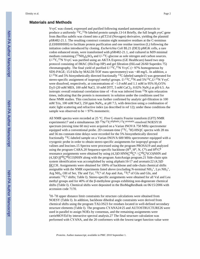
Materials and MethodsYvyC was cloned, expressed and purified following standard automated protocols toproduce a uniformly 13C,15N-labeled protein sample.13,14 Briefly, the full length yvyC genefrom Bacillus subtlis was cloned into a pET21d (Novagen) derivative, yielding the plasmidpSR482-21.1. The resulting construct contains eight nonnative residues at the C-terminus(LEHHHHHH) to facilitate protein purification and one residue insertion (L) following theinitiation codon introduced by cloning. Escherichia Coli BL21 (DE3) pMGK cells, a rarecodon enhanced strain, were transformed with pSR482-21.1, and cultured in MJ9 minimalmedium containing (15NH4)2SO4 and U-13C-glucose as sole nitrogen and carbon sources.U-13C,15N YvyC was purified using an AKTA Express (GE Healthcare) based two stepprotocol consisting of IMAC (HisTrap HP) and gel filtration (HiLoad 26/60 Superdex 75)chromatography. The final yield of purified U-13C,15N YvyC (> 97% homogeneous bySDS-PAGE; 15.0 kDa by MALDI-TOF mass spectrometry) was ∼48 mg/L. In addition, aU-15N and 5% biosynthetically directed fractionally 13C-labeled sample15 was generated forstereo-specific assignment of isopropyl methyl groups. U-13C,15N and 5%13C,U-15N YvyCwere dissolved, respectively, at concentrations of ∼1.0 mM and 1.1 mM in 95% H2O/5%D2O (20 mM MES, 100 mM NaCl, 10 mM DTT, 5 mM CaCl2, 0.02% NaN3) at pH 6.5. Anisotropic overall rotational correlation time of ∼8 ns was inferred from 15N spin relaxationtimes, indicating that the protein is monomeric in solution under the conditions used forthese NMR studies. This conclusion was further confirmed by analytic gel-filtration in 100mM Tris, 100 mM NaCl, 250 ppm NaN3, at pH 7.5, with detection using a combination ofstatic light scattering and refractive index (as described in ref 12); under these conditions thesample was observed to be > 97% monomeric.
All NMR spectra were recorded at 25 °C. Five G-matrix Fourier transform (GFT) NMRexperiments17 and a simultaneous 3D 15N/13Caliphatic/13Caromatic-resolved NOESY18spectrum (mixing time 60 ms) were acquired on a Varian INOVA 750 MHz spectrometerequipped with a conventional probe. 2D constant-time [13C, 1H]-HSQC spectra with 28 msand 56 ms constant-time delays were recorded for the 5% biosynthetically directedfractionally 13C-labeled sample on a Varian INOVA 600 MHz spectrometer equipped with acryogenic probe in order to obtain stereo-specific assignments for isopropyl groups ofvalines and leucines.15 Spectra were processed using the program PROSA19 and analyzedusing the program CARA.20 Sequence-specific backbone (HN, Hα, N, Cα) and Hβ/Cβ
resonance assignments were obtained by using (4,3)D HNNCαβCα / CαβCα(CO)NHN and(4,3)D HαβCαβ(CO)NHN along with the program AutoAssign program.21 Side-chain spinsystem identification was accomplished by using aliphatic16,17 and aromatic22 (4,3)DHCCH. Assignments were obtained for 100% of backbone and side-chain chemical shiftsassignable with the NMR experiments listed above (excluding N-terminal NH3
+, Lys NH3+,
Arg NH2, OH of Ser, Thr and Tyr, 13Cε of Asp and Asn, 13Cδ of Glu and Gln, andaromatic 13Cγ shifts; Table I). Stereo-specific assignments were obtained for all Val and Leumethyl groups and for 40% of the β-methylene groups exhibiting non-degenerate chemicalshifts (Table I). Chemical shifts were deposited in the BioMagResBank on 06/15/2006 withaccession code 7170.
1H–1H upper distance limit constraints for structure calculations were obtained fromNOESY (Table I). In addition, backbone dihedral angle constraints were derived fromchemical shifts using the program TALOS23 for residues located in well-defined secondarystructure elements (Table I). The programs CYANA24,25 and AUTOSTRUCTURE26 wereused in parallel to assign NOEs by consensus, and the remaining assignments werecarrieMOVEd by interactive spectral analysis.27 The final structure calculation wasperformed with CYANA, and the 20 conformers with the lowest target function value were
Eletsky et al. Page 2
Proteins. Author manuscript; available in PMC 2010 September 1.
NIH
-PA Author Manuscript
NIH
-PA Author Manuscript
NIH
-PA Author Manuscript

refined in an ‘explicit water bath’28 using the program CNS.29 The coordinates weredeposited in the Protein Data Bank on 06/15/2006 (accession code 2HC5).
Results and DiscussionA high-quality NMR structure of protein YvyC (Table I) was obtained. The structureconsists of three α-helices I-III (residues 9-22, 36-52 and 86-105) and three β-strands(residues 58-65, 68-75 and 81-85) forming one anti-parallel β-sheet with topology A(↑),B(↓), C(↑) (Fig. 1b). The helices form a three-helix bundle, which is attached to one side ofthe sheet. The secondary structure elements are locally and globally well-defined. Thesegment comprising residues 23-35 connecting helices I and II is largely disordered (Fig1a,c), which is manifested by comparably narrow NMR lines and lack of medium- and long-range NOEs.
A search of the CATH database using the CATHEDRAL30 server did not yield a matchingfold for protein YvyC, indicating that YvyC exhibits a distinct protein architecture.Moreover, a search of the PDB database for structurally similar proteins using both DALI31and SSM32 identified protein YkfF from E. coli (NESG target ER397, PDB code 2HJJ) asthe only significant (DALI Z-score = 3.3) hit. The structurally aligned fragment comprises56 residues (r.m.s.d. of Cα atoms = 2.9 Å; sequence identity 5%) and contains five regularsecondary structure elements (the β-sheet with strands A-C and α-helices II and III, Fig 1d).YkfF is the sole structural representative of Pfam family PF06006 comprising 38 proteins ofunknown function. Hence, structure comparison of proteins YvyC from B. Subtilis and YkfFfrom E. coli indicates a possible distant homology between the thus far unrelated PFAMfamilies PF03646 and PF06006.
Analysis of conserved surface features33 within the FlaG protein family PF03646 reveals asingle cluster of highly conserved residues on the surface of YvyC (Fig. 1e). These residuesbelong to β-strands B and C. Furthermore, a search for protein surface cavities using theProFunc34 server revealed three clefts (Fig. 1e,f) with volumes of ∼1,800 Å3, ∼800 Å3 and∼660 Å3. The calculation of the electrostatic protein surface potential calculated with theprogram MolMol35 shows that in protein YvyC cavity 2 is charged negatively while cavities1 and 3 exhibit a mixed charge distribution (Fig. 1f). Considering (i) that residues Leu 58,Glu 75, Ile 82 in cavity 1 and Glu 83 and Pro 86 in cavity 3 are highly conserved and (ii)that functional sites on protein surfaces are mostly located in the largest cavities36, one mayconclude that these two cavities are likely to be important for the molecular function.
Supplementary MaterialRefer to Web version on PubMed Central for supplementary material.
AcknowledgmentsThe authors thank Dr. Hunjoong Lee and Prof. Diana Murray for helpful discussions. Supported was obtained fromthe National Institutes of Health (U54 GM074958-01) and the University at Buffalo's Center for ComputationalResearch.
References1. Finn RD, Mistry J, Schuster-Bockler B, Griffiths-Jones S, Hollich V, Lassmann T, Moxon S,
Marshall M, Khanna A, Durbin R, Eddy SR, Sonnhammer ELL, Bateman A. Pfam: clans, web toolsand services. Nucleic Acids Res. 2006; 34:D247–D251. [PubMed: 16381856]
2. Macnab RM. How bacteria assemble flagella. Annual Review of Microbiology. 2003; 57:77–100.
Eletsky et al. Page 3
Proteins. Author manuscript; available in PMC 2010 September 1.
NIH
-PA Author Manuscript
NIH
-PA Author Manuscript
NIH
-PA Author Manuscript

3. Chevance FFV, Hughes KT. Coordinating assembly of a bacterial macromolecular machine. NatRev Micro. 2008; 6:455–465.
4. Minamino T, Namba K. Self-assembly and type III protein export of the bacterial flagellum. J MolMicrobiol Biotechnol. 2004; 7:5–17. [PubMed: 15170399]
5. Chen L, Helmann JD. The Bacillus subtilis sigma D-dependent operon encoding the flagellarproteins FliD, FliS, and FliT. J Bacteriol. 1994; 176:3093–3101. [PubMed: 8195064]
6. Homma M, Derosier DJ, Macnab RM. Flagellar hook and hook-associated proteins of Salmonellatyphimurium and their relationship to other axial components of the flagellum. J Mol Biol. 1990;213:819–832. [PubMed: 2193164]
7. Homma M, Fujita H, Yamaguchi S, Iino T. Excretion of unassembled flagellin by salmonella-typhimurium mutants deficient in hook-associated proteins. J Bacteriol. 1984; 159:1056–1059.[PubMed: 6384179]
8. Auvray F, Thomas J, Fraser GM, Hughes C. Flagellin polymerisation control by a cytosolic exportchaperone. J Mol Biol. 2001; 308:221–229. [PubMed: 11327763]
9. Bennett JCQ, Thomas J, Fraser GM, Hughes C. Substrate complexes and domain organization of theSalmonella flagellar export chaperones FlgN and FliT. Mol Microbiol. 2001; 39:781–791.[PubMed: 11169117]
10. Fraser GM, Bennett JCQ, Hughes C. Substrate-specific binding of hook-associated proteins byFlgN and FliT, putative chaperones for flagellum assembly. Mol Microbiol. 1999; 32:569–580.[PubMed: 10320579]
11. Capdevila S, Martinez-Granero FM, Sanchez-Contreras M, Rivilla R, Martin M. Analysis ofPseudomonas fluorescens F113 genes implicated in flagellar filament synthesis and their role incompetitive root colonization. Microbiology-Sgm. 2004; 150:3889–3897.
12. McGee K, Horstedt P, Milton DL. Identification and characterization of additional flagellin genesfrom Vibrio anguillarum. J Bacteriol. 1996; 178:5188–5198. [PubMed: 8752337]
13. Acton, TB.; Gunsalus, KC.; Xiao, R.; Ma, LC.; Aramini, J.; Baran, MC.; Chiang, YW.; Climent,T.; Cooper, B.; Denissova, NG.; Douglas, SM.; Everett, JK.; Ho, CK.; Macapagal, D.; Rajan, PK.;Shastry, R.; Shih, LY.; Swapna, GVT.; Wilson, M.; Wu, M.; Gerstein, M.; Inouye, M.; Hunt, JF.;Montelione, GT. Methods in Enzymology. Vol. 394. San Diego: Elsevier Academic Press Inc;2005. Robotic cloning and protein production platform of the Northeast Structural GenomicsConsortium. Nuclear Magnetic Resonance of Biological Macromolecules, Part C; p. 210-243.
14. Jansson M, Li YC, Jendeberg L, Anderson S, Montelione GT, Nilsson B. High-level production ofuniformly N-15- and C-13-enriched fusion proteins in Escherichia coli. J Biomol NMR. 1996;7:131–141. [PubMed: 8616269]
15. Neri D, Szyperski T, Otting G, Senn H, Wuthrich K. Stereospecific nuclear magnetic resonanceassignments of the methyl groups of valine and leucine in the DNA-binding domain of the 434repressor by biosynthetically directed fractional 13C labeling. Biochemistry. 1989; 28:7510–7516.[PubMed: 2692701]
16. Szyperski T, Yeh DC, Sukumaran DK, Moseley HNB, Montelione GT. Reduced-dimensionalityNMR spectroscopy for high-throughput protein resonance assignment. Proc Natl Acad Sci U S A.2002; 99:8009–8014. [PubMed: 12060747]
17. Atreya HS, Szyperski T. G-matrix Fourier transform NMR spectroscopy for complete proteinresonance assignment. Proc Natl Acad Sci U S A. 2004; 101:9642–9647. [PubMed: 15210958]
18. Shen Y, Atreya HS, Liu GH, Szyperski T. G-matrix Fourier transform NOESY-based protocol forhigh-quality protein structure determination. J Am Chem Soc. 2005; 127:9085–9099. [PubMed:15969587]
19. Guntert P, Dotsch V, Wider G, Wuthrich K. Processing of multidimensional NMR data with thenew software PROSA. J Biomol NMR. 1992; 2:619–629.
20. Keller, R. The Computer Aided Resonance Assignment Tutorial. Goldau: CANTINA Verlag;2004. p. 81
21. Zimmerman DE, Kulikowski CA, Huang YP, Feng WQ, Tashiro M, Shimotakahara S, Chien CY,Powers R, Montelione GT. Automated analysis of protein NMR assignments using methods fromartificial intelligence. J Mol Biol. 1997; 269:592–610. [PubMed: 9217263]
Eletsky et al. Page 4
Proteins. Author manuscript; available in PMC 2010 September 1.
NIH
-PA Author Manuscript
NIH
-PA Author Manuscript
NIH
-PA Author Manuscript

22. Eletsky A, Atreya HS, Liu GH, Szyperski T. Probing structure and functional dynamics of (large)proteins with aromatic rings: L-GFT-TROSY (4,3)D HCCH NMR spectroscopy. J Am Chem Soc.2005; 127:14578–14579. [PubMed: 16231903]
23. Cornilescu G, Delaglio F, Bax A. Protein backbone angle restraints from searching a database forchemical shift and sequence homology. J Biomol NMR. 1999; 13:289–302. [PubMed: 10212987]
24. Guntert P, Mumenthaler C, Wuthrich K. Torsion angle dynamics for NMR structure calculationwith the new program DYANA. J Mol Biol. 1997; 273:283–298. [PubMed: 9367762]
25. Herrmann T, Guntert P, Wuthrich K. Protein NMR structure determination with automated NOEassignment using the new software CANDID and the torsion angle dynamics algorithm DYANA.J Mol Biol. 2002; 319:209–227. [PubMed: 12051947]
26. Huang YJ, Tejero R, Powers R, Montelione GT. A topology-constrained distance networkalgorithm for protein structure determination from NOESY data. Proteins-Structure Function andBioinformatics. 2006; 62:587–603.
27. Liu GH, Shen Y, Atreya HS, Parish D, Shao Y, Sukumaran DK, Xiao R, Yee A, Lemak A,Bhattacharya A, Acton TA, Arrowsmith CH, Montelione GT, Szyperski T. NMR data collectionand analysis protocol for high-throughput protein structure determination. Proc Natl Acad Sci U SA. 2005; 102:10487–10492. [PubMed: 16027363]
28. Linge JP, Williams MA, Spronk C, Bonvin A, Nilges M. Refinement of protein structures inexplicit solvent. Proteins. 2003; 50:496–506. [PubMed: 12557191]
29. Brunger AT, Adams PD, Clore GM, DeLano WL, Gros P, Grosse-Kunstleve RW, Jiang JS,Kuszewski J, Nilges M, Pannu NS, Read RJ, Rice LM, Simonson T, Warren GL. Crystallography& NMR system: A new software suite for macromolecular structure determination. ActaCrystallogr Sect D-Biol Crystallogr. 1998; 54:905–921. [PubMed: 9757107]
30. Redfern OC, Harrison A, Dallman T, Pearl FMG, Orengo CA. CATHEDRAL: A fast and effectivealgorithm to predict folds and domain boundaries from multidomain protein structures. PLoSComput Biol. 2007; 3:2333–2347.
31. Holm L, Sander C. Dali - a network tool for protein structure comparison. Trends BiochemSci.1995; 20:478–480.
32. Krissinel E, Henrick K. Secondary-structure matching (SSM), a new tool for fast protein structurealignment in three dimensions. Acta Crystallogr Sect D-Biol Crystallogr. 2004; 60:2256–2268.[PubMed: 15572779]
33. Landau M, Mayrose I, Rosenberg Y, Glaser F, Martz E, Pupko T, Ben-Tal N. ConSurf 2005: theprojection of evolutionary conservation scores of residues on protein structures. Nucleic AcidsRes. 2005; 33:W299–W302. [PubMed: 15980475]
34. Laskowski RA, Watson JD, Thornton JM. ProFunc: a server for predicting protein function from3D structure. Nucl Acids Res. 2005; 33:W89–93. [PubMed: 15980588]
35. Koradi R, Billeter M, Wuthrich K. MOLMOL: A program for display and analysis ofmacromolecular structures. J Mol Graph. 1996; 14:51–55. [PubMed: 8744573]
36. Laskowski RA, Luscombe NM, Swindells MB, Thornton JM. Protein clefts in molecularrecognition and function. Protein Sci. 1996; 5:2438–2452. [PubMed: 8976552]
37. Laskowski RA, Rullmann JAC, MacArthur MW, Kaptein R, Thornton JM. AQUA andPROCHECK-NMR: Programs for checking the quality of protein structures solved by NMR. JBiomol NMR. 1996; 8:477–486. [PubMed: 9008363]
38. Word JM, Bateman RC, Presley BK, Lovell SC, Richardson DC. Exploring steric constraints onprotein mutations using MAGE/PROBE. Protein Sci. 2000; 9:2251–2259. [PubMed: 11152136]
39. Huang YJ, Powers R, Montelione GT. Protein NMR recall, precision, and F-measure scores (RPFscores): Structure quality assessment measures based on information retrieval statistics. J AmChem Soc. 2005; 127:1665–1674. [PubMed: 15701001]
40. Bhattacharya A, Tejero R, Montelione GT. Evaluating protein structures determined by structuralgenomics consortia. Proteins-Structure Function and Bioinformatics. 2007; 66:778–795.
Eletsky et al. Page 5
Proteins. Author manuscript; available in PMC 2010 September 1.
NIH
-PA Author Manuscript
NIH
-PA Author Manuscript
NIH
-PA Author Manuscript

Fig. 1.NMR structure of YvyC. (a) Backbone trace of residues 1-104 of the 20 representativeCYANA conformers after superposition of backbone N, Cα and C′ atoms of the regularsecondary structure elements for minimal root-mean-square deviation (RMSD). (b) Ribbondrawing of residues 1-104 of the conformer with the lowest CYANA target function. α-helices I-III are shown in red and yellow, β-strands A-C are shown in cyan, otherpolypeptide segments are shown in gray and the N- and C-termini are labeled as “N” and“C”. (c) Sausage representation of backbone and best defined side chains. A spline curvewas drawn through the mean positions of Cα atoms of residues 1-104 with the thicknessproportional to the mean global displacement of Cα atoms in the 20 conformerssuperimposed in (a). α-helices I-III are shown in red, β-strands A-C are shown in cyan, otherpolypeptide segments are shown in gray and a superposition of 34 side chains with thelowest global displacement is shown in blue. (d) Ribbon drawing of the conformers with the
Eletsky et al. Page 6
Proteins. Author manuscript; available in PMC 2010 September 1.
NIH
-PA Author Manuscript
NIH
-PA Author Manuscript
NIH
-PA Author Manuscript

lowest CYANA target function of YvyC (pale green) and YkfF (grey) after superposition ofCa atoms of residues 37-46, 60-63, 70-74, 81-85, 90-98 and 14-23, 27-30, 38-42, 48-52,59-67, respectively. For YvyC only residues 1-104 are shown. (e) Surface representation ofthe conformer with the lowest CYANA target function. The structure shown on the left hasthe same orientation as in (a-c), while the one on the right is rotated by 180° about thevertical axis. Surface colors represent sequence conservation among the seed sequences ofthe FlaG protein family calculated with ConSurf,33 with burgundy corresponding to thehighest conservation and cyan – to the highest variability. The cavities are labeled as C1, C2and C3. (f) Same as (e), but with the colors according to the electrostatic potential. Allfigures were prepared with the program MOLMOL.35
Eletsky et al. Page 7
Proteins. Author manuscript; available in PMC 2010 September 1.
NIH
-PA Author Manuscript
NIH
-PA Author Manuscript
NIH
-PA Author Manuscript

NIH
-PA Author Manuscript
NIH
-PA Author Manuscript
NIH
-PA Author Manuscript
Eletsky et al. Page 8
TABLE IStatistics of NMR Structure of Protein YvyC
Completeness of stereo-specific assignmentsa [%]
αCH2 of Gly 100 (1/1)
βCH2 40 (28/70)
Val and Leu methyl groups 100 (22/22)
Conformationally restricting distance constraints
Intraresidue [i = j] 548
Sequential [|i – j| = 1] 728
Medium Range [1 < |i – j| < 5] 697
Long Range [|i – j| ≥ 5] 916
Total 2889
Dihedral angle constraints
φ 35
ψ 35
Average number of constraints per residue 25.8
Average number of long-range distance constraints per residue 8.2
CYANA target function [Å2] 2.33±0.12
Average number of distance constraints violations per CYANA conformer
0.2 – 0.5 Å 0
> 0.5 Å 0
Average number of dihedral-angle constraint violations per CYANA conformer
> 5° 0
Average r.m.s.d. to the mean CYANA coordinates [Å]
Regular secondary structure elementsb, backbone heavy atoms 0.60±0.10
Regular secondary structure elementsb, all heavy atoms 1.00±0.11
Ordered residuesc, backbone heavy atoms 0.97±0.36
Ordered residuesc, all heavy atoms 1.43±0.35
Heavy atoms of molecular core including best-defined side chainsd 0.70±0.09
PROCHECK37 G-factors raw score (φ and ψ / all dihedral angles) c -0.15/-0.20
PROCHECK37 G-factors Z-score (φ and ψ / all dihedral angles) c -0.28/-1.18
MOLPROBITY38 clash score (raw / Z-score)c 24.03/-2.66
AutoQF R/P/DP scores39 [%] 95/95/85
Ramachandran plot summary (residues 2-17, 37-104) c [%]
most favored regions 91.2
Additionally allowed regions 7.8
generously allowed regions 0.4
disallowed regions 0.5
aRelative to pairs with non-degenerate chemical shifts.
bResidues 10-19, 37-49, 58-65, 68-75, 80-85, 90-101.
Proteins. Author manuscript; available in PMC 2010 September 1.

NIH
-PA Author Manuscript
NIH
-PA Author Manuscript
NIH
-PA Author Manuscript
Eletsky et al. Page 9
cResidues 3-5, 8-21, 38-55, 58-64, 67-104, 108-111. Ordered residues were defined based on dihedral angle order parameters, and Z-scores were
computed relative to corresponding structure quality measures for high resolution X-ray crystal structures, as described in reference.40
dBackbone and side-chain heavy atoms of residues 3, 6, 10-13, 16, 41, 43-45, 47, 48, 50, 51, 53, 54, 59, 62, 63, 71-74, 78, 81, 82, 85-87, 90, 94-97,
102, 103.
Proteins. Author manuscript; available in PMC 2010 September 1.