Mode of action of AraR, the key regulator of l-arabinose metabolism in Bacillus subtilis
Upload
sorbonne-frCategory
view
0download
0
The CymR Regulator in Complex with the Enzyme CysKControls Cysteine Metabolism in Bacillus subtilis*
Received for publication, August 1, 2008, and in revised form, October 15, 2008 Published, JBC Papers in Press, October 29, 2008, DOI 10.1074/jbc.M805951200
Catherine Tanous‡¶‡‡, Olga Soutourina‡¶1, Bertrand Raynal§�, Marie-Francoise Hullo‡¶, Peggy Mervelet**,Anne-Marie Gilles‡¶, Philippe Noirot**, Antoine Danchin‡¶, Patrick England§�2, and Isabelle Martin-Verstraete‡¶3
From the Institut Pasteur, ‡Unite de Genetique des Genomes Bacteriens, §Plate-forme de Biophysique des Macromolecules et deleurs Interactions, ¶CNRS, URA2171, and �CNRS, URA2185, 25 rue du Docteur Roux, 75724 Paris cedex 15, France, and INRA‡‡Mathematique, Informatique et Genome and **Unite de Genetique Microbienne, F-78352, Jouy-en-Josas, France
Several enzymes have evolved as sensors in signal transduc-tion pathways to control gene expression, thereby allowing bac-teria to adapt efficiently to environmental changes.We recentlyidentified the master regulator of cysteine metabolism in Bacil-lus subtilis, CymR, which belongs to the poorly characterizedRrf2 family of regulators. We now report that the signal trans-duction mechanism controlling CymR activity in response tocysteine availability involves the formation of a stable complexwith CysK, a key enzyme for cysteine biosynthesis. We carriedout a comprehensive quantitative characterization of this regu-lator-enzyme interaction by surface plasmon resonance andanalytical ultracentrifugation. We also showed that O-acetyl-serine plays a dual role as a substrate of CysK and as an effectormodulating the CymR-CysK complex formation. The ability ofB. subtilisCysK to bind to CymR appears to be correlated to theloss of its capacity to form a cysteine synthase complex withCysE.We propose an original model, supported by the determi-nation of the intracellular concentrations of the different part-ners, by which CysK positively regulates CymR in sensing thebacterial cysteine pool.
Bacteria have been particularly inventive to survive in adiversity of environments by developing mechanisms of regu-lation of gene expression as complex as those found in eukary-otic cells (1). They efficiently adapt to environmental changesby linking gene expression to intracellular pools of key metab-olites in various ways. In most cases, these effector moleculesact either by directly binding to transcriptional regulators or viaspecific receptors as observed for two-component regulatorysystems and sigma-antisigma factors. However, in the cell, theenzymes are themost suitable to inform aboutmetabolite avail-
ability. They specifically recognize their substrates or allostericeffectors and often undergo conformational changes uponbinding or during enzymatic reaction. Several enzymes haveevolved as sensors in signal transduction pathways controllinggene expression. These bifunctional proteins, which are activeas well in metabolism as in regulation, are called triggerenzymes (2). Some enzymes can directly bind to DNA (BirA,PutA, NadR, and PepA) or RNA (CitB), whereas phosphotrans-ferase system components control the activity of transcriptionfactors by phosphorylation events. However, the most abun-dant class of trigger enzymes modulates the transcriptionalactivity of regulators by protein-protein interactions. Forinstance, enzymes MalK, Aes, and MalY form complexes withthe MalT activator (3, 4). These enzymes antagonize the bind-ing of maltotriose, the co-activator of MalT. In the control ofnitrogen metabolism in Bacillus subtilis, glutamate dehydro-genase (RocG) and glutamine synthetase are able to interact,respectively, with the GltC activator or the TnrA repressor andto inactivate them concomitantly (5, 6). Here, we report thatCysK, the O-acetylserine thiol-lyase of B. subtilis, has acquireda regulatory function and is therefore a trigger enzyme acting asa repressor in the regulation of cysteine metabolism by an orig-inal mechanism.In bacteria, some archea and plants, cysteine biosynthesis pro-
ceeds via a two-step pathway involving CysK and CysE, the serineacetyl-transferase. CysE catalyzes the acetylation of L-serine byacetyl-CoA to give O-acetyl-L-serine (OAS)4. The OAS-thiol-lyase, CysK, then converts OAS and sulfide into L-cysteine andacetate.According to its catalyticmechanism,CysKbelongs to thefold type II group of pyridoxal 5�-phosphate (PLP)-dependentenzymes (7). In Salmonella typhimurium, Escherichia coli, Hae-mophilus influenzae, Mycobacterium tuberculosis, and higherplants, CysE and CysK form a bienzyme complex, called cysteinesynthase (8–10). The function of the complex is not metabolicchanneling but rather amodulation of the activity of each enzymein the complex. This association of serine acetyl-transferase andOAS-thiol-lyase provides a regulatory mechanism for cysteinebiosynthesis in plants (11).In line with the crucial role of cysteine in cellular physiology
and with the reactivity of its SH group, the transport, the bio-
* This work was supported by Centre National de la Recherche ScientifiqueGrant URA 2171, Agence National de la Recherche Grant NT05-2-44860,and Institut Pasteur Grant PTR256. The costs of publication of this articlewere defrayed in part by the payment of page charges. This article musttherefore be hereby marked “advertisement” in accordance with 18 U.S.C.Section 1734 solely to indicate this fact.
1 Assistant professor at the “Universite Paris 7.”2 To whom correspondence may be addressed: Plateforme de Biophy-
sique des Macromolecules et de leurs Interactions, Institut Pas-teur, France. Tel.: 33-1-44-38-94-74; Fax: 33-1-44-38-94-71; E-mail:[email protected].
3 Associate professor at the “Universite Paris 7.” To whom correspondencemay be addressed: Unite de Genetique des Genomes Bacteriens, URACNRS 2171, Institut Pasteur, France. Tel.: 33-1-40-61-35-61; Fax: 33-1-45-68-89-48; E-mail: [email protected].
4 The abbreviations used are: OAS, O-acetyl-L-serine; PLP, pyridoxal5�-phosphate; MBP, maltose-binding protein; Ni-NTA, nickel-nitrilotri-acetic acid; RU, resonance units; SPR, surface plasmon resonance; BD,DNA-binding domain; AD, activation domain; HPLC, high pressure liq-uid chromatography; HTH, helix turn helix.
THE JOURNAL OF BIOLOGICAL CHEMISTRY VOL. 283, NO. 51, pp. 35551–35560, December 19, 2008© 2008 by The American Society for Biochemistry and Molecular Biology, Inc. Printed in the U.S.A.
DECEMBER 19, 2008 • VOLUME 283 • NUMBER 51 JOURNAL OF BIOLOGICAL CHEMISTRY 35551
by guest on February 8, 2016http://w
ww
.jbc.org/D
ownloaded from

synthesis and the degradation of this amino acid are tightlycontrolled in response to environmental changes. A large vari-ety ofmolecularmechanisms participate in fine-tuning the reg-ulation of cysteine metabolism in bacteria (12). The CymR reg-ulator has recently been characterized as the central repressorof cysteine metabolism in B. subtilis (13). CymR belongs to thelarge andwidespreadRrf2 family of transcription factors, whichcomprises the iron-sulfur cluster biogenesis regulator IscR inE. coli (14, 15), and the global iron-responsive regulator RirA inRhizobiaceae (16). CymR controls the expression of genesinvolved either in cysteine synthesis from sulfide (cysK), sulfon-ates (ssu), ormethionine (mccAB) or in cystine uptake (tcyP), bydirectly binding a 27-bp conserved motif in their promoterregions (13, 17). Interestingly, a large set of genes is controlledby both CymR and CysK in B. subtilis (13, 18). Previous exper-iments suggested that CysK and CymR are required to induce aDNA electrophoreticmobility shift (19). Physiological data andgel shift experiments also show that OAS, the direct precursorof cysteine, is important in the signaling pathway controllingCymR-dependent repression in the presence of cysteine(19–21).We show in this work that CysK of B. subtilis interacts with
CymR in vivo and in vitro. This enzyme acts as a sensor ofcysteine availability in the signal transduction pathway modu-lating CymR activity.
EXPERIMENTAL PROCEDURES
Bacterial Strains and Plasmids—The bacterial strains andplasmids used are listed in Table 1. E. coliwas grown in LB and B.subtilis in minimal medium (20) supplemented with 1 mM L-me-thionine or 0.5 mM L-cystine. When required, antibiotics were
added: ampicillin and spectinomycin, 100 �g/ml; chlorampheni-col and kanamycin, 5 �g/ml (B. subtilis) or 20 �g/ml (E. coli).Yeast Two-hybrid Assay—The genes cysK and cysEwere fused
to the genes coding for the GAL4 DNA-binding (GAL4BD) orGAL4activation (GAL4AD)domains invectorspGBDU-C1 (bait)or pGAD-C1 (prey) (22), resulting in plasmids pDIA5653,pDIA5647, pDIA5778, and pDIA5779 (Table 1). Saccharomycescerevisiae strains PJ69-4a andPJ69-4� (22)were then transformedrespectively by the bait- and prey-derived vectors. Because wefailed to clone cymR into the pGADand pGBDUvectors inE. coli,we used gap repair recombination in S. cerevisiae (23) to directlyconstruct theplasmids containing the translational fusionof cymRwith Gal4BD or Gal4AD.The two-hybrid assays were then performed as previously
described (24). Positive protein-protein interaction betweenthe bait and a preywas detected by the ability of the cells to growon plates with synthetic completemediumdepleted for leucine,uracil, and histidine (SC-LUH) and for leucine, uracil, and ade-nine (SC-LUA). Interaction phenotypes were scored after 4 and22 days of growth at 30 °C.Overexpression and Purification of CysE, CysK, MccA, and
CymR—To obtain CysE fused to a His tag at the N terminus(His6-CysE) or at theC terminus (CysE-His6), the cysE gene (�1to �626) was amplified by PCR using primers introducing a5�-NdeI site and a 3�-XhoI site. This fragment was cloned intopET28a or pET22b (Novagen) to form a translational fusion ofcysE with six histidine codons at the 5� (pDIA5795) or 3�(pDIA5794) ends, respectively (Table 1). To obtain CysK fusedto a maltose-binding protein (MBP) at the N terminus (MBP-CysK), the cysK gene (�1 to �924) was amplified by PCR using
TABLE 1Strains and plasmids used in this studyspc is a spectinomycin resistance gene. ApR indicates ampicillin resistance; KmR indicates kanamycin resistance; and CmR indicates chloramphenicol resistance.
Genotypes and characteristics Source or referenceStrainsB. subtilis168 trpC2 Laboratory stockBSIP1304 trpC2 �cysK::spc Ref. 56BSIP1798 trpC2 �cymR amyE::aphA3 lacZ Ref. 20
E. coliDH5� F� Phi80dlacZ �M15 �(lacZYA-argF)U169 deoR recA1 endA1 hsdR17(rK-mK�)
phoA supE44 lambda- thi-1Commercial (Invitrogen)
BL21 F� ompT hsdSB(rB�mB�) gal dcm Commercial (Invitrogen)
S. cerevisiaePJ69-4 a/� MATa/� trp1-901 leu2-3,112 ura3-52 his3-200 gal4� gal80� Ref. 22
LYS2::GAL1-HIS3 GAL2-ADE2 met2::GAL7-lacZPlasmidspTYB11 Expression vector, T7 promoter N-terminal Intein-CBD fusion, ApR Commercial (Biolabs)pMAL-c2X Expression vector, tac promoter, N-terminal MBP fusion, ApR Commercial (Biolabs)pET22b Expression vector, T7 promoter, C-terminal His tag fusion, ApR Commercial (Novagen)pET28a Expression vector, T7 promoter, N-terminal His tag fusion KmR Commercial (Novagen)pDIA17 pACYC184 carrying lacI; CmR Ref. 25pGBDU-C1 Expression vector for use in two-hybrid analysis Ref. 22pGAD-C1 Expression vector for use in two-hybrid analysis Ref. 22pDIA5575 p�FytlI�-lacZ Ref. 20pDIA5647 pGAD-C1 derivative carrying cysK gene Ref. 20pDIA5653 pGBDU-C1 derivative carrying cysK gene This studypDIA5686 pET22b derivative carryingmccA gene Ref. 19pDIA5708 pET22b derivative carrying cysK gene Ref. 19pDIA5770 pTYB11 derivative carrying cymR gene Ref. 19pDIA5778 pGBDU-C1 derivative carrying cysE gene This studypDIA5779 pGAD-C1 derivative carrying cysE gene This studypDIA5794 pET22b derivative carrying cysE gene This studypDIA5795 pET28a derivative carrying cysE gene This studypDIA5796 pMAL-c2X derivative carrying cysK gene This study
CymR-CysK Regulatory Complex
35552 JOURNAL OF BIOLOGICAL CHEMISTRY VOLUME 283 • NUMBER 51 • DECEMBER 19, 2008
by guest on February 8, 2016http://w
ww
.jbc.org/D
ownloaded from

primers introducing a 5�-XbaI site or a 3�-PstI site, and thecorresponding fragmentwas cloned into pMAL-c2X (BioLabs),giving pDIA5796. These plasmids were introduced into E. coliBL21 strain (Novagen), which contains pDIA17 (25). Theresulting strains were grown at room temperature in LBmedium to anA600 of 1. Isopropyl�-D-thiogalactopyranoside (1mM) was then added, followed by incubations for 7 h at 20 °Cand 3 h at 37 °C. The cells were centrifuged and resuspended in50 mM sodium phosphate buffer (pH 8) containing 300 mMNaCl and protease inhibitor (Complete EDTA-free; RocheApplied Science) and lysed by sonication. E. coli crude extractscontaining His-tagged CysE were loaded on a Ni-NTA-agarosecolumn and eluted with 300 mM imidazole. The crude extractcontainingMBP-CysK was loaded on an amylose column (Bio-labs) and eluted with 10 mM maltose. MBP-CysK was thencleaved by the Factor X. The Factor X was removed using theFactor Xa cleavage capture kit (Novagen), whereas MBP wasretained by flowing the reaction mixture through an amylosecolumn.CysK-His6, MccA-His6, and native CymR were purified as
previously described (19). CymR, CysK-His6, and MBP-CysKwere subjected to a second step of purification by size exclusionchromatography on an Ultrogel AcA54 resin. The protein con-centration was determined bymeasuring the absorbance at 280nm using an extinction coefficient calculated from the aminoacid composition of each protein.Gel Mobility Shift Assays—The ytlI promoter region (�86 to
�22) was amplified using primers OS62 (5�-TGATTAGT-CAAAGATCAGAGTATGC-3�) and OS63 (5�-ATCAT-CACTCATTTTTGTCACTCCTC-3�), and pDIA5575 as tem-plate (20). The PCR product was labeled using [�-32P]ATP5�-end-labeled primer OS63. Unincorporated oligonucleotideswere removed using the QIAquick PCR purification kit (Qia-gen). Protein-DNA complexes were formed as previouslydescribed (19) and analyzed on 20% acrylamide gels. Whenspecified, OAS (Sigma) was added to the reaction mixture atvarious concentrations.In Vitro Pulldown Assays—His-tagged proteins were immo-
bilized on the Ni-NTA-agarose resin (Qiagen). 50 �l of Ni-NTA-agarose resin (50% (v/v) slurry) were incubated for 1 hwith or without 60 �g of CysK-His6, MccA-His6, or His6-CysEin a binding buffer 50 mM sodium phosphate, pH 8.0, contain-ing 300mMNaCl, and 0.1% (w/v) of bovine serum albumin. Themixtures were then incubated in the presence or absence of 30�g of purified CymR or CysK (obtained after cleavage of MBP).After washing, the bound proteins were eluted with 250 mMimidazole.Surface Plasmon Resonance Binding Assays—The assays
were performed at 25 °C in 20 mM sodium phosphate, pH 7.0,containing 200mMNaCl and 0.1 mM PLP. Tomonitor protein-protein interactions, monoclonal antibodies PentaHis (Qiagen)or anti-MBP mAb565 (26) were covalently coupled to a CM5sensorchip, using a Biacore 2000 instrument and the AmineCoupling Kit (Biacore). Immobilization densities of �10,000resonance units (RU; 1 RU � 1 pg�mm�2) were achieved. His6-CysE, CysE-His6, MccA-His6, MBP-CysK, or CysK-His6 wererespectively captured at densities of 150, 1800, 2000, and 1500RU. CymR (0.1–10.5 �M) was injected over immobilized CysK-
His6 or MBP-CysK at a flow rate of 50 �l�min�1. For the OASdissociation experiments, 800 or 250 RU of CymR were cap-tured on immobilized CysK-His6 or MBP-CysK, respectively,followed by a 3-min injection of various concentrations of OASor N-acetyl-serine from 0.05 to 1 mM.
A 109-bp ytlI biotinylated promoter region was immobilizedto the dextran-streptavidinmatrix of a SA sensorchip, at a den-sity of 340 RU. 1.75 �M of either CymR or CysK-His6, anequimolar CymR/CysK-His6 mixture or 18 �M of His6-CysE,were then injected at a flow rate of 5 �l�min�1. A streptavidinsurface devoid of DNA was used as reference. Each assay wasrepeated with three independent preparations of each protein.The association and dissociation profiles were double-refer-
enced using the Scrubber 2.0 software (BioLogic Software), i.e.both the signals from the reference surfaces and from blankexperiments using running buffer instead of protein were sub-tracted. The steady-state surface plasmon resonance (SPR)responses (Req) were plotted against the CymR concentration(C) and fitted according to a model corresponding to two bind-ing events to independent sites,
Req � �Rmax 1 � C�/�Kd1 � C� � �Rmax 2 � C�/�Kd2 � C�
(Eq. 1)
where Kd 1 and Kd 2 are the equilibrium dissociation constantsof the two successive binding events, and Rmax 1 and Rmax 2 arethe corresponding maximal binding capacities. The CymR-CysK dissociation profiles were analyzed using the followingdouble-exponential function of time,
R � R1 � e�koff 1 � t � �R0 � R1� � e�koff 2 � t (Eq. 2)
where R0 is the experimental SPR signal at the start of dissoci-ation, and R1 is the fitted amplitude of the first dissociationphase.The association profiles were finally fitted with an equation
describing a sequential CymR association process,
R � �kon 1 � C � Rmax 1/�kon 1 � C � koff 1�� � �1 � e��kon 1 � C � koff 1�t�
� ��kon 2 � C�/�kon 2 � C � koff 2���kon 1 � C � Rmax 1/�kon 1 � C � koff 1��
� �1 � e��kon 1 � C � koff 1�t� � �1 � e��kon 2 � C � koff 2�t� (Eq. 3)
where kon 1 and kon 2 are the association rate constants of thetwo successive binding events, koff 1 and koff 2 are the dissocia-tion rate constants (obtained fromEquation 2), andRmax 1 is themaximal binding capacity corresponding to the first bindingphase (obtained from Equation 1).Analytical Ultracentrifugation—Sedimentation velocity and
equilibrium experiments with CymR or CysK-His6 (3 �M or16.5 �M) and the equimolar CymR/CysK-His6 mixture (3 or16.5 �M each), were performed at 25 °C in a ProteomeLab XL-Ianalytical ultracentrifuge (Beckman-Coulter) equipped withboth UV-visible and Rayleigh interference detection systems,using an An60Ti rotor and 12 mm double-sector aluminumcenterpieces. The software Sednterp 1.09 was used to estimatethe partial specific volumes of CymR (0.745ml�g�1), CysK-His6(0.744 ml�g�1), and their 1:1 complex (0.744 ml�g�1), as well as
CymR-CysK Regulatory Complex
DECEMBER 19, 2008 • VOLUME 283 • NUMBER 51 JOURNAL OF BIOLOGICAL CHEMISTRY 35553
by guest on February 8, 2016http://w
ww
.jbc.org/D
ownloaded from
the viscosity ( � 0.903 cP) and density ( � 1.00587 g�ml�1) ofthe buffer used.For sedimentation velocity experiments, 300 �l of CymR or
CysK-His6 (3 or 16.5�M) were spun at 50,000 rpm, whereas theequimolar CymR/CysK-His6 mixture (3 or 16.5 �M each) wasspun at 30,000 rpm. Absorbance (276 nm) and interferencefringe displacement profiles were recorded every 2 min. Sedi-mentation coefficient continuous c(S) distributions and dimer-ization equilibrium dissociation constants were determinedusing the software Sedfit 11.3 (27).For sedimentation equilibrium experiments, 150�l of CymR
(3 �M), CysK-His6 (3 �M), or the equimolar CymR/CysK-His6mixture (3 �M each) were spun sequentially at 10,000 rpm for30 h, 13,000 rpm for 11 h, 17,000 rpm for 11 h, 21,000 rpm for11 h, and 50,000 rpm for 3 h (to measure the base-line value).The data were recorded for each speed after controlling thatsedimentation/diffusion equilibrium had been effectivelyreached. For each sample, the radial distributions for the twomost appropriate speeds were globally fitted with the Ultrascan9.5 software (28) using amodel corresponding to the number ofspecies detected in the sedimentation velocity experiments.Monte Carlo analysis was performed on each fit to estimate theerror on the measured values.Determination of CysK and CysE Activities—The OAS thiol-
lyase activity of CysK was determined as previously described(19). The serine acetyltransferase activity ofCysEwas assayed at30 °C by monitoring the cleavage of the thioester bond ofacetyl-CoA as previously described (29). The reaction rate wascalculated using a differential extinction coefficient betweenacetyl-CoA and CoA of 3.2 mM�cm�1. One unit of enzymeactivity was defined as the amount of enzyme that catalyzed theformation of 1 �mol CoA�min�1�(mg of protein)�1. All of theassays were performed in triplicate.Estimation of Intracellular Protein and Metabolite Con-
centrations—The intracellular concentrations of CymR, CysK,OAS, and cysteine inB. subtiliswere determined in strain 168 inMQ-S in the presence of methionine or cystine. Crude extractsand various amounts of purified CymR or CysK were loaded onan SDS-PAGE. Proteins were transferred to a polyvinylidenedifluoride membrane, probed with polyclonal anti CymR oranti-CysK antibodies followed by an anti-HP rabbit antibody,and revealed using the ECL-Plus Kit (Amersham). The quantityof CymR or CysK contained in a given volume of the crudeextract was estimated with the Quantity One software (Bio-Rad) by comparison with known amounts of purified proteins.Crude extracts of strain BSIP1798 (�cymR) and BSIP1304(�cysK) were used as negative controls. To determine metabo-lite concentrations, the cells were suspended in a sulfosalicylicacid buffer (3% final concentration) and disrupted by shak-ing for 40 s at power 5 in a FastPrep cell disruptor (FP120;Savant Instruments, Inc., Holbrook, NY). Supernatant sam-ples obtained after centrifugation were analyzed as described(19). Intracellular protein and metabolite concentrationswere estimated assuming a cell volume of 6.7 �l�(mg of totalcell protein) (30) or a B. subtilis intracellular volume of5 �m3.
RESULTS
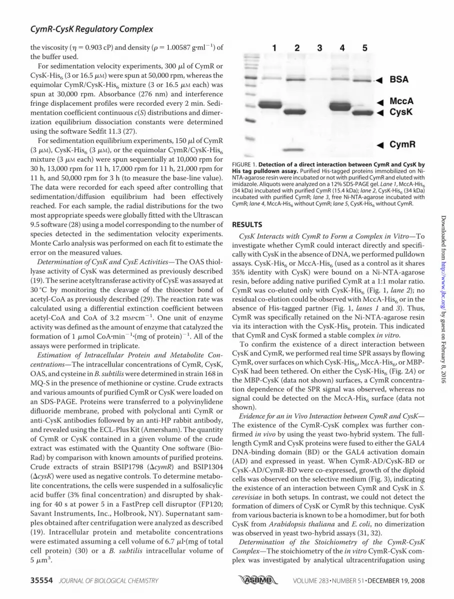
CysK Interacts with CymR to Form a Complex in Vitro—Toinvestigate whether CymR could interact directly and specifi-cally withCysK in the absence ofDNA,we performed pulldownassays. CysK-His6 or MccA-His6 (used as a control as it shares35% identity with CysK) were bound on a Ni-NTA-agaroseresin, before adding native purified CymR at a 1:1 molar ratio.CymR was co-eluted only with CysK-His6 (Fig. 1, lane 2); noresidual co-elution could be observedwithMccA-His6 or in theabsence of His-tagged partner (Fig. 1, lanes 1 and 3). Thus,CymR was specifically retained on the Ni-NTA-agarose resinvia its interaction with the CysK-His6 protein. This indicatedthat CymR and CysK formed a stable complex in vitro.To confirm the existence of a direct interaction between
CysK and CymR, we performed real time SPR assays by flowingCymR, over surfaces onwhichCysK-His6,MccA-His6, orMBP-CysK had been tethered. On either the CysK-His6 (Fig. 2A) orthe MBP-CysK (data not shown) surfaces, a CymR concentra-tion dependence of the SPR signal was observed, whereas nosignal could be detected on the MccA-His6 surface (data notshown).Evidence for an in Vivo Interaction between CymR and CysK—
The existence of the CymR-CysK complex was further con-firmed in vivo by using the yeast two-hybrid system. The full-length CymR and CysK proteins were fused to either the GAL4DNA-binding domain (BD) or the GAL4 activation domain(AD) and expressed in yeast. When CymR-AD/CysK-BD orCysK-AD/CymR-BD were co-expressed, growth of the diploidcells was observed on the selective medium (Fig. 3), indicatingthe existence of an interaction between CymR and CysK in S.cerevisiae in both setups. In contrast, we could not detect theformation of dimers of CysK or CymR by this technique. CysKfrom various bacteria is known to be a homodimer, but for bothCysK from Arabidopsis thaliana and E. coli, no dimerizationwas observed in yeast two-hybrid assays (31, 32).Determination of the Stoichiometry of the CymR-CysK
Complex—The stoichiometry of the in vitro CymR-CysK com-plex was investigated by analytical ultracentrifugation using
FIGURE 1. Detection of a direct interaction between CymR and CysK byHis tag pulldown assay. Purified His-tagged proteins immobilized on Ni-NTA-agarose resin were incubated or not with purified CymR and eluted withimidazole. Aliquots were analyzed on a 12% SDS-PAGE gel. Lane 1, MccA-His6(34 kDa) incubated with purified CymR (15.4 kDa); lane 2, CysK-His6 (34 kDa)incubated with purified CymR; lane 3, free Ni-NTA-agarose incubated withCymR; lane 4, MccA-His6 without CymR; lane 5, CysK-His6 without CymR.
CymR-CysK Regulatory Complex
35554 JOURNAL OF BIOLOGICAL CHEMISTRY VOLUME 283 • NUMBER 51 • DECEMBER 19, 2008
by guest on February 8, 2016http://w
ww
.jbc.org/D
ownloaded from

sedimentation velocity or equilibrium experiments, at variousconcentrations of CymR or CysK, alone or together (Fig. 4 andTable 2). The sedimentation coefficient distribution of CysK, at
both 3 and 16 �M, revealed the presence of a single species (4.7S) whose molecular mass (68.0 1.3 kDa) was determined bysedimentation equilibrium. This indicates that CysK is a dimerin this range of concentration, as previously observed in otherbacteria (33). Similar analyses were performed for CymR. At 3�M, they revealed one predominant species (2.2 S; 30.8 2.3
FIGURE 2. Monitoring of the binding of CymR to CysK-His6 by surfaceplasmon resonance. A, real time association and dissociation profiles corre-sponding to the injection of various CymR concentrations over immobilizedCysK-His6. B, CymR concentration dependence of steady-state SPR signals.Inset, deconvolution of the steady-state signal into two components, corre-sponding to the formation of (CymR)2-(CysK-His6)2 (dotted line) and (CymR)4-(CysK-His6)2 (dashed line), respectively. C, the decomposition of the experi-mental binding profile obtained for 2.25 �M of CymR, (in black), into its(CymR)2-(CysK-His6)2 (dotted line) and (CymR)4-(CysK-His6)2 (dashed line) com-ponents, and global fit assuming a sequential binding (in gray).
FIGURE 3. Detection of CymR/CysK interactions by yeast two-hybridassays. Diploid yeast cells expressing the indicated combinations of proteinsfused to Gal4BD (BD) and Gal4AD (AD) were selected for expression of the His3reporter. Binary interactions resulted in growing colonies on synthetic com-plete medium lacking leucine, uracil, and histidine. The Gal4AD and Gal4BDfusions are indicated on the top and left sides of the matrix, respectively. Con-trol matings with Gal4BD and Gal4AD alone were used as negative self-acti-vation controls.
FIGURE 4. Formation of the CymR-CysK complex studied by analyticalultracentrifugation. Continuous sedimentation coefficient distributionanalysis of CymR/CysK-His6 mixtures at 3 �M (dashed line) and 16 �M (solidline). Inset, experimental sedimentation equilibrium radial distributions at10,000 rpm (black) and 13,000 rpm (gray) of CymR/CysK-His6 at 3 �M andfitted curves corresponding to a 98.5-kDa (CymR)2-(CysK)2 complex. Sedi-mentation coefficients are expressed in Svedberg: 1 S � 10�13 s.
CymR-CysK Regulatory Complex
DECEMBER 19, 2008 • VOLUME 283 • NUMBER 51 JOURNAL OF BIOLOGICAL CHEMISTRY 35555
by guest on February 8, 2016http://w
ww
.jbc.org/D
ownloaded from

kDa), corresponding to a dimer of CymR. At higher concentra-tions (16 �M), small quantities of another sedimenting spe-cies were detected, with an S value of 4.4 S that would beconsistent with a tetrameric form of CymR. This is the firstreported data concerning the oligomerization state of amember of the poorly characterized Rrf2 family of repres-sors. The Kd of the (CymR)2 7 (CymR)4 equilibrium couldbe estimated at�200 �M.When CymR and CysK weremixedtogether at a 1:1 molar ratio, the sedimentation profilesrevealed two different species whose abundance varied withthe total CymR � CysK concentration. The mixtureobtained with 3 �M of each protein behaved as a homogene-ous species with a sedimentation coefficient of 6.2 S and amolecular mass of 98.4 5.6 kDa, corresponding to a com-plex constituted of two CymR and two CysK molecules.However, when 16 �M of each protein were mixed, wedetected the same (CymR)2-(CysK)2 complex together with anew majority species, which sediments with a standard Svalue of 8.6 S that would be consistent with a (CymR)4-(CysK)4 complex. A small proportion of unliganded CymR(either excess or inactive) could also be observed at this con-centration. The Kd of the (CymR)2-(CysK)2 7 (CymR)4-(CysK)4 equilibrium could be estimated at �4 �M.Determination of the Energetic and Kinetic Parameters of the
CymR-CysK Complex—SPR experiments were performed byflowing CymR over a CysK surface. The association and disso-ciation curves showed a markedly concentration-dependentbiphasic behavior, whether CysK was tethered by its N termi-nus (MBP-CysK; data not shown) or its C terminus (CysK-His6;Fig. 2A). The analysis of the dissociation curves revealed twoparallel dissociation phases, with respective dissociation ratesof 1.2 ( 0.2) 10�3 s�1 (koff 1) and 0.19 0.03 s�1 (koff 2). Thefirst slow phase (t1⁄2 � 600 s) reached a maximum amplitudeRmax 1 corresponding to a 1:1 stoichiometric ratio betweenCymR and CysK, consistent with the (CymR)2-(CysK)2 com-plex detected by analytical ultracentrifugation at low concen-trations. On the contrary, the second fast phase (t1⁄2 � 4 s) couldonly be detected for CymR concentrations higher than 250 nM,and its amplitude increased steadily reaching a maximal valueRmax 2, equal to Rmax 1, equivalent to the binding of a second(CymR)2 dimer. The steady-state SPR signals, when plottedagainst the CymR concentration (Fig. 2B), could be optimallyfitted with a two-term equation corresponding to two inde-pendent binding sites, with respective dissociation equilibrium
constants of 110 19 nM (Kd 1) and 3.5 0.6 �M (Kd 2). Byplotting the amplitudes of the two above-mentioned dissocia-tion phases against the CymR concentration, we deduced thatKd 1 corresponds to the slow dissociation phase (koff 1), andKd 2corresponds to the fast dissociation phase (koff 2) (Fig. 2B, inset).Finally, the association process of CymR to the CysK surfacewas optimally fitted, assuming it followed a sequential process,with a first (CymR)2 dimer binding to (CysK)2 with an associa-tion rate of 1.2 ( 0.3) 104 M�1�s�1 (kon 1), followed by asecond (CymR)2 binding to the (CymR)2-(CysK)2 complexwithan association rate of 4.3 ( 0.5) 104 M�1�s�1 (kon 2). Thedecomposition of the experimental binding profile for 2.25 �Mof CymR into these two components is shown in Fig. 2C. Thecontribution of preformed (CymR)4 tetramer to the bindingprocess could be neglected, because analytical ultracentrifuga-tion showed that this species was virtually absent in solution for3 �M of CymR (a concentration in the range of Kd 2). To ascer-tain whether the (CymR)4-(CysK)4 complex detected in theanalytical ultracentrifugation experiments at high concentra-tion could also be formed in the SPR experimental setup inwhich CysK is not in solution, a CymR/CysK-His6 mixture (2.5�M each) was injected onto a presaturated (CymR)2-(CysK)2surface. We consistently observed a stoichiometric binding ofpreformed (CymR)2-(CysK)2 onto the (CymR)2-(CysK)2 sur-face, showing that a (CymR)4-(CysK)4 complex could also beformed in the conditions used for the SPR experiments (datanot shown).Modulation of the CymR-CysK Interaction by OAS—OAS,
the substrate of CysK, is the signaling molecule controlling theCymR-dependent regulation (13). Although it has been shownthat OAS prevents the formation of a CymR-CysK-DNA ter-nary complex in vitro (19), the mechanism of action of OASremains unknown.We investigated by SPR the role ofOASon theformation of the CymR-CysK complex. The binding of CymR totheCysK-His6 surfacewas totally prevented in the presence of 100�MOAS.This effectwashighly specific, because5mMofN-acetyl-serine, a close derivative ofOAS, had no effect on the formation ofthe CymR-CysK complex (Fig. 5A).We also evaluated by SPR the effect ofOASon the preformed
(CymR)2-(CysK)2 complex by injecting a range of concentra-tions of OAS (6 �M to 5 mM) onto a saturated (CymR)2-(CysK-His6)2 surface. OAS increased the dissociation rate of CymR-CysK in a concentration-dependent manner (Fig. 5B). koffwas plotted against the concentration of OAS, allowing us todetermine an [OAS]50 of 211 51 �M, corresponding to theconcentration of OAS required to obtain half of the maximaldissociation rate. Similar results were obtained on a (CymR)2-(MBP-CysK)2 surface (data not shown).Binding of CymR and CysK to the ytlI Promoter Region—Al-
though CymR comprises a conserved winged helix-turn-helix(HTH) DNA-binding domain, as all the other repressors of theRrf2 family, previous gel mobility shift assay results suggestedthat both CysK and CymR were necessary to observe the for-mation of a protein-DNA complex with the yrrT promoterregion, one of the direct targets of CymR (13, 19). To obtainfurther insights on theDNAbinding properties of CymRand itsdependence on CysK, we determined, both by SPR and gel shiftassays, whether purified native CymR was able to interact with
TABLE 2Sedimentation characteristics of CysK, CymR, and the CymR-CysKcomplex
Concentration Sw,20 valueaPercentage oftotal mass
�M %CysK-His6 3 4.7 0.5 S 100
16 4.6 0.2 S 100CymR 3 2.2 0.2 S 100
16 2.4 0.3 S 893.8 0.5 S 11
CymR/CysK-His6 1:1 ratio 3 6.2 0.5 S 10016 2.5 0.2 S 16
6.0 0.5 S 278.4 0.6 S 57
a All of the detected species were determined by sedimentation coefficient distribu-tion c(S) analysis of the radial distributions using the software Sedfit 11.3 (27).
CymR-CysK Regulatory Complex
35556 JOURNAL OF BIOLOGICAL CHEMISTRY VOLUME 283 • NUMBER 51 • DECEMBER 19, 2008
by guest on February 8, 2016http://w
ww
.jbc.org/D
ownloaded from

another promoter region, ytlI, in the presence or absence ofCysK.For the SPR experiments, a biotinylated ytlI promoter region
fragment (�86 to �22) was captured on a streptavidin surface.We observed that both CymR alone and CymR-CysK, but notCysK alone, were able to bind to the ytlI surface (Fig. 6A). TheSPR signals obtained at saturating CymR andCymR-CysK con-centrations were consistent in both cases with a 4:1 stoichiom-etry ofCymRover ytlI. By comparing the dissociation rate of thebinary ytlI-(CymR)4 complex with that of the ternary ytlI-(CymR)4-(CysK)4 complex, we showed that the presence ofCysK increased 7-fold the stability of binding of CymR to theytlI promoter region. Indeed, the dissociation rate in theabsence of CysK was�3 10�3 s�1 (t1⁄2 � 4min), whereas thatin the presence of CysK was 4 10�4 s�1 (t1⁄2 � 30 min). TheDNA binding ability of CymR in the absence of CysK was alsoshown in gel shift experiments. However, important quantitiesof CymR (5 �g) were required to obtain complete ytlImobilityretardation, which is best detected in high percentage acrylam-ide gels (Fig. 6B). As expected, similar quantities of CysK alonedid not induce any ytlI mobility retardation (Fig. 6B, lane 5).Interestingly, when a preformed CymR-CysK complex wasincubated with the ytlI promoter fragment, full retardation wasobtained for protein amounts as low as 250 ng of CymR, mixedwith 500 ng of CysK (Fig. 6B, lane 7), in agreement with ourprevious observation for the yrrT promoter region.
Absence of Formation of a CysE-CysK Complex—In Gram-negative bacteria and plants, CysK forms a complex with CysE,called cysteine synthase (10, 31, 34–36). We therefore investi-gated whether such a complex occurred in B. subtilis, becausethismight influence the association of CysKwithCymR. In SPRexperiments, no interaction could be detected between MBP-CysK (or CysK, obtained after cleavage of MBP-CysK by factorX) and CysE, with anHis tag at either the N or C terminus (datanot shown). Similarly, we performed pulldown assays withHis6-CysE and CysK; no CysK was retained on the His6-CysEcolumn (data not shown). These results indicated thatCysE andCysK were not able to form a complex in vitro in our experi-mental conditions. The formation of cysteine synthase has beenreported to lead to a decrease of CysK activity in E. coli (35) andin plants (37). We however showed that in B. subtilis, asexpected, CysK activities were similar, whether His6-CysE waspresent (240 �mol of cysteine�min�1� (mg of protein)�1) orabsent (255 �mol of cysteine�min�1� (mg of protein)�1). More-over, unlike in E. coli (10), CysK-His6 had no impact on His6-CysE activity inB. subtilis; theCysE activities (1.6 0.5�mol ofCoA�min�1� (mg of protein)�1) were similar in the presence orabsence of CysK. No interaction between CysE and CysK wasdetected in yeast two-hybrid assays, in conditions inwhich boththe interaction of CysK with CymR and the dimerization of
FIGURE 5. Real time SPR analysis of the effect of OAS on the CymR-CysKcomplex. A, OAS or N-acetyl-serine (NAS) (5 mM) were co-injected with CymRover CysK-His6. B, OAS (6 –1000 �M) was injected during the dissociation ofCymR from CysK-His6 Inset: OAS concentration dependence of the dissocia-tion rate (koff) of the CymR-CysK complex.
FIGURE 6. Monitoring of the interaction of CymR with DNA in the pres-ence or absence of CysK by real time SPR (A) or by gel shift experiments(B). A, association and dissociation profiles corresponding to the injection ofCymR (1.75 �M) or preformed CymR-CysK complex (1.75 �M each), over theimmobilized ytlI promoter. B, CymR- and CysK-dependent retardation of theytlI promoter region mobility. Lane 1, free ytlI; lanes 2– 4, ytlI with increasingamounts (1, 2.5, and 5 �g, respectively) of purified CymR; lane 5, ytlI with 5 �gof purified CysK; lanes 6 – 8, ytlI with increasing amounts of 1:1 molar ratioCymR-CysK mixtures (50:100, 150:300, and 250:500 ng of CymR and CysK,respectively).
CymR-CysK Regulatory Complex
DECEMBER 19, 2008 • VOLUME 283 • NUMBER 51 JOURNAL OF BIOLOGICAL CHEMISTRY 35557
by guest on February 8, 2016http://w
ww
.jbc.org/D
ownloaded from

CysE could be readily detected (Fig. 3). Finally, we also verifiedby SPR that the addition of CysE had no effect on the CymR-CysK complex formation.Intracellular Concentrations of CymR, CysK, Cysteine, and
OAS in B. subtilis—To correlate the data obtained in vitrowiththe physiological situation present in B. subtilis, we performedWestern blot analyses to estimate the intracellular concentra-tion of CymR and CysK. We used crude extracts of the wild-type strain grown in minimal medium supplemented eitherwith cystine or with methionine. In both cases, we determineda CymR concentration of 0.4 0.3 �M, whereas we measuredCysK intracellular concentrations of 13 1 and 0.8 0.3�M incells grown in the presence of methionine or cystine, respec-tively. This is in agreement with previous results indicating thatthe expression of cysK, as well as that of other targets of CymR,is high in the presence ofmethionine and low in the presence ofcystine or sulfate (13, 38).The intracellular concentrations of OAS and cysteine were
determined by HPLC in crude extracts of the wild-type straingrown in the presence of either methionine or cystine. Theestimated OAS concentration was 184 43 �M in cells grownin the presence of methionine, whereas it was 10-fold lower(21 9 �M) in cells grown in the presence of cystine. In con-trast, the intracellular cysteine concentration was �20 8 �Min the presence of cystine, whereas it was nondetectable in thepresence of methionine. This result suggests that, in the pres-ence of cysteine, there is an inhibition of the synthesis and/orthe activity of CysE, which catalyzes the production of OASfrom serine and acetyl-CoA. It was previously demonstratedthat cysE expression is regulated by a cysteine-specific T-box(39). We therefore wanted to determine whether cysteine alsohad a direct impact on CysE activity. As previously demon-strated for other species (8), His6-CysE activity appeared to befeedback-inhibited by cysteine in B. subtilis, because itdecreased significantly in the presence of increasing L-cysteineconcentrations (Fig. 7). The concentration of cysteine thatinhibits 50% of CysE activity (IC50) was estimated at 8.1 1.5
�M (Fig. 7). This in vitro value of IC50 is in good correlationwiththe intracellular cysteine concentration (20 9 �M) measuredin cells grown in the presence of cystine. The OAS intracellularconcentration in the presence of methionine is also in agree-ment with the [OAS]50 determined in SPR experiments (seeabove).
DISCUSSION
We here report that CysK, a key enzyme of cysteine synthe-sis, is a new type of PLP-dependent enzyme playing a regulatoryrole. Proteins belonging to the ancient, very large, and widelydistributed family of PLP-dependent enzymes are mostlyinvolved in various fundamental metabolic pathways. Very fewexamples of direct involvement of PLP-dependent enzymes inregulatory functions have been described previously. Forinstance, the GabR regulator of B. subtilis contains both a HTHDNA-bindingmotif and a domainwithGABAand aminotrans-ferase activity required for regulation (40). Another example isthat of MalY from E. coli, which is a cystathione �-lyase actingas a repressor of the maltose regulon by direct interaction withthe MalT activator (3, 41). However, the involvement of MalYin the transduction of an unknown signal toMalT remains to beestablished. Finally, a short isoform of the human branchedchain aminotransferase is also a co-repressor for thyroid hor-mone receptor-mediated transcription by a physical interac-tion between these two proteins (42).We demonstrate for the first time that CysK of B. subtilis is
able to interact physically with the master repressor of cysteinemetabolism, CymR (13, 17) and is directly involved in signaltransduction to sense the pool of cysteine in the cell. The spe-cific formation of a stable CymR-CysK complex was observedboth in vitro using pulldown, SPR, and analytical ultracentrifu-gation and in vivo in yeast two-hybrid assays (Figs. 1–3). Theaffinity of (CymR)2 for (CysK)2 determined by real time SPR(Kd 1 � 100 nM) is slightly lower than that of the CysK-CysEinteractionwithin the plant or bacterial cysteine synthase (Kd�3–40 nM) (10, 37). CymR binds to CysK in a 1:1 molar ratio; a(CymR)2-(CysK)2 complex is observed at near physiologicalconcentrations, and a (CymR)4-(CysK)4 complex is observed athigher concentrations. The (CymR)2-(CysK)2 complex is ableto dimerize with a Kd � 10-fold higher than the intracellularconcentration of CymR (0.4 �M).
Each partner in the CymR-CysK complex appears to play aspecific role. Contrary to CysK, CymR has a typical HTHmotifand is the DNA-binding partner (Fig. 6). Accordingly, weshowed that CymR alone is able to bind to the ytlI promoterregion. However, CysK stabilized the CymR-DNA interaction,increasing 7-fold the half-life of this complex in SPR experi-ments and reducing 20-fold the amount of CymR necessary toobserve full retardation in gel shift assays. Per ytlI promoterregion, the interaction involves four CymRmolecules, alone orcomplexed with CysK. Surprisingly, the binding sequence rec-ognized by CymR is neither a direct repeat nor an invertedrepeat (13, 20). The mechanism of interaction of CymR withDNAmay differ from that ofmost prokaryotic regulators with aHTH-DNA-binding motif (43). The agreement between the invivo (18, 19) and the in vitro data strongly supports the conclu-sion thatCysKdirectly regulates theCymR-DNAbinding activ-
FIGURE 7. Feedback inhibition of CysE by L-cysteine. CysE activity wasassayed in the presence of 10 mM serine and increasing L-cysteine concentra-tions. The activity in the absence of L-cysteine was normalized to 100%.
CymR-CysK Regulatory Complex
35558 JOURNAL OF BIOLOGICAL CHEMISTRY VOLUME 283 • NUMBER 51 • DECEMBER 19, 2008
by guest on February 8, 2016http://w
ww
.jbc.org/D
ownloaded from

ity via the formation of the CymR-CysK complex. Usually, trig-ger enzymes negatively modulate the activity of transcriptionregulators by chelating them under particular growth condi-tions (2, 6). On the contrary, CysK positively regulates the con-trol of gene expression by CymR. Very recently, another exam-ple of positive control by an enzyme has been reported; theglutamine synthetase indeed acts as a chaperone for the GlnRrepressor, which controls gene expression in excess nitrogengrowth conditions, forming a transient protein-protein inter-action stabilizing the GlnR-DNA complexes (44). However,unlike the GlnR-glutamine synthetase system, the CymR-CysKsystem involves stable regulator-enzyme interactions, allowingus to identify both the binary CymR-CysK complex and theternary complex with DNA (Figs. 1–3 and 6). To this day, themechanism of control of CymR by CysK is therefore unique.We further showed that CysK plays a central role in the
response of B. subtilis to environmental changes, by signalingcysteine availability to the CymR repressor via the sensing ofthe concentration of its substrate, OAS. Physiological data andgel shift assays indeed suggest that OAS, the direct precursor ofcysteine, is the effector modulating CymR-dependent regula-tion (13, 19). The serine-transacetylase CysE produces OASfrom serine and acetyl-coA. In B. subtilis, cysE expression isregulated by transcription antitermination at a cysteine-spe-cific T-box (39). We demonstrated that CysE activity is feed-back-inhibited by cysteine with an IC50 of 8.4 �M, in the sameorder as that observed in E. coli and plants (Fig. 7) (10, 35).Accordingly, OAS concentration is inversely correlated withthat of cysteine in vivo. Moreover, SPR experiments indicatedthat OAS prevents the interaction of CymR with CysK andaccelerates the dissociation of preformed CymR/CysK (Fig. 5).A similar role of OAS in the dissociation of the bienzyme CysE-CysK complex has been demonstrated in E. coli and plants (10).Interestingly, the active site of CysK is involved in the interac-tion with CysE (45, 46). As in the case of the CysE-CysK com-
plex, OASmight induce the dissoci-ation of the CymR-CysK complexeither by directly competing withCymR for binding to the active siteof CysK or by altering the confor-mation of CysK indirectly prevent-ing its interaction with CymR. PLPhas been reported to stabilize thenative fold of most of PLP enzymes(47). In the case of MalY, PLP is alsorequired to maintain it in a confor-mation capable of interacting withits associated regulator MalT (41,48). The site of attachment of PLP inCysK, which is involved in OASbinding during catalysis, could alsoplay an essential role for the interac-tion between CymR and CysK.Several elements in this elaborate
regulation system involving CymRand CysK are interesting from anevolutionary point of view. Mem-bers of the Rrf2 family are sensitive
to various metabolites in cell (iron, nitric oxide, and iron-sulfurclusters) via the presence of three conserved cysteine residues,which likely correspond to a iron-sulfur cluster binding site inIscR, NsrR andRirA (14, 49). These cysteine residues are absentinCymR and in a recently identified Rrf2-type regulator, BadM,involved in the control of anaerobic benzoate degradation inRhodopseudomonas palustris (50). CymR-like regulators lack-ing the cysteine cluster are only found in Bacilli, Staphylococci,Listeria, and some Clostridia. In Clostridium acetobutylicum,Clostridium tetani, and Clostridium botulinum, cysK andcymR-like genes are adjacent on the chromosome.We proposethat CysK has been recruited by CymR to participate in signaltransduction of cysteine availability. The complex regulatorymechanism of cysteine metabolism deciphered in B. subtiliscould be conserved in Bacillales and some Clostridia.Interestingly, we failed to demonstrate the formation of a
bienzyme cysteine synthase complex between CysK and CysEin B. subtilis using a variety of approaches that have been usedto demonstrate this interaction in other bacteria and plants:pulldown, yeast two-hybrid, SPR, and enzymatic assays (11, 31,46, 51, 52). Furthermore, the addition of purified CysK to CysEhad no effect on the CysE activity, contrary to what is observedin E. coli (10). The only organism in which the absence of inter-action between CysK and CysE has been previously demon-strated is the protist parasite Entamoeba, in which the C-termi-nal helix of CysK occupies the groove that is involved in CysEbinding in other organisms (53). This could also be the case forCysK of B. subtilis. Another explanation might be the absenceof an Ile at the C terminus of CysE of B. subtilis, an amino acidthat is essential for its interactionwithCysK inmany organisms(36, 45). Whichever is the case, we propose that in B. subtilis,CysK may have lost the capacity to form a cysteine synthasecomplex with CysE and gained the ability to bind to CymR.In conclusion, we propose the following model for the regula-
tion of cysteine biosynthesis in B. subtilis taking into account the
FIGURE 8. Model of the regulation of the cysteine biosynthesis. The model is described in detail under“Discussion.” The reactions involved in metabolic pathways are indicated by thin arrows. cysE, cysS, and gltX,encode serine acetyltransferase and cysteinyl and glutamyl tRNA synthetases, respectively. The promoters andterminators are indicated. The motifs involved in premature termination of transcription are indicated by blackboxes. The intracellular concentrations of metabolites determined by HPLC are indicated in brackets. OAS,O-acetylserine; acetyl-CoA, acetyl-coenzyme A.
CymR-CysK Regulatory Complex
DECEMBER 19, 2008 • VOLUME 283 • NUMBER 51 JOURNAL OF BIOLOGICAL CHEMISTRY 35559
by guest on February 8, 2016http://w
ww
.jbc.org/D
ownloaded from

physiological concentrations of the different proteins and metab-olites and the quantitative in vitro parameters of the interactionsbetween the different partners (Fig. 8). Changes of OAS concen-trations in a narrow range in vivo significantly modulate the for-mationof theCymR-CysKcomplex.Whencysteine is present, thepool of OAS is low and the intracellular concentrations of CymRandCysK are of the same order ofmagnitude. Under these condi-tions the CymR-CysK complex is formed, and transcriptionalrepression of theCymRregulonoccurs. In the absence of cysteine,theOASpool is high. TheCymR-CysK complex ismostly dissoci-ated, leading to a faster dissociationofCymR from itsDNAtargetsand the lifting of CymR-dependent repression. This model is animportant step in understanding how B. subtilis efficiently adaptsto environmental changes by linking gene expression to the intra-cellular pool of a small number of keymetabolites as proposed forother regulatory networks (54, 55).
Acknowledgments—We are grateful to E. Courtois, E. Brito, and S.Hoos for technical assistance during this work and to P. Courtin forhelp with theHPLC analysis.We thankV. Fromion andA. Goelzer forhelpful advice on the cysteine metabolism model and stimulatingcollaboration.
REFERENCES1. Balaji, S., Babu,M.M., andAravind, L. (2007) J. Mol. Biol. 372, 1108–11222. Commichau, F. M., and Stulke, J. (2008)Mol. Microbiol. 67, 692–7023. Schreiber, V., Steegborn, C., Clausen, T., Boos, W., and Richet, E. (2000)
Mol. Microbiol. 35, 765–7764. Joly, N., Bohm, A., Boos, W., and Richet, E. (2004) J. Biol. Chem. 279,
33123–331305. Commichau, F. M., Herzberg, C., Tripal, P., Valerius, O., and Stulke, J.
(2007)Mol. Microbiol. 65, 642–6546. Wray, L. V., Jr., and Fisher, S. H. (2005) J. Biol. Chem. 280, 33298–333047. Mehta, P. K., and Christen, P. (2000) Adv. Enzymol. 74, 129–1848. Kredich, N. M. (1996) Biosynthesis of Cysteine. in Escherichia coli and
Salmonella: Cellular and Molecular Biology (Neidhardt, F. C. ed) pp.514–527, 2nd Ed., ASM Press, Washington, DC
9. Schnell, R., Oehlmann, W., Singh, M., and Schneider, G. (2007) J. Biol.Chem. 282, 23473–23481
10. Zhao, C., Moriga, Y., Feng, B., Kumada, Y., Imanaka, H., Imamura, K., andNakanishi, K. (2006) Biochem. Biophys. Res. Commun. 341, 911–916
11. Berkowitz,O.,Wirtz,M.,Wolf, A., Kuhlmann, J., andHell, R. (2002) J. Biol.Chem. 277, 30629–30634
12. Guedon, E., and Martin-Verstraete, I. (2007) in Amino Acid Biosynthesis:Pathways, Regulation andMetabolic Engineering. (Wendisch,V. F., ed) pp.195–218, Springer, Berlin Heidelberg, Germany
13. Even, S., Burguiere, P., Auger, S., Soutourina, O., Danchin, A., andMartin-Verstraete, I. (2006) J. Bacteriol. 188, 2184–2197
14. Schwartz, C. J., Giel, J. L., Patschkowski, T., Luther, C., Ruzicka, F. J.,Beinert, H., and Kiley, P. J. (2001) Proc. Natl. Acad. Sci. U. S. A. 98,14895–14900
15. Giel, J. L., Rodionov, D., Liu, M., Blattner, F. R., and Kiley, P. J. (2006)Mol.Microbiol. 60, 1058–1075
16. Viguier, C., P, O. C., Clarke, P., andO’Connell,M. (2005) FEMSMicrobiol.Lett. 246, 235–242
17. Choi, S. Y., Reyes, D., Leelakriangsak, M., and Zuber, P. (2006) J. Bacteriol.188, 5741–5751
18. Albanesi, D., Mansilla, M. C., Schujman, G. E., and deMendoza, D. (2005)J. Bacteriol. 187, 7631–7638
19. Hullo, M. F., Auger, S., Soutourina, O., Barzu, O., Yvon, M., Danchin, A.,and Martin-Verstraete, I. (2007) J. Bacteriol. 189, 187–197
20. Burguiere, P., Fert, J., Guillouard, I., Auger, S., Danchin, A., and Martin-Verstraete, I. (2005) J. Bacteriol. 187, 6019–6030
21. van der Ploeg, J. R., Barone, M., and Leisinger, T. (2001) FEMSMicrobiol.Lett. 201, 29–35
22. James, P., Halladay, J., and Craig, E. A. (1996) Genetics 144, 1425–143623. Noirot-Gros,M. F., Velten,M., Yoshimura,M.,McGovern, S., Morimoto,
T., Ehrlich, S. D., Ogasawara, N., Polard, P., and Noirot, P. (2006) Proc.Natl. Acad. Sci. U. S. A. 103, 2368–2373
24. Veiga, P., Bulbarela-Sampieri, C., Furlan, S., Maisons, A., Chapot-Chartier, M. P., Erkelenz, M., Mervelet, P., Noirot, P., Frees, D., Kuipers,O. P., Kok, J., Gruss, A., Buist, G., and Kulakauskas, S. (2007) J. Biol. Chem.282, 19342–19354
25. Munier, H., Gilles, A. M., Glaser, P., Krin, E., Danchin, A., Sarfati, R., andBarzu, O. (1991) Eur. J. Biochem. 196, 469–474
26. England, P., Bregegere, F., and Bedouelle, H. (1997) Biochemistry 36,164–172
27. Schuck, P. (2000) Biophys. J. 78, 1606–161928. Demeler, B. (2005) inModern Analytical Ultracentrifugation: Techniques
andMethods (Scott, D. J., Harding, S. E., and Rowe, A. J., eds) pp. 210–230,Royal Society of Chemistry, Cambridge, United Kingdom
29. Denk, D., and Bock, A. (1987) J. Gen. Microbiol. 133, 515–52530. Holtmann, G., and Bremer, E. (2004) J. Bacteriol. 186, 1683–169331. Bogdanova, N., and Hell, R. (1997) Plant J. 11, 251–26232. Liszewska, F., Lewandowska, M., Plochocka, D., and Sirko, A. (2007) Bio-
chim. Biophys. Acta 1774, 450–45533. Byrne, C. R., Monroe, R. S., Ward, K. A., and Kredich, N. M. (1988) J.
Bacteriol. 170, 3150–315734. Kredich, N. M., Becker, M. A., and Tomkins, G. M. (1969) J. Biol. Chem.
244, 2428–243935. Mino, K., Imamura, K., Sakiyama, T., Eisaki, N., Matsuyama, A., and
Nakanishi, K. (2001) Biosci. Biotechnol. Biochem. 65, 865–87436. Huang, B., Vetting, M. W., and Roderick, S. L. (2005) J. Bacteriol. 187,
3201–320537. Droux,M., Ruffet,M. L., Douce, R., and Job,D. (1998)Eur. J. Biochem. 255,
235–24538. Auger, S., Danchin, A., and Martin-Verstraete, I. (2002) J. Bacteriol. 184,
5179–518639. Gagnon, Y., Breton, R., Putzer, H., Pelchat, M., Grunberg-Manago, M.,
and Lapointe, J. (1994) J. Biol. Chem. 269, 7473–748240. Belitsky, B. R. (2004) J. Mol. Biol. 340, 655–66441. Clausen, T., Schlegel, A., Peist, R., Schneider, E., Steegborn, C., Chang,
Y. S., Haase, A., Bourenkov, G. P., Bartunik, H. D., and Boos, W. (2000)EMBO J. 19, 831–842
42. Lin, H. M., Kaneshige, M., Zhao, L., Zhang, X., Hanover, J. A., and Cheng,S. Y. (2001) J. Biol. Chem. 276, 48196–48205
43. Huffman, J. L., and Brennan, R. G. (2002) Curr. Opin. Struct. Biol. 12,98–106
44. Fisher, S. H., andWray, L. V., Jr. (2008) Proc. Natl. Acad. Sci. U. S. A. 105,1014–1019
45. Francois, J. A., Kumaran, S., and Jez, J. M. (2006) Plant Cell 18, 3647–365546. Bonner, E. R., Cahoon, R. E., Knapke, S. M., and Jez, J. M. (2005) J. Biol.
Chem. 280, 38803–3881347. Bettati, S., Benci, S., Campanini, B., Raboni, S., Chirico, G., Beretta, S.,
Schnackerz, K. D., Hazlett, T. L., Gratton, E., and Mozzarelli, A. (2000)J. Biol. Chem. 275, 40244–40251
48. Bertoldi, M., Cellini, B., Laurents, D. V., and Voltattorni, C. B. (2005)Biochemical J. 389, 885–898
49. Bodenmiller, D. M., and Spiro, S. (2006) J. Bacteriol. 188, 874–88150. Peres, C. M., and Harwood, C. S. (2006) J. Bacteriol. 188, 8662–866551. Mino, K., Yamanoue, T., Sakiyama, T., Eisaki, N., Matsuyama, A., and
Nakanishi, K. (1999) Biosci. Biotechnol. Biochem. 63, 168–17952. Wirtz, M., Berkowitz, O., Droux, M., and Hell, R. (2001) Eur. J. Biochem.
268, 686–69353. Chinthalapudi, K., Kumar, M., Kumar, S., Jain, S., Alam, N., and Gouri-
nath, S. (2008) Proteins 72, 1222–123254. Sonenshein, A. L. (2007) Nat. Rev. Microbiol. 5, 917–92755. Goelzer, A., Bekkal Brikci, F., Martin-Verstraete, I., Noirot, P., Bessieres,
P., Aymerich, S., and Fromion, V. (2008) BMC Syst. Biol. 2, 2056. Auger, S., Gomez, M. P., Danchin, A., and Martin-Verstraete, I. (2005)
Biochimie (Paris) 87, 231–238
CymR-CysK Regulatory Complex
35560 JOURNAL OF BIOLOGICAL CHEMISTRY VOLUME 283 • NUMBER 51 • DECEMBER 19, 2008
by guest on February 8, 2016http://w
ww
.jbc.org/D
ownloaded from
Isabelle Martin-VerstraeteMervelet, Anne-Marie Gilles, Philippe Noirot, Antoine Danchin, Patrick England and Catherine Tanous, Olga Soutourina, Bertrand Raynal, Marie-Françoise Hullo, Peggy
Bacillus subtilisMetabolism in The CymR Regulator in Complex with the Enzyme CysK Controls Cysteine
doi: 10.1074/jbc.M805951200 originally published online October 29, 20082008, 283:35551-35560.J. Biol. Chem.
10.1074/jbc.M805951200Access the most updated version of this article at doi:
Alerts:
When a correction for this article is posted•
When this article is cited•
to choose from all of JBC's e-mail alertsClick here
http://www.jbc.org/content/283/51/35551.full.html#ref-list-1
This article cites 53 references, 27 of which can be accessed free at
by guest on February 8, 2016http://w
ww
.jbc.org/D
ownloaded from
Copyright © 2022 FDOKUMEN