Fibronectin-Enriched Biomaterials, Biofunctionalization, and ...
Upload
independentCategory
view
3download
0
Cell Surface Molecules and Fibronectin-mediated Cell
Adhesion" Effect of Proteolytic Digestion of Membrane
Proteins
GUIDO TARONE, GIORGIO GALETTO, MARIA PRAT, and PAOLO M. COMOGLIO Chair of Histology, School of Medicine, University of Trieste, Trieste, and Department of Human Anatomy, School of Medicine, University of Turin, Turin, Italy
ABSTRACT Proteases have been used as a tool to investigate the role of surface molecules in fibronectin-mediated cell adhesion. Proteolytic digestion of membrane-proteins by pronase (1 mg/ml for 20 rain at 37°C) completely inhibited adhesion of baby hamster kidney (BHK) fibroblasts on fibronectin-coated plastic dishes. Various degrees of inhibition were also ob- tained after treatment with proteinase K, chymotrypsin, papain, subtilopeptidase A, and thermolysin.
Protein synthesis was required to restore the adhesive properties of pronase-treated cells, showing the protein nature of the molecules involved in adhesion to fibronectin.
A peculiar feature of these proteins was their resistance to cleavage by trypsin. After prolonged trypsin treatment (1 mg/ml for 20 min at 37°C), cells adhered and spread on fibronectin-coated dishes, even when protein synthesis was inhibited by 4/~M cycloheximide. Under these conditions only three glycoproteins (gp) of molecular weight 130,000, 120,000, and 80,000 were left on the cell surface. These were precipitated by a rabbit antiserum against BHK cells that also inhibited adhesion of trypsin-treated cells, gp120 and gp80 were left at the cell surface after mild pronase digestion (0.2 mg/ml for 20 min at 37°C), under conditions not affecting adhesion. These data suggest that these glycoproteins may be involved in fibronectin- mediated cell adhesion in some as yet unknown way.
Adhesion of fibroblasts to a growth substratum is mediated by fibronectin, a high molecular weight glycoprotein of the peri- cellular matrix (1-3). Fibronectin is organized in domains with distinct biological properties (4-6). A restricted region near the N-terminal end of the molecule contains a binding site for collagen (7, 8); on the C-terminal side, two regions with binding properties for glycosaminoglycans and the cell surface, respec- tively (9, 10), have been identified.
Interaction of fibronectin with the cell membrane can be studied by measuring the adhesion of fibroblasts to fibronectin- coated substrata (11). This system has already been employed in an attempt to elucidate the nature of the cell surface mole- cules involved. It has been reported that ricin-resistant baby hamster kidney (BHK) cell variants adhere poorly to fibronec- tin-coated dishes, suggesting that cell surface acceptors for ricin agglutinin are involved in the interaction with fibronectin (12). By means of reversible cross-linking experiments, a glycopro- rein with a molecular weight of 47,000 was found to be in
THE JOURNAL OF CELL BIOLOGY • VOLUme 94 JULY 1982 179-186 © The Rockefeller University Press - 0021-9525/82/07/0179/08 $1.00
closest proximity to substratum-bound fibronectin in adherent BHK cells. However, in poorly adherent ricin-resistant ceils, only very low amounts of the 47,000 tool wt glycoprotein were cross-linked to fibronectin (13). On the other hand, it has been postulated that highly sialylated gangliosides behave as cell surface receptors because of their inhibition of fibronectin- mediated cell adhesion (14--16).
In this work, we report the effect of proteolytic digestion of cell surface molecules on the ability of BHK fibroblasts to adhere to fibronectin-coated culture dishes. Adhesiveness was abolished by the proteases pronase, proteinase K, chymotryp- sin, papain, subtilopeptidase A, and thermolysin. Proteolytic digestion with trypsin, on the contrary, was without effect, suggesting that membrane proteins resistant to trypsin cleavage are involved in this process. By taking advantage of this peculiar behavior, we have identified two trypsin-resistant glycoproteins involved in adhesion to fibronectin-coated dishes.
179
on March 29, 2015
jcb.rupress.orgD
ownloaded from
Published July 1, 1982

MATERIALS AND METHODS
Materials
Fibronectin was purified from human plasma by alTmity chromatography on denatured co]]agen (gelatin). The original protocol (17) was modified as described below. 50 mi of plasma were chromatographed through an equal volume of underivatized Sepharose 4B to remove proteins that bind to the resin. The unbound fraction was applied to a gelatin-Sepharose column. The affinity column was washed with 2 M urea to remove loosely bound material and elnted with 6 M urea to release fibronectin. The eluate was immediately dialyzed against 100 mM sodium chloride, 1 mM calcium chloride, 10 mM cyclohexyl ammopropane sulfonic acid buffered at pH 11, and 0.02% sodium azide as a preservative. The protein obtained by this procedure was >95% pure, as judged by SDS acrylamide gel electrophoresis, and migrated as a single 250,000 mol wt band (see Fig. !).
Tosylaminophenylethylchloromethylketone (TPCK)-treated trypsin 209 U / mg was obtained from Worthington Biochemical Corp. (Freehold, N J). Protein- ase K from Serva Chemical Corp., and Protease type XIV from Streptomyces griseus (Pronase), thermolysin, subtilopeptidase A type VIII, papain twice crys- tallized, chymotrypsin thrice crystallized type I-S were all purchased from Sigma Chemical Co. (St. Louis, MO).
Cell Culture and Metabolic Labeling Early passage BHK2I/CI3 cells obtained from the stock of Dr. Macpherson
were employed in all the experiments and routinely grown in Dulbecco's modified Eagle's medium (DMEM) supplemented with 5% fetal calf serum, 10% tryptose phosphate broth, and antibiotics.
Metabolic labeling of glycoproteins was achieved by incubating cell mono- layers for ! 5 h in complete medium containing 30/~C/ml of [aH]6-D-glucosamine (20 Ci/mmol; Amersham Corp., Arlington Heights, IL). Whole cellular proteins were labeled by incubating cell monolayers for 6 h with 20/~C/mI of |35S]- methionine (800 Ci/mmol; Amersham Corp.) in methionine-free medium con- taining 10% fotal calf serum.
Proteolytic Digestion of Cell Surface Proteins
To cleave membrane proteins, subconfluent cell monolayers were incubated with the appropriate proteolytic enzyme at the indicated concentrations in serum- free medium. After 20 min at 37°C, the cells floating in the supematant were collected as a single-cell suspension, diluted in ice-cold DMEM, and extensively washed by centrifugation at 800 g for 5 min at 4°C. Controls not treated with proteases were obtained by suspending the cells in 5 mM EDTA in DMEM. Cell viability was checked by the trypan blue exclusion test (18), by refractility under the phase contrast microscope, and by measuring plating efficiency in the presence of 5% fetal calf serum. Only preparations containing >90% viable cells were employed.
Adhesion Assay
Adhesion to fibronectm-coated dishes was measured using an assay originally described by Grirmel et al. (19, 20). Culture plates (Costar, Cambridge, MA) containing 24 wells of 1-cm diameter were employed to simultaneously handle a
large number of samples. WeLls were coated by incubation for l h at room temperature in 150 mM sodium chloride, l0 mM sodium phosphate buffer pH 7.4 (PBS), containing 10 #g/ml of purified fibronectin. To saturate all the residual protein binding sites of the plastic, culture wells were further incubated with 0.2% bovine serum albumin (BSA) in PBS. Control plates were coated with BSA alone. 1 × l0 s cells suspended in 0.5 ml of serum-free DMEM, buffered to pH 7.3 with 20 mM hydroxy ethylpiperazine ethanesulfonic acid, were plated in each coated well and incubated for I h at 37°C.
The culture plates were then shaken for 1 min in a rotary shaker at 150 strokes/rain. To quantify cell adhesion, unbound cells, collected by rinsing the wells twice with PBS, were counted in an electronic cell counter. By this procedure, only cells firmly adhering to the dish were left. Only values measured on samples plated (in triplicate) in the same culture plate were compared.
Preparation and Adsorption of the Anti- BHK Serum
The antiserum was prepared by injecting rabbits with intact BHK ceils as described in detail elsewhere (21, 22). This antiserum recognized all major membrane glycoproteins (22; Fig. 2), and it contained a small amount of antibodies to hamster fibronectin. The antiserum was heated at 56°C for 30 rain to inactivate complement and adsorbed on insoluble fibronectiu to remove the corresponding antibodies. The latter reagent was prepared by binding hamster plasma fibroneetin to gelatin-Sepharose beads and by covalent cross-linking with 0.25% glutaraldehyde for 5 min at room temperature. 100 #1 of the anti-BHK serum were then incubated with 1 ml of packed fibronectin-gelatin-Sepharose for 2 h at room temperature. The unbound fraction was recovered and tested by immuaoprecipitation with [35S]methionine-labeled fibronectin (23).
The possible antiganglioside activities of the antiserum were tested in a competitive binding radioimmunoassay with purified ox brain gangliosides (Sigma Chemical Co.). BHK cells were plated on microtiter wells and fLxed with 0.25% glutaraldehyde; the anti-BHK serum, diluted 1/50 in PBS, was incubated with the appropriate concentrations of gangliusides and subsequently added to cells. Bound antibodies were quantified by L251-1abeled protein A.
Adsorption of the antiserum on BHK ceils was performed as follows: cells suspended by either EDTA or protease treatment (see above) were fixed for 10 rain at room temperature with a freshly prepared solution of 3.5% formaldehyde in PBS. After washing, fixed cells were suspended in PBS containing 10% calf serum to block residual aldehyde groups. 50 #1 of anti-BHK serum were then incubated with the appropriate amount of packed cells, for 1 h at 0°C. The adsorbed serum was recovered after sedimenting the cells in an Eppendorf microfuge.
F~GUR[ 1 Electrophoretic analysis of f ibro- nectin. Fibronectin was purified from hu- man plasma by affinity chromatography on gelatin as described in Materials and Meth- ods and analyzed in electrophoresis in 5- 15% polyacrylamide gradient gels in the presence of SDS. (A) Reference standards (mol wt x 10 -3 (B) 20 #g of purified fibro- nectin. The Coomassie Blue-stained pattern is shown.
180 THE }OURNAL OF CELL BSOLOGY • VOtUME 94, 1982
FIGURE 2 Cell surface radioiodine-labeled proteins of protease- treated BHK fibroblasts. Cells were labeled by lactoperoxidase-cat- alyzed radioiodination and were released from the culture dishes by EDTA or by proteases. Radiolabeled surface proteins are shown, before (A-C) or after ( D - F ) immunoprecipi tat ion with anti-BHK serum. Control cells treated with 5 mM EDTA (A and D), cells treated wi th 1 mg/ml of trypsin ( B and E), cells treated with 1 mg/ ml of pronase ( C and F). Fluorogram of the SDS-PAGE pattern.
on March 29, 2015
jcb.rupress.orgD
ownloaded from
Published July 1, 1982

Immunoprecipitation of Cell 1oo Surface Glycoproteins "~ eo
i i i = Selective precipitation of metabolically labeled cell surface glycoproteins with v
the anti-BHK serum was performed according to a previously published proce- u 60 dure (22). Briefly, the cell suspension was incubated with the anti-BHK serum (10 pl with 2 x 10 e ceils) for 1 h at 0°C with gentle agitation. Unbound antibodies ~ 40 were then removed by washing, and ceils were extracted by the NP40-DOC buffer (50 mM Tris-HCl, pH 7.4, 150 raM NaC1, 5 mM EDTA, 0.5% Nonidet P- 40, 0.5% sodium deoxycholate, and 2 mM phenylmethylsulfonyl fluoride as 20 protease inhibitor). After centrifugation at 10,000 g for 30 min, soluble immu- nocomplexes were recovered by adsorption on protein A-Sepharose beads (Phar- maeia Fine Chemicals, Piscataway, N J). After washing, bound material was eluted by boiling beads in 1% SDS and analyzed by SDS PAGE and fluorography (see below).
Radioiodination of Surface Proteins
The lactoperoxidase-glucose oxidase catalyzed radioiodination procedure was used (24). Cell monolayers were rinsed briefly with PBS and incubated with 2 ml of PBS, and 2.5 mM glucose, to which 40 gg of lactoperoxidase, 0.4 U of glucose oxidase, and 250 gCi of carrier-free 125I sodium iodide were added. The reaction was carried out for 15 min at room temperature. After labeling, cells were released from culture dishes by incubation with 5 mM EDTA, l rag/m1 trypsin, or 1 rag/ ml pronase, as described above. After repeated washings, cells were either solubihzed in 1% SDS, and analyzed by SDS PAGE (see below), or subjected to immunoprecipitation with the anti-BHK serum.
Electrophoresis and Fluorography
SDS PAGE was carried out in 5-15% acrylamide slabgels using the procedure described by LaemmLi (25). Gels were processed for fluorography as described by Laskey el al. (26), dried, and placed in contact with a Kodak X Omat R film. The following radioiodinated molecular weight markers were used: phosphorylase a (92,000), BSA (68,000), heavy (50,000) and light (25,000) chains of rabbit immunoglobulin G.
RESULTS
Adhesion Assay
An adhesion assay that allowed selective measurement of cell-substratum adhesion mediated by fibronectin (12) was used. Fibronectin was purified from human plasma according to the protocol described in Materials and Methods; the purity of the protein is documented in Fig. 1.
All experiments were performed with the BHK21/CI3 cell line growing as a monolayer adherent to polystyrene culture dishes. Before adhesion was assayed, cells were suspended by treatment either with various proteases (see below) or with EDTA. EDTA-treated cells plated in serum-free medium on fibronectin-coated dishes adhered rapidly. After 15 min of incubation at 37°C, ~20% of the cells were adherent and this number increased to more than 60% after 30 min. A plateau level was reached after 60 rain; at this time, 90--95% of the ceils adhered and displayed the typical flat fibroblastic morphology. By contrast, no adhesion occurred on dishes that were either uncoated or coated with BSA where the cells remained floating in the medium.
The optimal fibronectin concentration for coating the dishes was found to be 10 gg/ml and was used in all experiments.
Effect of Protease Treatment
Proteolysis of membrane proteins was performed as de- scribed in Materials and Methods. As shown in Fig. 3 a, digestion with pronase at a concentration of I mg/ml strongly inhibited cell adhesion: after a 1-h incubation at 37°C on fibronectin-coated dishes, treated cells were round (Fig. 4) and not adherent to the substratum. A dose-dependent effect was
15 30 45 6O time {rnin)
b
I I I I I
60 120 180 2,4.0 time (rnin t
FIGURE 3 Effect of pronase treatment on BHK fibroblast adhesion to fibronectin-coated dishes. (a) Membrane proteins were cleaved by incubating cells for 20 min at 37°C with: 1 mg/ml (O), 0.5 mg/rnl (El), 0.2 mg/ml (A) of pronase. The number of adherent cells was measured after the indicated times of incubation on fibroneclin- coated dishes. (b) Membrane proteins were cleaved with 0.2 (A) or 1 (0) mg/ml of pronase and adhesion was measured after prolonged incubation at 37°C on fibronectin-coated dishes. The experiment was repeated with cells treated with 1 mg/ml of pronase and incubated on coated dishes in the presence of 4 ttM cycloheximide to prevent resynthesis of the cleaved molecules (O). Asterisks indi- cate the number of adherent cells on BSA-coated dishes. Bars indicate standard error.
observed on employing lower enzyme concentration. Digestion with 0.5 mg/ml allowed -25% of cells to adhere, and this number increased to 60% when cells were treated with 0.2 mg/ ml (Fig. 3 a).
Control experiments were performed to test the possibility that trace amounts of the proteolytic enzyme adsorbed on the cell surface might have interfered with the assay by damaging the fibronectin layer coating the culture dish. Pronase-treated cells were incubated with 12~I-labeled fibronectin, and after 1 h at 37°C the TCA-soluble material released was quantified. The cell surface-associated proteolytic activity of pronase- treated ceils was almost undetectable and comparable to that of the EDTA-treated control cells.
As shown in Fig. 3b, after incubation at 37°C, pronase- treated cells regained their ability to adhere to the fibronectin- coated dishes; after 4 h, -50% of the cells treated with 1 mg/ ml were adherent, and after 15 h the number reached 80%. Under these conditions, no adhesion was observed on BSA- coated dishes, showing that interaction of the cells with the substratum after the recovery period was still mediated by fibronectin. The recovery of adhesive properties in pronase- treated cells was abolished by 4 gM cyclohexlmide (Fig. 3 b), indicating that protein synthesis was required.
These experiments led us to the conclusion that pronase inhibited cell adhesion by cleaving cell surface proteins that mediate adhesion to fibronectin, and that resynthesis of these proteins was necessary to reconstitute the adhesive properties.
Different results were obtained when membrane proteins were digested with TPCK-trypsin. Trypsinized cells, in fact, adhered to fibronectin-coated dishes with kinetics very similar to those of EDTA-treated control cells (Fig. 5). At 30 min, ~60% of the cells were firmly attached to the substratum, with a cytoplasmic lamina surrounding the nucleus; after 60 min, adhesion was completed and cells acquired a fiat morphology identical to that of untreated control cells (Fig. 4). Using trypsin concentrations ranging from 0.2 to 1 mg/ml, no signif- icant differences were observed either in the total number of
TARON[ ET AI.. Membrane Proteins end Fibronectin-mediated Adhesion 181
on March 29, 2015
jcb.rupress.orgD
ownloaded from
Published July 1, 1982

FIGURE 4 Adhesion o f BHK f ibroblasts on coated substrata after proteoly t ic cleavage of membrane proteins. Membrane proteins were cleaved as de- scribed in Mater ials and Methods wi th 1 mg/ml of trypsin, and cells were incubated for 1 h at 37°C on a dish coated wi th BSA (A) or wi th f ib ronect in (B). Picture C was taken under the same condi t ions as in B, but prote in synthesis was blocked wi th 4 #M cycloheximide. Cells were treated wi th 1 mg/ml of pronase for 20 min at 37°C, and incubated at 37°C on f ib ronect in -coated dishes for 1 h (D ) , 4 h (E), and 4 h wi th 4 #M cyc loheximide (F). All pictures were taken before removal of nonadheren t cells.
~ eO
=
8 60
!.° 20
I I I O I I
15 3 4 5 6 0 t ime (rain)
FIGURE 5 Effect o f trypsin t reatment on adhesion o f BHK f ibroblasts to f ibro- nect in-coated dishes. Membrane proteins were cleaved by incubat ing BHK cells wi th trypsin at the concentrat ions of I mg/ml (O) or 0.2 mg/ml (O). Af ter extensive washings, cells were plated on f ib ronec- t in-coated dishes and adhesion was assayed as descr ibed in Mater ials and Methods. Af ter cleavage o f membrane proteins, pro-
tein synthesis was b locked wi th 4 #,M cyc lohex imide (71). Trypsin- treated cells were also plated on BSA-coated dishes (A) as control . Each po in t represents the average o f three determinat ions.
adherent cells or in the kinetics of adhesion (Fig. 5). Moreover, adhesion of trypsin-treated cells was unaffected by 4 #M cycloheximide (Fig. 5) under conditions causing 90% inhibition of [aSS]methionine incorporation into TCA-precipitable mate- rial.
All these experiments indicate that cell surface molecules mediating adhesion on fibronectin-coated substratum are pro- tein in nature, sensitive to pronase but resistant to trypsin cleavage.
A set of different proteases was then tested: proteinase K, chymotrypsin, papain, subtilopeptidase A, and thermolysin. All these enzymes were used in association with trypsin since all were poorly effective in releasing cells from culture dishes. Cells were treated with 0.5 mg/ml of trypsin at 37°C for 20 min, and after extensive washing were further incubated at 37°C for 20 min with the second protease at the same concen- tration. The entire procedure was performed in the presence of 4 #M cycloheximide to avoid resynthesis of the cleaved cell surface molecules. As shown in Table I, when trypsin or pronase was used in the second incubation, the results reported for the single-step digestion were confirmed. This indicates that
182 THE JOURNAL OF CELL BIOLOGY-VOLUME 94, 1982
TABLE I
Fibronectin-mediated Adhesion of BHK Fibroblasts after Digestion of Membrane Proteins with Different Proteases
Adherent Enzyme* Inh ib i tor cells:~
%
Trypsin - - 85 _ 6 Pronase - - 3 :I: 0.5 Chymotryps in - - 35 :I: 3 Papain - - 20 :I: 3 Proteinase K - - 5 ± 1 Subt i lopept idase A - - 2 + 0.3 Thermolysin - - 4 :I: 0.7 Subt i lopept idase A 1 mM PMSF§ 80 :I: 4 Thermolysin 5 m M EDTA 82 :I: 5
* Cells, suspended by 0.5 rng/ml of trypsin for 20 rain at 37°C, were washed and treated with the indicated protease at a concentration of 0.5 mg/ml for 20 rain at 37°C.
~: Adhesion after I h at 37°C on fibronectin-coated dishes was quantified as described in Materials and Methods.
§ Phenylmethylsulfonyl fluoride.
cell surface proteins that mediate adhesion are resistant to trypsin in absolute terms since adhesion was unaffected by two cycles of digestion with high enzyme concentrations. When chymotrypsin and papain were employed, the ability of the cells to adhere to fibronectin-coated dishes was significantly reduced. Full inhibition was not obtained, probably due to partial cleavage of cell surface proteins: however, incubation of the cells in serum-free medium with higher concentrations of these enzymes resulted in loss of cell viability. Proteinase K, subtilopeptidase A, and thermolysin were more effective, giving complete inhibition (Table I). Control experiments showed that selective inactivation of subtilopeptidase A and thermol- ysin with 1 mM phenylmethylsulfonyl fluoride and 5 mM EDTA, respectively, abolished their ability to affect cell adhe- sion, indicating that their activity was due to proteolytic cleav- age of membrane proteins.
These data give further support to the conclusion that the surface molecules involved in fibronectin-mediated cell adhe- sion are protein in nature but are resistant to tryptic cleavage.
on March 29, 2015
jcb.rupress.orgD
ownloaded from
Published July 1, 1982
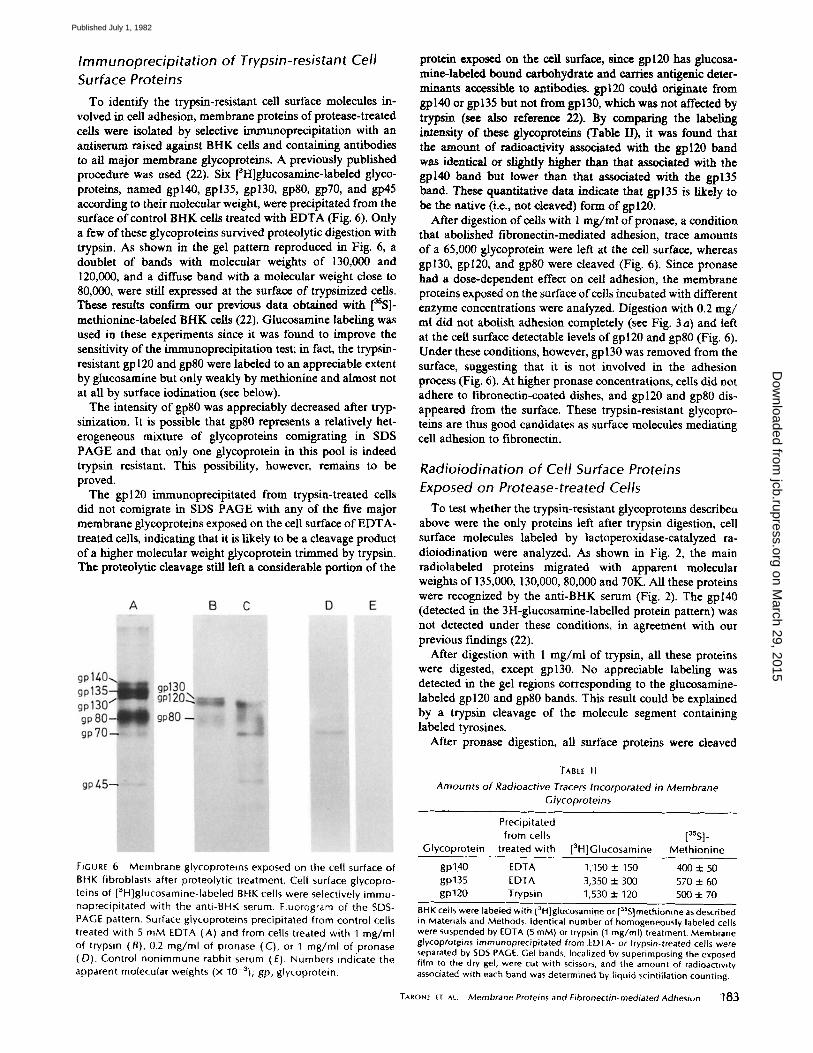
Immunoprecipitation of Trypsin-resistant Cell Surface Proteins
To identify the trypsin-resistant ceil surface molecules in- volved in cell adhesion, membrane proteins of protease-treated cells were isolated by selective immunoprecipitation with an antiserum raised against BHK cells and containing antibodies to all major membrane glycoproteins. A previously published procedure was used (22). Six [3H]glucosamine-labeled glyco- proteins, named gpl40, gp135, gpl30, gp80, gp70, and gp45 according to their molecular weight, were precipitated from the surface of control BHK cells treated with EDTA (Fig. 6). Only a few of these glycoproteins survived proteolytic digestion with trypsin. As shown in the gel pattern reproduced in Fig. 6, a doublet of bands with molecular weights of 130,000 and 120,000, and a diffuse band with a molecular weight close to 80,000, were still expressed at the surface of trypsinized cells. These results conftrm our previous data obtained with [~SS]- methionine-labeled BHK cells (22). Glucosamine labeling was used in these experiments since it was found to improve the sensitivity of the immunoprecipitation test; in fact, the trypsin- resistant gp 120 and gp80 were labeled to an appreciable extent by glucosamine but only weakly by methionine and almost not at all by surface iodination (see below).
The intensity of gp80 was appreciably decreased after tryp- sinization. It is possible that gp80 represents a relatively het- erogeneous mixture of glycoproteins comigrating in SDS PAGE and that only one glycoprotein in this pool is indeed trypsin resistant. This possibility, however, remains to be proved.
The gpl20 immunoprecipitated from trypsin-treated ceils did not comigrate in SDS PAGE with any of the five major membrane glyeoproteins exposed on the cell surface of EDTA- treated cells, indicating that it is likely to be a cleavage product of a higher molecular weight glycoprotein trimmed by trypsin. The proteolytie cleavage still left a considerable portion of the
protein exposed on the cell surface, since gpl20 has glucosa- mine-labeled bound carbohydrate and carries antigenic deter- minants accessible to antibodies, gpl20 could originate from gpl40 or gp135 but not from gpl30, which was not affected by trypsin (see also reference 22). By comparing the labeling intensity of these glycoproteins (Table II), it was found that the amount of radioactivity associated with the gpl20 band was identical or slightly higher than that associated with the gpl40 band but lower than that associated with the gp135 band. These quantitative data indicate that gp135 is likely to be the native (i.e., not cleaved) form of gpl20.
After digestion of cells with 1 mg/rnl ofpronase, a condition that abolished fibronectin-mediated adhesion, trace amounts of a 65,000 glycoprotein were left at the cell surface, whereas gpl30, gpl20, and gp80 were cleaved (Fig. 6). Since pronase had a dose-dependent effect on cell adhesion, the membrane proteins exposed on the surface ofcel/s incubated with different enzyme concentrations were analyzed. Digestion with 0.2 rag/ ml did not abolish adhesion completely (see Fig. 3 a) and left at the cell surface detectable levels of gpl20 and gp80 (Fig. 6). Under these conditions, however, gp 130 was removed from the surface, suggesting that it is not involved in the adhesion process (Fig. 6). At higher pronase concentrations, cells did not adhere to fibronectin-coated dishes, and gpl20 and gp80 dis- appeared from the surface. These trypsin-resistant glycopro- teins are thus good candidates as surface molecules mediating cell adhesion to fibronectin.
Radioiodination of Cell Surface Proteins Exposed on Protease-treated Cells
To test whether the trypsin-resistant glycoprotems describeu above were the only proteins left after trypsin digestion, cell surface molecules labeled by lactoperoxidase-catalyzed ra- dioiodination were analyzed. As shown in Fig. 2, the main radiolabeled proteins migrated with apparent molecular weights of 135,000, 130,000, 80,000 and 70K. All these proteins were recognized by the anti-BHK serum (Fig. 2). The gpl40 (detected in the 3H-glucosamine-labelled protein pattern) was not detected under these conditions, in agreement with our previous findings (22).
After digestion with 1 mg/ml of trypsin, all these proteins were digested, except gpl30. No appreciable labeling was detected in the gel regions corresponding to the glucosamine- labeled gpl20 and gp80 bands. This result could be explained by a trypsin cleavage of the molecule segment containing labeled tyrosines.
After pronase digestion, all surface proteins were cleaved
FIGURE 6 Membrane glycoprotems exposed on the cell surface of BHK fibroblasts after proteolytic treatment. Cell surface glycopro- teins of [3H]glucosamine-labeled BHK cells were selectively immu- noprecipitated with the anti-BHK serum. Fluorogram of the SDS- PAGE pattern. Surface glycoproteins precipitated from control cells treated with 5 mM EDTA (A) and from cells treated with 1 mg/ml of trypsin (B), 0.2 mg/ml of pronase (C), or 1 mg/ml of pronase (D). Control nonimmune rabbit serum (E). Numbers indicate the apparent molecular weights (x 10-3); gp, glycoprotein.
TABtE II
Amounts of Radioactive Tracers Incorporated in Membrane Gfycoproteins
Precipitated from cells [35S]-
Glycoprotein treated with [3H]Glucosamine Methionine
gpl40 EDTA 1,150 +_ 150 400 =1= 50 gp135 EDTA 3,350 + 300 570 :J: 60 gp120 Trypsin 1,530 + 120 500 ± 70
BHK cells were labeled with [~H]glucosami13e or [aSS]methionine as described in Materials and Methods. Identical number of homogeneously labeled cells were suspended by EDTA (5 raM) or trypsin (1 mg/ml) treatment. Membrane glycoprotfins immunoprecipitated from EDTA- or trypsin-treated cells were separated by SDS PAGE. Gel bands, localized by superimposing the exposed film to the dry gel, were cut with scissors, and the amount of radioactivity associated with each band was determined by liquid scintillation counting.
TARONE ET AL Membrane Proteins and Fibronectin-mediated Adhesion 183
on March 29, 2015
jcb.rupress.orgD
ownloaded from
Published July 1, 1982

and only trace amounts of a 65,000 mol wt protein were left, similar to that obtained with [3H]glucosamine-labeled ceils.
In conclusion, these experiments indicate that gpl30, gpl20, and gp80 were the only cell surface glycoproteins left on the BHK cell surface after trypsin treatment.
Inhibi t ion of Cell Adhesion by Antibodies
against Cell Surface Proteins
The antiserum used to precipitate the trypsin-resistant gly- coproteins from the BHK cell surface was then tested for its ability to prevent cell adhesion to fibronectin-coated dishes. Since the antiserum contained a small amount of antibodies to fibronectin that could interfere with the adhesion process (27, 28), this antibody fraction was removed by adsorption on fibronectin-Sepharose beads, as described in Materials and Methods.
The adsorbed and complement-inactivated serum was added to suspensions of trypsin-treated cells at the beginning of the test. As shown in Table III and Fig. 7, the anti-BHK serum displayed inhibition in a dose-dependent manner, while no effect was measured with the control nonimmune serum. The antiserum was not toxic, since cells (washed and left to recover for 2 h at 37°C in complete medium) fully regained their adhesive properties. Moreover, inhibition of cell adhesion was
TABLE Ill
Inhibition of Adhesion to Fibronectin-coated Dishes by Anti-BHK Serum
Antiserum* Di lut ion Adherent cells:l: %
Nonimmune 1/10 90 + 7 1/40 87 + 6
Anti-BHK 1/10 8 -t- 3 1/20 45 ± 5 1/40 7O ± 8
* Nonimmune rabbit serum or anti-BHK serum, which had been heated to inactivate complement and adsorbed to remove anti-fibronectin antibodies (see Materials and Methods), was added at the beginning of the assay at the indicated dilution.
:~Trypsin-treated BHK cells (1 mg/ml) were plated in fibronectin-coated dishes in the presence of the antisera, and adhesion was measured after 1 h at 37°C.
also obtained by pretreating trypsinized cells with different dilutions of the anti-BHK serum, washing, and plating on fibronectin-coated dishes in the absence of the antiserum (21).
Since gangliosides are possibly involved in the interaction of the cell surface with fibronectin (14), we tested whether the antiserum contained antiganglioside activities. No activity was found by testing the serum in a competitive radioimmunoassay with purified ox brain gangliosides in the concentration range of 30 ng/ml to 500/~g/ml (see Materials and Methods).
It is thus likely that inhibition of cell adhesion was due to binding of antibodies directed against the glycoproteins resist- ant to trypsin treatment. To test this possibility, adsorption experiments were performed. If these glycoproteins were in- deed the target of the antibodies inhibiting adhesion, adsorp- tion of the antiserum on trypsinized cells should remove the anti-adhesive activity, while adsorption on pronase-treated cells should not. The inhibitory activity of the adsorbed anti- serum was tested on cells suspended by EDTA, expressing the whole set of surface glycoproteins. As expected, adsorption of the antiserum on EDTA-treated cells (performed as a positive control) completely removed the inhibitory activity on adhe- sion (Table IV). Conversely, adsorption on cells treated with pronase was ineffective (Table IV and Fig. 7). Moreover, adsorption of the antiserum on cells treated with trypsin re- moved the inhibitory activity on adhesion almost as effectively as adsorption on untreated ceils (Table IV and Fig. 7).
These data give further support to the idea that trypsin- resistant glycoproteins recognized by the antiserum are in- volved in fibronectin-mediated cell adhesion.
DISCUSSION
A major problem in the elucidation of molecular mechanisms leading to cell-substratum adhesion is in understanding how the cell surface interacts with the extracellular matrix compo- nents. Fibronectin is a glycoprotein that mediates the interac- tion of the fibroblast surface with collagen and proteoglycans (29-32). Although a number of data have been obtained using adhesion mutants (33-35) or by chemical modification of the cell surface (36-39), the nature of the membrane molecules involved in the interaction with fibronectin is still uncertain.
The data presented in this paper indicate that adhesion of
FIGURE 7 Inhibit ion of cell adhesion with anti-BHK serum adsorbed on protease-treated cells. EDTA- treated BHK ceils were plated in f ibronectin-coated dishes and adhesion was measured after 1 h at 37°C. The fol lowing rabbit sera were added at final di lut ion of 1/10 at the beginning of the test: (A) control non- immune serum, (B) anti-BHK serum, (C) anti-BHK serum adsorbed on pronase-treated cells, and (D) anti-BHK serum adsorbed on trypsin-treated ceils. Pic- tures were taken after removing nonadherent ceils and staining with May-GrLinwald-Giemsa solution.
184 the JOURNAL Or CELL BIOLOGY - VOLUME 94, 1982
on March 29, 2015
jcb.rupress.orgD
ownloaded from
Published July 1, 1982

TABLE IV
Effect of Anti-BHK Serum Adsorbed by Protease-treated Cells on Fibronectin-mediated Adhesion
Number of cells used for Treatment of cells used for
adsorpt ion* adsorpt ion Adherent cells:l:
%
None - - S -4- 0.2 1 X 100 EDTA (5 mM) 80 + 7 1 X 10⁶ EDTA (5 mM) I00 ± 9 I X 106 Trypsin (I mg/ml ) 70 ± 8 1 x 100 Trypsin (1 mg/ml ) 75 ± 8 1 x 10 ° Pronase (1 mg/ml ) 10 + 2 1 X 10 ° Pronase (1 mg/ml ) 14 _+ 4
* 25 /al of anti-gHK serum were adsorbed with the indicated amount of formaldehyde-fixed cells as described in Materials and Methods.
:I: Adsorbed ser], at final dilution of 1/10, were added to EDTA-treated cells plated on fibronect~n-coated dishes. The percentage of adherent cells was determined after I h at 37°C.
BHK fibroblasts to fibronectin-coated dishes requires surface molecules that are protein in nature. This conclusion is based on the following findings: (a) digestion of the cell surface with a variety of proteolytic enzymes abolished the ability of cells to adhere to fibronectin; (b) recovery of adhesiveness after pro- teolytic cleavage required protein synthesis.
Complete inhibition of the adhesive properties required digestion with rather high enzyme concentrations. An unspe- cific toxic effect of the proteases employed was excluded by the cell viability tests performed and by the fact that selective inhibition of the proteolytic activity of subtilopeptidase and thermolysin abolished their ability to affect adhesion. More- over, the immunoprecipitation experiments indicated that there is a direct correlation between the loss of cell adhesive prop- erties and the disappearance of glycoproteins from the plasma membrane surface.
A peculiar feature of the glycoproteins involved in adhesion described in this paper is their resistance to enzymatic cleavage by trypsin. A different conclusion was reached by Grinnell in a recent study (40). This author studied fibroblast-substratum adhesion by measuring binding of fibronectin-coated latex beads to the cell surface. In this system, the binding of the coated beads was abolished by digestion of the cell surface with low concentrations of trypsin. No reason for this difference can be advanced as yet, apart from the fact that the assay conditions were different and a BHK cell variant growing in suspension culture was employed. A number of different in- vestigators, moreover, used trypsin-treated cells to study adhe- sion on dishes coated with serum (33, 34, 41), fibronectin (15), or fibronectin-collagen complexes (42), and no impairment of the adhesive properties by this treatment has been reported. Our data indicate that fibronectin-mediated cell adhesion is unaffected by digestion with high trypsin concentrations (up to 1 mg/ml) or by two digestion cycles with 0.5 mg/ml (see Table I). We thus conclude that the cell surface molecules mediating adhesion do not have functionally relevant, trypsin-sensitive sites facing the exterior side of the plasma membrane.
This property was exploited to identify the molecules in- volved. Using an antiserum to BHK cells, three typsin-resistant cell surface glycoproteins of 130,000, 120,000 and 80,000 have been identified by immunoprecipitation. Moreover, binding of antibodies to these proteins prevented adhesion of trypsinized cells to fibronectin. One of these glycoproteins, gpl30, was removed from the cell surface by treatment with low pronase
concentrations without affecting adhesion. By contrast, when gpl20 and gp80 were removed from the cell surface by com- plete pronase digestion, cell adhesion was fully inhibited. Taken together, these data suggest that gpl20 and/or gp80 are involved in cell adhesion mediated by fibronectin. We could not rule out, however, that other membrane proteins (repre- senting quantitatively minor cell surface components not de- tectable by the method employed) are involved as well.
Cell adhesion is a complex phenomenon occurring through a series of steps that involve different cellular functions: round cells first bind to the extracellular molecules adsorbed on the substratum; this binding triggers a cascade of intracellular events leading to spreading and acquisition of a fiat morphol- ogy (11, 43, 44). By studying adhesion mediated by glycosidases or lectins, it has been shown that the binding step is specific and mediated by cell surface receptors (43). Moreover, experi- ments with competitive inhibitors of glycosidases and lectins have shown that the cell surface receptors should interact with the extraceUular ligand also during the subsequent steps, when cell spreading takes place. When fibronectin is the extrac~llular ligand, the nature of the interaction with cell surface "receptors" is still a matter of controversy (13-16). The expres- sion at the cell surface of the trypsin-resistant glycoproteins described in this paper is sufficient to grant to the cell the ability to both initiate and complete the adhesion process mediated by fibronectin. The steps in which the molecules are required are not known, nor can their direct binding to fibro- nectin be demonstrated.
Recently, Aplin et al. (13) have shown that a 47,000 mol wt glycoprotein of BHK cells could be cross-linked to substratum- bound fibronectin in adherent ceils; it has been proposed that this protein may be a cell surface component interacting with fibronectin. As pointed out by these authors, the interaction of the 47,000 mol wt glycoprotein and fibronectin occurs in well- spread and fully adherent cells and it can not be excluded that other cell surface proteins are involved in earlier adhesion stages. A gp45 (with a molecular weight very close to that of the protein described by Aplin) was detected by our anti-BHK serum (see Fig. 6, lane A). This gp45, however, was absent on trypsinized cells (see Fig. 6, lane B, and Fig. 1, lanes B and E) that retained their adhesiveness to fibronectin.
Adhesive glycoproteins with molecular weights close to those of trypsin-resistant gpl20 and gp80 have been described by other laboratories. Hsieh and Sueoka (45) used an antiserum to a rat neuronal tumor cell line to induce rounding and detachment of a variety of mammalian cell lines. This antise- rum recognized two proteins, among the radiolabeled cell surface molecules, with molecular weights of 120,000 and 80,000 that were the target of the antibodies inhibiting cell adhesion. A glycoprotein with a molecular weight between 120,000 and 140,000 has also been identified in BHK ceils by Wylie et al. (46), again by using antisera to induce cell rounding and detachment from solid substrata.
We wish to thank Mrs. Maria Rosa Amedeo for technical assistance. This work was supported by a grant of the Progetto Finalizzato
Controllo Crescita Neoplastica (P. F. C. C. N.) of the Italian National Research Council.
Received for publication 14 January 1982, and in revised form 3 March 1982.
REFERENCES
L Yamada, K. M., and K. Olden. 1978. Fibronectin-adhesive glycoproteins of the cell surface and blood. Nature (Lond). 275:179-1M.
TARONE IT AL. Membrane Proteins and Fibronectin-mediated Adhesion 185
on March 29, 2015
jcb.rupress.orgD
ownloaded from
Published July 1, 1982

2, Mosesson, M. W. and D. L. Armani. 1980. The structure and biological activities of plasma fibronectin. Blood. 56:145-158.
3. Pearls~in, E. L., I. GoM, and A. Garcia-Pado. 1980. Fibronectin: a review of its structure and biological activity. Mot. Cell. Biochem. 29:103-128.
4. Colonna, G., S. S. Alexander. Jr., K. Yamada, I. Pastan, and H. Edethoch. 1978. Stability of cell surface protein to surfactants and denaturants. J. Biol. Chem. 253:7787-7790,
5. Fukuda, M., and S. I. Hakomori. 1979. Proteolytic and chemical fragmentation of galactoprot¢in a, a major transformation-sensitive glycoprotein released from hamster embryo fibroblasts. J. Biol. Chem. 254:5442-5450.
6. Wagner, D. D., and R, O. Hynes. 1979. Domain structure of fibronectin and its relation to function. J. Biol. Chem. 254:6746~754.
7. Ruoslahti, E., E. G. Hayman, P. Kuusela, J. E. Shively and E. Engvall. 1979. Isolation of a tryptic fragment containing the collagen-binding site of plasma fibronectin. J. Biol. Chem. 254:6054-6059.
8. Balian, G., E. M. Click, and P. Borstein. 1980. Location of a collagen-binding domain in fibronectin, J. Biol. Chem. 255:3234-3236.
9, Hahn, L.-H. E., and K. M. Yamada. 1979. Isolation and biological characterization of active fragments oftbe adhesive giycoprotein fibrnneetin. Cell. 18:1043-1051.
10. Sekignchi, K., and S. 1. Hakomori. 1980. Functional donmm structure of fibronectin. Proc. Natl. Acad. Sci. U. S. A. 77:2661-2665.
11. Grinnell, F. 1978. Cellular adhesiveness to extracelluiar substrata. Int. Rev. Cytol. 53:64- 144.
12. Penn, S. D. J., and R. C. Hughes. 1978. Fibronectin-piasma membrane interactions in the adhesion and spreading of hamster fibroblasts. Nature (Loud.) 276:80-83.
13. Aplin, J. D., R. C. Hughes, C. L. Jaffe, and N. Sharon. 1981. Reversible cross-linking of cellular components of adherent fibroblasts to fibronectin and lectin-coated substrata. Exp. Cell Res. 134:488~,94.
14. Kleinman, H. K., G. R. Martin, and P. H. Fishman. 1979. Ganglioside inhibition of fibronectin-mediated cell adhesion to collagen. Proc. Natl. Acad. Sci. U. S. A. 76:3367- 3371.
15. Ran~ala, H., W. G. Carter, and S. I. Hakomori. 1981. Studies on cell adhesion and recognition. Extent and specificity of cell adhesion triggered by carbohydrate-reactive proteins (glycosidas~ and lectins) and by fibronectin. £ Cell Biol. 88:127-137.
16. Yamada, K. M., D. W. Kennedy, G. R. Grotendorst, and T. Momoi. 1981. Glycolipids: receptors for fibronectin? J. Cell. Physiol. 109:343-351.
I7. Engvall, E., and E. Ruoslahti. 1977. Binding of soluble form of fibroblast surface protein, fibronectin, to collagen. Int. £ Cancer. 20:1-5.
18. Takahashi, T., L. J. Old, and E. A. Boyse. 1970. Surface alloantigens of plasma ceils. J. Exp. Med. 131:1325-1341.
19. Grinnell, F., and D. G. Hays. 1978. Induction of cell spreading by substratum adsorbed ligands directed against the cell surface. Exp. Cell. Res. 116:275-284.
20. Grinnell, F., D. G. Hays, and D. Minter. 1977. Cell adhesion and spreading factor: partial purification and properties. Exp. Cell. Res. 110:175-180.
21. Tarone, G., G. Galetto, and P. M. Comogho. 1982. Surface glycoproteins and fibroneetin- mediated cell adhesion. In Protides of Biological Fluids. H. Peeler editor. Pergamon Press, Oxford. 29:291-294.
22. Tarone, G., M. Prat, and P. M. Comoglio. 1980. Idantification and partial characterization of five major membrane giycoproteins of BHK fibroblasts, J. Membr, Biol. 53:55~51.
23. Tarone, G., P, Cesohi, M. Prat, and P. M. Comoglio. 198t. Transformation sensitive protein with molecular weight of 45,000 secreted by mouse fibroblasts. Cancer Res. 41:3648-365 I.
24. Hynes, R. O. 1973. Alteration of cell surface proteins by viral transformation and by proteolysis. Proc. Natl. Acad. Sci. U. S. A. 70:3170-3173.
25. Laemmli, U. K. 1970. Cleavage of structural proteins during the assembly of the head of the bacteriophage T4. Nature (Loud.). 227:680-683.
26. Laskey, R. A., and A. D. Mills. 1975. Quantitative film detection of 3-H and 14-C in polyacrylamide geh by fluorography. Eur. £ Biochem 56:335-340.
27. Yamada, K. M. 1978. Immunological characterization of a major transformation-sensitive fibroblast cell surface 81ycoprotein. J. Cell Biol. 78:520-541.
28. Grinnell, F., and M. K. Feld, 1979. Initial adhesion of human fibroblasts in serum-free medium: possible role of secreted fibronectin. Cell. 17: 117-129.
29. Klebe, R. J. 1974. Isolation of a collagen-dependent cell attachment factor. Nature (Loud.) 250:248-25 I.
30. Klebe, R. J, 1975. Cell attachment to collagen: the requirement of energy. £ Cell. PhysioL 86:231-236.
31. Perkins, M. E., T. H. JI, and R. O. Hynes, 1979. Cross-linking of fibronectin to sulphated proteoglycans at the cell surface. Cell. 16:941-952.
32. Borstein, P., D. Duksin, G. BMian, J. M. Davidson, and E. Crouch. 1978. Organization of extracelluiar proteins on the connective tissue cell surface: relevance to cell-matrix interactions "'in vitro" and "in vivo." Ann. N. E Acad Sci. 312:93-105.
33. Harper, P. A., and R. L. Juliauo. 1980. Isolation and characterization of Chinese hamster ovary cell variants defective in adhesion to fibronectin-coated collagen. £ Cell. BioL 87:755-763.
34. Harper. P. A., and R. L. Juliano. 1981. Two distinct mechanisms of fibroblast adhesion. Nature (Loud.). 290:136-138.
35. Pouyssegnr, J., M. Wlllingham, and I. Pastan. 1977. Role of cell surface carbohydrates and proteins in cell behavior: studies on the biochemical reversion of an N-ace/tylgiucos - amina-deficiem fibroblast mutant. Proc. Natl. Acad. Sci. U. S. A. 74:243-247.
36. Grinnell, F., M. Milam, and P, A. Srere. 1973. Studies on cell adhesion. III. Adhesion of baby hamster kidney cells. J. Cell Biol. 56:659~65.
37. Grinnell, F., and P. A. Srere. 1971. Inhibition of cellular adhesiveness by sulphydryl blocking reagents. £ Cell. Physiol. 78:153-160.
38. Grinnall, F., M. Mllam, and P. A. Srere. 1972. Studies on cell adhesion. II. Adhesion of cell to surfaces of diverse chemical composition and inhibition of adhesion by sulphydryl binding reagents. Arch. Biochem. Biophys. 152:241-250,
39. Aplin, J. D.. T. D. Butters, and R. C. Hughes. 1980. Effects of cell surface modifications on fibroblast-substratum adhesion. Cell Biol. Int. Rep. 4:735.
40. Grim-tell, F. 1980. Fibroblast receptor for cell-substratum adhesion: studies on the inter- action of baby hamster kidney ceils with latex beads coated by cold insoluble globulin (plasma fibronectin)..L Cell Biol. 86:104-112,
41. Juliano. R. L., and E. Gagalano. 1977. Adhesion of Chinese hamster cells: effects of temperature, metabolic inhibitors and proteolytic dissection of cell surface macromole- cules. J. Cell. Physiol. 92;209-220.
42. Pearlstein, E. L., and L. I. Gold. 1978. High molecular weight glycoprotein as a mediator of cellular adhesion. Ann. N. I". Acad. Sci. 312:278-292.
43. Carter, W. G., H. Rauvala, and S. I. Hakomori. 1981. Studies on cell adhesion and recognition. II, The kinetics of cell adhesion and cell spreading on surface coated with carbohydrate-reactive proteins (glycosidases and lectins) and fibronectin. ,£ Cell Biol. 88:138-148.
44. Rces, D. A., C. W. Lloyd, and D. Thom. 1977. Control of grip and stick in cell adhesion through lateral relationships of membrane glycoproteins. Nature (Land.) 267:124-128.
45. Hsieh, P,, and N, Sueoka. 1980. Antisera inhibiting mammalian cell spreading and possible cell surface antigens involved, J. Cell Biol. 86:866-873.
46. Wylie, D. E., C. H. Damsky, and C. A. Buck. 1979. Studies on the function of cell surface glycoproteins. Use of antisera to surface membranes in the identification of membrane componems relevant to cell-substrate adhesion. J. Cell BioL 80:385-402.
~ 8 6 THE IOURNAt O~ CELt BIOLOGY • VOLUME 94, 1982
on March 29, 2015
jcb.rupress.orgD
ownloaded from
Published July 1, 1982
Copyright © 2022 FDOKUMEN