Effect of processing on silk-based biomaterials: Reproducibility and biocompatibility
Upload
khangminh22Category
view
1download
0
applied sciences
Review
Fibronectin-Enriched Biomaterials, Biofunctionalization, andProactivity: A Review
Carla Palomino-Durand , Emmanuel Pauthe *,† and Adeline Gand *,†
�����������������
Citation: Palomino-Durand, C.;
Pauthe, E.; Gand, A. Fibronectin-
Enriched Biomaterials,
Biofunctionalization, and Proactivity:
A Review. Appl. Sci. 2021, 11, 12111.
https://doi.org/10.3390/
app112412111
Academic Editors: Barbara Nebe and
Karine Anselme
Received: 10 November 2021
Accepted: 12 December 2021
Published: 19 December 2021
Publisher’s Note: MDPI stays neutral
with regard to jurisdictional claims in
published maps and institutional affil-
iations.
Copyright: © 2021 by the authors.
Licensee MDPI, Basel, Switzerland.
This article is an open access article
distributed under the terms and
conditions of the Creative Commons
Attribution (CC BY) license (https://
creativecommons.org/licenses/by/
4.0/).
Equipe de Recherche sur les Relations Matrice Extracellulaire Cellules (ERRMECe), Institut des Matériaux,CY Cergy-Paris Université, CEDEX, 95302 Cergy-Pontoise, France; [email protected]* Correspondence: [email protected] (E.P.); [email protected] (A.G.)† A.G. & E.P. contributed equally to the supervision of the contribution.
Abstract: Modern innovation in reconstructive medicine implies the proposition of material-basedstrategies suitable for tissue repair and regeneration. The development of such systems necessitatesthe design of advanced materials and the control of their interactions with their surrounding cellularand molecular microenvironments. Biomaterials must actively engage cellular matter to direct andmodulate biological responses at implant sites and beyond. Indeed, it is essential that a true dialogueexists between the implanted device and the cells. Biomaterial engineering implies the knowledgeand control of cell fate considering the globality of the adhesion process, from initial cell attachmentto differentiation. The extracellular matrix (ECM) represents a complex microenvironment ableto meet these essential needs to establish a relationship between the material and the contactingcells. The ECM exhibits specific physical, chemical, and biochemical characteristics. Considering thecomplexity, heterogeneity, and versatility of ECM actors, fibronectin (Fn) has emerged among theECM protagonists as the most pertinent representative key actor. The following review focuses onand synthesizes the research supporting the potential to use Fn in biomaterial functionalization tomimic the ECM and enhance cell–material interactions.
Keywords: fibronectin; extracellular matrix; biofunctionalized biomaterials; interfacial properties;surface coatings; hydrogels
1. Introduction
Facing the challenge of increasing needs for the reparation of the human body andemerging innovative strategies to realize the modern concepts of regenerative and recon-structive medicine, many medical devices have been proposed by the scientific community.These are used in a wide range of clinical applications, and their performance is highlydependent on the nature of their surfaces and the latter’s interaction with defined proteinsand cells.
Commonly, once a biomaterial is implanted, protein adsorption from biological fluidsoccurs immediately, mediating the interaction between the surface and cells. Concomitantly,a tissue reaction occurs, which results in an inflammatory response driven, largely byimmune-related proteins and cell contact activation.
Although biomaterials were originally designed to be inert, contemporary applica-tions, such as tissue engineering and regenerative medicine, demand biomaterials that canactively engage cellular matter to direct and modulate biological responses at implant sitesand beyond [1–4]. Since biomaterials interact with their surroundings via their surfaces, anattractive strategy for producing truly bioactive materials is the design of biofunctionalizedcoatings. Of particular note are three-dimensional, cellularized, porous scaffold deviceswith interfaces optimized to support and implant viable cells, a promising breakthroughthat will facilitate biomedical advances in the future. An appropriately modulated innateimmune response would improve the acceptance of a given biomaterial by the host and its
Appl. Sci. 2021, 11, 12111. https://doi.org/10.3390/app112412111 https://www.mdpi.com/journal/applsci
Appl. Sci. 2021, 11, 12111 2 of 21
bio-integration with tissues. This principle is universal whatever the nature of the material,from metals and ceramics to polymers.
Currently, it is absolutely necessary to determine a way to promote a true and effectivedialogue between the material and its cellular and tissular microenvironment. The notionof material–cell engineering has emerged, and it leads global, multifactorial, and complexcell behavior at the site of implantation. Throughout the overall cell-adhesion process, fromthe initial phase of the cell-contacting response (i.e., cell attachment) to later cell actions,such as differentiation, the different phases of a cell’s fate must be taken into consideration.
This cell–material relationship is controlled and tuned by various parameters asfollowing: (i) adhesion control influenced by physical characteristics such as the mechanicalproperties of the materials and their topography; (ii) chemical/biochemical molecularreactions mediated by the availability of growth-factor (GF) receptors, the actions of solublefactors, and the role of extracellular-matrix (ECM) protagonists.
The ECM is composed of fibrous protein polymers, typically type I collagen fibernetworks, interspersed with strongly flexible and highly hydrated glycoproteins, polysac-charides, and other large molecules, from fibrin to heparin. Thus, there exist variousmolecular protagonists that, due to their specific and intrinsic properties, represent andrespond to various concepts and needs.
Biomaterial engineering science necessitates a simple, pertinent, representative so-lution to elucidate this ECM. Among the ECM protagonists, considering the complexity,heterogeneity, and versatility of ECM actors, fibronectin (Fn) has emerged as the mostpertinent key partner.
The aim of the following narrative review was to focus on the research supportingthis crucial role of Fn in representing the ECM and to explore many of the versatile andpredominant properties of biomaterials.
2. Fn as a Pertinent Key Actor in the ECM
The ECM, a material secreted by and surrounding cells, represents a complex networkof polysaccharides (such as proteoglycans) and proteins (such as collagen, elastin, and Fn).This physical barrier delimits organs and represents the structural component of tissuesthat influences their development and physiology. Fn plays a major role in the ECM and isinvolved in numerous physiological processes, especially owing to its modular structureand the numerous properties of its different regions.
2.1. Fn’s General Properties
Fn is a multifunctional glycoprotein expressed by many different cell types (hepato-cytes, fibroblasts, macrophages, and leukocytes, among others). It is found in the plasmaand the ECM. Its main biological functions are based on its affinity for different biologicalcompounds such as cell-surface receptors, ECM proteins (collagens, heparin, etc.), DNA,gangliosides, and immunoglobulins [5].
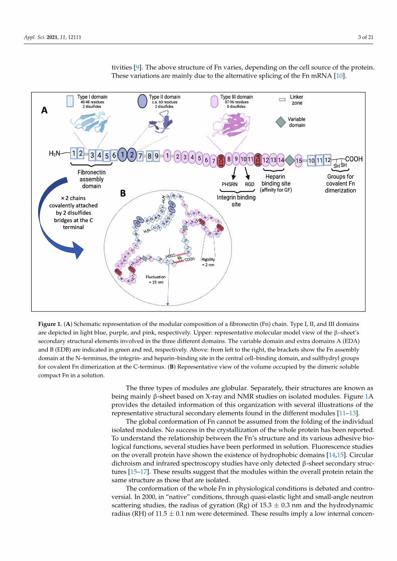
In vivo, Fn mediates cell adhesion to the ECM and contributes to phagocytosis regula-tion, wound healing, cell proliferation, differentiation, and locomotion [6]. At the structurallevel, Fn is composed of two identical polypeptide subunits covalently linked via twodisulfide bonds near their carboxy termini. Each subunit contains nearly 2500 aminoacids (with a molecular weight of approximately 250,000 Da). It consists of a repetition of56 modules of three types having a well-defined structure and a high degree of internalhomology. Modules of types I, II, and III consist of 45, 60, and 90 amino acids, respectively.Type I modules make up the NH2-terminal and COOH-terminal ends of the polypeptide.Two type II modules interrupt a row of nine type I modules at the NH2-terminus. Fifteentype III modules make up the middle of the polypeptide [7] (Figure 1A). Type I, II, andIII modules are also found in many other proteins, with type III even being ubiquitous inanimal proteins [8]. These modules are connected by short polypeptide segments sensitiveto proteinases and are grouped into functional domains that express specific binding ac-
Appl. Sci. 2021, 11, 12111 3 of 21
tivities [9]. The above structure of Fn varies, depending on the cell source of the protein.These variations are mainly due to the alternative splicing of the Fn mRNA [10].
Appl. Sci. 2021, 11, x FOR PEER REVIEW 3 of 23
segments sensitive to proteinases and are grouped into functional domains that express specific binding activities [9]. The above structure of Fn varies, depending on the cell source of the protein. These variations are mainly due to the alternative splicing of the Fn mRNA [10].
The three types of modules are globular. Separately, their structures are known as being mainly β-sheet based on X-ray and NMR studies on isolated modules. Figure 1A provides the detailed information of this organization with several illustrations of the representative structural secondary elements found in the different modules [11–13].
Figure 1. (A) Schematic representation of the modular composition of a fibronectin (Fn) chain. Type I, II, and III domains are depicted in light blue, purple, and pink, respectively. Upper: representative molecular model view of the β–sheet’s secondary structural elements involved in the three different domains. The variable domain and extra domains A (EDA) and B (EDB) are indicated in green and red, respectively. Above: from left to the right, the brackets show the Fn assembly domain at the N–terminus, the integrin- and heparin–binding site in the central cell–binding domain, and sulfhydryl groups for covalent Fn dimerization at the C-terminus. (B) Representative view of the volume occupied by the dimeric soluble compact Fn in a solution.
The global conformation of Fn cannot be assumed from the folding of the individual isolated modules. No success in the crystallization of the whole protein has been reported. To understand the relationship between the Fn’s structure and its various adhesive biological functions, several studies have been performed in solution. Fluorescence studies on the overall protein have shown the existence of hydrophobic domains [14,15]. Circular dichroism and infrared spectroscopy studies have only detected β-sheet secondary structures [15–17]. These results suggest that the modules within the overall protein retain the same structure as those that are isolated.
The conformation of the whole Fn in physiological conditions is debated and controversial. In 2000, in “native” conditions, through quasi-elastic light and small-angle neutron scattering studies, the radius of gyration (Rg) of 15.3 ± 0.3 nm and the
Figure 1. (A) Schematic representation of the modular composition of a fibronectin (Fn) chain. Type I, II, and III domainsare depicted in light blue, purple, and pink, respectively. Upper: representative molecular model view of the β–sheet’ssecondary structural elements involved in the three different domains. The variable domain and extra domains A (EDA)and B (EDB) are indicated in green and red, respectively. Above: from left to the right, the brackets show the Fn assemblydomain at the N–terminus, the integrin- and heparin–binding site in the central cell–binding domain, and sulfhydryl groupsfor covalent Fn dimerization at the C-terminus. (B) Representative view of the volume occupied by the dimeric solublecompact Fn in a solution.
The three types of modules are globular. Separately, their structures are known asbeing mainly β-sheet based on X-ray and NMR studies on isolated modules. Figure 1Aprovides the detailed information of this organization with several illustrations of therepresentative structural secondary elements found in the different modules [11–13].
The global conformation of Fn cannot be assumed from the folding of the individualisolated modules. No success in the crystallization of the whole protein has been reported.To understand the relationship between the Fn’s structure and its various adhesive bio-logical functions, several studies have been performed in solution. Fluorescence studieson the overall protein have shown the existence of hydrophobic domains [14,15]. Circulardichroism and infrared spectroscopy studies have only detected β-sheet secondary struc-tures [15–17]. These results suggest that the modules within the overall protein retain thesame structure as those that are isolated.
The conformation of the whole Fn in physiological conditions is debated and contro-versial. In 2000, in “native” conditions, through quasi-elastic light and small-angle neutronscattering studies, the radius of gyration (Rg) of 15.3 ± 0.3 nm and the hydrodynamicradius (RH) of 11.5 ± 0.1 nm were determined. These results imply a low internal concen-

Appl. Sci. 2021, 11, 12111 4 of 21
tration (M/R3g/H) compared to those of the usual globular proteins. This is also confirmed
by the RH/Rg ratio of 0.75 ± 0.02, consistent with a Gaussian chain, whereas the RH/Rgratio of 1.3 was identified for spherical-shaped molecules. However, adding a denaturingagent (8 M urea) increases Rg by a factor of two. This means that the Fn “native” chainis not completely unfolded. The average shape of the Fn conformation was also probedby small-angle neutron scattering performed for a reverse scattering vector q-1 smallerthan Rg (0.2 < q-1 < 15 nm). The measured form factor is in complete agreement withthe form factor of a random string of 56 beads with 5 nm in diameter (Figure 1B). It rulesout the possibility of an unfolded chain as well as globular structures [18]. This studydemonstrates not only the complexity, but also the versatility offered by Fn in modulatingand adapting its structural and conformational organization. In other words, consideringthe nature of Fn, it is crucial to make a distinction between structure and conformation andto deal with the two definitions to clearly explain and discuss some specificity. For instance,conformation is used more to refer to the overall global whole-chain organization, whereasthe structural level concerns the intrinsic remarkable structural elements in the modules.For example, without compromising the presence of the β-sheet in the individual modules,which provides the structure of the modules, the overall Fn molecule can transition from ahighly extended and almost linear conformation to a globular and compact state. In otherwords, the molecule appears as a highly flexible and extendable string of beads capable oflarge conformational modulations.
Moreover, the global molecular organization of the whole Fn, at interfaces versus insolutions, is under control and mediated by the specificity and the nature (hydrophobic,charged, etc.) of the elements found along the chain.
First, the beta structures found in the three types of modules are composed of hy-drophobic and large amino acids (Figure 1A). The structures of these apolar and hydropho-bic elements are strongly influenced by polar or apolar environments, which, in turn,strongly impact the Fn’s structure and its subsequent organization. The nature of cer-tain solvents and the physicochemical properties of materials strongly affect the overallconformation and structure of the Fn [19].
Second, the charges of the different modules and the subsequent electrostatic effectsalso play a crucial role in the structuration and conformation of the molecule. The completeamino acid sequence of human Fn has been known for decades [20]. The protein databank contains a specific amino acid sequence for the 32 modules of each chain. Theattribution of one positive charge for arginine and lysine residues and one negative chargefor aspartic acid and glutamic acid residues gives 192 positive charges and 259 negativecharges, forming an overall net charge per chain equal to −259 + 192 = −67. With morethan 450 negative and positive residues charged among 2500, Fn is a true polyelectrolyte.Moreover, with 134 (67 × 2) more negative charges than positive ones, Fn, owing to thisexcess of negative charges, can be globally considered a polyanion. At a more detailed level,a fine analysis of the contained positively charged residues in each module, comparedto the quantity of negatively charges, permits the calculation of the net charge for eachmodule. The differences range between −11 (for the III-2 and the EDA modules with−14/+3 and −17/+6, respectively) and +6 (for the III-14 with −7/+13) (see Figure 2A).
This heterogeneous repartition of charges toward the different modules, preciselyin the cell-binding region (the middle of the chain that contains the type III modules),gives rise, led by electrostatic interactions, to the specific folding of the chain in solution(Figure 2A). Moreover, the other part of the chains also adopts a specific orientation. TheN-terminal part must present a favorable orientation of the Fn assembly domain, whichallows the stabilization of the globular conformation of the dimer. At the C-terminal part,the orientation of this segment is led by the constraint of the double disulfide cross-linksthat covalently binds the two Fn chains (Figure 2B). This “natural” folding and orientationof the chains is conserved and stabilized in the dimer structure (Figure 2C).

Appl. Sci. 2021, 11, 12111 5 of 21Appl. Sci. 2021, 11, x FOR PEER REVIEW 5 of 23
Figure 2. (A) Representation of the modular composition of an Fn chain with details of the electrostatic net charge for each of individual 32 domains. A red–blue scale illustrates the gradual change in the charge from –11 to +11. (B) Schematic representation of the structural organization adopted by the chain in solutions, essentially mediated by electrostatic domain interactions in the type III–1–III–14 central domains. (C) Representation of an Fn dimer stabilized by disulfide cross-links at the C-terminus and Fn–Fn interactions in the N-terminal domain.
This heterogeneous repartition of charges toward the different modules, precisely in the cell-binding region (the middle of the chain that contains the type III modules), gives rise, led by electrostatic interactions, to the specific folding of the chain in solution (Figure 2A). Moreover, the other part of the chains also adopts a specific orientation. The N-terminal part must present a favorable orientation of the Fn assembly domain, which allows the stabilization of the globular conformation of the dimer. At the C-terminal part, the orientation of this segment is led by the constraint of the double disulfide cross-links that covalently binds the two Fn chains (Figure 2B). This “natural” folding and orientation of the chains is conserved and stabilized in the dimer structure (Figure 2C).
2.2. Fn’s Role and the Structure–Function Relationship within the ECM and Cells Cells’ behaviors are under the control of their interactions with their surrounding
extracellular microenvironment. Cells are very sensitive to the mechanical and biophysicochemical properties of this ECM environment, and cell receptors and integrins are mainly solicited. Via the integrins, cells literally “feel” the specificity of their environment and respond to crucial parameters such as mechanical properties. For instance, a lack of rigidity, as in a very soft environment, compromises cell adhesion and, subsequently, induces a lack of intracellular tension and non-performant intrinsic signaling pathway. Conversely, the ECM that presents a relatively high stiffness plays a pivotal role in provoking appropriate cell behaviors such as correct differentiation, the generation of intrinsic signals, the leading of focal adhesions and, thus, cell engagement in appropriate signaling pathways.
Moreover, in addition to the fact that cells respond to their biochemical environments via integrins, they also mainly employ GF receptors. Indeed, GFs, delivered to cells, are essential factors that govern part of their response. These GFs must be delivered specifically to the cells. In physiological conditions, they are present throughout the ECM, where they bind to the GF receptors.
Figure 2. (A) Representation of the modular composition of an Fn chain with details of the electrostatic net charge for eachof individual 32 domains. A red–blue scale illustrates the gradual change in the charge from –11 to +11. (B) Schematicrepresentation of the structural organization adopted by the chain in solutions, essentially mediated by electrostatic domaininteractions in the type III–1–III–14 central domains. (C) Representation of an Fn dimer stabilized by disulfide cross-links atthe C-terminus and Fn–Fn interactions in the N-terminal domain.
2.2. Fn’s Role and the Structure–Function Relationship within the ECM and Cells
Cells’ behaviors are under the control of their interactions with their surroundingextracellular microenvironment. Cells are very sensitive to the mechanical and biophysico-chemical properties of this ECM environment, and cell receptors and integrins are mainlysolicited. Via the integrins, cells literally “feel” the specificity of their environment andrespond to crucial parameters such as mechanical properties. For instance, a lack of rigidity,as in a very soft environment, compromises cell adhesion and, subsequently, induces a lackof intracellular tension and non-performant intrinsic signaling pathway. Conversely, theECM that presents a relatively high stiffness plays a pivotal role in provoking appropriatecell behaviors such as correct differentiation, the generation of intrinsic signals, the leadingof focal adhesions and, thus, cell engagement in appropriate signaling pathways.
Moreover, in addition to the fact that cells respond to their biochemical environmentsvia integrins, they also mainly employ GF receptors. Indeed, GFs, delivered to cells, areessential factors that govern part of their response. These GFs must be delivered specificallyto the cells. In physiological conditions, they are present throughout the ECM, where theybind to the GF receptors.
When scientists and engineers design and generate biomaterials with interfacialproperties capable of controlling cell behavior, it is of crucial interest to implicate andmobilize specific receptors’ integrins and GF receptors, respectively. In other words, ECMmodels must consider both integrins and GF receptors. Moreover, integrins mediateadhesion related to the mechanical or viscoelastic properties of the environment. TheECM presents dynamic viscoelasticity, and biomaterials developed by engineers must alsointegrate these viscoelastic properties.
Thus, there are two crucial questions regarding the aim of satisfying and optimizingthe dialogue between cells and their surrounding environments: (i) How do cells sense thisviscoelasticity, and how can this viscoelasticity be incorporated into materials? (ii) Howcan GFs’ presentation and their accessibility be maximized? Dalby et al., in a recent review,synthetized the different possibilities for presenting GFs [21], from uploading GFs in

Appl. Sci. 2021, 11, 12111 6 of 21
interfacial films to increasing GFs’ affinity, specific chemical binding, or affinity in the ECM.Among these strategies, the development of materials with both integrin binding and GFbinding represents a highly interesting way to solve these problems.
Fn appears to be the most pertinent protagonist of the ECM for satisfying the latestrequirements and addressing this interesting dual specificity. First, as presented above,among the different modules that contain specific properties, the modules III-10 and III-9contain the well-known RGD and PHSRN sequences that are, highly specific and a well-known region for integrin binding, respectively (especially αvβ3 and α5β1). Second, inthe vicinity of this region, there exists a row of three specific type III modules (III-12–III-14heparin-binding site) that exhibit a specific affinity for binding GFs (Figure 1A). Martinoand Hubbell showed that Fn III-12–III-14 from Fn promote the binding of GFs includingBMP2 [22]. Thus, within Fn, integrin binding can be simultaneously optimized withGF binding.
The accessibility of these regions necessitates a specific Fn dynamic conformationand structural rearrangement. Initially secreted from cells and, in solution, organized as acompressed, soluble dimer connected by disulfide linkages, Fn is naturally stimulated inan active state by interactions between its integrin-affinity sequences and synergy domainsof the integrin present on the cell surface. This initiates a gathering of several Fn moleculesnear the cell surface. Although each molecule has a row of five Fn-specific binding sites,the primary matrix-assembly domain, located near the disulfide bonds, is essential forfiber formation. The high concentration of activated Fn molecules promotes the linkagesbetween each of these and molecules from surrounding cells. The process generates anetwork of interconnected, stabilized, and insoluble proteins connecting cells throughoutthe matrix (Figure 3).
Appl. Sci. 2021, 11, x FOR PEER REVIEW 7 of 23
Figure 3. Schematic representation of the major steps that mediate an Fn fibrillar assembly at the interfaces and lead to a structural Fn reorganization from a soluble compact form to aligned Fn molecules with fibrils. First, the III-9 and III-10 modules of each chain of the soluble Fn bind to the integrins clustered at the surface (1). This triggers a reorganization of both the type III modules around the cell-binding domain and that of the disulfide bridge cross-links at the C-terminal part of each chain in a double-folded loop orientated to face the integrins and a dissociation of the N-terminal dimer assembly with a motion of the row of the six type I modules at the N-terminal site that now become aligned in parallel to the membrane, opposite to the orientation in a solution (2). This opening and new orientation of the N-terminal Fn-assembly domain favors interactions with the N-terminal domain of another Fn molecule and, consequently, initiates Fn fibrillogenesis (3).
3. Fn Application and Involvement in Biomaterials and Medical Device Engineering
As already established, one of the main challenges in material implantation is appropriate bio-integration that conditions the successful performance of the implant in the human body. Implant–host tissue crosstalk must be improved through interfacial strategies. In recent years, there has been increasing interest in the functionalization of surfaces with protein coatings, since the first event to occur right after implantation is the interaction between the implant and circulating blood components, leading to the adsorption of ECM proteins. Consequently, cells do not interact directly with the implant but interact with protein coatings [23] that influence cell behavior (recruitment, adhesion, and differentiation). In this context, proteins from the ECM are of particular interest. As described above, due to its intrinsic biochemical properties and specific biophysical multi-modular structuration, Fn plays a key role in mediating cell behavior, from adhesion, growth, and migration to differentiation.
Many studies have described strategies based on the selection of a discriminant part of Fn. For instance, studies report the extensive use of functional fragments of Fn, such as specific Fn peptides or moieties containing integrin-binding domains (RGD and PHSRN),
Figure 3. Schematic representation of the major steps that mediate an Fn fibrillar assembly at the interfaces and lead to astructural Fn reorganization from a soluble compact form to aligned Fn molecules with fibrils. First, the III-9 and III-10modules of each chain of the soluble Fn bind to the integrins clustered at the surface (1). This triggers a reorganization ofboth the type III modules around the cell-binding domain and that of the disulfide bridge cross-links at the C-terminal partof each chain in a double-folded loop orientated to face the integrins and a dissociation of the N-terminal dimer assemblywith a motion of the row of the six type I modules at the N-terminal site that now become aligned in parallel to the membrane,opposite to the orientation in a solution (2). This opening and new orientation of the N-terminal Fn-assembly domain favorsinteractions with the N-terminal domain of another Fn molecule and, consequently, initiates Fn fibrillogenesis (3).

Appl. Sci. 2021, 11, 12111 7 of 21
3. Fn Application and Involvement in Biomaterials and Medical Device Engineering
As already established, one of the main challenges in material implantation is ap-propriate bio-integration that conditions the successful performance of the implant in thehuman body. Implant–host tissue crosstalk must be improved through interfacial strategies.In recent years, there has been increasing interest in the functionalization of surfaces withprotein coatings, since the first event to occur right after implantation is the interactionbetween the implant and circulating blood components, leading to the adsorption of ECMproteins. Consequently, cells do not interact directly with the implant but interact withprotein coatings [23] that influence cell behavior (recruitment, adhesion, and differenti-ation). In this context, proteins from the ECM are of particular interest. As describedabove, due to its intrinsic biochemical properties and specific biophysical multi-modularstructuration, Fn plays a key role in mediating cell behavior, from adhesion, growth, andmigration to differentiation.
Many studies have described strategies based on the selection of a discriminant partof Fn. For instance, studies report the extensive use of functional fragments of Fn, such asspecific Fn peptides or moieties containing integrin-binding domains (RGD and PHSRN),to improve implant integration [24–26]. Despite numerous opportunities, these strategiespresent drawbacks due to the lack of synergy and cooperation between different modules.
A full-length Fn protein, instead of small peptides, could provide secondary actionslinked to the presence of other binding sites, and it is more biomimetic of the ECM andalso fully integrates the dimension of the protein dynamic which is essential for repre-senting the structural molecular rearrangement involved during the remodeling of thecell microenvironment.
Different strategies can be used to functionalize materials with Fn:
• Simple molecular two-dimensional (2D) coatings at interfaces via covalent binding oradsorption and the use of aptamers to favor Fn adsorption in monolayers;
• Complex coated interfaces, where Fn is combined with other molecules in orderto form bioengineered multilayered interfaces generating thin films and interfacesin 2.5D;
• Fn distribution in a three-dimensional (3D) volume entrapped in hydrogels via physi-cal dispersion and covalent cross-linking.
These biofunctionalized interfaces recreate, at different levels, various pertinent andrepresentative biomimetic cell microenvironments between the implant and the surround-ing tissues.
3.1. Fn 2D Molecular Coatings3.1.1. Fn Covalent Binding
The covalent binding of Fn presents many advantages such as the strong and stablelinkage of molecules to the surface. Nevertheless, the grafting process is delicate, asit should proceed in mild conditions to preserve the protein structure and requires thepresence of functional groups at the surface of the biomaterial able to react with reactiveprotein groups, which often requires a preliminary activation step at the surface. Thefollowing section describes the main published papers regarding surface activation andFn-grafting modes on various substrates, which are summarized in Table 1.

Appl. Sci. 2021, 11, 12111 8 of 21
Table 1. Fn surface functionalization of different substrates using different strategies and related biological activities.
Surface Activation/Fn Grafting Substrate Biological Activity References
Modification with isocyanates Polyvinyl alcohol (PVA) Increasing cell adhesion and proliferation in rabbit cornealepithelial cells [27–29]
Carbonyl diimidazole activation PVAEnhancing cell adhesion and proliferation in murine (NIH3T3) and
human fibroblasts, murine chondrocytes, and porcine radial arterial andendothelial cells
[30–33]
Avidin–biotin Polyethylene glycol diacrylate (PEGDA)/polyacrylamide (PA) Improving cell adhesion and supporting long-term survival of ratastroglioma cells [34]
Hydrazine hydrate activation PA Leading to higher cell adhesion of human marrow stromal cells(hMSCs); enhancing the secretion of proangiogenic factors [35]
Polydopamine coating Poloxamine Enhancing cell adhesion and the proliferation of humandermal fibroblasts [36]
Modification with cyanate ester NanocelluloseIncreasing cell adhesion and proliferation in static and dynamic culture
conditions of human saphenous vein endothelial and endothelialprogenitor cells
[37]
Carbodiimide cross-linker
Chitosan
Increasing cell adhesion and proliferation in osteoblasts, murinemyoblasts (C2C12), hMSCs, NIH3T3, and pancreatic tumor cells
[38]Fibrinogen [39]
PEGDA/PA [40]Poly(N-isopropylacrylamide) [41]
PVA [42]
Polymer modification PEG–NHS
PEG Improving cell adhesion and proliferation in human aortic smoothmuscle cells and human umbilical vein endothelial cells (HUVECs) [43]
PEGDA Increasing cell proliferation and metabolic activity in porcine hepatocytecells; enhancing albumin secretion [44]
Modification with Sulfo-SANPAHPA
Enhancing osteoblast differentiation in bone marrow stromal cells;influencing translocation of yes-associated protein (YAS) in hMSCs. [45]
[46]
PVA Enhancing optimal migratory behavior in human hepatocytes [47]
GA cross-linking Plasma-treated silica and polytetrafluoroethylene (PTFE) Increasing CBD accessibility and bovine aortic endothelial cell adhesion [48,49]
Modification with phosphonate Titanium Increasing dermal fibroblast adhesion, spreading, and proliferation;enhancing the strength of adhesion to bioengineered dermal tissue [50]
Appl. Sci. 2021, 11, 12111 9 of 21
One of the first approaches to Fn-covalent immobilization was reported by Kobayashiand Ikacia on polyvinyl alcohol (PVA) hydrogels for contact lens applications [29]. In thiscase, PVA, known to be a highly bio-inactive polymer, was modified by the addition ofisocyanate groups able to react with the amino and hydroxyl groups of Fn to form ureaand urethane bonds, respectively [29]. The resulting Fn-functionalized PVA enhanced theadhesion of corneal epithelial cells and their proliferation [27,28]. PVA chains can alsobe activated via a three-step reaction: the linkage of alkyl chains containing acids groupfollowed by acid-group activation with carbonyl diimidazole (CDI) and, finally, a PVA–CDI reaction with Fn amine groups [30–33]. This Fn-covalent-grafting strategy permitsthe obtention of a normal cell morphology, higher cell adhesion, and the proliferation ofNIH3T3 fibroblast cells [30], primary human foreskin fibroblasts [31], chondrocytes [32],and porcine radial arterial and endothelial cells [33].
Other strategies for Fn covalent immobilization on hydrogel surfaces using an avidin–biotin-binding system have also been described in the literature [34], as have those involv-ing surface activation with hydrazine hydrate [35] or via a polydopamine-coating pro-cess [36]. Recently, Wacker et al. reported the biofunctionalization of bacterial nanocellulose-hydrogel-based vascular grafts (BNCs) with human albumin, Fn, or heparin–chitosan [37].Briefly, BCN surfaces were activated with 1-cyano-4-dimethylamino pyridinium tetrafluo-roborate (CDAP). Bioconjugation with proteins was induced by the formation of isoureabonds with amino acids. The Fn bioactivity regarding human saphenous vein endothelialcell (VEC) and endothelial progenitor cell (EPC) adhesion and proliferation was remarkablecompared to that of albumin or heparin coatings [37].
The grafting processes described above remain difficult to implement, as they in-volve many complex steps. Therefore, Custodio et al. developed a simple and low-costmethod for the immobilization of Fn on chitosan using N-(3-dimethylaminopropyl)-N′-ethylcarbodiimide (EDC) as a cross-linker [38]. They showed that human-osteoblast-likecell adhesion is promoted on chitosan functionalized with immobilized Fn and that thelong-term proliferation of cells is enhanced compared to that on surfaces with adsorbedFn. On the contrary, adsorbed Fn has been shown to desorb over time, as only weak andunspecific forces are involved in the protein’s immobilization on the surface (hydrogenbonds, Van der Waals forces, and electrostatic or hydrophobic interactions). This sim-ple technique was successfully used to functionalize different hydrogel surfaces such asfibrinogen [39], polyethylene glycol diacrylate (PEGDA)/polyacrylamide (PA) [40], poly(N-isopropylacrylamide) [41], and PVA [42]. A homogeneous distribution of Fn on all surfacesand a good cell–hydrogel interaction via improved cell adhesion and proliferation hasbeen highlighted.
Another similar and simple approach using N-hydroxy succinimide (NHS) to cova-lently attach Fn has also been described. The cross-linking between the NHS-ester of modi-fied PEG-NHS and free amino groups on Fn has also been reported [43,44]. Shinoara et al.demonstrated a better cell adhesion and proliferation of human fetal lung fibroblasts,human aortic smooth muscle cells (HASMCs), and human aortic endothelial cells (HAECs)in a PEG–Fn hydrogel compared to that in a bare PEG hydrogel or PEG–gelatin hydro-gel [43]. Recently, the modification of a hydrogel surface with sulfo-SANPAH (a moleculecontaining an NHS-ester and a photoactivatable nitrophenyl azide) to cross-link Fn hasbeen reported [45–47]. Stanton et al. showed the biofunctionalization of a PA hydrogelsurface with ECM proteins such as Fn, type I collagen, type IV collagen, and laminin usingsulfo-SANPAH. They reported that Fn and laminin biofunctionalization results in a morehomogeneous distribution than collagen type I or IV, using immunostaining. Moreover, thequantity of Fn immobilized at the surface is higher than those for other ECM proteins [46].
The covalent binding of Fn to the surface can affect Fn’s conformation and, thus,bioactivity. Vallières et al. showed that Fn can be anchored via its lysyl or cysteyl moieties ona PTFE or silica treated with ammonia plasma via two conjugation strategies using glutaricanhydride (GA) and sulfosuccinimidyl 4-(p-maleimidophenyl) butyrate (SMPB) cross-

Appl. Sci. 2021, 11, 12111 10 of 21
linking agents. They showed that Fn adopts different conformations and that the covalentbinding of Fn via its lysyl group is more favorable for cell adhesion [48,49]. A recent studyshowed that the hydrophilic/hydrophobic nature of the linker, its length, and the bindingsite on the protein influenced the Fn conformation and RGD accessibility [51]. Fn graftinghas also been used to functionalize titanium surfaces in the context of transcutaneousimplants to enhance soft-tissue sealing around the implant [50]. In this study, the authorsshowed that Fn grafted via phosphonate-linking arms increases dermal fibroblast adhesion,spreading and proliferation, and the strength of the adhesion between the functionalizedmaterial and the bioengineered dermal tissue.
Even if the covalent binding of proteins leads to stable surfaces and locks the proteinin a defined conformation, the grafting process remains difficult and involves many steps,and it remains difficult to control the protein’s conformation and, therefore, to maintainits bioactivity.
3.1.2. Fn Physical Adsorption
Physical adsorption can lead to unstable coatings; however, it is less damaging forthe conformation of the adsorbed molecules. Zhang et al. investigated the bioactivityof adsorbed Fn vs. Fn covalently immobilized on polyethylene terephthalate (PET) [52].They showed that adsorbed Fn results in higher accessibility for the RGD domain andbioactivity. Over the last few decades, Fn adsorption on various biomaterial surfaces hasbeen analyzed and garnered great interest in different contexts.
The overall surface properties of a biomaterial, such as the chemical composition,topography and roughness, hydrophobicity, and surface energy, influence the adsorption,structure, and function of proteins and have impacts on cell responses [53–55]. Severalstudies have shown the influence of surface topography on Fn adsorption. Microtopog-raphy leads to a heterogeneous adsorption of the protein: on rough or micro-groovedsurfaces, Fn is preferentially adsorbed on peaks or onto groove/ridge boundaries [56,57].Nanostructures at the surface also influence the amount of Fn adsorbed, but in this case,a homogeneous deposition of 10–20 µg/mL Fn solution has been observed [58]. In allcases, great impacts on cell spreading, adhesion, and the size of focal adhesion havebeen observed.
Interactions between Fn and polymeric surfaces have been demonstrated to be influ-enced by surface hydrophobicity. Early studies revealed that the amount of Fn adsorbedon hydrophobic surfaces is higher, but the protein is less active [59,60]. The loss of Fn’sbioactivity could be directly linked to Fn’s conformation: on a hydrophilic substrate, atomicforce microscopy (AFM) images highlighted an elongated conformation of Fn and, thus, theexposure of specific binding sequences were obtained, whereas on hydrophobic substrates,a more compact structure was observed [61]. Baujard-Lamotte et al. also demonstratedthat, even though the surface densities of Fn adsorbed onto a hydrophilic silica surfaceand a hydrophobic polystyrene surface are identical, Fn retains a native conformation onthe hydrophilic substrate while exhibiting an alteration of the globular module on thehydrophobic material [19]. To assess the impact of surface chemistry on Fn adsorption andconformation, self-assembled monolayers presenting different terminated groups wereused as model surfaces. Fn adsorption on gold surfaces modified by self-assembled mono-layers (SAMs) of alkanethiols terminated with CH3, OH, COOH, and NH2 functionalitiesled to differences in the accessibility of the integrin-binding domain [62] and, therefore,had a direct impact on cell adhesion and spreading, with higher cell responses on OH–SAMs [54]. Protein conformation and integrin-binding site accessibility play key roles inearly cell adhesion; however, Lin et al. showed that the force of protein adsorption on abiomaterial and protein reorganization carried out by cells affect long-term cell adhesionand are directly related to surface chemistry [63].
Recently, surfaces exhibiting a specific chemistry—poly(ethyl acrylate) (PEA)—havebeen shown to induce Fn fibrillogenesis, a process usually driven by cells, leading to theformation of a physiological fibrillar network [64,65]. On these surfaces, Fn adsorbs in a

Appl. Sci. 2021, 11, 12111 11 of 21
conformation that permits the simultaneous exposure of the integrin-binding domain (FnIII-9 and -10) and GF-binding domain (Fn III-12–III-14), leading to a synergistic presentationof the GFs BMP2 and VEGF for bone regeneration or vascularization [66,67].
Biomaterial biofunctionalization with Fn is often used to enhance implant biointegra-tion and biocompatibility, modulating the cell response. Fn adsorption on biomaterialshas also been shown to modulate the inflammatory and wound-healing process that oc-curs after biomaterial implantation. Monocytes adhere to the material surface rapidlyafter implantation and become activated macrophages able to release proinflammatory cy-tokines [68]. Shen et al. showed that Fn adsorption on TCPS increases monocyte adhesion,but the release of the proinflammatory cytokine TNFα and the level of foreign body giantcell formation are lower compared to those for TCPS coated with IgG [69]. Fn coatingson specific polyurethanes were also explored in the context of vascular applications. Theresults showed that the specific cell-binding sequences of the Fn are less exposed to cellreceptors that could reduce the immune cell response and inflammation [70].
To enrich biomaterial surfaces with Fn, another strategy consists of the use of ap-tamers to enhance Fn retention. Aptamers are small single-stranded oligonucleotides thatovercome a specific and defined 3D conformation and specifically bind a target molecule.They are thus good candidates for therapeutic purposes in the same way as antibodiesbut without their disadvantages, which are their immunogenicity and low stability [71].The functionalization of biomaterials with aptamers could thus control the adsorptionof proteins that occurs shortly after implantation by selectively binding a target protein.Parisi et al. used aptamers directed against Fn to promote the specific adsorption of theprotein and to enhance the cell colonization of chitosan films. They showed that theuse of aptamers had no influence on the amount of Fn adsorbed on the surface but im-proved osteoblastic and human epithelial cell adhesion, proliferation, and spreading [72,73].Aptamers thus help to maintain Fn in a functional conformation that enhances the cellresponse at the interface.
To obtain multifunctional coatings, Fn can also be co-immobilized with other molecules.In the context of cardiovascular implants, for a family of biomaterials in direct contact withblood, the main issues are long-term implantation and implant failure due to thrombusformation and the absence of endothelialization. Fn is co-adsorbed or co-immobilizedon a fluorocarbon polymer or titanium in association with phosphorylcholine or heparin,known for their antithrombogenic and anticoagulant properties. It has been shown thatthe association of Fn with these molecules could improve the endothelialization processand prevent thrombus formation [74,75]. Fn can also be co-adsorbed with plasma proteinsand generate a synergetic effect. This is the case, when Fn is co-adsorbed with albumin; theassociation of the two proteins modulates preosteoblastic cell adhesion, spreading, andproliferation and may enhance fibroblast adhesion [76–78].
3.2. Fn in Complex Coated Interfaces
Surface biofunctionalization can occur via Fn 2D simple coatings, but increasingly,studies have focused on complex interfaces in order to form bioengineered interfaces. Inthe context of biomedical applications, coatings formed by layer-by-layer (LbL) assembliesare of growing interest due to their high versatility [79]. This technique is based onthe alternate deposition of oppositely charged polyelectrolytes [80], which results in theformation of thin coatings, ranging from a few nanometers to a few microns, whichare easy to fabricate and can be deposited on any type of surface irrespective of theirgeometry and chemistry, making them attractive for implant functionalization. Thin films’properties can be easily modulated in terms of thickness, rigidity, and roughness andcan be functionalized with bioactive molecules or serve as reservoirs for the controlleddelivery of biomolecules [81–86]. Fn can be considered a polyampholyte with negativelyand positively charged patches and an acidic isoelectric point (5.5–6) (Figure 2). It is thusnegatively charged at physiological pH and can be used as a polyanion to functionalize thesurfaces of polyelectrolyte multilayer films or directly incorporated during film assembly.

Appl. Sci. 2021, 11, 12111 12 of 21
Polyelectrolyte multilayer films often present low mechanical rigidity, which is notfavorable for cell adhesion and spreading. To overcome this drawback, several authorsdecided to functionalize an LbL film with Fn adsorbed on top of the film. Wittmer et al.showed that it is possible to adsorb Fn as a top layer on a poly-L-lysine (PLL) (+)/dextransulfate (−) nanofilm and that it positively affects cell spreading [87]. They noted that thecharge of the terminated layer could influence Fn adsorption, with a higher amount ofprotein adsorbed on films exhibiting a positively charged terminated layer. Surprisingly, itis also possible to adsorb Fn on poly(sodium 4-styrenesulfonate) (PSS) in a higher amountthan on non-functionalized surfaces.
The nature of the terminated layer, thus, has an impact on Fn adsorption and couldalso impact the protein conformation. Ngankam et al. studied the adsorption of Fn on PSS(−) and poly(allylamine hydrochloride) (PAH, (+)) thin films. They found that, on PAH-terminated films, the Fn adsorption, density, and thickness are higher than those on a PSS-terminated layer. However, integrin-binding sites are more accessible on PSS-terminatedthin films, suggesting a potential higher bioactivity [88]. It has been clearly shown that theadsorption of an Fn monolayer on the top of thin films positively influences cell adhesionand spreading [87,89,90]; the properties of the underlying thin films (i.e., thickness androughness) are also of great importance [90].
Recently, hybrid cell adhesive materials (hCAMs) have been developed where abilayer of Fn and PAH is deposited on top of polyelectrolyte multilayer films. hCAMsallow the long-term anchorage of P19 cells, and pluripotent cells are able to differentiateinto neuronal skeletal muscle and cardiac muscle. Their proliferation and differentiationinto neuron-like cells have been studied under dielectrophoretic conditions and continuousfluid flow [91]. hCAM constructs have also been used to develop an in vitro model to studytumor–endothelial cell interactions. The co-culture of hepatocarcinoma cells (HepG2) andendothelial cells (HUVECs) on hCAMs shows that HepG2 tends to form 3D structureswhile endothelial cells form cords [92]. When hCAMs are treated with transglutaminase,known to play a role in the cross-linking of ECM proteins, Hep2G cells change from3D to 2D morphology, suggesting that Fn remodeling plays a key role in 3D structuremorphology formation.
Fn has also been directly integrated in polyelectrolyte multilayer films during filmassembly in place of an anionic polyelectrolyte in order to generate interfaces biomimeticof the ECM. Enriched-Fn thin-film construction in association with PLL or collagen followsa novel growth regime, as the film seems to stall after a few layers, whereas polyelectrolytemultilayer films usually adopt linear or exponential growth [93,94]. Nevertheless, filmassembly includes proteins that are non-trivial and is highly influenced by the constructionconditions, i.e., type of buffer and pH. Fn-based multilayer thin films (PLL-Fn)5 are ableto enhance murine MC3T3-E1 preosteoblastic cell adhesion, spreading and proliferationcompared to an Fn monolayer. Cells can reorganize Fn into fibrils, and after seven daysof culture, Fn remains in a thin film. Thin films enriched in Fn, therefore, constitutea reservoir of Fn, which is able to be used and remodeled by cells, thereby improvingcell proliferation [93].
Finally, LbL assembly can also be performed on the cell surface to protect and controlcell function [95,96]. Fn in association with gelatin creates a protective barrier aroundcells in a stressful environment, and the protection is greater with Fn–gelatin thin filmsthan with films constructed with synthetic polyelectrolytes, the protection being directlydependent on the thickness of the film [96].
3.3. Fn in Volume
Hydrogels are 3D polymeric networks that can mimic some specific ECM character-istics due to their properties such as porosity, swelling, and viscoelasticity [97]. As 3Dbiomimetic artificial matrices, hydrogels must be tuned to favor appropriate cell behaviorand to provide an appropriate microenvironment. There is thus a need for hydrogels toexhibit specific mechanical properties and appropriate biological cues [97,98]. Hydrogels

Appl. Sci. 2021, 11, 12111 13 of 21
could be obtained with different types of hydrophilic polymers (natural or synthetic).However, there may be some drawbacks, such as insufficient cell-anchorage cues, whichcan lead to minimal cell adhesion and spreading [99–101] or a decrease in cell viability inthe case of cell encapsulation [102,103].
As previously described, surface biofunctionalization with full-length Fn has beenshown to greatly enhance cell–material interactions on substrates of various natures.
Fn can also been included within hydrogels to generate a 3D biomimetic matrix andrealize ECM-like biological functions. Different strategies can be employed for Fn incorpo-ration and entrapment within the 3D network, such as simple physical entrapment and/orcovalent linking within the hydrogel’s matrices. Numerous parameters and characteristicsmust be taken into consideration and tested. The nature of the polymers, from natural tosynthetic ones, is of primary interest. Over the last few decades, following different aimsand perspectives, numerous results have been obtained regarding the generation of thedesired cell response related to desired applications, etc. The following section reportssome of the most significant results obtained, summarized in Table 2, which show thesynthesis of the published work classified according to the mechanism involved in Fnaddition, i.e., physical dispersion or covalent cross-linking.
3.3.1. Fn Physical Dispersion
Some remarkable studies dealing with Fn physical dispersion have been reportedfor alginate-based hydrogels [98,100,101,113]. For instance, Mosahebi et al. investigatedthe addition of Fn in an alginate solution before physical gelation with Ca2+ and itsimpact on nerve regeneration. Schwann cells (SCs) were encapsulated in an alginateor alginate–Fn hydrogel. The alginate–Fn hydrogel appeared to clearly improve cellproliferation and metabolic activity. In vivo studies in a peripheral-nerve-injury model ofSCs encapsulated in an alginate–Fn hydrogel demonstrated a steady progression of axonsup to six weeks [100]. Interestingly, similar results were obtained by Novikova et al., withan increase in the metabolic activity of olfactory ensheathing cells and the improvement ofneurite sprouting from dorsal root ganglion in an alginate–Fn hydrogel compared to thatin a bare alginate hydrogel [101]. The differentiation of stem cells in alginate–Fn physicalhydrogel beads has also been evaluated. Gilmozzi et al. described the differentiationof human-induced pluripotent stem cells (hiPSCs) to dopaminergic (DA) neurons. Theyshowed higher cell viability for the long-term culture (20 days) of encapsulated hiPSCs inalginate–Fn hydrogel beads compared to alginate alone and an increase in the transcription-factor (i.e., LMX1A and FOXA2) expression of DA neurons over time [98]. The advantageof using this 3D culture system was demonstrated by the presence of synaptic connectionsformed by 3D neurons. An interpenetrating polymer network hydrogel composed ofalginate/gelatin/hyaluronic acid polymers was fabricated through dual cross-linking witha temperature change and the addition of Ca2+ for a 3D impression [102]. Fn was added tothe mixture before gelation, and chondrogenic progenitor cells (CPCs) were encapsulatedinto the hydrogel. Although the Fn was released from the hydrogels (a burst release within8 days was observed, followed by a sustained release up to 14 days), the cells encapsulatedin the Fn-functionalized hydrogel showed an increase in chondrogenic marker (COL2,PRG4, and SOX9) expression compared to those in a bare hydrogel [102].
The potential of using a collagen-based system was also explored. Several studiesreport Fn entrapped within collagen hydrogels [103,104,114,115]. For example, Gonzalez-Perez et al. investigated the regeneration of a critical sciatic nerve gap (15 mm) in rats.In this case, they compared the effect of the functionalization of laminin or Fn in a col-lagen type I hydrogel within chitosan conduits and showed an improvement in nerveregeneration in the collagen–Fn hydrogel [103]. Recently, Norris et al. evaluated the pro-tein organization and biological activity of a collagen–Fn hydrogel using an ultrasoundmethodology. Fn was added to a neutralized collagen solution before ultrasound exposureat a constant frequency. A pattern of radially aligned collagen fibers with co-localized Fnwas observed in the ultrasound-exposed samples. Fn-null mouse embryonic fibroblasts
Appl. Sci. 2021, 11, 12111 14 of 21
were seeded on top of these hydrogels, and the reorganization of Fn in fibrils was onlyobserved in the ultrasound-exposed samples [104]. A composite collagen/gelatin hydrogelwas biofunctionalized with Fn in order to improve the chemotactic and bioactive action onhuman apical papilla cells for dental-pulp regeneration [105]. The study showed that theentrapment of Fn at a concentration of 10 µg/mL promoted cell viability and cell migrationthrough a membrane compared to the collagen/gelatin hydrogel alone. The higher geneexpression of α5 integrin (ITGA5), αV integrin (ITGV), type I collagen (COL1A1), and typeIII collagen (COL3A1) was also observed.
In other studies, a PEG-based system was considered. PEG-diacrylate (PEGDA) hy-drogels were combined with natural polymers, such as collagen [106] and fibrinogen [107].In these cases, Fn was physically entrapped within hydrogels to provide an appropriate 3Denvironment for stem cells. Nii et al. reported the encapsulation of human adipose-derivedstem cells (hADSCs) in PEGDA–collagen hydrogels. They showed that the addition ofa specific concentration of Fn (10 µg/mL) combined with an intermediate matrix stiff-ness (~55 kPa) led to cell differentiation into the osteoblastic phenotype associated witha significant increase in osteocalcin expression (130-fold) [106]. Furthermore, Goldshmidand Seliktar described the encapsulation of human mesenchymal stem cells (hMSCs) in aPEGDA–fibrinogen hydrogel. A combined addition of Fn and the Von Willebrand factorinto the hydrogel (5% and 2%, respectively) resulted in higher cell proliferation and animprovement in cell pluripotency [107].
The results presented above demonstrate the potential of using simple physical linksto foster the effects of Fn within hydrogels. However, some undesired effects, such asuncontrolled burst release and the instability of 3D systems, were frequently observed.This suggests that the better control of Fn stabilization inside the gel may represent a routefor improving Fn-based 3D hydrogel applications. These considerations have pushedresearchers to explore the drawbacks and opportunities of covalently immobilizing Fninside hydrogels. Some of the major results are presented in the following section.
3.3.2. Fn Covalent Cross-Linking
Fn has been incorporated using different chemical cross-linking strategies in hydro-gels made with natural compounds. First, for example, fibrin hydrogels containing Fncovalently cross-linked by factor XIII and thrombin have been reported [117]. Associatedwith vitronectin, fibrin–Fn hydrogels have been used as a platform to deliver autologousmesenchymal cells in an emphysema sheep model. The authors demonstrated the capacityof this system to promote tissue regeneration after four weeks [112]. Moreover, Ao et al.reported the benefit of using a fibrin–Fn hydrogel system to specifically bind heparin (Hep)and subsequently improve the presentation of bone morphogenic protein (BMP2) to cells.A fibrin/Fn/Hep–BMP2 hydrogel led to mineralization after 21 days of MC3T3 cell cultureand the expression of osteogenic markers (ALP, RUNX2, OPN, OCN, and COL-I). In vivotests in a rat calvaria critical-sized defect model revealed significantly improved boneregeneration compared to that achieved with a local administration of high-concentrationBMP2 [113]. Complementary to the enzymatic natural cross-linking approach describedabove, other chemical strategies have been explored. For instance, the covalent immobiliza-tion of Fn inside hyaluronic acid hydrogels has been performed by a photopolymerizationreaction. Seidlits et al. investigated this method for functionalizing hyaluronic acid hydro-gels and showed that encapsulated HUVECs presented increased viability and migratedinto aligned structures organized in tubule-like networks [101]. In addition, Trujillo et al.developed an HA hydrogel that contained Fn covalently cross-linked by thiolene additionmediated by photopolymerization. MSCs seeded on the top of or encapsulated in theHA–Fn hydrogel presented well-defined actin fibers and high cell adhesion compared to inthe HA hydrogel [99].

Appl. Sci. 2021, 11, 12111 15 of 21
Table 2. Fn in a three-dimensional (3D) matrix using different strategies and related biological activities.
Type of Binding Mechanism of Fn Addition Polymer Biological Activity References
Physical entrapment Dispersed into a hydrogel
Alginate
Improving cell proliferation and metabolic activity in rat Schwann cells andolfactory ensheathing cells; enhancing nerve reparation in vivo [104,105]
Increasing cell viability for the long-term culture of human inducedpluripotent stem cells (hiPSCs) and cell differentiation [102]
Alginate/gelatin/hyaluronic acid Enhancing the proliferation, migration, and chondrogenic differentiation ofrat chondrogenic progenitor cells [106]
CollagenImproving nerve regeneration in adult rats [107]
Co-localizing and reorganizing Fn in fibrils after the seeding offibronectin-null mouse embryonic fibroblasts [108]
Collagen/gelatinImproving the adhesion, spreading, and viability and migration of humanapical papilla cells; gene expression of α5 integrin, αV integrin, and type I
and type III collagens[109]
Agarose Enhancing cell survival, adhesion, and the metabolic activity of hMSCs incomplex with fibrinogen [103]
PEGDA/collagen Enhancing the differentiation of human adipose-derived stem cells (hADSCs)and cell proliferation and pluripotency of hMSCs
[110]PEGDA/fibrinogen [111]
Covalent cross-linking
Cross-linking with XIII factor Fibrin
Increasing cell adhesion and spreading in sheep lung mesenchymal cells andtissue regeneration in vivo [112]
Sustaining the release of bone morphogenic protein (BMP2) and theexpression of osteogenic markers in preosteoblast cells; improving tissue
regeneration in vivo[113]
Photo cross-linking Hyaluronic acidIncreasing cell viability and tubular organization of HUVECs [101]
Improving the cell adhesion and proliferation of MSCs; expressing YASprotein in encapsulated cells [99]
Maleimide reaction PEG Increasing cell adhesion and proliferation in human cardiovascular cell types [114]
Enzymatic cross-linking Poloxamine Enhancing the cell adhesion and proliferation of hMSCs and HUVECs [100,115]
Cross-linking by radicals Plasma immersion PA Improving the cell adhesion and proliferation of human dermal fibroblast [116]

Appl. Sci. 2021, 11, 12111 16 of 21
Moreover, Fn can also be covalently linked to synthetic-material-based hydrogels. Forexample, maleimide-functionalized PEG can react with cysteine residues of Fn and formcovalent bonds with sulfhydryl groups [114,118]. The resulting PEG–Fn, associated withPEG–heparin, formed a hydrogel that significantly enhanced the adhesion of cardiovascularcells to a greater extent than when PEG–RGD was used in place of PEG–Fn [114]. Otherstudies report Fn-covalent incorporation in Tetronic®-tyramide polymer via a one-stepenzymatic reaction with H2O2 and horseradish peroxidase (HRP) [100,115]. The celladhesion and proliferation of hMSCs were remarkably influenced by the Fn concentrationand covalent immobilization; at the highest concentration tested (50 µg/mL), mature F-actinfibers and higher cell proliferation were observed, whereas no improvement was observedwhen Fn was physically adsorbed on hydrogels [115]. Similar results were obtained in a PAhydrogel obtained by embedding radicals from plasma immersion ion implantation [116].Fn was mixed into an acrylamide precursor prior to polymerization or coated on the surfaceof the formed hydrogel. The presence of radicals inside the hydrogels generated by plasmaimmersion is believed to covalently cross-link Fn to the hydrogel matrix. The researchersdemonstrated significantly higher human dermal fibroblast adhesion and proliferation onboth hydrogels functionalized with Fn compared to those on a bare hydrogel.
3.4. Potential for the Use of Fn in Medical Applications
The sections above present different state-of-the-art strategies developed to gener-ate Fn implants that can be considered for potential medical applications. It is evidentthat, from synthetic or recombinant fragments to whole Fn purified from animal or hu-man plasma, these strategies have biological potential for directing tissue responses andpromoting repair in the human body.
Recently, the use of Fn-modified biomaterials in surgical applications has been re-ported in different studies, especially in animal models (from rodents to dogs) [119–122].Some opportunities can also be explored for human applications, and the clinical useof Fn-modified biomaterials can now be seriously considered. However, in these cases,aspects beyond the biological tissular response benefits need to be considered, such asconcerns about safety and regulatory affairs.
Synthetic or recombinant strategies using Fn fragments are powerful; however, asexplained above, the use of whole animal or human Fn may represent a pertinent alternativefor fully harnessing the potential of this molecule. With produced fragments, the issues ofimmunocompatibility and the risk of contamination are less prominent. Nevertheless, inthe case of animal or human purified molecules, the control of the source and the originof the material have to be carefully considered, and questions about quality assuranceand regulatory affairs are central. For human medical applications, we can envision, asdeveloped previously with platelet-rich plasma, the use of Fn purified from the patientthemself. This approach necessitates some optimization of the purification process at thebedside. Allogenic xenogenic sources may also be considered; however, in that case, theperfect control of the purification and the analysis of the elimination of immunologicalmarkers and viruses would be essential.
4. Conclusions
Biomaterial bio-integration constitutes one of the main challenges in regenerativemedicine, as it conditions the success of medical device implantation in the human body.Over the last few decades, several strategies have been developed to improve cell–materialinteractions, and in particular, biomaterial biofunctionalization with ECM proteins hasreceived particular interest. Among these ECM proteins, Fn has caught the attention ofresearchers due to the presence of binding domains specific for cell integrins, heparin,and GFs. However, Fn is a very large and complex protein able to adopt different confor-mations, from a compact globular form in circulating fluids to an extended and fibrillarconformation at interfaces, which makes it difficult to handle. In this review, we focused

Appl. Sci. 2021, 11, 12111 17 of 21
on the descriptions of different strategies developed for Fn-material functionalization,from simple coatings at interfaces to complex Fn-enriched materials in bulk materials.Fn addition is not trivial, and the method of supplying the protein to a material, fromsimple physical coating or physical entrapment to covalent grafting, strongly affects theprotein’s dynamic conformation and, thus, bioactivity. The ideal choice of strategy highlydepends on the material physico-chemical and surface properties. Despite this complexity,Fn functionalization is promising and presents numerous advantages for synergy with theproperties of other molecules from the ECM or GFs.
Author Contributions: C.P.-D., A.G. and E.P. contributed equally to the writing of this review. A.G.and E.P. contributed equally to the supervision of the manuscript. All authors have read and agreedto the published version of the manuscript.
Funding: This research received no external funding.
Conflicts of Interest: The authors declare no conflict of interest.
References1. Davis, H.E.; Leach, J.K. Designing Bioactive Delivery Systems for Tissue Regeneration. Ann. Biomed. Eng. 2011, 39, 1–13.
[CrossRef] [PubMed]2. Gabriel, D.; Dvir, T.; Kohane, D.S. Delivering Bioactive Molecules as Instructive Cues to Engineered Tissues. Expert. Opin. Drug
Deliv. 2012, 9, 473–492. [CrossRef] [PubMed]3. DeForest, C.A.; Anseth, K.S. Advances in Bioactive Hydrogels to Probe and Direct Cell Fate. Annu. Rev. Chem. Biomol. Eng.
2012, 3, 421–444. [CrossRef] [PubMed]4. Montoya, C.; Du, Y.; Gianforcaro, A.L.; Orrego, S.; Yang, M.; Lelkes, P.I. On the Road to Smart Biomaterials for Bone Research:
Definitions, Concepts, Advances, and Outlook. Bone Res. 2021, 9, 1–16. [CrossRef]5. Ruoslahti, E. Fibronectin and its receptors. Annu. Rev. Biochem. 1988, 57, 375–413. [CrossRef]6. Mosher, D.F. Physiology of Fibronectin. Annu. Rev. Med. 1984, 35, 561–575. [CrossRef]7. Potts, J.R.; Campbell, I.D. Fibronectin Structure and Assembly. Curr. Opin. Cell Biol. 1994, 6, 648–655. [CrossRef]8. Tsyguelnaia, I.; Doolittle, R.F. Presence of a Fibronectin Type III Domain in a Plant Protein. J. Mol. Evol. 1998, 46, 612–614.
[CrossRef]9. Yamada, K.M. Fibronectin Domains and Receptors. In Fibronection; Elsevier: Amsterdam, The Netherlands, 1989; pp. 47–121.
ISBN 978-0-12-508470-3.10. Kornblihtt, A.R.; Pesce, C.G.; Alonso, C.R.; Cramer, P.; Srebrow, A.; Werbajh, S.; Muro, A.F. The Fibronectin Gene as a Model for
Splicing and Transcription Studies. FASEB J. 1996, 10, 248–257. [CrossRef]11. Dickinson, C.D.; Veerapandian, B.; Dai, X.-P.; Hamlin, R.C.; Xuong, N.; Ruoslahti, E.; Ely, K.R. Crystal Structure of the Tenth Type
III Cell Adhesion Module of Human Fibronectin. J. Mol. Biol. 1994, 236, 1079–1092. [CrossRef]12. Williams, M.J.; Phan, I.; Harvey, T.S.; Rostagno, A.; Gold, L.I.; Campbell, I.D. Solution Structure of a Pair of Fibronectin Type 1
Modules with Fibrin Binding Activity. J. Mol. Biol. 1994, 235, 1302–1311. [CrossRef]13. Sticht, H.; Pickford, A.R.; Potts, J.R.; Campbell, I.D. Solution Structure of the Glycosylated Second Type 2 Module of Fibronectin.
J. Mol. Biol. 1998, 276, 177–187. [CrossRef]14. Alexander, S.S.; Colonna, G.; Yamada, K.M.; Pastan, I.; Edelhoch, H. Molecular Properties of a Major Cell Surface Protein from
Chick Embryo Fibroblasts. J. Biol. Chem. 1978, 253, 5820–5824. [CrossRef]15. Alexander, S.S.; Colonna, G.; Edelhoch, H. The Structure and Stability of Human Plasma Cold-Insoluble Globulin. J. Biol. Chem.
1979, 254, 1501–1505. [CrossRef]16. Mosesson, M.W.; Chen, A.B.; Huseby, R.M. The Cold-Insoluble Globulin of Human Plasma: Studies of Its Essential Structural
Features. Biochim. Biophys. Acta (BBA)-Protein Struct. 1975, 386, 509–524. [CrossRef]17. Koteliansky, V.E.; Glukhova, M.A.; Benjamin, M.V.; Smirnov, V.N.; Filimonov, V.V.; Zalite, O.M.; Venyaminov, S.Y. A Study of the
Structure of Fibronectin. Eur. J. Biochem. 1981, 119, 619–624. [CrossRef]18. Pelta, J.; Berry, H.; Fadda, G.C.; Pauthe, E.; Lairez, D. Statistical Conformation of Human Plasma Fibronectin. Biochemistry
2000, 39, 5146–5154. [CrossRef] [PubMed]19. Baujard-Lamotte, L.; Noinville, S.; Goubard, F.; Marque, P.; Pauthe, E. Kinetics of Conformational Changes of Fibronectin
Adsorbed onto Model Surfaces. Colloids Surf. B Biointerfaces 2008, 63, 129–137. [CrossRef]20. Kornblihtt, A.R.; Umezawa, K.; Vibe-Pedersen, K.; Baralle, F.E. Primary Structure of Human Fibronectin: Differential Splicing
May Generate at Least 10 Polypeptides from a Single Gene. EMBO J 1985, 4, 1755–1759. [CrossRef] [PubMed]21. Dalby, M.J.; García, A.J.; Salmeron-Sanchez, M. Receptor Control in Mesenchymal Stem Cell Engineering. Nat. Rev. Mater.
2018, 3, 1–14. [CrossRef]22. Martino, M.M.; Hubbell, J.A. The 12th–14th Type III Repeats of Fibronectin Function as a Highly Promiscuous Growth Factor-
Binding Domain. FASEB J. 2010, 24, 4711–4721. [CrossRef]

Appl. Sci. 2021, 11, 12111 18 of 21
23. Wilson, C.J.; Clegg, R.E.; Leavesley, D.I.; Pearcy, M.J. Mediation of Biomaterial–Cell Interactions by Adsorbed Proteins: A Review.Available online: https://www.liebertpub.com/doi/abs/10.1089/ten.2005.11.1 (accessed on 4 October 2021).
24. Davis, D.H.; Giannoulis, C.S.; Johnson, R.W.; Desai, T.A. Immobilization of RGD to <1 1 1> Silicon Surfaces for Enhanced CellAdhesion and Proliferation. Biomaterials 2002, 23, 4019–4027. [CrossRef]
25. Jin Yoon, J.; Ho Song, S.; Sung Lee, D.; Park, T.G. Immobilization of Cell Adhesive RGD Peptide onto the Surface of HighlyPorous Biodegradable Polymer Scaffolds Fabricated by a Gas Foaming/Salt Leaching Method. Biomaterials 2004, 25, 5613–5620.[CrossRef] [PubMed]
26. García, A.J.; Schwarzbauer, J.E.; Boettiger, D. Distinct Activation States of A5β1 Integrin Show Differential Binding to RGD andSynergy Domains of Fibronectin. Biochemistry 2002, 41, 9063–9069. [CrossRef]
27. Kobayashi, H.; Ikacia, Y. Corneal Cell Adhesion and Proliferation on Hydrogel Sheets Bound with Cell-Adhesive Proteins. Curr.Eye Res. 1991, 10, 899–908. [CrossRef]
28. Kobayashi, H.; Ikada, Y.; Moritera, T.; Ogura, Y.; Honda, Y. Collagen-Immobilized Hydrogel as Material for Lamellar Keratoplasty.J. Appl. Biomater. 1991, 2, 261–267. [CrossRef]
29. Kobayashi, H.; Ikada, Y. Covalent Immobilization of Proteins on to the Surface of Poly(Vinyl Alcohol) Hydrogel. Biomaterials1991, 12, 747–751. [CrossRef]
30. Nuttelman, C.R.; Mortisen, D.J.; Henry, S.M.; Anseth, K.S. Attachment of Fibronectin to Poly(Vinyl Alcohol) Hydrogels PromotesNIH3T3 Cell Adhesion, Proliferation, and Migration. J. Biomed. Mater. Res. 2001, 57, 217–223. [CrossRef]
31. Zajaczkowski, M.B.; Cukierman, E.; Galbraith, C.G.; Yamada, K.M. Cell–Matrix Adhesions on Poly(Vinyl Alcohol) Hydrogels.Tissue Eng. 2003, 9, 525–533. [CrossRef] [PubMed]
32. Grant, C.; Twigg, P.; Egan, A.; Moody, A.; Smith, A.; Eagland, D.; Crowther, N.; Britland, S. Poly(Vinyl Alcohol) Hydrogel as aBiocompatible Viscoelastic Mimetic for Articular Cartilage. Biotechnol. Prog. 2006, 22, 1400–1406. [CrossRef]
33. Millon, L.E.; Padavan, D.T.; Hamilton, A.M.; Boughner, D.R.; Wan, W. Exploring Cell Compatibility of a Fibronectin-Functionalized Physically Crosslinked Poly(Vinyl Alcohol) Hydrogel. J. Biomed. Mater. Res. Part B Appl. Biomater. 2012, 100B, 1–10.[CrossRef]
34. Hynd, M.R.; Frampton, J.P.; Dowell-Mesfin, N.; Turner, J.N.; Shain, W. Directed Cell Growth on Protein-Functionalized HydrogelSurfaces. J. Neurosci. Methods 2007, 162, 255–263. [CrossRef] [PubMed]
35. Abdeen, A.A.; Weiss, J.B.; Lee, J.; Kilian, K.A. Matrix Composition and Mechanics Direct Proangiogenic Signaling from Mes-enchymal Stem Cells. Tissue Eng. Part A 2014, 20, 2737–2745. [CrossRef]
36. Lee, Y.B.; Lee, J.; Byun, H.; Ahmad, T.; Akashi, M.; Matsusaki, M.; Shin, H. One-Step Delivery of a Functional Multi-LayeredCell Sheet Using a Thermally Expandable Hydrogel with Controlled Presentation of Cell Adhesive Proteins. Biofabrication2018, 10, 025001. [CrossRef] [PubMed]
37. Wacker, M.; Riedel, J.; Walles, H.; Scherner, M.; Awad, G.; Varghese, S.; Schürlein, S.; Garke, B.; Veluswamy, P.; Wippermann, J.; et al.Comparative Evaluation on Impacts of Fibronectin, Heparin–Chitosan, and Albumin Coating of Bacterial Nanocellulose Small-Diameter Vascular Grafts on Endothelialization In Vitro. Nanomaterials 2021, 11, 1952. [CrossRef] [PubMed]
38. Custódio, C.A.; Alves, C.M.; Reis, R.L.; Mano, J.F. Immobilization of Fibronectin in Chitosan Substrates Improves Cell Adhesionand Proliferation. J. Tissue Eng. Regen. Med. 2010, 4, 316–323. [CrossRef] [PubMed]
39. Rajangam, T.; An, S.S.A. Improved Fibronectin-Immobilized Fibrinogen Microthreads for the Attachment and Proliferation ofFibroblasts. Int. J. Nanomed. 2013, 8, 1037–1049. [CrossRef]
40. Warner, J.; Soman, P.; Zhu, W.; Tom, M.; Chen, S. Design and 3D Printing of Hydrogel Scaffolds with Fractal Geometries. ACSBiomater. Sci. Eng. 2016, 2, 1763–1770. [CrossRef]
41. Yamaki, K.; Harada, I.; Goto, M.; Cho, C.-S.; Akaike, T. Regulation of Cellular Morphology Using Temperature-ResponsiveHydrogel for Integrin-Mediated Mechanical Force Stimulation. Biomaterials 2009, 30, 1421–1427. [CrossRef]
42. Schulte, A.; Alhusaini, Q.F.M.; Schönherr, H. Anodic Aluminum Oxide Nanopore Template-Assisted Fabrication of Nanostruc-tured Poly(Vinyl Alcohol) Hydrogels for Cell Studies. ACS Appl. Bio. Mater. 2020, 3, 2419–2427. [CrossRef]
43. Shinohara, S.; Kihara, T.; Sakai, S.; Matsusaki, M.; Akashi, M.; Taya, M.; Miyake, J. Fabrication of in Vitro Three-DimensionalMultilayered Blood Vessel Model Using Human Endothelial and Smooth Muscle Cells and High-Strength PEG Hydrogel. J. Biosci.Bioeng. 2013, 116, 231–234. [CrossRef] [PubMed]
44. Wu, L.; Ferracci, G.; Wang, Y.; Fan, T.F.; Cho, N.-J.; Chow, P.K.H. Porcine Hepatocytes Culture on Biofunctionalized 3D InvertedColloidal Crystal Scaffolds as an in Vitro Model for Predicting Drug Hepatotoxicity. RSC Adv. 2019, 9, 17995–18007. [CrossRef]
45. Sharma, R.I.; Snedeker, J.G. Biochemical and Biomechanical Gradients for Directed Bone Marrow Stromal Cell Differentiationtoward Tendon and Bone. Biomaterials 2010, 31, 7695–7704. [CrossRef] [PubMed]
46. Stanton, A.E.; Tong, X.; Yang, F. Extracellular Matrix Type Modulates Mechanotransduction of Stem Cells. Acta Biomater.2019, 96, 310–320. [CrossRef] [PubMed]
47. Xia, T.; Zhao, R.; Feng, F.; Yang, L. The Effect of Matrix Stiffness on Human Hepatocyte Migration and Function—An In VitroResearch. Polymers 2020, 12, 1903. [CrossRef]
48. Vallières, K.; Petitclerc, E.; Laroche, G. Covalent Grafting of Fibronectin onto Plasma-Treated PTFE: Influence of the ConjugationStrategy on Fibronectin Biological Activity. Macromol. Biosci. 2007, 7, 738–745. [CrossRef] [PubMed]
49. Vallières, K.; Chevallier, P.; Sarra-Bournet, C.; Turgeon, S.; Laroche, G. AFM Imaging of Immobilized Fibronectin: Does theSurface Conjugation Scheme Affect the Protein Orientation/Conformation? Langmuir 2007, 23, 9745–9751. [CrossRef]
Appl. Sci. 2021, 11, 12111 19 of 21
50. Ghadhab, S.; Bilem, I.; Guay-Bégin, A.-A.; Chevallier, P.; Auger, F.A.; Ruel, J.; Pauthe, E.; Laroche, G. Fibronectin Grafting toEnhance Skin Sealing around Transcutaneous Titanium Implant. J. Biomed. Mater. Res. Part A 2021. [CrossRef]
51. Vanslambrouck, S.; Chevallier, P.; Guay-Bégin, A.-A.; Laroche, G. Effect of Linking Arm Hydrophilic/Hydrophobic Nature,Length and End-Group on the Conformation and the RGD Accessibility of Surface-Immobilized Fibronectin. Mater. Sci. Eng. C2020, 107, 110335. [CrossRef]
52. Zhang, Y.; Chai, C.; Jiang, X.S.; Teoh, S.H.; Leong, K.W. Fibronectin Immobilized by Covalent Conjugation or Physical AdsorptionShows Different Bioactivity on Aminated-PET. Mater. Sci. Eng. C 2007, 27, 213–219. [CrossRef]
53. Allen, L.T.; Tosetto, M.; Miller, I.S.; O’Connor, D.P.; Penney, S.C.; Lynch, I.; Keenan, A.K.; Pennington, S.R.; Dawson, K.A.;Gallagher, W.M. Surface-Induced Changes in Protein Adsorption and Implications for Cellular Phenotypic Responses to SurfaceInteraction. Biomaterials 2006, 27, 3096–3108. [CrossRef]
54. Barrias, C.C.; Martins, M.C.L.; Almeida-Porada, G.; Barbosa, M.A.; Granja, P.L. The Correlation between the Adsorption ofAdhesive Proteins and Cell Behaviour on Hydroxyl-Methyl Mixed Self-Assembled Monolayers. Biomaterials 2009, 30, 307–316.[CrossRef]
55. Rabe, M.; Verdes, D.; Seeger, S. Understanding Protein Adsorption Phenomena at Solid Surfaces. Adv. Colloid Interface Sci.2011, 162, 87–106. [CrossRef] [PubMed]
56. Pegueroles, M.; Aparicio, C.; Bosio, M.; Engel, E.; Gil, F.J.; Planell, J.A.; Altankov, G. Spatial Organization of Osteoblast FibronectinMatrix on Titanium Surfaces: Effects of Roughness, Chemical Heterogeneity and Surface Energy. Acta Biomater. 2010, 6, 291–301.[CrossRef] [PubMed]
57. De Luca, A.C.; Zink, M.; Weidt, A.; Mayr, S.G.; Markaki, A.E. Effect of Microgrooved Surface Topography on OsteoblastMaturation and Protein Adsorption. J. Biomed. Mater. Res. A 2015, 103, 2689–2700. [CrossRef] [PubMed]
58. González-García, C.; Sousa, S.R.; Moratal, D.; Rico, P.; Salmerón-Sánchez, M. Effect of Nanoscale Topography on FibronectinAdsorption, Focal Adhesion Size and Matrix Organisation. Colloids Surf. B Biointerfaces 2010, 77, 181–190. [CrossRef]
59. Grinnell, F.; Feld, M.K. Fibronectin Adsorption on Hydrophilic and Hydrophobic Surfaces Detected by Antibody Binding andAnalyzed during Cell Adhesion in Serum-Containing Medium. J. Biol. Chem. 1982, 257, 4888–4893. [CrossRef]
60. Toworfe, G.K.; Composto, R.J.; Adams, C.S.; Shapiro, I.M.; Ducheyne, P. Fibronectin Adsorption on Surface-ActivatedPoly(Dimethylsiloxane) and Its Effect on Cellular Function. J. Biomed. Mater. Res. A 2004, 71, 449–461. [CrossRef]
61. Bergkvist, M.; Carlsson, J.; Oscarsson, S. Surface-Dependent Conformations of Human Plasma Fibronectin Adsorbed to Silica,Mica, and Hydrophobic Surfaces, Studied with Use of Atomic Force Microscopy. J. Biomed. Mater. Res. Part A 2003, 64A, 349–356.[CrossRef] [PubMed]
62. Keselowsky, B.G.; Collard, D.M.; García, A.J. Surface Chemistry Modulates Fibronectin Conformation and Directs IntegrinBinding and Specificity to Control Cell Adhesion. J. Biomed. Mater. Res. Part A 2003, 66A, 247–259. [CrossRef]
63. Lin, M.; Wang, H.; Ruan, C.; Xing, J.; Wang, J.; Li, Y.; Wang, Y.; Luo, Y. Adsorption Force of Fibronectin on Various SurfaceChemistries and Its Vital Role in Osteoblast Adhesion. Biomacromolecules 2015, 16, 973–984. [CrossRef] [PubMed]
64. Rico, P.; Hernández, J.C.R.; Moratal, D.; Altankov, G.; Pradas, M.M.; Salmerón-Sánchez, M. Substrate-Induced Assemblyof Fibronectin into Networks: Influence of Surface Chemistry and Effect on Osteoblast Adhesion. Tissue Eng. Part A2009, 15, 3271–3281. [CrossRef]
65. Salmerón-Sánchez, M.; Rico, P.; Moratal, D.; Lee, T.T.; Schwarzbauer, J.E.; García, A.J. Role of Material-Driven FibronectinFibrillogenesis in Cell Differentiation. Biomaterials 2011, 32, 2099–2105. [CrossRef]
66. Llopis-Hernández, V.; Cantini, M.; González-García, C.; Cheng, Z.A.; Yang, J.; Tsimbouri, P.M.; García, A.J.; Dalby, M.J.;Salmerón-Sánchez, M. Material-Driven Fibronectin Assembly for High-Efficiency Presentation of Growth Factors. Sci. Adv.2016, 2, e1600188. [CrossRef] [PubMed]
67. Moulisová, V.; Gonzalez-García, C.; Cantini, M.; Rodrigo-Navarro, A.; Weaver, J.; Costell, M.; Sabater I Serra, R.; Dalby, M.J.;García, A.J.; Salmerón-Sánchez, M. Engineered Microenvironments for Synergistic VEGF—Integrin Signalling during Vascular-ization. Biomaterials 2017, 126, 61–74. [CrossRef]
68. Anderson, J.M.; Rodriguez, A.; Chang, D.T. Foreign body reaction to biomaterials. Semin Immunol. 2008, 20, 86–100. [CrossRef]69. Shen, M.; Garcia, I.; Maier, R.V.; Horbett, T.A. Effects of Adsorbed Proteins and Surface Chemistry on Foreign Body Giant
Cell Formation, Tumor Necrosis Factor Alpha Release and Procoagulant Activity of Monocytes. J. Biomed. Mater. Res. Part A2004, 70A, 533–541. [CrossRef] [PubMed]
70. Gossart, A.; Gand, A.; Ollivier, V.; Boissière, M.; Santerre, J.P.; Letourneur, D.; Pauthe, E. Coating of Cobalt Chrome Substrateswith Thin Films of Polar/Hydrophobic/Ionic Polyurethanes: Characterization and Interaction with Human Immunoglobulin Gand Fibronectin. Colloids Surf. B Biointerfaces 2019, 179, 114–120. [CrossRef]
71. Keefe, A.D.; Pai, S.; Ellington, A. Aptamers as Therapeutics. Nat. Rev. Drug Discov. 2010, 9, 537–550. [CrossRef] [PubMed]72. Parisi, L.; Galli, C.; Bianchera, A.; Lagonegro, P.; Elviri, L.; Smerieri, A.; Lumetti, S.; Manfredi, E.; Bettini, R.; Macaluso, G.M.
Anti-Fibronectin Aptamers Improve the Colonization of Chitosan Films Modified with D-(+) Raffinose by Murine OsteoblasticCells. J. Mater. Sci. Mater. Med. 2017, 28, 136. [CrossRef]
73. Parisi, L.; Toffoli, A.; Bianchi, M.G.; Bergonzi, C.; Bianchera, A.; Bettini, R.; Elviri, L.; Macaluso, G.M. Functional FibronectinAdsorption on Aptamer-Doped Chitosan Modulates Cell Morphology by Integrin-Mediated Pathway. Materials 2019, 12, 812.[CrossRef] [PubMed]

Appl. Sci. 2021, 11, 12111 20 of 21
74. Montaño-Machado, V.; Chevallier, P.; Mantovani, D.; Pauthe, E. On the Potential for Fibronectin/Phosphorylcholine Coatings onPTFE Substrates to Jointly Modulate Endothelial Cell Adhesion and Hemocompatibility Properties. Biomatter 2015, 5, e979679.[CrossRef]
75. Li, G.; Yang, P.; Qin, W.; Maitz, M.F.; Zhou, S.; Huang, N. The Effect of Coimmobilizing Heparin and Fibronectin on Titanium onHemocompatibility and Endothelialization. Biomaterials 2011, 32, 4691–4703. [CrossRef] [PubMed]
76. Sousa, S.R.; Lamghari, M.; Sampaio, P.; Moradas-Ferreira, P.; Barbosa, M.A. Osteoblast Adhesion and Morphology on TiO2Depends on the Competitive Preadsorption of Albumin and Fibronectin. J. Biomed. Mater. Res. Part A 2008, 84A, 281–290.[CrossRef] [PubMed]
77. Zelzer, M.; Albutt, D.; Alexander, M.R.; Russell, N.A. The Role of Albumin and Fibronectin in the Adhesion of Fibroblasts toPlasma Polymer Surfaces. Plasma Process. Polym. 2012, 9, 149–156. [CrossRef]
78. Hindié, M.; Camand, E.; Agniel, R.; Carreiras, F.; Pauthe, E.; Van Tassel, P. Effects of Human Fibronectin and Human SerumAlbumin Sequential Adsorption on Preosteoblastic Cell Adhesion. Biointerphases 2014, 9, 029008. [CrossRef]
79. Gribova, V.; Auzely-Velty, R.; Picart, C. Polyelectrolyte Multilayer Assemblies on Materials Surfaces: From Cell Adhesion toTissue Engineering. Chem. Mater. 2012, 24, 854–869. [CrossRef]
80. Decher, G.; Hong, J.D.; Schmitt, J. Buildup of Ultrathin Multilayer Films by a Self-Assembly Process: III. Consecutively AlternatingAdsorption of Anionic and Cationic Polyelectrolytes on Charged Surfaces. Thin Solid Film. 1992, 210–211, 831–835. [CrossRef]
81. Wu, C.; Aslan, S.; Gand, A.; Wolenski, J.S.; Pauthe, E.; Van Tassel, P.R. Porous Nanofilm Biomaterials Via Templated Layer-by-LayerAssembly. Adv. Funct. Mater. 2013, 23, 66–74. [CrossRef]
82. Gand, A.; Hindié, M.; Chacon, D.; Van Tassel, P.R.; Pauthe, E. Nanotemplated Polyelectrolyte Films as Porous BiomolecularDelivery Systems. Application to the Growth Factor BMP-2. Biomatter 2014, 4, e28823. [CrossRef]
83. Guan, B.; Wang, H.; Xu, R.; Zheng, G.; Yang, J.; Liu, Z.; Cao, M.; Wu, M.; Song, J.; Li, N.; et al. Establishing Antibacterial MultilayerFilms on the Surface of Direct Metal Laser Sintered Titanium Primed with Phase-Transited Lysozyme. Sci. Rep. 2016, 6, 36408.[CrossRef]
84. Shi, Q.; Qian, Z.; Liu, D.; Liu, H. Surface Modification of Dental Titanium Implant by Layer-by-Layer Electrostatic Self-Assembly.Front. Physiol. 2017, 8, 574. [CrossRef]
85. Hartmann, H.; Krastev, R. Biofunctionalization of Surfaces Using Polyelectrolyte Multilayers. BioNanoMaterials 2017, 18. [CrossRef]86. Ouni, O.A.; Subbiahdoss, G.; Scheberl, A.; Reimhult, E. DNA Polyelectrolyte Multilayer Coatings Are Antifouling and Promote
Mammalian Cell Adhesion. Materials 2021, 14, 4596. [CrossRef]87. Wittmer, C.R.; Phelps, J.A.; Saltzman, W.M.; Van Tassel, P.R. Fibronectin Terminated Multilayer Films: Protein Adsorption and
Cell Attachment Studies. Biomaterials 2007, 28, 851–860. [CrossRef] [PubMed]88. Ngankam, A.P.; Mao, G.; Van Tassel, P.R. Fibronectin Adsorption onto Polyelectrolyte Multilayer Films. Langmuir
2004, 20, 3362–3370. [CrossRef]89. Kreke, M.R.; Badami, A.S.; Brady, J.B.; Akers, R.M.; Goldstein, A.S. Modulation of Protein Adsorption and Cell Adhesion by
Poly(Allylamine Hydrochloride) Heparin Films. Biomaterials 2005, 26, 2975–2981. [CrossRef]90. Li, M.; Mills, D.K.; Cui, T.; McShane, M.J. Cellular Response to Gelatin- and Fibronectin-Coated Multilayer Polyelectrolyte
Nanofilms. IEEE Trans. NanoBioscience 2005, 4, 170–179. [CrossRef]91. Reyes, D.R.; Hong, J.S.; Elliott, J.T.; Gaitan, M. Hybrid Cell Adhesive Material for Instant Dielectrophoretic Cell Trapping
and Long-Term Cell Function Assessment. Available online: https://pubs.acs.org/doi/pdf/10.1021/la200762j (accessed on20 October 2021).
92. Bhadriraju, K.; Hong, J.S.; Lund, S.P.; Reyes, D.R. Fibronectin in Layer-by-Layer Assembled Films Switches Tumor Cellsbetween 2D and 3D Morphology. Available online: https://pubs.acs.org/doi/pdf/10.1021/acsbiomaterials.7b00608 (accessed on18 October 2021).
93. Gand, A.; Tabuteau, M.; Chat, C.; Ladam, G.; Atmani, H.; Van Tassel, P.R.; Pauthe, E. Fibronectin-Based Multilayer Thin Films.Colloids Surf B Biointerfaces 2017, 156, 313–319. [CrossRef] [PubMed]
94. Mauquoy, S.; Dupont-Gillain, C. Combination of Collagen and Fibronectin to Design Biomimetic Interfaces: Do These ProteinsForm Layer-by-Layer Assemblies? Colloids Surf. B Biointerfaces 2016, 147, 54–64. [CrossRef]
95. Kadowaki, K.; Matsusaki, M.; Akashi, M. Control of Cell Surface and Functions by Layer-by-Layer Nanofilms. Langmuir2010, 26, 5670–5678. [CrossRef]
96. Matsuzawa, A.; Matsusaki, M.; Akashi, M. Effectiveness of Nanometer-Sized Extracellular Matrix Layer-by-Layer AssembledFilms for a Cell Membrane Coating Protecting Cells from Physical Stress. Available online: https://pubs.acs.org/doi/pdf/10.1021/la303459v (accessed on 19 October 2021).
97. Akther, F.; Little, P.; Li, Z.; Nguyen, N.-T.; Ta, H.T. Hydrogels as Artificial Matrices for Cell Seeding in Microfluidic Devices. RSCAdv. 2020, 10, 43682–43703. [CrossRef]
98. Ahearne, M. Introduction to Cell–Hydrogel Mechanosensing. Interface Focus 2014, 4, 20130038. [CrossRef] [PubMed]99. Trujillo, S.; Vega, S.L.; Song, K.H.; Félix, A.S.; Dalby, M.J.; Burdick, J.A.; Salmeron-Sanchez, M. Engineered Full-Length Fibronectin–
Hyaluronic Acid Hydrogels for Stem Cell Engineering. Adv. Healthc. Mater. 2020, 9, 2000989. [CrossRef]100. Jun, I.; Ahmad, T.; Bak, S.; Lee, J.-Y.; Kim, E.M.; Lee, J.; Lee, Y.B.; Jeong, H.; Jeon, H.; Shin, H. Spatially Assembled Bilayer Cell
Sheets of Stem Cells and Endothelial Cells Using Thermosensitive Hydrogels for Therapeutic Angiogenesis. Adv. Healthc. Mater.2017, 6, 1601340. [CrossRef]

Appl. Sci. 2021, 11, 12111 21 of 21
101. Seidlits, S.K.; Drinnan, C.T.; Petersen, R.R.; Shear, J.B.; Suggs, L.J.; Schmidt, C.E. Fibronectin–Hyaluronic Acid CompositeHydrogels for Three-Dimensional Endothelial Cell Culture. Acta Biomater. 2011, 7, 2401–2409. [CrossRef]
102. Gilmozzi, V.; Gentile, G.; Riekschnitz, D.A.; Von Troyer, M.; Lavdas, A.A.; Kerschbamer, E.; Weichenberger, C.X.; Rosato-Siri, M.D.;Casarosa, S.; Conti, L.; et al. Generation of HiPSC-Derived Functional Dopaminergic Neurons in Alginate-Based 3D Culture.Front. Cell Dev. Biol. 2021, 9, 2013. [CrossRef] [PubMed]
103. Karoubi, G.; Ormiston, M.L.; Stewart, D.J.; Courtman, D.W. Single-Cell Hydrogel Encapsulation for Enhanced Survival of HumanMarrow Stromal Cells. Biomaterials 2009, 30, 5445–5455. [CrossRef] [PubMed]
104. Mosahebi, A.; Wiberg, M.; Terenghi, G. Addition of Fibronectin to Alginate Matrix Improves Peripheral Nerve Regeneration inTissue-Engineered Conduits. Tissue Eng. 2003, 9, 209–218. [CrossRef] [PubMed]
105. Novikova, L.N.; Mosahebi, A.; Wiberg, M.; Terenghi, G.; Kellerth, J.-O.; Novikov, L.N. Alginate Hydrogel and Matrigel asPotential Cell Carriers for Neurotransplantation. J. Biomed. Mater. Res. Part A 2006, 77A, 242–252. [CrossRef]
106. Zhou, Y.; Qin, R.; Chen, T.; Zhang, K.; Gui, J. 3D Bioprinting Modified Autologous Matrix-Induced Chondrogenesis(AMIC)Technique for Repair of Cartilage Defects. Mater. Des. 2021, 203, 109621. [CrossRef]
107. Gonzalez-Perez, F.; Hernández, J.; Heimann, C.; Phillips, J.B.; Udina, E.; Navarro, X. Schwann Cells and Mesenchymal Stem Cellsin Laminin- or Fibronectin-Aligned Matrices and Regeneration across a Critical Size Defect of 15 Mm in the Rat Sciatic Nerve. J.Neurosurg. Spine 2017, 28, 109–118. [CrossRef] [PubMed]
108. Norris, E.G.; Dalecki, D.; Hocking, D.C. Acoustic Fabrication of Collagen–Fibronectin Composite Gels Accelerates MicrotissueFormation. Appl. Sci. 2020, 10, 2907. [CrossRef] [PubMed]
109. Leite, M.L.; Soares, D.G.; Anovazzi, G.; Anselmi, C.; Hebling, J.; Costa, C.A. de S. Fibronectin-Loaded Collagen/Gelatin HydrogelIs a Potent Signaling Biomaterial for Dental Pulp Regeneration. J. Endod. 2021, 47, 1110–1117. [CrossRef] [PubMed]
110. Nii, M.; Lai, J.H.; Keeney, M.; Han, L.-H.; Behn, A.; Imanbayev, G.; Yang, F. The Effects of Interactive Mechanical and BiochemicalNiche Signaling on Osteogenic Differentiation of Adipose-Derived Stem Cells Using Combinatorial Hydrogels. Acta Biomater.2013, 9, 5475–5483. [CrossRef] [PubMed]
111. Goldshmid, R.; Seliktar, D. Hydrogel Modulus Affects Proliferation Rate and Pluripotency of Human Mesenchymal Stem CellsGrown in Three-Dimensional Culture. ACS Biomater. Sci. Eng. 2017, 3, 3433–3446. [CrossRef] [PubMed]
112. Ingenito, E.P.; Sen, E.; Tsai, L.W.; Murthy, S.; Hoffman, A. Design and Testing of Biological Scaffolds for Delivering ReparativeCells to Target Sites in the Lung. J. Tissue Eng. Regen. Med. 2010, 4, 259–272. [CrossRef]
113. Ao, Q.; Wang, S.; He, Q.; Ten, H.; Oyama, K.; Ito, A.; He, J.; Javed, R.; Wang, A.; Matsuno, A. Fibrin Glue/Fibronectin/Heparin-Based Delivery System of BMP2 Induces Osteogenesis in MC3T3-E1 Cells and Bone Formation in Rat Calvarial Critical-SizedDefects. ACS Appl. Mater. Interfaces 2020, 12, 13400–13410. [CrossRef]
114. Nie, T.; Akins, R.E.; Kiick, K.L. Production of Heparin-Containing Hydrogels for Modulating Cell Responses. Acta Biomater.2009, 5, 865–875. [CrossRef] [PubMed]
115. Jun, I.; Lee, Y.B.; Choi, Y.S.; Engler, A.J.; Park, H.; Shin, H. Transfer Stamping of Human Mesenchymal Stem Cell Patches UsingThermally Expandable Hydrogels with Tunable Cell-Adhesive Properties. Biomaterials 2015, 54, 44–54. [CrossRef]
116. Walia, R.; Akhavan, B.; Kosobrodova, E.; Kondyurin, A.; Oveissi, F.; Naficy, S.; Yeo, G.C.; Hawker, M.; Kaplan, D.L.;Dehghani, F.; et al. Hydrogel−Solid Hybrid Materials for Biomedical Applications Enabled by Surface-Embedded Radicals. Adv.Funct. Mater. 2020, 30, 2004599. [CrossRef]
117. Okada, M.; Blombäck, B.; Chang, M.D.; Horowitz, B. Fibronectin and Fibrin Gel Structure. J. Biol. Chem. 1985, 260, 1811–1820.[CrossRef]
118. Robinson, K.G.; Nie, T.; Baldwin, A.D.; Yang, E.C.; Kiick, K.L.; Akins, R.E. Differential Effects of Substrate Modulus on HumanVascular Endothelial, Smooth Muscle, and Fibroblastic Cells. J. Biomed. Mater. Res. Part A 2012, 100A, 1356–1367. [CrossRef]
119. Chang, Y.-C.; Ho, K.-N.; Feng, S.-W.; Huang, H.-M.; Chang, C.-H.; Lin, C.-T.; Teng, N.-C.; Pan, Y.H.; Chang, W.-J. Fibronectin-Grafted Titanium Dental Implants: An In Vivo Study. BioMed Res. Int. 2016, 2016, e2414809. [CrossRef] [PubMed]
120. Agarwal, R.; González-García, C.; Torstrick, B.; Guldberg, R.E.; Salmerón-Sánchez, M.; García, A.J. Simple Coating withFibronectin Fragment Enhances Stainless Steel Screw Osseointegration in Healthy and Osteoporotic Rats. Biomaterials2015, 63, 137–145. [CrossRef] [PubMed]
121. Zhang, B.G.X.; Myers, D.E.; Wallace, G.G.; Brandt, M.; Choong, P.F.M. Bioactive Coatings for Orthopaedic Implants—RecentTrends in Development of Implant Coatings. Int. J. Mol. Sci. 2014, 15, 11878–11921. [CrossRef] [PubMed]
122. Assmann, A.; Struß, M.; Schiffer, F.; Heidelberg, F.; Munakata, H.; Timchenko, E.V.; Timchenko, P.E.; Kaufmann, T.; Huynh, K.;Sugimura, Y.; et al. Improvement of the in Vivo Cellular Repopulation of Decellularized Cardiovascular Tissues by a Detergent-Free, Non-Proteolytic, Actin-Disassembling Regimen. J. Tissue Eng. Regen. Med. 2017, 11, 3530–3543. [CrossRef]
Copyright © 2022 FDOKUMEN