Cooperative nucleotide binding to the human erythrocyte sugar transporter
Upload
independentCategory
view
1download
0
S T E C K , R A M O S , A N D S T R A P A Z O N
Proteolytic Dissection of Band 3, the Predominant Transmembrane Polypeptide of the Human Erythrocyte Membrane?
Theodore L. Steck,* Benita Ramos, and Ernest Strapazon
ABSTRACT: Band 3 is the major, membrane-spanning, -90 000 dalton polypeptide of the human erythrocyte membrane. To facilitate the analysis of its structural inte- gration into the membrane, we have cleaved this protein in situ into large fragments and ascertained their disposition. Digestion of intact cells with chymotrypsin yielded band 3 fragments with apparent molecular weights of 38 000 and 55 000. Both fragments resisted elution by NaOH and ace- tic acid, suggesting that they are anchored in the apolar core of the membrane. Both pieces communicate with the extracellular space, and the 55 000 dalton species extends to the cytoplasmic surface as well. Digestion of unsealed ghosts with chymotrypsin produced a hydrophobic 17 000 dalton species, a segment of the 55 000 dalton fragment, which spans and is firmly anchored in the core of the mem-
B a n d 3 is the predominant -90000 dalton polypeptide component of the human erythrocyte membrane.' It is a glycoprotein (Tanner and Boxer, 1972; Ho and Guidotti, 1975) which spans the membrane asymmetrically (cf. Steck, 1974a; Shin and Carraway, 1974; Whiteley and Berg, 1974), with carbohydrate groups confined to its outer surface domain (Steck and Dawson, 1974; Yu and Steck, 1975a) and binding sites for G3PD2 (Yu and Steck, 1975b) and aldolase (Strapazon and Steck, 1976) a t its cytoplasmic pole. It appears to be organized into noncovalent dimers both in situ (Steck, 1972a; Wang and Richards, 1974) and when isolated in nondenaturing detergents (Yu and Steck, 1975b). This configuration is appropriate to its suggested role in facilitating the transfer of anions (cf. Cabantchik and Rothstein, 1974a; H o and Guidotti, 1975), glucose (Taverna and Langdon, 1973; Lin and Spudich, 1975), and perhaps water (Brown et al., 1975) across the membrane. It is possible that band 3 is a set of closely related polypeptides specialized for several (transport) functions (cf. Steck, 1 974a).
We seek an understanding of the structural basis for band 3 function and integration in the red cell membrane. We used three proteases to dissect band 3 while still mem-
From the Departments of Biochemistry and Medicine, The Univer- sity of Chicago, Chicago, Illinois 60637. Receiced September 22, 1975. This research was supported by American Cancer Society Grants BC- 95B and C. and U S . Public Health Service Grant CA-14599 (Univer- sit) of Chicago Cancer Research Center). E.S. was supported by U S . Public Health Service Training Grant No. GM-424. ' The major membrane polypeptides are enumerated according to their characteristic electrophoretic mobilities and staining pattern ac- cording to Fairbanks et ai. (1971). as modified b j Steck (1972a, 197421). ' Abbreviations used are: G3PD, glyceraldehyde-3-phosphate dehy-
drogenase (EC 1.2.1.12); PAGE, polyacrylamide gel electrophoresis; UTCB. 2-nitro-5-thiocyanoben7oate.
brane. Trypsin and papain a t low concentration generated integral band 3 fragments of 52 000 daltons and released major band 3 fragments of 5 41 000 daltons from the cyto- plasmic side of the membrane. The latter water-soluble po- lypeptides remained associated in discrete complexes which retained the capacity to bind glyceraldehyde-3-phosphate dehydrogenase. An interchain disulfide bond, which can be induced only a t the cytoplasmic surface, cross-linked intact band 3, and certain of its water-soluble fragments. Finally, fragments of 23 000 daltons were generated from the inner- surface domain by reacting disulfide-linked band 3 dimers with cyanide or reduced polypeptides with 2-nitro-5-thiocy- anobenzoate. A provisional ordering of these fragments is proposed.
brane-bound, since digestion in situ was highly restricted and topographically defined (Bender et al., 197 1 : Bretscher, 1971; Triplett and Carraway, 1972; Steck, 1972b; Cab- antchik and Rothstein. 1974b), while digestion of purified band 3 was extensive. The large and distinctive fragments identified in this way help to define band 3 architecture in terms of outer surface, inner surface, and membrane-span- ning domains.
Experimental Procedures
Materials. Papain (twice crystallized) and a-chymotryp- sin (Type 11) were obtained from Sigma, and trypsin (three times crystallized) was from Worthington. NTCB was the generous gift of John Russell and Robert Heinrikson.
Membranes. Unless otherwise noted, all procedures were performed at 0-5 O C and all centrifugations were at 15 000 rpm in a Sorvall SS-34 rotor. Human erythrocytes and u n - sealed ghosts were prepared from fresh or freshly out-dated bank blood by the method of Fairbanks et al. (1971). Intact cells were invariably suspended in 150 mM NaCI-5 mM so- dium phosphate (pH 8.0). Sealed inside-out and right-side- out membrane vesicles were prepared and assayed for sidedness according to Steck et al. (1970) and Steck (1974b).
Depleted membranes were prepared by dissociating band 6 (the protomer of G3PD) from ghosts with I50 mM NaCI- 5 mM sodium phosphate (pH 8.0) (Kant and Steck, 1973). washing twice in 5 mM sodium phosphate (pH 8.0), and in- cubating the ghosts i n 0.1 mM EDTA (pH 8.0) at 37 "c for 15 min to elute bands I , 2. and 5 (Fairbanks et ai., 1971). The ghosts are reduced to vesicles during this step (cf. Steck, 1974b).
Stripped membranes were prepared by mixing unsealed ghosts with I O volumes of ice-cold 0.1 N NaOH and imme- diately centrifuging for 30 min to collect the membranes,
1154 B I O C H E M I S T R Y , V O L . 1 5 . K O . 5 , 1 9 7 6

P R O T E O L Y T I C D I S S E C T I O N O F A T R A N S M E M B R A N E P R O T E I N
now lacking bands 1, 2.4.1, 4.2, 5,6, and the other (minor) peripheral proteins (Steck and Yu, 1973). Ghosts stripped with 10% acetic acid (Schubert, 1973) gave identical re- sults. The various membrane derivatives were routinely di- luted to a concentration equivalent to that found in pelleted, unsealed ghosts (approximately 7 X IO9 ghosts/ml or 4 mg of proteinjml).
PAGE. Except where noted, electrophoresis was per- formed on gels containing 5.0% acrylamide (including 0.19% N,N’-methylenebisacrylamide) and 0.2% dodecyl sulfate, following the procedure of Fairbanks et al. (1971), as modified by Steck and Yu (1973). Samples were pre- pared for electrophoresis by adding 1 volume of the fol- lowing “PAGE concentrate” to 4 volumes of sample: 5% dodecyl sulfate, 50% sucrose, 50 mM Tris-HCI (pH 8.0), 5 m M EDTA, 200 mM dithiothreitol, and 50 rrgjml of py- ronin Y, as in Fairbanks et al. (1971). The stain was Coom- assie Blue R. Staining intensities and apparent molecular weights were estimated by densitometry (Fairbanks et al., 1971), using 11 pure polypeptide standards ( I 4 700-69 000 daltons) run in two parallel gels (Weber and Osborn, 1969). Molecular weight estimates are given f the standard devia- tion: n is the number of determinations. Calculated values are taken to be useful estimates rather than true molecular weights, due to limitations in the dodecyl sulfate PAGE method (Steck and Fox, 1972).
Electrophoresis in the absence of dodecyl sulfate (Figure 3) was performed on 4.0% polyacrylamide gels in 5 mM so- dium phosphate (pH 7) buffer for 60-90 min at room tem- perature (-4 mA/gel). Samples were prepared by the addi- tion of solid sucrose to 10% (wjw). The anodal (+) migra- tion pattern is shown.
Digestions. Enzyme solutions were freshly prepared. Pa- pain was activated by incubating a stock of 50 pgjml of pa- pain in 5 mM dithiothreitol-l mM EDTA-IO mM sodium phosphate (pH 8.0) on ice, and diluting just prior to use. Digestions were terminated with diisopropyl phosphofluo- ridate (for trypsin and chymotrypsin: 0.05 mM, final) or so- dium iodoacetate (for papain; 10 mM, final).
NTCB cleavage was performed according to Jacobson et al. (1973).
Cross-Linking. Ghosts or vesicles were incubated with o-phenanthroline plus CuSO4, which catalyzes interchain disulfide bond formation via air oxidation of band 3 sulfhy- dryl groups (Steck, 1972a; Wang and Richards, 1974). The reaction was terminated by an excess of EDTA. Cross- linked polypeptides were visualized by omitting dithiothrei- to1 from the PAGE concentrate; including the reducing agent reversed the cross-linking pattern entirely (Steck, 1972a).
Results Asymmetry of Band 3 Disulfide Cross-Linking. Band 3
was not cross-linked when intact erythrocytes or resealed ghosts were treated with o-phenanthroline and CuS04 but was converted to a covalent dimer in unsealed ghosts. The disulfide cross-linking reaction thus appeared to be confined to the cytoplasmic domain of the molecule. This premise was confirmed in Figure I . Whereas right-side-out vesicles resisted band 3 dimer formation. inside-out vesicles, like un- sealed ghosts, were readily cross-linked. (The trace of hand 3 dimer in the right-side-out vesicles is consistent with the measured 23% accessibility of the inner surface in that preparation.) Treatment of right-side-out vesicles with Tri-
FIGURE I: Sidedness of band 3 disulfide cross-linking. Aliquots of un- sealed ghosts (GHO), sealed right-side-out (RO) vesicles (77% pure). and sealed inside-out (IO) vesicles (88% pure) were incubated with (+) and without (-) o-phenanthroline and CuSOn according to Steck (1972). The reaction was terminated by the addition of PAGE concen- trate lacking dithiothreitol. Aliquots derived from IO-pl ghosts were electrophoresed on 4.0% polyacrylamide gels in 0.2% dodecyl sulfate. (3)2 denotes the cross-linked band 3 dimer (Steck. 1972a). H . protom- ers of hemoelobin. and TD, the tracking dye.
ton X-100 disrupted the membrane permeability barrier and permitted extensive cross-linking.
Digestion of Bond 3 with Chymotrypsin. When intact erythrocytes were incubated overnight with sufficient chy- motrypsin, band 3 was selectively cleaved (Figure 2A). (It is uncertain whether the trace material which persists in its place should be called hand 3 molecules or not.) As pre- viously noted (Steck, 1972b; Cabantchik and Rothstein, 1974b). two new bands were generated by chymotrypsin treatment: a conspicuous component of 55 000 i 2000 dal- tons ( n = 14) and a minor band of 38 000 f 1000 daltons (n = 14). Both fragments must arise from band 3 since no other major component of the Coomassie Blue profile is cleaved. [The persistence of some band 6 and hemoglobin chains in gel 2 may reflect a partial resealing of the digested ghosts (cf. Steck, 1972b): they should not be mistaken for proteolytic fragments or new bands.]
In ghosts stripped of their peripheral (i.e., nonhydropho- bically bound) proteins with 0.1 N NaOH (or 10% acetic acid), band 3 becomes the predominant component of the Coomassie Blue stained profile (Figure 2, gel 3 and Steck and Yu, 1973). Both the 55 000 dalton and the 38 000 dal- ton band 3 fragments generated by chymotrypsin digestion of intact cells were also retained in the NaOH residue (Fig- ure 2, gel 5 ) . While the 5 5 000 dalton fragment appeared in nearly stoichiometric yield (compare gels 3 and 5 ) . the re-
B I O C H E M I S T R Y . V O L . 1 5 , N O . 5 . 1 9 7 6 1155

S T E C K , R A M O S , A N D S T R A P A Z O N
out vesicles, but is very sensitive to trypsinization of un- sealed ghosts and inside-out vesicles (Steck, 1972b). In the present study, an overnight incubation of red blood cells a t room temperature with 100 fig/ml of trypsin caused no ob- servable loss of band 3, whereas a parallel incubation with chymotrypsin (Figure 2) or digestion of unsealed ghosts or inside-out vesicles with 5 fig/ml of trypsin on ice for I h re- sulted in the cleavage of nearly every hand 3 molecule. Mild trypsin treatment thus seems to cleave band 3 in situ only at inner membrane surface sites.
A similar selectivity was shown here for mild papain di- gestion: degradation of band 3 occurred in unsealed ghosts and inside-out vesicles but not in resealed ghosts or right- side-out vesicles (see also Steck, 1972b). Overnight incuha- tion of intact erythrocytes with 100 F g of papain/ml. how- ever, produced a partial band 3 digestion pattern indistin- guishable from that seen in Figure 2A with chymotrypsin.
Cytoplasmic-Surface Complexes Released from Band 3 by Trypsin and Papain. “Depleted” vesicles (see Experi- mental Procedures) were a good source of band 3 cyto- plasmic surface fragments, since they lack contaminants de- rived from bands l , 2, 5, and 6, they retain “native” band 3 topography (Steck, I972b), their cytoplasmic surfaces are fully accessible, and they are readily prepared. Unsealed ghosts, in contrast, break down upon proteolysis to form sealed right-side-out vesicles (Avruch et al., 1973; T. L. Steck, unpublished) which curtails digestion and fragment release from the inner surface.
Water-soluble proteolytic fragments were released from depleted vesicles by trypsin and papain digestion. Figure 3, gels 1-4, show their electrophoretic profiles in the absence of dodecyl sulfate. Single broad bands predominate in each case; their mobilities were similar, except for samples gen- erated by papain at 23 “C. No stained proteins migrated toward the cathode, and no material was released from the vesicles in the absence of enzymic digestion.
Dodecyl sulfate PAGE analysis was performed on both the unfractionated water-soluble fragments (Figure 3, gels 5, 7, 9, 11) and on the protein peak sliced from each un- stained duplicate of gels 1-4 (Figure 3, gels 6, 8, 10, 12). Only a few major polypeptide species were released by each enzyme, the most abundant of which were associated with the predominant band observed in the gels lacking dodecyl sulfate. Trypsin treatment (Figure 3, gels 5-8) released fragments of 41 000 & 3000,22 000 f 1000, and 16 000 f 400 daltons (n = 4,11, and 7). Papain (gels 9-12) produced major bands of 40 000 f 1000 and 31 000 i 1000 daltons (n = 12 and 17), and a minor and variable fragment of -22 000 daltons. (We have disregarded the smaller proteo- lytic fragments for now, because of their variability and un- certain molecular weight and homogeneity.)
Figure 3 indicated that the principal water-soluble frag- ments generated by trypsin and papain might remain asso- ciated in “native” complexes comprised of the bulk of the inner-surface domain of band 3. Since the cytoplasmic as- pect of hand 3 is the binding site of G3PD to human ghosts (Kant and Steck, 1973; Yu and Steck, 1975bj. we investi- gated the association of G3PD with these fragments. We found by rate zonal sedimentation analysis on sucrose den- sity gradients that the tryptic fragments formed specific 1:l complexes with G3PD molecules (Yu and Steck, 1975b). We have now obtained the analogous result for soluble pa- pain fragments. When human erythrocyte G3PD was prein- cubated with papain fragments, a complex formed which sedimented more rapidly than either species alone in su-
4. f t .-
3- 41- 4 2-
H
-17K
. I TD- ... 1 2 3 4 5 6 7 8 9
A B C D
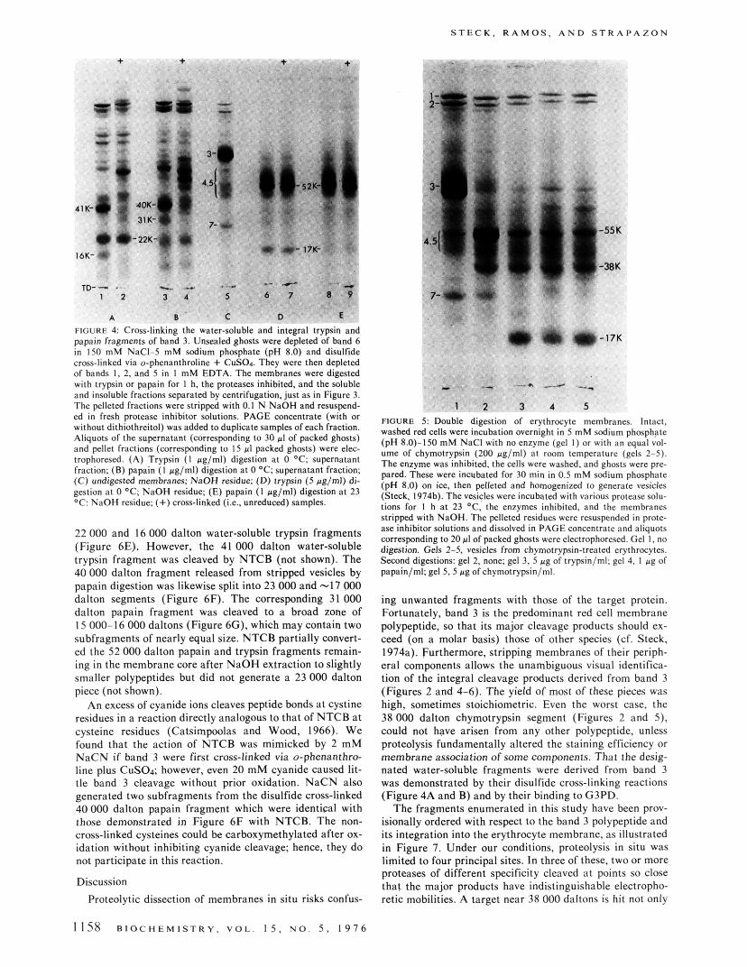
FIGURE 2 Integral fragments of band 3 generated by chymotrypsin. (A) Intact. washed red cells were shaken gently overnight at room tem- perature (23 “C) with an equal volume of 5 mM sodium phosphate (pH S.O)-lSO m M NaCl lacking (gel I) or containing 200 pg/ml of chymotrypsin (gel 2). The enzyme was inhibited, the cells were washed, and ghosts (depleted of band 6) were prepared and cross- linked via o-phenanthraline + CuSOn. Aliguots were dissolved and re- duced in PAGE concentrate and electrophoresed. (B) Undigested ghosts (as in gel I ) were stripped of their peripheral proteins with NaOH and electrophoresed with (gel 3) and without (gel 4) reduction of disulfide linkages. (C) Ghosts from erythrocytes digested as in gel 2 were stripped and electraphomed with (gel 5) and without (gel 6) re- duction. (D) Fresh, unsealed ghosts were cross-linked, then digested for I h at 23 ‘C with chymotrypsin. 100 rg/ml, in 5 mM sodium phos- phate (pH 8.0)-1 m M EDTA. Aliquots were electrophoresed following reduction (gel 7). stripping, and reduction (gel 8). or stripping without reduction (gel 9). Proteins derived from 20 pl of packed ghosts were run on each gel. (+) Cross-linked (Le.. unreduced) ~ a m ~ l e s . 55K. 38K. and 17K denote the fragments of interest.
covery of the 38 000 dalton band seemed low and more variable, presumably because of more extensive degrada- tion. Glycosylation could also contribute to the poor Coom- assie Blue staining of this fragment (Steck and Fox, 1972).
The covalently cross-linked dimer of band 3 persists in NaOH-extracted membranes (Figure 2, gel 4). When ghosts from chymotrypsin-treated erythrocytes were cross- linked and stripped with NaOH, the 38 000 dalton frag- ment was unaffected but the 5 5 000 dalton band dimin- ished; new bands appeared in the 130 000 dalton region (Figure 2, gel 6).
In contrast to intact red cells, the polypeptides of un- sealed ghosts were extensively digested by a brief chymo- trypsin treatment (Figure 2, gel 7). The NaOH residue of these ghosts contained a major fragment of 17 000 & 500 daltons ( n = IO), whose staining intensity was close to that predicted for a cleavage product of band 3 (compare gels 8 and 3). This band was not cross-linked by o-phenanthroline plus CuSO4 (gel 9). Mild treatment of unsealed ghosts with chymotrypsin (e&, 5 fig/ml for I h) also produced a 38 000 dalton integral fragment which appeared to be converted to a -34 000 dalton unit and then to disappear as digestion progressed.
Selectiue Digestion of Band 3 at the Cytoplasmic Sur- face with Trypsin and Papain. Along with the entire Coom- assie Blue profile, band 3 is quite resistant to trypsin diges- tion of intact erythrocytes, resealed ghosts, and right-side-
1156 B I O C H E M I S T R Y , V O L . 1 5 , N O . 5 , 1 9 7 6

P R O T E O L Y T I C D I S S E C T I O N O F A T R A N S M E M B R A N E P R O T E I N
FIGURE 3 Water-soluble fraements releast I from bat bated for I hr in 5 mM sodium phosphate (p 8) with I 0 ' C , (0) papain. I ag/ml, 23 OC. The proteases were of each supernatant fraction were electrophoresed: one sence. One of the latter sets was stained (gels 1-4). whi 50 PI of PAGE concentrate (5 mi") and frozen. The gt min). placed onto fresh SDS gels, and electrophoresed ( ic fragments.
3 by trypsin and papain. Membranes depleted of bands 1. 2, 5, and 6 (gel 13) were incu- se proteases: (A) trypsin, 1 ag/ml. 0 O C ; (B) trypsin. 5 #g/ml, 0 'C: (C) papain, I pglml. hibited and the supernatant fractions collected by a 30-min centrifugation. Three aliquots tin the presence of sodium dodecyl sulfate (SDS) (gels 5 . 7, 9. 11). and two sets in its ab- the unstained duplicates were sliced transversely into 4-mm sections. These were bailed in ections corresponding to the protein peaks visualized in gels 1-4 were thawed (37 OC, IS Is 6.8, IO, 12). Note that more vigorous digestion favored production of Smaller prateolyt-
crose gradients containing only 5 m M sodium phosphate (pH 7.0). This shift was abolished by the presence in the gradient of 150 mM NaCl or 2 m M NADH, agents known to dissociate the G3PD-band 3 complex (Kant and Steck, 1973: Yu and Steck, 1975b).
We verified these results using gel electrophoresis a t low ionic strength in the absence of dodecyl sulfate. The water- soluble fragments generated by trypsin and papain had a high anodal mobility, while purified G3PD remained near the origin or traveled slowly toward the cathode. Mixtures of the fragments and G3PD assumed an intermediate mo- bility. which we attribute to their complexation.
Integral Fragments Generated from Band 3 by Trypsin and Papain. Trypsin and papain digestion of ghosts or de- pleted vesicles also produced fragments of 52 000 f 4000 ( n = 6) and 52 000 f 2000 (n = 4) daltons, respectively. These fragments remained associated with the membranes even after stripping with NaOH or acetic acid (Figure 4, gels 6-9). Their staining intensity and molecular weights indicate that they must be a major split product of hand 3 (which is completely digested under these conditions).
Cross-Linking the Trypsin and Papain Fragments of Band 3. We tested the trypsin and papain fragments for in- terchain disulfide links following oxidation. The 52 000 dal- ton integral fragments were not cross-linked (Figure 4, gels 6-9). The 41 000 and 16 000 dalton trypsin fragments and the 40 000 and 31 000 dalton papain fragments disappeared following 0-phenanthroline-CuSOA treatment and new bands of higher apparent molecular weight appeared reci- procally (Figure 4, gels 1-4). The 22 000 dalton trypsin and papain fragments were not affected by this cross-linking treatment.
Double Digestion of Erythrocyte Membranes. Cbymo- trypsin digestion of intact erythrocytes cleaves band 3 at the
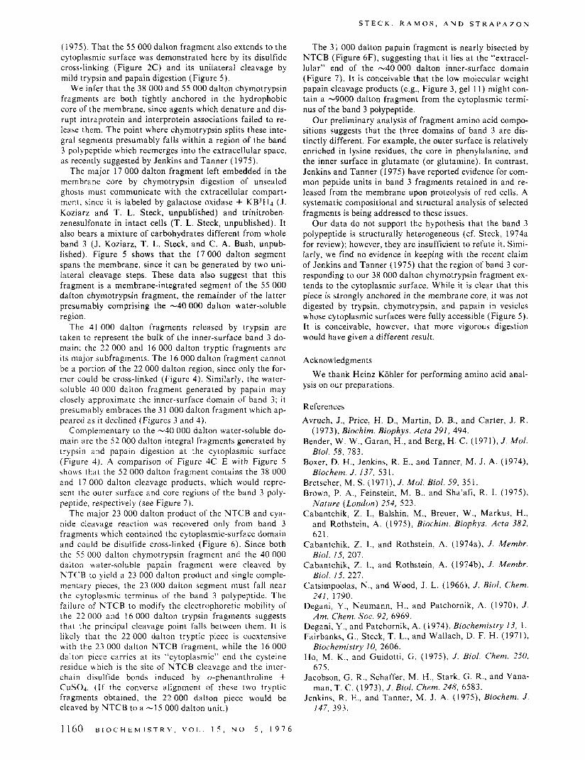
outer surface, while mild trypsin and papain treatment cleaves only at the cytoplasmic surface. We obtained addi- tional information by performing both treatments sequen- tially on the same membranes (Figure 5). As in Figure 2, the first digestion generated 55 000 dalton and 38 000 dal- ton integral fragments from band 3. Trypsin, papain, and chymotrypsin, a t concentrations insufficient to cleave band 3 a t the extracellular surface, split the 55 000 dalton frag- ment and generated a 17 000 dalton integral band indistin- guishable from the 17 000 dalton segment created by chy- motrypsin digestion of unsealed ghosts. The 38 000 dalton chymotrypsin fragment resisted all three proteases in the second digestion.
Cleauage of Band 3 by S-Cyanylation. 2-Nitro-5-thiocy- anobenzoate (NTCB) has been shown to S-cyanylate cyste- ine residues so as to cleave polypeptides a t the N-peptide bond by forming the 2-iminothiazolidine derivative (Jacob- son et al., 1973: Degani and Patchornik. 1974). Band 3 was split by 1 mM NTCB, generating a prominent band of 23 000 f 1000 daltons ( n = 6) in high yield and minor products, particularly a band cluster a t -66 000 daltons (Figure 6A). Increasing the reagent concentration to 45 mM did not alter the cleavage profile significantly, while prior treatment with N-ethylmaleimide or iodoacetate blocked the cleavage reaction.
We delineated the location of the 23 000 dalton NTCB product in terms of the various proteolytic fragments. Of the 55 000 and 38 000 dalton segments generated by chy- motrypsin digestion of intact cells (Figure 2), NTCB cleaved the former (to -35 000 and 23 000 dalton seg- ments, Figure 68) but not the latter (Figure 6C). NTCB did not detectably alter the electrophoretic mobility of the 17 000 dalton fragment left embedded in the membrane fol- lowing chymotrypsin digestion of ghosts (Figure 6D) or the
B I O C H E M I S T R Y , V O L . 1 5 , N O . 5 , 1 9 7 6 1157
S T E C K , R A M O S . A N D S T R A P A Z O N
41
16
FIGURE 4 Cross-linking the water-soluble and integral trypsin and papain fragmenrs of band 3. Unsealed ghosts were depleted of band 6 in 150 m M NaCI-5 m M sodium phosphate (pH 8.0) and disulfide cross-linked via o-phenanthroline + CuSOa. They were then depleted of bands I , 2, and 5 in I mM EDTA. The membranes were digested with trypsin or papain for 1 h, the proteases inhibited, and the soluble and insoluble fractions separated by centrifugation. just as in Figure 3. The pelleted fractions were stripped with 0.1 N NaOH and resuspend- cd in fresh protease inhibitor solutions. PAGE concentrate (with or without dithiothreitol) was added to duplicate samples of each fraction. Aliquots of the supernatant (corresponding to 30 p1 of packed ghosts) and pellet fractions (corresponding to I5 pl packed ghosts) were elec- trophoresed. (A) Trypsin ( I pg/ml) digestion at 0 'C: supernatant fraction: (B) papain ( I pg/ml) digestion a t 0 'C , supernatant fraction; (C) undigested membranes: NaOH residue; (D) trypsin (5 @g/ml) di- gestion a t 0 'C; NaOH residue; (E) papain ( I pglml) digestion a t 23 O C : NaOH residue: (+)cross-linked (i.e.. unreduced) samples.
22 000 and 16 000 dalton water-soluble trypsin fragments (Figure 6E). However, the 41 000 dalton water-soluble trypsin fragment was cleaved by NTCB (not shown). The 40 000 dalton fragment released from stripped vesicles by papain digestion was likewise split into 23 000 and -I7 000 dalton segments (Figure 6F). The corresponding 31 000 dalton papain fragment was cleaved to a broad zone of 15 000-16 000 daltons (Figure 6G), which may contain two subfragments of nearly equal size. NTCB partially convert- ed the 52 000 dalton papain and trypsin fragments remain- ing in the membrane core after NaOH extraction to slightly smaller polypeptides but did not generate a 23 000 dalton piece (not shown).
An excess of cyanide ions cleaves peptide bonds at cystine residues in a reaction directly analogous to that of NTCB a t cysteine residues (Catsimpoolas and Wood, 1966). We found that the action of NTCB was mimicked by 2 mM NaCN if band 3 were first cross-linked via o-phenanthro- line plus CuS04: however, even 20 mM cyanide caused lit- tle band 3 cleavage without prior oxidation. NaCN also generated two subfragments from the disulfide cross-linked 40 000 dalton papain fragment which were identical with those demonstrated in Figure 6 F with NTCB. The non- cross-linked cysteines could be carboxymethylated after ox- idation without inhibiting cyanide cleavage: hence, they do not participate in this reaction.
Discussion Proteolytic dissection of membranes in situ risks confus-
1158 B I O C H E M I S T R Y . V O L 1 5 , N O 5 , 1 9 7 6
FIGURE 5: Double digestion of erythrocyte membranes. Intact. washed red cells were incubation overnight in 5 mM sodium phasphge (pH 8.0)-150 mM NaCl with no enzyme (gel I ) or with an q u a l vol- ume of chymotrypsin (200 pglml) at room temperature (gels 2-5). Thc enzyme was inhibited, the cells were washed. and ghosts were pre- pared. These were incubated for 30 mi" in 0.5 mM scdium phosphate (pH 8.0) on ice. then pelleted and homogenized to generate vesicles (Steck. 1974b). The vesicles were incubated with various protease s o h t ims for I h at 23 'C. the enzymes inhibited. and the membranes stripped with NaOH. The pelleted residues were resuspended in prote- ase inhibitor solutions and dissolved in PAGE concentrate and aliquots corresponding to 20 pl of packed ghosts were electrophoresed. Gel I, no digestion. Gels 2-5. vesicles from chymotrypsin-treatcd erythrocytes. Second digestions: gel 2, none; gel 3, 5 ,tg of trypsin/ml: gel 4, I pg of papain/ml: gel 5, 5 pg of chymotrypsin/ml
ing unwanted fragments with those of the target protein. Fortunately, band 3 is the predominant red cell membrane polypeptide, so that its major cleavage products should ex- ceed (on a molar basis) those of other species (cf. Steck, 1974a). Furthermore, stripping membranes of their periph- eral components allows the unambiguous visual identifica- tion of the integral cleavage products derived from band 3 (Figures 2 and 4-6). The yield of most of these pieces was high, sometimes stoichiometric. Even the worst case, the 38 000 dalton chymotrypsin segment (Figures 2 and 5) . could not have arisen from any other polypeptide, unless proteolysis fundamentally altered the staining efficiency or membrane association of some components. That the desig- nated water-soluble fragments were derived from band 3 was demonstrated by their disulfide cross-linking reactions (Figure 4A and B) and by their binding to G3PD.
The fragments enumerated in this study have been prov- isionally ordered with respect to the band 3 polypeptide and its integration into the erythrocyte membrane, as illustrated in Figure 7. Under our conditions, proteolysis in situ was limited to four principal sites. In three of these, two or more proteases of different specificity cleaved a t points so close that the major products have indistinguishable electropho- retic mobilities. A target near 38 000 daltons is hit not only

P R O T E O L Y T I C D I S S E C T I O N O F A T R A N S M E M B R A N E P R O T E I N
FIGURE 6: 2-Nitr~5-thiocyanobenroate (NTCB) cleavage of band 3 and its proteolytic fragments. (A) Band 3: ghosts were stripped and 100-pl al- iquots of the neutralized residue were dissolved by the addition of 25 pl of 4% dadecyl sulfate. 50 rl of 10 mM sodium phosphate (pH 8.0) contain- ing 0 (gel I ) or 4 mM NTCB (gel 2) was added and the mixture incubated a t 23 for 15 min. Then 25 pl of 500 mM sodium borate (pH 9.0) was added and the mixtures were incubated overnight a t 31 OC (final NTCB concentration, I mM). The reaction was terminated by the addition of 50 pl of PAGE concentrate (which contains an excess of dithiothreitol) and 100 pl of each mixture was electrophoresed. (B) The 55 000 daltun frag- ment generated by chymotrypsin digestion of intact cells (Figure 2, gel 3) was purified by preparative PAGE in dodecyl sulfate. dialyzed and react- ed as in (A). (C) The 38 W O dalton fragment generated by chymotrypsin digestion of intact cells (see Figure 2, gel 3) was prepared and rcacted as in (B). (D) The 17 000 dalton fragment generated by chymotrypsin digestion of unsealed ghosts (see Figure 2. gel 8) was prepared and reacted as i n (B). (E) The soluble fragments released from depleted vesicles by trypsin (as in Figure 3B) were reacted with 0 (gel 9) or 0.5 mM NTCB (gel IO) as in (A). (A similar negative result is obtained using higher NTCB concentrations.) (F) The soluble fragments released by papain digestion (as in Figure 4C) were treated with iodaacetate (gel 1 I ) or NTCB (0.5 mM final: gel 12) as in (A). NTCB inhibited the papain (Degani et al., 1970) while it modified the fragments for cleavage. (G) The soluble fragments released from depleted vesicles by papain digestion (as in Figure 3D) were treated with iadoaeetate (gel 13) or 1 mM NTCB (gcl 14) as in (A). (+) NTCB treatment.
by chymotrypsin (Figures 2 and 5) . but also by Pronase (Bender et al., 1971: Bretscher, 1971; Steck, 1972b; Cab- antchik and Rothstein, 1974b). papain (this study), thermo- lysin, and subtilisin (Jenkins and Tanner, 1975). While mo- lecular weight assignments differed among laboratories, the fragments generated by two or more of these enzymes in a given laboratory coelectrophoresed. Similarly, mild chymo- trypsin (not shown), trypsin, and papain digestion of the cy- toplasmic surface (Figure 4) created indistinguishable 52000 dalton integral fragments. The third case is the water-soluble 40 000-41 000 dalton and 22 000 dalton fragments generated by both trypsin and papain. This pat- tern of restricted cleavage in situ is lost upon solubilization in nondenaturing or denaturing detergents. These data suggest that regions of high accessibility within band 3 con- nect portions under conformational or steric protection (e.g., by the head groups or apolar tails of membrane lip- ids).
The 55 000 dalton and 38 000 dalton segments generated by chymotrypsin attack on intact erythrocytes sum to the molecular weight of band 3 [91 000 i 4000 (n = 14) in this study]. Both fragments must communicate with the extra- cellular space, if the protease does not penetrate the mem- brane barrier. In support of this inference, we found (T. L. Steck and B. Ramos, unpublished data) that both frag- ments were labeled by galactose oxidase + KB’H.,, which tritiates band 3 carbohydrates only a t the extracellular sur- face (Steck and Dawson, 1974; Yu and Steck, 1975a). Both were also labeled in intact cells by trinitrobenzenesulfonate (as in Steck, 1972b). while the polypeptides and band 3 fragments confined to the cytoplasmic surface were not
. . I , I , , . , . OUTSIDE CORE INSIDE
3AND 3 91 SH
CHYMOTRYPSIN -38- -55- -17-
I-CYANYLATION -23-
4 4 4 4 0 20 40 60 80
MOLECULAR WEIGHT x IO-’ FIGURE 1 A possible ordering of band 3 fragments. Eleven fragments of interest were aligned without gaps or overlap to an oriented. 93 000 dalton band 3 polypeptide. Bar lengths as drawn deviate by no more than +2WO from measured molecular weights. Arrows designate the principal p i n t s of proteolytic attack: the -SH group on band 3 suggests a likely p i t i o n for the cysteine residue($) involved in cross- linking and S-eyanylation. Certain fragments observed in this study are not designated here, but arc compatible with this formulation.
tagged. These labeling studies are consistent with those of Bender et al. (1971). Bretscher (1971). Cabantchik and Rothstein (1974b). Whiteley and Berg (1974). Boxer et al. (1974). Cabantchik et al. (1975). and Jenkins and Tanner
B I O C H E M I S T R Y , V O L . 1 5 . N O . 5 . 1 9 7 6 1159
(1975). That the 55 000 dalton fragment also extends to the cytoplasmic surface was demonstrated here by its disulfide cross-linking (Figure 2C) and its unilateral cleavage by mild trypsin and papain digestion (Figure 5).
We infer that the 38 000 and 55 000 dalton chymotrypsin fragments are both tightly anchored in the hydrophobic core of the membrane, since agents which denature and dis- rupt intraprotein and interprotein associations failed to re- lease them. The point where chymotrypsin splits these inte- gral segments presumably falls within a region of the band 3 polypeptide which reemerges into the extracellular space, as recently suggested by Jenkins and Tanner (1975).
The major 17 000 dalton fragment left embedded in the membrane core by chymotrypsin digestion of unsealed ghosts must communicate with the extracellular compart- ment, since it is labeled by galactose oxidase + KB3H4 (J. Koziarz and T. L. Steck, unpublished) and trinitroben- zenesulfonate in intact cells (T. L. Steck, unpublished). It also bears a mixture of carbohydrates different from whole band 3 (J. Koziarz, T. L. Steck, and C. A. Bush, unpub- lished). Figure 5 shows that the 17 000 dalton segment spans the membrane, since it can be generated by two uni- lateral cleavage steps. These data also suggest that this fragment is a membrane-integrated segment of the 55 000 dalton chymotrypsin fragment, the remainder of the latter presumably comprising the -40 000 dalton water-soluble region.
The 41 000 dalton fragments released by trypsin are taken to represent the bulk of the inner-surface band 3 do- main; the 22 000 and 16 000 dalton tryptic fragments are its major subfragments. The 16 000 dalton fragment cannot be a portion of the 22 000 dalton region, since only the for- mer could be cross-linked (Figure 4). Similarly, the water- soluble 40 000 dalton fragment generated by papain may closely approximate the inner-surface domain of band 3; it presumably embraces the 3 1 000 dalton fragment which ap- peared as it declined (Figures 3 and 4).
Complementary to the -40 000 dalton water-soluble do- main are the 52 000 dalton integral fragments generated by trypsin and papain digestion a t the cytoplasmic surface (Figure 4). A comparison of Figure 4C-E with Figure 5 shows that the 52 000 dalton fragment contains the 38 000 and 17 000 dalton cleavage products, which would repre- sent the outer surface and core regions of the band 3 poly- peptide, respectively (see Figure 7).
The major 23 000 dalton product of the NTCB and cya- nide cleavage reaction wac recovered only from band 3 fragments which contained the cytoplasmic-surface domain and could be disulfide cross-linked (Figure 6). Since both the 55 000 dalton chymotrypsin fragment and the 40 000 dalton water-soluble papain fragment were cleaved by NTCB to yield a 23 000 dalton product and single comple- mentary pieces, the 23 000 dalton segment must fall near the cytoplasmic terminus of the band 3 polypeptide. The failure of NTCB to modify the electrophoretic mobility of the 22 000 and 16 000 dalton trypsin fragments suggests that the principal cleavage point falls between them. It is likely that the 22 000 dalton tryptic piece is coextensive with the 23 000 dalton NTCB fragment, while the 16 000 dalton piece carries a t its “cytoplasmic” end the cysteine residue which is the site of NTCB cleavage and the inter- chain disulfide bonds induced by o-phenanthroline + CuSO4. (If the converse alignment of these two tryptic fragments obtained, the 22 000 dalton piece would be cleaved by NTCB to a -I5 000 dalton unit.)
S T E C K , R A M O S , A N D S T R A P A Z O N
The 31 000 dalton papain fragment is nearly bisected by NTCB (Figure 6F), suggesting that it lies a t the “extracel- lular’’ end of the -40 000 dalton inner-surface domain (Figure 7). It is conceivable that the low molecular weight papain cleavage products (e.g., Figure 3, gel 1 1 ) might con- tain a -9000 dalton fragment from the cytoplasmic termi- nus of the band 3 polypeptide.
Our preliminary analysis of fragment amino acid compo- sitions suggests that the three domains of band 3 are dis- tinctly different. For example, the outer surface is relatively enriched in lysine residues, the core in phenylalanine, and the inner surface in glutamate (or glutamine). In contrast, Jenkins and Tanner (1975) have reported evidence for com- mon peptide units in band 3 fragments retained in and re- leased from the membrane upon proteolysis of red cells. A systematic compositional and structural analysis of selected fragments is being addressed to these issues.
Our data do not support the hypothesis that the band 3 polypeptide is structurally heterogeneous (cf. Steck, l974a for review); however, they are insufficient to refute it. Simi- larly, we find no evidence in keeping with the recent claim of Jenkins and Tanner (1975) that the region of band 3 cor- responding to our 38 000 dalton chymotrypsin fragment ex- tends to the cytoplasmic surface. While it is clear that this piece is strongly anchored in the membrane core, it was not digested by trypsin, chymotrypsin, and papain in vesicles whose cytoplasmic surfaces were fully accessible (Figure 5). It is conceivable, however, that more vigorous digestion would have given a different result.
Acknowledgments
ysis on our preparations. We thank Heinz Kohler for performing amino acid anal-
References Avruch, J., Price, H. D., Martin, D. B., and Carter, J . R.
Bender, W . W., Garan, H., and Berg, H . C. (1971), J . Mol.
Boxer, D. H., Jenkins, R. E., and Tanner, M. J. A. (1974),
Bretscher, M. S. (1971), J . Mol. Biol. 59, 351. Brown, P. A,, Feinstein, M. B., and Sha’afi, R . I . (1975),
Nature (London) 254, 523. Cabantchik, Z. I., Balshin, M., Breuer, W., Markus, H.,
and Rothstein, A. (1975), Biochim. Biophys. Acta 382, 621.
Cabantchik, Z. I., and Rothstein, A. (1974a), J . Membr. Biol. 15, 207.
Cabantchik, Z. I., and Rothstein, A. (1974b), J . Membr. Biol. 15, 227.
Catsimpoolas, N., and Wood, J . L. (1966), J . Biol. Chem. 241, 1790.
Degani, Y. , Neumann, H., and Patchornik, A. (1970), J . Am. Chem. SOC. 92, 6969.
Degani, Y . , and Patchornik, A. (1974), Biochemistry 13, 1 . Fairbanks, G., Steck, T. L., and Wallach, D. F. H. (1971),
Ho, M. K., and Guidotti, G. (1975), J . Biol. Chem. 250,
Jacobson, G . R., Schaffer, M. H., Stark, G. R., and Vana-
Jenkins, R. E., and Tanner, M. J. A. (1975), Biochem. J .
(1973), Biochim. Biophys. Acta 291, 494.
Biol. 58, 783.
Biochem. J . 137, 53 1.
Biochemistry 10, 2606.
675.
man, T. C. (1973), J . Biol. Chem. 248, 6583.
147, 393.
1160 B I O C H E M I S T R Y , V O L . 1 5 , U O . 5 , 1 9 7 6
S E L F - A S S O C I A T I O N O F A P O L I P O P R O T E I N A - I 1
Kant, J. A., and Steck, T. L. (1973), J . Biol. Chem. 248,
Lin, S., and Spudich, J. A. (1975), Biochem. Biophys. Res.
Schubert, D. (1973), Hoppe-Seyler's Z . Physiol. Chem.
Shin, B. C., and Carraway, K. L. (1974), Biochim. Bio-
Steck, T. L. (1972a), J . Mol. Biol. 66, 295. Steck, T. L. (1972b), in Membrane Research, Fox, C. F.,
Steck, T. L. (1974a), J . Cell Biol. 62, 1. Steck, T. L. (1974b), Methods Membr. Biol. 2, 245. Steck, T. L., and Dawson, G. (1974), J . Biol. Chem. 249,
2135. Steck, T. L., and Fox, C. F. (1972), in Membrane Molecu-
lar Biology, Fox, C. F. and Keith, A. D., Ed., Stamford, Conn., Sinauer Assoc., p 27.
Steck, T. L., Weinstein, R. S., Straus, J. H., and Wallach,
8457.
Commun. 61, 147 1.
354, 781.
phys. Acta 345, 141.
Ed., New York, N.Y., Academic Press, p 71.
D. F. H. (1970), Science 168, 255.
220.
in press.
129, 333.
phys. Res. Commun. 54. 593.
1 1 , 2897.
8005.
4406.
541.
Steck, T. L., and Yu, J. (1973), J . Supramol. Struct. I ,
Strapazon, E., and Steck, T. L. (1976), Biochemistry 15,
Tanner, M. J. A,, and Boxer, D. H. (1972), Biochem. J .
Taverna, R. D., and Langdon, R. G. (1973), Biochem. Bio-
Triplett, R. B., and Carraway, K. L. (1972), Biochemistry
Wang, K., and Richards, F. (1974), J . Biol. Chem. 249,
Weber, K., and Osborn, M. (1969), J . Biol. Chem. 244.
Whiteley, N. M., and Berg, H. C. (1974), J . Mol. Biol. 87,
Yu, J., andSteck, T. L. (1975a), J. Biol. Chem. 250, 9170. Yu, J., and Steck, T. L. (1975b), J . Biol. Chem. 250, 9176,
Studies on Human Serum High-Density Lipoproteins. Self- Association of Human Serum Apolipoprotein A-I1 in Aqueous Solutionst
Lidia B. Vitellot and Angelo M. Scanu*s§
ABSTRACT: Some of the solution properties of pure prepa- rations of human serum high-density apolipoprotein A-I1 were studied by sedimentation equilibrium ultracentrifuga- tion, conducted at different apoprotein concentrations and at several speeds. The concentration dependence of the ap- parent weight average molecular weight indicated that apo- lipoprotein A-11, when dissolved in 0.02 M EDTA (pH 8.6),
Apo l ipopro te in A-II (apo-A-11)' is one of the major components of the human serum high-density lipoprotein (HDL) class (Morrisett et al., 1975; Scanu et al., 1975).
+ From the Departments of Medicine and Biochemistry, the Univer- sity of Chicago Pritzker School of Medicine, and the Franklin McLean Memorial Research Institute (operated by the University of Chicago for the United States Energy Research and Development Administra- tion), Chicago, Illinois 60637. Received October I, 1975. Supported by Grants HL-08727 from the United States Public Health Service, A72-6 from the Chicago and Illinois Heart Association, and Contract E( 1 1 - l)-69 from the U S . Energy Research and Development Admin- istration. Presented in part at the 59th Annual Meeting of the Federa- tion of American Societies for Experimental Biology, Atlantic City, N.J., April 13-18, 1975 (Vitello et al., 1975). * Recipient of US. Public Health Service Postdoctoral Fellowship HL-52,752. Present address: Northwestern University School of Medi- cine, Chicago, Ill. , and the VA Hospital, Research-in-Aging Laborato- ry. Downey, 111.
During the period of this work, A.M.S. was the recipient of U S . Public Health Service Research Career Development Award No. HL- 24,867.
' Abbreviations used are: HDL, high-density lipoproteins; apo-HDL, apolipoprotein of HDL; apo A-11, one of the two major polypeptides of apo-H DL.
undergoes self-association. Over a protein concentration range between 0.8 and 1.5 mg/ml, the self-association could best be described by a monomer-dimer-trimer step associa- tion, although indefinite self-association could not be ruled out. The equilibrium constants obtained were sufficient to describe the system over the concentration range investi- gated.
The chemical properties of this apoprotein are known, in- cluding its amino acid sequence (Brewer et al., 1972); it has a molecular weight of 17 400 and is made up of two identi- cal polypeptide chains linked together at the sixth position from the amino terminal by a single disulfide bridge. It is now established that apo A-I1 retains binding capacity for lipids in vitro. Several investigations have been conducted on the definition of the molecular properties of this apopro- tein with reference to its lipid binding (Assmann and Brew- er, 1974; Makino et al., 1974; Morrisett et al., 1975; Reyn- olds and Simon, 1974; Stoffel et al., 1974; Vitello et al., 1975).
Even though a large amount of information is available on the properties of apo A-11, very little is known on the be- havior of this polypeptide in aqueous solutions. In this re- port, we present data indicating that apo A-I1 tends to self- associate in aqueous solution.
Materials and Methods
Preparation of Apo A-I I and Assessment of Purity. Human serum HDL ( p 1.063-1.21 g/ml) were separated
B I O C H E M I S T R Y , V O L . 1 5 , N O . 5 , 1 9 7 6 1161
Copyright © 2022 FDOKUMEN