Liposomes for malaria management: the evolution from 1980 ...
Upload
independentCategory
view
2download
0
EFFECT OF STREPTOLYSIN O ON ERYTHROCYTE
MEMBRANES, LIPOSOMES, AND LIPID DISPERSIONS
A Protein-Cholesterol Interaction
JAMES L. DUNCAN and RICHARD SCHLEGEL
From the Departments of Microbiology and Pathology, Northwestern University Medical and Dental Schools, Chicago, Illinois 60611. Dr. Schlegel's present address is the Department of Pathology, Peter Bent Brigham Hospital, Harvard University Medical School, Boston, Massachusetts 02115.
ABSTRACT
The effect of the bacterial cytolytic toxin, streptolysin O (SLO), on rabbit erythrocyte membranes, liposomes, and lipid dispersions was examined. SLO produced no gross alterations in the major erythrocyte membrane proteins or lipids. However, when erythrocytes were treated with SLO and examined by electron microscopy, rings and "C"-shaped structures were observed in the cell membrane. The rings had an electron-dense center, 24 nm in diameter, and the overall diameter of the structure was 38 nm. Ring formation also occurred when erythrocyte membranes were fixed with glutaraldehyde and OsO4 before the addition of toxin. In contrast, rings were not seen when erythrocytes were treated with toxin at 0~ indicating that adsorption of SLO to the membrane is not sufficient for ring formation since toxin is known to bind to erythrocytes at that temperature. The ring structures were present on lecithin-cholesterol-dicetylphos- phate liposomes after SLO treatment, but there was no release of the trapped, internal markers, K2CrO4 or glucose. The crucial role of cholesterol in the formation of rings and C's was demonstrated by the fact that these structures were present in toxin-treated cholesterol dispersions, but not in lecithin-dicetylphosphate dispersions nor in the SLO preparations alone. The importance of cholesterol was also shown by the finding that no rings were present in membranes or cholesterol dispersions which had been treated with digitonin before SLO was added. Al- though the rings do not appear to be "holes" in the membrane, a model is pro- posed which suggests that cholesterol molecules are sequestered during ring and C-structure formation, and that this process plays a role in SLO-induced hemoly- sis.
Streptolysin O (SLO) is a bacterial toxin produced by virtually all strains of Streptococcus pyogenes. The toxin is a protein with a molecular weight of approximately 60,000 daltons, and is characteristic of a group of cytolytic toxins known as the oxygen-labile toxins (3). The toxins in this group are produced by several different gram-positive
bacteria, and possess a number of common proper- ties: they are activated by SH compounds: they appear to be antigenically related; and their bio- logical activity is completely inhibited by low concentrations of cholesterol and certain related sterols.
Hemolysis occurs within minutes after the addi-
160 THE JOURNAL OF CELL BIOLOGY �9 VOLUME 67, 1975 �9 pages 160-173
on July 30, 2015jcb.rupress.org
Dow
nloaded from
Published October 1, 1975

tion of SLO to erythrocytes, and toxic effects of SLO on several types of mammal ian cells in culture have been demonstrated (13). The speed with which sensitive cells are affected by the toxin suggests that the cell membrane is the primary site of action. Several lines of indirect evidence suggest that membrane cholesterol is the binding site and /o r target of S L O action. Only those cells which contain cholesterol in their membranes are susceptible to the toxin, SLO is " inact ivated" only by the membrane lipid fraction which contains cholesterol, and the addition of exogenous choles- terol to SLO inhibits toxin action. Shany and co-workers (24) recently provided evidence that SLO does not adsorb to erythrocyte membranes which have been treated with alfalfa saponin or the polyene antibiotic, filipin, agents known to bind to
cholesterol in the membrane. Despite these obser- vations, it is not known how SLO produces the membrane alterations which result in lysis or death of sensitive cells.
When red ceils are treated with certain hemo- lytic agents such as saponin, filipin, or antibody plus complement, characteristic rings or holes are formed in the erythrocyte membrane (reviewed by Seeman, reference 22). The rings are readily seen in electron micrographs of negatively stained membrane preparations, but their relationship to the lytic process is not clear. Freeze-etch studies on saponin-treated erythrocytes, however, suggest that this substance produces an actual hole in the membrane (23). Several years ago, Dourmashkin and Rosse (9) reported that SLO produced "holes" with a 50-nm diameter in erythrocyte membranes. The toxin preparation which they used was not described, however, and the conditions necessary for the formation of the membrane lesions were not reported.
We have studied the effects of SLO on the major erythrocyte membrane proteins and lipids, and have examined the lesions produced by the toxin on red cells, ghosts, liposomes, and cholesterol dispersions. A model is proposed to account for the ringlike structures which are observed.
M A T E R I A L S A N D M E T H O D S
Toxin Preparation
The Richards strain (type 3) of S. pyogenes was used for SLO production. Culture supernates (20-24 h) of this organism, grown in trypticase soy broth-yeast extract (3:1) dialysate medium supplemented with 0.5% glucose and Na~HPO, were harvested and brought to 70% saturation with solid (NH4)3SO,. The precipitate was
collected and resuspcnded in 0.05 M Tris buffer, pH 8.4. The crude toxin preparation was applied to a 2.5 • 100-cm G-100 Sephadex column with a bed volume of 400 ml, which had been equilibrated with Tris buffer at 4oC. The toxin was eluted and the active fractions were pooled and applied to a 1.5 • 30-cm DEAE-Sephadex column equilibrated in the same Tris buffer. The column was washed with 400-500 ml of buffer, and SLO was eluted by the addition of a 0.3 M NaCI gradient. The toxin had a specific activity of approximately 200,000 hemolytic units (HU) per mg protein, and gave a single protein band on disc gel electrophoresis. The toxin was activated by the addition of 10 mM cysteine, and stored at -70~ SLO activity was assayed as previously described (10).
Erythrocytes
Rabbit blood was collected from New Zealand white rabbits or purchased locally from a veterinarian. The blood was centrifuged at 2,000 g and the plasma and buffy coat were removed by aspiration. The cells were washed at least three times with phosphate-buffered saline (PBS; NaC1, 137 raM; Na2HPO,, 8 raM; KH2PO,, 2 raM; pH 7.2).
Analysis of Erythrocyte Membrane Components
Washed rabbit erythrocytes were suspended in PBS to a concentration of 40-50%, and 0.1-0.2 ml of SLO (500-2,000 HU) or control solution (consisting of Tris buffer plus cysteine) was added to 2-4 ml of the cell suspension. Other controls included the addition of cholesterol-inactivated SLO, SLO neutralized by an- titoxin, or heat-inactivated toxin. The cell suspensions were incubated at 37~ for 0.5, I, 3, 5, or 17 h; lysis of suspensions treated with active toxin was apparent in less than 5 rain. After incubation, the erythrocytc membranes were isolated and washed in 5 mM Na2HPO~ buffer, pH 8, as described by Fairbanks et al. (l l).
MEMBRANE PROTEINS
The washed membranes were solubilized in 2% sodium dodecylsulfate (SDS) at 100~ for 3 rain (16). A 0.l-ml sample of solubilized membranes was added to an equal volume of a solution containing 5% (wt/vol) SDS, 160 mM dithiothreitol, 50 mM Tris-HCl (pH 8). 20 .g /ml pyronin Y, 50% (wt/vol) sucrose, and 5 mM EDTA, and the major erythrocyte membrane proteins were analyzed by electrophoresis on 5.6% polyacrylamide gels as de- scribed by Fairbanks and co-workers (11).
In some experiments, the membrane proteins were cross-linked by incubating toxin-treated or control mem- branes in 5 mM phosphate buffer with an equal volume of phosphate buffer containing 0, 2, or 4 mM glutaralde- hyde. After 20 min at room temperature, the membranes were washed and solubilized by the procedure of Steck (28). The proteins were analyzed on 3% polyacrylamide gels.
DUNCAN AND SCHLEGEL Effects of Streptolysin 0 161
on July 30, 2015jcb.rupress.org
Dow
nloaded from
Published October 1, 1975

MEMBRANE LIPIDS
Washed membranes of SLO-treated or control red cells were extracted in CHCl3-methanol with procedure III of Ways and Hanahan (30). The final lipid extract was resuspended in 1 ml benzene and stored at -20~
The membrane lipids were analyzed by thin-layer chromatography (TLC) on silica gel 60 plates (Merck), by the procedures described by Skipsky (26). Phospho- lipids were analyzed with a solvent system of CHCls- methanol-acetic acid-H~O (25:15:4:2). For neutral lipids, the plates were run first in isopropyl ether-acetic acid (96:4), then in petroleum ether-diethyl ether-acetic acid (90:10:1). The plates were developed by spraying with H2SO~ and charring at 300~ or 15 min.
Liposome Preparation
Liposomes composed of phosphatidylcholine (30 mR), cholesterol (15 mR), and dicetylphosphate (3 mR) were prepared by mixing CHCIs solutions of the lipid compo- nents and drying them under a vacuum. I ml of 0.1 M K2CrO, or 0.3 M glucose was added to the dried lipids and the solution was incubated at room temperature with occasional Vortexing. The mixture was sonicated for 5 s with a Model L Branson sonifier at a setting of 4. The liposomes were separated from free K~CrO4 or glucose by passing the mixture through a small G-50 Sephadex column equilibrated with PBS. The liposomes, which are eluted in the void volume, were collected and dialyzed against PBS.
The effect of SLO on the leakage of chromate or glucose from the liposomes was tested by placing 0.5 ml of liposomes in a small dialysis bag suspended in a scintillation vial containing 10 ml PBS. The escape of the markers from within the liposomes after SLO or Triton X-100 addition was assayed by taking periodic samples of the PBS surrounding the dialysis bag. Chromate was determined by absorbance at 370 nm, and glucose with the Glucostat reagent. Liposomes which were used in electron microscopy studies were prepared in 0.1 M K~CrO, or PBS as described above.
Dispersions of cholesterol (35 mg/ml) or phos- phatidylcholine (15 mg/ml) plus dicetylphosphate (3 mg/ml) were prepared by drying CHCIs solutions of the lipids under a vacuum and resuspending in PBS. The suspensions were sonicated for 3 min at a setting of 4.
Electron Microscopy Studies
SAMPLE PREPARATION
The sequence of steps in sample preparation, fixing and staining for each experiment is shown in Table I.
ERYT~ROCYTES: A 40-~1 sample of a 50% suspen- sion of washed red cells in PBS was incubated with 20/~1 of SLO (80-100 HU) or control solution at 37~ for 10 rain. A 20-ul sample of the mixture was then added to a l-ml drop ofdistilled water, by the technique of Nicolson and Singer (17), in which some of the red cell ghosts are
dispersed over the air-water interface. A 400-mesh, Formvar-carbon-coated copper grid was gently applied to the surface of the drop for I min. Excess fluid was removed from the grid by touching the edge with filter paper.
GHOSTS: A 20-#1 sample of an untreated 50% red cell suspension was added to a 1-ml drop of water, and the ghosts were taken up on grids as described above. The ghosts were then treated with toxin by floating the grids on 20-#1 droplets of SLO solutions for 10 rain at 37~
LIPOSOMES AND LIPID DISPERSIONS: Liposomes were diluted 1:50 in PBS, and a 20-~1 sample was mixed with 40 #1 of SLO (200 HU) or control solution and incubated at 37 C for 10 min. Grids were then allowed to float on the mixture for 30 s and the excess fluid was removed with filter paper. In some experiments the reaction mixture was added directly to the grids. Cholesterol dispersions diluted 1:50 in PBS, or undiluted phos- phatidylcholine-dicetylphosphate dispersions were treated with SLO and applied to the grids in the same way.
FIXATION AND STAINING: The samples to be fixed were incubated with 2.5% glutaraldehyde and/or 1.0% osmium tetroxide (in 0.1 M phosphate buffer, pH 7.3) for 20 min at room temperature. The grids were then washed exhaustively by 120 immersions in 100 ml PBS, and negatively stained for 30 s with 1.0% phosphotungstic acid (PTA) adjusted to pH 6.5 with KOH. In some experiments the samples were stained with 0.1% uranyl acetate (pH unadjusted).
In several experiments, washed postfixation samples were exposed to biological substances, e.g., trypsin or SLO, before negative staining with PTA.
ELECTRON MICROSCOPY Sample grids were examined in a Philips EM-200
electron microscope at 60 keV with an aperture size of 20 #m. A liquid nitrogen sample anticontamination de- vice was utilized. Micrographs were usually taken at a magnification of 41,000 with slight underfocus settings to enhance contrast, and were further magnified on a Durst enlarger.
MICROGRAPH MEASUREMENTS
The size of the rings observed in SLO-treated prepara- tions was estimated by using micrographs taken at a magnification of 41,000 and then enlarged 3.5 times. The inside diameter of 10 well-visualized rings was used to compute the average inside diameter. Likewise, the length of the "C" structures observed in toxin-cholesterol preparations was determined by measuring 10 "C'" structures (using dental floss to achieve accurate mea- surement of the semicircular forms) and then computing the average length.
Other Materials
Chromatographically pure egg lecithin, phosphatidyl- ethanolamine, phosphatidyiserine, phosphatidylinositol,
162 THE JOURNAL OF CELL BIOLOGY . VOLUME 67, 1975
on July 30, 2015jcb.rupress.org
Dow
nloaded from
Published October 1, 1975

lysolecithin, and sphingomyelin were purchased from General Biochemicals, Chagrin Falls, Ohio. Cholesterol, dicetylphosphate, trypsin, and pronase were obtained from Sigma Chemical Co., St. Louis, Mo. Glucostat reagent was purchased from Worthington Biochemical Corp., Freehold, N. J.
RESULTS
Effect o f SLO on Erythrocyte Membrane Proteins and Lipids
Although the mechanism of action of most cytolytic toxins is not known, a few have been shown to possess enzymatic activity for mem- brane lipids (3). The possibility that SLO enzymatically alters or in some way extracts a major membrane constituent was examined by treating erythrocyte suspensions with high concen- trations of SLO for up to 17 h. In the experiment shown in Fig. l a, erythrocytes were treated with 1,000 or 2,000 HU of SLO or control solution for 0.5 h. The cell membranes were solubilized in SDS and the membrane proteins analyzed by PAGE. It can be seen that there is no alteration in the membrane proteins of toxin-treated erythrocytes. Similar results were obtained with different toxin concentrations and incubation times of 1, 3, 5, or 17 h. The fastest moving band, more prominent in the control membrane preparation, is hemoglobin; SLO-lysed cells were consistently found to have less hemoglobin associated with the washed mem- branes.
Although SLO did not affect the structure of the membrane polypeptides themselves, the possibility that the spatial arrangements of these proteins in the membrane might be altered was examined. Membranes from red cells treated with 440 HU SLO or control solution for 1.25 h were isolated and the membrane proteins cross-linked by the addition of glutaraldehyde (28). The results in Fig. 1 b reveal no differences in the toxin-treated and control membrane proteins.
The effect of SLO on erythrocyte membrane lipids was examined in similar experiments. The lipids were extracted from toxin-treated and con- trol cells and analyzed by thindayer chromatogra- phy. There appeared to be no alterations in the membrane lipid constituents of red cells treated with 400 HU of SLO for 1 h; similar results were obtained with cells incubated with higher concen- trations of toxin for up to 17 h.
Effect of SLO on Erythrocyte
Membrane Structure
Despite the absence of gross alterations in the membrane components, the effect of SLO on red cell membrane structure was investigated. Red cells were treated with SLO or control solutions; the membranes were placed on a water droplet and applied to a grid as described in Materials and Methods. Membranes from control cells which had been osmotically lysed on the water droplet, then fixed and stained, had a normal appearance, as seen in Fig. 2c. In toxin-lysed red cells, however, ringlike structures were readily apparent on the membrane surface (Fig. 2 b, c). The number of rings observed in the membranes was related to the activity of the toxin preparations and not to the fixation or staining procedures used. The rings appeared to have a raised periphery with an electron-dense center which was 24 nm in diame- ter. When the erythrocytes were treated with SLO which had been inactivated by heating for l0 rain at 95~ or by the addition of I mM cholesterol, no ringlike structures were observed; the membranes were completely normal.
SLO will adsorb to erythrocytes at 0-4~ but hemolysis does not occur at those low tempera- tures (1, 18). To determine whether the adsorption of toxin to the cell membrane was sufficient to produce the ringlike structures, red cells and toxin were incubated together at 0~ Glutaraldehyde fixation of the sample preparation was carried out at 0-4~ Upon examination, the membranes were normal with no evidence of ring formation. These and other electron microscopy experiments are summarized in Table I.
In a second series of experiments, erythrocyte ghosts were prepared by osmotically lysing un- treated red cells on the water droplet. The ghosts were then treated with SLO or control solutions. Ringlike structures were apparent in the toxin- treated ghosts, and this procedure was then used to study the effects of membrane fixation on ring formation. When erythrocyte ghosts were fixed with glutaraldehyde or glutaraldehyde plus 0s0~ before the addition of SLO, ring structures were readily visible in the membrane. Rings were also seen when ghosts were fixed with glutaraldehyde before, and with 0s04 after toxin treatment. Since SLO is thought to interact with cholesterol in the membrane, the effect of SLO on ghosts treated with digitonin, an agent which reacts stoichiomet- rically with cholesterol, was examined. The mem-
DUNCAN ANn SCtiLEGEL Effects of Streptolysin 0 163
on July 30, 2015jcb.rupress.org
Dow
nloaded from
Published October 1, 1975

m
Z r-
t" g < 0 Q
m
FIG
UR
E I
SDS
gel
elec
trop
hore
sis
of e
ryth
rocy
te m
embr
ane
prot
eins
. (a
). P
rote
ins
from
con
trol
cel
ls (
left
) or
cel
ls t
reat
ed w
ith
1,00
0 (c
ente
r) o
r 2,
000
(rig
ht)
HU
SL
O w
ere
run
on 5
.6%
acr
ylam
ide
gels
. (b
) M
embr
anes
fro
m c
ontr
ol o
r to
xin-
trea
ted
cells
wer
e cr
oss-
linke
d w
ith
0 (l
eft
pair
), 2
mM
(ce
nter
pai
r),
or 4
mM
(r
ight
pai
r) g
luta
rald
ehyd
e an
d ru
n on
3%
acr
ylam
ide
gels
. C
ontr
ol i
s on
lef
t, SL
O-t
reat
ed o
n ri
ght
of e
ach
pair
.
on July 30, 2015jcb.rupress.orgDownloaded from
Pub
lishe
d O
ctob
er 1
, 197
5

FIGURE 2 Electron micrographs of membranes from control and toxin-treated erythrocytes. • 151,700. Bars = 0.1 ~m. (a) Untreated cells fixed with glutaraldehyde and stained with PTA. (b) Toxin-treated cells fixed with glutaraldehyde and OsO4, then stained with PTA. (c) Toxin-treated cells, unfixed and stained with PTA.
DUNCAN AND SCHLEGEL Effects of Streptolysin 0 165
on July 30, 2015jcb.rupress.org
Dow
nloaded from
Published October 1, 1975
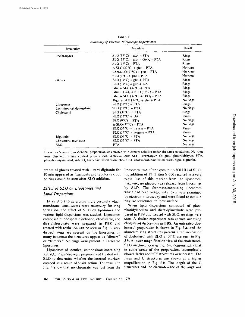
TABLE I
Summary Of Electron Microscopy Experiments
Preparation Procedure Result
Erythrocytes SLO (37~ + glut + PTA Rings SLO (37~ + glut + OsO~ + PTA Rings SLO (37~ + PTA Rings A-SLO (37~ + glut + PTA No rings ChoI-SLO (37~ + glut + PTA No rings SLO (0~ + glut + PTA No rings
Ghosts SLO (37~ + glut + PTA Rings SLO (37~ + glut + UA Rings Glut + SLO (37~ + PTA Rings Glut + OsO, + SLO (37~ + PTA Rings Glut + SLO (37~ + OsO~ + PTA Rings Digit + SLO (37~ + glut + PTA No rings
Liposomes SLO (37~ + PTA Rings Lecithin-dicetylphosphate SLO (37~ + PTA No rings Cholesterol SLO (37~ + PTA Rings
SLO (37~ + UA Rings SLO (0~ + PTA No rings A-SLO (37~ + PTA No rings SLO (37~ + trypsin + PTA Rings SLO (37~ + pronase + PTA Rings
Digitonin SLO (37~ + PTA No rings Cholesterol myristate SLO (37~ + PTA No rings SLO PTA No rings
In each experiment, an identical preparation was treated with control solution under the same conditions. No rings were observed in any control preparations. Abbreviations: SLO, streptolysin O; glut, glutaraldehyde; PTA, phosphotungstic acid; A-SLO, heat-inactivated toxin; chol-SLO, cholesterol-inactivated toxin; digit, digitonin.
branes of ghosts treated with 1 mM digitonin for 10 min appeared as fragments and tubules (8), but no rings could be seen after SLO addition.
Effect of SLO on Liposomes and Lipid Dispersions
In an effort to determine more precisely which membrane constituents were necessary for ring formation, the effect of SLO on liposomes and various lipid dispersions was studied. Liposomes composed of phosphatidylcholine, cholesterol, and dicetylphosphate were prepared in PBS and treated with toxin. As can be seen in Fig. 3, very distinct rings are present on the liposomes; in many instances the structures appear as "dimers'" or " t r imers ." No rings were present in untreated liposomes.
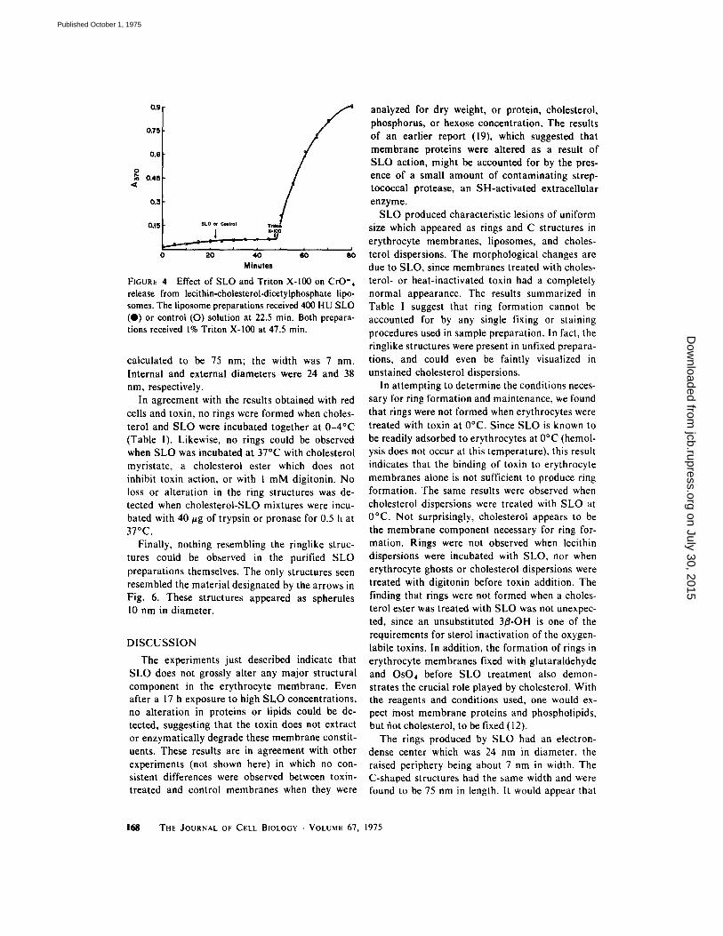
Liposomes of identical composition containing K~CrO4 or glucose were prepared and treated with SLO to determine whether the internal markers escaped as a result of toxin action. The results in Fig. 4 show that no chromate was lost from the
liposomes even after exposure to 800 HU of SLO; the addition of 1% Triton X-100 resulted in a very rapid loss of this marker from the liposomes. Likewise, no glucose was released from liposomes by SLO. The chromate-containing liposomes which had been treated with toxin were examined by electron microscopy and were found to contain ringlike structures on their surface.
When lipid dispersions composed of ptaos- phatidylcholine and dicetylphosphate were pre- pared in PBS and treated with SLO, no rings were seen. A similar experiment was carried out using cholesterol dispersions in PBS. An untreated cho- lesterol preparation is shown in Fig. 5 a, and the abundant ring structures present after incubation of cholesterol with SLO at 37 C are seen in Fig. 5 b. A lower magnification view of the cholesterol- SLO mixture, seen in Fig. 6 a, demonstrates that in some areas of the preparation, incompletely closed circles and " C " structures were present. The rings and C structures are shown at a higher magnification in Fig. 6b. The length of the C structures and the circumference of the rings was
166 THE JOURNAL OF CELL BIOLOGY �9 VOLUME 67, 1975
on July 30, 2015jcb.rupress.org
Dow
nloaded from
Published October 1, 1975

FIGURE 3 Lccithin-cholesteroi-dicetylphosphate liposomes treated with SLO. x 114,800. Bar = 0. I , m .
DUNCAN AND SCHLEGEL Effects of Streptolysin 0 167
on July 30, 2015jcb.rupress.org
Dow
nloaded from
Published October 1, 1975
0.9
0.75
0.6
0 o.4s
0.3
0.15 $LO ~ r Tri~ X-IO0
i i i , ,
e'o ' ' 0 20 40 80
Minutes
FIGURE 4 Effect of SLO and Triton X-100 on CrO'4 release from lecithin-cholesterol-dicetylphosphate lipo- somes. The liposome preparations received 400 HU SLO (@) or control (O) solution at 22.5 min. Both prepara- tions received 1% Triton X-100 at 47.5 min.
calculated to be 75 nm; the width was 7 nm. Internal and external diameters were 24 and 38 nm, respectively.
In agreement with the results obtained with red cells and toxin, no rings were formed when choles- terol and SLO were incubated together at 0-4~ (Table I). Likewise, no rings could be observed when SLO was incubated at 37~ with cholesterol myristate, a cholesterol ester which does not inhibit toxin action, or with 1 mM digitonin. No loss or alteration in the ring structures was de- tected when cholesterol-SLO mixtures were incu- bated with 40 #g of trypsin or pronase for 0.5 h at 37~
Finally, nothing resembling the ringlike struc- tures could be observed in the purified SLO preparations themselves. The only structures seen resembled the material designated by the arrows in Fig. 6. These structures appeared as spherules 10 nm in diameter.
DISCUSSION
The experiments just described indicate that SLO does not grossly alter any major structural component in the erythrocyte membrane. Even after a 17 h exposure to high SLO concentrations, no alteration in proteins or lipids could be de, tected, suggesting that the toxin does not extract or enzymatically degrade these membrane constit- uents. These results are in agreement with other experiments (not shown here) in which no con- sistent differences were observed between toxin- treated and control membranes when they were
analyzed for dry weight, or protein, cholesterol, phosphorus, or hexose concentration. The results of an earlier report (19), which suggested that membrane proteins were altered as a result of SLO action, might be accounted for by the pres- ence of a small amount of contaminating strep- tococcal protease, an SH-activated extraceilular enzyme.
SLO produced characteristic lesions of uniform size which appeared as rings and C structures in erythrocyte membranes, liposomes, and choles- terol dispersions. The morphological changes are due to SLO, since membranes treated with choles- terol- or heat-inactivated toxin had a completely normal appearance. The results summarized in Table I suggest that ring formation cannot be accounted for by any single fixing or staining procedures used in sample preparation. In fact, the ringlike structures were present in unfixed prepara- tions, and could even be faintly visualized in unstained cholesterol dispersions.
In attempting to determine the conditions neces- sary for ring formation and maintenance, we found that rings were not formed when erythrocytes were treated with toxin at 0~ Since SLO is known to be readily adsorbed to erythrocytes at 0~ (hemol- ysis does not occur at this temperature), this result indicates that the binding of toxin to erythrocyte membranes alone is not sufficient to produce ring formation. The same results were observed when cholesterol dispersions were treated with SLO at 0~ Not surprisingly, cholesterol appears to be the membrane component necessary for ring for- mation. Rings were not observed when lecithin dispersions were incubated with SLO, nor when erythrocyte ghosts or cholesterol dispersions were treated with digitonin before toxin addition. The finding that rings were not formed when a choles- terol ester was treated with SLO was not unexpec- ted, since an unsubstituted 3B-OH is one of the requirements for sterol inactivation of the oxygen- labile toxins. In addition, the formation of rings in erythrocyte membranes fixed with glutaraldehyde and OsO, before SLO treatment also demon- strates the crucial role played by cholesterol. With the reagents and conditions used, one would ex- pect most membrane proteins and phospholipids, but riot cholesterol, to be fixed (12).
The rings produced by SLO had an electron- dense center which was 24 nm in diameter, the raised periphery being about 7 nm in width. The C-shaped structures had the same width and were found to be 75 nm in length. It would appear that
168 THE JOURNAL OF CELL BIOLOGY . VOLUME 67, 1975
on July 30, 2015jcb.rupress.org
Dow
nloaded from
Published October 1, 1975

FIGURE 5 (a) Untreated cholesterol dispersion, x 35,000. Bar ~ 0.2 ~,m. (b) SLO-treated cholesterol dispersion, x 114,800. Bar = 0.1 tzm.
DUNCAN AND SCHLEGEL Effects of Streptolysin 0 169
on July 30, 2015jcb.rupress.org
Dow
nloaded from
Published October 1, 1975
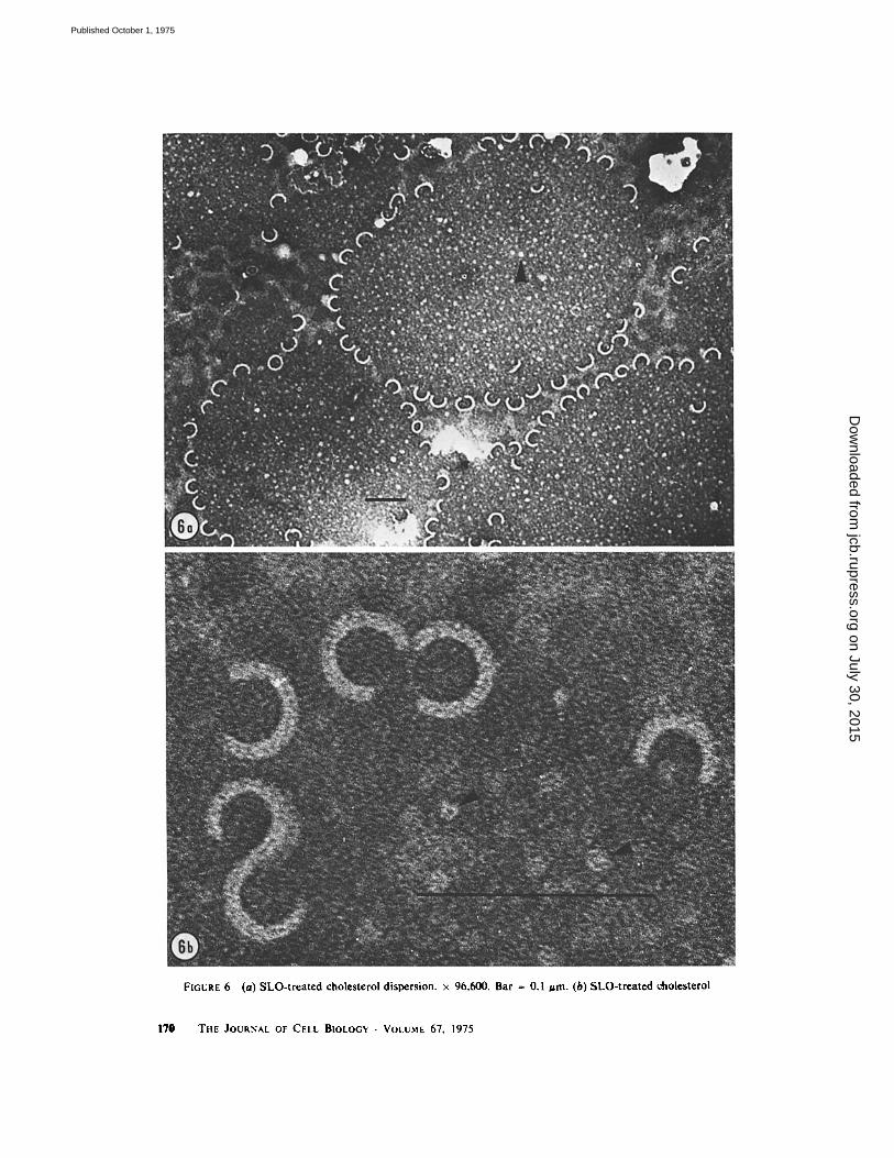
FIGURE 6 (a) SLO- t r ea t ed choles terol dispers ion, x 96,600. Bar ~ 0.1 jam. (b) SLO- t rea t cd choles terol
170 THE JOURNAL OF CELL BIOLOGY �9 VOLUME 67, 1975
on July 30, 2015jcb.rupress.org
Dow
nloaded from
Published October 1, 1975

the rings represent "C" structures which have circularized, since the calculated circumference of the dense ring centers closely approximated the measured length of the C's.
Considerable evidence from several lines of research points to cholesterol as the adsorption site or target of SLO action; the finding that choles- terol is necessary (and sufficient) for ring forma- tion supports this concept. A hypothetical model which might account for the SLO-cholesterol relationship is presented in Fig. 7. According to this proposal, the toxin-cholesterol interaction is such that the SLO molecule undergoes a configu- rational change and assumes an unfolded, more linear C structure, surrounded by adsorbed choles- terol molecules. The molecular weight of SLO is in the range of 60,000 daltons (2, 25, 29), and if the protein existed in its a-helical form in this situa- tion, it would have a length of approximately 78 nm, assuming an average amino acid molecular weight of 120, and a distance of 1.57 A between adjacent amino acids. As suggested in Fig. 8, most of the C structures circularize to form the charac- teristic rings, but some aggregates to form dimers, trimers, or "S"-shaped structures (Figs. 3 and 6). The failure of trypsin and pronase to alter the morphology of the rings and C's suggests that if the toxin molecule is present in these structures as proposed, it is protected in some way, perhaps by the cholesterol molecules associated with the toxin molecule. Both enzymes rapidly destroyed the hemolytic activity of SLO in solution.
The model as given is one possible explanation
for the results and is not meant to exclude other interpretations. For example, the structures ob- served may represent aggregations of SLO and cholesterol molecules, or simply a reorganization or packing of the cholesterol molecules alone. However, the presence primarily of rings and C's, and the absence of large numbers of intermediate forms and of structures larger than 75 nm would seem to argue against the aggregation proposal. In any event, the remarkable morphological altera- tions (with implied change in protein secondary structure) consequent to SLO exposure to choles- terol suggests that this toxin may be a useful tool in understanding protein-cholesterol interactions.
The experiments reported here describe the conditions necessary for ring formation, but the mechanism of SLO-induced hemolysis and the role of the ring structures in this process are not clear. The evidence from other studies suggests that perhaps saponin, but not antibody-complement, or filipin, produces transverse holes in the membrane (14, 22, 23). We observed a good correlation between ring formation and cell lysis. However, rings were observed in SLO-treated lecithin- cholesterol-dicetylphosphate liposomes, even though the toxin did not release either of the trapped internal markers, CrO4" or glucose. These markers were released from the liposomes by Triton X - 100 (Fig. 4), saponin, SDS, and the unrelated streptococcal toxin, streptolysin S (not shown). This finding suggests that the ring struc- tures are not actually "holes" in erythrocyte or liposome membranes, but one must be cautious sL0
+
Cholesterol �9 0 0 0 g �9 observed length = 75 nm
calculated length = ?8 nm
( for a-helix conformation assuming tool wt=60.OOO )
FIGURE 7 Hypothetical model of C-structure formation as a result of conformationai changes in the SLO molecule and protein-cholesterol interaction.
DUNCAN AND SCHLEGEL Effects of Streptolysin 0 171
on July 30, 2015jcb.rupress.org
Dow
nloaded from
Published October 1, 1975

"C" complexes Cholesterol with "sticky ends" SLO + 1" ~ C 75 nm length
ircularizatlon
( ~ dlmer 0
@ ~ trlmer 0 0 diameter = 24 nm
"S" circumstance �9 75 nm FIGURE 8 Hypothetical model of ring formation and aggregation of C's to form dimers, trimers, and S-shaped structures.
regarding the results obtained from a heterogene- ous population of liposomes. It is conceivable that the ring structures were present only in a unique subpopulation of membrane-like structures which contain high concentrations of cholesterol, but little trapped chromate or glucose marker.
According to the proposal shown in Figs. 7 and 8, the ring structures represent cholesterol mole- cules sequestered around extended SLO molecules. High concentrations of sequestered cholesterol may weaken certain areas of the membrane, leading to large holes or tears in the structure. Cholesterol is known to affect the mean molecular area of lecithin molecules (7) as well as the permeability characteristics of liposomes (6, 7, 20) and natural membranes (4). Cholesterol has also been shown to decrease the energy content of the phase transition of lipids in artificial (5, 15) and biological membranes (4, 21). Whatever the expla- nation for the disruption of erythrocytes by SLO, one must take into account the observation that a leakage of intracellular ions does not significantly precede the escape of hemoglobin (10), a result suggesting that an osmotic mechanism of lysis is not involved in this hemolytic process.
Recently, Smyth et al. (27) demonstrated that the membrane lesions produced by commercial phospholipase C (9) were due to contamination by the Clostridium perfringens 0-hemolysin. The 0- hemolysin is also an oxygen-labile toxin, and
Smyth and co-workers found that it produced ring- and arc-shaped structures in erythrocyte mem- branes, lipid dispersions containing cholesterol, and cholesterol dispersions very similar to the structures described here. They also found arc- shaped structures in hemolysin-treated lecithin dispersions and a few such structures were seen in electron micrographs of their toxin preparations. The authors suggested that these effects might be due to contamination with nanogram amounts of cholesterol. In general, their results with 0-toxin, and our own with SLO are in good agreement.
We appreciate the excellent technical assistance of Lela Buckingham. In addition, we thank Dr. Charles Ganote (Department of Pathology, Northwestern University) for helpful suggestions and Dr. Robert Jennings (Chairman, Department of Pathology) for offering access to the electron microscopy facilities.
This investigation was supported by United States Public Health Services grant AI 11644. Received for publication 14 April 1975, and in revised form 16 June 1975.
R E F E R E N C E S
1. A L O U F , J. E., AND M. RAYNAUD. 1968. Action de la streptolysine O sur les membranes cellulaires. I, Fixation sur la membrane ~:rythrocytaire. Ann. Inst. Pasteur (Paris). 114:812-827.
172 THE JOURNAL OF CELL BIOLOGY . VOLUME 67, 1975
on July 30, 2015jcb.rupress.org
Dow
nloaded from
Published October 1, 1975

2. ALOUF, J. E., AND M. RAYNAUD. 1973. Purification and some properties of streptolysin O. Biochimie. 55:1187-1193.
3. BERNHEIMER, A. W. 1974. Interactions between membranes and cytolytic bacterial toxins. Biochim. Biophys. Acta. 344:27-50.
4. DE KRUYFF, B., R. A. DEMEL, AND L. L. M. VAN DEENEN, 1972. The effect of cholesterol and epi- cholesterol incorporation on the permeability and on the phase transition of intact Acholeplasma laidlawii cell membranes and derived liposomes. Biochim. Biophys. Acta. 255:331-347.
5. DE KRUYFF, B., P. W. M. VAN DIJCK, R. A. DEMEL, A. SCHUIJFF, F. BRANTS, AND L. L. M. VAN DEENEN. 1974. Non-random distribution of cholesterol in phosphatidylcholine bilayers. Biochim. Biophys. Acta. 356:1-7.
6. DEMEL, R. A., K. R. BRUCKDORFER, AND L. L. M. VAN DEENEN. 1972. The effects of sterol structure on the permeability of liposomes to glucose, glycerol, and Rb +. Biochim. Biophys. Acta. 255:321-330.
7. DEMEL, R. A., W. S. M. GEURTS VAN KESSEL, AND L. L. M. VAN DEENEN. 1972. The properties of polyunsaturated lecithins in monolayers and lipo- somes and the interactions of these lecithins with cholesterol. Biochim. Biophys. Acta. 266:26-40.
8. DOURMASHKIN, R. R., R. M. DOUGHERTY, AND R. ,I. C. HARRIS. 1962. Electron microscopic observa- tions on Rous sarcoma virus and cell membranes. Nature (Load.). 194:1116-1119.
9. DOURMASHKIN, R. R., AND W. F. ROSSE. 1966. Morphologic changes in the membranes of red blood cells undergoing hemolysis. Am. J. Med. 41:699 - 710.
10. DUNCAN, J. L. 1974. Characteristics of strep- tolysin O hemolysis: kinetics of hemoglobin and 86-rubidium release. Infect. lmmun. 9:1022-1027.
11. FAIRBANKS, G., T. L. STECK, AND D. F. H. WALLACH. 1971. Electrophoretic analysis of the major polypeptides of the human erythrocyte mem- brane. Biochemistry. I0:2606-2617.
12. HAGAT, M. A. 1970. Principles and techniques of electron microscopy. Biological applications. Vol. I. Van Nostrand Reinhold Co., New York.
13. HALBERT, S. P. 1970. Streptolysin O. In Microbial Toxins. Vol 3. T. C. Montie, S. Kadis, and S. J. Ajl, editors. Academic Press Inc., New York. 69-95.
14. ILES, G. H., P. SEEMAN, D. NAYLOR, AND B. CINADER. 1973. Membrane lesions in immune lysis. Surface rings, globule aggregates, and transient openings. J. Cell Biol. 56:528-539.
15. LADBROOKE, B. D., R. M. WILLIAMS, AND D. CHAPMAN. 1968. Studies on lecithin-cholesterol- water interactions by differential scanning calorime- try and x-ray diffraction. Biochim. Biophys. Acta. 150:333-340.
16. LENARD, J. 1970. Protein and glycolipid components of human erythrocyte membranes. Biochemistry. 9:1129-1132.
17. NICOLSON, G, L., AND S. J. SINGER. 1971. Ferritin- conjugated plant agglutinins as specific saccharide stains for electron microscopy: application to saccha- rides bound to cell membranes. Proc. Nat. Acad. Sci. U. S. A. 68:942-945.
18. OBERLEY, T. D., AND J. L. DUNCAN. 1971. Charac- teristics of streptolysin O action. Infect. lmmun. 4:683-687.
19. OBERLEY, T. D., AND J. L. DUNCAN. 1972. Strep- tolysin O induced alterations in rabbit erythrocyte membrane polypeptides. Biochem. Biophys. Res. Commun. 48:1339-1347.
20. PAPAHADJOPOULOS, D., M. COWDEN, AND H. KIMELBERG. 1973. Role of cholesterol in membranes. Effects on phospholipid-protein interactions, mem- brane permeability, and enzymic activity. Biochim. Biophys. Acta. 330:8-26.
21. ROTTEM, S. V., P. CIRILLO, B. DE DRUYFF, M. SHINITZKY, AND S. RAZIN. 1973. Cholesterol in mycoplasma membranes. Correlation of enzymic and transport activities with physical state of lipids in membranes of Mycoplasma mycoides var. capri adapted to grow with low cholesterol concentrations. Biochim. Biophys. Acta. 323: 509-519.
22. SEEMAN P. 1974. Ultrastructure of membrane lesions in immune lysis, osmotic lysis and drug- induced lysis. Fed. Proc. 33:2116-2124.
23. SEEMAN, P., D. CHENG, ANt) G. H. ILES. 1973. Structure of membrane holes in osmotic and saponin hemolysis. J. Cell Biol. 56:519-527.
24. SHANY, S., A. W. BERNHEIMER, P. S. GRUSHOFF, AND K. S. Kim. 1974. Evidence for membrane cholesterol as the common binding site for cereoly- sin, streptolysin O and saponin. Mol. Cell. Biochem. 3:179 186.
25. SHANY, S., P. GRUSHOFF, AND A. W. 8ERNHEIMER. 1973. Physical separation of streptococcal nicotina- mide adenine dinucleotide glycohydrolase from streptolysin O. Infect. lmmun. 7:731-734.
26. SKIPSKI, V. P., AND M. BARCLAY. 1969. Thin-layer chromatography of lipids. In Methods in Enzymol- DRy. Vol. 14. J. M. Lowenstein, editor. Academic Press, Inc., New York. 530-598.
27. SMYTH, C. J., J. H. FREER, AND J. P. ARBUTHNOTT. 1975. Interaction of Clostridium perfringens 0-ha- emolysin, a contaminant of commercial phospholi- pase C, with erythrocyte ghost membranes and lipid dispersions: A morphological study. Biochim. BiD- phys. Acta. 382:479-493.
28. STECK, T. L. 1972. Cross-linking the major proteins of the isolated erythrocyte membrane. J. Mol. Biol. 66:295-305.
29. VAN EPPS, D. E., AND B. R. ANDERSEN. 1969. Streptolysin O: Sedimentation coefficient and mo- lecular weight determinations. J. BacterioL 100:526 527.
30. WAYS, P., AND D. J. HANAHAN. 1964. Characteriza- tion and quantification of red cell lipids in normal man. J. Lipid Res. 5:318 328.
DUNCAN AND SCHLEGEL Effects of Streptolysin 0 173
on July 30, 2015jcb.rupress.org
Dow
nloaded from
Published October 1, 1975
Copyright © 2022 FDOKUMEN