Methods of Liposomes Preparation - MDPI
49
Citation: Lombardo, D.; Kiselev, M.A. Methods of Liposomes Preparation: Formation and Control Factors of Versatile Nanocarriers for Biomedical and Nanomedicine Application. Pharmaceutics 2022, 14, 543. https://doi.org/10.3390/ pharmaceutics14030543 Academic Editors: Lucimara Gaziola de la Torre and Mariano Michelon Received: 23 January 2022 Accepted: 24 February 2022 Published: 28 February 2022 Publisher’s Note: MDPI stays neutral with regard to jurisdictional claims in published maps and institutional affil- iations. Copyright: © 2022 by the authors. Licensee MDPI, Basel, Switzerland. This article is an open access article distributed under the terms and conditions of the Creative Commons Attribution (CC BY) license (https:// creativecommons.org/licenses/by/ 4.0/). pharmaceutics Review Methods of Liposomes Preparation: Formation and Control Factors of Versatile Nanocarriers for Biomedical and Nanomedicine Application Domenico Lombardo 1, * and Mikhail A. Kiselev 2,3,4 1 Consiglio Nazionale delle Ricerche, Istituto per i Processi Chimico-Fisici, 98158 Messina, Italy 2 Frank Laboratory of Neutron Physics, Joint Institute for Nuclear Research, 141980 Dubna, Moscow Region, Russia; [email protected] 3 Department of Nuclear Physics, Dubna State University, 141980 Dubna, Moscow Region, Russia 4 Physics Department, Lomonosov Moscow State University, 119991 Moscow, Moscow Region, Russia * Correspondence: [email protected]; Tel.: +39-090-39762222 Abstract: Liposomes are nano-sized spherical vesicles composed of an aqueous core surrounded by one (or more) phospholipid bilayer shells. Owing to their high biocompatibility, chemical composition variability, and ease of preparation, as well as their large variety of structural properties, liposomes have been employed in a large variety of nanomedicine and biomedical applications, including nanocarriers for drug delivery, in nutraceutical fields, for immunoassays, clinical diagnostics, tissue engineering, and theranostics formulations. Particularly important is the role of liposomes in drug- delivery applications, as they improve the performance of the encapsulated drugs, reducing side effects and toxicity by enhancing its in vitro- and in vivo-controlled delivery and activity. These applications stimulated a great effort for the scale-up of the formation processes in view of suitable industrial development. Despite the improvements of conventional approaches and the development of novel routes of liposome preparation, their intrinsic sensitivity to mechanical and chemical actions is responsible for some critical issues connected with a limited colloidal stability and reduced entrapment efficiency of cargo molecules. This article analyzes the main features of the formation and fabrication techniques of liposome nanocarriers, with a special focus on the structure, parameters, and the critical factors that influence the development of a suitable and stable formulation. Recent developments and new methods for liposome preparation are also discussed, with the objective of updating the reader and providing future directions for research and development. Keywords: liposome formation; lipid-based nanocarriers; phospholipids self-assembly; drug delivery; nanomedicine 1. Introduction Liposomes represent versatile nanoplatforms for the improved delivery of pharma- ceutical drugs and active compounds in a large variety of biomedical and nanomedicine applications [1,2]. They are characterized by easily controllable properties such as lipid composition, size, structure and morphology, surface charge, and the possibility of function- alizing their surfaces with polymers or ligands [3–5]. Particularly interesting is the ability of liposomal systems to encapsulate both hydrophilic and lipophilic active compounds as well as various biomolecules, including carbohydrates [6], proteins and peptides [7], DNA [8], or imaging compounds [9]. Liposomes’ structure is regulated by soft interactions and self-assembly phenomena that regulate their structural properties and their stability within the environments of biological tissues [10–12]. The inclusion of drugs within the vesicles’ nanostructure favors the active compounds’ solubilization in solution and protects against their chemical and biological degradation. The use of liposome nanoformulations also causes a sensitive enhancement of their therapeutic performances [12–15]. Particu- larly interesting is the development of new liposome nano-platforms for biomedical and Pharmaceutics 2022, 14, 543. https://doi.org/10.3390/pharmaceutics14030543 https://www.mdpi.com/journal/pharmaceutics
-
Upload
khangminh22 -
Category
Documents
-
view
8 -
download
0
Transcript of Methods of Liposomes Preparation - MDPI

�����������������
Citation: Lombardo, D.; Kiselev, M.A.
Methods of Liposomes Preparation:
Formation and Control Factors of
Versatile Nanocarriers for Biomedical
and Nanomedicine Application.
Pharmaceutics 2022, 14, 543.
https://doi.org/10.3390/
pharmaceutics14030543
Academic Editors: Lucimara
Gaziola de la Torre and
Mariano Michelon
Received: 23 January 2022
Accepted: 24 February 2022
Published: 28 February 2022
Publisher’s Note: MDPI stays neutral
with regard to jurisdictional claims in
published maps and institutional affil-
iations.
Copyright: © 2022 by the authors.
Licensee MDPI, Basel, Switzerland.
This article is an open access article
distributed under the terms and
conditions of the Creative Commons
Attribution (CC BY) license (https://
creativecommons.org/licenses/by/
4.0/).
pharmaceutics
Review
Methods of Liposomes Preparation: Formation and ControlFactors of Versatile Nanocarriers for Biomedical andNanomedicine ApplicationDomenico Lombardo 1,* and Mikhail A. Kiselev 2,3,4
1 Consiglio Nazionale delle Ricerche, Istituto per i Processi Chimico-Fisici, 98158 Messina, Italy2 Frank Laboratory of Neutron Physics, Joint Institute for Nuclear Research,
141980 Dubna, Moscow Region, Russia; [email protected] Department of Nuclear Physics, Dubna State University, 141980 Dubna, Moscow Region, Russia4 Physics Department, Lomonosov Moscow State University, 119991 Moscow, Moscow Region, Russia* Correspondence: [email protected]; Tel.: +39-090-39762222
Abstract: Liposomes are nano-sized spherical vesicles composed of an aqueous core surrounded byone (or more) phospholipid bilayer shells. Owing to their high biocompatibility, chemical compositionvariability, and ease of preparation, as well as their large variety of structural properties, liposomeshave been employed in a large variety of nanomedicine and biomedical applications, includingnanocarriers for drug delivery, in nutraceutical fields, for immunoassays, clinical diagnostics, tissueengineering, and theranostics formulations. Particularly important is the role of liposomes in drug-delivery applications, as they improve the performance of the encapsulated drugs, reducing sideeffects and toxicity by enhancing its in vitro- and in vivo-controlled delivery and activity. Theseapplications stimulated a great effort for the scale-up of the formation processes in view of suitableindustrial development. Despite the improvements of conventional approaches and the developmentof novel routes of liposome preparation, their intrinsic sensitivity to mechanical and chemicalactions is responsible for some critical issues connected with a limited colloidal stability and reducedentrapment efficiency of cargo molecules. This article analyzes the main features of the formation andfabrication techniques of liposome nanocarriers, with a special focus on the structure, parameters,and the critical factors that influence the development of a suitable and stable formulation. Recentdevelopments and new methods for liposome preparation are also discussed, with the objective ofupdating the reader and providing future directions for research and development.
Keywords: liposome formation; lipid-based nanocarriers; phospholipids self-assembly; drug delivery;nanomedicine
1. Introduction
Liposomes represent versatile nanoplatforms for the improved delivery of pharma-ceutical drugs and active compounds in a large variety of biomedical and nanomedicineapplications [1,2]. They are characterized by easily controllable properties such as lipidcomposition, size, structure and morphology, surface charge, and the possibility of function-alizing their surfaces with polymers or ligands [3–5]. Particularly interesting is the abilityof liposomal systems to encapsulate both hydrophilic and lipophilic active compoundsas well as various biomolecules, including carbohydrates [6], proteins and peptides [7],DNA [8], or imaging compounds [9]. Liposomes’ structure is regulated by soft interactionsand self-assembly phenomena that regulate their structural properties and their stabilitywithin the environments of biological tissues [10–12]. The inclusion of drugs within thevesicles’ nanostructure favors the active compounds’ solubilization in solution and protectsagainst their chemical and biological degradation. The use of liposome nanoformulationsalso causes a sensitive enhancement of their therapeutic performances [12–15]. Particu-larly interesting is the development of new liposome nano-platforms for biomedical and
Pharmaceutics 2022, 14, 543. https://doi.org/10.3390/pharmaceutics14030543 https://www.mdpi.com/journal/pharmaceutics

Pharmaceutics 2022, 14, 543 2 of 49
nanomedicine applications, which are stimulated by the liposomes’ special properties, suchas their colloidal stability, efficient targeting, and site-specific delivery via various routes ofadministration [1–4].
The industrial applications of liposome nanoplatforms include their use as drug-delivery vehicles in nanomedicine, cancer, antimicrobial therapy, as signal carriers inbiomedical diagnostics and biochemistry, as adjuvants in vaccination, and as solubilizersand support matrices for various active compounds and macromolecules [13–15]. Moreover,owing to their high biocompatibility and non-toxicity, liposomes are the most importantcategory of clinically approved therapeutic drug nanocarriers for cancer treatment [16–18].Those systems play a crucial role also for the encapsulation of unstable bioactive substances(including antioxidants, antimicrobials, phytochemicals, and nutraceuticals) due to theirstrong enhancement of the colloidal stability [19–21].
The modern generation of liposomes includes lipid-based targeted and theranosticnanoplatforms, obtained by the engineering of the phospholipid nanostructures [22–26].All those varieties of liposome nanoplatforms stimulated a great effort for the scale-up ofthe fabrication methods in view of industrial developments. Concerning the manufacturingmethods, the main critical issues are the low colloidal stability, low entrapment efficiency,toxicity of organic solvents residue, and high cost for large-scale production. Despite thelarge success in nanomedicine applications, a number of critical issues have been identifiedwhich are mainly connected with the poor colloidal stability in biological environments,caused by lipids’ hydrolysis and oxidation processes, particle fission and fusion, and theconsequent loss of their active cargo.
In this article, we discuss the main features of the formation and fabrication techniquesof liposome nanocarriers with a special focus on the structural properties as well as thecrucial factors that influence the development of suitable and stable formulations. Wealso describe the main positive (and negative) aspects of each approach, as well as theirpotential for large-scale industrial production.
2. Structural Features and Main Control Factors of Liposomes
Liposomes are composed of a spherical hollow structure formed by phospholipidsdispersed in aqueous solution. The liposomes’ final organization, structure, and physico-chemical properties depends on the types, size, morphology, concentration, and chargeof the constituent lipids, as well as the solution properties (such as the ionic strength,pH, temperature) [27,28]. According to the theory of the lipid bilayers elasticity proposedby Helfrich [29], the curvature energy of a vesicle bilayer is higher than in the (stacked)multilamellar (liquid-crystal) phase (in water excess). Therefore, an energy cost is requestedin order to stimulate the curvature of the lipids’ bilayer into a vesicle structure (i.e., a closedlipid bilayer). Therefore, liposomes are metastable nanostructured systems that depend onthe methods of preparation (i.e., stirring, sonication, evaporation, extrusion) [29,30].
A crucial parameter for preparing liposomes by a self-assembly process is the criticalmicelles concentration (CMC), whereby the amphiphilic solution exhibits sensitive changesin their physico-chemical properties [31–33], while the aggregation of the amphiphilic lipidmolecules produce micelle-like aggregates. The CMC value generally depends on severalparameters, such as the hydrophobic/hydrophilic balance of the component lipids, thetemperature, and the solvent’s composition and properties (ionic strength, pH) [31,32].Other important factors of liposome nanocarriers are their size and lamellarity.
The size of liposome nanoformulations (that, for biomedical applications, rangespreferably between 50 and 500 nm) strongly influence their drug-delivery process [34,35].Liposomes with diameters in the range of 100–150 nm favor the cell uptake and are able toescape from the blood vessels’ capillaries within the diseased tissues (such as kidney, heart,lung) and enter through the (fenestrated) vessels into the tumor environments [35,36].Moreover, liposomes within 50–100 nm (or less) in size are able to avoid immune sys-tem phagocytosis clearance and exhibit longer blood circulation times [35,36]. The mainstructural features of liposomes are reported in Figure 1.

Pharmaceutics 2022, 14, 543 3 of 49Pharmaceutics 2022, 14, x 4 of 50
Figure 1. Schematic representation a DMPC unilamellar liposome (A). Typical onion-like structure composed of concentric bilayer surfaces (hydrated multilayers) of a multilamellar vesicle (MLV) (B). Characteristic phases of a water solution of DMPC phospholipids (C).
Another important factor of the self-assembled lipids’ nanostructures is given by the critical packing parameter CPP = V0/A0lc (where V0 is the effective volume occupied by hy-drophobic chains, A0 is the effective hydrophilic headgroup surface area, and lc is the max-imum effective chain length) [31,44]. The CPP of a certain lipid (or lipids’ composition) allows for predicting the preferred lipid aggregates’ structure, which can be spherical or ellipsoidal (CPP ≤ 1/3), cylindrical (CPP ≤ 1/2), or lamellar (CPP = 1). For (1/2 ≤ CPP ≤ 1), vesicles are generally generated.
The suitable combination of phospholipids with different CPPs or the modulation of the lipid/cholesterol ratio allows for obtaining the optimum size of liposomes. For exam-ple, the lipids 1-palmitoyl-2-oleoyl-sn-glycero-3-phosphocholine (POPC), which have CPPs near to 1, are able to form liposomes. Most of the naturally occurring phosphatidyl-cholines form planar bilayers (CPP = 1), but when mixed with conically shaped phospho-lipids that favor the bilayer curvature, (e.g., 1,2-dioleoyl-sn-glycero-3-phosphoethanola-mine (DOPE)), liposome nanostructures are also formed [43–45]. It is worth pointing out that, for amphiphilic building blocks with a more complex geometry or in the presence of a complex combination of (short- or long-range) interactions, the size and shape of am-phiphilic nanoassemblies is hard to predict with high precision. However, the analysis of the CPP still remains a valid approach for a qualitative estimation of the main morpho-logical features of soft-interacting macromolecules, and a versatile tool for the design of nanoscale drug carriers [45–49].
An important lipid that can sensitively influence the structural properties of lipo-somes is cholesterol. When incorporated into liposomes, cholesterol decreases the lipid bi-layer packing defects by distributing itself with its hydroxyl group close to the head lipids’ group region, while the aromatic rings are aligned with the hydrophobic alkyl chains. This configuration causes an increased fluidity in the bilayer core, but an increase of the vis-cosity (and rigidity) in the headgroups’ region. This causes a decrease of the fluidity and water permeability of liposomes, while the bilayer is less inclined to penetration (absorp-tion) by external nanoparticles. This increase in the mechanical rigidity results is im-portant for the liposome stability and prevents its interaction with proteins (such as trans-ferrin, albumin, and high-density lipoproteins), thus avoiding a possible reduction of their performances [34,35]. Moreover, the ability of the cholesterol to control the phospholipid packing, membrane fluidity, and the liposomes’ surface charge, produces an effect also
Figure 1. Schematic representation a DMPC unilamellar liposome (A). Typical onion-like structurecomposed of concentric bilayer surfaces (hydrated multilayers) of a multilamellar vesicle (MLV) (B).Characteristic phases of a water solution of DMPC phospholipids (C).
Concerning the lamellarity of the underlying vesicle structures, liposomes may beclassified into small unilamellar vesicles < 100 nm (SUVs), large unilamellar vesicles100–1000 nm (LUVs), and giant unilamellar vesicles > 1 µm (GUVs). Finally, the multil-amellar vesicles (MLVs) present an onion-like structure composed of concentric bilayersurfaces (hydrated multilayers) (Figure 1B). ULVs present a more rapid drug-release ratethan MLVs, which, on the other side, exhibit a larger entrapped volume.
Another important property of liposomes is given by the fluidity (and rigidity) of theirlipid bilayer structure. This property facilitates the crossing of the bilayers by the drug(macro-) molecules and strongly influences the rate of the drug-release process. Owingto their high flexibility, the self-assembled bilayers’ structures undergo a large varietyof structural and dynamic transitions that depend on various parameters such as thelipids’ temperature and composition [37–40]. With increasing temperature. several lipidbilayers pass from an ordered, crystalline (or gel) phase to a fluid state. For example, inFigure 1C, we report the structural feature of dimyristoylphosphatidylcholine (DMPC)MLVs in water solution at different temperature intervals as revealed by small-angle SANSand SAXS experiments [39,40]. This liposome system undergoes structural transitions atthe so called “pre-transition” (at temperature Tp = 15.0 ◦C) and the main phase transition(at the temperature Tm = 23.4 ◦C), which identifies the border between the gel Lβ’, ripplePβ’, and liquid-crystalline Lα phases, respectively (Figure 1C).
The presence of an intermediate ripple phase (formed by domains of liquid-crystallineordered phases within the gel phase) depends mainly on the liposomes’ aggregation state,and is directly related to the phospholipid composition and temperature. The characteristictemperature (TC) at which phospholipids undergo the transition from the gel to the liquid-crystalline phase is an important parameter in the formation of liposomes, as it is indicativeof liposomes’ fluidity (and permeability) and depends on the alkyl chains’ lengths, theirsaturation degree, head group species, and the associated charge [27,28]. For T < TC, thelipid bilayers are in the gel phase and exhibit lower fluidity (and lower permeability),while for T > TC, they are in a liquid-crystalline state and have a larger fluidity (and largerpermeability) [37–40]. Therefore, the phase transition behaviour of the constituent lipidscan be exploited to improve liposome structural modification (or integrity). With this aim, aproper lipid composition can be designed to preserve the liposome structure characteristics

Pharmaceutics 2022, 14, 543 4 of 49
and their physico-chemical properties, or to stimulate a structural modification (such as anaggregation or drug release) close to body temperature (T = 37 ◦C).
During the transition from the (more ordered) gel phase to the (less ordered) liquid-crystalline phase, drug molecules are less impeded when crossing the lipids’ bilayer, ex-hibiting an increase of the permeation rate (with a peak near Tm). For this reason, liposomes’nanoformulations, containing lipids with high Tm, such as the saturated-phospholipidsdipalmitoyl phosphatidylcholine (DPPC) or the fully saturated distearoylphosphatidyl-choline (DSPC), exhibit a more rigid and stable bilayer structure and a reduced leakage ofthe encapsulated drugs (weak permeability) [41–43]. On the contrary, liposomes containingunsaturated phospholipids (such as the egg or soybean phosphatidylcholine) provideless-stable bilayer nanostructures, caused by the disruption of the packing effect of adjacentacyl chains, and exhibit higher flexibility (and a higher permeability) of the whole lipidbilayer, and then a decrease of Tm [41–43]. The proper combination of lipids with acylchains of different types favors, then, the design of temperature-sensitive liposomes (withrequired Tm).
Another important factor of the self-assembled lipids’ nanostructures is given by thecritical packing parameter CPP = V0/A0lc (where V0 is the effective volume occupiedby hydrophobic chains, A0 is the effective hydrophilic headgroup surface area, and lcis the maximum effective chain length) [31,44]. The CPP of a certain lipid (or lipids’composition) allows for predicting the preferred lipid aggregates’ structure, which can bespherical or ellipsoidal (CPP ≤ 1/3), cylindrical (CPP ≤ 1/2), or lamellar (CPP = 1). For(1/2 ≤ CPP ≤ 1), vesicles are generally generated.
The suitable combination of phospholipids with different CPPs or the modulation ofthe lipid/cholesterol ratio allows for obtaining the optimum size of liposomes. For example,the lipids 1-palmitoyl-2-oleoyl-sn-glycero-3-phosphocholine (POPC), which have CPPsnear to 1, are able to form liposomes. Most of the naturally occurring phosphatidylcholinesform planar bilayers (CPP = 1), but when mixed with conically shaped phospholipidsthat favor the bilayer curvature, (e.g., 1,2-dioleoyl-sn-glycero-3-phosphoethanolamine(DOPE)), liposome nanostructures are also formed [43–45]. It is worth pointing out that, foramphiphilic building blocks with a more complex geometry or in the presence of a complexcombination of (short- or long-range) interactions, the size and shape of amphiphilicnanoassemblies is hard to predict with high precision. However, the analysis of the CPPstill remains a valid approach for a qualitative estimation of the main morphologicalfeatures of soft-interacting macromolecules, and a versatile tool for the design of nanoscaledrug carriers [45–49].
An important lipid that can sensitively influence the structural properties of liposomesis cholesterol. When incorporated into liposomes, cholesterol decreases the lipid bilayerpacking defects by distributing itself with its hydroxyl group close to the head lipids’group region, while the aromatic rings are aligned with the hydrophobic alkyl chains. Thisconfiguration causes an increased fluidity in the bilayer core, but an increase of the viscosity(and rigidity) in the headgroups’ region. This causes a decrease of the fluidity and waterpermeability of liposomes, while the bilayer is less inclined to penetration (absorption)by external nanoparticles. This increase in the mechanical rigidity results is importantfor the liposome stability and prevents its interaction with proteins (such as transferrin,albumin, and high-density lipoproteins), thus avoiding a possible reduction of their perfor-mances [34,35]. Moreover, the ability of the cholesterol to control the phospholipid packing,membrane fluidity, and the liposomes’ surface charge, produces an effect also on the li-posome size, final morphology, and encapsulation efficiency [50]. Due to its low, flexible,hydrophobic ring structure, cholesterol can interact (through hydrophobic interactions andcooperative hydrogen bonds) with the phospholipid hydrophobic acyl chains, while itspresence stabilizes the straight-chain arrangement of saturated fatty acids (through the vander Waals interactions) [50,51]. Recent results evidenced that the inclusion of cholesterol inliposomes causes a sensitive increase of the incorporation efficiency of retinol, as well asan increase of the mean size and the colloidal stability of the liposome nanocarriers [51].

Pharmaceutics 2022, 14, 543 5 of 49
Recently, cholesterol proved to be a crucial component in modulating the release of en-capsulated hydrophilic (fluorescent dye) sulforhodamine B (SRB) molecules. The increaseof cholesterol concentration induced a decrease in the bilayer fluidity and an increasein the mean liposome size, (with a transition from irregular to regular spherical-shapevesicles) [52].
One approach to enhance the colloidal stability of liposomes consists of the incorpora-tion of charged components (such as anionic/cationic lipids or charged macromolecules).The charged surface creates, in fact, an electrostatic repulsion (a so-called ζ-potential)among liposomes that prevents possible coagulation (or aggregation) effects. Several inves-tigations evidenced that a negative charge (induced by the inclusion of negatively chargedmacromolecules or nanoparticles) can provide an enhancement of the colloidal stability ofneutral liposomes, due to the generated electrostatic repulsive forces [53–58]. Moreover,negatively charged lipids (such as DOPS and DMPG) are recognized by macrophages (byan aggregation-dependent phagocytic uptake mechanism) and are able to enter the cells viaendocytosis (with a faster rate than the neutral lipids), thus resulting in a shorter circulationtime. Within the blood circulation, the liposomes interact with the biological fluids andundergo an opsonization process with the circulating proteins, followed by the uptakeby the MPS. [59]. On the contrary, for gene delivery, the cationic liposomes are generallypreferred, as in the case of the charge interaction between positively charged lipids, suchas dioleoylphosphatidylethanolamine (DOPE), which have an amine head group (NH3
+),and the nucleic acids (negatively charged) [57]. Cationic liposome nanocarriers favor theinteractions with glycoproteins, which are present on the endothelial cells’ membranes,thus exhibiting longer circulation half-lives [57,58]. However, cationic liposomes mayinteract with the anionic components of the blood (such as plasma proteins), and may in-duce an enhanced uptake by the mononuclear phagocytic system (MPS), thus favoring theclearance process by the liver, lung, or spleen [59]. This causes a diminished accumulationin tumor tissues.
The enhancement of the liposomes’ colloidal stability can be obtained through theincorporation of specific polymers into their surface that hinder (sterically) the componentsof the blood from the interaction with the surface of the liposomes. This effect can beobtained through the liposome PEGylation process, which consists in the liposome surfacefunctionalization with polyethylene glycol (PEG), thus improving the colloidal stabilityand the blood-circulation time of active therapeutics [60,61]. However, a number of investi-gations have reported adverse immune responses, such the accelerated blood clearance(ABC), consisting in the rapid clearance of PEGylated nanocarriers upon repeat administra-tion [62,63], or the complement activation-related pseudo-allergy (CARPA), consisting inan adverse reactivity (hypersensitivity), which is correlated with side effects caused by theaction of the PEGylated nanocarriers [63]. These (adverse) immune responses stimulatethe investigation of alternative (natural or synthetic) polymers that are able to propose thesame properties and functions of PEG.
Finally, it is possible to functionalize the surface of the liposomes with a large varietyof ligands (including monoclonal antibodies, peptides, aptamers, and growth factors) thefavors the specificity of the liposome interaction (targeted drug delivery) and the controlleddrug release to specific target sites (such as diseased tissues or tumors) [1,64]
In Figure 2A, we report the main interactions exhibited by liposomes. We also re-port the main features of the (first FDA-approved) Doxil anticancer nanoformulation, aPEGylated liposome doxorubicin nanoformulation, for the treatment of epithelial ovarianKaposi’s sarcoma [16–18]. The energy barrier which results from the balance betweenattractive and repulsive forces prevents the aggregation (adhesion) of two nanocarrierswhile approaching one another. The control over the nanocarriers’ soft interactions rep-resents, then, a fundamental step for the design and engineering of the colloidal stabilityand biocompatibility of the liposomes, in view of overcoming the number of obstacles andbiological barriers found in biological system [3,58].

Pharmaceutics 2022, 14, 543 6 of 49
Pharmaceutics 2022, 14, x 6 of 50
Kaposi’s sarcoma [16–18]. The energy barrier which results from the balance between at-tractive and repulsive forces prevents the aggregation (adhesion) of two nanocarriers while approaching one another. The control over the nanocarriers’ soft interactions repre-sents, then, a fundamental step for the design and engineering of the colloidal stability and biocompatibility of the liposomes, in view of overcoming the number of obstacles and biological barriers found in biological system [3,58].
Figure 2. Schematic representation of the main interactions exhibited by liposomes (A). Main struc-tural characteristics of the anticancer drug Doxil (B).
In summary, the design and engineering of all the factors and parameters mentioned above, such as the lipid chemical nature and headgroup charge, the length and degree of unsaturation of the alkyl hydrophobic chains, the transition temperature (Tc), as well as the liposome surface functionalization (with PEG, ligands, proteins, or antibodies), make liposomes versatile tools in a large variety of biomedical applications.
3. Conventional Methods for the Preparation of Liposomes The main goals of a method for liposome nanoformulation formation is the formation
of monodisperse particles (with a narrow size distribution) and the requested degree of lamellarity, efficient drug inclusion, and long-term colloidal stability of the products. In the conventional methods, liposomes, initially dissolved in a volatile organic solvent, are subsequently mixed with an aqueous phase. The presence of an organic solvent may per-turbate the chemical properties of the incorporated active compounds, or influence the stability (or toxicity) of the generated nanoformulation [65]. The conventional methods for liposomes preparation involve the following main stages: 1. Dissolution of lipids in an organic solvent; 2. Drying-down of the resultant lipidic solution from the organic solvent; 3. Hydrating the lipid with an aqueous media (followed by agitation/stirring); 4. Downsizing (and/or change in lamellarity); 5. Post-formation processing (purification, sterilization); 6. Characterization of the final nanoformulation product.
Figure 2. Schematic representation of the main interactions exhibited by liposomes (A). Mainstructural characteristics of the anticancer drug Doxil (B).
In summary, the design and engineering of all the factors and parameters mentionedabove, such as the lipid chemical nature and headgroup charge, the length and degree ofunsaturation of the alkyl hydrophobic chains, the transition temperature (Tc), as well asthe liposome surface functionalization (with PEG, ligands, proteins, or antibodies), makeliposomes versatile tools in a large variety of biomedical applications.
3. Conventional Methods for the Preparation of Liposomes
The main goals of a method for liposome nanoformulation formation is the formationof monodisperse particles (with a narrow size distribution) and the requested degree oflamellarity, efficient drug inclusion, and long-term colloidal stability of the products. Inthe conventional methods, liposomes, initially dissolved in a volatile organic solvent, aresubsequently mixed with an aqueous phase. The presence of an organic solvent mayperturbate the chemical properties of the incorporated active compounds, or influence thestability (or toxicity) of the generated nanoformulation [65]. The conventional methods forliposomes preparation involve the following main stages:
1. Dissolution of lipids in an organic solvent;2. Drying-down of the resultant lipidic solution from the organic solvent;3. Hydrating the lipid with an aqueous media (followed by agitation/stirring);4. Downsizing (and/or change in lamellarity);5. Post-formation processing (purification, sterilization);6. Characterization of the final nanoformulation product.
Depending on the specific formation process, the hydration of the lipid (stage 3) mayanticipate the dry-down of the lipid solution from the organic solvent (stage 2).
3.1. Thin-Film Hydration (TFH) Method (Bangham Method)
The thin-film hydration technique (the so-called Bangham method) is the oldest, mostcommon, and simplest method used for the preparation of MLVs [66–68] (Figure 3). Toensure a homogeneous mixture, the main phospholipid ingredients are dissolved in anorganic solvent (such as dichloromethane, chloroform, ethanol, or a chloroform–methanol
Pharmaceutics 2022, 14, 543 7 of 49
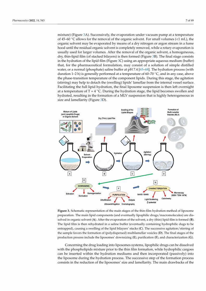
mixture) (Figure 3A). Successively, the evaporation under vacuum pump at a temperatureof 45–60 ◦C allows for the removal of the organic solvent. For small volumes (<1 mL), theorganic solvent may be evaporated by means of a dry nitrogen or argon stream in a fumehood until the residual organic solvent is completely removed, while a rotary evaporation isusually used for larger volumes. After the removal of the organic solvent, a homogeneous,dry, thin-lipid film (of stacked bilayers) is then formed (Figure 3B). The final stage consistsin the hydration of the lipid film (Figure 3C) using an appropriate aqueous medium (buffer)that, for the pharmaceutical formulation, may consist of a solution of simple distilledwater, or a normal (phosphate) saline buffer at pH 7.4 [65–68]. The hydration process (withduration 1–2 h) is generally performed at a temperature of 60–70 ◦C, and in any case, abovethe phase-transition temperature of the component lipids. During this stage, the agitation(stirring) may help to detach the (swelling) lipids’ lamellae from the internal vessel surface.Facilitating the full lipid hydration, the final liposome suspension is then left overnightat a temperature of T = 4 ◦C. During the hydration stage, the lipid becomes swollen andhydrated, resulting in the formation of a MLV suspension that is highly heterogeneous insize and lamellarity (Figure 3D).
Pharmaceutics 2022, 14, x 7 of 50
Depending on the specific formation process, the hydration of the lipid (stage 3) may anticipate the dry-down of the lipid solution from the organic solvent (stage 2).
3.1. Thin-Film Hydration (TFH) Method (Bangham Method) The thin-film hydration technique (the so-called Bangham method) is the oldest,
most common, and simplest method used for the preparation of MLVs [66–68] (Figure 3). To ensure a homogeneous mixture, the main phospholipid ingredients are dissolved in an organic solvent (such as dichloromethane, chloroform, ethanol, or a chloroform–meth-anol mixture) (Figure 3A). Successively, the evaporation under vacuum pump at a tem-perature of 45–60 °C allows for the removal of the organic solvent. For small volumes (<1 mL), the organic solvent may be evaporated by means of a dry nitrogen or argon stream in a fume hood until the residual organic solvent is completely removed, while a rotary evaporation is usually used for larger volumes. After the removal of the organic solvent, a homogeneous, dry, thin-lipid film (of stacked bilayers) is then formed (Figure 3B). The final stage consists in the hydration of the lipid film (Figure 3C) using an appropriate aqueous medium (buffer) that, for the pharmaceutical formulation, may consist of a solu-tion of simple distilled water, or a normal (phosphate) saline buffer at pH 7.4 [65–68]. The hydration process (with duration 1–2 h) is generally performed at a temperature of 60–70 °C, and in any case, above the phase-transition temperature of the component lipids. Dur-ing this stage, the agitation (stirring) may help to detach the (swelling) lipids’ lamellae from the internal vessel surface. Facilitating the full lipid hydration, the final liposome suspension is then left overnight at a temperature of T = 4 °C. During the hydration stage, the lipid becomes swollen and hydrated, resulting in the formation of a MLV suspension that is highly heterogeneous in size and lamellarity (Figure 3D).
Figure 3. Schematic representation of the main stages of the thin-film hydration method of liposome preparation. The main lipid components (and eventually lipophilic drugs/macromolecules) are dis-solved in organic solvent (A). After the evaporation of the solvent, a dry (thin) lipid film is formed (B). The lipid film is then rehydrated in a saline buffer (eventually containing hydrophilic dugs to be entrapped), causing a swelling of the lipid bilayers’ stacks (C). The successive agitation/stirring of the sample favors the formation of (polydispersed) multilamellar vesicles (D). The final stages of the production process include the liposomes’ downsizing (E), purification (F), and characterization (G).
Figure 3. Schematic representation of the main stages of the thin-film hydration method of liposomepreparation. The main lipid components (and eventually lipophilic drugs/macromolecules) are dis-solved in organic solvent (A). After the evaporation of the solvent, a dry (thin) lipid film is formed (B).The lipid film is then rehydrated in a saline buffer (eventually containing hydrophilic dugs to beentrapped), causing a swelling of the lipid bilayers’ stacks (C). The successive agitation/stirring ofthe sample favors the formation of (polydispersed) multilamellar vesicles (D). The final stages of theproduction process include the liposomes’ downsizing (E), purification (F), and characterization (G).
Concerning the drug loading into liposomes systems, lipophilic drugs can be dissolvedwith the phospholipids mixture prior to the thin film formation, while hydrophilic cargoescan be inserted within the hydration mediums and then incorporated (passively) intothe liposome during the hydration process. The successive step of the formation processconsists in the reduction of the liposomes’ size and lamellarity. The main drawbacks of the

Pharmaceutics 2022, 14, 543 8 of 49
Bangham method are the difficulty of removing the organic solvent, the low entrapmentefficiency, and the small-scale production.
By means of the TFH method, liposome nanoformulations have been employed toencapsulate a large variety of lipophilic drug molecules, such as Docetaxel (DTX), Paclitaxel(PTX), Quercetin, Resveratrol (RES), as well a variety of hydrophilic ingredients (such astargeted protein, small interfering RNA, siRNA) [66–68]. Recently, Jeon et al. [69] developeda theranostic multilayered nanomaterial by inserting an additional liposomal layer (LAL)to the gold (Au)-coated liposome prepared by a TFH method. The additional liposomallayer enabled further functionalization with PEG groups (to enhance in vivo stability) andradiolabeling (for in vivo imaging). In vivo photothermal therapy (PTT) investigations evi-denced that the suitable combination of intravenous injection of LAL and laser irradiationwere able to suppress the tumor progression in 4T1 orthotopic tumor mouse model [69].This liposomal nanocarrier could be a promising theranostic PTT nanoplatform for thetreatment of metastatic lesions, as it exhibited high stability, tumor targeting efficiency, andimaging ability. Wang et al. [70], developed a (pH-responsive) betulinic acid-loaded lipo-some (pH-BA-LP), coated with Eudragit S100 by means of the TFH method and the (easilyscalable) pH-driven method. The prepared liposomes showed advantages such as largeencapsulation efficiency (of 90%), low size (<100 nm), and high stability. Concerning thein vivo antitumor functions, it was shown that the tumor proliferation and cell migrationwere significantly inhibited in colorectal cancer after the action of the pH-BA-LP nanocarri-ers. The pH-BA-LP also inhibited tumor growth, with potential antitumor effects connectedwith the enhancement of the autoimmunity level in tumor-bearing mice. This study ev-idences that TFH method still represents an effective technique for the development ofpH-responsive liposome nanoformulations for the delivery of biologically active drugs,with potential improvements of the therapeutic index in chemotherapy treatments [70].
3.2. Detergent Removal (Depletion) Method
With the detergent removal method, lipids are hydrated (and solubilized) by using adetergents solution [71]. Upon mixing, the detergent will associate with the phospholipids(shielding the hydrophobic portions from the direct interaction with the aqueous phase),and thus, mixed (detergent/lipids) micelles are formed. With the successive (progres-sive) removal of the detergent, the mixed micelles become richer in lipids and give riseto the formation of unilamellar vesicles [65,72]. Commonly used detergents are thosewith a high CMC, such as sodium cholate, Triton X-100, sodium deoxycholate, and alkylglycoside [65,72]. Detergent removal can be obtained through different routes.
The simplest method for detergent removal is the dilution method (by 10- to 100-fold)by means of a buffer. Upon dilution with a buffer of the aqueous solution of a mixedlipid–detergent system, the size and polydispersity of the initial micelles increases [30].Finally, a spontaneous transition from polydispersed (elongated) micelles to vesicles occurs,as the system is diluted beyond the mixed micellar phase boundary [30]. In the aqueoussolution of a mixed system composed of lecithin-bile salt (detergent), with the increase ofthe dilution factor, the aggregates progressively passed from spherical micelles, to longer(flexible) cylindrical micelles, until becoming nearly monodisperse unilamellar vesicles (atthe higher dilution factors) [73]. This sequence can be explained on the basis of the conceptof spontaneous curvature (and the critical packing factor). While lecithin alone formsaggregates of low spontaneous curvatures, bile salt alone forms highly curved (spherical)micelles. At high bile salt contents, therefore, spherical (or elongated) mixed micelles areformed within the mixed lecithin-bile salt system. Because bile salt is far more water-solublethan lecithin, a subsequent dilution causes a reduction of the bile salt content within theaggregates, and this causes a decrease of the spontaneous monolayer curvature (whichleads to the formation of liposomes) [73].
In conclusion, in the final stage of the detergent removal method, when the totaldetergent concentration becomes lower than the detergent’s CMC, (proteo-) liposomes willform, while other methods should be used to remove the residual detergent remaining in

Pharmaceutics 2022, 14, 543 9 of 49
the nanoformulation. The detergent removal method has the main drawbacks of a final lowconcentration of liposomes, and a low entrapment efficiency of hydrophobic compounds.
An alternative approach for detergent removal is the detergent dialysis method, whichfurnishes an excellent reproducibility, with the final formation of homogenous size popu-lations of liposomes. However, with this approach, traces of detergent(s) are still presentwithin the liposomal nanoformulation. Finally, column gel chromatography, centrifugation,and the adsorption onto hydrophobic resin beads have been used as alternative efficientapproaches for detergent removal [65,72].
The self-assembly process which underlies the detergent removal method is drivenby the molecular structure of the involved amphiphiles (Figure 4A). Most of phospho-lipids have a cylindrical molecular conformation (and then a critical packing parameterof CPP = 1), and in aqueous solution, they likely form bilayers (curvature = 0). On thecontrary, most detergents have a cone structure (with CPP = 1/3), and favor the formationof micellar aggregates in solution (Figure 4B). When lipid bilayers (which can includeproteins) interact with a (micellar) detergent solution, lipid–protein–detergent mixed mi-celles are formed (solubilization). The formation process can be reversed by removingthe detergent (reconstitution) (Figure 4A). The characteristic molecular geometries’ andaggregates’ structures of (pure) lipids and detergents are also reported (Figure 4B). InFigure 4C–F, we report the main stages of the detergent removal method.
Pharmaceutics 2022, 14, x 9 of 50
water-soluble than lecithin, a subsequent dilution causes a reduction of the bile salt con-tent within the aggregates, and this causes a decrease of the spontaneous monolayer cur-vature (which leads to the formation of liposomes) [73].
In conclusion, in the final stage of the detergent removal method, when the total de-tergent concentration becomes lower than the detergent’s CMC, (proteo-) liposomes will form, while other methods should be used to remove the residual detergent remaining in the nanoformulation. The detergent removal method has the main drawbacks of a final low concentration of liposomes, and a low entrapment efficiency of hydrophobic compounds.
An alternative approach for detergent removal is the detergent dialysis method, which furnishes an excellent reproducibility, with the final formation of homogenous size populations of liposomes. However, with this approach, traces of detergent(s) are still present within the liposomal nanoformulation. Finally, column gel chromatography, cen-trifugation, and the adsorption onto hydrophobic resin beads have been used as alterna-tive efficient approaches for detergent removal [65,72].
The self-assembly process which underlies the detergent removal method is driven by the molecular structure of the involved amphiphiles (Figure 4A). Most of phospholip-ids have a cylindrical molecular conformation (and then a critical packing parameter of CPP = 1), and in aqueous solution, they likely form bilayers (curvature = 0). On the con-trary, most detergents have a cone structure (with CPP = 1/3), and favor the formation of micellar aggregates in solution (Figure 4B). When lipid bilayers (which can include pro-teins) interact with a (micellar) detergent solution, lipid–protein–detergent mixed micelles are formed (solubilization). The formation process can be reversed by removing the de-tergent (reconstitution) (Figure 4A). The characteristic molecular geometries’ and aggre-gates’ structures of (pure) lipids and detergents are also reported (Figure 4B). In Figure 4C–F, we report the main stages of the detergent removal method.
Figure 4. Self-assembly process in mixtures of lipids and detergents. (A) Membrane solubilization and reconstitution by addition (or removal) of detergents. (B) Characteristic molecular geometries’ and aggregates’ structures of (pure) lipids and detergents. (C–F) Main stages of the detergent re-moval method. Initially, the lipid hydration with a detergent solution allows for the formation of mixed (detergent/lipids) micelles (C). The successive dilution of mixed micellar solution with aque-ous buffer favors an increase of the mixed micelles’ size (and polydispersity) (D), followed by a transition to the vesicles’ structures (E). The formation process is completed by a complementary method for the removal of the residual detergent inside the liposomal nanoformulation (F).
Figure 4. Self-assembly process in mixtures of lipids and detergents. (A) Membrane solubilizationand reconstitution by addition (or removal) of detergents. (B) Characteristic molecular geometries’and aggregates’ structures of (pure) lipids and detergents. (C–F) Main stages of the detergent removalmethod. Initially, the lipid hydration with a detergent solution allows for the formation of mixed(detergent/lipids) micelles (C). The successive dilution of mixed micellar solution with aqueousbuffer favors an increase of the mixed micelles’ size (and polydispersity) (D), followed by a transitionto the vesicles’ structures (E). The formation process is completed by a complementary method forthe removal of the residual detergent inside the liposomal nanoformulation (F).
The detergent removal technique permits the vesicles’ formation with no degradationof their relevant biological activity, and represents one of most employed methods for thereconstitution of (poorly soluble) membrane proteins [65,71].

Pharmaceutics 2022, 14, 543 10 of 49
Different studies have investigated the micelle-to-vesicle transition (MVT) process, bywhich mixed micelles transform into vesicles, and their effect on the reconstituted (or en-capsulated) components onto liposomes, by describing the (molecular and supramolecular)out-of-equilibrium processes and providing quantitative information on the intermediate(unstable) aggregates, partition coefficients, etc. [72–77].
Recently, proteoliposomes (complex composed by integral membrane proteins (IMPs)inserted within unilamellar liposomes) have been employed as model systems to investigatethe structure/function relationships between proteins and biological membranes [78]. Pro-teoliposomes are formed by removing the detergent from solubilized lipid/membrane pro-tein mixtures or from mixtures of detergent-solubilized membrane proteins and preformedliposomes [78]. Proteoliposomes mimic isolated cells, while the specific bio-environmentsof compounds (such as ions, or pH gradients) can be created inside (and/or outside)the liposome system. This approach favors in vitro biomembrane studies, and providesimportant information about the integral membrane proteins (IMPs) structure–functionrelationship, thus stimulating pharmaceutical developments concerning the protein ac-tivity. Different liposome nanoplatforms (prepared by mixing anionic and conical lipids)were developed to investigate the activity of mammalian glucose transporters and thecorrelated IMPs’ functional conformations [78]. Recently, Neves et al. [79] reported thereconstitution of OmpF in preformed DMPC and E. coli liposomes using two differenttechniques for detergent removal: (1) exclusion chromatography and (2) incubation withdetergent-adsorbing beads. The study evidenced that protein insertion in membranesstrongly depends both on the lipid composition used for the liposomes’ formation and theapproach used for the detergent removal. Despite the extensive investigations and diverseapplications of the reconstitution process, the mechanism of liposome reconstitution (i.e.,insertion process of the membrane proteins into liposomes) is still not fully understood.
Finally, the main advantages of the detergent removal method are the good controlover the particle dimension and the product homogeneity, which strongly depend onthe detergent removal rate and the initial detergent/phospholipid ratio. Some potentialdisadvantages of this method are connected with the slow equilibration process of theintermediate micellar aggregates, the presence of detergent residues, and the difficulty ofremoving the organic solvent.
3.3. Solvent Injection Method
The solvent injection methods consist in the lipid dissolution into an organic solvent,and the injection of the solution into aqueous phase. Two main solvents (ethanol and ether)have been employed for the preparation of liposomal nanoformulation [65–68].
3.3.1. Ethanol Injection Method
In the ethanol injection method, the phospholipids (dissolved in ethanol) are rapidlyinjected to a (pre-heated) distilled water (or TRIS-HCl) buffer. The dilution of ethanol inthe water solution below a critical concentration favors the self-assembly of the dissolvedlipids in the aqueous phase [80,81]. The rapid ethanol dilution (in the aqueous phase) alsofavors the lipid molecules’ precipitation and the successive formation of bilayer planarfragments (stacks), which encapsulate the aqueous phase. Finally, the ethanol depletion(evaporation) favors the fusion of the lipids’ fragments and the successive formation ofclosed unilamellar vesicles. In Figure 5, a schematic representation of the main stages ofthe ethanol injection method is reported.
The volume of added ethanol represents a crucial factor of the liposome formation. Ifthe ethanol does not exceed 7.5% of the whole formulation volume, homogenous SUVs areformed. Conversely, if ethanol is rapidly injected (to a huge excess of buffer) a heteroge-neous population of MLVs are formed. [81]. The residual ethanol is separated by a dialysismembrane, while the use of a filtration tube (under the pressure of nitrogen gas) allowsfor obtaining the concentration of the sample [67]. With this method, both LUV and SUV

Pharmaceutics 2022, 14, 543 11 of 49
liposomes are spontaneously formed. Finally, the ethanol can be removed by using a rotaryevaporator (under nitrogen gas at reduced pressure, and T = 40 ◦C) [81].
Pharmaceutics 2022, 14, x 11 of 50
Figure 5. Schematic representation of the main stages of the ethanol injection method. A composi-tion of lipids dissolved in alcohol solution is injected into an aqueous phase (buffer) (A). The dilution of ethanol in the water solution favors the self-assembly of lipid components and the formation of bilayer planar fragments (B). Finally, the ethanol evaporation (depletion) favors the fusion of the lipids’ fragments and the formation of closed unilamellar vesicles (SUL and LUV) (C).
The volume of added ethanol represents a crucial factor of the liposome formation. If the ethanol does not exceed 7.5% of the whole formulation volume, homogenous SUVs are formed. Conversely, if ethanol is rapidly injected (to a huge excess of buffer) a hetero-geneous population of MLVs are formed. [81]. The residual ethanol is separated by a di-alysis membrane, while the use of a filtration tube (under the pressure of nitrogen gas) allows for obtaining the concentration of the sample [67]. With this method, both LUV and SUV liposomes are spontaneously formed. Finally, the ethanol can be removed by using a rotary evaporator (under nitrogen gas at reduced pressure, and T = 40 °C) [81].
Recently, an automated high-throughput version of the ethanol injection method has been developed, which uses a dedicated pipetting robot (for measuring and mixing vol-umes, mixing reservoir) in connection with a dynamic light scattering plate reader to char-acterize the liposomes in terms of size/distribution. This automated version favors the op-timization of the amount of used materials, decreases the liposomes’ production time (and costs), and facilitates the screening of many liposome properties in a shortened time [81].
The ethanol injection method was employed for the encapsulation into liposomes of the hydrophobic beclomethasone dipropionate (BDP) and hydrophilic cytarabine (Ara-C) drugs, with the aim of realizing an efficient nanocarrier to be administered via the pulmo-nary route [82]. The drug-loaded liposomes were characterized in terms of size, encapsu-lation efficiency (EE), release study, cell uptake, and aerodynamic behavior, as a function
Figure 5. Schematic representation of the main stages of the ethanol injection method. A compositionof lipids dissolved in alcohol solution is injected into an aqueous phase (buffer) (A). The dilutionof ethanol in the water solution favors the self-assembly of lipid components and the formation ofbilayer planar fragments (B). Finally, the ethanol evaporation (depletion) favors the fusion of thelipids’ fragments and the formation of closed unilamellar vesicles (SUL and LUV) (C).
Recently, an automated high-throughput version of the ethanol injection methodhas been developed, which uses a dedicated pipetting robot (for measuring and mixingvolumes, mixing reservoir) in connection with a dynamic light scattering plate reader tocharacterize the liposomes in terms of size/distribution. This automated version favorsthe optimization of the amount of used materials, decreases the liposomes’ productiontime (and costs), and facilitates the screening of many liposome properties in a shortenedtime [81].
The ethanol injection method was employed for the encapsulation into liposomes ofthe hydrophobic beclomethasone dipropionate (BDP) and hydrophilic cytarabine (Ara-C) drugs, with the aim of realizing an efficient nanocarrier to be administered via thepulmonary route [82]. The drug-loaded liposomes were characterized in terms of size,encapsulation efficiency (EE), release study, cell uptake, and aerodynamic behavior, as afunction of the main formulation parameters. The results evidenced the formation of smallmultilamellar vesicles, with sizes ranging from about 80 to 170 nm, and with an higherencapsulation efficiency of about 100% for the hydrophobic BDP drug, and about 16%for the hydrophilic (Ara-C) drugs. The in vitro release study showed a prolonged release

Pharmaceutics 2022, 14, 543 12 of 49
profile for BDP, in contrast with Ara-C, which was released more rapidly. The cell-uptakeexperiments evidenced that the (fluorescent) liposomes have been well internalized into thecytoplasm of SW-1573 human lung carcinoma cells, thus confirming the possibility of usingliposomes for lung cell targeting. Finally, the nebulized Ara-C and BDP liposomes presentedaerodynamic diameters compatible with deep lung deposition, thus confirming that theformed liposomes’ nanoformulation represents an efficient nanocarrier for both Ara-C andBDP pulmonary delivery [82]. Recently, the liposome formulation, consisting of a 1:1 ratio oforganic:aqueous phase (v/v), and the phospholipids DOPE/cholesterol/DSPE-mPEG2000,was used to develop a novel Methotrexate (MTX)-loaded nanocarrier for rheumatoidarthritis therapy by using the ethanol injection method [83]. The study investigated anovel pre-concentration approach, based on the use of an initial aqueous volume of only20%, and the addition of the remaining 80% after the ethanol evaporation stage. Theproposed approach evidenced the formation of small liposomes (130 ± 10 nm) with a smallpolydispersity index (<0.1), without the need of the successive extrusion process, and a highMTX encapsulation (about 40%). On the contrary, liposome-encapsulated MTX producedby the conventional ethanol injection method exhibited a high value of size (>150 nm)and PDI polydispersity index (>0.1) and were considered not suitable for further in vivoapplications, thus requesting a further extrusion process to achieve liposomes suitablefor biomedical applications. Moreover, nuclear magnetic resonance studies evidenced themutual interactions (via hydrogen bonding) between the main phospholipids and the drug,while the in vivo experiments revealed an increased biological benefit in arthritic mice [83].This approach contributes to a significant advance in rheumatoid arthritis treatments andtherapies by using the liposomal nanoformulation of MTX [83].
In conclusion, the main advantages of the ethanol injection technique are the simplicity,the high level of reproducibility, the use of a non-harmful solvent such as ethanol, as wellas the easy scale-up of the method. The main drawbacks are connected with the difficultyof removing the residual ethanol (as it forms azeotrope with water), and the final formationof a (very diluted) heterogeneous (30–110 nm) population of liposomes. Finally, there is therisk of an inactivation of (biologically active) macromolecules in the presence of (even lowamounts of) ethanol.
3.3.2. Ether Injection Method
In the ether injection approach, lipids dissolved in ether (or diethyl ether/methanolmixture), are (slowly) injected to an aqueous phase containing the components to beencapsulated, which are heated to a temperature range of 55–65 ◦C (in order to facilitateevaporation of the solvent from the liposomal product). The successive removal of theorganic solvent (under reduced pressure) favors the generation of LUVs [84,85]. Theinjection of an ether solution of lipids into the water phase causes the formation of SUVsfrom the evaporation of the ether solvent (the so-called ether vaporization method) [84]. Anadvantage of this approach (compared to the ethanol injection method) consists in the moreefficient removal of the organic solvent from the final product. This favors the formationof concentrated liposome solutions with high entrapment efficiencies. The main limits ofthis method are the high polydispersity of the final population of liposomes (60 to 200 nm)and the fact that the active (or therapeutic) agents are exposed to organic solvents and hightemperatures. This circumstance might compromise both the safety and stability of theliposomes’ formulation.
Recently, different liposomes loaded with tamoxifen (a hormone used to treat breastcancer) were prepared by modified ether injection (MEIM) and thin-film hydration meth-ods (TFHM) [86]. The prepared liposomes, characterized by using optical microscopy,evidenced an increased encapsulation efficiency (from about 60% to 86%) as a function ofthe increasing amount of phospholipids and cholesterol, while in vitro (by means of thedialysis membrane) and ex vivo (by means of the chicken intestinal sac,) diffusion studiesevidenced an efficient and controlled release process. The study evidenced a similar per-formance of the liposome system prepared by the two different methods [86]. A variation
Pharmaceutics 2022, 14, 543 13 of 49
of this method is given by the inkjet method, based on the employment of commercialinkjet printers (and cartridges), which are used to inject the lipid solution into the waterphase [87]. This approach allows for the formation of highly reproducible SUVs (in therange of 50–200 nm) with high levels of control on particle dimension (with narrow distri-bution) and efficient drug incorporation within the nanovesicles, as well as a high potentialfor scaling up [87].
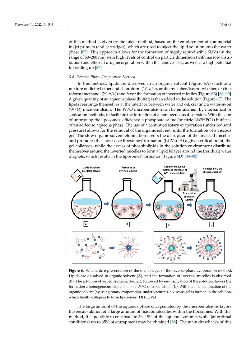
3.4. Reverse-Phase Evaporation Method
In this method, lipids are dissolved in an organic solvent (Figure 6A) (such as amixture of diethyl ether and chloroform (1:1 v/v), or diethyl ether/isopropyl ether, or chlo-roform/methanol (2:1 v/v)) and favor the formation of inverted micelles (Figure 6B) [88–90].A given quantity of an aqueous phase (buffer) is then added to the solution (Figure 6C). Thelipids rearrange themselves at the interface between water and oil, creating a water-in-oil(W/O) microemulsion. The W/O microemulsion can be emulsified, by mechanical orsonication methods, to facilitate the formation of a homogeneous dispersion. With the aimof improving the liposomes’ efficiency, a phosphate saline (or citric-Na2HPO4) buffer isoften added to aqueous phase. The use of a continued rotary evaporation (under reducedpressure) allows for the removal of the organic solvent, until the formation of a viscousgel. The slow organic solvent elimination favors the disruption of the inverted micellesand promotes the successive liposomes’ formation (LUVs). At a given critical point, thegel collapses, while the excess of phospholipids in the solution environment distributethemselves around the inverted micelles to form a lipid bilayer around the (residual) waterdroplets, which results in the liposomes’ formation (Figure 6D) [88–90].
Pharmaceutics 2022, 14, x 13 of 50
ods (TFHM) [86]. The prepared liposomes, characterized by using optical microscopy, ev-idenced an increased encapsulation efficiency (from about 60% to 86%) as a function of the increasing amount of phospholipids and cholesterol, while in vitro (by means of the dialysis membrane) and ex vivo (by means of the chicken intestinal sac,) diffusion studies evidenced an efficient and controlled release process. The study evidenced a similar per-formance of the liposome system prepared by the two different methods [86]. A variation of this method is given by the inkjet method, based on the employment of commercial inkjet printers (and cartridges), which are used to inject the lipid solution into the water phase [87]. This approach allows for the formation of highly reproducible SUVs (in the range of 50–200 nm) with high levels of control on particle dimension (with narrow distribution) and efficient drug incorporation within the nanovesicles, as well as a high potential for scaling up [87].
3.4. Reverse-Phase Evaporation Method In this method, lipids are dissolved in an organic solvent (Figure 6A) (such as a mix-
ture of diethyl ether and chloroform (1:1 v/v), or diethyl ether/isopropyl ether, or chloro-form/methanol (2:1 v/v)) and favor the formation of inverted micelles (Figure 6B) [88–90]. A given quantity of an aqueous phase (buffer) is then added to the solution (Figure 6C). The lipids rearrange themselves at the interface between water and oil, creating a water-in-oil (W/O) microemulsion. The W/O microemulsion can be emulsified, by mechanical or son-ication methods, to facilitate the formation of a homogeneous dispersion. With the aim of improving the liposomes’ efficiency, a phosphate saline (or citric-Na2HPO4) buffer is of-ten added to aqueous phase. The use of a continued rotary evaporation (under reduced pressure) allows for the removal of the organic solvent, until the formation of a viscous gel. The slow organic solvent elimination favors the disruption of the inverted micelles and promotes the successive liposomes’ formation (LUVs). At a given critical point, the gel collapses, while the excess of phospholipids in the solution environment distribute themselves around the inverted micelles to form a lipid bilayer around the (residual) wa-ter droplets, which results in the liposomes’ formation (Figure 6D) [88–90].
Figure 6. Schematic representation of the main stages of the reverse-phase evaporation method. Lipids are dissolved in organic solvent (A), and the formation of inverted micelles is observed (B). The addition of aqueous media (buffer), followed by emulsification of the solution, favors the for-mation a homogeneous dispersion of a W/O microemulsion (C). With the final elimination of the
Figure 6. Schematic representation of the main stages of the reverse-phase evaporation method.Lipids are dissolved in organic solvent (A), and the formation of inverted micelles is observed(B). The addition of aqueous media (buffer), followed by emulsification of the solution, favors theformation a homogeneous dispersion of a W/O microemulsion (C). With the final elimination of theorganic solvent (by using rotary evaporation, under vacuum), a viscous gel is formed in the solution,which finally collapses to form liposomes (D) (LUVs).
The large amount of the aqueous phase encapsulated by the microemulsions favorsthe encapsulation of a large amount of macromolecules within the liposomes. With thismethod, it is possible to encapsulate 30–45% of the aqueous volume, while (at optimalconditions) up to 65% of entrapment may be obtained [88]. The main drawbacks of this

Pharmaceutics 2022, 14, 543 14 of 49
approach is connected with the presence of residual solvent (which can be removed bymeans of the dialysis and centrifugation methods) and with the difficulties of scaling-upthe process. A further disadvantage of this method is that the process is not suitable forfragile molecules (such as peptides), as the drugs to be loaded within the liposomes are indirect contact with an organic solvent. Finally, biomolecules such as enzymes, proteins, oroligonucleotides may undergo a conformational change due to the mechanical agitationand the direct exposure to the organic solvent (such as protein denaturation, breakage ofDNA strands) [88–90].
Recently, the reverse-phase evaporation method has been employed to combine thera-peutic and diagnostic agents in the same lipid (theranostic) nanoformulation for advancedbiomedical applications. Ultra-magnetic liposomes (UMLs), prepared by means of thereverse-phase evaporation method, exhibited a higher magnetic nanoparticle (MNP) load-ing efficiency (about 100-fold), compared to the classical thin-film hydration method [91].Do et al. [92] developed nucleic acid-delivery nano-formulation systems based on mag-netic cationic liposomes (MCLs), by means of reverse-phase evaporation and cosolventsonication techniques. The new MCLs’ nano-formulation composed of the lipids DPPC,DSPC, DOPE, 18:0 PEG2000 PE, 14:0 PEG750/1000/2000 PE, and cationic lipid DMAPAP,showed high capacity and efficiency to form complexes and transfect (CT-26) cells (us-ing the antibiotic-free pFAR4-luc plasmid), thanks to their ability to transfect cells withhigh efficiency. The constructed MCLs (of <200 nm) offer a magnetic resonance imagingcontrast enhancement agent (due to the encapsulated magnetic nanoparticles), and canbe considered a promising nanovector for image-guided gene-delivery therapy. Recently,a (pressure-controlled) encapsulation of graphene quantum dots (GQDs) into liposomenanocarriers has been obtained by the reverse-phase evaporation method [93]. The GQDs-loaded liposomes exhibited a high loading of ultra-small (~4 nm) GQDs into the aqueousliposomes’ cores (45.68 ± 1.44%), which was controllable by the pressure, and exhibiteda very good stability for over a month. Furthermore, the inclusion of the indocyaninegreen (an near-infrared photothermal agent) could convert NIR laser energy into ther-mal energy and break down the liposomes, causing the release of GQDs in 6 min. ThisNIR light-controlled drug-release nanoformulation exhibited a good in vitro (photother-mal) therapeutic performance, and 75% of cancer cells were killed at a concentration of200 µg/mL [93]. The successful development of these controlled-release nanocarriers bythe reverse-phase evaporation method may stimulate future biomedical applications ofadvanced liposome theranostic systems.
4. Downsizing and Post-Formation Processing
For specific biomedical applications, the precise control of particle size (and polydis-persity index—PDI), lamellarity, and homogeneity, is a crucial step in their manufacturingand a fundamental parameter in the products’ specifications. For this purpose, a post-formation processing is required, with the aim of breaking down the initial large MLVsobtained as the final product. Three main procedures, namely, the sonication, extrusion, andthe high-pressure homogenization methods, represent the most employed post-formationtreatments of size reduction (downsizing) within the liposome formation approaches.
4.1. Sonication Method
The sonication method consists in the application of a high (ultrasonic)-energy inputbased on cavitation to the MLVs liposome solution under a passive (inert) atmosphere. Twotypes of sonication techniques are used on an aqueous dispersion of a phospholipid system:namely, the bath sonication and probe sonication techniques [94]. In the probe sonicationmethod (generally used for small volumes), a sonicator tip is immersed into the liposomesolution. The bath vessel is immersed into a water/ice bath to avoid high energy deliveredby the tip, which causes a local warming-up and degradation of the lipidic solution [65,94].For this reason, The main disadvantages of this method are connected with the possiblerelease of metal (titanium) particles from probe tip, which may cause contamination of the

Pharmaceutics 2022, 14, 543 15 of 49
lipid solution. Moreover, with prolonged sonication times (≥1 h), sensitive amounts oflipids can be de-esterified (≥3%) [65,94]. In the bath sonication method (generally used forlarge volumes), the liposome dispersion is placed into a sterile vessel (with a temperature-control system), or under an inert atmosphere. The main disadvantages of this approachare the low encapsulation efficacy, possible phospholipid (or encapsulated compound)degradation, and the high size polydispersity [65,94]. Finally, although sonication is oneof the most used approaches for the formation of SUVs (with diameters in the range of15–25 µm), it does not seem optimal in those cases in which precise physical liposomeproperties are needed.
4.2. Extrusion Method
The extrusion method consists in the extrusion through pore-containing membranes(with sizes ranging from 1 mm down to 25 nm). A heating block set around the extruderallows for performing the extrusion above the phase-transition temperature of the phospho-lipids. Several passes through the polycarbonate membrane filters allow for the formationof (narrow-size distribution) LUV liposomes with dimensions close to the membrane pores’sizes. This method allows for a reproducible result of the final liposome product, as ev-idenced by several investigations performed on various lipid formulations [95–97]. Avariation of this method is given by the maximator device, an extrusion setup consisting ofa thermo-stable supply vessel connected to a high-pressure pumping system [96].
The extrusion methods have a high reproducibility of downsizing. The main disadvan-tage of this method is the sensitive product losses, which represent a limit for large-scaleproductions. A different extrusion process for the production of liposome nanoformu-lations, called the French press method, is based on the extrusion, at high pressures, ofsuspensions of MLV through a small orifice, which results in the formation of SUVs [98].Liposomes formed with this setup are larger than those obtained by means of the son-ication of MLVs [98]. This technique was originally developed to break up cells undermore appropriate (milder) conditions than those used with ultrasound methods (to avoidthe degradation of lipids, proteins, or other sensitive biomolecules during the sonicationprocess). The drawbacks of the method are connected with the difficulty of reaching hightemperatures, and the relatively small working volumes (<50 mL), which are not suitablefor large-scale production [98].
4.3. High-Pressure Homogenization Method
In the homogenization method, the initial liposome suspension (composed of mul-tilamellar liposomes) is continuously injected with a constant high pressure through anorifice, and collides with a fixed stainless-steel wall that causes downsizing of the lipo-somes [99]. The formation of the liposomes’ structure takes place due to cavitation, shearphenomena, and turbulence. With this method, the liposome size distribution may stillbe broad and variable. More specifically, the properties and the size distribution of theliposomes depends on the pressure, temperature, and the number of times that the lipidicsystem is processed within the homogenizer setup. A key role is also played by the initialproperties (and factors) associated with the processed sample, including the lipids’ (andbulk medium’s) composition and ionic strength, and the initial liposomes’ size-distributionand lamellarity. The major drawbacks of method are connected with the use of very high(operating) pressures and possible metal and oil contamination.
5. Novel Technologies for Liposome Preparation
The main drawbacks of conventional liposome formation approaches include the diffi-culty in achieving an easily scalable process for (large-mass) production, and the difficultiesof obtaining elevated encapsulation efficiencies. Moreover, conventional methods may benot suitable for the processing of many (bio-) molecules, as they undergo structural (orfunctional) perturbations/alterations, due their exposure to detergents, organic solventresidues (with sensitive toxicity), and (high) shear homogenization (or sonication) pro-
Pharmaceutics 2022, 14, 543 16 of 49
cesses, which may severely affect the clinical applications. With the aim of overcomingthose critical issues, recently, novel technologies were developed for the production ofliposome nano-formulations.
5.1. Freeze-Drying (Lyophilization) Method
Water-soluble drugs with lipid-based nanoformulations are generally subjected toleakage during preparation and storage on the shelf. Moreover, active drugs may bedegraded because of possible oxidation phenomena and other chemical reactions beforetheir use in drug-delivery applications. Those circumstances represent limiting factors inthe commercial development of liposome nanoformulations. An approach to overcomethese problems consists in the removal of water (ice) from the liposome systems in thefrozen state (and at low pressures) [65,100].
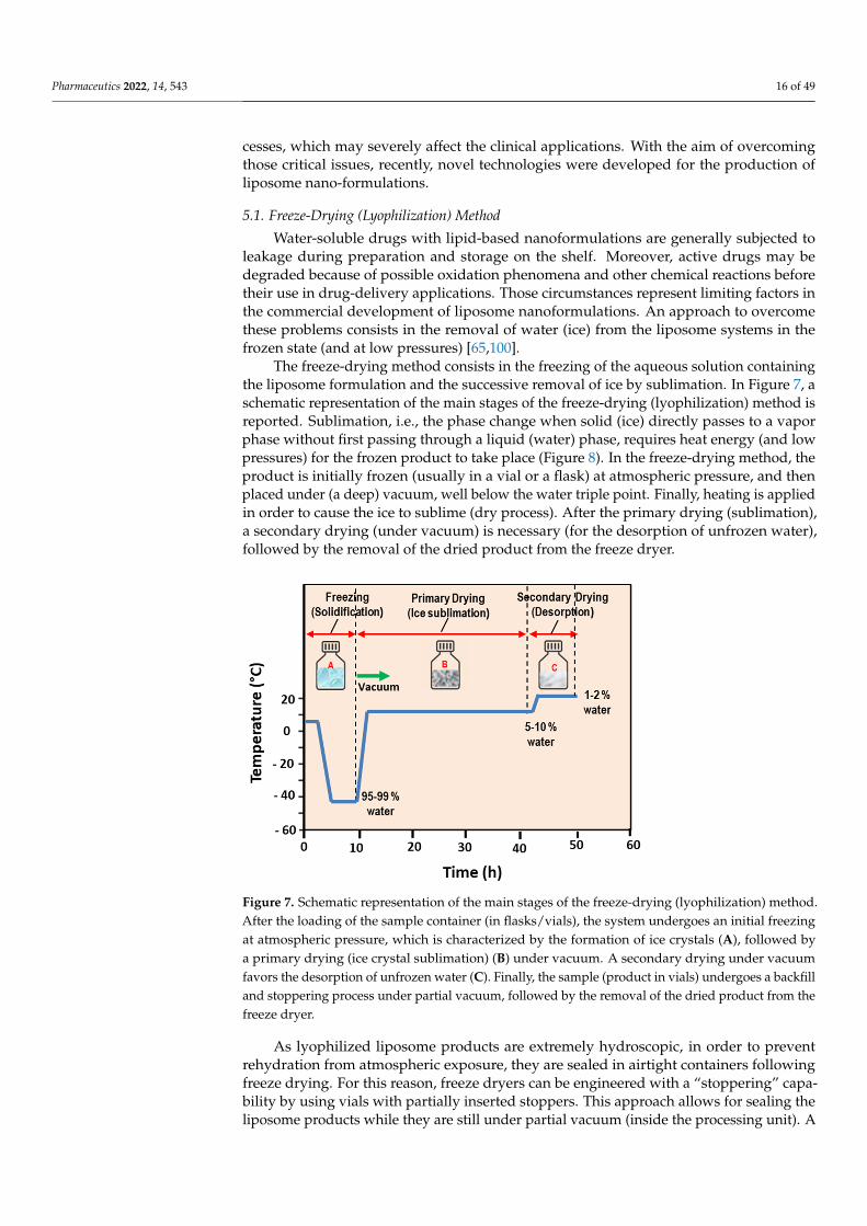
The freeze-drying method consists in the freezing of the aqueous solution containingthe liposome formulation and the successive removal of ice by sublimation. In Figure 7, aschematic representation of the main stages of the freeze-drying (lyophilization) method isreported. Sublimation, i.e., the phase change when solid (ice) directly passes to a vaporphase without first passing through a liquid (water) phase, requires heat energy (and lowpressures) for the frozen product to take place (Figure 8). In the freeze-drying method, theproduct is initially frozen (usually in a vial or a flask) at atmospheric pressure, and thenplaced under (a deep) vacuum, well below the water triple point. Finally, heating is appliedin order to cause the ice to sublime (dry process). After the primary drying (sublimation),a secondary drying (under vacuum) is necessary (for the desorption of unfrozen water),followed by the removal of the dried product from the freeze dryer.
Pharmaceutics 2022, 14, x 16 of 50
bulk medium’s) composition and ionic strength, and the initial liposomes’ size-distribu-tion and lamellarity. The major drawbacks of method are connected with the use of very high (operating) pressures and possible metal and oil contamination.
5. Novel Technologies for Liposome Preparation The main drawbacks of conventional liposome formation approaches include the dif-
ficulty in achieving an easily scalable process for (large-mass) production, and the diffi-culties of obtaining elevated encapsulation efficiencies. Moreover, conventional methods may be not suitable for the processing of many (bio-) molecules, as they undergo struc-tural (or functional) perturbations/alterations, due their exposure to detergents, organic solvent residues (with sensitive toxicity), and (high) shear homogenization (or sonication) processes, which may severely affect the clinical applications. With the aim of overcoming those critical issues, recently, novel technologies were developed for the production of liposome nano-formulations.
5.1. Freeze-Drying (Lyophilization) Method Water-soluble drugs with lipid-based nanoformulations are generally subjected to
leakage during preparation and storage on the shelf. Moreover, active drugs may be de-graded because of possible oxidation phenomena and other chemical reactions before their use in drug-delivery applications. Those circumstances represent limiting factors in the commercial development of liposome nanoformulations. An approach to overcome these problems consists in the removal of water (ice) from the liposome systems in the frozen state (and at low pressures) [65,100].
The freeze-drying method consists in the freezing of the aqueous solution containing the liposome formulation and the successive removal of ice by sublimation. In Figure 7, a schematic representation of the main stages of the freeze-drying (lyophilization) method is reported. Sublimation, i.e., the phase change when solid (ice) directly passes to a vapor phase without first passing through a liquid (water) phase, requires heat energy (and low pressures) for the frozen product to take place (Figure 8). In the freeze-drying method, the product is initially frozen (usually in a vial or a flask) at atmospheric pressure, and then placed under (a deep) vacuum, well below the water triple point. Finally, heating is ap-plied in order to cause the ice to sublime (dry process). After the primary drying (subli-mation), a secondary drying (under vacuum) is necessary (for the desorption of unfrozen water), followed by the removal of the dried product from the freeze dryer.
Figure 7. Schematic representation of the main stages of the freeze-drying (lyophilization) method. After the loading of the sample container (in flasks/vials), the system undergoes an initial freezing Figure 7. Schematic representation of the main stages of the freeze-drying (lyophilization) method.After the loading of the sample container (in flasks/vials), the system undergoes an initial freezingat atmospheric pressure, which is characterized by the formation of ice crystals (A), followed bya primary drying (ice crystal sublimation) (B) under vacuum. A secondary drying under vacuumfavors the desorption of unfrozen water (C). Finally, the sample (product in vials) undergoes a backfilland stoppering process under partial vacuum, followed by the removal of the dried product from thefreeze dryer.
As lyophilized liposome products are extremely hydroscopic, in order to preventrehydration from atmospheric exposure, they are sealed in airtight containers followingfreeze drying. For this reason, freeze dryers can be engineered with a “stoppering” capa-bility by using vials with partially inserted stoppers. This approach allows for sealing theliposome products while they are still under partial vacuum (inside the processing unit). A

Pharmaceutics 2022, 14, 543 17 of 49
backfilling with the dry nitrogen (inert gas) before the products’ sealing (or stoppering) is,finally, performed [100,101]. Although water is the main solvent that must be eliminatedfrom the liposomes solution by the freeze-drying process, there are several nanoformu-lations that are manufactured via freeze-drying processes that require the use of organicco-solvent systems.
Pharmaceutics 2022, 14, x 17 of 50
at atmospheric pressure, which is characterized by the formation of ice crystals (A), followed by a primary drying (ice crystal sublimation) (B) under vacuum. A secondary drying under vacuum fa-vors the desorption of unfrozen water (C). Finally, the sample (product in vials) undergoes a backfill and stoppering process under partial vacuum, followed by the removal of the dried product from the freeze dryer.
Figure 8. Phase diagram of carbon dioxide.
As lyophilized liposome products are extremely hydroscopic, in order to prevent re-hydration from atmospheric exposure, they are sealed in airtight containers following freeze drying. For this reason, freeze dryers can be engineered with a “stoppering” capa-bility by using vials with partially inserted stoppers. This approach allows for sealing the liposome products while they are still under partial vacuum (inside the processing unit). A backfilling with the dry nitrogen (inert gas) before the products’ sealing (or stoppering) is, finally, performed [100,101]. Although water is the main solvent that must be elimi-nated from the liposomes solution by the freeze-drying process, there are several nanofor-mulations that are manufactured via freeze-drying processes that require the use of or-ganic co-solvent systems.
Frozen products present a crystalline or an amorphous glass nanostructure. While crystalline materials present a “eutectic” freezing/melting point (called “collapse temper-ature”), the amorphous formulations exhibit a corresponding “glass transition” tempera-ture and, for this reason, are difficult to be processed with the freeze-drying method.
The freeze-drying method is very useful for the preservation of the shelf stability of the liposome systems, since water can favor unwanted chemical reactions, thus leading to the modification (or even degradation) of the drugs contained in the nanoformulation. The approach is suited to dry thermo-labile liposome products that would be degraded by the heat-drying process. This method, therefore, preserves a large variety of heat-sen-sitive biomaterials, including proteins, pharmaceuticals, tissues, and plasma components. The lyophilized form of the lipid-based pharmaceuticals ensure an increased shelf-life, especially when the inclusion cargoes are given by drugs that are not stable in the aqueous phase [100,101].
With the aim of ensuring the (cryo-) protection of the liposomes’ structure, during the freezing stage of the lyophilization process, sugar macromolecules such as sucrose, lactose, and trehalose are usually incorporated to the liposome systems [100–105]. In this case, during the use of the lipid nanoformulation, upon a rehydration process, the water molecules are able to replace the sugars and liposomes reconstituted without significant
Figure 8. Phase diagram of carbon dioxide.
Frozen products present a crystalline or an amorphous glass nanostructure. Whilecrystalline materials present a “eutectic” freezing/melting point (called “collapse tempera-ture”), the amorphous formulations exhibit a corresponding “glass transition” temperatureand, for this reason, are difficult to be processed with the freeze-drying method.
The freeze-drying method is very useful for the preservation of the shelf stability ofthe liposome systems, since water can favor unwanted chemical reactions, thus leadingto the modification (or even degradation) of the drugs contained in the nanoformulation.The approach is suited to dry thermo-labile liposome products that would be degraded bythe heat-drying process. This method, therefore, preserves a large variety of heat-sensitivebiomaterials, including proteins, pharmaceuticals, tissues, and plasma components. Thelyophilized form of the lipid-based pharmaceuticals ensure an increased shelf-life, espe-cially when the inclusion cargoes are given by drugs that are not stable in the aqueousphase [100,101].
With the aim of ensuring the (cryo-) protection of the liposomes’ structure, duringthe freezing stage of the lyophilization process, sugar macromolecules such as sucrose,lactose, and trehalose are usually incorporated to the liposome systems [100–105]. In thiscase, during the use of the lipid nanoformulation, upon a rehydration process, the watermolecules are able to replace the sugars and liposomes reconstituted without significantchanges to their size. Certain sugars (such as trehalose) are able to mimic the presence ofwater, and are capable of preserving the integrity of dry liposomes and membranes [104]. Liand Deng [100] performed a freeze-drying process on a phospholipid t-butyl alcohol water–sucrose solution by initially freezing at the temperature T = −40 ◦C (for 8 h), successivelydrying (for 48 h at the same temperature), and finally drying the product for 10 h atT = 25 ◦C. They evidenced that the liposomes’ size and polydispersity were decreased withthe increased sucrose concentration, while the dry liposomes could be stored in a sealedcontainer for a long period [100]. Those results confirm, in part, the previous observationsof Kiselev et al. [102,103], who observed a decrease of the vesicles polydispersity, andincreases of the vesicles lifetime in an aqueous solution, extruded DMPC vesicles upon the

Pharmaceutics 2022, 14, 543 18 of 49
addition of sucrose (in the concentration range of 0–40% w/w). In that study, no effects weredetected on the membranes’ thickness and the hydrocarbon chains’ packing. It is worthnoticing that the degree of water absorption strongly depends on the hydrophilic characterof the phospholipid’s head group and, in part, on the specific composition and length ofthe hydrocarbon chain [102,103]. Liposomes formed with this method have a small size(<200 nm); with the use of suitable cryoprotectants, a highly efficient encapsulation (80%)with high stability and reproducibility can be obtained [102,103].
5.2. Dense Gas Technology: Supercritical Fluid-Assisted Methods
Traditional methods of liposome preparation have various shortcomings connectedwith the poor stability, high polydispersity, and high amounts of residual organic solvent. Inparticular, organic solvents used in the conventional preparation of liposomes may degradethe encapsulated active ingredients and may represent a cause of toxicity for human healthand the environment. In this respect, the peculiar properties of dense gases have been usedto substitute many organic solvents, thus enabling novel processing approaches, includingpurification (separation) and size-reduction processes. Dense gases in the region above thecritical point, in fact, have mass transport characteristics similar to gases, and dissolutioncharacteristics similar to liquids and traditional solvents [106,107]. More specifically, su-percritical fluids are (non-condensable) very dense fluids at temperatures and pressuresabove the critical values (i.e., at the point where we observe the disappearance of the linebetween the gas and the liquid phase).
Several new liposome formation methods employ a supercritical fluid (SCF), i.e., afluid maintained under supercritical temperature Tc and pressure Pc (see Figure 8). Inthis state, SCFs are excellent solvents for many lipids components. The variations in theirtemperature (or pressure) lead to sensitive modifications in their density and facilitatethe solubility of a large variety of active components in the SCF. For those main reasons,SCFs are progressively replacing the organic solvents, as they favor a high-performingpurification and separation procedure. [106,107]. More specifically, supercritical carbondioxide (SC-CO2) represents a widely used organic solvent substitute, and is one of themost widely used non-toxic and environmentally friendly dense gases. Moreover, it hasvalues of the critical parameters (i.e., temperature Tc = 31.1 ◦C and pressure Pc = 73.8 bar)that are easily accessible for a large variety of labile bio-materials.
The general supercritical fluid method is characterized by two stages [106,107]. Ini-tially, in the high-pressure (P = 250 bar) part, the dissolution of the lipids in supercriticalcarbon dioxide is performed. Successively, the obtained (supercritical) homogeneous solu-tion is successively expanded at the temperature of T = 60 ◦C, with the addition of (a smallamount, of about 7% v/v) of ethanol. Interestingly, the ethanol total amount is 15-timeslower than in the ethanol injection method. The expanded liquid is then mixed (injectedthrough a nozzle) with a water phase, and liposomes (with encapsulated water-solubledrugs) are formed. This method produces large liposomes with sizes in the range of0.2–4 µm, while only 3% of the liposomes are usually subjected to degradation [107,108].In a variation of this method, the phospholipid(s) are initially mixed, while the SCF andthe co-solvent mixture with the aqueous phase is successively decompressed by spraying it(through a nozzle) [108].
5.2.1. Supercritical Reverse-Phase Evaporation (SC-RPE) Method
Otake et al. [109] reported the first supercritical reverse-phase evaporation technique,using SC-CO2 as the solvent for the phospholipids. The employed experimental setup isschematically reported in Figure 7 [109].
A variable volume chamber was used to progressively decrease the pressure of thesystem. Initially, the lipids, the compressed gas, and an organic co-solvent were combinedand inserted in a (variable-volume) viewing cell, and stirred with a magnetic tip above thelipid phase-transition temperature (Figure 9). The temperature was then raised above thespecific (phospho-) lipid phase-transition temperature and the supercritical temperature of

Pharmaceutics 2022, 14, 543 19 of 49
carbon dioxide. After a given time of equilibration, an aqueous solution (containing thedrug’s molecules) was introduced to the viewing cell through a high-performance liquidchromatography (HPLC) pump. Finally, after the reduction of the pressure through therelease of the compressed CO2 gas, a homogeneous dispersion of LUVs (0.1–1.2 µm) wasformed [109].
Pharmaceutics 2022, 14, x 19 of 50
5.2.1. Supercritical Reverse-Phase Evaporation (SC-RPE) Method Otake et al. [109] reported the first supercritical reverse-phase evaporation technique,
using SC-CO2 as the solvent for the phospholipids. The employed experimental setup is schematically reported in Figure 7 [109].
A variable volume chamber was used to progressively decrease the pressure of the system. Initially, the lipids, the compressed gas, and an organic co-solvent were combined and inserted in a (variable-volume) viewing cell, and stirred with a magnetic tip above the lipid phase-transition temperature (Figure 9). The temperature was then raised above the specific (phospho-) lipid phase-transition temperature and the supercritical tempera-ture of carbon dioxide. After a given time of equilibration, an aqueous solution (contain-ing the drug’s molecules) was introduced to the viewing cell through a high-performance liquid chromatography (HPLC) pump. Finally, after the reduction of the pressure through the release of the compressed CO2 gas, a homogeneous dispersion of LUVs (0.1–1.2 μm) was formed [109].
Figure 9. Schematic representation of the apparatus used in the supercritical reverse-phase evapo-ration method [109].
The SC-RPE method is similar to the decompression method of Castor and Chu [108], since the lipid, aqueous phase, dense gas, and modifier are first combined and then de-pressurized (decompressed) to form liposomes. The main differences is that, in the de-compression method, the depressurization is obtained by spraying the solution through a nozzle, whereas in the SC-RPE method, it is given by the release of the dense gas from a variable-volume cell containing the sample.
An investigation of Imura et al. evidenced that, by varying the pressure and organic co-solvent concentration, it is possible to control the size and trapping efficiency of the formed liposomes [110]. More specifically, liposomes prepared at pressures below 200 bar are resultingly larger than those prepared at >200 bar. The minimum ethanol concentra-tion to obtain water in a CO2 emulsion at 200 bar is 6.8 wt %. This represents the “optimal” and the ethanol “limit concentration” to obtain large unilamellar vesicles with the highest trapping efficiency, while at ethanol concentrations less than 6.8 wt %, multilamellar vesicles are formed since the (osmotic) shrinkage velocities and trapping efficiencies are low [110].
Recently, liposomes of various phospholipids prepared using an improved SC-RPE method (with supercritical CO2) exhibited the formation of a unilamellar vesicle structure with loosely packed phospholipids, with high stability (for one month) at room tempera-ture [111]. Moreover, the SC-RPE method ensured a maximum trapping efficiency of glu-cose of 36% (for 20 mM l-α-dioleoylphosphatidylcholine (DOPC)), compared to less than 10% using the traditional (Bangham) method [111]. The SC-RPE method (with supercriti-cal CO2) was also used for the construction of a novel liposome system consisting of
Figure 9. Schematic representation of the apparatus used in the supercritical reverse-phase evapora-tion method [109].
The SC-RPE method is similar to the decompression method of Castor and Chu [108],since the lipid, aqueous phase, dense gas, and modifier are first combined and thendepressurized (decompressed) to form liposomes. The main differences is that, in thedecompression method, the depressurization is obtained by spraying the solution througha nozzle, whereas in the SC-RPE method, it is given by the release of the dense gas from avariable-volume cell containing the sample.
An investigation of Imura et al. evidenced that, by varying the pressure and organic co-solvent concentration, it is possible to control the size and trapping efficiency of the formedliposomes [110]. More specifically, liposomes prepared at pressures below 200 bar areresultingly larger than those prepared at >200 bar. The minimum ethanol concentration toobtain water in a CO2 emulsion at 200 bar is 6.8 wt %. This represents the “optimal” and theethanol “limit concentration” to obtain large unilamellar vesicles with the highest trappingefficiency, while at ethanol concentrations less than 6.8 wt %, multilamellar vesicles areformed since the (osmotic) shrinkage velocities and trapping efficiencies are low [110].
Recently, liposomes of various phospholipids prepared using an improved SC-RPEmethod (with supercritical CO2) exhibited the formation of a unilamellar vesicle structurewith loosely packed phospholipids, with high stability (for one month) at room temper-ature [111]. Moreover, the SC-RPE method ensured a maximum trapping efficiency ofglucose of 36% (for 20 mM l-α-dioleoylphosphatidylcholine (DOPC)), compared to lessthan 10% using the traditional (Bangham) method [111]. The SC-RPE method (with su-percritical CO2) was also used for the construction of a novel liposome system consistingof DPPC/Cholesterol/C16-Arg2 lipopeptides (LPs)/DSPE-PEG2000 (60/30/5/5), for thedelivery of bovine serum albumin (BSA) protein [112]. The constructed liposome nanofor-mulation (with diameter of about 1000 nm) evidenced an enhanced colloidal stability (of90% over 40 h) and up to 70% entrapment efficiency for BSA, which was six-times higherthan that obtained with the Bangham method. Small-angle X-ray scattering (SAXS) anddifferential scanning calorimetry (DSC) experiments evidenced that some amounts of LPsinduced structural changes and phase transitions in the DPPC lamellar structure, withan improvement of the nanocarrier properties [112]. Those studies evidence the possi-bility of developing novel organic-solvent-free liposome nanoformulations with desiredperformance, which allows for addressing important environmental concerns.

Pharmaceutics 2022, 14, 543 20 of 49
5.2.2. Supercritical Anti-Solvent (SAS) Method
In this method, a solution containing an organic solvent and the solute (lipids andactive drugs) is placed in contact with a SCF, (such as SC-CO2) which is (completely)miscible with the organic solvent but acts as anti-solvent for the solute [113]. The dissolutionof SC-CO2 in the liquid phase and the successive organic solvent extraction favors theprecipitation of the lipidic nanoparticles. The processed solution is successively hydrated inan aqueous buffer solution, leading to the formation of liposomes. To remove any organicsolvent in excess, a washing stage (with pure CO2) is finally performed. In Figure 10, wereport a schematic representation of the SAS method for liposome preparation.
Pharmaceutics 2022, 14, x 20 of 50
DPPC/Cholesterol/C16-Arg2 lipopeptides (LPs)/DSPE-PEG2000 (60/30/5/5), for the deliv-ery of bovine serum albumin (BSA) protein [112]. The constructed liposome nanoformu-lation (with diameter of about 1000 nm) evidenced an enhanced colloidal stability (of 90% over 40 h) and up to 70% entrapment efficiency for BSA, which was six-times higher than that obtained with the Bangham method. Small-angle x-ray scattering (SAXS) and differ-ential scanning calorimetry (DSC) experiments evidenced that some amounts of LPs in-duced structural changes and phase transitions in the DPPC lamellar structure, with an improvement of the nanocarrier properties [112]. Those studies evidence the possibility of developing novel organic-solvent-free liposome nanoformulations with desired perfor-mance, which allows for addressing important environmental concerns.
5.2.2. Supercritical Anti-Solvent (SAS) Method In this method, a solution containing an organic solvent and the solute (lipids and
active drugs) is placed in contact with a SCF, (such as SC-CO2) which is (completely) mis-cible with the organic solvent but acts as anti-solvent for the solute [113]. The dissolution of SC-CO2 in the liquid phase and the successive organic solvent extraction favors the precipitation of the lipidic nanoparticles. The processed solution is successively hydrated in an aqueous buffer solution, leading to the formation of liposomes. To remove any or-ganic solvent in excess, a washing stage (with pure CO2) is finally performed. In Figure 10, we report a schematic representation of the SAS method for liposome preparation.
The SAS method represents, then, a relatively simple, efficient, and environmentally friendly process that enables very-low residual solvent contents. The SAS approach also allows for the processing of molecules with poor solubility, for the production of high quantities of liposomes [113]. With the SAS method, both hydrophobic and hydrophilic drugs (that do not need to be dissolved in SC-CO2) can be entrapped in lipid vesicles.
Figure 10. Schematic representation of the SAS method for liposome preparation. The SCF CO2 is pumped to the top of the high-pressure vessel until the system reaches a constant temperature and pressure. Subsequently, an organic solution containing the lipids and active substance is sprayed (through an atomization nozzle) as fine droplets into the above SCF bulk phase. Liposomes are finally formed in a successive hydration step. In the continuous antisolvent (CAS) method, the addition of the hydration unit allows for the hydration of the lipid suspension in the same autoclave under pressure.
Lesoin et al. [113] used the SAS method to produce liposomes from lecithin liposomes and compared it with the TFH (Bangham) method. The liposomes produced with the SAS method exhibited a narrower and more reproducible particle size distribution (ranging
Figure 10. Schematic representation of the SAS method for liposome preparation. The SCF CO2 ispumped to the top of the high-pressure vessel until the system reaches a constant temperature andpressure. Subsequently, an organic solution containing the lipids and active substance is sprayed(through an atomization nozzle) as fine droplets into the above SCF bulk phase. Liposomes arefinally formed in a successive hydration step. In the continuous antisolvent (CAS) method, theaddition of the hydration unit allows for the hydration of the lipid suspension in the same autoclaveunder pressure.
The SAS method represents, then, a relatively simple, efficient, and environmentallyfriendly process that enables very-low residual solvent contents. The SAS approach alsoallows for the processing of molecules with poor solubility, for the production of highquantities of liposomes [113]. With the SAS method, both hydrophobic and hydrophilicdrugs (that do not need to be dissolved in SC-CO2) can be entrapped in lipid vesicles.
Lesoin et al. [113] used the SAS method to produce liposomes from lecithin liposomesand compared it with the TFH (Bangham) method. The liposomes produced with the SASmethod exhibited a narrower and more reproducible particle size distribution (rangingfrom 0.1 µm to 100 µm). The encapsulation efficiency was, however, lower (10–20%) thanthat obtained using the TFH method (20%); the stability in suspension was low (after onemonth of storage at 4 ◦C) in both preparation methods. The SAS process was upgraded tothe continuous anti-solvent (CAS) process, whereby the micronization and the successiveliposome hydration process are performed in the same autoclave under pressure [114].
5.2.3. Rapid Expansion of a Supercritical Solution (RESS) Method
The RESS technique is carried out in two steps: initially, a solution containing lipids isdissolved in ethanol (5–10% of v/v) and supercritical CO2 within an extractor. The resulting

Pharmaceutics 2022, 14, 543 21 of 49
solution is depressurized through a heated nozzle in a low-pressure chamber [115–117].The rapid expansion/decompression (at supersonic speeds) through a nozzle favors thedecrease of the pressure and the evaporation of CO2, thus leading to the supersaturation,and then, to the precipitation of the solid (that will be collected from the gaseous stream).In Figure 11, we report a schematic representation of the RESS method (adapted) forliposome preparation.
Pharmaceutics 2022, 14, x 21 of 50
from 0.1 μm to 100 μm). The encapsulation efficiency was, however, lower (10–20%) than that obtained using the TFH method (20%); the stability in suspension was low (after one month of storage at 4 °C) in both preparation methods. The SAS process was upgraded to the continuous anti-solvent (CAS) process, whereby the micronization and the successive liposome hydration process are performed in the same autoclave under pressure [114].
5.2.3. Rapid Expansion of a Supercritical Solution (RESS) Method The RESS technique is carried out in two steps: initially, a solution containing lipids
is dissolved in ethanol (5–10% of v/v) and supercritical CO2 within an extractor. The re-sulting solution is depressurized through a heated nozzle in a low-pressure chamber [115–117]. The rapid expansion/decompression (at supersonic speeds) through a nozzle favors the decrease of the pressure and the evaporation of CO2, thus leading to the supersatura-tion, and then, to the precipitation of the solid (that will be collected from the gaseous stream). In Figure 11, we report a schematic representation of the RESS method (adapted) for liposome preparation.
Figure 11. Schematic representation of the RESS method for liposome preparation. Initially, lipids are dissolved in supercritical CO2 and ethanol (5–10% of v/v) within an extractor. The resulting so-lution is depressurized through a heated nozzle in a low-pressure chamber. Finally, the formation of particles is generated, due to the supersaturation.
The rapid depressurization causes the lipids’ desolvation, which favors the formation of layers around the droplets, and finally, the formation of liposomes. The obtained lipo-somes exhibit a oligolamellar vesicle structure with a broad size distribution (ranging from 50 nm to nearly 1 μm). Either hydrophobic or hydrophilic drugs can be encapsulated by this method. The size (size distribution) and encapsulation efficiency (EE) of the lipo-somes can be regulated by modifying the pre-expansion pressure, the flow rate, and the nozzle position in the low-pressure chamber (reactor). The main drawbacks of this ap-proach are connected with the poor solubility of most of the biomaterials (e.g., polymer-based macromolecules) in SC-CO2, and the difficulty of the separation between co-sol-vents and vesicles during the depressurization process, which causes a sensitive increase of the liposome production costs [115–117].
A modified RESS technique was applied to encapsulate an essential oil (extracted from Atractylodes macrocephala Koidz) into liposomal nanocarriers [118]. Lipid compo-nents and the drugs (essential oil) were dissolved in the mixture with SC-CO2/ethanol, and successively, the solution was sprayed into an aqueous medium through a nozzle to
Figure 11. Schematic representation of the RESS method for liposome preparation. Initially, lipidsare dissolved in supercritical CO2 and ethanol (5–10% of v/v) within an extractor. The resultingsolution is depressurized through a heated nozzle in a low-pressure chamber. Finally, the formationof particles is generated, due to the supersaturation.
The rapid depressurization causes the lipids’ desolvation, which favors the formationof layers around the droplets, and finally, the formation of liposomes. The obtained lipo-somes exhibit a oligolamellar vesicle structure with a broad size distribution (ranging from50 nm to nearly 1 µm). Either hydrophobic or hydrophilic drugs can be encapsulated bythis method. The size (size distribution) and encapsulation efficiency (EE) of the liposomescan be regulated by modifying the pre-expansion pressure, the flow rate, and the nozzleposition in the low-pressure chamber (reactor). The main drawbacks of this approach areconnected with the poor solubility of most of the biomaterials (e.g., polymer-based macro-molecules) in SC-CO2, and the difficulty of the separation between co-solvents and vesiclesduring the depressurization process, which causes a sensitive increase of the liposomeproduction costs [115–117].
A modified RESS technique was applied to encapsulate an essential oil (extracted fromAtractylodes macrocephala Koidz) into liposomal nanocarriers [118]. Lipid componentsand the drugs (essential oil) were dissolved in the mixture with SC-CO2/ethanol, andsuccessively, the solution was sprayed into an aqueous medium through a nozzle to forma liposome suspension. By changing the expansion processing conditions (such as thetemperature and pressure of SC-CO2 and the amount of ethanol), it was possible to controlthe performances of the formation process. Under optimum conditions (P = 30 MPa,T = 338 ◦K and 15% ethanol mole fraction in SC-CO2), the formed liposomes appeared asdouble-layered colloidal spheres (size = 173 nm) with a narrow size distribution, and anentrapment efficiency (EE) of 82.18% (and drug loading of 5.18%) [118].

Pharmaceutics 2022, 14, 543 22 of 49
The RESS method was recently used to produce melatonin-loaded liposomes (MLL) [119].The results evidenced that 140 bar was the best pressure to obtain a maximum value ofEE (82.2%). MLL characterizations, performed using infrared spectroscopy, transmissionelectron microscopy, light scattering, and gas chromatograph-mass spectrometry, evidencedan average nanocarrier diameter of 66 nm with a uniform size distribution. Stabilitytests evidenced that MLL maintained a good preservation duration, and residual solventexperiments indicated that only 1.03% (mass ratio) of ethanol remained in the products.Simulated release (through an in vitro simulated digestion experiment) indicated a slowrelease feature in early digestive stages and a more thorough characteristic in the late stagesof digestion [119]. Finally, phosphatidylcholine (PC) liposomes encapsulating vitamin C(VC) were investigated by the rapid expansion of the supercritical solution process (RESS),through the combined effects of temperature, pressure, and feeding ratio of VC against thedrug-loading content of the prepared liposomes [120]. Under optimal operating conditions(pressure of 25 MPa, temperature of 48 ◦C, and feed ratio of VC against PC of 0.25), theVC-loaded liposomes exhibited the loading content of 75.38 ± 1.03%, and well-definedand reproducible particle sizes (270.4 ± 5.2 nm), PDI (0.254 ± 0.010), and zeta-potential(−41.7 ± 0.9 mv) [120].
5.2.4. Supercritical-Assisted Liposome Formation (SuperLip) Method
In the SuperLip method, the ethanol solution and CO2 are continuously fed to ahomogenizer, forming an expanded liquid which is then delivered to a precipitationvessel. In this vessel, water with droplets containing drugs are produced by means of anatomization process (inside a high-pressure vessel). The droplets are then surrounded bya lipid layer (thus favoring the formation of w/CO2 emulsion) which falls into the waterpool placed at the bottom of the vessel, where liposomes are formed (Figure 12) [121].
The SuperLip technique allows for a sensitive control of the liposomes’ size (in thenanometric and sub-micrometric range) and size distributions with a sensitive reduction ofthe residual solvent. Moreover, the high encapsulation efficiencies (from about 80% up to99% for hydrophilic compounds) allow for the entrapment of a large variety of active drugs,including antibodies, proteins, antibiotics, antioxidants, essential oils, and dyes [121]. Theentrapment of additives (such as cholesterol and phosphatidylethanolamine) on the lipidicdouble layer compartment of liposomes allows for obtaining a more compact structure withencapsulation efficiencies of 96% (cholesterol) and 95% (phosphatidylethanolamine) [121].The versatility of this method may stimulate the industrial applications in a large variety oftechnology fields, such as pharmaceutics, cosmetics, textiles, and nutraceuticals.
By using the SuperLip process, lutein (a lipophilic ophthalmic nano-drug) has beenencapsulated in liposomes with mean diameters between 153 ± 38 and 267 ± 56 nm, withhigh lutein encapsulation efficiencies (between 86.5 ± 0.4% and 97.8 ± 1.2%) [122]. Thevariation of temperature for the production of liposomes showed a significant impact onlutein retention time (within the double lipid layer). Lutein drug release from liposomesproduced at 35 ◦C ended after almost 4.5 days, whereas liposomes produced at 40 ◦Cshowed a faster lutein release in 3 days. Then, vesicles obtained at 45 ◦C released theirlutein content in only 2 days [122]. Moreover, the SuperLip method has been employed toproduce ampicillin-loaded liposomes that were successively entrapped into alginate gels,while a final treatment with supercritical CO2 drying allowed for obtaining an aerogel [123].This combined approach allowed for obtaining a meta-carrier (i.e., a carrier within anothercarrier) with a drug release that works with two mass-transfer resistances (in series).The first mass resistance was given by the structure of the aerogel, during the release ofampicillin not entrapped into liposomes. The second mass resistance was given by thelipidic double layer of liposomes. The initial liposomes (with a diameter of 200 ± 77 nmand EE of 69.5 ± 1.2%) were successfully entrapped into the aerogels (as confirmed by EDXanalysis), while the antibiotic entrapped into simple liposomes alone was totally releasedafter about 3 days. The antibiotic entrapped into a meta-carrier reached a plateau afterabout 5 days [123].

Pharmaceutics 2022, 14, 543 23 of 49
Pharmaceutics 2022, 14, x 23 of 50
another carrier) with a drug release that works with two mass-transfer resistances (in se-ries). The first mass resistance was given by the structure of the aerogel, during the release of ampicillin not entrapped into liposomes. The second mass resistance was given by the lipidic double layer of liposomes. The initial liposomes (with a diameter of 200 ± 77 nm and EE of 69.5 ± 1.2%) were successfully entrapped into the aerogels (as confirmed by EDX analysis), while the antibiotic entrapped into simple liposomes alone was totally re-leased after about 3 days. The antibiotic entrapped into a meta-carrier reached a plateau after about 5 days [123].
Figure 12. SuperLip method and mechanism of liposome formation. A high-pressure vessel is filled with an expanded liquid mixture (formed by PLs/ethanol/CO2 containing drugs). Water droplets are produced by atomization inside a high-pressure vessel. These droplets are rapidly surrounded by a lipid layer, forming a w/CO2 emulsion (inverted micelle). Finally, liposomes (w/w emulsion) are formed when they fall in the water pool located at the bottom of the vessel.
5.2.5. Depressurization of an Expanded Liquid Organic Solution into Aqueous Suspen-sion (DELOS) Method
In the DELOS method [124,125], the lipids (and active drugs) are first dissolved in an organic solvent (such as ethanol) contained in a vessel at fixed temperature and pressure, and then mixed with SC-CO2 (used as a co-solvent). The mixture (depressurized at 35–55 bar) is then expanded into CO2, and (through a nozzle) is successively injected in a vessel containing water bath and active drugs. The evaporation of part of ethanol allows the contact between lipids (transported by bubbles) and water, and favors the formation of liposomes. (Figure 13). The main advantages of the DELOS method are that the resulting liposomes are small in size, have relatively high uniformity and homogeneity (of spherical
Figure 12. SuperLip method and mechanism of liposome formation. A high-pressure vessel is filledwith an expanded liquid mixture (formed by PLs/ethanol/CO2 containing drugs). Water dropletsare produced by atomization inside a high-pressure vessel. These droplets are rapidly surrounded bya lipid layer, forming a w/CO2 emulsion (inverted micelle). Finally, liposomes (w/w emulsion) areformed when they fall in the water pool located at the bottom of the vessel.
5.2.5. Depressurization of an Expanded Liquid Organic Solution into Aqueous Suspension(DELOS) Method
In the DELOS method [124,125], the lipids (and active drugs) are first dissolvedin an organic solvent (such as ethanol) contained in a vessel at fixed temperature andpressure, and then mixed with SC-CO2 (used as a co-solvent). The mixture (depressurizedat 35–55 bar) is then expanded into CO2, and (through a nozzle) is successively injected ina vessel containing water bath and active drugs. The evaporation of part of ethanol allowsthe contact between lipids (transported by bubbles) and water, and favors the formation ofliposomes. (Figure 13). The main advantages of the DELOS method are that the resultingliposomes are small in size, have relatively high uniformity and homogeneity (of sphericalshape), and enhanced colloidal stability, as well as the possibility of reducing the usage ofsterols [124,125]. However, the residual ethanol concentration does not always ensure thefull biocompatibility and safety of the liposome nanoformulation products [124,125].

Pharmaceutics 2022, 14, 543 24 of 49
Pharmaceutics 2022, 14, x 24 of 50
shape), and enhanced colloidal stability, as well as the possibility of reducing the usage of sterols [124,125]. However, the residual ethanol concentration does not always ensure the full biocompatibility and safety of the liposome nanoformulation products [124,125].
Figure 13. Schematic representation of the DELOS method. The lipids dissolved in organic solvent contained in a vessel at desired temperature and pressure, are mixed with SC-CO2 (used as a co-solvent to the organic solvent). Then, the mixture (depressurized at 35–55 bar) is expanded into CO2, and injected through a nozzle into in a vessel containing water bath and active drugs, where the liposomes are formed.
The engineering of new (pH-sensitive) lipid nano-vesicles, for the delivery of miR-NAs (and other small RNAs) for the triggering of a suppressive response into tumor cells, was recently reported [126]. The nanocarriers, developed by means of the DELOS tech-nique, exhibited an enhanced stability upon storage that preserved their (unilamellar) morphology, size (<150 nm), and low polydispersity (<0.25), for at least up to 6 months. The functionality of the developed pH-sensitive nanocarrier was tested for miRNA deliv-ery against selected neuroblastoma (a common aggressive extracranial solid tumor in chil-dren). The high stability, together with their simple and effective (one-step, green, and scalable) manufacturing procedure, makes these sRNA nanocarriers highly attractive for the translation to the clinical phases, compared to other nanocarriers with low stability and with challenging (multistep) scalability [126]. Recently, the DELOS method has been employed to prepare α-galactosidase-loaded nanoliposomes (nanoGLA) for the treatment of Fabry disease (a lysosomal storage disease that arises from a deficiency of the enzyme α-galactosidase A (GLA)) [127]. Within the investigation, a nanoformulation for (preclin-ical) in vivo studies was developed by implementing a Quality by Design (QbD) ap-proach. This methodology allows for developing efficient drug-manufacturing and con-trol methods. Through a risk analysis and a Design of Experiments (DoE), it was possible to obtain the critical parameters (such as the GLA and lipid concentration) for achieving a stable nanoformulation, and to optimize the production process for in vivo preclinical testing [127].
In conclusion, apart from being organic solvent-free methods, the supercritical fluidic methods offer, in general, many other advantages—such as the use of CO2 as a cheap and environmentally harmless solvent, the possibility of controlling particle size, in situ steri-lization, and large-scale production. However, the disadvantages of the supercritical fluid technique are mainly connected with the high cost, low yield, and the use of high pres-sures (i.e., 200–350 bar), which require special infrastructures. Those circumstances dis-courage their applications for the industrial development of liposomal technologies.
Figure 13. Schematic representation of the DELOS method. The lipids dissolved in organic solventcontained in a vessel at desired temperature and pressure, are mixed with SC-CO2 (used as a co-solvent to the organic solvent). Then, the mixture (depressurized at 35–55 bar) is expanded into CO2,and injected through a nozzle into in a vessel containing water bath and active drugs, where theliposomes are formed.
The engineering of new (pH-sensitive) lipid nano-vesicles, for the delivery of miRNAs(and other small RNAs) for the triggering of a suppressive response into tumor cells, wasrecently reported [126]. The nanocarriers, developed by means of the DELOS technique,exhibited an enhanced stability upon storage that preserved their (unilamellar) morphology,size (<150 nm), and low polydispersity (<0.25), for at least up to 6 months. The functionalityof the developed pH-sensitive nanocarrier was tested for miRNA delivery against selectedneuroblastoma (a common aggressive extracranial solid tumor in children). The high stabil-ity, together with their simple and effective (one-step, green, and scalable) manufacturingprocedure, makes these sRNA nanocarriers highly attractive for the translation to theclinical phases, compared to other nanocarriers with low stability and with challenging(multistep) scalability [126]. Recently, the DELOS method has been employed to pre-pare α-galactosidase-loaded nanoliposomes (nanoGLA) for the treatment of Fabry disease(a lysosomal storage disease that arises from a deficiency of the enzyme α-galactosidase A(GLA)) [127]. Within the investigation, a nanoformulation for (preclinical) in vivo studieswas developed by implementing a Quality by Design (QbD) approach. This methodologyallows for developing efficient drug-manufacturing and control methods. Through a riskanalysis and a Design of Experiments (DoE), it was possible to obtain the critical parameters(such as the GLA and lipid concentration) for achieving a stable nanoformulation, and tooptimize the production process for in vivo preclinical testing [127].
In conclusion, apart from being organic solvent-free methods, the supercritical fluidicmethods offer, in general, many other advantages—such as the use of CO2 as a cheapand environmentally harmless solvent, the possibility of controlling particle size, in situsterilization, and large-scale production. However, the disadvantages of the supercriticalfluid technique are mainly connected with the high cost, low yield, and the use of highpressures (i.e., 200–350 bar), which require special infrastructures. Those circumstancesdiscourage their applications for the industrial development of liposomal technologies.
5.3. Microfluidic (Channel) Methods
In the microfluidic method, lipids dissolved in ethanol (or isopropanol) solvent, are suc-cessively propelled within microscopic channels (with 5–500-µm cross-section) [128–130].The alcoholic phospholipids solution, focused between two aqueous streams in a microflu-idic channel (microchannel), generate a (hydrodynamic) laminar flow and a (diffusive)

Pharmaceutics 2022, 14, 543 25 of 49
mixing at the two (liquid) interfaces that favors the lipids’ self-assembly into vesicles. Withthe precise control of mixing and the fluid flow rates, this method allows for the productionof small (monodisperse) liposome nanoformulations with controllable sizes and distribu-tions, with the use of low-toxicity solvents (such as ethanol). Compared to traditional bulkmethods, the final product does not require post-production processing (i.e., extrusion,sonication). Although the microfluidic method offers high versatility and flexibility, themain disadvantages of this approach are connected with use of organic solvents, sensitivemechanic agitation, and the difficulty of large-scale production [128–130].
In a recent investigation of Jahn et al. [129], the liposome system (consisting of thelipids DMPC, cholesterol, and dihexadecyl phosphate (DCP)) formed by the microflu-idic process evidenced that, by modifying the alcohol-to-aqueous (volumetric) flow rateratio, the vesicle sizes (and distributions) were tunable over a mean diameter (between50–150 nm). The authors also observed that the formation of liposomes strongly dependson the (focused) alcohol stream width and its mixing with the aqueous stream [129].
In Figure 14A, we report a three-inlet microfluidic setup representation, illustrating the(SUV) liposome self-assembly. We also report (Figure 14B) the confocal microscopy imagesevidencing the hydrodynamic focusing of an isopropyl alcohol (IPA) stream containingsulforhodamine B, by two adjacent aqueous buffer streams (not visible), for 7 different flowrate ratios (FRRs). Finally, the corresponding liposome size distributions at different FRRsare reported in Figure 14C [129].
Pharmaceutics 2022, 14, x 25 of 50
5.3. Microfluidic (Channel) Methods In the microfluidic method, lipids dissolved in ethanol (or isopropanol) solvent, are
successively propelled within microscopic channels (with 5–500-μm cross-section) [128–130]. The alcoholic phospholipids solution, focused between two aqueous streams in a microfluidic channel (microchannel), generate a (hydrodynamic) laminar flow and a (dif-fusive) mixing at the two (liquid) interfaces that favors the lipids’ self-assembly into vesi-cles. With the precise control of mixing and the fluid flow rates, this method allows for the production of small (monodisperse) liposome nanoformulations with controllable sizes and distributions, with the use of low-toxicity solvents (such as ethanol). Compared to traditional bulk methods, the final product does not require post-production processing (i.e., extrusion, sonication). Although the microfluidic method offers high versatility and flexibility, the main disadvantages of this approach are connected with use of organic sol-vents, sensitive mechanic agitation, and the difficulty of large-scale production [128–130].
In a recent investigation of Jahn et al. [129], the liposome system (consisting of the lipids DMPC, cholesterol, and dihexadecyl phosphate (DCP)) formed by the microfluidic process evidenced that, by modifying the alcohol-to-aqueous (volumetric) flow rate ratio, the vesicle sizes (and distributions) were tunable over a mean diameter (between 50–150 nm). The authors also observed that the formation of liposomes strongly depends on the (focused) alcohol stream width and its mixing with the aqueous stream [129].
In Figure 14A, we report a three-inlet microfluidic setup representation, illustrating the (SUV) liposome self-assembly. We also report (Figure 14B) the confocal microscopy images evidencing the hydrodynamic focusing of an isopropyl alcohol (IPA) stream con-taining sulforhodamine B, by two adjacent aqueous buffer streams (not visible), for 7 dif-ferent flow rate ratios (FRRs). Finally, the corresponding liposome size distributions at different FRRs are reported in Figure 14C [129].
Figure 14. Schematic representation of a three-inlet microfluidic setup, (A). Confocal microscopy (false-color) pictures of the hydrodynamic focusing of an isopropyl alcohol (IPA) stream (that con-tains sulforhodamine (B) by two (aqueous buffer) streams, for 7 flow rate ratios (FRRs), increasing from 5 to 35 (increments of 5 from left to right) at a total constant volumetric flow rate VFR = 100 μL/min (B). Final liposome size distribution at different FRRs (C). Adapted with permission from Jahn et al. [129], Copyright 2007 American Chemical Society.
Figure 14. Schematic representation of a three-inlet microfluidic setup, (A). Confocal microscopy(false-color) pictures of the hydrodynamic focusing of an isopropyl alcohol (IPA) stream (that containssulforhodamine (B) by two (aqueous buffer) streams, for 7 flow rate ratios (FRRs), increasing from 5to 35 (increments of 5 from left to right) at a total constant volumetric flow rate VFR = 100 µL/min (B).Final liposome size distribution at different FRRs (C). Adapted with permission from Jahn et al. [129],Copyright 2007 American Chemical Society.
The effects of the nano-formulation parameters on the colloidal stability and pharma-ceutical properties of nano-liposomes remotely loaded with dexamethasone were recentlyinvestigated by using the microfluidic and the TFH methods [131]. The liposomes gen-erated by the microfluidic method showed a unilamellar structure, while the liposomesproduced by the TFH technique were multilamellar. Under the same remote loading condi-tions (using a calcium acetate gradient), both formulations released the drug for almost onemonth, while a higher loading capacity and low batch-to-batch differences were observed

Pharmaceutics 2022, 14, 543 26 of 49
for liposomes obtained by the microfluidic method. In vitro studies showed that bothformulations exhibited a non-toxic behavior, associated to human adult retinal pigmentepithelial cell line-19 (ARPE-19) cells, and efficiently reduced the inflammation, with the li-posomes obtained by the microfluidic technique slightly outperforming [131]. Those resultsevidence that the microfluidic technique offers advantages for the generation of liposomalnanoformulations with an enhanced drug-controlled release and biopharmaceutical profileand with high scalability.
In the last decades, a variety of new microfluidic methods have been developed for theformation of liposome nanoformulations for biomedical applications. Among many varia-tions of this method, the micro hydrodynamic focusing (MHF) method developed by Jahnet al. [129] is able to produces(40–140 nm) homogeneous SUVs and LUVs with excellentcontrol of the flow and mixing conditions. Moreover, high-throughput novel microfluidicarchitectures can be engineered for the mass production of liposome nanoformulation.Finally, on-chip liposome loading with (both hydrophilic and lipophilic) bio-active com-pounds represent an exciting research field to develop using this method [129,130].
In the microfluidic droplets (MD) method, two immiscible phases (such as waterand oil) are forced to flow (under specific conditions) into a microchannel and generate(uniformly sized) small droplets of one phase. With this method, the dissolution of lipidsin hexane allow for the formation of giant (4–20-µm) liposomes. In the work of Shumet al. [132] a water-in-oil-in-water (w/o/w) double emulsion was prepared by the microflu-idic technique with the lipid dissolved in the central oil phase (a volatile mixture of tolueneand chloroform). By evaporating the organic layer, the monolayers transform, in lipidbilayers, into giant vesicles [132].
The pulsed jet flow microfluidic method [133] (Figure 9) consists in drying the solutioncontaining phospholipid in microtubes (micro-capillary). The formed lipid film is then hy-drated (within the microtubes) through a perfusion process that forms giant (200–500-µm)vesicles of uniform size with high encapsulation efficiency. The lipid bilayer is formed bybringing in contact two aqueous drops coated with a lipid monolayer within an oil phasecontaining the phospholipids. The draining of oil film between the two drops favors theformation of a planar bilayer, into which a periodic pulse of a fluid jet of aqueous solution(buffer) is injected (using a micro-dispenser). The pulsed jet flow, directed in the interfaceregion between the two water drops, results in the formation of giant vesicles (Figure 15).One limitation of this method is connected with the difficulty of automation process, asthe control of the position of the microcapillary (close to the bilayer) should be performedmanually [133].
Recent innovations of the microfluidic technique includes the continuous flow lipo-somes formation, based on the transmembrane pH (or ion) concentration gradient, which iscreated by using an on-chip microdialysis membrane [134,135]. By using a thermoplastic mi-crofabrication method, it has been possible to develop fully integrated microfluidic devicesthat favors a low-cost (scale-up) technology for the production of liposomal nanocarriers, ina (continuous) flow process. This integrated method (called pharmacy-on-a-chip) allows forthe large-scale production (at about 100 mg/h lipid) of a new generation of fully optimized,multi-agent, and targeted liposomal nanoformulations [134,135].
5.4. Membrane Contactor Method
The membrane contactor technique is based on a modified ethanol injection method.The reference setup consists of two pressurized vessels, one for an organic phase containinglipids, and the other for an aqueous phase, separated by a special porous glass membrane,having pore sizes that allow for the flow of the organic phase [68,136,137]. Often, the(alternative) polypropylene hollow fibers are employed as the membrane, as they allowfor larger areas and uniform flows. A lipid phase dissolved in alcohol (such as ethanol), ispressed through the porous membrane and extruded into the aqueous phase, which flowsin a tangential direction to the membrane surface (Figure 16). Upon the contact betweenthe organic solution with the aqueous phase flow, the lipid molecules self-assemble into

Pharmaceutics 2022, 14, 543 27 of 49
liposomes, and are finally collected at the exit portal of the membrane. At the end of theprocess, the ethanol is removed by rotary evaporation (under reduced pressure), while thegenerated liposomes can be stabilized by magnetic stirring. The porous membrane modulecan be regenerated by washing (flushing) with a water/ethanol mixture [65,136–138].
Pharmaceutics 2022, 14, x 27 of 50
Figure 15. Schematic representation of the pulsed jet flow method. The lipids initially dissolved in the organic phase self-assemble (at the oil–water interface) as monolayers. The periodic pulses of a fluid jet (of a buffer) is directed at the interface of two nearby W/O microemulsions, and favors the formation of giant unilamellar vesicles [133].
Recent innovations of the microfluidic technique includes the continuous flow lipo-somes formation, based on the transmembrane pH (or ion) concentration gradient, which is created by using an on-chip microdialysis membrane [134,135]. By using a thermoplastic microfabrication method, it has been possible to develop fully integrated microfluidic de-vices that favors a low-cost (scale-up) technology for the production of liposomal nanocar-riers, in a (continuous) flow process. This integrated method (called pharmacy-on-a-chip) allows for the large-scale production (at about 100 mg/h lipid) of a new generation of fully optimized, multi-agent, and targeted liposomal nanoformulations [134,135].
5.4. Membrane Contactor Method The membrane contactor technique is based on a modified ethanol injection method.
The reference setup consists of two pressurized vessels, one for an organic phase contain-ing lipids, and the other for an aqueous phase, separated by a special porous glass mem-brane, having pore sizes that allow for the flow of the organic phase [68,136,137]. Often, the (alternative) polypropylene hollow fibers are employed as the membrane, as they al-low for larger areas and uniform flows. A lipid phase dissolved in alcohol (such as etha-nol), is pressed through the porous membrane and extruded into the aqueous phase, which flows in a tangential direction to the membrane surface (Figure 16). Upon the con-tact between the organic solution with the aqueous phase flow, the lipid molecules self-assemble into liposomes, and are finally collected at the exit portal of the membrane. At the end of the process, the ethanol is removed by rotary evaporation (under reduced pres-sure), while the generated liposomes can be stabilized by magnetic stirring. The porous membrane module can be regenerated by washing (flushing) with a water/ethanol mix-ture [65,136–138].
Figure 15. Schematic representation of the pulsed jet flow method. The lipids initially dissolved inthe organic phase self-assemble (at the oil–water interface) as monolayers. The periodic pulses of afluid jet (of a buffer) is directed at the interface of two nearby W/O microemulsions, and favors theformation of giant unilamellar vesicles [133].
Pharmaceutics 2022, 14, x 28 of 50
Figure 16. Liposome formation process in the membrane contactor method.
The advantages of this new technique are the high EE of drug molecules and the precise control over the mean size (and size distribution) of the prepared liposomes by tuning the process parameters (such as the lipid concentration, aqueous-flow rate, and pressure of the organic phase) [136–138]. Moreover, the scaling-up abilities of the method allow for continuous, large-scale (multilamellar) liposome preparation for industrial pro-duction purposes.
A novel method based on the use of a polypropylene hollow fiber module has been successfully used for the formation of liposomes filled with a hydrophobic drug model, namely, spironolactone (used for pediatric medication application) [137]. The initial lipo-some suspension was composed of phospholipids (DPPC, EPC-3 or Lipoid® E80, and cho-lesterol (20%, w/w)) dissolved in 250 mL ethanol. Transmission electron microscopy ex-periments evidenced the formation of spherical oligo-lamellar vesicles with a mean size of 113 nm (and zeta potential of ζ = −43 mV) for drug-free, and 123 nm (ζ = −23 mV) for drug-loaded liposomes. The entrapment efficiency was high (93%), while the (rapid) re-lease profile showed a complete release within about 5 h, and good stability for 2 months. The study also evidenced a liposome size (distribution) decrease with the decrease of the organic phase pressure and the phospholipid concentration, and the (relative) aqueous phase increases [137]. A novel application of micro-engineered membranes (using nickel micro-engineered flat disc membranes with a uniform pore size of 5–40 μm and a pore spacing of 80 or 200 μm) was used for the investigation of the process parameters during the encapsulation of vitamin E in a liposome system composed of POPC or Lipoid®E80 and cholesterol stabilizer [138]. Under optimal conditions, TEM images evidenced the spherical multi-lamellar structure of vesicles with a mean size of 84 nm (for Lipoid E80) and 59 nm (for POPC), which increased with increasing membrane pore size and decreas-ing pore spacing. A high entrapment efficiency of 99.87% was achieved when Lipoid E80 liposomes were loaded with vitamin E. Lipoid E80 (or stearic acid) liposomes stabilized by cholesterol maintained their initial size within 3 months, while the reproducibility of the formation technique was high. These results evidence that the hollow fiber module-based technique has potential for the (fast) continuous production of nanosized liposome suspensions at large scales [138]. In Table 1, we report the main features obtained by using different formation methods.
Figure 16. Liposome formation process in the membrane contactor method.
The advantages of this new technique are the high EE of drug molecules and the precisecontrol over the mean size (and size distribution) of the prepared liposomes by tuning theprocess parameters (such as the lipid concentration, aqueous-flow rate, and pressure of theorganic phase) [136–138]. Moreover, the scaling-up abilities of the method allow for contin-uous, large-scale (multilamellar) liposome preparation for industrial production purposes.
A novel method based on the use of a polypropylene hollow fiber module has beensuccessfully used for the formation of liposomes filled with a hydrophobic drug model,namely, spironolactone (used for pediatric medication application) [137]. The initial li-posome suspension was composed of phospholipids (DPPC, EPC-3 or Lipoid® E80, andcholesterol (20%, w/w)) dissolved in 250 mL ethanol. Transmission electron microscopyexperiments evidenced the formation of spherical oligo-lamellar vesicles with a mean sizeof 113 nm (and zeta potential of ζ = −43 mV) for drug-free, and 123 nm (ζ = −23 mV)for drug-loaded liposomes. The entrapment efficiency was high (93%), while the (rapid)

Pharmaceutics 2022, 14, 543 28 of 49
release profile showed a complete release within about 5 h, and good stability for 2 months.The study also evidenced a liposome size (distribution) decrease with the decrease of theorganic phase pressure and the phospholipid concentration, and the (relative) aqueousphase increases [137]. A novel application of micro-engineered membranes (using nickelmicro-engineered flat disc membranes with a uniform pore size of 5–40 µm and a porespacing of 80 or 200 µm) was used for the investigation of the process parameters duringthe encapsulation of vitamin E in a liposome system composed of POPC or Lipoid®E80 andcholesterol stabilizer [138]. Under optimal conditions, TEM images evidenced the sphericalmulti-lamellar structure of vesicles with a mean size of 84 nm (for Lipoid E80) and 59 nm(for POPC), which increased with increasing membrane pore size and decreasing porespacing. A high entrapment efficiency of 99.87% was achieved when Lipoid E80 liposomeswere loaded with vitamin E. Lipoid E80 (or stearic acid) liposomes stabilized by cholesterolmaintained their initial size within 3 months, while the reproducibility of the formationtechnique was high. These results evidence that the hollow fiber module-based techniquehas potential for the (fast) continuous production of nanosized liposome suspensions atlarge scales [138]. In Table 1, we report the main features obtained by using differentformation methods.
Table 1. Summary of the main features obtained by using different formation methods.
Method Advantages Disadvantages Liposomes Ref.
Thin-Film Hydration(Bangham method)
Simple procedure,good encapsulation efficiency (EE)(both with small and large drugs)
Large use of organic solvent(difficult to remove)
lower EE for water-soluble drugs,small-scale production,no particle size control,
time consuming,sterilization needed
PolydisperseMLVs [66–68]
Detergent Removal(Depletion) Method
Simple procedure, good EE(both with small and large drugs)
Need of large amount oforganic (and residual) solvent,poor EE (for lipophilic drugs),
low final liposome concentrations(low yield), time consuming,
sterilization issue
MLVs, LUVs [71–76]
Solvent(Ethanol/Ether)
Injection
Simple, rapid, reproducibleEther injection gives greater EE.
Removal of ethanol is difficult, asit forms azeotrope with water,difficult to handle biologically
active macromolecules in ethanol,possible nozzle blockage (ether
system), sterilization issue
SMVs, SUVs [81–85]
Reverse-PhaseEvaporation
Simple process,suitable EE
Large quantity of organic solvent,not suitable for fragile (bio-) drugs,
time consuming,sterilization issue
MLVs, LUVs [88–90]
Novel Technologies
Freeze Drying(Lyophilization)
Low organic solvent residue,suitable for large-scale production,prevents the physical degradation
of liposomes during storage,increases liposomes’ shelf-life
Time- and energy-consuming,may induce (structural/size)alterations in formed vesicle,loss of encapsulated material,
sterilization issue
MLVs, LUVsSMVs, SUVs [100–105]
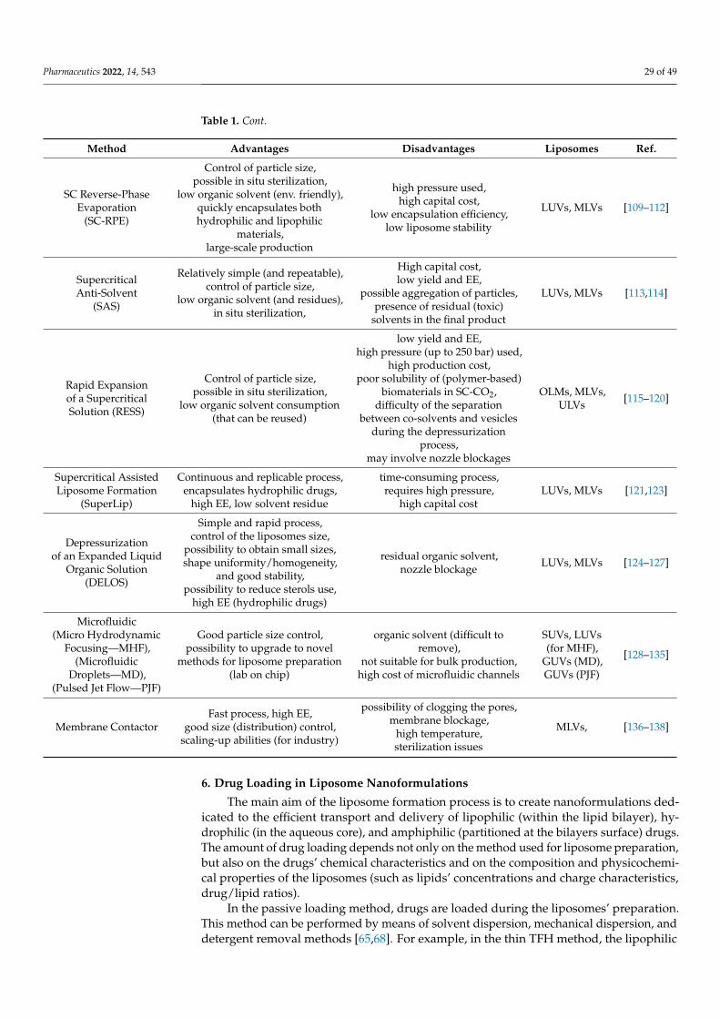
Pharmaceutics 2022, 14, 543 29 of 49
Table 1. Cont.
Method Advantages Disadvantages Liposomes Ref.
SC Reverse-PhaseEvaporation
(SC-RPE)
Control of particle size,possible in situ sterilization,
low organic solvent (env. friendly),quickly encapsulates bothhydrophilic and lipophilic
materials,large-scale production
high pressure used,high capital cost,
low encapsulation efficiency,low liposome stability
LUVs, MLVs [109–112]
SupercriticalAnti-Solvent
(SAS)
Relatively simple (and repeatable),control of particle size,
low organic solvent (and residues),in situ sterilization,
High capital cost,low yield and EE,
possible aggregation of particles,presence of residual (toxic)
solvents in the final product
LUVs, MLVs [113,114]
Rapid Expansionof a SupercriticalSolution (RESS)
Control of particle size,possible in situ sterilization,
low organic solvent consumption(that can be reused)
low yield and EE,high pressure (up to 250 bar) used,
high production cost,poor solubility of (polymer-based)
biomaterials in SC-CO2,difficulty of the separation
between co-solvents and vesiclesduring the depressurization
process,may involve nozzle blockages
OLMs, MLVs,ULVs [115–120]
Supercritical AssistedLiposome Formation
(SuperLip)
Continuous and replicable process,encapsulates hydrophilic drugs,
high EE, low solvent residue
time-consuming process,requires high pressure,
high capital costLUVs, MLVs [121,123]
Depressurizationof an Expanded Liquid
Organic Solution(DELOS)
Simple and rapid process,control of the liposomes size,
possibility to obtain small sizes,shape uniformity/homogeneity,
and good stability,possibility to reduce sterols use,
high EE (hydrophilic drugs)
residual organic solvent,nozzle blockage LUVs, MLVs [124–127]
Microfluidic(Micro Hydrodynamic
Focusing—MHF),(Microfluidic
Droplets—MD),(Pulsed Jet Flow—PJF)
Good particle size control,possibility to upgrade to novel
methods for liposome preparation(lab on chip)
organic solvent (difficult toremove),
not suitable for bulk production,high cost of microfluidic channels
SUVs, LUVs(for MHF),
GUVs (MD),GUVs (PJF)
[128–135]
Membrane ContactorFast process, high EE,
good size (distribution) control,scaling-up abilities (for industry)
possibility of clogging the pores,membrane blockage,
high temperature,sterilization issues
MLVs, [136–138]
6. Drug Loading in Liposome Nanoformulations
The main aim of the liposome formation process is to create nanoformulations ded-icated to the efficient transport and delivery of lipophilic (within the lipid bilayer), hy-drophilic (in the aqueous core), and amphiphilic (partitioned at the bilayers surface) drugs.The amount of drug loading depends not only on the method used for liposome preparation,but also on the drugs’ chemical characteristics and on the composition and physicochemi-cal properties of the liposomes (such as lipids’ concentrations and charge characteristics,drug/lipid ratios).
In the passive loading method, drugs are loaded during the liposomes’ preparation.This method can be performed by means of solvent dispersion, mechanical dispersion, anddetergent removal methods [65,68]. For example, in the thin TFH method, the lipophilic

Pharmaceutics 2022, 14, 543 30 of 49
drugs are incorporated (in large amounts) within the lipid bilayers during the first stagesof the liposomes’ self-assembly process. On the contrary, the hydrophilic drugs, which areusually incorporated during the (aqueous buffer) hydration stage, present lower incorpo-ration efficiencies, as only part of the drugs are present within the liposome core region,while part of them still remain in the hydration volume (outside the liposomes). For thisreason, strategies can be developed to increase the aqueous phase within the core volume,by means of the different methods of liposome formation.
Active loading methods, which incorporate drugs after liposome preparation, gener-ally exploit the diffusion properties of a gradient which is established across the surfaceof the liposomes (gradient loading) by involving the addition of suitable buffers. Activeloading can be obtained with lyophilization, whereby the formed liposomes are generatedby transfer of the lipids from organic to aqueous phases [65,80]. The use of active-loadingapproaches generally shows higher EE as compared to the passive methods. Particularlyinteresting is the (transmembrane) pH-gradients method, as it can be used to encapsu-late a variety of active drugs (including anticancer, anti-anesthetics, antimalarials, etc.).Interestingly, several amine-functionalized drugs (including doxorubicin, daunorubicin,chlorpromazine, quinine, quinidine, dopamine, serotonin, and chloroquine) exhibit a pH-gradient effect after liposome preparations by accumulating protons in the vesicles’ interior(and exhibiting an acidic character in their internal region). In order to create a pH gradientacross the liposome membrane, the pH of the exterior aqueous compartment can be in-creased through the incorporation of an alkylating agent (alkaline buffer) or by exchanging(by gel filtration or dialysis methods) the external media with the desired buffer. Accordingto this approach, the liposomes are first hydrated with a given buffer (of a fixed pH),followed by dialysis in an excess of another buffer (with a different pH), in order to replacethe buffer in the liposomes outside region (and to create a trans-membrane pH gradient).In this case, uncharged drugs entering the liposomes become charged inside the first buffer(of the liposome aqueous core) and remain entrapped in the liposomes (as the chargeddrugs cannot diffuse through the lipid layer) [65,68].
7. Post-Formation Processing of Liposomes7.1. Purification of Liposomes Nanoformulations
Irrespective of the adopted formation method, non-encapsulated compounds (such asnon-entrapped drugs, small molecules, or contaminant molecules) are generally presentin the external (liquid) environment of the generated liposomes, and must be removedthrough a purification process from the final nanoformulation. The liposomes purificationprocess represents, then, a further stage of the liposomes’ manufacturing process. Themain techniques employed for the removal of the non-encapsulated materials include theultra-filtration, ultra-centrifugation, dialysis, and (size exclusion, gel-permeation, or ion-exchange) chromatography. Due to column equilibration and dilution processes connectedwith those techniques, the purification process requires lots of time and can cause a sensitivedecrease of the final liposomes’ production.
Another very important step of the post formation process is connected with theremoval of residual organic solvents from the final liposomes. The use of the organicsolvents (such as ethanol, methanol, chloroform, ether, and methylene chloride) represents acrucial stage in the liposomes’ formation processes, as it facilitates the molecular dispersionprocess of lipids and prevents the oxidation during the lipids’ storage. However, theresidual solvents which are present in the final products may destabilize the liposomes,thus posing some risks upon application [65,68]. Although the (organic) solvents are usuallyremoved by evaporation techniques, this process causes a concentration of the lipids (and ofunwanted the contaminants) in the residual solvents that is not easy to further remove. Forthis reason, the manufacturer should clearly indicate (on the nanoformulation specifications)the residual levels of solvents in the final product (and the allowable safety level).
Finally, liposome nanoformulations should be protected from oxidation. Most of thelipid nanocarriers contain unsaturated lipids (acyl chains). During preparation, storage,

Pharmaceutics 2022, 14, 543 31 of 49
or normal use, unsaturated lipids may undergo oxidative degradation (lipid peroxida-tion), a chemical process that involves some free radical reactions with the formationof cyclic-peroxides and hydro-peroxides. The lipid peroxidation process may be mini-mized by protecting them by keeping them under inert gases such as nitrogen or argon(in order to have minimal exposure to oxygen). Peroxidation can be minimized also bykeeping liposome formulations in light-resistant containers or by the removal of heavymetals (adding ethylenedinitrilotetraacetic—EDTA). Finally, the addition of antioxidantssuch as alpha-tocopherol or butylated hydroxytoluene can also minimize lipid oxidationprocesses [65,68].
7.2. Sterilization of Liposomes
As parenteral route is the most frequent way of administration, liposomes nanoformu-lations should be free of viable microorganisms (such as bacteria, fungi, spores, etc.) thatare capable of altering their properties. For this reason, it is important to remove all possiblemicroorganisms through a sterilization process. The sterilization process can be achievedthrough various approaches, including steam heating (autoclaving), ultraviolet and gammaionizing irradiation, chemicals, and filtration methods. However, as liposomes have highsensitivity and a tendency suffer from (physico-chemical) alterations, the efficient use ofsterilization processes still remains a difficult challenge [139]. We will briefly analyze themain characteristics of the sterilization processes, highlighting their critical aspects.
The steam (autoclaving) sterilization method consists of the combination of saturatedsteam under heat and pressure that causes the germs’ destruction by hydrolysis of proteins.Many investigations have evidenced that this method is responsible for several liposomealterations that involve the oxidation and hydrolysis of lipids, phase transition and ag-gregation, and degradation (or leakage) of the encapsulated drugs. For this reason, thistechnique is suitable only for a limited number of liposome nanoformulations.
Heat sterilization may cause structural phase transitions (and correlated degradationsand/or drug leakages), as well as the oxidation or hydrolysis of the component phos-pholipids (at higher temperatures (T > 121 ◦C)). For this reason, this method cannot beconsidered a reference method for liposome sterilization.
Although the gamma ionizing irradiation has a high-energy ionizing power (andthen a strong penetration capacity), the sterilization process with this radiation may causeliposomes’ degradation by lipid peroxidation (of unsaturated lipids), hydrolysis or frag-mentation of component lipids, and changes in pH. On the contrary, unlike gamma rays,ultraviolet (UV) radiation is a non-ionizing (low-energy) radiation with poor penetrationcapacity in materials, and for this reason, is not effective to cause the sensitive sterilityof liposomes.
Ethylene oxide (chemical) sterilization uses ethylene oxide gas as a sterilizing antimi-crobial agent whose sterilizing mechanism consists of the alkylation of the side chains ofDNA, RNA, and enzymes, thus causing a strong metabolism inhibition and avoiding themultiplication of microorganisms. However, due to the flammable and explosive nature ofethylene oxide, and the toxic, carcinogenic, mutagenic character of its residues, the use ofthis sterilization method is very limited.
Sterilization by filtration is a relatively time-consuming method, based on the use of asterile, disposable filtration unit consisting of an aseptic bacterial-free membrane (0.22-µm)or depth filters for the removal of the microorganisms present in gaseous or liquid products.This method, which is not applicable for liposomes greater than 0.2 µm, are ineffective forthe filtration of smaller viruses or bacteria.
Aseptic manufacturing consists in the preparation and the filling of a product ina controlled sterile environment (class A environment) equipped with sterile materialsand equipment. This method is quite complex and expensive, as several stages have tobe performed in sterile environments. Additionally, the sterility level cannot be easilyevaluated, and a potential risk of contamination (connected with initial raw materials thatare not effectively sterilized) still remains.

Pharmaceutics 2022, 14, 543 32 of 49
In view of the drawbacks of all these conventional approaches for liposome steril-ization, it is crucial to search for an alternative method in order to ensure the sterility ofliposomes in a green, effective, and inexpensive fashion [139]. In this respect, an interestingalternative method for the liposomes’ sterilization can be connected with the use of thesupercritical carbon dioxide (SC-CO2) technology [140]. This method allows for producingand sterilizing liposomes’ nanoformulations (in a single step). However, these approachesrequire further investigations and optimization using the standard biological indicators.
7.3. Microfluidic Lab-on-Chip Nanodevices for the Combined Formation, Drug Loading, andPurification of LiposomesMicro- and Nanofabrication Techniques
The preparation of versatile nanoplatforms, based on microfluidic chips, have stimu-lated the development of multitask lab-on-chip devices for biomedical and nanomedicineapplications [141]. With the successful development of the micro- and nanotechnolo-gies’ fabrication processes, the liposome preparation techniques have been revolutionizedwith the development of novel smart devices. Recently, microfluidic (continuous-flow)formation of liposomes was combined with in-line drug loading and final sample purifica-tion [141–143].
Using on-chip microdialysis elements, it is possible to develop multitask systemsthat allow for the encapsulation efficiencies of a variety of lipids and that (through thecreation of suitable transmembrane pH and ion gradients) favor the remote loading of drugsinto the liposomes. The novel approach also allows for post-formation refinement, andfor the determination of the liposomal nanoformulation properties (such as size, particlenumber, and surface potential). This novel microfluidic technology promises the precisecontrol of the size and lamellarity of liposomes, and the reduction of the volumes (andcosts) of the processed chemical components (and reagents), thus allowing for potentialupgrades for the lab-on-chip-scale production and integration of process within analyticaltechnologies [141–143]. In a recent investigation, a continuous-process flow system for thesimultaneous preparation, drug loading, and purification of liposomes from contaminantshas been developed [143]. The purification step, which is based on a tangential flow-filtration (TFF) device, allows for purifying a large variety of liposome nanoformulations(with very large recovery of lipids > 98%) and for the efficient removal of the non-entrappedcompounds (>95%) and organic solvents (>95% reduction) in a very short time (i.e., less than4 minutes). Those studies demonstrate the feasibility of the advanced on-chip formationand purification of liposome batches for development within industrial processes [143].
8. Characterization Methods of Liposome Nanocarriers
The behaviour of liposome nanoformulations in solution depends on their (nano-)structural organization at the molecular (and supramolecular) level. The performance ofliposomes in biomedical and nanomedicine applications is strongly related to a numberof control parameters, such as dimension, shape, morphology, lamellarity, and surfacefunctionalization. In this respect, the structural characterization of the conformation of theunderlying bilayer vesicles is crucial for the study of the performance of these nanocarrierssystems, including their biodistribution and passive/active targeting capacity. For thisreason, after their formation, liposome nanoformulations are subjected to characterizationin order to evaluate the physico-chemical properties that influence their colloidal stabilityand their therapeutic and biological performances. More specifically, the main liposomeproperties should be investigated in specific solution conditions that resemble the standardconditions of administration. The study of the self-assembly processes involving lipidcomponents (and amphiphilic molecules in general) represents also a crucial step and arelevant topic for the investigation of the interaction of liposomes (or nanoparticles) withbiomembranes, and for the elucidation of various mechanisms connected with importantphysiological bio-processes involving biomembranes (such as phase changes, fusion, sep-aration, biomembrane perforation, and endocytosis), as well as a variety of pathological

Pharmaceutics 2022, 14, 543 33 of 49
processes regulated by specialized biomembrane nanodomains (lipid rafts) [144–146]. Inthe next section, we highlight the importance of some structural parameters of liposomesystems, and shortly describe the main techniques used to measure them.
8.1. Small-Angle X-ray/Neutron Scattering (SAXS/SANS) and Diffraction Techniques
Among the experimental methods, the small-angle X-ray scattering (SAXS), small-angle neutron scattering (SANS), and diffraction methods are the most widely used for anon-destructive characterization of the structural properties of liposomes [147–150]. Scatter-ing techniques also provide the fundamental tools for investigating the main interactions ofnanocarriers in different solution environments, and for investigating conformational mod-ifications and structural transitions which are relevant for the prediction of the structure–function relationship in many biological processes [151–153].
Small-angle X-ray scattering (SAXS), furnishes useful information on lipid phases(and domains), size, spacing, and bilayer thicknesses of liposome systems under differentsolution conditions [153–156]. The addition of a further detector on the same SAXS exper-imental station allows for performing wide-angle X-ray scattering (WAXS) experimentsthat provide complementary information (at the Å scale) on the hydrophobic lipid chainpacking and their lamellar phase characteristics, when ordered crystalline-like phases arepresent in the liposome system.
Diffraction is a powerful technique for the structural investigation of lipid-basedordered (crystalline-like) systems, such as lipid multilayers, bilayers, or monolayers, andtheir structural organization within the liposome nanostructures [38,40]. The sharp peaksdetected in the low-angle region of a diffraction pattern are connected with the (long-range)organization of the lipid nanostructures and indicate the specific crystalline lattice (andsymmetry) and the unit cell repeat distance of the detected lipid phase. The signals in thehigh-angle region furnish indications of the short-range organization, which is connectedwith the lipids’ hydrocarbon chains conformation. Lipid hydrocarbon alkyl chains show apeculiar liquid-like conformation (called type α) that exhibits only a broad peak in the high-angle region of X-ray diffraction (XRD) spectra. In liposome and lipid-based nanostructures,the reference unit cell can be described by only one parameter, such as in the lamellar (1),hexagonal (2), and cubic (3-D)-phase systems. However, more complex phase diagramscan be detected when a variety of different phases coexist at the equilibrium (especially inmulticomponent lipid mixtures). In any case, the crystalline structural properties of lipidbilayers and multilayers composed of different phospholipids (with saturated/unsaturatedalkyl chains) exhibit similar dimensions in their unit cell (of about 5.0 × 8.0 Å2). In thelamellar phase, the unit cell indicates the total thickness of the lipid and water layers, whilein the hexagonal phase, it corresponds to the distance between the axes of the cylinders.The diffraction technique plays a crucial role in the study of the interaction of liposomeswith biological membranes, thus furnishing useful insights in important bio-processes suchas physiological (or pathological) crystallizations or phase separations/transitions inducedby nanoparticle interactions [37,148].
A XRD study of Kiselev et al. [156] investigated the influence of the ceramide lipidson the hydration and internal nanostructure of DMPC lipid vesicles. The analysis of theXRD peaks (Figure 17) evidenced that the DMPC/CER[AP] vesicles exhibit one phase, witha membrane repeat distance of d1 = 6.53 ± 0.06 nm, which is larger than those observedfor the pure DMPC (d0 = 6.27 nm) [40]. However, the DMPC/CER[NP] vesicles exhibitedtwo phases with repeat distances of d1 = 6.43 ± 0.06 nm and d2 = 3.85 ± 0.04 nm, whichcorrespond to the two peaks in the diffraction spectra (Figure 17). This circumstance isprobably caused by the characteristic V-shaped form of CER[NP] lipids, which favors aphase separation from the DMPC lipid membrane and the formation of a new ceramide-enriched domain with a shorter periodicity (or pure CER[NP] crystal) [40].

Pharmaceutics 2022, 14, 543 34 of 49Pharmaceutics 2022, 14, x 35 of 50
Figure 17. X-ray diffraction patterns for the mixed lipid system composed of DMPC/ α-hydroxy-N-stearoyl phytosphingosine CER[AP] and of DMPC/ N-stearoyl phytosphingosine CER[NP] multila-mellar vesicles (MLVs) at the temperature of T = 30 °C Adapted by permission from Springer Na-ture: [156] (Appl. Phys. A) by Kiselev et al., Copyright 2013.
With the use of the very intense X-ray beam available at synchrotron radiation facil-ities, and the dedicated instruments, it is possible to perform time-resolved experiments for the study of rapid transient processes (over millisecond time resolution and in a large range of length scales) [157,158]. More specifically, the combination of SAXS/WAXS time-resolved investigations are particularly important for the investigation of the structural properties and phase transitions in liposome nanostructures. As the lipid systems un-dergo complex self-assembly processes, typical of amphiphilic systems [146,159], the small-angle scattering technique allows for monitoring the structures and structural changes and the transitions of many biological amphiphilic macromolecules [152,160]. Moreover, the analysis of scattering structure factor S(q), by means of advanced theoreti-cal approaches and computer simulation methods, allows for investigating the nanoparti-cles’ interactions (at the molecular level) in very different solution environments [161–166].
Small-angle neutron scattering (SANS) shares the same basic principles with SAXS. While SAXS spectra originates from the scattering of the electrons present in the materials system under investigation, and is sensitive to the hydrophilic region of a lipid bilayer, the SANS technique furnishes information on the lipids’ hydrophobic tails region, as neu-trons furnish a better “contrast” for that region [151–153]. Moreover, neutron diffraction measurements performed on (selectively) deuterated lipids furnish an important ap-proach for the determination of the conformation of the lipid molecules at different posi-tions [27,40]. Therefore, SAXS and SANS represent complementary methods for a detailed description of the structure of liposomes systems.
It is worth pointing that the investigation of the structural features in lipid-based nanostructures involves the investigation of the collective behavior of a large number of interacting molecules, and helps to understand the structural and dynamic behavior of biological membranes, representing, then, a fundamental interdisciplinary topic in fields of biotechnology and pharmaceutical science [27,167].
Figure 17. X-ray diffraction patterns for the mixed lipid system composed of DMPC/ α-hydroxy-N-stearoyl phytosphingosine CER[AP] and of DMPC/N-stearoyl phytosphingosine CER[NP] mul-tilamellar vesicles (MLVs) at the temperature of T = 30 ◦C Adapted by permission from SpringerNature: [156] (Appl. Phys. A) by Kiselev et al., Copyright 2013.
With the use of the very intense X-ray beam available at synchrotron radiation facilities,and the dedicated instruments, it is possible to perform time-resolved experiments forthe study of rapid transient processes (over millisecond time resolution and in a largerange of length scales) [157,158]. More specifically, the combination of SAXS/WAXS time-resolved investigations are particularly important for the investigation of the structuralproperties and phase transitions in liposome nanostructures. As the lipid systems undergocomplex self-assembly processes, typical of amphiphilic systems [146,159], the small-anglescattering technique allows for monitoring the structures and structural changes andthe transitions of many biological amphiphilic macromolecules [152,160]. Moreover, theanalysis of scattering structure factor S(q), by means of advanced theoretical approachesand computer simulation methods, allows for investigating the nanoparticles’ interactions(at the molecular level) in very different solution environments [161–166].
Small-angle neutron scattering (SANS) shares the same basic principles with SAXS.While SAXS spectra originates from the scattering of the electrons present in the materialssystem under investigation, and is sensitive to the hydrophilic region of a lipid bilayer,the SANS technique furnishes information on the lipids’ hydrophobic tails region, asneutrons furnish a better “contrast” for that region [151–153]. Moreover, neutron diffractionmeasurements performed on (selectively) deuterated lipids furnish an important approachfor the determination of the conformation of the lipid molecules at different positions [27,40].Therefore, SAXS and SANS represent complementary methods for a detailed description ofthe structure of liposomes systems.
It is worth pointing that the investigation of the structural features in lipid-basednanostructures involves the investigation of the collective behavior of a large number ofinteracting molecules, and helps to understand the structural and dynamic behavior ofbiological membranes, representing, then, a fundamental interdisciplinary topic in fields ofbiotechnology and pharmaceutical science [27,167].
8.2. Electron Microscopy and Atomic Force Microscopy (AFM) Techniques
Electron microscopy (EM) techniques allow for high-resolution visualizations of lipo-somes in different environments, with the possibility of resolving the details of liposomes

Pharmaceutics 2022, 14, 543 35 of 49
(and nanoparticles) of varying sizes, morphologies, and lamellarities [168–170]. In EM,an electron beam is focused by a set of electromagnetic lenses onto the sample surface.A further set of electromagnetic lenses refocus (and magnify) the electrons scattered bythe sample, thus furnishing a projected image of the sample. Sample preparation in EMtechniques often requires the liposomes’ removal from their original environment, whichcan cause some morphological artifacts or induce structural perturbations in liposomesystems [168–170].
In scanning electron microscopy (SEM) experiments, the (dried) sample is scanned(point by point) with an electron beam, while a detector reveals the secondary electronsthat are emitted by the investigated sample. In this way, it is possible to obtain a detailedthree-dimensional (3-D) picture of the nanostructures (or of the surface) present in thesample. The SEM technique also provides crucial information on the size and morphologyand layered (concentric) liposomes structures [168–170].
In transmission electron microscopy (TEM) experiments, a small amount of sample(<100-nm thick) is placed in a vacuum chamber, while the solvent is dried prior to themicroscopic analysis. This circumstance may affect the sample morphology or orienta-tion before the analysis of their images. TEM provides the image of nanoparticles witha resolution below 1 nm in size and with enhanced contrast and image contours, thusproviding important information on the surface modifications of liposomes (such as inthe case of surface coating or conjugation with ligands). In TEM experiments, liposomesmostly appear as black spherical nanoparticles on a white background. TEM also offersthe advantage of differentiating individual vesicles from aggregates and provides infor-mation about the lipid phase transitions, allowing for a critical and complete assessmentof the liposome structural properties [169,170]. Nevertheless, TEM has several drawbacksconnected with the sample pre-treatments that may cause artifacts and changes in theliposomal structure/morphology (such as vesicle swelling, shrinkage, or deformation).
To overcome many of the limitations of the TEM method, the (cryo-) transmission elec-tron microscopy (Cryo-TEM) technique was developed [171,172]. This method allows foravoiding the liposome structure perturbation by making use of a (flash-)freezing treatmentfor the direct visualization of the specimen in the solid-state (without the need to eliminatethe solvent). Thin liposome-hydrated films (vitrified in liquid ethane) are used prior toimaging, thus allowing for the analysis in their most native state. This rapid freezinglimits the ice crystals’ formation and preserves the integrity of proteins and other biologicalmaterials. Cryo-TEM techniques provide detailed insights into lipid size, shape, internalstructure (lamellarity), lipid packing, phase behavior, and drug inclusion characteristics,with resolutions in the range of 5–500 nm (defined by the film thickness). Cryo-TEM alsoprovides high-resolution images for the investigation of complex self-assembly processes,phase changes, and dynamic processes in lipid-based nanoformulations. Moreover, thedevelopment of the cryo-tomography allows for obtaining 3D information about the struc-tural properties of bio-materials and bio-molecules, (drug-loaded) liposomes, cubosomes,hexosomes, and other biomembrane phases [171,172].
Another important microscopic method used to analyze the liposomes’ sizes andmorphologies is given by the atomic force microscopy (AFM). This technique is a type ofscanning probe microscopy (SPM) that provides high resolution (of the order of fractions ofa nanometer) on the 3D profile of liposome nanostructures within their native solution envi-ronment and without the need of applying vacuum [170,173]. The structural information isobtained by exploring the materials’ surface with a mechanical probe (diamond tip) whichis in direct contact with the explored surface and measures the surface forces between theprobe and the investigated sample (with the interatomic van der Waals forces that providethe interaction mechanism). Piezoelectric elements (sensors) facilitate precise and accuratemovements of the probe. This favors the precise scanning and imaging of a large varietyof surfaces, including polymers, glass, composites, and biological systems [173,174]. TheAFM method can provide a 3D image of liposomes with details on size (distribution andhomogeneity), shape (with a spatial resolution in the order of fractions of a nanometer), and

Pharmaceutics 2022, 14, 543 36 of 49
surface modifications (due to the presence of ligands, antibodies, or polymers conjugatedat the liposomes’ surface) [173,174].
8.3. Light (Fluorescence and Confocal) Microscopy Techniques
Light (or optical) microscopy utilizes visible light as source and a relatively simplesetup of lenses, and furnishes a magnified image of the sample with a limited resolutionof ~250 nm (defined by the smallest diffraction-limited spot size achievable). It furnishesuseful information on the dimension, shape (homogeneity), and degree of aggregation of aliposome system or of giant unilamellar vesicles (GUVs) of hundreds of micrometers, butis unable to directly provide details on the vesicles’ lamellarity and on the structures of theSUVs [169,170]. The incorporation of low concentrations (≤1 mol%) of fluorescent probes(dyes) placed within the aqueous phase (or the lipid bilayers) permits the visualizationof the liposomes’ structure (and dynamics), with a negligible impact on the structuralproperties of the lipid membrane [174,175]. This technique allows for the visualizationof the size, shape, and fluidity of lipidic GUVs [175], while by incorporating specificprobes (such as rhodamine) into the lipid bilayer allows for identifying the liposomes’lamellarity [174], or for detecting the different conformations of lipid lateral packing(rhodamine-labeled lipids) [175].
Among the various optical imaging techniques that allow for the observation oflipid-based nanostructures, confocal laser scanning microscopy (CLSM) is a method ofchoice, since it facilitates the structural investigation (in an almost non-invasive way)by visualizing the liposomes’ internal structures [170]. It also allows for localizing (andquantifying) the extent of liposomes’ subcellular distribution during the drug-deliveryprocesses [176–179]. CLSM is a crucial technique for the characterization of biomaterialsand biological specimens with high-resolution images. The technique provides an imagewhich is built by scanning one (or more) focused light beams across a slice of a sample. Itgives a sharp image, as it allows for focusing on a particular focal point without interferingwith the out-of-focus adjacent planes. The confocal optical system is composed of twosystems of lenses: the objective lens (which views onto the image plane), and the collectorlens (that acts rather as an imaging lens by using a small area point detector rather thanas collectors) [174,177]. Confocal microscopy provides imaging and detection systemsthat are on the same focal plane (plane of interest). Recent setup upgrades allow forfurther processing into a 3D specimen representation by means of the volume visualizationtechniques. To selectively mark the view of a nanocarrier within a biological sample, afluorescence dye (such as rhodamine 123, coumarin 6, fluorescein 5-isothiocyanate (FITC),etc.) is inserted during the sample preparation. Significant advances in the technique allowfor generating high-resolution images (in a noninvasive manner) at various depths of theskin layer without the need of any sample mechanical sectioning [180].
In conclusion, no single experimental approach can combine high spatial resolutionimage acquisition in one instrument, while the combination of different microscopy meth-ods allows for studying many structural features of the liposomes’ nanoformulations atdifferent stages of the drug-delivery process [174].
For example, heparin-loaded liposomes (HLp) composed of L-α-phosphatidyl choline,cholesterol, and stearylamine (PC/Chol/SA, 7:3:1) have been recently prepared by thethin-film hydration method (followed by the extrusion) [181]. The nanocarrier was finallyformulated as an ibuprofen-containing gel to obtain a nano-spray formulation applicablefor topical drug-delivery treatments (HLp-Ibu-NSG). The blank liposome (BLp) had a meandiameter of (181 ± 23), polydispersity index (PDI) of (0.278 ± 0.093), and zeta potentialof (+41.6 ± 3.98) mV, while upon the encapsulation of heparin, the HLp exhibited a slightincrease in the size (204 ± 12 nm) and PDI (0.41 ± 0.072), and a slight decrease in the zetapotential of (+38.3 ± 4.3) mV. Those results obtained by the dynamic light scattering wereconfirmed by means of the SEM, TEM, and AFM techniques (Figure 18A), which evidencedan average liposome diameter of around d = 200 nm. Moreover, fluorescently labelledHLp were prepared for cellular uptake studies. The accumulation ability of cationic HLp

Pharmaceutics 2022, 14, 543 37 of 49
in mouse fibroblast cells was investigated by using confocal laser scanning microscopy(Figure 18B). Fluorescent confocal micrographs showed that fibroblasts exposed to the HLp(green fluorescence) for 3 h accumulated liposomes (in significant numbers) inside thecytoplasm of the cells (Figure 18B). No significant difference in cellular accumulation wasobserved between HLp and BLp [181].
Pharmaceutics 2022, 14, x 38 of 50
were confirmed by means of the SEM, TEM, and AFM techniques (Figure 18A), which evidenced an average liposome diameter of around d = 200 nm. Moreover, fluorescently labelled HLp were prepared for cellular uptake studies. The accumulation ability of cati-onic HLp in mouse fibroblast cells was investigated by using confocal laser scanning mi-croscopy (Figure 18B). Fluorescent confocal micrographs showed that fibroblasts exposed to the HLp (green fluorescence) for 3 h accumulated liposomes (in significant numbers) inside the cytoplasm of the cells (Figure 18B). No significant difference in cellular accu-mulation was observed between HLp and BLp [181].
Figure 18. (A) Images of heparin-loaded liposomes (HLp), obtained by means of the SEM, TEM, and AFM techniques. (B) Confocal laser scanning microscopy images of cellular uptake of HLd (top) and the blank liposomes (BLp) (bottom) by fibroblast cell lines after 3 h of incubation. Blue (DAPI): nucleus; green (fluorescent HLp). Adapted with permission from Vaghasiya et al. [181], Copyright 2019 American Chemical Society.
HLp exhibited significant healing of a wound in vitro (scratch assay, fibroblast cells) and in vivo (wound healing in Sprague–Dawley rats) at a low dose. In the rat model of frostbite injury, the HLp-Ibu-NSG formulation demonstrated significant reductions in the wound area (up to ~96%), and improvements of histopathology in 14 days, as compared to the control groups, while no edema or erythema were detected (in the affected area) during the post-treatment of HLp-Ibu-NSG [181].
8.4. Dynamic Light Scattering Technique Dynamic light scattering (DLS) furnishes important information on the structure and
dynamic properties of nanoparticles in solution, such as bio-macromolecules, colloids, nanoemulsions, liposomes, and gels [182,183]. In a DLS experiment, the fluctuations in the scattered light intensity is connected with the scattered electric field correlation function, which yields information on the nanoparticles’ Brownian motion through the Stokes–Ein-stein relation D = kBT/6πρRH, which expresses the (translational) diffusion coefficient D as a function of the particles’ hydrodynamic radius RH, the absolute temperature T, and the solvent
Figure 18. (A) Images of heparin-loaded liposomes (HLp), obtained by means of the SEM, TEM, andAFM techniques. (B) Confocal laser scanning microscopy images of cellular uptake of HLd (top)and the blank liposomes (BLp) (bottom) by fibroblast cell lines after 3 h of incubation. Blue (DAPI):nucleus; green (fluorescent HLp). Adapted with permission from Vaghasiya et al. [181], Copyright2019 American Chemical Society.
HLp exhibited significant healing of a wound in vitro (scratch assay, fibroblast cells)and in vivo (wound healing in Sprague–Dawley rats) at a low dose. In the rat model offrostbite injury, the HLp-Ibu-NSG formulation demonstrated significant reductions in thewound area (up to ~96%), and improvements of histopathology in 14 days, as compared tothe control groups, while no edema or erythema were detected (in the affected area) duringthe post-treatment of HLp-Ibu-NSG [181].
8.4. Dynamic Light Scattering Technique
Dynamic light scattering (DLS) furnishes important information on the structure anddynamic properties of nanoparticles in solution, such as bio-macromolecules, colloids,nanoemulsions, liposomes, and gels [182,183]. In a DLS experiment, the fluctuations in thescattered light intensity is connected with the scattered electric field correlation function,which yields information on the nanoparticles’ Brownian motion through the Stokes–Einstein relation D = kBT/6πρRH, which expresses the (translational) diffusion coefficient Das a function of the particles’ hydrodynamic radius RH, the absolute temperature T, and thesolvent viscosity ρ (where kB is the Boltzmann constant) [182,183]. Analysis of the scatteringintensity also allows for obtaining the liposomes’ nanoparticles size distribution [152,183].

Pharmaceutics 2022, 14, 543 38 of 49
8.5. Zeta (ζ) Potential Technique
The zeta (ζ) potential measurement represent a useful tool for addressing the elec-trostatic effects in charged nanocarriers [184]. It is regulated by the nature and distribu-tion of the surface charge of liposomes and depends on the lipid composition and theirheadgroup charges. The zeta potential is an important factor that strongly regulates theliposomes’ colloidal stability, their biodistribution, pharmacokinetics, cellular affinity, anddrug-internalization processes. A liposome nanocarrier dispersed in an aqueous solutionacquires a surface charge caused by the ionization of surface end-groups (or the adsorptionof charged species on its surface) (Figure 19). The dispersed counter-ions will surround thesurface of the particle (Stern layer), while the zeta potential represents the electrical poten-tial at the nanoparticle interface. The zeta potential is a key indicator of the colloidal stabilityof a liposome’s dispersions, as it expresses the degree of electrostatic repulsion betweennanoparticles in a dispersion. Nanoparticles with high (negative or positive) zeta potentialsare electrically stabilized (and have high colloidal stability), while the nanoparticles thatexhibit low zeta potentials tend to aggregate or flocculate.
Pharmaceutics 2022, 14, x 39 of 50
viscosity ρ (where kB is the Boltzmann constant) [182,183]. Analysis of the scattering in-tensity also allows for obtaining the liposomes’ nanoparticles size distribution [152,183].
8.5. Zeta (ζ) Potential Technique The zeta (ζ) potential measurement represent a useful tool for addressing the electro-
static effects in charged nanocarriers [184]. It is regulated by the nature and distribution of the surface charge of liposomes and depends on the lipid composition and their head-group charges. The zeta potential is an important factor that strongly regulates the lipo-somes’ colloidal stability, their biodistribution, pharmacokinetics, cellular affinity, and drug-internalization processes. A liposome nanocarrier dispersed in an aqueous solution acquires a surface charge caused by the ionization of surface end-groups (or the adsorp-tion of charged species on its surface) (Figure 19). The dispersed counter-ions will sur-round the surface of the particle (Stern layer), while the zeta potential represents the elec-trical potential at the nanoparticle interface. The zeta potential is a key indicator of the colloidal stability of a liposome’s dispersions, as it expresses the degree of electrostatic repulsion between nanoparticles in a dispersion. Nanoparticles with high (negative or positive) zeta potentials are electrically stabilized (and have high colloidal stability), while the nanoparticles that exhibit low zeta potentials tend to aggregate or flocculate.
Figure 19. Diagram indicating the electrical potential as a function of the ionic concentration (and distance from the charged surface) of a liposome suspended in a dispersion medium.
The zeta potential ζ of a colloidal nanoparticle is obtained by measuring the electro-phoretic mobility μE using principles of phase analysis light scattering (PALS) [184,185]. It is worth noticing that a suitable combination of light-scattering and zeta-potential tech-niques allows for a detailed investigation of electrostatic interactions in charged lipo-somes, and furnishes useful information on the interactions involved between complex charged liposome nanocarriers and target cells or tissues [186–189].
Electric Double Layer+
-+
+
+
+
+
+
+
++
+
+++
+
+
++++
+
+
+
+
+
----
- - - ------
+
+
+
+
+
+
+
-
+
+
+
++
+
+
+
-
-
-
--
--
-
-
-
-
-
---
--
-
-
-
-
-
-
-
+
+
Diffuse Layer
Stern plane
Electrical Potential
Surface Potential
Stern Potential
Slipping Plane
ZETA TOTENTIAL
Distance from surface0
Figure 19. Diagram indicating the electrical potential as a function of the ionic concentration (anddistance from the charged surface) of a liposome suspended in a dispersion medium.
The zeta potential ζ of a colloidal nanoparticle is obtained by measuring the elec-trophoretic mobility µE using principles of phase analysis light scattering (PALS) [184,185].It is worth noticing that a suitable combination of light-scattering and zeta-potential tech-niques allows for a detailed investigation of electrostatic interactions in charged liposomes,and furnishes useful information on the interactions involved between complex chargedliposome nanocarriers and target cells or tissues [186–189].
8.6. Other Complementary Characterization Techniques
Detailed information on the liposome population over a wide range of sizes can bedetermined by means of the size-exclusion chromatography (SEC) technique [190]. SEC and

Pharmaceutics 2022, 14, 543 39 of 49
high-performance size exclusion chromatography (HPSEC) allows for physically separating(and quantifying) liposomes from small solutes, or for subdividing different liposomesubpopulations. Those techniques allow for the determination of the size (distributionand polydispersity), stability, bilayer permeabilization, as well as liposome formationand reconstitution processes. SEC also represents a powerful method for the study ofthe encapsulation/interaction of a large variety of compounds, including small solutes(such as drugs, surfactants, etc.) or large macromolecules (such as peptides, proteins, andnucleic acids) in liposomes [190]. Alternatively, the field-flow fractionation (FFF) separationtechniques offer a physical separation method of complex and inhomogeneous liposomessamples, which potentially cannot be characterized by other separation methods, such asSEC. The absence of a stationary phase in the FFF technique causes a reduced interactionwith surfaces or column packing materials [191,192].
Many other complementary techniques can be employed for the determination of theaffinity of active drugs (and macromolecules) for liposomes’ inclusion, including variousspectroscopy techniques such as ultraviolet (UV), FT-infrared (FT-IR), electron paramag-netic resonance (EPR), electron spin resonance (ESR), and circular dichroism [191–193].Furthermore, complementary biophysical techniques, such as the differential scanningcalorimetry (DSC) [193] and the isothermal titration calorimetry (ITC) [194], allow forthe study of the thermodynamics of drug–lipid interactions (and the associated bindingprocesses). Moreover, nuclear magnetic resonance (NMR) is a quantitative spectroscopytechnique for the (non-destructive and non-invasive) study of the drug–liposomes interac-tions, and furnishes crucial information on changes in the bilayer structure or dynamics oflipid molecules and lipid-based nanostructures (at a sub-Ångstrom resolution) [195,196].
In conclusion, the use of high-performance characterization techniques, combinedwith advanced structural modeling, have an important impact on the quality control inlipid nanocarriers’ post-formation processes.
9. Technology Transfer and Regulatory Perspectives
The assessment of the quality of pharmaceutical nanoformulations and health careproducts is a mandatory step before their technology transfer and their introduction intothe market. Although a growing number of liposome systems are able to prove theirbeneficial action in preclinical trials, only liposome nanoformulations that are efficientin clinical trials will enter into the clinic. In the last decades, a wide range of liposomeformulations were successfully translated into the clinic, while others are in differentphases of their clinical investigation [17,18]. The quality of pharmaceutical products isintimately connected with their performance, stability, and absence of contamination,and it is certified through their efficacy in obtaining the therapeutic benefits indicatedon the products’ labels [197]. As many liposome nanoformulations involve the use ofnovel drugs and complex interactions between the various molecular components, a setof physicochemical and biopharmaceutical characterization methods are required beforeliposomes nanoformulations are recognized (and marketed) as biomedical products [198].The high costs of production and the stringent requirements for assessing their benefits(or risks) can represent a limit to an efficient technology transfer. The specific requirementsof a liposome nanoformulation depend on the pharmaceutical type of intervention, thetarget patient population, and the corresponding route of administration. In this respect,the regulatory agencies outline the number of requirements needed in terms the quality,safety, and effectiveness of the liposome-based products [197,199].
In the last decades, the European and US regulatory agencies have promoted guide-lines to push manufacturers to perform accurate pre-authorization investigations for assess-ing the quality, safety, and efficacy profile of the new proposed pharmaceutical products.Those regulatory agencies publish periodic scientific guidelines on human medicines thatare harmonized by the International Council for Harmonisation of Technical Requirementsfor Registration of Pharmaceuticals for Human Use (ICH) [200–202]. Concerning the newliposome nanoformulations, useful guidelines can be found in the reflection paper released

Pharmaceutics 2022, 14, 543 40 of 49
by the European Medicine Agency (EMA) (for intravenous liposomal products) [201] andthe draft guidance of the Food and Drug Administration (FDA) (for liposome drug prod-ucts) [202]. The information provided by those agencies helps to define (harmonized)quality standards concerning:
- the assessment of the benefit/risk of the liposomal systems;- definition of the critical quality attributes of the final product;- guide for the formulation studies in the early stages of development (designing
preclinical/clinical trials);- support to the pharmaceutical quality control system and sustaining the post-marketing
variations.
In particular, those agencies require a detailed description of the liposomes’ compo-sition (quality/purity of lipids used), a characterization of their physicochemical proper-ties, manufacturing process (and controls), and stability in physiological environments,a characterization of their functionality (related to the specific application), and clinicalpharmacology studies (pharmacokinetics, bioavailability, bioequivalence tests).
Concerning the future perspectives for an efficient technology transfer and regulatoryactivities, a synergistic action of joint research centers may contribute to stimulate ex-ploratory research for the translation to the clinic of liposome products and nanomedicines.More specifically, the new discoveries could stimulate the identification of the main reg-ulatory issues and the corresponding design strategies, in collaboration with the worldregulation authorities. In this respect, the creation of a regulatory consortium, by includinga wide community of laboratories, industries, stakeholders in regulation, and end users,could help establish a permanent forum for the efficient transfer of the new scientificresults into regulatory strategies, and to update the regulatory issues (including theirharmonization). The network between scientists, regulation agencies, and product devel-opers can stimulate the advancement of regulatory issues in the field of pharmaceutics andnanomedicine, through the identification of the safety issues induced by nanomedicines, thedevelopment (and validation) of new standards for analytical (or experimental) methods,and assays requested by the regulators.
The Quality by Design (QbD) Method
Recently, a Quality by Design (QbD) approach has been proposed to improve thequality of pharmaceutical products [203,204]. QbD is a new perspective based on theanalysis and study of potential factors that influence the quality of the pharmaceuticalproducts through a risk-assessment process. The application of the QbD strategy in thepharmaceutical developments helps to influence the quality, the achievements, and thesuccess of the pharmaceutical nanoformulation products. For this reason, the QbD approachhas been recommended by drug regulatory agencies such as the FDA and EMA for thedevelopment and manufacturing of better-quality drug formulations.
A QbD approach includes the following crucial stages, as follows [203,204]:
(1) A definition of the quality target product profile (QTPP), based on some specificproperties of the product, in order to ensure the desired quality, safety, and efficacy ofthe drug product, and by considering the critical factors of the administration (such asspecific route, dosage, bioavailability, strength, and stability);
(2) The identification of the main factors, such as the critical quality attributes (CQAs) ofthe targeted product, the critical material attributes (CMAs), and the critical processparameters (CPPs), which are related to the selected production method;
(3) The identification of the risk assessment (RA) through a science-based process thatranks the parameters’ impact on the CQAs of the product, and identifies (betweenthem) the critical material attributes (CMAs) and critical process parameters (CPPs);
(4) The development of a design space (DS), i.e., a study of the combination and interac-tion (in a mathematical form) of the input variables (such as the CMAs and CPPs) andtheir impact on CQAs and the process parameters (to ensure desired product quality).

Pharmaceutics 2022, 14, 543 41 of 49
To establish the design space with a minimum number of experiments, the Design ofExperiments (DoE) strategy is used;
(5) The definition (and implementation) of a control strategy (CS), with the aim of stimu-lating the continuous products’ improvement;
(6) A life cycle management.
In recent years, a growing number of investigations have used the QbD approach tofacilitate the identification of the main risk factors that may impact liposome nanoformula-tion formation processes, in order to improve the quality of product, through the analysisand the control of the formulation’s materials and manufacturing variables [204–206].
10. Conclusions
In this article, we analyzed the main features of the formation techniques of liposomenanocarriers with a special focus on the structural parameters and the critical factors that in-fluence the development of a suitable and stable nanoformulation. Traditional methods andnovel approaches for liposome preparation are discussed, with the objective of updating thereader on recent developments and providing future directions for research and develop-ment. Conventional techniques for liposome preparation still remain very popular, as theyare simple to implement and do not require sophisticated equipment for their instrumentalupgrade. However, these techniques may not be suitable for the processing of several (bio-)molecules, as they may produce structural (and then functional) alterations. With the recentprogress in nanotechnology and biomedicine, novel manufacturing methods for the facileencapsulation of both hydrophobic and hydrophilic molecules (with no need for organicsolvents and/or sophisticated equipment) are highly desired. Moreover, the critical issue ofsystem instability for liposomes loaded with hydrophilic materials requires new strategiesthat will achieve acceptable loading while aiming at targeting cargo delivery at the siteof interest. Furthermore, the difficulties of achieving an easily scalable process for massproduction, and of achieving elevated encapsulation efficiencies, still represent the furtherdrawbacks of conventional liposome formation processes. In this respect, the study of novelformation approaches, together with the introduction of new technologies, may stimulatethe production of advanced liposome nanoformulations from the laboratory, which will besuitable for industrial-scale production and even more effective clinical applications.
Author Contributions: Conceptualization, D.L.; methodology, D.L. and M.A.K.; formal analysis,D.L. and M.A.K.; investigation, D.L. and M.A.K.; writing—original draft preparation, D.L.; writing—review and editing, D.L.; supervision, D.L.; project administration, D.L. All authors have read andagreed to the published version of the manuscript.
Funding: This research received no external funding.
Institutional Review Board Statement: Not applicable.
Informed Consent Statement: Not applicable.
Data Availability Statement: Not applicable.
Conflicts of Interest: The authors declare no conflict of interest.
References1. Bozzuto, G.; Molinari, A.A. Liposomes as nanomedical devices. Int. J. Nanomed. 2015, 10, 975–999. [CrossRef]2. Allen, T.M.; Cullis, P.R. Liposomal drug delivery systems: From concept to clinical applications. Adv. Drug Deliver. Rev. 2013,
65, 36–48. [CrossRef] [PubMed]3. Lombardo, D.; Calandra, P.; Barreca, D.; Magazù, S.; Kiselev, M.A. Soft interaction in liposome nanocarriers for therapeutic drug
delivery. Nanomaterials 2016, 6, 125. [CrossRef] [PubMed]4. Lee, M.-K. Liposomes for Enhanced Bioavailability of Water-Insoluble Drugs: In Vivo Evidence and Recent Approaches.
Pharmaceutics 2020, 12, 264. [CrossRef] [PubMed]5. Ewert, K.K.; Scodeller, P.; Simón-Gracia, L.; Steffes, V.M.; Wonder, E.A.; Teesalu, T.; Safinya, C.R. Cationic Liposomes as Vectors
for Nucleic Acid and Hydrophobic Drug Therapeutics. Pharmaceutics 2021, 13, 1365. [CrossRef]

Pharmaceutics 2022, 14, 543 42 of 49
6. Boons, G.-J. Liposomes Modified by Carbohydrate Ligands Can Target B-Cells for the Treatment of B-Cell Lymphomas. ExpertRev. Vaccines 2010, 9, 1251–1256. [CrossRef]
7. Kumar Giri, T.; Giri, A.; Kumar Barman, T.; Maity, S. Nanoliposome is a Promising Carrier of Protein and Peptide Biomolecule forthe Treatment of Cancer. Anti-Cancer Agents Med. Chem. 2016, 16, 816–831. [CrossRef]
8. Blanken, D.; Foschepoth, D.; Serrão, A.C.; Danelon, C. Genetically controlled membrane synthesis in liposomes. Nat. Commun.2020, 11, 4317. [CrossRef]
9. Xia, Y.; Xu, C.; Zhang, X.; Ning, P.; Wang, Z.; Tian, J.; Chen, X. Liposome-based probes for molecular imaging: From basic researchto the bedside. Nanoscale 2019, 11, 5822–5838. [CrossRef]
10. Shao, X.R.; Wei, X.Q.; Zhang, S.; Fu, N.; Lin, Y.-F.; Cai, X.-X. Effects of Micro-environmental pH of Liposome on Chemical Stabilityof Loaded Drug. Nanoscale Res. Lett. 2017, 12, 504. [CrossRef]
11. Lombardo, D.; Calandra, P.; Caccamo, M.T.; Magazù, S.; Kiselev, M.A. Colloidal stability of liposomes. AIMS Mater. Sci. 2019,6, 200–213. [CrossRef]
12. Pattni, B.S.; Chupin, V.V.; Torchilin, V.P. New Developments in Liposomal Drug Delivery. Chem. Rev. 2015, 115, 10938–10966.[CrossRef] [PubMed]
13. Gu, Z.; Da Silva, C.G.; Van der Maaden, K.; Ossendorp, F.; Cruz, L.J. Liposome-Based Drug Delivery Systems in CancerImmunotherapy. Pharmaceutics 2020, 12, 1054. [CrossRef]
14. Salehi, B.; Calina, D.; Docea, A.O.; Koirala, N.; Aryal, S.; Lombardo, D.; Pasqua, L.; Taheri, Y.; Salgado Castillo, C.M.; Martorell,M.; et al. Curcumin’s Nanomedicine Formulations for Therapeutic Application in Neurological Diseases. J. Clin. Med. 2020,9, 430. [CrossRef] [PubMed]
15. Ba, S.; Qiao, M.; Jia, L.; Zhang, J.; Zhao, X.; Hu, H.; Chen, D. Construction of Hierarchical-Targeting pH-Sensitive Liposomes toReverse Chemotherapeutic Resistance of Cancer Stem-like Cells. Pharmaceutics 2021, 13, 1205. [CrossRef]
16. Bobo, D.; Robinson, K.J.; Islam, J.; Thurecht, K.J.; Corrie, S.R. Nanoparticle-based medicines: A review of FDA approved materialsand clinical trials to date. Pharm. Res 2016, 33, 2373–2387. [CrossRef]
17. Bulbake, U.; Doppalapudi, S.; Kommineni, N.; Khan, W. Liposomal Formulations in Clinical Use: An Updated Review. Pharma-ceutics 2017, 9, 12. [CrossRef]
18. Lamichhane, N.; Udayakumar, T.S.; D’Souza, W.D.; Simone, C.B., 2nd; Raghavan, S.R.; Polf, J.; Mahmood, J. Liposomes: ClinicalApplications and Potential for Image-Guided Drug Delivery. Molecules 2018, 23, 288. [CrossRef]
19. Noble, G.T.; Stefanick, J.F.; Ashley, J.D.; Kiziltepe, T.; Bilgicer, B. Ligand-targeted liposome design: Challenges and fundamentalconsiderations. Trends Biotechnol. 2014, 32, 32–45. [CrossRef]
20. Dutta, S.; Moses, J.A.; Anandharamakrishnan, C. Encapsulation of Nutraceutical Ingredients in Liposomes and Their Potentialfor Cancer Treatment. Nutr. Cancer 2018, 70, 1184–1198. [CrossRef]
21. Subramani, T.; Ganapathyswamy, H. An overview of liposomal nano-encapsulation techniques and its applications in food andnutraceutical. J. Food Sci. Technol. 2020, 57, 3545–3555. [CrossRef] [PubMed]
22. Lee, W.; Im, H. Theranostics Based on Liposome: Looking Back and Forward. Nucl. Med. Mol. Imaging 2019, 53, 242–246.[CrossRef]
23. Seleci, M.; Ag Seleci, D.; Scheper, T.; Stahl, F. Theranostic Liposome-Nanoparticle Hybrids for Drug Delivery and Bioimaging. Int.J. Mol. Sci. 2017, 18, 1415. [CrossRef] [PubMed]
24. Cheng, X.; Gao, J.; Ding, Y.; Lu, Y.; Wei, Q.; Cui, D.; Fan, J.; Li, X.; Zhu, E.; Lu, Y.; et al. Multi-Functional Liposome: A PowerfulTheranostic Nano-Platform Enhancing Photodynamic Therapy. Adv. Sci. 2021, 8, e2100876. [CrossRef]
25. Fahmy, S.A.; Azzazy, H.M.E.-S.; Schaefer, J. Liposome Photosensitizer Formulations for Effective Cancer Photodynamic Therapy.Pharmaceutics 2021, 13, 1345. [CrossRef] [PubMed]
26. Fobian, S.-F.; Cheng, Z.; ten Hagen, T.L.M. Smart Lipid-Based Nanosystems for Therapeutic Immune Induction against Cancers:Perspectives and Outlooks. Pharmaceutics 2022, 14, 26. [CrossRef]
27. Sackmann, E. Physical basis of self-organization and function of membranes: Physics of vesicles. In Handbook of Biological Physics;Lipowsky, R., Sackmann, E., Eds.; Elsevier: Amsterdam, The Netherlands, 1995; Volume 1, pp. 213–303.
28. Katsaras, J.; Gutberlet, T. Lipid Bilayers. Structure and Interactions; Springer: Berlin/Heidelberg, Germany, 2000.29. Helfrich, W. Elastic properties of lipid bilayers: Theory and possible experiments. Z. Nat. C 1973, 28, 693–703. [CrossRef]30. Akbarzadeh, A.; Rezaei-Sadabady, R.; Davaran, S.; Joo, S.W.; Zarghami, N.; Hanifehpour, Y.; Samiei, M.; Kouhi, M.; Nejati-Koshki,
K. Liposome: Classification, preparation, and applications. Nanoscale Res. Lett. 2013, 8, 102. [CrossRef]31. Israelachvili, J.N. Physics of Amphiphiles: Micelles, Vesicles and Microemulsions; Degiorgio, V., Corti, M., Eds.; North-Holland:
Amsterdam, The Netherlands, 1985.32. Israelachvili, J.; Wennerström, H. Role of hydration and water structure in biological and colloidal interactions. Nature 1996,
379, 219–225. [CrossRef]33. Lombardo, D.; Munaò, M.; Calandra, P.; Pasqua, L.; Caccamo, M.T. Evidence of pre-micellar aggregates in water solution of
amphiphilic PDMS-PEO block copolymer. Phys. Chem. Chem. Phys. 2019, 21, 11983–11991. [CrossRef]34. Ishida, T.; Harashima, H.; Kiwada, H. Liposome clearance. Biosci. Rep. 2012, 22, 197–224. [CrossRef] [PubMed]35. Kraft, J.C.; Freeling, J.P.; Wang, Z.; Ho, R.J.Y. Emerging research and clinical development trends of liposome and lipid nanoparticle
drug delivery systems. J. Pharm. Sci. 2014, 103, 29–52. [CrossRef] [PubMed]

Pharmaceutics 2022, 14, 543 43 of 49
36. Merino, M.; Zalba, S.; Garrido, M.J. Immunoliposomes in clinical oncology: State of the art and future perspectives. J. Control.Release 2018, 275, 162–176. [CrossRef] [PubMed]
37. Luzzati, V.; Tardieu, A. Lipid Phases: Structure and Structural Transitions. Annu. Rev. Phys. Chem. 1974, 25, 79–94. [CrossRef]38. Nagle, J.F.; Tristram-Nagle, S. Structure of lipid bilayers. Biochim. Biophys. Acta. 2000, 1469, 159–195. [CrossRef]39. Lesieur, P.; Kiselev, M.A.; Barsukov, L.I.; Lombardo, D. Temperature-induced micelle to vesicle transition: Kinetic effects in the
DMPC/NaC system. J. Appl. Cryst. 2000, 33, 623–627. [CrossRef]40. Kiselev, M.A.; Lombardo, D. Structural characterization in mixed lipid membrane systems by neutron and X-ray scattering.
Biochim. Biophys. Acta Gen. Subj. 2017, 1861, 3700–3717. [CrossRef]41. Rawicz, W.; Olbrich, K.C.; Mcintosh, T.; Needham, D.; Evans, E. Effect of Chain Length and Unsaturation on Elasticity of Lipid
Bilayers. Biophys. J. 2000, 79, 1–12. [CrossRef]42. Cullis, P.R.; De Kruijff, B. Lipid polymorphism and the functional roles of lipids in biological membranes. Biochim. Biophys. Acta
BBA Rev. Biomembr. 1979, 559, 399–420. [CrossRef]43. Mouritsen, O.G. Lipids, curvature, and nano-medicine. Eur. J. Lipid Sci. Technol. 2011, 113, 1174–1187. [CrossRef]44. Lombardo, D.; Kiselev, M.A.; Magazu, S.; Calandra, P. Amphiphiles Self-Assembly: Basic Concepts and Future Perspectives of
Supramolecular Approaches. Adv. Cond. Matter Phys. 2015, 2015, 151683. [CrossRef]45. Kashapov, R.; Gaynanova, G.; Gabdrakhmanov, D.; Kuznetsov, D.; Pavlov, R.; Petrov, K.; Zakharova, L.; Sinyashin, O. Self-
Assembly of Amphiphilic Compounds as a Versatile Tool for Construction of Nanoscale Drug Carriers. Int. J. Mol. Sci. 2020,21, 6961. [CrossRef] [PubMed]
46. Lombardo, D.; Longo, A.; Darcy, R.; Mazzaglia, A. Structural properties of nonionic cyclodextrin colloids in water. Langmuir 2004,20, 1057–1064. [CrossRef] [PubMed]
47. Schäfer, K.; Kolli, H.B.; Killingmoe Christensen, M.; Bore, S.L.; Diezemann, G.; Gauss, J.; Milano, G.; Lund, R.; Cascella,M. Supramolecular packing drives morphological transitions of charged surfactant micelles. Angew. Chem. Int. Ed. 2020,59, 18591–18598. [CrossRef]
48. Mallamace, F.; Beneduci, R.; Gambadauro, P.; Lombardo, D.; Chen, S.H. Glass and percolation transitions in dense attractivemicellar system. Phys. A Stat. Mech. Appl. 2001, 302, 202–219. [CrossRef]
49. Chen, S.H.; Mallamace, F.; Faraone, A.; Gambadauro, P.; Lombardo, D.; Chen, W.R. Observation of a re-entrant kinetic glasstransition in a micellar system with temperature-dependent attractive interaction. Eur. Phys. J. E Soft Matter 2002, 9, 283–286.[CrossRef]
50. Nakhaei, P.; Margiana, R.; Bokov, D.O.; Abdelbasset, W.K.; Kouhbanani, M.A.J.; Varma, R.S.; Marofi, F.; Jarahian, M.; Beheshtkhoo,N. Liposomes: Structure, Biomedical Applications, and Stability Parameters With Emphasis on Cholesterol. Front. Bioeng.Biotechnol. 2021, 9, 705886. [CrossRef]
51. Lee, S.-C.; Lee, K.-E.; Kim, J.-J.; Lim, S.-H. The Effect of Cholesterol in the Liposome Bilayer on the Stabilization of IncorporatedRetinol. J. Liposome Res. 2005, 15, 157–166. [CrossRef]
52. Kaddah, S.; Khreich, N.; Kaddah, F.; Charcosset, C.; Greige-Gerges, H. Cholesterol modulates the liposome membrane fluidityand permeability for a hydrophilic molecule. Food Chem. Toxicol. 2018, 113, 40–48. [CrossRef]
53. Hernández-Caselles, T.; Villalaín, J.; Gómez-Fernández, J.C. Influence of liposome charge and composition on their interactionwith human blood serum proteins. Mol. Cell. Biochem. 1993, 120, 119–126. [CrossRef]
54. Jobin, M.L.; Alves, I.D. On the importance of electrostatic interactions between cell penetrating peptides and membranes: Apathway toward tumor cell selectivity? Biochimie 2014, 107, 154–159. [CrossRef] [PubMed]
55. Pöyry, S.; Vattulainen, I. Role of charged lipids in membrane structures—Insight given by simulations. Biochim. Biophys. Acta2016, 1858, 2322–2333. [CrossRef]
56. Claessens, M.M.; van Oort, B.F.; Leermakers, F.A.; Hoekstra, F.A.; Cohen Stuart, M.A. Charged lipid vesicles: Effects of salts onbending rigidity, stability, and size. Biophys. J. 2004, 87, 3882–3893. [CrossRef] [PubMed]
57. Miller, C.R.; Bondurant, B.; McLean, S.D.; McGovern, K.A.; O’Brien, D.F. Liposome-cell interactions in vitro: Effect of lipo-some surface charge on the binding and endocytosis of conventional and sterically stabilized liposomes. Biochemistry 1998,37, 12875–12883. [CrossRef] [PubMed]
58. Campbell, R.B.; Ying, B.; Kuesters, G.M.; Hemphill, R. Fighting cancer: From the bench to bedside using second generationcationic liposomal therapeutics. J. Pharm. Sci. 2009, 98, 411–429. [CrossRef]
59. Inglut, C.T.; Sorrin, A.J.; Kuruppu, T.; Vig, S.; Cicalo, J.; Ahmad, H.; Huang, H.C. Immunological and Toxicological Considerationsfor the Design of Liposomes. Nanomaterials 2000, 10, 190. [CrossRef]
60. Gref, R.; Lück, M.; Quellec, P.; Marchand, M.; Dellacherie, E.; Harnisch, S.; Blunk, T.; Müller, R.H. ‘Stealth’ corona-corenanoparticles surface modified by polyethylene glycol (PEG): Influences of the corona (PEG chain length and surface density)and of the core composition on phagocytic uptake and plasma protein adsorption. Colloids Surf. B Biointerfaces 2000, 18, 301–313.[CrossRef]
61. Immordino, M.L.; Dosio, F.; Cattel, L. Stealth liposomes: Review of the basic science, rationale, and clinical applications, existingand potential. Int. J. Nanomed. 2006, 1, 297–315.
62. Mohamed, M.; Abu Lila, A.S.; Shimizu, T.; Alaaeldin, E.; Hussein, A.; Sarhan, H.A.; Szebeni, J.; Ishida, T. PEGylated liposomes:Immunological responses. Sci. Technol. Adv. Mater. 2019, 20, 710–724. [CrossRef]

Pharmaceutics 2022, 14, 543 44 of 49
63. Thi, T.T.H.; Pilkington, E.H.; Nguyen, D.H.; Lee, J.S.; Park, K.D.; Truong, N.P. The Importance of Poly(ethylene glycol) Alternativesfor Overcoming PEG Immunogenicity in Drug Delivery and Bioconjugation. Polymers 2020, 12, 298. [CrossRef]
64. Dai, Y.; Wang, B.; Sun, Z.; Cheng, J.; Zhao, H.; Wu, K.; Sun, p.; Shen, Q.; Li, M.; Fan, Q. Multifunctional Theranostic LiposomesLoaded with a Hypoxia-Activated Prodrug for Cascade-Activated Tumor Selective Combination Therapy. ACS Appl. Mater.Interfaces 2019, 11, 39410–39423. [CrossRef] [PubMed]
65. New, R.C.C. Preparation of liposomes. In Liposomes: A Practical Approach; New, R.C.C., Ed.; Oxford University Press: New York,NY, USA, 1990; pp. 33–104.
66. Zhang, H. Thin-Film Hydration Followed by Extrusion Method for Liposome Preparation. In Liposomes. Methods in MolecularBiology; D’Souza, G., Ed.; Humana Press: New York, NY, USA, 2017; Volume 1522. [CrossRef]
67. Xiang, B.; De-ying, C. Liposome-Based Drug Delivery Systems. In Biomaterial Engineering; Lu, W.-L., Qi, X.-R., Eds.; Springer:Berlin/Heidelberg, Germany, 2018. [CrossRef]
68. Lasch, J.; Weissig, V.; Brandl, M. Preparation of liposomes. In Liposomes: A Practical Approach, 2nd ed.; Torchilin, V., Weissig, V.,Eds.; Oxford University Press: New York, NY, USA, 2003; pp. 3–29.
69. Jeon, M.; Kim, G.; Lee, W.; Im, H.-J. Development of theranostic PEGylated liposomal Au-liposome for effective tumor passivetargeting and photothermal therapy. J. Nucl. Med. 2020, 61 (Suppl. 1), 1076.
70. Wang, G.; Yang, Y.; Yi, D.; Yuan, L.; Yin, P.-H.; Ke, X.; Jun-Jie, W.; Tao, M.-F. Eudragit S100 prepared pH-responsive liposomes-loaded betulinic acid against colorectal cancer in vitro and in vivo. J. Liposome Res. 2021, 13, 1–15. [CrossRef] [PubMed]
71. Jiskoot, W.; Teerlink, T.; Beuvery, E.C.; Crommelin, D.J. Preparation of liposomes via detergent removal from mixed micellesby dilution. The effect of bilayer composition and process parameters on liposome characteristics. Pharm. Weekbl. Sci. 1986,8, 259–265. [CrossRef]
72. Ollivon, M.; Lesieur, S.; Grabielle-Madelmont, C.; Paternostre, M. Vesicle reconstitution from lipid-detergent mixed micelles.Biochim. Biophys. Acta 2000, 1508, 34–50. [CrossRef]
73. Leng, J.; Egelhaaf, S.U.; Cates, M.E. Kinetics of the micelle-to-vesicle transition: Aqueous lecithin-bile salt mixtures. Biophys. J.2003, 85, 1624–1646. [CrossRef]
74. Kiselev, M.A.; Lombardo, D.; Lesieur, P.; Kisselev, A.M.; Borbely, S.; Simonova, T.N.; Barsukov, L.I. Membrane Self Assembly inMixed DMPC/NaC Systems by SANS. Chem. Phys. 2008, 345, 173–180. [CrossRef]
75. Kiselev, M.; Janich, M.; Hildebrand, A.; Strunz, P.; Neubert, R.; Lombardo, D. Structural transition in aqueous lipid/bile salt[DPPC/NaDC] supramolecular aggregates: SANS and DLS study. Chem. Phys. 2013, 424, 93–99. [CrossRef]
76. Niroomand, H.; Venkatesan, G.A.; Sarles, S.A.; Mukherjee, D.; Khomami, B. Lipid-Detergent Phase Transitions during Detergent-Mediated Liposome Solubilization. J. Membr. Biol. 2016, 249, 523–538. [CrossRef]
77. Juan-Colás, J.; Dresser, L.; Morris, K.; Lagadou, H.; Ward, R.H.; Burns, A.; Tear, S.P.; Johnson, S.D.; Leake, M.C.; Quinn, S.D. TheMechanism of Vesicle Solubilization by the Detergent Sodium Dodecyl Sulfate. Langmuir 2020, 36, 11499–11507. [CrossRef]
78. Majeed, S.; Ahmad, A.B.; Sehar, U.; Georgieva, E.R. Lipid Membrane Mimetics in Functional and Structural Studies of IntegralMembrane Proteins. Membranes 2021, 11, 685. [CrossRef]
79. Neves, P.; Lopes, S.C.; Sousa, I.; Garcia, S.; Eaton, P.; Gameiro, P. Characterization of membrane protein reconstitution in LUVs ofdifferent lipid composition by fluorescence anisotropy. J. Pharm. Biomed. Anal. 2009, 49, 276–281. [CrossRef] [PubMed]
80. Lasic, D.D. Mechanisms of liposome formation. J. Liposome Res. 1995, 5, 431–441. [CrossRef]81. Gouda, A.; Sakr, O.S.; Nasr, M.; Sammour, O.A. Ethanol injection technique for liposomes formulation: An insight into
development, influencing factors, challenges and applications. J. Drug Deliv. Sci. Technol. 2020, 61, 102174. [CrossRef]82. Jaafar-Maalej, C.; Diab, R.; Andrieu, V.; Elaissari, A.; Fessi, H. Ethanol injection method for hydrophilic and lipophilic drug-loaded
liposome preparation. J. Liposome Res. 2009, 20, 228–243. [CrossRef]83. Guimarães, D.; Noro, J.; Loureiro, A.; Lager, F.; Renault, G.; Cavaco-Paulo, A.; Nogueira, E. Increased Encapsulation Efficiency of
Methotrexate in Liposomes for Rheumatoid Arthritis Therapy. Biomedicines 2020, 8, 630. [CrossRef]84. Deamer, D.; Bangham, A.D. Large volume liposomes by an ether vaporization method. Biochim. Biophys. Acta 1976, 443, 629–634.
[CrossRef]85. DDeamer, D.W. Preparation and properties of ether-injection liposomes. Ann. N. Y. Acad. Sci. 1978, 308, 250–258. [CrossRef]86. Hasan, M.; Hamiduzzaman; Jahan, I.; Hasan, A. Asaduzzaman Formulation Development, Characterization and In-Vitro
Evaluation of Tamoxifen Loaded Liposomes. J. Pharm. Res. Int. 2020, 32, 64–82. [CrossRef]87. Hauschild, S.; Lipprandt, U.; Rumplecker, A.; Borchert, U.; Rank, A.; Schubert, R.; Förster, S. Direct preparation and loading of
lipid and polymer vesicles using inkjets. Small 2005, 1, 1177–1180. [CrossRef]88. Szoka, F., Jr.; Papahadjopoulos, D. Procedure for preparation of liposomes with large internal aqueous space and high capture by
reverse-phase evaporation. Proc. Natl. Acad. Sci. USA 1978, 75, 4194–4198. [CrossRef] [PubMed]89. Pidgeon, C.; McNeely, S.; Schmidt, T.; Johnson, J.E. Multilayered vesicles prepared by reverse-phase evaporation: Liposome
structure and optimum solute entrapment. Biochemistry 1987, 26, 17–29. [CrossRef] [PubMed]90. Shi, N.-Q.; Qi, X.-R. Preparation of Drug Liposomes by Reverse-Phase Evaporation. In Liposome-Based Drug Delivery Systems.
Biomaterial Engineering; Lu, W.L., Qi, X.R., Eds.; Springer: Berlin/Heidelberg, Germany, 2021. [CrossRef]91. Béalle, G.; Di Corato, R.; Kolosnjaj-Tabi, J.; Dupuis, V.; Clément, O.; Gazeau, F.; Wilhelm, C.; Ménager, C. Ultra Magnetic
Liposomes for MR Imaging, Targeting, and Hyperthermia. Langmuir 2012, 28, 11834–11842. [CrossRef] [PubMed]
Pharmaceutics 2022, 14, 543 45 of 49
92. Do, H.D.; Ménager, C.; Michel, A.; Seguin, J.; Korichi, T.; Dhotel, H.; Marie, C.; Doan, B.-T.; Mignet, N. Development of TheranosticCationic Liposomes Designed for Image-Guided Delivery of Nucleic Acid. Pharmaceutics 2020, 12, 854. [CrossRef] [PubMed]
93. Liu, C.; Liu, Y.-Y.; Chang, Q.; Shu, Q.; Shen, N.; Wang, H.; Xie, Y.; Deng, X. Pressure-Controlled Encapsulation of GrapheneQuantum Dots into Liposomes by the Reverse-Phase Evaporation Method. Langmuir 2021, 37, 14096–14104. [CrossRef]
94. Mendez, R.; Banerjee, S. Sonication-Based Basic Protocol for Liposome Synthesis. Methods Mol. Biol. 2017, 1609, 255–260.[CrossRef]
95. Olson, F.; Hunt, C.A.; Szoka, F.; Vail, W.; Papahadjopoulos, D. Preparation of liposomes of defined size distribution by extrusionthrough polycarbonate membranes. Biochim. Biophys. Acta 1979, 557, 9–23. [CrossRef]
96. Schneider, T.; Sachse, A.; Rößling, G.; Brandl, M. Generation of contrast-carrying liposomes of defined size with a new continuoushigh pressure extrusion method. Int. J. Pharm. 1995, 117, 1–12. [CrossRef]
97. Ong, S.S.G.; Chitneni, M.; Lee, K.S.; Ming, L.C.; Yuen, K.H. Evaluation of Extrusion Technique for Nanosizing Liposomes.Pharmaceutics 2016, 8, 36. [CrossRef]
98. Hamilton, R.L., Jr.; Goerke, J.; Guo, L.S.; Williams, M.C.; Havel, R.J. Unilamellar liposomes made with the French pressure cell: Asimple preparative and semiquantitative technique. J. Lipid. Res. 1980, 21, 981–992. [CrossRef]
99. Brandl, M.; Bachmann, D.; Drechsler, M.; Bauer, K.H. Liposome preparation by a new high pressure homogenizer Gaulin MicronLab 40. Drug Dev. Ind. Pharm. 1990, 16, 2167–2191. [CrossRef]
100. Li, C.; Deng, Y. A novel method for the preparation of liposomes: Freeze drying of monophase solutions. J. Pharm. Sci. 2004,93, 1403–1414. [CrossRef] [PubMed]
101. Franzé, S.; Selmin, F.; Samaritani, E.; Minghetti, P.; Cilurzo, F. Lyophilization of Liposomal Formulations: Still Necessary, StillChallenging. Pharmaceutics 2018, 10, 139. [CrossRef]
102. Kiselev, M.A.; Lesieur, P.; Kisselev, A.M.; Lombardo, D.; Killany, M.; Lesieur, S.; Ollivon, M. A sucrose solutions application to thestudy of model biological membranes. Nucl. Instrum. Methods Phys. Res. A 2001, 470, 409–416. [CrossRef]
103. Kiselev, M.; Lesieur, P.; Kisselev, A.M.; Lombardo, D.; Killany, M.; Lesieur, S. Sucrose solutions as prospective medium to studythe vesicle structure: SAXS and SANS study. J. Alloys Compd. 2001, 328, 71–76. [CrossRef]
104. Crowe, J.H.; Spargo, B.J.; Crowe, L.M. Preservation of dry liposomes does not require retention of residual water. Proc. Natl. Acad.Sci. USA 1987, 84, 1537–1540. [CrossRef]
105. Stark, B.; Pabst, G.; Prassl, R. Long-term stability of sterically stabilized liposomes by freezing and freeze-drying: Effects ofcryoprotectants on structure. Eur. J. Pharm. Sci. 2010, 41, 546–555. [CrossRef]
106. Meure, L.A.; Foster, N.R.; Dehghani, F. Conventional and dense gas techniques for the production of liposomes: A review. AAPSPharmSciTech 2008, 9, 798–809. [CrossRef]
107. Maja, L.; Željko, K.; Mateja, P. Sustainable technologies for liposome preparation. J. Supercrit. Fluids 2000, 165, 104984. [CrossRef]108. Castor, T.P.; Chu, L. Methods and Apparatus for Making Liposomes Containing Hydrophobic Drugs. U.S. Patent 5,776,486,
7 July 1998.109. Otake, K.; Imura, T.; Sakai, H.; Abe, M. Development of a new preparation method of liposomes using supercritical carbon
dioxide. Langmuir 2001, 17, 3898–3901. [CrossRef]110. Imura, T.; Gotoh, T.; Otake, K.; Yoda, S.; Takebayashi, Y.; Yokoyama, S.; Takebayashi, H.; Sakai, H.; Yuasa, M.; Abe, M. Control of
physicochemical properties of liposomes using a supercritical reverse phase evaporation method. Langmuir 2003, 19, 2021–2025.[CrossRef]
111. Otake, K.; Shimomura, T.; Goto, T.; Imura, T.; Furuya, T.; Yoda, S.; Takebayashi, Y.; Sakai, H.; Abe, M. Preparation of liposomesusing an improved supercritical reverse phase evaporation method. Langmuir 2006, 22, 2543–2550. [CrossRef] [PubMed]
112. Aburai, K.; Yagi, N.; Yokoyama, Y.; Okuno, H.; Sakai, K.; Sakai, H.; Sakamoto, K.; Abe, M. Preparation of liposomes modified withlipopeptides using a supercritical carbon dioxide reverse-phase evaporation method. J. Oleo Sci. 2011, 60, 209–215. [CrossRef][PubMed]
113. Lesoin, L.; Crampon, C.; Boutin, O.; Badens, E. Preparation of liposomes using the supercritical anti-solvent (SAS) process andcomparison with a conventional method. J. Supercrit. Fluids 2011, 57, 162–174. [CrossRef]
114. Lesoin, L.; Crampon, C.; Boutin, O.; Badens, E. Development of a continuous dense gas process for the production of liposomes. J.Supercrit. Fluids 2011, 60, 51–62. [CrossRef]
115. Soh, S.H.; Lee, L.Y. Microencapsulation and Nanoencapsulation Using Supercritical Fluid (SCF) Techniques. Pharmaceutics 2019,11, 21. [CrossRef]
116. Türk, M. Particle synthesis by rapid expansion of supercritical solutions (RESS): Current state, further perspectives and needs. J.Aerosol Sci. 2022, 161, 105950. [CrossRef]
117. Chakravarty, P.; Famili, A.; Nagapudi, K.; Al-Sayah, M.A. Using Supercritical Fluid Technology as a Green Alternative During thePreparation of Drug Delivery Systems. Pharmaceutics 2019, 11, 629. [CrossRef]
118. Wen, Z.; Liu, B.; Zheng, Z.; You, X.; Pua, Y.; Li, Q. Preparation of liposomes entrapping essential oil from Atractylodesmacrocephala Koidz by modified RESS technique. Chem. Eng. Res. Des. 2010, 88, 1102–1107. [CrossRef]
119. Zhang, Q.; Ou, C.; Ye, S.; Song, X.; Luo, S. Construction of nanoscale liposomes loaded with melatonin via supercritical fluidtechnology. J. Microencapsul. 2017, 34, 687–698. [CrossRef]
120. Jiao, Z.; Wang, X.; Han, S.; Zha, X.; Xia, J. Preparation of vitamin C liposomes by rapid expansion of supercritical solution process:Experiments and optimization. J. Drug Deliv. Sci. Technol. 2019, 51, 1–6. [CrossRef]

Pharmaceutics 2022, 14, 543 46 of 49
121. Trucillo, P.; Campardelli, R.; Reverchon, E. A versatile supercritical assisted process for the one-shot production of liposomes. J.Supercrit. Fluids 2019, 146, 136–143. [CrossRef]
122. Trucillo, P.; Martino, M.; Reverchon, E. Supercritical Assisted Production of Lutein-Loaded Liposomes and Modelling of DrugRelease. Processes 2021, 9, 1162. [CrossRef]
123. Trucillo, P.; Cardea, S.; Baldino, L.; Reverchon, E. Production of liposomes loaded alginate aerogels using two supercritical CO2assisted techniques. J. CO2 Util. 2020, 39, 101161. [CrossRef]
124. Meure, L.A.; Knott, R.; Foster, N.R.; Dehghani, F. The Depressurization of an Expanded Solution into Aqueous Media for the BulkProduction of Liposomes. Langmuir 2009, 25, 326–337. [CrossRef]
125. Zhao, L.; Temelli, F. Preparation of liposomes using a modified supercritical process via depressurization of liquid phase. J.Supercrit. Fluids 2015, 100, 110–120. [CrossRef]
126. Boloix, A.; Feiner-Gracia, N.; Köber, M.; Repetto, J.; Pascarella, R.; Soriano, A.; Masanas, M.; Segovia, N.; Vargas-Nadal, G.; Merlo-Mas, J.; et al. Engineering pH-Sensitive Stable Nanovesicles for Delivery of MicroRNA Therapeutics. Small 2021, 18, 2101959.[CrossRef]
127. Merlo-Mas, J.; Tomsen-Melero, J.; Corchero, J.-L.; González-Mira, E.; Font, A.; Pedersen, J.N.; García-Aranda, N.; Cristóbal-Lecina,E.; Alcaina-Hernando, M.; Mendoza, R.; et al. Application of Quality by Design to the robust preparation of a liposomal GLAformulation by DELOS-susp method. J. Supercrit. Fluids 2021, 173, 105204. [CrossRef]
128. Van Swaay, D.; Demello, A. Microfluidic methods for forming liposomes. Lab Chip 2013, 13, 752–767. [CrossRef]129. Jahn, A.; Vreeland, W.N.; DeVoe, D.L.; Locascio, L.E.; Gaitan, M. Microfluidic directed formation of liposomes of controlled size.
Langmuir 2007, 23, 6289–6293. [CrossRef]130. Carugo, D.; Bottaro, E.; Owen, J.; Stride, E.; Nastruzzi, C. Liposome production by microfluidics: Potential and limiting factors.
Sci. Rep. 2016, 6, 25876. [CrossRef]131. Al-Amin, M.D.; Bellato, F.; Mastrotto, F.; Garofalo, M.; Malfanti, A.; Salmaso, S.; Caliceti, P. Dexamethasone Loaded Liposomes by
Thin-Film Hydration and Microfluidic Procedures: Formulation Challenges. Int. J. Mol. Sci. 2020, 21, 1611. [CrossRef] [PubMed]132. Shum, H.C.; Lee, D.; Yoon, I.; Kodger, T.; Weitz, D.A. Double Emulsion Templated Monodisperse Phospholipid Vesicles. Langmuir
2008, 24, 7651–7653. [CrossRef] [PubMed]133. Funakoshi, K.; Suzuki, H.; Takeuchi, S. Formation of giant lipid vesiclelike compartments from a planar lipid membrane by a
pulsed jet flow. J. Am. Chem. Soc. 2007, 129, 12608–12609. [CrossRef] [PubMed]134. Bruna, G.; Carvalho, B.G.; Ceccato, B.T.; Michelon, M.; Han, S.W.; de la Torre, L.G. Advanced Microfluidic Technologies for Lipid
Nano-Microsystems from Synthesis to Biological Application. Pharmaceutics 2022, 14, 141. [CrossRef]135. Zhang, G.; Sun, J. Lipid in Chips: A Brief Review of Liposomes Formation by Microfluidics. Int. J. Nanomed. 2021, 16, 7391–7416.
[CrossRef]136. Jaafar-Maalej, C.; Charcosset, C.; Fessi, H. A new method for liposome preparation using a membrane contactor. J. Liposome Res.
2010, 21, 213–220. [CrossRef]137. Laouini, A.; Jaafar-Maalej, C.; Sfar, S.; Charcosset, C.; Fessi, H. Liposome preparation using a hollow fiber membrane contactor—
Application to spironolactone encapsulation. Int. J. Pharm. 2011, 415, 53–61. [CrossRef]138. Laouini, A.; Charcosset, C.; Fessi, H.; Holdich, R.; Vladisavljevic, G. Preparation of liposomes: A novel application of microengi-
neered membranes-investigation of the process parameters and application to the encapsulation of vitamin E. RSC Adv. 2013,3, 4985–4994. [CrossRef]
139. Delma, K.L.; Lechanteur, A.; Evrard, B.; Semdé, R.; Piel, G. Sterilization methods of liposomes: Drawbacks of conventionalmethods and perspectives. Int. J. Pharm. 2021, 597, 120271. [CrossRef]
140. Soares, G.; Learmonth, D.A.; Vallejo, M.; Davila, S.P.; González, P.; Sousa, R.A.; Oliveira, A. Supercritical CO2 technology: Thenext standard sterilization technique? Mater. Sci. Eng. C 2019, 99, 520–540. [CrossRef] [PubMed]
141. Hood, R.R.; Vreeland, W.N.; DeVoe, D. Microfluidic remote loading for rapid single-step liposomal drug preparation. Lab Chip2014, 14, 3359–3367. [CrossRef] [PubMed]
142. Kastner, E.; Verma, V.; Lowry, D.; Perrie, Y. Microfluidic-controlled manufacture of liposomes for the solubilisation of a poorlywater soluble drug. Int. J. Pharm. 2015, 485, 122–130. [CrossRef] [PubMed]
143. Dimov, N.; Kastner, E.; Hussain, M.; Perrie, Y.; Szita, N. Formation and purification of tailored liposomes for drug delivery usinga module-based micro continuous-flow system. Sci. Rep. 2017, 7, 12045. [CrossRef]
144. Yadav, S.; Sharma, A.K.; Kumar, P. Nanoscale Self-Assembly for Therapeutic Delivery. Front. Bioeng. Biotechnol. 2020, 8, 127.[CrossRef]
145. Lombardo, D.; Calandra, P.; Pasqua, L.; Magazù, S. Self-Assembly of Organic Nanomaterials and Biomaterials: The Bottom-UpApproach for Functional Nanostructures Formation and Advanced Applications. Materials 2020, 13, 1048. [CrossRef]
146. Calandra, P.; Caschera, D.; Liveri, V.T.; Lombardo, D. How self-assembly of amphiphilic molecules can generate complexity in thenanoscale. Colloids Surf. A Physicochem. Eng. 2015, 484, 164–183. [CrossRef]
147. Zemb, T.; Lindner, P. Neutron, X-rays and Light Scattering Methods Applied to Soft Condensed Matter; North-Holland: Amsterdam,The Netherlands, 2002.
148. Di Cola, E.; Grillo, I.; Ristori, S. Small Angle X-ray and Neutron Scattering: Powerful Tools for Studying the Structure ofDrug-Loaded Liposomes. Pharmaceutics 2016, 8, 10. [CrossRef]

Pharmaceutics 2022, 14, 543 47 of 49
149. Fitter, J.; Gutberlet, T.; Katsaras, J. Neutron scattering in Biology. In Techniques and Applications; Springer: Berlin/Heidelberg,Germany, 2006.
150. Belloni, L. Interacting monodisperse and polydisperse spheres. In Neutron X-ray and Light Scattering; Lindner, P., Zemb, T., Eds.;Elsevier Science Publishers B. V.: New York, NY, USA, 1991.
151. Schmidt, P.W. Modern Aspects of Small-Angle Scattering; Brumberger, H., Ed.; Kluwer Academic: Dordrecht, The Netherlands, 1995.152. Lombardo, D.; Calandra, P.; Kiselev, M.A. Structural Characterization of Biomaterials by Means of Small Angle X-rays and
Neutron Scattering (SAXS and SANS), and Light Scattering Experiments. Molecules 2020, 25, 5624. [CrossRef]153. Kinnun, J.J.; Scott, H.L.; Ashkar, R.; Katsaras, J. Biomembrane Structure and Material Properties Studied with Neutron Scattering.
Front. Chem. 2021, 9, 642851. [CrossRef] [PubMed]154. Kiselev, M.A.; Zemlyanaya, E.V.; Ryabova, N.Y.; Hauss, T.; Dante, S.; Lombardo, D. Water distribution function across the curved
lipid bilayer: SANS study. Chem. Phys. 2008, 345, 185–190. [CrossRef]155. Dorrell, M.; Heberle, F.A.; Katsaras, J.; Lyman, E.; Sodt, A.J. Nanoscale Structure of Lipid Bilayers Revealed by In-Silico and
Experimental Small Angle Neutron Scattering. Biophys. J. 2019, 116, 90a. [CrossRef]156. Kiselev, M.; Zemlyanaya, E.V.; Ryabova, N.Y.; Hauß, T.; Almásy, L.; Funari, S.S.; Zbytovská, J.; Lombardo, D. Influence of ceramide
on the internal structure and hydration of the phospholipid bilayer studied by neutron and X-ray scattering. Appl. Phys. A 2014,116, 319–325. [CrossRef]
157. Amenitsch, H.; Bernstorff, S.; Kriechbaum, M.; Lombardo, D.; Mio, H.; Rappolt, M.; Laggner, P. Performance and first results ofthe ELETTRA high-flux beamline for small-angle X-ray scattering. J. Appl. Crystallogr. 1997, 30, 872–876. [CrossRef]
158. Narayanan, T.; Sztucki, M.; Van Vaerenbergh, P.; Léonardon, J.; Gorini, J.; Claustre, L.; Sever, F.; Morse, J.; Boesecke, P.A multipurpose instrument for time-resolved ultra-small-angle and coherent X-ray scattering. J. Appl. Crystallogr. 2018,51, 1511–1524. [CrossRef]
159. Turco Liveri, V.; Lombardo, D.; Pochylski, M.; Calandra, P. Molecular association of small amphiphiles: Origin of ionic liquidproperties in dibutyl phosphate/propylamine binary mixtures. J. Mol. Liq. 2018, 263, 274–281. [CrossRef]
160. Koch, M.H.; Vachette, P.; Svergun, D.I. Small-angle scattering: A view on the properties, structures and structural changes ofbiological macromolecules in solution. Q. Rev. Biophys. 2003, 36, 147–227. [CrossRef]
161. Weyerich, B.; Brunner-Popela, J.; Glatter, O. Small-angle scattering of interacting particles. II. Generalized indirect Fouriertransformation under consideration of the effective structure factor for polydisperse systems. J. Appl. Crystallogr. 1999, 32, 197–209.[CrossRef]
162. Micali, N.; Scolaro, L.M.; Romeo, A.; Lombardo, D.; Lesieur, P.; Mallamace, F. Structural properties of methanol-polyamidoaminedendrimer solutions. Phys. Rev. E 1998, 58, 6229–6235. [CrossRef]
163. Sachs, J.; Petrache, H.; Woolf, T. Interpretation of small angle X-ray measurements guided by molecular dynamics simulations oflipid bilayers. Chem. Phys. Lipids 2003, 26, 211–223. [CrossRef]
164. Kucerka, N.; Nagle, J.F.; Sachs, J.N.; Feller, S.E.; Pencer, J.; Jackson, A.; Katsaras, J. Lipid bilayer structure determined by thesimultaneous analysis of neutron and X-ray scattering data. Biophys. J. 2008, 95, 2356–2367. [CrossRef] [PubMed]
165. Lombardo, D.; Calandra, P.; Bellocco, E.; Laganà, G.; Barreca, D.; Magazù, S.; Wanderlingh, U.; Kiselev, M.A. Effect of anionicand cationic polyamidoamine (PAMAM) dendrimers on a model lipid membrane. Biochim. Biophys. Acta 2016, 1858, 2769–2777.[CrossRef] [PubMed]
166. Lombardo, D.; Calandra, P.; Magazù, S.; Wanderlingh, U.; Barreca, D.; Pasqua, L.; Kiselev, M.A. Soft nanoparticles chargeexpression within lipid membranes: The case of amino terminated dendrimers in bilayers vesicles. Colloids Surf. B Biointerfaces2018, 170, 609–616. [CrossRef]
167. Lombardo, D.; Calandra, P.; Caccamo, M.T.; Magazù, S.; Pasqua, L.; Kiselev, M.A. Interdisciplinary approaches to the study ofbiological membranes. AIMS Biophys. 2020, 7, 267. [CrossRef]
168. Klang, V.; Valenta, C.; Matsko, N.B. Electron microscopy of pharmaceutical systems. Micron 2013, 44, 45–74. [CrossRef]169. Bibi, S.; Kaur, R.; Henriksen-Lacey, M.; McNeil, S.E.; Wilkhu, J.; Lattmann, E.; Christensen, D.; Mohammed, A.R.; Perrie, Y.
Microscopy imaging of liposomes: From coverslips to environmental SEM. Int. J. Pharm. 2011, 417, 138–150. [CrossRef]170. Ruozi, B.; Belletti, D.; Tombesi, A.; Tosi, G.; Bondioli, L.; Forni, F.; Vandelli, M.A. AFM, ESEM, TEM, and CLSM in liposomal
characterization: A comparative study. Int. J. Nanomed. 2011, 6, 557–563. [CrossRef]171. Helvig, S.; Azmi, I.D.M.; Moghimi, S.M.; Yaghmur, A. Recent advances in cryo-TEM imaging of soft lipid nanoparticles. AIMS
Biophys. 2015, 2, 116–130. [CrossRef]172. Almgren, M.; Edwards, K.; Karlsson, G.B. Cryo transmission electron microscopy of liposomes and related structures. Colloids
Surf. A Physicochem. Eng. 2000, 174, 3–21. [CrossRef]173. Spyratou, E.; Mourelatou, E.A.; Makropoulou, M.; Demetzos, C. Atomic force microscopy: A tool to study the structure, dynamics
and stability of liposomal drug delivery systems. Expert Opin. Drug Deliv. 2009, 6, 305–317. [CrossRef]174. Robson, A.L.; Dastoor, P.C.; Flynn, J.; Palmer, W.; Martin, A.; Smith, D.W.; Woldu, A.; Hua, S. Advantages and Limitations of
Current Imaging Techniques for Characterizing Liposome Morphology. Front. Pharmacol. 2018, 9, 80. [CrossRef] [PubMed]175. Bouvrais, H.; Pott, T.; Bagatolli, L.A.; Ipsen, J.H.; Meleard, P. Impact of membrane-anchored fluorescent probes on the mechanical
properties of lipid bilayers. Biochim. Biophys. Acta 2010, 1798, 1333–1337. [CrossRef] [PubMed]176. Mertins, O.; Dimova, R. Insights on the interactions of chitosan with phospholipid vesicles. Part II: Membrane stiffening and pore
formation. Langmuir 2013, 29, 14552–14559. [CrossRef] [PubMed]

Pharmaceutics 2022, 14, 543 48 of 49
177. Murphy, D.B.; Davidson, M.W. Confocal laser scanning microscopy. In Fundamentals of Light Microscopy and Electronic Imaging,2nd ed.; Murphy, D.B., Ed.; John Wiley and Sons: New York, NY, USA, 2012; pp. 265–305. [CrossRef]
178. Solomon, M.A. Determination of the Subcellular Distribution of Liposomes Using Confocal Microscopy. Methods Mol. Biol. 2017,1522, 119–130. [CrossRef]
179. Bruun, K.; Hille, C. Study on intracellular delivery of liposome encapsulated quantum dots using advanced fluorescencemicroscopy. Sci. Rep. 2019, 9, 10504. [CrossRef]
180. Sattler, E.C.; Maier, T.; Hoffmann, V.S.; Hegyi, J.; Ruzicka, T.; Berking, C. Noninvasive in vivo detection and quantification ofDemodex mites by confocal laser scanning microscopy. Br. J. Dermatol. 2012, 167, 1042–1047. [CrossRef]
181. Vaghasiya, K.; Sharma, A.; Kumar, K.; Ray, E.; Adlakha, S.; Katare, O.P.; Hota, S.K.; Verma, R.K. Heparin-Encapsulated Metered-Dose Topical “Nano-Spray Gel” Liposomal Formulation Ensures Rapid On-Site Management of Frostbite Injury by InflammatoryCytokines Scavenging, ACS Biomater. Sci. Eng. 2019, 5, 6617–6631. [CrossRef]
182. Berne, B.J.; Pecora, R. Dynamic Light Scattering; John Wiley: New York, NY, USA, 1976.183. Brown, W. Dynamic Light Scattering: The Method and some Applications; Clarendon Press: Oxford, UK, 1993.184. Hunter, R.J. Foundations of Colloid Science; Oxford University Press: New York, NY, USA, 1986; Volume I–II.185. McNeil-Watson, F.; Tscharnuter, W.; Miller, J. A new instrument for the measurement of very small electrophoretic mobilities
using phase analysis light scattering (PALS). Colloids Surf. A 1998, 140, 53–57. [CrossRef]186. Carvalho, P.M.; Felício, M.R.; Santos, N.C.; Gonçalves, S.; Domingues, M.M. Application of Light Scattering Techniques to
Nanoparticle Characterization and Development. Front. Chem. 2018, 6, 237. [CrossRef]187. Mazzaglia, A.; Angelini, N.; Darcy, R.; Donohue, R.; Lombardo, D.; Micali, N.; Sciortino, M.T.; Villari, V.; Scolaro, L.M. Novel
heterotopic colloids of anionic porphyrins entangled in cationic amphiphilic cyclodextrins: Spectroscopic investigation andintracellular delivery. Chemistry 2003, 9, 5762–5769. [CrossRef]
188. Mazzaglia, A.; Angelini, N.; Lombardo, D.; Micali, N.; Patané, S.; Villari, V.; Scolaro, L.M. Amphiphilic cyclodextrin carriersembedding porphyrins: Charge and size modulation of colloidal stability in heterotopic aggregates. J. Phys. Chem. B 2005,109, 7258–7265. [CrossRef] [PubMed]
189. Lebrón, J.A.; López-López, M.; García-Calderón, C.B.; Rosado, I.V.; Balestra, F.R.; Huertas, P.; Rodik, R.V.; Kalchenko, V.I.; Bernal,E.; Moyá, M.L.; et al. Multivalent Calixarene-Based Liposomes as Platforms for Gene and Drug Delivery. Pharmaceutics 2021,13, 1250. [CrossRef] [PubMed]
190. Grabielle-Madelmont, C.; Lesieur, S.; Ollivon, M. Characterization of loaded liposomes by size exclusion chromatography. J.Biochem. Biophys. Methods 2003, 56, 189–217. [CrossRef]
191. Nieh, M.P.; Heberle, F.A.; Katsaras, J. Characterization of Biological Membranes Structure and Dynamics; De Gruyter: Berlin,Germany, 2019.
192. Pignatello, R.; Musumeci, T.; Basile, L.; Carbone, C.; Puglisi, G. Biomembrane models and drug-biomembrane interaction studies:Involvement in drug design and development. J. Pharm. Bioallied Sci. 2011, 3, 4–14. [CrossRef] [PubMed]
193. Pignatello, R. Drug-Biomembrane Interaction Studies, the Application of Calorimetric Techniques; Elsevier: Amsterdam,The Netherlands, 2013.
194. Abraham, T.; Lewis, R.N.; Hodges, R.S.; McElhaney, R.N. Isothermal titration calorimetry studies of the binding of a rationallydesigned analogue of the antimicrobial peptide gramicidin s to phospholipid bilayer membranes. Biochemistry 2005, 44, 2103–2112.[CrossRef]
195. Zhang, X.M.; Patel, A.B.; de Graaf, R.A.; Behar, K.L. Determination of liposomal encapsulation efficiency using proton NMRspectroscopy. Chem. Phys. Lipids 2004, 127, 113–120. [CrossRef]
196. Cruciani, O.; Mannina, L.; Sobolev, A.; Cametti, C.; Segre, A. An improved NMR study of liposomes using 1-palmitoyl-2-oleoyl-sn-glycero-3-phospatidylcholine as model. Molecules 2006, 11, 334–344. [CrossRef]
197. Sainz, V.; Conniot, J.; Matos, A.I.; Peres, C.; Zupanoio, E.; Moura, L.; Silva, L.C.; Florindo, H.F.; Gaspar, R.S. Regulatory aspects onnanomedicines. Biochem. Biophys. Res. Commun. 2015, 468, 504–510. [CrossRef]
198. Raval, N.; Maheshwari, R.G.; Kalyane, D.; Youngren-Ortiz, S.R.; Chougule, M.B.; Tekade, R.K. Importance of Physicochemical Char-acterization of Nanoparticles in Pharmaceutical Product Development; Elsevier BV: Amsterdam, The Netherlands, 2019; pp. 369–400.[CrossRef]
199. Danaei, M.; Dehghankhold, M.; Ataei, S.; Davarani, F.H.; Javanmard, R.; Dokhani, A.; Khorasani, S.; Mozafari, M. Impact ofParticle Size and Polydispersity Index on the Clinical Applications of Lipidic Nanocarrier Systems. Pharmaceutics 2018, 10, 57.[CrossRef] [PubMed]
200. Németh, Z.; Pallagi, E.; Dobó, D.G.; Csóka, I. A Proposed Methodology for a Risk Assessment-Based Liposome DevelopmentProcess. Pharmaceutics 2020, 12, 1164. [CrossRef]
201. European Medicine Agency (EMA). Reflection Paper on the Data Requirements for Intravenous Liposomal Products Developedwith Reference to an Innovator Liposomal Product. 2013. Available online: https://www.ema.europa.eu/en/documents/scientific-guideline/reflection-paper-data-requirements-intravenous-liposomal-products-developed-reference-innovator_en.pdf (accessed on 18 February 2022).
202. Food and Drug Administration (FDA). Liposome Drug Products Chemistry, Manufacturing, and Controls; Human Pharmacoki-netics and Bioavailability; and Labeling Documentation Guidance for Industry. 2018. Available online: http://www.fda.gov/Drugs/GuidanceComplianceRegulatoryInformation/Guidances/default.htm (accessed on 18 February 2022).

Pharmaceutics 2022, 14, 543 49 of 49
203. Sabir, F.; Katona, G.; Pallagi, E.; Dobó, D.G.; Akel, H.; Berkesi, D.; Kónya, Z.; Csóka, I. Quality-by-design-based development ofn-propyl-gallate-loaded hyaluronic-acid-coated liposomes for intranasal administration. Molecules 2021, 26, 1429. [CrossRef][PubMed]
204. Porfire, A.; Achim, M.; Barbalata, C.; Rus, I.; Tomuta, I.; Cristea, C. Pharmaceutical Development of Liposomes Using the QbDApproach. Liposomes Adv. Perspect 2019, 2019, 1–20. [CrossRef]
205. Lu, B.; Ma, Q.; Zhang, J.; Liu, R.; Yue, Z.; Xu, C.; Li, Z.; Lin, H. Preparation and characterization of bupivacaine multivesicularliposome: A QbD study about the effects of formulation and process on critical quality attributes. Int. J. Pharm. 2021, 598, 120335.[CrossRef] [PubMed]
206. Xu, X.; Khan, M.A.; Burgess, D.J. A quality by design (QbD) case study on liposomes containing hydrophilic API: I. Formulation,processing design and risk assessment. Int. J. Pharm. 2011, 419, 52–59. [CrossRef]