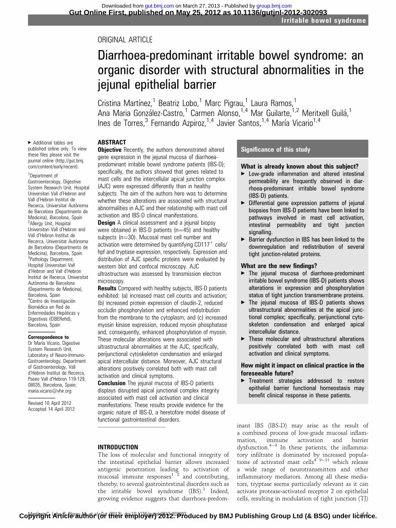
Diarrhoea-predominant irritable bowel syndrome: an organic disorder with structural abnormalities in...
10
ORIGINAL ARTICLE Diarrhoea-predominant irritable bowel syndrome: an organic disorder with structural abnormalities in the jejunal epithelial barrier Cristina Martı ´nez, 1 Beatriz Lobo, 1 Marc Pigrau, 1 Laura Ramos, 1 Ana Maria Gonza ´lez-Castro, 1 Carmen Alonso, 1,4 Mar Guilarte, 1,2 Meritxell Guila ´, 1 Ines de Torres, 3 Fernando Azpiroz, 1,4 Javier Santos, 1,4 Marı ´a Vicario 1,4 ABSTRACT Objective Recently, the authors demonstrated altered gene expression in the jejunal mucosa of diarrhoea- predominant irritable bowel syndrome patients (IBS-D); specifically, the authors showed that genes related to mast cells and the intercellular apical junction complex (AJC) were expressed differently than in healthy subjects. The aim of the authors here was to determine whether these alterations are associated with structural abnormalities in AJC and their relationship with mast cell activation and IBS-D clinical manifestations. Design A clinical assessment and a jejunal biopsy were obtained in IBS-D patients (n¼45) and healthy subjects (n¼30). Mucosal mast cell number and activation were determined by quantifying CD117 + cells/ hpf and tryptase expression, respectively. Expression and distribution of AJC specific proteins were evaluated by western blot and confocal microscopy. AJC ultrastructure was assessed by transmission electron microscopy. Results Compared with healthy subjects, IBS-D patients exhibited: (a) increased mast cell counts and activation; (b) increased protein expression of claudin-2, reduced occludin phosphorylation and enhanced redistribution from the membrane to the cytoplasm; and (c) increased myosin kinase expression, reduced myosin phosphatase and, consequently, enhanced phosphorylation of myosin. These molecular alterations were associated with ultrastructural abnormalities at the AJC, specifically, perijunctional cytoskeleton condensation and enlarged apical intercellular distance. Moreover, AJC structural alterations positively correlated both with mast cell activation and clinical symptoms. Conclusion The jejunal mucosa of IBS-D patients displays disrupted apical junctional complex integrity associated with mast cell activation and clinical manifestations. These results provide evidence for the organic nature of IBS-D, a heretofore model disease of functional gastrointestinal disorders. INTRODUCTION The loss of molecular and functional integrity of the intestinal epithelial barrier allows increased antigenic penetration leading to activation of mucosal immune responses 1 2 and contributing, thereby, to several gastrointestinal disorders such as the irritable bowel syndrome (IBS). 3 Indeed, growing evidence suggests that diarrhoea-predom- inant IBS (IBS-D) may arise as the result of a combined process of low-grade mucosal inflam- mation, immune activation and barrier dysfunction. 4e9 In these patients, the inflamma- tory infiltrate is dominated by increased popula- tions of activated mast cells 4 9e11 which release a wide range of neurotransmitters and other inflammatory mediators. Among all these media- tors, tryptase seems particularly relevant as it can activate protease-activated receptor 2 on epithelial cells, resulting in modulation of tight junction (TJ) < Additional tables are published online only. To view these files please visit the journal online (http://gut.bmj. com/content/early/recent). 1 Department of Gastroenterology, Digestive System Research Unit, Hospital Universitari Vall d’Hebron and Vall d’Hebron Institut de Recerca, Universitat Auto `noma de Barcelona (Departmento de Medicina), Barcelona, Spain 2 Allergy Unit, Hospital Universitari Vall d’Hebron and Vall d’Hebron Institut de Recerca, Universitat Auto `noma de Barcelona (Departmento de Medicina), Barcelona, Spain 3 Pathology Department, Hospital Universitari Vall d’Hebron and Vall d’Hebron Institut de Recerca, Universitat Auto `noma de Barcelona (Departmento de Medicina), Barcelona, Spain 4 Centro de Investigacio ´n Biome ´dica en Red de Enfermedades Hepa ´ticas y Digestivas (CIBERehd), Barcelona, Spain Correspondence to Dr Marı ´a Vicario, Digestive System Research Unit, Laboratory of Neuro-Immuno- Gastroenterology, Department of Gastroenterology, Vall d’Hebron Institut de Recerca, Paseo Vall d’Hebron 119-129, 08035, Barcelona, Spain; [email protected] Revised 10 April 2012 Accepted 14 April 2012 Significance of this study What is already known about this subject? < Low-grade inflammation and altered intestinal permeability are frequently observed in diar- rhoea-predominant irritable bowel syndrome (IBS-D) patients. < Differential gene expression patterns of jejunal biopsies from IBS-D patients have been linked to pathways involved in mast cell activation, intestinal permeability and tight junction signalling. < Barrier dysfunction in IBS has been linked to the downregulation and redistribution of several tight junction-related proteins. What are the new findings? < The jejunal mucosa of diarrhoea-predominant irritable bowel syndrome (IBS-D) patients shows alterations in expression and phosphorylation status of tight junction transmembrane proteins. < The jejunal mucosa of IBS-D patients shows ultrastructural abnormalities at the apical junc- tional complex; specifically, perijunctional cyto- skeleton condensation and enlarged apical intercellular distance. < These molecular and ultrastructural alterations positively correlated both with mast cell activation and clinical symptoms. How might it impact on clinical practice in the foreseeable future? < Treatment strategies addressed to restore epithelial barrier functional homeostasis may benefit clinical response in these patients. Martı ´nez C, Lobo B, Pigrau M, et al. Gut (2012). doi:10.1136/gutjnl-2012-302093 1 of 8 Irritable bowel syndrome Gut Online First, published on May 25, 2012 as 10.1136/gutjnl-2012-302093 Copyright Article author (or their employer) 2012. Produced by BMJ Publishing Group Ltd (& BSG) under licence. group.bmj.com on March 27, 2013 - Published by gut.bmj.com Downloaded from
-
Upload
independent -
Category
Documents
-
view
0 -
download
0
Transcript of Diarrhoea-predominant irritable bowel syndrome: an organic disorder with structural abnormalities in...
ORIGINAL ARTICLE
Diarrhoea-predominant irritable bowel syndrome: anorganic disorder with structural abnormalities in thejejunal epithelial barrier
Cristina Martınez,1 Beatriz Lobo,1 Marc Pigrau,1 Laura Ramos,1
Ana Maria Gonzalez-Castro,1 Carmen Alonso,1,4 Mar Guilarte,1,2 Meritxell Guila,1
Ines de Torres,3 Fernando Azpiroz,1,4 Javier Santos,1,4 Marıa Vicario1,4
ABSTRACTObjective Recently, the authors demonstrated alteredgene expression in the jejunal mucosa of diarrhoea-predominant irritable bowel syndrome patients (IBS-D);specifically, the authors showed that genes related tomast cells and the intercellular apical junction complex(AJC) were expressed differently than in healthysubjects. The aim of the authors here was to determinewhether these alterations are associated with structuralabnormalities in AJC and their relationship with mast cellactivation and IBS-D clinical manifestations.Design A clinical assessment and a jejunal biopsywere obtained in IBS-D patients (n¼45) and healthysubjects (n¼30). Mucosal mast cell number andactivation were determined by quantifying CD117+ cells/hpf and tryptase expression, respectively. Expression anddistribution of AJC specific proteins were evaluated bywestern blot and confocal microscopy. AJCultrastructure was assessed by transmission electronmicroscopy.Results Compared with healthy subjects, IBS-D patientsexhibited: (a) increased mast cell counts and activation;(b) increased protein expression of claudin-2, reducedoccludin phosphorylation and enhanced redistributionfrom the membrane to the cytoplasm; and (c) increasedmyosin kinase expression, reduced myosin phosphataseand, consequently, enhanced phosphorylation of myosin.These molecular alterations were associated withultrastructural abnormalities at the AJC, specifically,perijunctional cytoskeleton condensation and enlargedapical intercellular distance. Moreover, AJC structuralalterations positively correlated both with mast cellactivation and clinical symptoms.Conclusion The jejunal mucosa of IBS-D patientsdisplays disrupted apical junctional complex integrityassociated with mast cell activation and clinicalmanifestations. These results provide evidence for theorganic nature of IBS-D, a heretofore model disease offunctional gastrointestinal disorders.
INTRODUCTIONThe loss of molecular and functional integrity ofthe intestinal epithelial barrier allows increasedantigenic penetration leading to activation ofmucosal immune responses1 2 and contributing,thereby, to several gastrointestinal disorders such asthe irritable bowel syndrome (IBS).3 Indeed,growing evidence suggests that diarrhoea-predom-
inant IBS (IBS-D) may arise as the result ofa combined process of low-grade mucosal inflam-mation, immune activation and barrierdysfunction.4e9 In these patients, the inflamma-tory infiltrate is dominated by increased popula-tions of activated mast cells4 9e11 which releasea wide range of neurotransmitters and otherinflammatory mediators. Among all these media-tors, tryptase seems particularly relevant as it canactivate protease-activated receptor 2 on epithelialcells, resulting in modulation of tight junction (TJ)
< Additional tables arepublished online only. To viewthese files please visit thejournal online (http://gut.bmj.com/content/early/recent).1Department ofGastroenterology, DigestiveSystem Research Unit, HospitalUniversitari Vall d’Hebron andVall d’Hebron Institut deRecerca, Universitat Autonomade Barcelona (Departmento deMedicina), Barcelona, Spain2Allergy Unit, HospitalUniversitari Vall d’Hebron andVall d’Hebron Institut deRecerca, Universitat Autonomade Barcelona (Departmento deMedicina), Barcelona, Spain3Pathology Department,Hospital Universitari Valld’Hebron and Vall d’HebronInstitut de Recerca, UniversitatAutonoma de Barcelona(Departmento de Medicina),Barcelona, Spain4Centro de InvestigacionBiomedica en Red deEnfermedades Hepaticas yDigestivas (CIBERehd),Barcelona, Spain
Correspondence toDr Marıa Vicario, DigestiveSystem Research Unit,Laboratory of Neuro-Immuno-Gastroenterology, Departmentof Gastroenterology, Valld’Hebron Institut de Recerca,Paseo Vall d’Hebron 119-129,08035, Barcelona, Spain;[email protected]
Revised 10 April 2012Accepted 14 April 2012
Significance of this study
What is already known about this subject?< Low-grade inflammation and altered intestinal
permeability are frequently observed in diar-rhoea-predominant irritable bowel syndrome(IBS-D) patients.
< Differential gene expression patterns of jejunalbiopsies from IBS-D patients have been linked topathways involved in mast cell activation,intestinal permeability and tight junctionsignalling.
< Barrier dysfunction in IBS has been linked to thedownregulation and redistribution of severaltight junction-related proteins.
What are the new findings?< The jejunal mucosa of diarrhoea-predominant
irritable bowel syndrome (IBS-D) patients showsalterations in expression and phosphorylationstatus of tight junction transmembrane proteins.
< The jejunal mucosa of IBS-D patients showsultrastructural abnormalities at the apical junc-tional complex; specifically, perijunctional cyto-skeleton condensation and enlarged apicalintercellular distance.
< These molecular and ultrastructural alterationspositively correlated both with mast cellactivation and clinical symptoms.
How might it impact on clinical practice in theforeseeable future?< Treatment strategies addressed to restore
epithelial barrier functional homeostasis maybenefit clinical response in these patients.
Martınez C, Lobo B, Pigrau M, et al. Gut (2012). doi:10.1136/gutjnl-2012-302093 1 of 8
Irritable bowel syndrome Gut Online First, published on May 25, 2012 as 10.1136/gutjnl-2012-302093
Copyright Article author (or their employer) 2012. Produced by BMJ Publishing Group Ltd (& BSG) under licence.
group.bmj.com on March 27, 2013 - Published by gut.bmj.comDownloaded from

proteins and increases in permeability through paracellularpathways.12
Studies investigating the molecular basis of these abnormali-ties have largely focused on the colon,13e15 partly due to the easyaccess to distal areas of the gut. However, some data indicatethat mucosal inflammation in IBS-D patients is not limited tothe lower gut,4 16 and that increased intestinal permeabilityoccurs along the whole intestine.6 Furthermore, several lines ofevidence point to the small bowel as the site of origin of IBSsymptoms.17e20 The general hypothesis of our researchprogramme is that IBS-D is a disorder which primarily affectsthe small bowel and, hence, our studies focused on the jejunum.
In previous studies, we demonstrated increased number andactivation of mast cells in a cohort of IBS-D patients (n¼25) ascompared with healthy subjects (n¼23).11 We further observedsignificant differences in global gene expression in both groups,and TJ signalling pathways came out as the most significantcanonical pathways associated with altered gene expression inthe jejunal mucosa of IBS-D patients.11 Having shown thisabnormal expression of genes related to TJs, we now hypoth-esised that these alterations are associated with structuralabnormalities of the apical junction complex (AJC) at theintestinal mucosal barrier. The relevance of this hypothesis isthat it questions the paradigmatic functional nature of thisdisorder. In the first place, we wished to confirm in a signifi-cantly larger cohort of patients and controls the mast cellabnormalities and their correlation with symptoms previouslyobserved. The original aims of the present study were to deter-mine in this cohort: (1) the molecular mechanisms of AJCdisassembly; (2) the impact of these molecular alterations onAJC ultrastructure; and (3) whether AJC disassembly is relatedto mast cell activation and IBS-D clinical manifestations.
METHODSParticipantsNaive patients (n¼45) referred for the evaluation of IBSsymptom who presented daily watery diarrhoea or mushy stoolsand meeting Rome II IBS-D criteria21 were prospectivelyrecruited from the gastroenterology outpatient clinic. Healthysubjects (n¼30) without gastrointestinal symptoms wererecruited only by public advertising. Prior to entering the study,candidates were asked to fill out clinical questionnaires (tocharacterise the symptoms in patients and verify the lack ofsymptoms healthy subjects), and all underwent a battery of skinprick tests for 22 common food allergens (Leti SA, Barcelona,Spain) to exclude allergy. In all patients a broad biochemical andserological profile, including antitransglutaminase antibodies andthyroid hormones were determined. Previous history of acutegastroenteritis in relation to the initiation of IBS symptoms wasspecifically excluded. Moreover, reasonable exclusion of gastro-intestinal comorbidities, such as microscopic colitis and celiacdisease, was achieved by means of upper and lower fiberoptic andsmall bowel capsule endoscopy, abdominal sonography andbarium studies, when considered pertinent. Other exclusioncriteria for all participants were: regular use of medication orherbal preparation in the previous 3 months, active smoking,major psychiatric and organic diseases. The study protocol wasapproved by the Ethics Committee of the Hospital Vall d’Hebron.Written informed consent was obtained from each participant.
General proceduresClinical evaluationUsing daily questionnaires over a 10-day period, the followingparameters were recorded in all participants: (a) severity of
abdominal pain by a 100-point visual analogue scale; (b)frequency of abdominal pain (number of days with pain); (c)stool frequency (day with maximum number of bowel move-ments); and (d) stool consistency assessed by the Bristol StoolForm Score22 (if more than one bowel movement per day, theBristol Stool Form Score was averaged, and the grand mean overthe 10-day period was calculated). Background stress over thelast year was evaluated by the Modified Social ReadjustmentScale of HolmeseRahe.23
Jejunal biopsyA single mucosal biopsy was obtained from the proximaljejunum (5 cm distal to the angle of Treitz) using a Watson’scapsule as previously described.4 11 Tissue samples were imme-diately split into two similar pieces with a sterile scalpel. Onefragment was fixed in formalin and embedded in paraffin forfurther microscopic examination (histology, immunohisto-chemistry and immunofluorescence). The other fragment wassnap frozen in liquid nitrogen for protein isolation and westernblot analysis or placed in RNAse free tubes containing 500 ml ofRNA Later Solution (Ambion, Madrid, Spain) for RNA isolationand analysis by quantitative real-time PCR (Q-RT-PCR) (seeExperimental design).
Experimental design and rationaleClinical evaluation and jejunal biopsy were obtained at studyentry in all participants. Analyses performed in each participantare detailed in supplementary table 1. Routine histology andimmunohistochemistry to assess the number of mast cells andintraepithelial lymphocytes were performed in all participants(45 IBS-D patients and 30 healthy subjects). The followinganalyses were also performed in randomly selected subjects (seeStatistical analysis below and supplementary table 1):a. To evaluate mast cell activation, tryptase RNA and protein
were analysed by Q-RT-PCR (17 IBS-D patients and 12healthy subjects) and by western blot (seven IBS-D patientsand seven healthy subjects), respectively.
b. To evaluate the expression of TJ transmembrane proteins, theprotein was isolated and analysed by western blot analysis(seven IBS-D patients and seven healthy subjects).
c. To evaluate the mechanisms of AJC disassembly throughmodulation of cytoskeleton, protein expression of phosphor-ylated myosin light chain (pMLC), myosin light chain kinase(MLCK) and the catalytic subunit of myosin phosphatase(PP1cd) was assessed by immunofluorescence (14 IBS-Dpatients and 14 healthy subjects) and western blot (sevenIBS-D patients and seven healthy subjects).
d. To assess the impact of the above molecular alterations onAJC ultrastructure, tissue samples were analysed by trans-mission electron microscopy (TEM) (15 IBS-D patients and10 healthy subjects).
Analytical proceduresHistology and immunohistochemistryTissue sections were processed for routine H&E staining to assessepithelial morphology. In addition, the number of mast cells wasdetermined at 3400 magnification using antihuman c-kit(CD117) (Dako, Barcelona, Spain), as previously described.4 11
Specimens were blindly examined by one experiencedpathologist (IT).
ImmunofluorescenceTissue sections were deparaffinised and hydrated followinggeneral procedures and blocked with Dako Blocking Solution
2 of 8 Martınez C, Lobo B, Pigrau M, et al. Gut (2012). doi:10.1136/gutjnl-2012-302093
Irritable bowel syndrome
group.bmj.com on March 27, 2013 - Published by gut.bmj.comDownloaded from
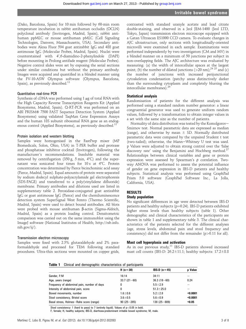
(Dako, Barcelona, Spain) for 10 min followed by 60-min roomtemperature incubation in rabbit antihuman occludin (OCLN)polyclonal antibody (Invitrogen, Madrid, Spain), rabbit anti-human ppMLC or mouse antihuman pMLC (Cell SignalingTechnologies, Danvers, Massachusetts, USA). Secondary anti-bodies were Alexa Fluor 594 goat antirabbit IgG and 488 goatantimouse IgG (Molecular Probes, Madrid, Spain). Nuclei werecounterstained with 4’,6-diamidino-2-phenylindole (DAPI)before mounting in Prolong antifade reagent (Molecular Probes).Negative control slides were set by exposing the serial sectionsunder similar conditions but without the primary antibody.Images were acquired and quantified in a blinded manner usingthe FV-10-ASW Olympus software (Olympus, Barcelona,Spain), as previously described.11
Quantitative real-time PCRSynthesis of cDNAwas performed using 1 mg of total RNAwiththe High Capacity Reverse Transcription Reagents Kit (AppliedBiosystems, Madrid, Spain). Q-RT-PCR was performed on anABI PRISM� 7500 FAST Sequence Detection System (AppliedBiosystems) using validated TaqMan Gene Expression Assaysand the human 18S subunit ribosomal RNA gene as an endog-enous control (Applied Biosystems), as previously described.11
Protein isolation and western blottingSamples were homogenised in the FastPrep mixer (MPBiomedicals, Solon, Ohio, USA) in T-PER buffer and proteaseand phosphatase inhibitor cocktail (Invitrogen), following themanufacturer ’s recommendations. Insoluble material wasremoved by centrifugation (350 g, 5 min, 48C) and the super-natant was sonicated four times for 10 s at 48C. Proteinconcentration was determined by Pierce bicinchoninic acid assay(Pierce, Madrid, Spain). Equal amounts of protein were separatedby sodium dodecyl sulphate-polyacrylamide gel electrophoresis(SDS-PAGE) and transferred to a poly(vinylidene difluoride)membrane. Primary antibodies and dilutions used are listed insupplementary table 2. Peroxidase-conjugated goat antirabbitIgG or goat antimouse IgG (Pierce) and the chemiluminescencedetection system SuperSignal West Femto (Thermo Scientific,Madrid, Spain) were used to detect bound antibodies. All blotswere probed with mouse antihuman b-actin (Sigma-Aldrich,Madrid, Spain) as a protein loading control. Densitometriccomparison was carried out on the same immunoblot using theImageJ software (National Institutes of Health; http://rsb.info.nih.gov/ij/).
Transmission electron microscopySamples were fixed with 2.5% glutaraldehyde and 2% para-formaldehyde and processed for TEM following standardprocedures. Ultra-thin sections were mounted on copper grids,
contrasted with standard uranyle acetate and lead citratedouble-staining, and observed in a Jeol JEM-1400 (Jeol LTD,Tokyo, Japan) transmission electron microscope equipped witha Gatan Ultrascan ES1000 CCD camera. To evaluate changes inAJC ultrastructure, only sections with longitudinally-orientedmicrovilli were examined in each sample. Examinations wereperformed independently by two investigators (CM and MV) ina blinded manner on a minimum of 50 junctions per subject innon-overlapping fields. The AJC architecture was evaluated bymeasuring: (a) the width of intercellular spaces at the largestpoint; (b) the number of dilated junctions (>20 nm);24 25 and (c)the number of junctions with increased perijunctionalcytoskeleton condensation (patchy areas distinctively darkerthan the surrounding cytoplasm and completely blurring theintercellular membranes).26
Statistical analysisRandomisation of patients for the different analysis wasperformed using a standard random number generator: a linearcongruential generator was used to generate uniform randomvalues, followed by a transformation to obtain integer values ina set with the same size as the number of patients.Normality of data distributionwas tested by the Komolgorove
Smirnov test. Normal parametric data are expressed as median(range), and otherwise by mean 6 SD. Normally distributedparametric data were compared by the unpaired Student t test(two-tailed); otherwise, the ManneWhitney U test was used.p Values were adjusted to obtain strong control over the ‘falsediscovery rate’ using the Benjamini and Hochberg method.27
Relationships between clinical variables and gene or proteinexpression were assessed by Spearman’s r correlation. Two-way ANOVA was performed to assess the potential influenceof gender on gene expression in IBS-D patients and healthysubjects. Statistical analysis was performed using GraphPadPrism 5.0 software (GraphPad Software Inc., La Jolla,California, USA).
RESULTSStudy populationNo significant differences in age were detected between IBS-Dpatients and healthy subjects (p¼0.24). IBS-D patients exhibitedhigher stress levels than healthy subjects (table 1). Otherdemographic and clinical characteristics of the participants areshown in table 1 and supplementary table 3. The clinical char-acteristics of the patients selected for the different analyses(age, stress levels, abdominal pain and stool frequency andconsistency) did not differ from the remainder (p>0.11 for all).
Mast cell hyperplasia and activationAs in our previous study,11 IBS-D patients showed increasedmast cell counts (IBS-D: 26.2611.1; healthy subjects: 17.268.8
Table 1 Clinical and demographic characteristics of participants
H (n[30) IBS-D (n[45) p Value
Gender, F:M 16:14 34:11 e
Age, years (range) 33.7 (21e60) 36.3 (18e60) 0.24
Frequency of abdominal pain, number of days 0 5.562.9 e
Intensity of abdominal pain, score 0 51.3625.0 e
Bowel movements, number 1.660.6 5.262.8 <0.0001Stool consistency, Bristol score 3.660.5 5.660.9 <0.0001Basal stress, HolmeseRahe score (range) 90 (25e285) 138 (25e889) <0.05
Stool consistency: 1 (hard to pass) to 7 (entirely liquid). Values of p<0.05 in bold.F, female; H, healthy subjects; IBS-D, diarrhoea-predominant irritable bowel syndrome; M, male.
Martınez C, Lobo B, Pigrau M, et al. Gut (2012). doi:10.1136/gutjnl-2012-302093 3 of 8
Irritable bowel syndrome
group.bmj.com on March 27, 2013 - Published by gut.bmj.comDownloaded from

CD117+ cells/hpf; p¼0.0004) and tryptase mRNA (IBS-D:1.960.5; healthy subjects: 1.160.2 fold; p<0.0001). The presentstudy further showed that tryptase protein was overexpressed inpatients (IBS-D: 1.460.3; healthy subjects: 1.060.2 fold;p¼0.03).
Expression and distribution of transmembrane TJ proteinsOur previous study11 found several genes involved in the TJsignalling pathway differentially expressed in IBS-D. In order tofurther assess TJ dysregulation, the expression of trans-membrane TJ proteins was now analysed. No differences inprotein expression of claudins (CLDNs) 1, 3 and 4 were
observed, while CLDN2 was significantly upregulated in IBS-D(figure 1). Protein expression of OCLN was similar in bothgroups; however, OCLN phosphorylation (p-OCLN) (which isidentified by the upper 85 KDa band) was significantly reducedin IBS-D (figure 1). Of note, p-OCLN correlated with theexpression of CLDN3 (r¼0.84, p¼0.0001) and CLDN4 (r¼0.73,p¼0.003).
As the phosphorylation status of OCLN has been linked to itsdistribution in epithelial cells,28 we assessed OCLN staining byconfocal immunofluorescence. IBS-D samples displayed signifi-cantly increased staining of OCLN in the cytoplasm of jejunalenterocytes, suggesting intensive internalisation of this protein,
Figure 1 Transmembrane tight junction protein expression in jejunal mucosa. CLDN and OCLN protein expression was measured by western blot inseven IBS-D patients and seven healthy subjects (left panel). Protein fold-change (right panel) was calculated for each sample with reference to theaverage of the target protein to b-actin ratio of the healthy group. Intergroup comparisons were performed by the ManneWhitney U test (significant pvalues shown). CLDN, claudin; IBS-D, diarrhoea-predominant irritable bowel syndrome; OCLN, occludin.
Figure 2 OCLN distribution in jejunalmucosa. Upper panels showrepresentative micrographs of jejunalepithelium (high-power field, 31000)with OCLN labelled byimmunofluorescence in red and nuclearlabelling with DAPI in blue. Lowerpanels show quantitative analyses offluorescence intensity performed blindlyusing the FV-10-ASW Olympussoftware. Data are average offluorescence intensity in 10 non-overlapping fields per subject.Intergroup comparisons wereperformed by the unpaired Student ttest (significant p values shown). IBS-D,diarrhoea-predominant irritable bowelsyndrome; OCLN, occludin; TJ, tightjunction.
4 of 8 Martınez C, Lobo B, Pigrau M, et al. Gut (2012). doi:10.1136/gutjnl-2012-302093
Irritable bowel syndrome
group.bmj.com on March 27, 2013 - Published by gut.bmj.comDownloaded from

as opposed to the healthy group where OCLN was predomi-nantly located in the TJ (figure 2).
Phosphorylation of MLCIn an attempt to identify mechanisms regulating AJC function,we evaluated MLC phosphorylation. IBS-D patients showeda significant increase in MLC phosphorylation at Ser19 (pMLC)(figure 3), which has been related to the contraction of the acto-myosin cytoskeleton and disassembly of TJ.1 2 By contrast, MLCphosphorylation at Thr18+Ser19 (ppMLC) remained unchanged(figure 3), suggesting that de novo TJ assembly is not impaired inIBS-D.
In concordance with the increased pMLC, IBS-D showedincreased expression of MLCK (4.963.7 vs 1.060.8 fold-changein healthy subjects, p¼0.01), while PP1cd was decreased (IBS-D:0.760.2 vs1.060.2 fold-change in healthy subjects, p¼0.02).
Ultrastructural alterations at the apical junctionsAs previously described,11 routine processing of jejunal samplesfor H&E staining in the present study revealed no differences in
epithelial architecture between IBS-D patients and healthysubjects. Using TEM, we were now able to demonstrate that themolecular alterations described above were associated withultrastructural changes of intercellular junctions featuring a realdisassembly (figure 4). Indeed, compared with healthy subjects,IBS-D patients exhibited a larger apical intercellular distancewith a higher proportion of dilated (>20 nm) junctions(figure 4). Patients also had a higher percentage of junctions withperijunctional cytoskeleton condensation (figure 4).
Correlation of molecular and ultrastructural AJC alterations withIBS-D clinical manifestationsAs previously observed,11 mast cell activation, measured bytryptase mRNA expression, correlated with stool frequency andconsistency in IBS-D patients (table 2), whereas their correlationwith mast cell number was poor, suggesting that mast cellactivation rather than number influences bowel habit. In thepresent study, we further demonstrated that tryptase proteinexpression, the end result of mast cell activation, also exhibitedgood correlation with bowel habit (table 2). Furthermore,
Figure 3 MLC phosphorylation injejunal mucosa. Upper panels showrepresentative micrographs of jejunalepithelium (high-power field, 3400)with Ser19-phosphorylated MLC (pMLC)labelled by immunofluorescence ingreen, Ser19/Thr18-phosphorylated MLC(ppMLC) in red and nuclear labellingwith DAPI in blue. Lower panels showquantitative analyses of fluorescenceintensity performed blindly using theFV-10-ASW Olympus software. Dataare average of fluorescence intensity in10 non-overlapping fields per subject.Intergroup comparisons wereperformed by the unpaired Student ttest (significant p value shown). IBS-D,diarrhoea-predominant irritable bowelsyndrome; MLC, myosin light chain.
Martınez C, Lobo B, Pigrau M, et al. Gut (2012). doi:10.1136/gutjnl-2012-302093 5 of 8
Irritable bowel syndrome
group.bmj.com on March 27, 2013 - Published by gut.bmj.comDownloaded from

tryptase protein expression correlated with AJC impairmentmeasured by CLDN2 protein overexpression (r¼0.76, p¼0.002)and by increased OCLN cytoplasmic staining (r¼0.75, p¼0.003).Conversely, AJC impairment correlated with stool frequencyand consistency (table 2). Ultrastructural AJC alterationscorrelated with abnormal bowel habit, as well as with abdom-inal pain intensity (table 2).
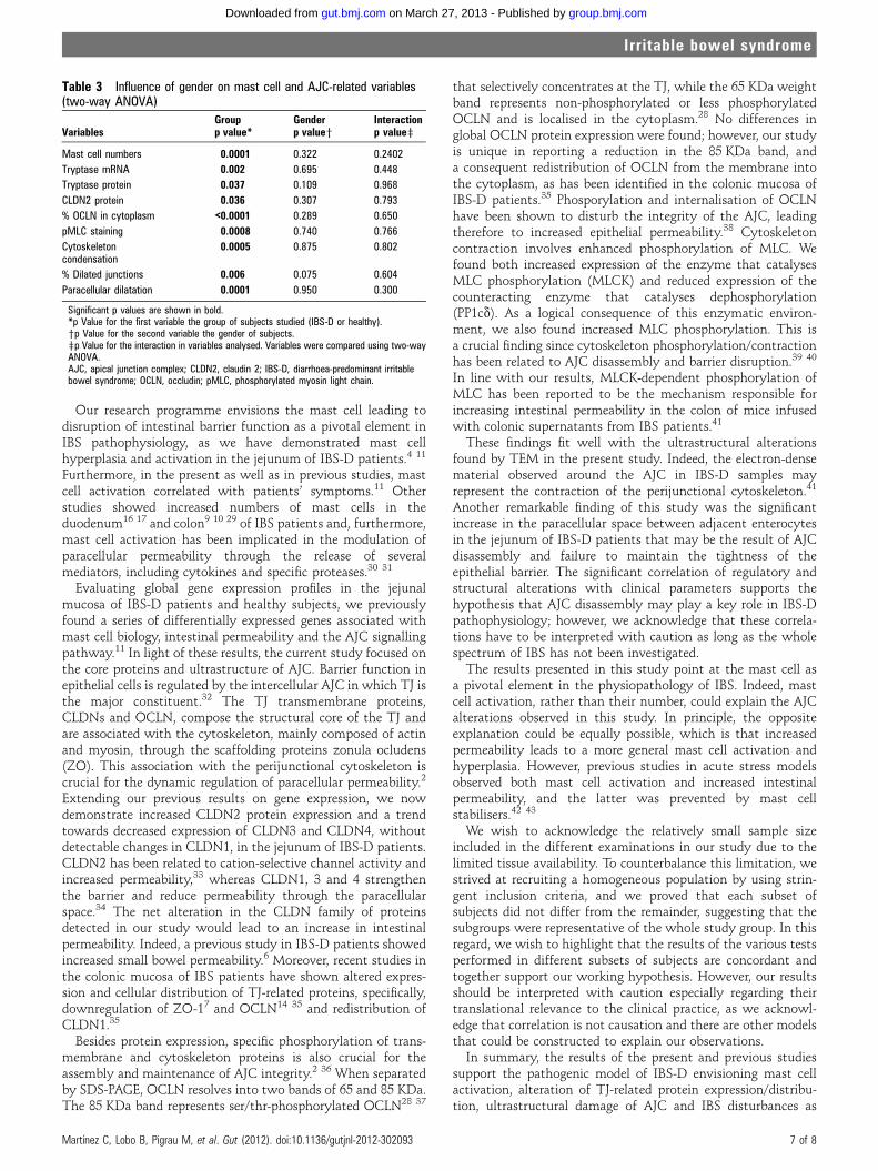
Correlation of mast cell-related variables and AJC alterationswith gender and stress levelsTo assess the potential influence of gender on mast cell infiltra-tion/activation and on AJC alterations, we included gender asa variable in the analysis and did not find significant differencesin any of the parameters evaluated (table 3).
To assess the influence of basal stress levels on biologicalparameters, we applied the Spearman r correlation to pooled
data of patients and healthy subjects. Stress level was correlatedwith increased mast cell activation, measured by tryptaseprotein expression (r¼0.53, p¼0.05). However, no significantassociation between basal stress and the other biologicalparameters measured was detected, including mast cell counts(r¼0.12, p¼0.33), molecular alterations in TJ-related proteins(r<0.27, p>0.35 for all) and ultrastructural AJC alterations(r<0.32, p>0.12 for all).
DISCUSSIONFor the first time, we demonstrated structural abnormalities inthe jejunal mucosa of IBS-D patients, which suggests that this isan organic rather than a functional disorder. Indeed, in contrastto healthy subjects, IBS-D patients exhibited disrupted archi-tecture of the apical junctional complex, which was associatedwith mast cell activation and IBS symptoms.
Figure 4 Ultrastructure of apicaljunctional complex in the jejunalmucosa. Upper panels showrepresentative micrographs of thejejunal epithelium (originalmagnification 360 000). Noteintercellular junctions in healthysubjects (black arrow, in upper leftpanel), widening of the intercellularspace (white arrow, in upper middlepanel) and perijunctional cytoskeletoncondensation (dashed circles, in upperright panel) in diarrhoea-predominantirritable bowel syndrome (IBS-D)patients. The maximum width ofintercellular spaces (at the largest point)was measured in a minimum of 50junctions per subject in non-overlappingfields. The proportion of dilatedjunctions (>20 nm) per subject isshown in lower left panel and theaverage intercellular distance persubject in lower middle panel. Theproportion in each subject of junctionswith electron-dense material around theapical junctional complex is shown in lowerright panel. Intergroup comparisons were performedby the unpaired Student t test (significant p values shown).
Table 2 Clinicalebiological correlations in the jejunal mucosa of IBS-D patients and healthy subjects
Abdominal pain* Bowel movements Stool consistencyyr p FDR r p FDR r p FDR
Mast cell-related variables
CD117+ cells/hpf (45 IBS-D, 30 HS) �0.24 0.12 0.16 0.21 0.08 0.11 0.34 0.004 0.01
Tryptase mRNA (17 IBS-D, 12 HS) 0.35 0.17 0.21 0.65 0.0001 0.001 0.68 0.0001 0.002
Tryptase protein (7 IBS-D, 7 HS) 0.28 0.56 0.62 0.87 0.03 0.05 0.61 0.02 0.04
TJ-related proteins
CLDN2 protein (7 IBS-D, 7 HS) 0.20 0.66 0.68 0.56 0.04 0.06 0.71 0.004 0.01
% OCLN in the cytoplasm (14 IBS-D, 10 HS) �0.10 0.76 0.76 0.67 0.001 0.007 0.55 0.02 0.04
pMLC staining (14 IBS-D 14 HS) 0.18 0.55 0.64 0.58 0.002 0.006 0.60 0.001 0.004
Ultrastructural TJ alterations
Cytoskeleton condensation (15 IBS-D, 10 HS) 0.16 0.56 0.60 0.50 0.01 0.02 0.67 0.0002 0.002
Proportion of dilated junctions (15 IBS-D, 10 HS) 0.52 0.05 0.08 0.50 0.01 0.02 0.66 0.0004 0.002
Paracellular dilatation (15 IBS-D, 10 HS) 0.74 0.002 0.008 0.65 0.0004 0.002 0.72 0.0001 0.003
An FDR <15% was accepted as significant.*Severity score.yBristol scale score. Values of p<0.05 in bold.CLDN2, claudin 2; FDR, false discovery rate; hpf, high power field; HS, healthy subjects; IBS-D, diarrhoea-predominant irritable bowel syndrome; OCLN, occludin; pMLC, phosphorylated myosinlight chain; TJ, tight junction.
6 of 8 Martınez C, Lobo B, Pigrau M, et al. Gut (2012). doi:10.1136/gutjnl-2012-302093
Irritable bowel syndrome
group.bmj.com on March 27, 2013 - Published by gut.bmj.comDownloaded from
Our research programme envisions the mast cell leading todisruption of intestinal barrier function as a pivotal element inIBS pathophysiology, as we have demonstrated mast cellhyperplasia and activation in the jejunum of IBS-D patients.4 11
Furthermore, in the present as well as in previous studies, mastcell activation correlated with patients’ symptoms.11 Otherstudies showed increased numbers of mast cells in theduodenum16 17 and colon9 10 29 of IBS patients and, furthermore,mast cell activation has been implicated in the modulation ofparacellular permeability through the release of severalmediators, including cytokines and specific proteases.30 31
Evaluating global gene expression profiles in the jejunalmucosa of IBS-D patients and healthy subjects, we previouslyfound a series of differentially expressed genes associated withmast cell biology, intestinal permeability and the AJC signallingpathway.11 In light of these results, the current study focused onthe core proteins and ultrastructure of AJC. Barrier function inepithelial cells is regulated by the intercellular AJC in which TJ isthe major constituent.32 The TJ transmembrane proteins,CLDNs and OCLN, compose the structural core of the TJ andare associated with the cytoskeleton, mainly composed of actinand myosin, through the scaffolding proteins zonula ocludens(ZO). This association with the perijunctional cytoskeleton iscrucial for the dynamic regulation of paracellular permeability.2
Extending our previous results on gene expression, we nowdemonstrate increased CLDN2 protein expression and a trendtowards decreased expression of CLDN3 and CLDN4, withoutdetectable changes in CLDN1, in the jejunum of IBS-D patients.CLDN2 has been related to cation-selective channel activity andincreased permeability,33 whereas CLDN1, 3 and 4 strengthenthe barrier and reduce permeability through the paracellularspace.34 The net alteration in the CLDN family of proteinsdetected in our study would lead to an increase in intestinalpermeability. Indeed, a previous study in IBS-D patients showedincreased small bowel permeability.6 Moreover, recent studies inthe colonic mucosa of IBS patients have shown altered expres-sion and cellular distribution of TJ-related proteins, specifically,downregulation of ZO-17 and OCLN14 35 and redistribution ofCLDN1.35
Besides protein expression, specific phosphorylation of trans-membrane and cytoskeleton proteins is also crucial for theassembly and maintenance of AJC integrity.2 36 When separatedby SDS-PAGE, OCLN resolves into two bands of 65 and 85 KDa.The 85 KDa band represents ser/thr-phosphorylated OCLN28 37
that selectively concentrates at the TJ, while the 65 KDa weightband represents non-phosphorylated or less phosphorylatedOCLN and is localised in the cytoplasm.28 No differences inglobal OCLN protein expression were found; however, our studyis unique in reporting a reduction in the 85 KDa band, anda consequent redistribution of OCLN from the membrane intothe cytoplasm, as has been identified in the colonic mucosa ofIBS-D patients.35 Phosporylation and internalisation of OCLNhave been shown to disturb the integrity of the AJC, leadingtherefore to increased epithelial permeability.38 Cytoskeletoncontraction involves enhanced phosphorylation of MLC. Wefound both increased expression of the enzyme that catalysesMLC phosphorylation (MLCK) and reduced expression of thecounteracting enzyme that catalyses dephosphorylation(PP1cd). As a logical consequence of this enzymatic environ-ment, we also found increased MLC phosphorylation. This isa crucial finding since cytoskeleton phosphorylation/contractionhas been related to AJC disassembly and barrier disruption.39 40
In line with our results, MLCK-dependent phosphorylation ofMLC has been reported to be the mechanism responsible forincreasing intestinal permeability in the colon of mice infusedwith colonic supernatants from IBS patients.41
These findings fit well with the ultrastructural alterationsfound by TEM in the present study. Indeed, the electron-densematerial observed around the AJC in IBS-D samples mayrepresent the contraction of the perijunctional cytoskeleton.41
Another remarkable finding of this study was the significantincrease in the paracellular space between adjacent enterocytesin the jejunum of IBS-D patients that may be the result of AJCdisassembly and failure to maintain the tightness of theepithelial barrier. The significant correlation of regulatory andstructural alterations with clinical parameters supports thehypothesis that AJC disassembly may play a key role in IBS-Dpathophysiology; however, we acknowledge that these correla-tions have to be interpreted with caution as long as the wholespectrum of IBS has not been investigated.
The results presented in this study point at the mast cell asa pivotal element in the physiopathology of IBS. Indeed, mastcell activation, rather than their number, could explain the AJCalterations observed in this study. In principle, the oppositeexplanation could be equally possible, which is that increasedpermeability leads to a more general mast cell activation andhyperplasia. However, previous studies in acute stress modelsobserved both mast cell activation and increased intestinalpermeability, and the latter was prevented by mast cellstabilisers.42 43
We wish to acknowledge the relatively small sample sizeincluded in the different examinations in our study due to thelimited tissue availability. To counterbalance this limitation, westrived at recruiting a homogeneous population by using strin-gent inclusion criteria, and we proved that each subset ofsubjects did not differ from the remainder, suggesting that thesubgroups were representative of the whole study group. In thisregard, we wish to highlight that the results of the various testsperformed in different subsets of subjects are concordant andtogether support our working hypothesis. However, our resultsshould be interpreted with caution especially regarding theirtranslational relevance to the clinical practice, as we acknowl-edge that correlation is not causation and there are other modelsthat could be constructed to explain our observations.In summary, the results of the present and previous studies
support the pathogenic model of IBS-D envisioning mast cellactivation, alteration of TJ-related protein expression/distribu-tion, ultrastructural damage of AJC and IBS disturbances as
Table 3 Influence of gender on mast cell and AJC-related variables(two-way ANOVA)
VariablesGroupp value*
Genderp valuey
Interactionp valuez
Mast cell numbers 0.0001 0.322 0.2402
Tryptase mRNA 0.002 0.695 0.448
Tryptase protein 0.037 0.109 0.968
CLDN2 protein 0.036 0.307 0.793
% OCLN in cytoplasm <0.0001 0.289 0.650
pMLC staining 0.0008 0.740 0.766
Cytoskeletoncondensation
0.0005 0.875 0.802
% Dilated junctions 0.006 0.075 0.604
Paracellular dilatation 0.0001 0.950 0.300
Significant p values are shown in bold.*p Value for the first variable the group of subjects studied (IBS-D or healthy).yp Value for the second variable the gender of subjects.zp Value for the interaction in variables analysed. Variables were compared using two-wayANOVA.AJC, apical junction complex; CLDN2, claudin 2; IBS-D, diarrhoea-predominant irritablebowel syndrome; OCLN, occludin; pMLC, phosphorylated myosin light chain.
Martınez C, Lobo B, Pigrau M, et al. Gut (2012). doi:10.1136/gutjnl-2012-302093 7 of 8
Irritable bowel syndrome
group.bmj.com on March 27, 2013 - Published by gut.bmj.comDownloaded from
a chain of events. These results challenge the classical concep-tion of IBS-D as a functional disorder and provide evidence oforganicity in a significant proportion of patients.
Acknowledgements The authors thank Milagros Gallart, Montse Casellas andCarmen Alastrue for help in sample analysis; Anna Aparici, Maria Teresa Casaus andPurificacion Rodrıguez for their invaluable assistance in the performance of jejunalbiopsies; Dr Marta Valeri from the Scientific and Technical Support Unit of Valld’Hebron Research Institute for excellent technical support in confocal microscopy;Servei de Microscopia of Universitat Autonoma de Barcelona for excellenttechnical support; Christine O’Hara for English editing of the manuscript; Dr. MarıaAntolin for critically reviewing the manuscript; and Gloria Santaliestra for secretarialassistance.
Contributors CM designed the experiments, analysed the data and wrote the paper;BL, LR, CA and MP recruited the participants, performed the biopsies andcreated/maintained the participant database; MeG contributed to data acquisition;MaG evaluated the allergy status of the participants; IT performed thehistopathological analysis; AMG contributed to maintenance of the database and finaledition of the paper; FA gave clinical advice and critically reviewed the manuscript; JSrecruited patients and collected the clinical data, supervised the project and wrote thepaper; MV designed the experiments, supervised the project and wrote the paper. Allauthors discussed the results and implications and commented on the manuscript atall stages. JS and MV share senior coauthorship.
Funding Supported in part by the Fondo de Investigacion Sanitaria and CIBERehd,Instituto Carlos III, Subdireccion General de Investigacion Sanitaria, Ministerio deCiencia e Innovacion: CM08/00229 (B Lobo); CM05/00055 (L Ramos); CM10/00155(M Pigrau); CM04/00019 (C Alonso); PI05/1423, EC07/90148 & PI/080940 (J Santos);CP10/00502 (M Vicario); Ministerio de Educacion, Direccion General de Investigacion:SAF 2009-07416 (F Azpiroz); Agencia de Gestio d’Ajuts Universitaris i de Recerca, dela Generalitat de Catalunya: 2009 SGR 219 (F Azpiroz and J Santos); the InternationalFoundation for Functional Gastrointestinal Disorders (2008 IFFGD) and the 2010 RomeFoundation Award (J Santos); and Centro de Investigacion Biomedica en Red deEnfermedades Hepaticas y Digestivas: CB06/04/0021 (F Azpiroz and J Santos).CIBERehd is funded by the Instituto de Salud Carlos III.
Competing interests None.
Ethics approval Approval provided by the Ethics Committee of the Hospital Valld’Hebron.
Provenance and peer review Not commissioned; externally peer reviewed.
REFERENCES1. Shen L, Turner JR. Role of epithelial cells in initiation and propagation of intestinal
inflammation. Eliminating the static: tight junction dynamics exposed. Am J PhysiolGastrointest Liver Physiol 2006;290:G577e82.
2. Turner JR. Intestinal mucosal barrier function in health and disease. Nat RevImmunol 2009;9:799e809.
3. Spiller R. Clinical update: irritable bowel syndrome. Lancet 2007;369:1586e8.4. Guilarte M, Santos J, de Torres I, et al. Diarrhoea-predominant IBS patients show
mast cell activation and hyperplasia in the jejunum. Gut 2007;56:203e9.5. Gwee KA, Collins SM, Read NW, et al. Increased rectal mucosal expression of
interleukin 1beta in recently acquired post-infectious irritable bowel syndrome. Gut2003;52:523e6.
6. Dunlop SP, Hebden J, Campbell E, et al. Abnormal intestinal permeability insubgroups of diarrhea-predominant irritable bowel syndrome. Am J Gastroenterol2006;101:1288e94.
7. Piche T, Barbara G, Aubert P, et al. Impaired intestinal integrity in the colon ofirritable bowel syndrome patients: involvement of soluble mediators. Gut2009;58:196e201.
8. Dinan TG, Quigley EM, Ahmed SM, et al. Hypothalamic-pituitary-gut axisdysregulation in irritable bowel syndrome: plasma cytokines as a potential biomarker?Gastroenterology 2006;130:304e11.
9. Chadwick VS, Chen W, Shu D, et al. Activation of the mucosal immune system inirritable bowel syndrome. Gastroenterology 2002;122:1778e83.
10. Barbara G, Stanghellini V, De Giorgio R, et al. Activated mast cells in proximity tocolonic nerves correlate with abdominal pain in irritable bowel syndrome.Gastroenterology 2004;126:693e702.
11. Martinez C, Vicario M, Ramos L, et al. The jejunum of diarrhea-predominant irritablebowel syndrome shows molecular alterations in the tight junction signaling pathwaythat are associated with mucosal pathobiology and clinical manifestations. Am JGastroenterol. Published Online First: 13 March 2012. doi:10.1038/ajg.2011.472
12. Bueno L, Fioramonti J. Protease-activated receptor 2 and gut permeability: a review.Neurogastroenterol Motil 2008;20:580e7.
13. Zhou Q, Zhang B, Verne GN. Intestinal membrane permeability and hypersensitivityin the irritable bowel syndrome. Pain 2009;146:41e6.
14. Coeffier M, Gloro R, Boukhettala N, et al. Increased proteasome-mediateddegradation of occludin in irritable bowel syndrome. Am J Gastroenterol2010;105:1181e8.
15. Aerssens J, Camilleri M, Talloen W, et al. Alterations in mucosal immunity identifiedin the colon of patients with irritable bowel syndrome. Clin Gastroenterol Hepatol2008;6:194e205.
16. Walker MM, Talley NJ, Prabhakar M, et al. Duodenal mastocytosis, eosinophilia andintraepithelial lymphocytosis as possible disease markers in the irritable bowelsyndrome and functional dyspepsia. Aliment Pharmacol Ther 2009;29:765e73.
17. Foley S, Garsed K, Singh G, et al. Impaired uptake of serotonin by platelets frompatients with irritable bowel syndrome correlates with duodenal immune activation.Gastroenterology 2011;140:1434e43.
18. Salvioli B, Serra J, Azpiroz F, et al. Origin of gas retention and symptoms in patientswith bloating. Gastroenterology 2005;128:574e9.
19. Salvioli B, Serra J, Azpiroz F, et al. Impaired small bowel gas propulsion inpatients with bloating during intestinal lipid infusion. Am J Gastroenterol2006;101:1853e7.
20. Malagelada C, De Lorio F, Segui S, et al. Functional gut disorders or disordered gutfunction? Small bowel dysmotility evidenced by an original technique.Neurogastroenterol Motil 2012;24:223e8, e104-5.
21. Talley NJ, Stanghellini V, Heading RC, et al. Functional gastroduodenal disorders.Gut 1999;45:1137e42.
22. Lewis SJ, Heaton KW. Stool form scale as a useful guide to intestinal transit time.Scand J Gastroenterol 1997;32:920e4.
23. Holmes TH, Rahe RH. The social readjustment rating scale. J Psychosom Med1967;11:213e18.
24. Demaude J, Salvador-Cartier C, Fioramonti J, et al. Phenotypic changes incolonocytes following acute stress or activation of mast cells in mice: implications fordelayed barrier dysfunction. Gut 2006;55:655e61.
25. Soderholm JD, Olaison G, Peterson KH, et al. Augmented increase in tight junctionpermeability by luminal stimuli in the non-inflamed ileum of Crohn’s disease. Gut2002;50:307e13.
26. Graham WV, Marchiando AM, Shen L, et al. No static at all. A new perspective onmolecular architecture of the tight junction. Ann N Y Acad Sci 2009;1165:314e22.
27. Benjamini Y, Hochberg Y. Controlling the false discovery rate: a practical andpowerful approach to multiple testing. J R Statist Soc B 1995;57:289e300.
28. Simonovic I, Rosenberg J, Koutsouris A, et al. Enteropathogenic Escherichia colidephosphorylates and dissociates occludin from intestinal epithelial tight junctions.Cell Microbiol 2000;2:305e15.
29. Barbara G, Wang B, Stanghellini V, et al. Mast cell-dependent excitation of visceral-nociceptive sensory neurons in irritable bowel syndrome. Gastroenterology2007;132:26e37.
30. Jacob C, Yang PC, Darmoul D, et al. Mast cell tryptase controls paracellularpermeability of the intestine. Role of protease-activated receptor 2 and beta-arrestins. J Biol Chem 2005;280:31936e48.
31. Lee JW, Park JH, Park DI, et al. Subjects with diarrhea-predominant IBS haveincreased rectal permeability responsive to tryptase. Dig Dis Sci 2010;55:2922e8.
32. Balda MS, Matter K. Tight junctions at a glance. J Cell Sci 2008;121:3677e82.33. Amasheh S, Meiri N, Gitter AH, et al. Claudin-2 expression induces cation-selective
channels in tight junctions of epithelial cells. J Cell Sci 2002;115:4969e76.34. Van Itallie CM, Anderson JM. Claudins and epithelial paracellular transport. Annu
Rev Physiol 2006;68:403e29.35. Bertiaux-Vandaele N, Youmba SB, Belmonte L, et al. The expression and the
cellular distribution of the tight junction proteins are altered in irritable bowelsyndrome patients with differences according to the disease subtype. Am JGastroenterol 2011;106:2165e73.
36. Rao R. Occludin phosphorylation in regulation of epithelial tight junctions. Ann N YAcad Sci 2009;1165:62e8.
37. Sakakibara A, Furuse M, Saitou M, et al. Possible involvement of phosphorylation ofoccludin in tight junction formation. J Cell Biol 1997;137:1393e401.
38. Marchiando AM, Shen L, Graham WV, et al. Caveolin-1-dependent occludinendocytosis is required for TNF-induced tight junction regulation in vivo. J Cell Biol2010;189:111e26.
39. Clayburgh DR, Barrett TA, Tang Y, et al. Epithelial myosin light chain kinase-dependent barrier dysfunction mediates T cell activation-induced diarrhea in vivo. JClin Invest 2005;115:2702e15.
40. Turner JR. Show me the pathway! Regulation of paracellular permeability by Na+/glucose cotransport. Adv Drug Deliv Rev 2000;41:265e81.
41. Gecse K, Roka R, Ferrier L, et al. Increased faecal serine protease activity indiarrhoeic IBS patients: a colonic lumenal factor impairing colonic permeability andsensitivity. Gut 2008;57:591e9.
42. Santos J, Saunders PR, Hanssen NP, et al. Corticotropin-releasing hormone mimicsstress-induced colonic epithelialpathophysiology in the rat. Am J Physiol 1999;277:G391e9.
43. Santos J, Yang PC, Soderholm JD, et al. Role of mast cells in chronicstress inducedcolonic epithelial barrier dysfunction in the rat. Gut 2001;48:630e6.
PAGE fraction trail=8
8 of 8 Martınez C, Lobo B, Pigrau M, et al. Gut (2012). doi:10.1136/gutjnl-2012-302093
Irritable bowel syndrome
group.bmj.com on March 27, 2013 - Published by gut.bmj.comDownloaded from

doi: 10.1136/gutjnl-2012-302093 published online May 25, 2012Gut
Cristina Martínez, Beatriz Lobo, Marc Pigrau, et al. epithelial barrierstructural abnormalities in the jejunalsyndrome: an organic disorder with Diarrhoea-predominant irritable bowel
http://gut.bmj.com/content/early/2012/05/24/gutjnl-2012-302093.full.htmlUpdated information and services can be found at:
These include:
Data Supplement http://gut.bmj.com/content/suppl/2012/05/24/gutjnl-2012-302093.DC1.html
"Supplementary Data"
References http://gut.bmj.com/content/early/2012/05/24/gutjnl-2012-302093.full.html#ref-list-1
This article cites 42 articles, 13 of which can be accessed free at:
P<P Published online May 25, 2012 in advance of the print journal.
serviceEmail alerting
the box at the top right corner of the online article.Receive free email alerts when new articles cite this article. Sign up in
CollectionsTopic
(263 articles)Irritable bowel syndrome � Articles on similar topics can be found in the following collections
(DOIs) and date of initial publication. publication. Citations to Advance online articles must include the digital object identifier citable and establish publication priority; they are indexed by PubMed from initialtypeset, but have not not yet appeared in the paper journal. Advance online articles are Advance online articles have been peer reviewed, accepted for publication, edited and
http://group.bmj.com/group/rights-licensing/permissionsTo request permissions go to:
http://journals.bmj.com/cgi/reprintformTo order reprints go to:
http://group.bmj.com/subscribe/To subscribe to BMJ go to:
group.bmj.com on March 27, 2013 - Published by gut.bmj.comDownloaded from
Notes
(DOIs) and date of initial publication. publication. Citations to Advance online articles must include the digital object identifier citable and establish publication priority; they are indexed by PubMed from initialtypeset, but have not not yet appeared in the paper journal. Advance online articles are Advance online articles have been peer reviewed, accepted for publication, edited and
http://group.bmj.com/group/rights-licensing/permissionsTo request permissions go to:
http://journals.bmj.com/cgi/reprintformTo order reprints go to:
http://group.bmj.com/subscribe/To subscribe to BMJ go to:
group.bmj.com on March 27, 2013 - Published by gut.bmj.comDownloaded from