Alterations in Expression of p11 and SERT in Mucosal Biopsy Specimens of Patients With Irritable...
16
ALTERATIONS IN EXPRESSION OF p11 and SERT IN MUCOSAL BIOPSIES OF PATIENTS WITH IRRITABLE BOWEL SYNDROME Michael Camilleri, M.D., Clinical Enteric Neuroscience Translational and Epidemiological Research, (C.E.N.T.E.R.) Group, and Gastroenterology Research Unit, Mayo Clinic College of Medicine, Rochester, Minnesota Christopher N. Andrews, M.D., Clinical Enteric Neuroscience Translational and Epidemiological Research, (C.E.N.T.E.R.) Group, and Gastroenterology Research Unit, Mayo Clinic College of Medicine, Rochester, Minnesota Adil E. Bharucha, M.D., Clinical Enteric Neuroscience Translational and Epidemiological Research, (C.E.N.T.E.R.) Group, and Gastroenterology Research Unit, Mayo Clinic College of Medicine, Rochester, Minnesota Paula J. Carlson, B.S., Clinical Enteric Neuroscience Translational and Epidemiological Research, (C.E.N.T.E.R.) Group, and Gastroenterology Research Unit, Mayo Clinic College of Medicine, Rochester, Minnesota Irene Ferber, B.S., Clinical Enteric Neuroscience Translational and Epidemiological Research, (C.E.N.T.E.R.) Group, and Gastroenterology Research Unit, Mayo Clinic College of Medicine, Rochester, Minnesota Debra Stephens, R.N., Clinical Enteric Neuroscience Translational and Epidemiological Research, (C.E.N.T.E.R.) Group, and Gastroenterology Research Unit, Mayo Clinic College of Medicine, Rochester, Minnesota Thomas C. Smyrk, M.D., Clinical Enteric Neuroscience Translational and Epidemiological Research, (C.E.N.T.E.R.) Group, and Gastroenterology Research Unit, Mayo Clinic College of Medicine, Rochester, Minnesota Raul Urrutia, M.D., Clinical Enteric Neuroscience Translational and Epidemiological Research, (C.E.N.T.E.R.) Group, and Gastroenterology Research Unit, Mayo Clinic College of Medicine, Rochester, Minnesota Jeroen Aerssens, Ph.D., Johnson & Johnson Pharmaceutical Research & Development, Beerse, Belgium Leen Thielemans, Ph.D., Johnson & Johnson Pharmaceutical Research & Development, Beerse, Belgium Hinrich Göhlmann, Ph.D., Johnson & Johnson Pharmaceutical Research & Development, Beerse, Belgium Ilse Van Den Wyngaert, B.S., and Johnson & Johnson Pharmaceutical Research & Development, Beerse, Belgium Address for correspondence and reprint requests: Michael Camilleri, M.D., C.E.N.T.E.R., Mayo Clinic, Charlton 8-110, 200 First St. SW, Rochester, MN 55905, Tel: 507-266-2305, Email: [email protected]. Publisher's Disclaimer: This is a PDF file of an unedited manuscript that has been accepted for publication. As a service to our customers we are providing this early version of the manuscript. The manuscript will undergo copyediting, typesetting, and review of the resulting proof before it is published in its final citable form. Please note that during the production process errors may be discovered which could affect the content, and all legal disclaimers that apply to the journal pertain. NIH Public Access Author Manuscript Gastroenterology. Author manuscript; available in PMC 2008 July 18. Published in final edited form as: Gastroenterology. 2007 January ; 132(1): 17–25. NIH-PA Author Manuscript NIH-PA Author Manuscript NIH-PA Author Manuscript
-
Upload
independent -
Category
Documents
-
view
1 -
download
0
Transcript of Alterations in Expression of p11 and SERT in Mucosal Biopsy Specimens of Patients With Irritable...

ALTERATIONS IN EXPRESSION OF p11 and SERT IN MUCOSALBIOPSIES OF PATIENTS WITH IRRITABLE BOWEL SYNDROME
Michael Camilleri, M.D.,Clinical Enteric Neuroscience Translational and Epidemiological Research, (C.E.N.T.E.R.) Group,and Gastroenterology Research Unit, Mayo Clinic College of Medicine, Rochester, Minnesota
Christopher N. Andrews, M.D.,Clinical Enteric Neuroscience Translational and Epidemiological Research, (C.E.N.T.E.R.) Group,and Gastroenterology Research Unit, Mayo Clinic College of Medicine, Rochester, Minnesota
Adil E. Bharucha, M.D.,Clinical Enteric Neuroscience Translational and Epidemiological Research, (C.E.N.T.E.R.) Group,and Gastroenterology Research Unit, Mayo Clinic College of Medicine, Rochester, Minnesota
Paula J. Carlson, B.S.,Clinical Enteric Neuroscience Translational and Epidemiological Research, (C.E.N.T.E.R.) Group,and Gastroenterology Research Unit, Mayo Clinic College of Medicine, Rochester, Minnesota
Irene Ferber, B.S.,Clinical Enteric Neuroscience Translational and Epidemiological Research, (C.E.N.T.E.R.) Group,and Gastroenterology Research Unit, Mayo Clinic College of Medicine, Rochester, Minnesota
Debra Stephens, R.N.,Clinical Enteric Neuroscience Translational and Epidemiological Research, (C.E.N.T.E.R.) Group,and Gastroenterology Research Unit, Mayo Clinic College of Medicine, Rochester, Minnesota
Thomas C. Smyrk, M.D.,Clinical Enteric Neuroscience Translational and Epidemiological Research, (C.E.N.T.E.R.) Group,and Gastroenterology Research Unit, Mayo Clinic College of Medicine, Rochester, Minnesota
Raul Urrutia, M.D.,Clinical Enteric Neuroscience Translational and Epidemiological Research, (C.E.N.T.E.R.) Group,and Gastroenterology Research Unit, Mayo Clinic College of Medicine, Rochester, Minnesota
Jeroen Aerssens, Ph.D.,Johnson & Johnson Pharmaceutical Research & Development, Beerse, Belgium
Leen Thielemans, Ph.D.,Johnson & Johnson Pharmaceutical Research & Development, Beerse, Belgium
Hinrich Göhlmann, Ph.D.,Johnson & Johnson Pharmaceutical Research & Development, Beerse, Belgium
Ilse Van Den Wyngaert, B.S., andJohnson & Johnson Pharmaceutical Research & Development, Beerse, Belgium
Address for correspondence and reprint requests: Michael Camilleri, M.D., C.E.N.T.E.R., Mayo Clinic, Charlton 8-110, 200 First St.SW, Rochester, MN 55905, Tel: 507-266-2305, Email: [email protected]'s Disclaimer: This is a PDF file of an unedited manuscript that has been accepted for publication. As a service to our customerswe are providing this early version of the manuscript. The manuscript will undergo copyediting, typesetting, and review of the resultingproof before it is published in its final citable form. Please note that during the production process errors may be discovered which couldaffect the content, and all legal disclaimers that apply to the journal pertain.
NIH Public AccessAuthor ManuscriptGastroenterology. Author manuscript; available in PMC 2008 July 18.
Published in final edited form as:Gastroenterology. 2007 January ; 132(1): 17–25.
NIH
-PA Author Manuscript
NIH
-PA Author Manuscript
NIH
-PA Author Manuscript

Bernard Coulie, M.D., Ph.D.Johnson & Johnson Pharmaceutical Research & Development, Beerse, Belgium
AbstractBackground/Aim—The pathophysiology of irritable bowel syndrome (IBS) remains enigmatic;abnormalities in serotonin metabolism have been implicated. Two proteins that influence the functionof serotonin and serotonergic receptors are serotonin transporter protein (SERT or soluble carrierprotein SLC6A4) and p11 (S-100A10, or calpactin I light chain). Both proteins are reported to beassociated with depression-like states, a frequent co-morbid condition in IBS. We explored thehypothesis that expression of these two proteins in colonic and rectal mucosa is abnormal in patientswith IBS as compared to healthy controls.
Methods—mRNA expression of SLC6A4 and p11 was measured in sigmoid and rectal mucosalbiopsies. Genotype of the promoter for SLC6A4 was also assessed in all participants. Validationstudies explored reproducibility of two biopsies taken from the same region, and biopsies taken anaverage of ~3 months apart.
Results—We found normal colonic mucosal expression of SLC6A4 in diarrhea (IBS-D) orconstipation predominant IBS (IBS-C). On the other hand, p11 expression was increased in IBS. Nosignificant effect on p11 mRNA expression in sigmoid colon or rectum was noted from antidepressanttreatment in any of the analyzed subgroups.
Conclusion—Colonic mucosal expression of SLC6A4 in IBS is normal. Given that overexpressionof p11 can increase serotonergic receptor functions (e.g. 5-HT1B receptors), these data support theneed for further study of the interaction between p11 expression in health and disease and its role inthe therapeutic response to serotonergic agents, including antidepressants.
KeywordsGenotype; mucosal gene expression; IBS phenotype; SERT; SLC6A4; p11
INTRODUCTIONThere is increasing evidence for disordered enteric serotoninergic signaling in IBS. Serotoninis produced in enteroendocrine cells in the gastrointestinal mucosa, and the enteric nervoussystem. After food ingestion or passage of a bolus through the intestine, there is release ofserotonin and increased levels are observed in IBS-D (1,2). This biogenic amine is inactivatedby the serotonin transporter protein (SERT), which belongs to the family of SLC 6A4transporters (3). In one study, mRNA for SERT and tryptophan hydroxylase protein levels inrectal mucosa were found to be markedly decreased in rectal mucosa from patients with IBS,whether associated with diarrhea or constipation (4). This led to two intriguing hypotheses:first, that diarrhea resulted from the failure to inactivate serotonin and activation of receptorsfor serotonin that accelerate transit (e.g. 5-HT4 receptors); second, that constipation in IBSresulted from down-regulation of the receptor secondary to failure to inactivate the serotonin(4).Serotoninergic 5-HT3 receptor antagonists are used to treat diarrhea-predominant IBS while5-HT4 receptor agonists are used to treat constipation-predominant IBS. However, it is unclearwhy only a subgroup of patients responds to these agents. It is conceivable that the genotypeinfluences the response to these agents (5).
Most attention has focused on the role of 5-HT3 and 5-HT4 receptor subtypes in the entericserotoninergic signaling. Serotoninergic 5-HT1B receptors also play a crucial role in regulatingserotonin neurotransmission (6,7) The 5-HT1B/1D receptor agonist sumatriptan retarded gastricemptying in healthy subjects (8). Sumatriptan also increased gastric accommodation (9) andreduced perception of gastric distension in patients with functional dyspepsia and
Camilleri et al. Page 2
Gastroenterology. Author manuscript; available in PMC 2008 July 18.
NIH
-PA Author Manuscript
NIH
-PA Author Manuscript
NIH
-PA Author Manuscript

hypersensitivity (10). A recent study found that a protein (i.e., p11), which is a member of theS100 EF-hand protein family, increased the localization of 5-HT1B receptors to the cell surface(11). Moreover, p11 expression was reduced in humans and in animal models of depressionand p11 knockout mice had features of depression (11). Conversely, mice with overexpressionof p11 had increased 5-HT1B receptor function and acted as is they were treated with anantidepressant. p 11 also facilitates the translocation of annexin II to the cell surface and thefunctional expression of ion channels (NaV1.8, TASK-1, TRPV5/6) was shown earlier (12–15).
pH protein belongs to a family of calcium-binding proteins. Among these, S100A1, S100A4,S100A6 and S100A10 (also known as p11), but not S100B, are expressed in guinea-pig smoothmuscle, and could be potentially involved in the regulation of cytoplasmic Ca(2+)-concentration and/or in signal transduction in smooth muscle (16). S100-mediated signaltransduction pathways also play an important role in nervous system function, and studiesimplicate S100A1 in the neuronal cell dysfunction or death that occurs in Alzheimer's disease(17). Given these functions and the association between depression and IBS, it is conceivablethat this protein may be relevant to functional gastrointestinal and motility disorders.
Therefore, our aim was to assess the mRNA expression of SLC6A4 and p11 in mucosal biopsiesfrom IBS patients. A better understanding of the expression of SLC6A4 and p11 may providemechanistic clues to IBS and improve our understanding of the variation in the efficacy ofserotonergic agents in this disorder.
PATIENTS AND METHODSPatients with Irritable Bowel Syndrome and Healthy Participants
Sixty-five participants (19–73 years old) completed a validated bowel disease questionnaire[BDQ (18) including questions that corresponded to Rome II criteria (19)]. Forty IBSparticipants were selected from an administrative database of 752 patients with IBS residingwithin 150 miles radius of Rochester, Minnesota, U.S.A, and were recruited by mailing. AllIBS patients had already been evaluated by a staff gastroenterologist by clinically indicatedtests including endoscopy, biopsies and tests of rectal evacuation. Patients were selected basedon their predominant bowel dysfunction which was confirmed at the time of study by meansof a standard questionnaire (18). Healthy volunteers were recruited by public advertisement inRochester, MN. All participants signed informed consent for the study, which was approvedby the Mayo Clinic Institutional Review Board.
Collection and RNA Isolation from Rectal and Sigmoid Colon Mucosal BiopsiesParticipants attended the Mayo General Clinical Research Center to undergo a flexiblesigmoidoscopy and biopsies in order to isolate mRNA from the biopsies. Due to the risk ofbleeding from the biopsy procedure, participants taking aspirin, platelet inhibitors oranticoagulants were excluded if these medications could not be stopped at least one week priorto the endoscopy. Two phosphosoda enemas (Fleet® enema, C.B. Fleet, Lynchburg, VA) wereadministered one hour prior to sigmoidoscopy. One rectal and two sigmoid colon mucosalbiopsies were collected from the 14 IBS-C, 26 IBS-D patients and 25 controls using standardlarge size biopsy forceps.
Ten of these subjects (5 controls and 5 IBS patients) were randomly selected to have a second,IRB-approved sigmoidoscopy two to three months after the first collection in order to assessdata reproducibility. At the second examination, one rectal and one sigmoid biopsy werecollected. Biopsies were taken from normal appearing mucosa only, i.e., areas with edema dueto endoscope pressure were avoided. All sigmoidoscopies were performed without sedation,
Camilleri et al. Page 3
Gastroenterology. Author manuscript; available in PMC 2008 July 18.
NIH
-PA Author Manuscript
NIH
-PA Author Manuscript
NIH
-PA Author Manuscript

as is the clinical standard at Mayo Clinic, and the subjects were monitored for 60 minutes afterthe biopsies to ensure that they were stable for dismissal from the research center without signsof any bleeding or other complications. Immediately after collection, biopsies were submergedin 5 volumes of RNA Later Solution (Ambion Inc., Austin, TX) and stored at −20°C untilanalyzed. Tissue was homogenized in a mixer mill 501 (Qiagen, Venlo, The Netherlands) inRLT cell lysis buffer (Qiagen), followed by RNA extraction from the disrupted cells using theRNeasy Kit (Qiagen) with DNase treatment on the column.
Measurement of the Expression of SERT-P and p11 mRNA in Mucosal BiopsiescDNA synthesis was performed using 500 nanograms of total RNA as template, randomhexamer primers and Superscript III reverse transcriptase in a volume of 20µl during 1 hourat 50°C. This was followed by inactivation of the enzyme at 70°C for 10 minutes, accordingto the recommendations of the manufacturer (Invitrogen, Carlsbad, CA). Finally, real-timequantitative PCR (RTQ-PCR) was performed using the ABI Prism® 7900HT SequenceDetection System (Applied Biosystems, Foster City, CA) and the qPCR Core Kit (Eurogentec,Seraing, Belgium), with primers and probes from validated fluorogenic TaqMan geneexpression assays-on-demand (Applied Biosystems). The thermal cycling conditions were 10minutes at 95°C, followed by 45 cycles of 15 seconds at 95°C and 1 minute at 60°C. Thefollowing assays-on-demand were applied: Hs00169010_m1 (SERT, SLC6A4),Hs00741221_m1 (p11, S100A10, exon 1–2), Hs00237010_m1 (p11, S100A10, exon 2–3), andassays for control genes Hs99999903_m1 (β-actin, ACTB), Hs00193002_m1 (SART1,encoding the squamous cell carcinoma antigen recognized by T cells), and Hs99999907_m1(β2-microglobulin, B2M). Threshold cycle (Ct) values representing the PCR cycle at which anincrease in reporter fluorescence above a baseline signal was first detected were automaticallycalculated by the Sequence Detector Software SDS 2.1 (Applied Biosystems). Transcriptquantification was performed in triplicate for every sample.
The relative quantity of our genes of interest (SLC6A4 and S100A10) was then determined bythe comparative cycle threshold method for relative quantification, as described in a userbulletin of Applied Biosystems (20). Briefly, relative gene expression within one sample wasexpressed as ΔCt = Ctgene-of-interest − Ctcontrol-gene. Using a sigmoid colon mucosal samplefrom a healthy subject as the calibrator, relative gene expression was then calculated as ΔΔCt(=ΔCtsample − ΔCtcalibrator) to allow for comparison of gene expression levels of all samplesin the study. Finally, the relative gene expression levels were converted and expressed as folddifference (= 2−ΔΔCt).
In order to ensure the validity of our analyses, we expressed each of the genes of interest relativeto a control gene with a similar level of expression. Therefore, SLC6A4 was expressed relativeto SART1, and S100A10 relative to β-2 microglobin (B2M). Because SLC6A4 was expressedrelative to ACTB (β actin) in the original paper of Coates et al. (4), we also performed thisanalysis, despite the large difference in expression level between SLC6A4 and ACTB. A batchcorrection of the data was performed to avoid bias caused by technical variations during sampleprocessing at different time points.
Genotyping SLC6A4-PromoterVenous blood drawn from a forearm vein was stored as de-identified samples for geneticanalysis. Genomic DNA was isolated from whole blood using the Purgene DNA purificationperformed on the Autopure LS (Gentra Systems, Minneapolis, MN). Molecular assays togenotype the SERT-P (SLC6A4) promoter were performed as described earlier (21).
Camilleri et al. Page 4
Gastroenterology. Author manuscript; available in PMC 2008 July 18.
NIH
-PA Author Manuscript
NIH
-PA Author Manuscript
NIH
-PA Author Manuscript

Histological Assessment of Sigmoid and Rectal BiopsiesA single expert pathologist (T.S.) did a conventional histological assessment on formalin-fixedrectal and sigmoid biopsy tissue from each subject using hematoxylin and eosin–stainedsections and standardized, published criteria (22).
Data and Statistical AnalysisBecause the RTQ-PCR analyses were performed independently for both of the two collectedbiopsy samples of sigmoid mucosa per subject, the average fold difference value was calculatedfor each subject and used for the statistical analyses. The Kruskal-Wallis, or non-parametricANOVA test, was used for comparing the gene expression in the three groups: controls, IBS-C, and IBS-D. When the p-value for the three-group comparison was 0.05 or less, we comparedthe content in each IBS group versus the control group separately using the Mann-Whitneytest. All comparisons were two-tailed.
RESULTSPatient demographics are listed in Table 1. All participants completed studies withoutcomplications.
Histological Assessment of Mucosal BiopsiesUsing established criteria, hematoxylin- and eosin-stained sections of rectal and sigmoidbiopsies were normal in most healthy subjects and patients with C-IBS and D-IBS. One healthysubject and 2 patients with D-IBS had focal acute colitis affecting one of two sites (i.e., eithersigmoid colon or rectum) in each subject. Melanosis coli were observed in 3 patients with C-IBS and 1 patient with D-IBS. The thickness of the subepithelial collagen layer was at the upperlimit of normal (i.e., 10 µm) in 1 healthy subject, 1 patient with C-IBS, and 2 patients with D-IBS. Differences among groups were not statistically significant.
Expression of SLC6A4 mRNAThe expression of SLC6A4 mRNA was not significantly different in the three groups (healthycontrol, IBS-C, IBS-D) in either sigmoid or rectal mucosa, whether expressed as the ratiorelative to SART1 or ACTB (Figure 1A and 1B). As this was a surprising result, we performedvalidation studies, assessing reproducibility of SLC6A4 mRNA measurements in the twosigmoid mucosal biopsies obtained in the same biopsy session and in biopsy session severalweeks apart. Thus, Figure 2 shows Bland-Altman plots of SLC6A4 mRNA expression level intwo sigmoid biopsies (taken at the same sigmoidoscopy) in controls and IBS, using SART1 orACTB as control gene. Overall Pearson correlation coefficients were 0.75 (p<0.0001) and 0.71(p<0.0001) for SLC6A4/SART1 and SLC6A4/ACTB, respectively.
Second, we determined variation in mucosal SLC6A4 mRNA content in rectal and sigmoidcolon biopsies over time in 10 randomly selected subjects (5 healthy controls and 5 IBS).Median time between the two endoscopies was 82 days (IQR 71–97, range 57–105). TheSpearman correlation (rs) analysis, demonstrated also in Bland-Altman plots in Figure 3, showsthat SLC6A4 expression is stable over time in sigmoid colon samples (rs > 0.7, p<0.02). Thisreproducibility was not observed in rectal mucosal samples (rs <0.15, p= NS). Together, theseanalyses demonstrate that the lack of a difference in mRNA expression of SLC6A4 in IBSpatients versus healthy controls cannot be attributed to sampling error or day-to-day variability.
Camilleri et al. Page 5
Gastroenterology. Author manuscript; available in PMC 2008 July 18.
NIH
-PA Author Manuscript
NIH
-PA Author Manuscript
NIH
-PA Author Manuscript

Association of SLC6A4 Promoter Genotype and Mucosal SLC6A4 mRNA in IBSNo overall significant association was found between SLC6A4 promoter genotype andSLC6A4 mRNA expression in the entire study population (Figure 4) or in any of the phenotypegroups (data not shown).
p11 mRNA ExpressionIn contrast to the lack of difference in expression of SLC6A4, a significant increase wasobserved in the mRNA expression of p11 in the sigmoid mucosa of IBS patients. Figure 5shows the mRNA expression of p11 (S100A10) relative to β2 microglobulin (B2M) as measuredby two different RTQ-PCR assays. According to both assays, Hs00741221_m1, spanning p11exons 1 and 2 and Hs00237010_m1, spanning p11 exons 2 and 3, a significant increase wasfound in IBS-C patients compared to controls. It should be noted that several IBS-D individualsalso had higher p11 mRNA expression on both assays, though statistical significance was notachieved when comparing median group values. However, when IBS-D and IBS-C patientswere pooled into one group of IBS patients and compared to the group of healthy controls, p11mRNA was significantly increased relative to controls for both RTQ-PCR assays ( p<0.016and p<0.013 respectively for p11 exon 1–2 and p11 exon 2–3).
In rectal mucosal samples, a significantly higher p11 expression compared to the control groupwas observed in the exon 1–2 assay for the IBS-D subgroup, but no statistical significance wasreached for the IBS-C group‥ When all IBS patients were pooled into one group, a significantly(p<0.018) higher expression was found relative to controls. No significant effect on p11 mRNAexpression in sigmoid colon or rectum was noted from antidepressant treatment in any of theanalyzed subgroups [healthy, IBS-C, IBS-D (Figure 5)].
DISCUSSIONThis is the first demonstration that the sigmoid mucosal expression of p11, a protein criticalto 5-HT1B receptor functions, was increased in IBS. However, in contrast to a previous study(4), mRNA expression of SLC6A4 in the sigmoid and rectal mucosa, assessed on two separateoccasions in a subset of subjects, was not significantly different in patients with IBS-D or IBS-C relative to controls. Moreover, there was no statistically significant relationship between thegerm line DNA (SLC6A4 promoter genotype) and sigmoid or rectal mucosal SLC6A4 mRNAexpression.
We considered several potential explanations for these differences between this and theprevious study in the published literature (4).
First, we considered whether the control gene’s expression may have influenced the relativeexpression of the mRNA for SLC6A4. In our study, the SLC6A4 mRNA expression data wereanalyzed using two control genes, SART1 and ACTB. We noted somewhat greater deviationfrom the line of identity in the ratio of SLC6A4 to ACTB than the ratio to SART1 in thesemucosal samples, as seen in Figure 2. The higher correlations that are found when usingSART1 as the control gene may be explained by the large difference in the expression levelbetween SLC6A4 and ACTB. This is also demonstrated by Coates et al, who measured a 105–106 difference in rectal mucosal concentrations of SLC6A4 and ACTB mRNA. In contrast, thisdifference in mucosal concentration between SCL6A4 and SART1 is much smaller (~102).However, irrespective of the control gene (ACTB or SART1), mRNA for SCL6A4 was notdifferent between IBS and controls.
Second, we considered the possibility that, in contrast to the observation of Coates et al (4),our controls may have had low levels of expression of SCL6A4 mRNA, and this low level mayhave resulted from low grade inflammation as a result of coincidental prior gastroenteritis or
Camilleri et al. Page 6
Gastroenterology. Author manuscript; available in PMC 2008 July 18.
NIH
-PA Author Manuscript
NIH
-PA Author Manuscript
NIH
-PA Author Manuscript

intake of nonsteroidal agents. Thus, we carefully examined the rectal and sigmoid mucosalbiopsies for evidence of inflammation. The histopathological examination, performed by asingle histopathologist who was blinded to the clinical diagnosis, suggests that there were nofeatures to suggest low grade inflammation in the IBS or control participants. Hence, the normallevel of expression of SCL6A4 mRNA in IBS patients relative to healthy controls cannot beattributed to subclinical inflammation reducing expression of SCL6A4 in the controls.
Third, we considered the possibility that SLC6A4 expression fluctuates over time. Thereforewe repeated biopsies in a subset of subjects. Taken together, the results demonstrate that theresults for SLC6A4 expression from the two initial sigmoid biopsies were highly correlated.Moreover, SLC6A4 expression within subjects was highly correlated between the initial andsubsequent biopsies taken almost three months later.
Fourth, we considered the possibility that concomitant medications influenced expression ofSLC6A4. Thus, SSRIs affect SLC6A4 activity; however, in our study, an equal proportion ofparticipants in each group was taking an SSRI. Moreover, Coates et al (4) observed that theuse of an SSRI, a 5-HT4 receptor agonist, or a non-serotonin-specific antidepressant had noeffect on any of the elements of 5-HT signaling evaluated in their study.
It is unclear why mucosal SLC6A4 mRNA expression differed between the sigmoid and rectalmucosa in our study. Participants did receive a sodium phosphate enema two hours prior to theprocedure in our study. Details on bowel preparation of participants were not provided in thepaper by Coates et al (4). Overall, we observed that data from biopsies of the sigmoid mucosaare more reproducible, and these data suggest that this region may represent a favorable sitefor monitoring changes in SLC6A4 expression levels, for example in response to drugtreatment. Further studies of the expression of key functional proteins in sigmoid and rectalmucosa are awaited to determine whether there are systematic differences in mucosalexpression of these proteins.
We observed over-expression of p11 mRNA in sigmoid mucosa from patients with IBScompared to controls. This is in contrast to the deficit in p11 expression in depression (11,23) and of SLC6A4 expression or genetics in anxiety, life event stress responses and depression(3,24–26). Though the effects of increased p11 mRNA expression on gastrointestinalsensorimotor functions are unknown, p11 is co-localized with 5-HT1B receptors, stimulationof which is known to relax the gastric fundus and delay stomach emptying in humans. Thus,it is certainly conceivable that increased p11 might retard colonic transit through activation ofthe serotonergic receptors by serotonin released in response to luminal chemical or mechanicalstimuli. Also, the physiological significance of increased p11 mRNA expression in at least asubset of IBS-D patients is, unclear and awaits further study of the potential role of p11 inmediating response to stimulation of other serotonergic receptors.
p11 is known to be involved in trafficking members of the voltage-gated sodium and potassiumchannel families as well as transient receptor potential and chloride channels. It plays a selectiverole in enhancing functional expression of ASIC1a (27). ASICs are involved in visceralnociception and, hence, p11 signaling may also be relevant to the pain component of IBS(28). Thus, over-expression of p11 may conceivably contribute to visceral hypersensitivity orpain experienced in IBS.
Antidepressants regulate p11 expression to restore normal functions. In view of the normal orelevated expression of p11 in individual IBS patients, this variation may also explain therelative variation in efficacy of antidepressants in the treatment of IBS (29) whose function inother neural tissues is altered by the expression of p11 mRNA. Further studies are needed toexplore whether concomitant anxiety or depression influence p11 expression and the responseto antidepressants. However, our preliminary analysis on the effect of treatment with
Camilleri et al. Page 7
Gastroenterology. Author manuscript; available in PMC 2008 July 18.
NIH
-PA Author Manuscript
NIH
-PA Author Manuscript
NIH
-PA Author Manuscript

antidepressants in this observational study did not reveal any significant effect on mucosal p11mRNA expression. We also perceive that these data should stimulate further studies of the p11expression at the protein level, the physiological correlates of this expression, the effects ofserotonergic agents and antidepressants on p11 expression, and its effects on colonic motorand sensory functions.
In summary, over-expression of p11 in IBS has the potential to modulate the function ofserotonergic receptors, including 5-HT1B receptors, to induce symptoms. The normal mRNAexpression of SLC6A4 in rectal and sigmoid mucosa of well characterized patients with IBSraises questions about the association reported in another study.
Acknowledgment
This study was supported in part by General Clinical Research Center grant RR00585. Dr. Camilleri is supported bygrants R01 DK54681 and K24 DK02638 from National Institutes of Health. This work was also supported by aResearch grant from Johnson & Johnson Pharmaceutical Research & Development. We thank Mrs. Cindy Stanislavfor excellent secretarial assistance and Mr. Thomas Ford for assistance with questionnaire analysis.
Conflict of Interest Disclosure
The research was supported by a grant from Johnson & Johnson Pharmaceutical Research & Development. Thefollowing authors are employees of Johnson & Johnson Pharmaceutical Research & Development: Jeroen Aerssens,Leen Thielemans, Hinrich Göhlmann, Ilse Van Den Wyngaert, and Bernard Coulie.
REFERENCES1. Dunlop SP, Coleman NS, Blackshaw E, Perkins AC, Singh G, Marsden CA, Spiller RC. Abnormalities
of 5-hydroxytryptamine metabolism in irritable bowel syndrome. Clin Gastroenterol Hepatol2005;3:349–357. [PubMed: 15822040]
2. Atkinson W, Lockhart S, Whorwell PJ, Keevil B, Houghton LA. Altered 5-hydroxytryptaminesignaling in patients with constipation- and diarrhea-predominant irritable bowel syndrome.Gastroenterology 2006;130:34–43. [PubMed: 16401466]
3. Lesch KP, Bengel D, Heils A, Sabol SZ, Greenberg BD, Petri S, Benjamin J, Muller CR, Hamer DH,Murphy DL. Association of anxiety-related traits with a polymorphism in the serotonin transportergene regulatory region. Science 1996;274:1527–1531. [PubMed: 8929413]
4. Coates MD, Mahoney CR, Linden DR, Sampson JE, Chen J, Blaszyk H, Crowell MD, Sharkey KA,Gershon MD, Mawe GM, Moses PL. Molecular defects in mucosal serotonin content and decreasedserotonin reuptake transporter in ulcerative colitis and irritable bowel syndrome. Gastroenterology2004;126:1657–1664. [PubMed: 15188158]
5. Camilleri M, Atanasova E, Carlson PJ, Ahmad U, Kim HJ, Viramontes BE, McKinzie S, Urrutia R.Serotonin-transporter polymorphism pharmacogenetics in diarrhea-predominant irritable bowelsyndrome. Gastroenterology 2002;123:425–432. [PubMed: 12145795]
6. Maroteaux L, Saudou F, Amlaiky N, Boschert U, Plassat JL, Hen R. Mouse 5HT1B serotonin receptor:cloning, functional expression, and localization in motor control centers. Proc Natl Acad Sci USA1992;89:3020–3024. [PubMed: 1557407]
7. Moret C, Briley M. The possible role of 5-HT(1B/D) receptors in psychiatric disorders and theirpotential as a target for therapy. Eur J Pharmacol 2000;404:1–12. [PubMed: 10980257]
8. Coulie B, Tack J, Maes B, Geypens B, De Roo M, Janssens J. Sumatriptan, a selective 5-HT1 receptoragonist, induces a lag phase for gastric emptying of liquids in humans. Am J Physiol 1997;272:G902–G908. [PubMed: 9142924]
9. Boeckxstaens GE, Hirsch DP, Kuiken SD, Heisterkamp SH, Tytgat GN. The proximal stomach andpostprandial symptoms in functional dyspeptics. Am J Gastroenterol 2002;97:40–48. [PubMed:11811168]
10. Tack J, Caenepeel P, Corsetti M, Janssens J. Role of tension receptors in dyspeptic patients withhypersensitivity to gastric distention. Gastroenterology 2004;127:1058–1066. [PubMed: 15480984]
Camilleri et al. Page 8
Gastroenterology. Author manuscript; available in PMC 2008 July 18.
NIH
-PA Author Manuscript
NIH
-PA Author Manuscript
NIH
-PA Author Manuscript

11. Svenningsson P, Chergui K, Rachleff I, Flajolet M, Zhang X, El Yacoubi M, Vaugeois JM, NomikosGG, Greengard P. Alterations in 5-HT1B receptor function by p11 in depression-like states. Science2006;311:77–80. [PubMed: 16400147]
12. Deora AB, Kreitzer G, Jacovina AT, Hajjar KA. An annexin 2 phosphorylation switch mediates p11-dependent translocation of annexin 2 to the cell surface. J Biol Chem 2004;279:43411–43418.[PubMed: 15302870]
13. Okuse K, Malik-Hall M, Baker MD, Poon WY, Kong H, Chao MV, Wood JN. Annexin II light chainregulates sensory neuron-specific sodium channel expression. Nature 2002;417:653–656. [PubMed:12050667]
14. van de Graaf SF, Hoenderop JG, Gkika D, Lamers D, Prenen J, Rescher U, Gerke V, Staub O, NiliusB, Bindels RJ. Functional expression of the epithelial Ca(2+) channels (TRPV5 and TRPV6) requiresassociation of the S100A10-annexin 2 complex. EMBO J 2003;22:1478–1487. [PubMed: 12660155]
15. Girard C, Tinel N, Terrenoire C, Romey G, Lazdunski M, Borsotto M. p11, an annexin II subunit, anauxiliary protein associated with the background K+ channel, TASK-1. EMBO J 2002;21:4439–4448. [PubMed: 12198146]
16. Daub B, Schroeter M, Pfitzer G, Ganitkevich V. Expression of members of the S100 Ca2+-bindingprotein family in guinea-pig smooth muscle. Cell Calcium 2003;33:1–10. [PubMed: 12526882]
17. Zimmer DB, Chaplin J, Baldwin A, Rast M. S100-mediated signal transduction in the nervous systemand neurological diseases. Cell Mol Biol 2005;51:201–214. [PubMed: 16171556]
18. Talley NJ, Phillips SF, Wiltgen CM, Zinsmeister AR, Melton LJ 3rd. Assessment of functionalgastrointestinal disease: the bowel disease questionnaire. Mayo Clin Proc 1990;65:1456–1479.[PubMed: 2232900]
19. Thompson WG, Longstreth GF, Drossman DA, Heaton KW, Irvine EJ, Muller-Lissner SA. Functionalbowel disorders and functional abdominal pain. Gut 1999;45:II43–II47. [PubMed: 10457044]
20. Livak KJ. ABI Prism 7700 Sequence Detection System, User Bulletin 2, PE Applied Biosystems.1997
21. Kim HJ, Camilleri M, Carlson PJ, Cremonini F, Ferber I, Stephens D, McKinzie S, Zinsmeister AR,Urrutia R. Association of distinct alpha(2) adrenoceptor and serotonin transporter polymorphismswith constipation and somatic symptoms in functional gastrointestinal disorders. Gut 2004;53:829–837. [PubMed: 15138209]
22. Jenkins D, Balsitis M, Gallivan S, et al. Guidelines for the initial biopsy diagnosis of suspected chronicidiopathic inflammatory bowel disease. The British Society of Gastroenterology Initiative. J ClinPathol 1997;50:93–105. [PubMed: 9155688]
23. Sharp T. Neuroscience. A new molecule to brighten the mood. Science 2006;311:45–46. [PubMed:16400139]
24. Caspi A, Sugden K, Moffitt TE, Taylor A, Craig IW, Harrington H, McClay J, Mill J, Martin J,Braithwaite A, Poulton R. Influence of life stress on depression: moderation by a polymorphism inthe 5-HTT gene. Science 2003;301:386–389. [PubMed: 12869766]
25. Hariri AR, Mattay VS, Tessitore A, Kolachana B, Fera F, Goldman D, Egan MF, Weinberger DR.Serotonin transporter genetic variation and the response of the human amygdala. Science2002;297:400–403. [PubMed: 12130784]
26. Sibille E, Lewis DA. SERT-ainly involved in depression, but when? Am J Psychiatry 2006;163:8–11. [PubMed: 16390880]
27. Donier E, Rugiero F, Okuse K, Wood JN. Annexin II light chain p11 promotes functional expressionof acid-sensing ion channel ASIC1a. J Biol Chem 2005;280:38666–38672. [PubMed: 16169854]
28. Cervero F, Laird JM. Understanding the signaling and transmission of visceral nociceptive events. JNeurobiol 2004;61:45–54. [PubMed: 15362152]
29. Quartero AO, Meineche-Schmidt V, Muris J, Rubin G, de Wit N. Bulking agents, antispasmodic andantidepressant medication for the treatment of irritable bowel syndrome. Cochrane Database SystRev 2005;2:CD003460. [PubMed: 15846668]
Camilleri et al. Page 9
Gastroenterology. Author manuscript; available in PMC 2008 July 18.
NIH
-PA Author Manuscript
NIH
-PA Author Manuscript
NIH
-PA Author Manuscript

Abbreviations usedIBS, irritable bowel syndrome; IBS-C, irritable bowel syndrome with predominantconstipation; IBS-D, irritable bowel syndrome with predominant diarrhea; NS, not significant;SERT, serotonin transporter; SERT-P, serotonin transporter promoter; SLC6A4, solute carrierfamily 6 (neurotransmitter transporter, serotonin), member 4.
Camilleri et al. Page 10
Gastroenterology. Author manuscript; available in PMC 2008 July 18.
NIH
-PA Author Manuscript
NIH
-PA Author Manuscript
NIH
-PA Author Manuscript

Figure 1.Relative SLC6A4 mRNA expression in mucosal biopsies from (A) sigmoid colon and (B)rectum in healthy controls, IBS-D and IBS-C patients. Filled circles refer to participantsreceiving antidepressant medications. Data are expressed relative to the control genesSART1 (left) and ACTB (right). For sigmoid colon, data points represent the average value oftwo samples per subject. Median values per group are shown as horizontal lines.
Camilleri et al. Page 11
Gastroenterology. Author manuscript; available in PMC 2008 July 18.
NIH
-PA Author Manuscript
NIH
-PA Author Manuscript
NIH
-PA Author Manuscript

Figure 2.Bland-Altman plots of the relative SLC6A4 mRNA expression level in two sigmoid biopsiestaken at the same sigmoidoscopy in controls and IBS. Data are expressed relative to the controlgenes SART1 (left) and ACTB (right). Units of both axes are fold difference of SLC6A4 overcontrol gene. Dashed lines represent the 95% confidence intervals for the mean differencebetween the biopsies.
Camilleri et al. Page 12
Gastroenterology. Author manuscript; available in PMC 2008 July 18.
NIH
-PA Author Manuscript
NIH
-PA Author Manuscript
NIH
-PA Author Manuscript

Figure 3.Bland-Altman plots of the relative SLC6A4 mRNA expression level within 10 subjects (5controls and 5 IBS) in rectal and sigmoid colon mucosal biopsies over time. Median timebetween endoscopies was 82 days. Data are expressed relative to the control genes SART1 (toprow) and ACTB (bottom row). Units of both axes are fold difference of SLC6A4 over controlgene. The averaged values from the initial two sigmoid biopsies were used for this analysis.
Camilleri et al. Page 13
Gastroenterology. Author manuscript; available in PMC 2008 July 18.
NIH
-PA Author Manuscript
NIH
-PA Author Manuscript
NIH
-PA Author Manuscript

Figure 4.Comparison of mucosal SLC6A4 mRNA expression between SLC6A4 promoter genotypes.Data of SLC6A4 mRNA are expressed in relation to the control genes SART1 (upper panel)and ACTB (lower panel). Results from sigmoid and rectal biopsies are shown on the left andright, respectively. Horizontal lines represent medians.
Camilleri et al. Page 14
Gastroenterology. Author manuscript; available in PMC 2008 July 18.
NIH
-PA Author Manuscript
NIH
-PA Author Manuscript
NIH
-PA Author Manuscript
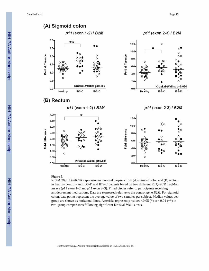
Figure 5.S100A10 (p11) mRNA expression in mucosal biopsies from (A) sigmoid colon and (B) rectumin healthy controls and IBS-D and IBS-C patients based on two different RTQ-PCR TaqManassays (p11 exon 1–2 and p11 exon 2–3). Filled circles refer to participants receivingantidepressant medications. Data are expressed relative to the control gene B2M. For sigmoidcolon, data points represent the average value of two samples per subject. Median values pergroup are shown as horizontal lines. Asterisks represent p-values <0.05 (*) or <0.01 (**) intwo-group comparisons following significant Kruskal-Wallis tests.
Camilleri et al. Page 15
Gastroenterology. Author manuscript; available in PMC 2008 July 18.
NIH
-PA Author Manuscript
NIH
-PA Author Manuscript
NIH
-PA Author Manuscript

NIH
-PA Author Manuscript
NIH
-PA Author Manuscript
NIH
-PA Author Manuscript
Camilleri et al. Page 16
Table 1Phenotypic Characteristics and Relevant Medication Use of IBS Patients and Controls
Controls IBS-C IBS-D
No. of participants 25 16 24Caucasian (n, %) 24 (96) 16 (100) 24 (100)Gender (n, % female) 23 (92) 16 (100) 21 (88)Age (mean ± SEM) (range) 39 ± 2 (19 – 60) 47 ± 3 (27 – 73) 40 ± 3 (22 – 64)BMI (mean ± SEM) (range) 26.1 ± 1.2 (18.3 – 40.2) 25.7 ± 1.4 (20.0 – 42.6) 29.2 ± 1.3 (20.9 – 43.6)Medication TypeLaxatives/Fiber (%) 0 9 (56) 1 (4)Antidiarrheals/antispasmodics (%) 0 1 (6) 4 (14)Clonidine (%) 0 0 2 (8)SSRI (%) 4 (16) 4 (25) 8 (33)5-HT3 Antagonists (alosetron) (%) 0 0 2 (8)5-HT4 Agonists (tegaserod) (%) 0 2 (13) 0Other Antidepressants (%) 2 (8) (buproprion) 4 (25) (3 buproprion, 1
amitriptyline)2 (8) (buproprion,
amitriptyline)
Gastroenterology. Author manuscript; available in PMC 2008 July 18.


![Nucleotide supplementation: a randomised double-blind placebo controlled trial of IntestAidIB in people with Irritable Bowel Syndrome [ISRCTN67764449]](https://static.fdokumen.com/doc/165x107/6342ffc5ef1ebf453b0dbbeb/nucleotide-supplementation-a-randomised-double-blind-placebo-controlled-trial-of.jpg)