Modulation of enteric neurons by interleukin-6 and corticotropin-releasing factor contributes to...
16
J Physiol 592.23 (2014) pp 5235–5250 5235 The Journal of Physiology Modulation of enteric neurons by interleukin-6 and corticotropin-releasing factor contributes to visceral hypersensitivity and altered colonic motility in a rat model of irritable bowel syndrome Maria M. Buckley 1,2 , Ken D. O’Halloran 2 , Mark G. Rae 2 , Timothy G. Dinan 1,3 and Dervla O’Malley 1,2 1 Alimentary Pharmabiotic Centre, University College Cork, Cork, Ireland 2 Department of Physiology, University College Cork, Cork, Ireland 3 Department of Psychiatry, University College Cork, Cork, Ireland Key points Hyperactivity of the stress system and low-grade immune activation characterize the functional bowel disorder irritable bowel syndrome (IBS). These studies show that interleukin (IL)-6 and IL-8 and the stress hormone corti- cotropin-releasing factor (CRF), present in IBS plasma, have functional effects on gastro- intestinal activity by stimulating myenteric neurons and colonic contractions. Moreover, in the Wistar Kyoto rat model of IBS, which exhibits altered gastrointestinal motility and visceral pain sensitivity, blocking IL-6 and/or CRF1 receptors alleviates these IBS-like symptoms. Underlying these effects are altered colonic protein expression of tight junction proteins which regulate gut barrier function and the T-type calcium channel Ca V 3.2, which has been linked to visceral pain. These findings demonstrate the importance of the enteric nervous system and intestinal physio- logy in bowel dysfunction. Abstract The search for effective therapeutic strategies for irritable bowel syndrome (IBS) is hampered by an incomplete understanding of its underlying pathophysiology. Stress and altered plasma cytokine profiles indicative of immune activation are characteristic of the disorder. The neuromodulatory effects of interleukin-6 (IL-6) and corticotropin-releasing factor receptor (CRFR) 1 in visceral pain and stress-induced defecation in the Wistar Kyoto (WKY) rat model of IBS were investigated. Sprague Dawley and WKY rats were administered anti-IL-6 receptor anti- bodies (xIL-6R, 0.5 mg kg −1 I.P) with or without the CRFR1 antagonist antalarmin (10 mg kg −1 I.P). Post-intervention, the pain threshold to colorectal distension and stress-induced faecal output were compared and changes in colonic mucosal protein expression were investigated. The neuro-stimulatory effects of IBS plasma on the myenteric plexus is mediated by IL-6, IL-8 and CRF. The stimulatory effects of these soluble factors on myenteric neuron excitability and colonic contractility were additive. Moreover, inhibition of IL-6 and CRF1 receptors in vivo in the WKY IBS rat model normalized stress-induced defecation (P < 0.01) and visceral pain sensitivity (P < 0.001) with associated changes in protein expression of the tight junction proteins occludin and claudin 2, the visceral pain-associated T-type calcium channel Ca V 3.2 and intracellular signalling molecules STAT3, SOCS3 and ERK1/2. These studies demonstrate the additive effects of immune and stress factors on myenteric neuronal excitability. Moreover, combined targeting of peripheral IL-6 and CRF1 receptors is effective in alleviating IBS-like symptoms in the WKY C 2014 The Authors. The Journal of Physiology C 2014 The Physiological Society DOI: 10.1113/jphysiol.2014.279968
-
Upload
independent -
Category
Documents
-
view
0 -
download
0
Transcript of Modulation of enteric neurons by interleukin-6 and corticotropin-releasing factor contributes to...

J Physiol 592.23 (2014) pp 5235–5250 5235
The
Jou
rnal
of
Phys
iolo
gy
Modulation of enteric neurons by interleukin-6 andcorticotropin-releasing factor contributes to visceralhypersensitivity and altered colonic motility in a rat modelof irritable bowel syndrome
Maria M. Buckley1,2, Ken D. O’Halloran2, Mark G. Rae2, Timothy G. Dinan1,3 and Dervla O’Malley1,2
1Alimentary Pharmabiotic Centre, University College Cork, Cork, Ireland2Department of Physiology, University College Cork, Cork, Ireland3Department of Psychiatry, University College Cork, Cork, Ireland
Key points
� Hyperactivity of the stress system and low-grade immune activation characterize the functionalbowel disorder irritable bowel syndrome (IBS).
� These studies show that interleukin (IL)-6 and IL-8 and the stress hormone corti-cotropin-releasing factor (CRF), present in IBS plasma, have functional effects on gastro-intestinal activity by stimulating myenteric neurons and colonic contractions.
� Moreover, in the Wistar Kyoto rat model of IBS, which exhibits altered gastrointestinal motilityand visceral pain sensitivity, blocking IL-6 and/or CRF1 receptors alleviates these IBS-likesymptoms.
� Underlying these effects are altered colonic protein expression of tight junction proteins whichregulate gut barrier function and the T-type calcium channel CaV3.2, which has been linkedto visceral pain.
� These findings demonstrate the importance of the enteric nervous system and intestinal physio-logy in bowel dysfunction.
Abstract The search for effective therapeutic strategies for irritable bowel syndrome (IBS)is hampered by an incomplete understanding of its underlying pathophysiology. Stress andaltered plasma cytokine profiles indicative of immune activation are characteristic of the disorder.The neuromodulatory effects of interleukin-6 (IL-6) and corticotropin-releasing factor receptor(CRFR) 1 in visceral pain and stress-induced defecation in the Wistar Kyoto (WKY) rat model ofIBS were investigated. Sprague Dawley and WKY rats were administered anti-IL-6 receptor anti-bodies (xIL-6R, 0.5 mg kg−1 I.P) with or without the CRFR1 antagonist antalarmin (10 mg kg−1
I.P). Post-intervention, the pain threshold to colorectal distension and stress-induced faecaloutput were compared and changes in colonic mucosal protein expression were investigated.The neuro-stimulatory effects of IBS plasma on the myenteric plexus is mediated by IL-6, IL-8and CRF. The stimulatory effects of these soluble factors on myenteric neuron excitability andcolonic contractility were additive. Moreover, inhibition of IL-6 and CRF1 receptors in vivo in theWKY IBS rat model normalized stress-induced defecation (P < 0.01) and visceral pain sensitivity(P < 0.001) with associated changes in protein expression of the tight junction proteins occludinand claudin 2, the visceral pain-associated T-type calcium channel CaV3.2 and intracellularsignalling molecules STAT3, SOCS3 and ERK1/2. These studies demonstrate the additive effectsof immune and stress factors on myenteric neuronal excitability. Moreover, combined targetingof peripheral IL-6 and CRF1 receptors is effective in alleviating IBS-like symptoms in the WKY
C© 2014 The Authors. The Journal of Physiology C© 2014 The Physiological Society DOI: 10.1113/jphysiol.2014.279968

5236 M. M. Buckley and others J Physiol 592.23
rat. Thus, crosstalk between stress and immune factors during IBS flares may underlie symptomexacerbation.
(Received 23 June 2014; accepted after revision 19 September 2014; first published online 25 September 2014)Corresponding author D. O’Malley: Department of Physiology, 4.23 Western Gateway Building, University CollegeCork, Cork, Ireland. Email: [email protected]
Abbreviations CRD, colorectal distension; CRF, corticotropin-releasing factor; CRFR, CRF receptor; CRP, C-reactiveprotein; ELISA, enzyme-linked immunosorbent assay; ERK-MAPK, extracellular signal-regulated kinase-mitogenactivated protein kinase; GI, gastrointestinal; HPA, hypothalamic-pituitary-adrenal; IBS, irritable bowel syndrome;IKK2, IkappaB kinase2; IL, interleukin; IL-6R, interleukin-6 receptor; JAK-STAT, janus tyrosine kinase and signaltransducers and activators of transcription; LMMP, longitudinal muscle myenteric plexus; NFκB, nuclear factorκ-light-chain-enhancer of activated B cells; OF, open field; SD, Sprague Dawley; SOCS, suppressor of cytokine signalling;TER, trans-epithelial resistance; WKY, Wistar Kyoto; xIL-6R, anti-IL-6R.
Introduction
Irritable bowel syndrome (IBS) is a highly prevalentfunctional gastrointestinal (GI) disorder affecting upto 15–20% of the population (Lovell & Ford, 2012).Characterized by chronic episodic bouts of abdominalpain, bloating and altered bowel habit (Longstreth et al.2006), IBS substantially impairs sufferers’ quality of lifeand confers a heavy economic and social burden on society.The underlying pathophysiology of this functional boweldisorder is poorly understood and a lack of reproduciblebiomarkers hampers diagnosis and therapeutic targetidentification. Nonetheless it is generally accepted thatdysfunction of the bidirectional communication systembetween the brain and the gut contributes to symptommanifestation (Shanahan & Anton, 1988; Cryan &O’Mahony, 2011). Integral to the initiation, prolongationand persistence of IBS symptom flares is activation ofthe hypothalamic–pituitary–adrenal (HPA) stress axis(Bohmelt et al. 2005; Dinan et al. 2006; Chang et al. 2009).Indeed, mimicking the effects of stress, the hypothalamichormone corticotropin-releasing factor (CRF) inducespathophysiological changes in GI function (Mawdsley &Rampton, 2005). CRF exerts its biological effects throughactivation of the CRF1 and CRF2 receptors (CRFR1 andCRFR2), which are expressed in both the CNS and theenteric nervous system of the GI tract (Tache & Bonaz,2007; O’Malley et al. 2010b, 2011b).
The importance of immune system activation inthe development of IBS is well recognized with about of inflammatory gastroenteritis conferring increasedsusceptibility to develop IBS (Spiller & Garsed, 2009). IBSmucosal biopsies display more immune infiltrates suchas T-cells, intra-epithelial lymphocytes and mast cells(Chadwick et al. 2002) and soluble mediators secretedfrom IBS biopsies have excitatory actions on humanenteric neurons (Buhner et al. 2009). Moreover, we pre-viously demonstrated that colonic secretions from theWistar Kyoto (WKY) rat model of IBS stimulated naı̈vesubmucosal neurons to a greater extent than secretionsfrom low anxiety Sprague Dawley (SD) controls, an
effect mediated in part by the pro-inflammatory cyto-kine interleukin-6 (IL-6) (O’Malley et al. 2011a). IL-6,along with other cytokines such as IL-1β and tumournecrosis factor α, are elevated in IBS peripheral mono-nuclear cells (Liebregts et al. 2007) and circulating levelsof IL-6 and IL-8 have reproducibly been found to be raisedin IBS patients (Dinan et al. 2006; Liebregts et al. 2007;Dinan et al. 2008). However, more than simply being abiomarker for IBS (Clarke et al. 2009), IL-6 activates sub-mucosal secretomotor neurons (Xia et al. 1999; O’Malleyet al. 2011c) and modulates mucosal ion transport andepithelial permeability (Natale et al. 2003; Kindt et al.2010) suggesting a causative role for IL-6 in this disorder.
Both psychological stress and immune activation havebeen proposed as contributory factors in symptomflares, and therefore crosstalk between these systems mayexacerbate symptoms (O’Malley et al. 2011d, 2013). Insupport of this, low-grade inflammatory changes correlatewith alterations in the stress axis in IBS (Heitkemper et al.1996; Bohmelt et al. 2005; Dinan et al. 2006) and IBSpatients often exhibit concurrent increases in markers ofa hyperactive stress response and immune upregulation(Dinan et al. 2006). To test the hypothesis that crosstalkbetween stress and immune factors results in exacerbationof IBS-like symptoms, such as visceral pain and alteredbowel habit, the WKY rat model of IBS was treated withmonoclonal anti-IL-6 receptor (xIL-6R) antibodies withand without the CRFR1 antagonist antalarmin.
Methods
Ethical approval
All experiments involving animals were conducted infull accordance with the European Community CouncilDirective (86/609/EEC) following University College Corkanimal ethics committee approval.
The study protocol (APC020, 2009) for collecting bloodsamples from IBS patients and healthy volunteers wasapproved by the University College Cork Clinical Research
C© 2014 The Authors. The Journal of Physiology C© 2014 The Physiological Society
J Physiol 592.23 IL-6 and CRFR1 contribute to IBS-like symptoms 5237
Ethics committee of the Cork University Hospital.Informed consent was obtained from all participants.
Animals and chronic treatments
SD and WKY rats (200–250 g), purchased from Harlan,Derbyshire, UK, were group-housed six per cage andmaintained on a 12/12 h light–dark cycle (08:00–20:00 h)with a room temperature of 22 ± 1°C. Rats were handledfor at least 5 days by the same researcher prior tobehavioural assessment. Food and water were availablead libitum. For the in vivo open field and colorectaldistension (CRD) studies, SD and WKY rats were allocatedto one of three groups: control, xIL-6R or xIL-6R plusantalarmin (n = 12 per group). All animal groups receivedan I.P. injection of anti-IgG (Jackson Immunoresearch,Westgrove, PA, USA, 1 mg kg−1) on day 0 to induceantibody tolerance. Group 1 were administered salineand acted as the control group. Group 2 received an I.P.injection of monoclonal xIL-6R antibodies (1 mg kg−1
on day 0 and 0.5 mg kg−1 on days 3 and 10). Group 3also received xIL-6R on days 0, 3 and 10 but additionallyreceived the CRFR1 antagonist antalarmin (10 mg kg−1)1 h prior to the open field trial on day 10 and CRD on day14. Following CRD, all animals were killed by decapitation.Trunk blood samples were centrifuged at 1600 g. andplasma samples were stored at −80 °C for later analysis.Sections of colon (2 cm long) were excised and snap frozenin liquid nitrogen for Western blotting analysis. Tissue wasstored at −80 °C for later processing.
Study participants and plasma samples
IBS patients aged 18–65 years who satisfied Rome IIcriteria for the diagnosis of IBS (Thompson et al.1999) were recruited from gastroenterology clinics atCork University Hospital, Cork, Ireland. Healthy controlswere recruited from the research institute (AlimentaryPharmabiotic Centre) or hospital staff. Bowel habit forIBS patients was defined as constipation-predominant(IBS-C), diarrhoea-predominant (IBS-D) or alternating(IBS-A). Individuals with a history of psychiatricillness, inflammatory bowel disease, celiac disease, lactoseintolerance, immunodeficiency or abdominal surgerywere excluded. No patient was categorized as havingpost-infectious IBS. Each individual was evaluated witha full review of their family history, details of currentand recent medications, a physical examination anddocumentation of body mass index. Plasma sampleswere selected from a larger study previously published(McKernan et al. 2011b).
Twenty millilitres of venous blood was donated between11:00 and 13:00 h to avoid diurnal variations. Whole blood(15 ml) was added to an equal volume of Histopaque1077 (Sigma, St Louis, MO, USA) and centrifuged at
400 g (30 min, room temperature). Consistent with pre-vious studies in the laboratory (Dinan et al. 2006, 2008),plasma was collected and stored at −80 °C. To maintaina consistency of response, pooled plasma samples (n = 6samples in each group) from both healthy volunteers andIBS patients (IBS-D, IBS-C and IBS-A) were applied to theneurons and the responses collated.
Behavioural measurements
Open field trial. As previously described (O’Mahony et al.2009, 2010a), animals were exposed to the psychologicalstress of being placed in the centre of a brightly illuminated(�800 lux), white open field (OF) arena (0.9 m indiameter, 38 cm in height) for a 10 min trial. Trials wereconducted daily between 09:00 and 13:00 h and the arenawas cleaned with 70% ethanol between trials. The numberof faecal pellets excreted during the 10 min exposure wasrecorded as an indication of stress-induced defecation.Trials were recorded and the distance and velocity ofmovement was analysed offline.
Colorectal distension. Rats were fasted for 24 h beforeCRD and acclimatized to the testing room for 30 min priorto being lightly anaesthetized (Isoflurane, Abbott AnimalResearch, Ireland). As previously described (O’Mahonyet al. 2009), a latex balloon (6 cm in length, Durex, Union,NJ, USA) was inserted into the colon, 1 cm from the anus.The animals were permitted a recovery time of 10 minbefore the CRD procedure was initiated. A ramp distensionprotocol was used where the balloon was distended from 0to 80 mmHg over an 8 min period (increasing by 10 mmHgeach minute). During this period two parameters weremeasured. The first was the threshold pressure that evokedvisibly identifiable pain behaviours. Behaviours includedwere stretching, abdominal retractions and/or abdominalwithdrawal reflex as previously described (O’Mahony et al.2010). Secondly, the cumulative number of visceral painbehaviours over the course of the trial was counted. Theanimals were tested in a random fashion by a single blindedinvestigator.
Immunoassays
Western blotting. As previously described (O’Malleyet al. 2011b), the supernatants from homogenized colonicmucosal samples taken from rats 1 h post-CRD wereseparated on 12% SDS polyacrylamide gels. Proteinswere electro-transferred to polyvinylidene fluoride(PVDF) membranes, which were blocked and incubatedwith primary antibodies against phosphoERK1/2, totalERK1/2, phosphoSTAT3, total STAT3 (all Cell SignalingTechnology, Danvers, MA, USA), occludin (Abcam,Cambridge, UK), claudin 2 (Santa Cruz Biotechnology,Dallas, TX, USA) (all at 1:1000), suppressor of cytokine
C© 2014 The Authors. The Journal of Physiology C© 2014 The Physiological Society

5238 M. M. Buckley and others J Physiol 592.23
signalling 3 (SOCS3; Everest Biotech, Upper Heyford, UK)(1:500) and CaV3.2 (Merck Millipore, Temecula, CA, USA)(1:200, overnight at 4 °C). Complimentary horseradishperoxidase-conjugated secondary antibodies (JacksonImmunoResearch) (1:5000) and an ECL detection system(Pierce ECL Western Blotting Substrate, Rockford, IL,USA) were used to visualize the protein bands. β-Actin(Cell Signaling Technology) (1:1000) was used as areference protein for occludin, claudin 2, SOCS3 andCaV3.2. Images were captured using a GelDoc ImageReader (Las3000; Fujifilm, Tokyo, Japan) and analysedusing Multigauge v2.2 (Fujifilm) software. Proteindensitometry was used to measure expression levels inarbitrary units as a ratio of the loading controls. Proteinbands are comparable only between each sub-group whichwas analysed at the same time, under identical conditions.Protein samples for each experimental group (SD saline,WKY saline, WKY xIL-6R, WKY xIL-6R & antalarmin)were run in a single tank and probed under identicalconditions.
ELISA. A sandwich ELISA (Legend MaxTM Rat IL-6 ELISAkit; BioLegend, San Diego, CA, USA) was carried outto determine IL-6 levels in plasma samples from controlWKY (n = 7) and SD (n = 7) animals, and from WKYrats treated with either anti-IL-6R (n = 8) or anti-IL-6Rand antalarmin (n = 12), according to the manufacturer’sguidelines. The test was carried out in duplicate and theplate read on a fluorescent plate reader (Biotek SynergyHT plate reader, Gen5 data analysis software). A standardcurve was generated from known IL-6 concentrations andsample IL-6 concentrations were extrapolated from thecurve.
Calcium imaging in myenteric neurons. Duringan action potential, Ca2+ enters the neuron viavoltage-gated channels and at the post-synapticmembrane neurotransmitters induce an influx of calciumvia receptor-mediated receptors such as N-methyl-D-aspartate (NMDA) receptors, thereby contributingto dendritic action potentials. Thus, neuronal changesin calcium levels as imaged using a calcium-sensitivedye (Fura-2AM) are indicative of changes in neuronalexcitability. Changes in intracellular calcium wererecorded from myenteric neuronal cell bodies as reported(O’Malley et al. 2011c), SD rats were killed via an overdoseof CO2 and the distal colon (<4 cm from the anus) wasexcised and placed in ice-cold, 95% O2/5% CO2 bubbledKrebs saline solution consisting of (mmol l−1): NaCl, 117;KCl, 4.8; CaCl2, 2.5; MgCl2, 1.2; NaHCO3, 25; NaH2PO4,1.2; and D-glucose (11 with 1 μM nifedipine to inhibitsmooth muscle contractions). Whole mount preparationsof longitudinal muscle myenteric plexus (LMMP) wereprepared by removing the mucosa and circular muscle,pinning the tissue out in Sylgard-lined petri dishes and
loading with Fura-2AM (7 μM, 1 h). Images were acquiredat 3 Hz using a xenon/mercury arc burner (Olympus,Melville, NY, USA), a charge-coupled device digitalcamera (F-view II; Olympus Soft Imaging Solutions,Munster, Germany) and a 40× water-immersion objectiveon a fixed stage upright microscope (Olympus BX51WI).Ganglionic neurons were identified based on morphologyand responsiveness to 75 mM KCl. Cell R software(Olympus Soft Imaging Solutions, 1986–2009) wasused to record excitation and emission wavelengths of340/380 and 510 nm, respectively, permitting ratiometricimaging. Responses are reported as a change in ratio.Responding neurons were defined as those with increasesin intracellular calcium [Ca2+]i greater than two standarddeviations from baseline (calculated as the average ratioduring the 150 s preceding stimulus application). Aperfusion system continuously superfused the colonictissue with carbogen-bubbled Krebs-buffered saline.
Antibody neutralization of IBS plasma (pooled from allIBS subtypes) with human neutralizing anti-IL-6 (1:100),anti-IL-8 (1:250), anti-C-reactive protein (anti-CRP,1:200) and anti-IgG (1:200, all from R&D systems,Minneapolis, MN, USA) antibodies (1 h at 37 °C) wascarried out prior to neuronal application.
Colonic contractile function. To measure circular musclecontractile activity from SD distal colons, mucosawas removed and, with the circular muscle orientatedlengthwise, the tissue was suspended from a tension trans-ducer under 1 g of tension in a bath of carbogen-bubbledKrebs saline at 37°C and allowed to equilibrate(20–30 min). Changes in tension were amplified, recordedand analysed using Powerlab and LabChart7 (both ADInstruments, Colorado Springs, CO, USA). Responses arereported as a percentage of the maximal response evokedby the muscarinic agonist carbachol (100 μM) in eachexperiment.
Statistics
Data are represented as mean ± SEM. Student’s t-testsand repeated measures one-way or two-way ANOVAswith Neumann Keuls post-hoc tests and Chi squared testswere used where appropriate. P � 0.05 was consideredsignificant.
Results
IL-6 and CRF activate myenteric neurons andstimulate colonic contractility
We have previously demonstrated that thepro-inflammatory cytokine IL-6 stimulates increases in[Ca2+]i in the submucosal plexus (O’Malley et al. 2011c)and that this is further modulated by the presence ofCRF (O’Malley et al. 2013). We have interpreted this
C© 2014 The Authors. The Journal of Physiology C© 2014 The Physiological Society

J Physiol 592.23 IL-6 and CRFR1 contribute to IBS-like symptoms 5239
rise in Ca2+, which regulates the release of synapticneurotransmitters and post-synaptic excitability, as anindicator of neuronal excitability. In total, 46% of LMMPneurons (n = 35 neurons from three rats) preparedfrom naı̈ve SD rats were activated by IL-6 (1 nM, 3 min).Moreover, CRF (100 nM, 3 min), which has a higheraffinity for CRFR1, activated 34% of the same neuronsand co-application of IL-6 plus CRF activated 54% ofthese neurons and evoked a larger calcium response onaverage than either reagent alone (Fig. 1A).
The functional consequences of activating myentericneurons on colonic contractility were investigated.Control experiments using the cholinergic agonistcarbachol (100 μM) at the beginning and the end of theexperiment demonstrated that the evoked contractionswere not diminished following multiple stimulations(0.074 ± 0.004 vs. 0.076 ± 0.008, P > 0.05). IL-6- andCRF-evoked contractions were expressed as a percentageof this maximal carbachol response. IL-6 (10 nM, 20 min)
induced a small but robust increase in circular musclecontractile activity (n = 5), whereas CRF evoked a largercontraction (n = 4). However, unlike the results inthe LMMP, addition of both agonists did not have anadditive effect on muscle contractility (n = 4, P > 0.05,Fig. 1B). Consistent with a neuronal contribution tocontractile activity, the Na+ channel blocker TTX (100 nM,20 min) diminished the IL-6 (n = 5, P < 0.01, Fig. 1C)and CRF (n = 4, P < 0.01, Fig. 1D) effects, althoughincomplete abolition of the responses suggests additionaldirect actions of IL-6 and CRF on the musculature.
IL-8 activates myenteric neurons and stimulatescolonic contractility
The chemokine IL-8 is also elevated in IBS plasma (Dinanet al. 2006; McKernan et al. 2011b) and like IL-6 it alsohas functional effects on colonic neurons. IL-8 (1 nM,3 min) increased [Ca2+]i in 58% of myenteric neurons
B
A
C
DTTX
CRF CRF
5 min0.01
mV
0.01
mV
5min
TTX
IL-6
0.01
mV
5 min
0.1
ratio5 min
IL-6 & CRF**
*
Cha
nge
in ra
tio
**
% M
axim
al re
spon
se%
Max
imal
resp
onse
% M
axim
al re
spon
se **
0
20
40
60
0
20
40
60
0
20
40
60
0.0
0.1
0.2
0.3
**
CRFIL-6
IL-6 & CRFCRFIL-6
IL-6 & CRFCRFIL-6
IL-6 & TTX
CRF CRF & TTX
IL-6
IL-6 & CRFCRFIL-6
IL-6
Figure 1. IL-6 and CRF activatemyenteric neurons and stimulatecolonic contractilityA, bar chart and representative traceillustrating calcium responses in myentericneurons from SD distal colon in response toIL-6, CRF and IL-6 & CRF (n = 35). B–D, barcharts and representative traces illustratingchanges in circular muscle contractileactivity evoked by IL-6, CRF and IL-6 & CRF(B, n = 4), IL-6 (C, n = 4) CRF (D, n = 4) inthe absence or presence of TTX, as apercentage of a maximal contractionevoked by carbachol (100 μM). Asterisksindicate ∗P < 0.05 and ∗∗P < 0.01.
C© 2014 The Authors. The Journal of Physiology C© 2014 The Physiological Society

5240 M. M. Buckley and others J Physiol 592.23
(n = 33) tested. The amplitude of the response wassimilar to CRF-mediated effects (P > 0.05). In total,34% of neurons responded to CRF alone, whereas 71%of neurons were activated by the co-application of IL-8and CRF, resulting in a significantly enhanced response(P < 0.01, Fig. 2A). When compared with recombinantIL-6, IL-8 evoked a calcium response of similar amplitudebut the number of neurons sensitive to IL-8 (58%, n = 33)was greater than the percentage sensitive to IL-6 (46%;χ2 = 4.8, d.f. = 2, P < 0.05). However, the combinationof IL-8 and IL-6 activated an even greater percentage ofneurons (65%; χ2 = 6.2, d.f. = 2, P < 0.05) and increasedthe amplitude of the calcium response compared witheither IL-6 (P < 0.001) or IL-8 (P < 0.01, Fig. 2B) alone.Application of IL-6, IL-8 and CRF stimulated 55% of the
neurons (n = 33) and, although the amplitude of theresponse was greater than IL-6 plus IL-8 (P < 0.05), it wassimilar to the response evoked by IL-8 plus CRF (P > 0.05).
IL-8 evoked large, repetitive colonic contractions (n=5,Fig. 2C). While the amplitude of contractions wasattenuated in the presence of TTX, the frequency ofcontractions increased from 0.48 ± 0.1 to 0.84 ± 0.1 Hz(n = 5, P < 0.01). The amplitude of IL-8-evoked contra-ctions (n = 5) was greater than IL-6-evoked contractions(n = 5, P < 0.001) but no further change was notedwhen IL-8 and IL-6 were co-applied (n = 5, P > 0.05,Fig. 2D). However, the amplitude of contractions evokedby co-application of IL-6, IL-8 and CRF together (n = 4)was larger than IL-6 (P < 0.001), IL-8 (P < 0.001) or CRF(P < 0.001) responses alone (Fig. 2E).
A
B
C
D
E IL-6 & IL-8 & CRF
******
IL-8 CRF IL-8 & CRF0.0
0.1
0.2
0.3
Cha
nge
in ra
tio
**
******
***
IL-6 IL-8 IL-6&IL-8
IL-6&IL-8&CRF
0.000.050.100.150.200.25
Cha
nge
in ra
tio
***
IL-8 IL-8&TTX0
20
40
60
80
100
% M
axim
al re
spon
se%
Max
imal
resp
onse
% M
axim
al re
spon
se
******
IL-6 IL-8 IL-6&IL-8
0
20
40
60
80
100
******
*****
***
IL-6 IL-8 IL-6&IL-8&CRF
020
40
60
80
100
5 min
IL-6 IL-8IL-6 &IL-8
IL-6 & IL-8& CRF
0.1
ratio
IL-8 IL-8TTX
0.01
mV
5 min
IL-8 IL-8 & CRFCRF
IL-8
5 min
IL-6
IL-6 & IL-8
0.01
mV
5 min
0.1
ratio
5 min0.01
mV
IL-6 IL-8 CRF
CRF
Figure 2. IL-8 activates myentericneurons and stimulates coloniccontractilityA and B, bar charts and traces illustratingcalcium responses in SD myenteric neuronsfollowing exposure to IL-8 and/or CRF (A,n = 24) and IL-6 and/or IL-8 and/or CRF (B,n = 33). C and D, bar charts and originaltraces illustrating circular muscle contractileresponses to IL-8 in the absence orpresence of TTX (C, n = 5) and IL-6 ± IL-8(D, n = 5) as a percentage of a maximalcontraction evoked by carbachol (100 μM).E, bar chart and original trace illustratingcircular muscle contractile responses to IL-6,IL-8, CRF or all three (n = 4). Asterisksindicate ∗∗P < 0.01 and ∗∗∗P < 0.001.
C© 2014 The Authors. The Journal of Physiology C© 2014 The Physiological Society
J Physiol 592.23 IL-6 and CRFR1 contribute to IBS-like symptoms 5241
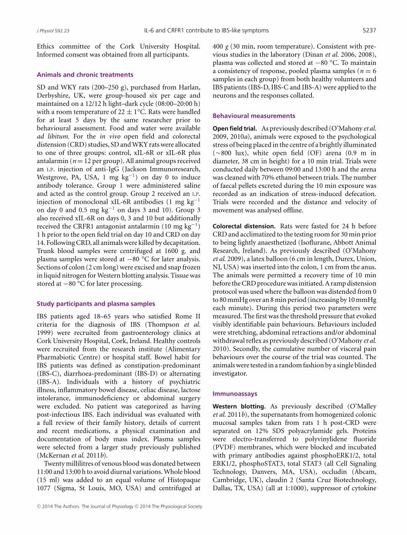
Soluble factors in IBS plasma activate myentericneurons
Thus far, we have demonstrated neuro-immune andneuro-endocrine interactions between IL-6, IL-8 and CRFin myenteric neurons and on colonic contractile activity.To relate these findings to human IBS, SD rat myentericneurons were exposed to patient and healthy controlplasma samples. Soluble factors in plasma samples from
healthy controls (n = 6 pooled samples) induced a smallincrease in [Ca2+]i in naı̈ve rat myenteric neurons (n = 43,Fig. 3A). However, IBS plasma (n = 6 pooled samples fromall subtypes) evoked a larger response (n = 43, P < 0.001,Fig. 3A).
As cytokine profiles in plasma from IBS patients arealtered (McKernan et al. 2011b), we investigated whetherthe aforementioned immune molecules contributed to theneuroexcitatory effect of IBS plasma. Control experiments,
A
B
C
D
0.1
ratio
5 min
Healthyplasma(1:250)
IBS plasma(1:250)
***
Healthyplasma
IBS plasma0.0
0.1
0.2
0.3
Cha
nge
in ra
tio
E
0.1
ratio
5 min
IBS plasmaIBS plasma
& xIgG
5 min0.1
ratio
IBS plasma
IBS plasma& xIL-6
IBS plasmaIBS plasma
& xIL-8
0.1
ratio
5 min
IBS plasma
0.1
ratio
5 min
IBS plasma
xCRP
***
IBS plasma IBS plasma & xIL-6
0.00
0.05
0.10
0.15
Cha
nge
in ra
tio
IBS plasma IBS plasma & xIgG
0.0
0.1
0.2
0.3
Cha
nge
in ra
tio
***
IBS plasma IBS plasma& xIL-8
0.00
0.05
0.10
0.15
Cha
nge
in ra
tio
*
IBS plasma IBS plasma & xCRP
0.0
0.1
0.2
0.3
Cha
nge
in ra
tio
Figure 3. Immune factors in human IBSplasma activates myenteric neuronA, bar chart and original trace showingcalcium responses in SD myenteric neuronsto pooled plasma samples from healthycontrols and IBS patients (n = 43). B–E, barcharts and original traces showing theeffects of antibody neutralization of IgG (B,n = 45), IL-6 (C, n = 71), IL-8 (D, n = 70)and CRP (E, n = 38) on IBS plasma-evokedcalcium responses in SD myenteric neurons.Asterisks indicate ∗P < 0.05 and∗∗∗P < 0.001.
C© 2014 The Authors. The Journal of Physiology C© 2014 The Physiological Society

5242 M. M. Buckley and others J Physiol 592.23
where IgG was neutralized, had no effect on the calciumresponses evoked by IBS plasma (n = 45, P > 0.05, Fig. 3B).However, neutralization of IL-6 (n = 71, P < 0.001,Fig. 3C) and IL-8 (n = 70, P < 0.001, Fig. 3D) attenuatedthe initial IBS plasma-evoked response. The inflammatorymarker CRP is also elevated in these plasma samples(McKernan et al. 2011a) and neutralizing antibodiesdirected against this protein also reduced the IBS-evokedresponse (n = 38, P < 0.05, Fig. 3E) although to a lesserextent than the cytokines. Antalarmin (1 μM, 10 min), theCRFR1 antagonist, blocked IBS plasma-evoked neuronalactivation (n = 44, P < 0.001, Fig. 4A), whereas astressin2B, a CRFR2 inhibitor, had no effect (n = 53, P > 0.05,Fig. 4B).
Incubation of myenteric neurons with the specificSTAT3 inhibitor WP1006 (10 μM, 10 min) attenuated theIBS plasma-evoked response (n = 48, P < 0.01, Fig. 5A)as did the extracellular signal-regulated kinase-mitogenactivated protein kinase (ERK-MAPK) inhibitor PD98059(10 μM, 10 min, n = 40, P < 0.001, Fig. 5B). In contrast,the phosphatidyl 3-kinase inhibitor wortmannin (10 μM,10 min) had no effect on the IBS plasma responses (n = 48,P > 0.05, Fig. 5C). Finally the role of nuclear factorκ-light-chain-enhancer of activated B cells (NFκB) wastested using the NFκB antagonist IκB Kinase2 (IKK2)(5 μM, 10 min). Interestingly, IKK2 stimulated an increasein the amplitude of the IBS plasma-evoked calciumresponse (n = 37, P < 0.05, Fig. 5D).
Monoclonal xIL-6R antibodies and antalarmin inhibitdefecation in WKY rats
The above studies demonstrate a functional role for IL-6and CRF at a cellular level, but in vivo studies werenecessary to determine if these observations had trans-lational potential. Thus, stress-sensitive WKY rats, which
defecate more in the anxiogenic OF arena compared withSD controls (O’Malley et al. 2010a) and display visceralhypersensitivity to CRD (Gunter et al. 2000; O’Mahonyet al. 2010) were treated with neutralizing xIL-6R anti-bodies and antalarmin. Control IgG- and saline-treatedWKY rats excreted more boli in the OF than SD rats(n = 12, P < 0.001, Fig. 6A). The previously describedanxious phenotype of WKY rats (O’Malley et al. 2010a)was further evidenced by decreased exploration of theexposed inner zone of the OF arena compared with SD(P < 0.001). Targeting IL-6 signalling with xIL-6R mono-clonal antibodies did not affect stress-induced defecationin SD rats in the OF, but the number of faecal pelletsexcreted by WKY rats was reduced (n = 12, P < 0.05).However, this remained higher than the low-anxiety SDcomparator (n = 12, P < 0.01, Fig. 6A). Co-application ofxIL-6R with the CRFR1 antagonist antalarmin, which canmodulate GI function (O’Malley et al. 2013), decreasedfaecal output further (n = 12, P < 0.001) to levelsequivalent to SD controls (P > 0.05, Fig. 6A).
xIL-6R and antalarmin improve visceral painsensitivity
CRD (0–80 mmHg over 8 min) evoked pain behavioursat lower pressures in control WKY as compared with SDrats (n = 10, P < 0.001, Fig. 6B). However, neutralizingIL-6Rs increased the pain threshold to CRD in WKY rats(n = 9, P < 0.05, Fig. 6B) and this was further improvedby co-application of antalarmin with xIL-6R (n = 12,P < 0.01), such that visceral sensitivity in WKY rats wasequivalent to SD controls (n = 10, P > 0.05). Interestingly,pain thresholds in SD rats following treatment with xIL-6R(n = 8, P < 0.01) and xIL-6R plus antalarmin (n = 8,P < 0.01) were also increased (Fig. 6B).
A
B
IBS plasma
IBS plasma
0.1
ratio
5 min
Antalarmin
0.1
ratio
5 min
IBS plasma IBS plasma
Astressin 2B
***
IBS plasma IBS plasma & antalarmin
0.0
0.1
0.2
0.3
Cha
nge
in ra
tio
IBS plasma IBS plasma& Astressin 2B
0.00
0.05
0.10
0.15
Cha
nge
in ra
tio
Figure 4. Stress factors in human IBSplasma activate myenteric neuronsBar charts and original traces illustrating theeffects of the CRFR1 antagonist antalarmin(A, n = 44) and the CRFR2 antagonistastressin 2B (B, n = 53) on IBSplasma-evoked calcium responses in SDmyenteric neurons. ∗∗∗P < 0.001.
C© 2014 The Authors. The Journal of Physiology C© 2014 The Physiological Society

J Physiol 592.23 IL-6 and CRFR1 contribute to IBS-like symptoms 5243
ELISA analysis of plasma IL-6 concentrations revealedno differences between saline-treated control WKY and SDrats (P > 0.05) and no changes following treatment withxIL-6R (P > 0.05) or xIL-6R plus antalarmin (P > 0.05,data not shown).
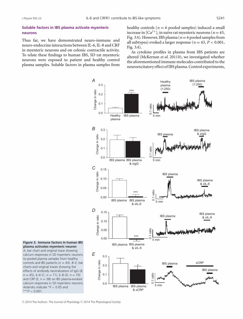
Colonic mucosal protein expression is altered byxIL-6R and antalarmin treatment
Underlying the beneficial effects of treatment of GIfunction were changes in colonic protein expression.Depletion of the tight junction protein occludin is linkedwith increased intestinal barrier permeability (Al-Sadiet al. 2011), and thus the observed increase in occludinexpression in WKY control tissues (P < 0.05) suggeststighter barrier control. This was unchanged by combinedtreatment with xIL-6R plus antalarmin, although xIL-6R
treatment alone reduced occludin expression levels(P < 0.05, n = 5 rats, Fig. 7A). In contrast, increasedexpression of another tight junction protein, claudin 2,is thought to reduce barrier tightness (Amasheh et al.2002) and expression of this protein was also elevated inWKY colons (P < 0.05) but reduced following treatmentwith xIL-6R alone or xIL-6R and antalarmin (P < 0.05,n = 5 rats, Fig. 7B). Given that we have previouslyfound that transepithelial resistance, as an indicator ofcolonic permeability, is comparable between SD andWKY rats (O’Malley et al. 2012) it may be that the sumeffect of increased expression of claudin 2 and occludinis no net change in permeability. Consistent with theviscerally sensitive phenotype of the WKY rat, Cav3.2,T-type calcium channels linked to visceral pain sensitivity(Marger et al. 2011), were also elevated in WKY tissue(P < 0.01). Moreover, consistent with amelioration of thevisceral sensitivity following treatment with xIL-6R alone
A
B
C
D
IBS plasma
IBS plasma
PD98059
0.1
ratio
5 min
IBS plasmaIBS plasma
WP1006
0.1
ratio
5 min0.
1 ra
tio
5 min
IBS plasmaIBS plasma
Wortmannin
0.1
ratio
5 min
IBS plasmaIBS plasma
IKK2
***
IBS plasma IBS plasma & PD98059
0.00
0.05
0.10
0.15
0.20
Cha
nge
in ra
tio
**
IBS plasma IBS plasma & WP1006
0.00
0.05
0.10
0.15
Cha
nge
in ra
tio
IBS plasma IBS plasma & wortmannin
0.00
0.05
0.10
0.15
0.20
Cha
nge
in ra
tio
*
IBS plasma IBS plasma& IKK2
0.00
0.05
0.10
0.15
Cha
nge
in ra
tio
Figure 5. Intracellular signalling moleculesactivated by IBS plasmaBar charts and original traces illustrating theeffects of the STAT3 inhibitor WP1006 (A,10 μM, n = 48), the ERK-MAPK inhibitorPD98059 (B, 10 μM, n = 40), the PI 3-kinaseinhibitor wortmannin (C, 10 μM, n = 48) andthe NFκB antagonist IKK2 (D, 5 μM, n = 37) oncalcium signals evoked by IBS plasma in SDmyenteric neurons. Asterisks indicate∗P < 0.05, ∗∗P < 0.01 and ∗∗∗P < 0.001.
C© 2014 The Authors. The Journal of Physiology C© 2014 The Physiological Society

5244 M. M. Buckley and others J Physiol 592.23
A
B
* **
*
0.5
1.5
2.5
3.5
4.5
5.5
6.5
No
Of P
elle
ts
xIL-6R xIL-6R & antalarmin
Saline xIL-6R xIL-6R &antalarmin
Saline
SDWKY
***vs SD
**
*
**vs SD
0
10
20
30
40
50
Pai
n th
resh
old
(mm
/Hg)
SDWKY
xIL-6R xIL-6R & antalarmin
Saline xIL-6R xIL-6R & antalarmin
Saline
**
***
C
Ca2+CRF
ERK1/2
P PSTAT3
SOCS3
Nucleus
OccludinClaudin 2
IL-6: interleukin-6JAK: Janus tyrosine kinaseSTAT3: Signal transducer andactivator of transcription 3P: phosphorylatedCRF: corticotropin-releasing factorCRFRI: CRF receptor 1ERK1/2: Extrace signal-regulatedkinase 1/2SOCS3: suppressor of cytokinesignalling 3CaV3.2: T-type clacium channel
AAAAA
CRFR1CaV3.2
STAT3
IL-6
JAK
JAK
P
WKY colonic mucosa
Visceral Pain
Alteredbarrierfunction
Figure 6. IL-6R neutralization and antalarmin inhibit defecation in WKY ratsA, bar chart illustrating the number of faecal pellets excreted by SD (n = 7, 7 and 9) and WKY (n = 10, 9 and11) rats in the OF when administered saline, anti-IL-6 receptor antibodies (xIL-6R) or xIL-6R and antalarmin. B,bar chart showing the pressure (mmHg) at which SD (n = 7, 7 and 9) and WKY (n = 10, 9 and 11) rats displaypain behaviours in response to colorectal distension when treated with saline, xIL-6R or xIL-6R and antalarmin. C,schematic illustration of potential intracellular signalling mechanisms activated by CRF and IL-6 binding to theirreceptors. Asterisks indicate ∗P < 0.05, ∗∗P < 0.01 and ∗∗∗P < 0.001.
C© 2014 The Authors. The Journal of Physiology C© 2014 The Physiological Society
J Physiol 592.23 IL-6 and CRFR1 contribute to IBS-like symptoms 5245
A
B
C
D
E
F
SOCS3
~45kDa
~24kDa
~ 42kDa
pERK1/2
Total ERK1/2
~ 42kDa
pSTAT3
Total STAT3
~85kDa
~85kDa
*
Saline xIL-6R xIL-6R & antSaline0
1
2
3
4
SDWKY
******
*
*
0.0
0.5
1.0
1.5
2.0
Saline xIL-6R & antxIL-6RSaline
SDWKY
****
0
2
4
6
8
Saline xIL-6R xIL-6R & antSaline
SDWKY
**
Occludin
β-actin
β-actin
β-actin
β-actin
~45kDa
~59kDa
WKYCon
WKY xIL-6R xIL-6R & Ant
WKY SDCon
WKYCon
WKY xIL-6R xIL-6R & Ant
WKY SDCon
WKYCon
WKY xIL-6R xIL-6R & Ant
WKY SDCon
WKYCon
WKY xIL-6R xIL-6R & Ant
WKY SDCon
WKYCon
WKY xIL-6R xIL-6R & Ant
WKY SDCon
WKYCon
WKY xIL-6R xIL-6R & Ant
WKY SDCon
CaV3.2
~250kDa
~45kDa
Claudin 2
~25kDa
~45kDa
* *
0
1
2
3
Rat
io (A
U)
Rat
io (A
U)
Rat
io (A
U)
Rat
io (A
U)
Rat
io (A
U)
Rat
io (A
U)
xIL-6R xIL-6R & antSalineSaline
SDWKY
**
0
2
4
6
xIL-6R & antxIL-6RSalineSaline
SDWKY
*
****
0
2
4
6
8
Saline xIL-6R & antxIL-6RSaline
SDWKY
**
Figure 7. IL-6R neutralization and antalarmin alter colonic mucosal protein expressionBar charts illustrating the ratios of expression of occludin (A, n = 5), claudin 2 (B, n = 5) and Cav3.2 (C, n = 5)over the β actin loading control, phosphoSTAT3 as a fraction of total STAT3 (D, n = 5), SOCS3 as a fraction of theβ actin loading control (E, n = 5) and phosphoERK1/2 over total ERK1/2 (F, n = 5) in mucosal samples from thedistal colon of SD and WKY rats treated with saline, anti-IL-6 receptor antibody (xIL-6R) and xIL-6R with antalarmin(ant). Asterisks indicate ∗P < 0.05, ∗∗P < 0.01 and ∗∗∗P < 0.001.
C© 2014 The Authors. The Journal of Physiology C© 2014 The Physiological Society

5246 M. M. Buckley and others J Physiol 592.23
and xIL-6R plus antalarmin, expression of Cav3.2 was alsoreduced (P < 0.01, n = 5 rats, Fig. 7C).
IL-6 activates the intracellular janus tyrosinekinase and signal transducers and activators oftranscription (JAK-STAT) signalling cascade and indeedphosphorylation of STAT3 was elevated in WKY rats whichhave elevated levels of IL-6 as compared with SD colons(P < 0.05). Treatment with xIL-6R had no effect onpSTAT3 expression (P > 0.05), whereas co-administrationof xIL-6R and antalarmin suppressed pSTAT3 expression(P < 0.05, n = 5 rats, Fig. 7D). SOCS3, a suppressor ofcytokine signalling, was also elevated in WKY controls(P < 0.01), and was reduced to levels equivalent to SDcontrols following xIL-6R (P > 0.05) and xIL-6R plusantalarmin (P > 0.05, n = 5 rats, Fig. 7E) treatment. IL-6binding also activates the intracellular signalling moleculepERK1/2 and this was greater in control WKY comparedwith SD colons (P < 0.05), but was markedly reduced byxIL-6R (P<0.001) and xIL-6R plus antalarmin (P<0.001,n = 5 rats, Fig. 7F) treatment.
Discussion
Neuroimmune and neuroendocrine interactionscontribute to IBS pathophysiology
The importance of immune factors such as IL-6 (Liebregtset al. 2007), IL-8 (Dinan et al. 2006, 2008; McKernanet al. 2011b) and the stress hormone CRF (Mayer, 2000;Bohmelt et al. 2005; Dinan et al. 2006; Chang et al.2009) in the aetiology of human IBS has been reported.Moreover, we have previously reported the effects of thesefactors on the excitability of submucosal neurons leadingto functional changes in secretory activity (O’Malley et al.2011c, 2013). The studies reported here have identifiedthat the pro-inflammatory cytokines IL-6 and IL-8 areacting as neuromodulators, which is consistent withthe work of Kelles et al. (2000), who described thestimulation of guinea-pig ileum myenteric neurons byIL-1β and IL-6. However, we have also determined thatthe stress factor CRF activates myenteric neurons andthat interaction between the immune and stress factorsmay occur, thereby contributing to bowel symptom flares.Myenteric neurons are associated with GI motility andin terms of pathophysiology, GI spasm and abdominalpain (Sarna, 2007). Indeed, consistent with recent reportsdescribing how recombinant IL-6 (Zhang et al. 2013) andan acute stressor (Liang et al. 2012) increased contra-ctile activity, we found that IL-6, IL-8 and CRF inducedcolonic contractions. IL-8, which had minimal effects onsubmucosal excitability (O’Malley et al. 2011c), evokedrobust responses in myenteric neurons and stimulatedlarge colonic contractions. The amplitude of contractionsevoked by IL-6 and IL-8 was greatly reduced by TTX,whereas the inhibitory effect was less for CRF-evoked
contractions, which may be acting directly on smoothmuscle cells or interstitial cells of Cajal. The observedincrease in the frequency of GI smooth muscle basalcontractility in the presence of TTX may due to end-ogenous inhibitory neural tone (Huizinga et al. 1990).Furthermore, the stimulatory effects of the immune andstress factors were additive, with the largest neuronal andcontractile response being evoked by the combined pre-sence of all three factors. These findings are consistentwith our proposal that hyperactivity of the stress response,in conjunction with low-grade immune activation, mayunderlie symptom flares in functional bowel disorders(O’Malley et al. 2011d).
To test this hypothesis further, an LMMP preparationwas exposed to clinical plasma samples from IBS patientsand healthy controls. The much larger neuronal responsesto IBS plasma indicates that certain soluble mediatorspresent in this plasma have neuroexcitatory effects.Neutralizing IL-6 and IL-8 and the CRFR1 antagonistantalarmin attenuated the IBS plasma-evoked neuronalresponses, thereby demonstrating the importance of thesefactors to the neuroexcitatory actions of IBS plasma.Indeed, IL-6 has previously been shown to act as a neuro-modulator in myenteric S and AH neurons (Kelles et al.2000). Although others have described the neuroexcitatoryeffects of other mucosally secreted soluble factors, such asserotonin, histamine and proteases in submucosal neurons(Buhner et al. 2009, 2012), our findings demonstrate thatblood-borne IL-6, CRF, IL-8 and, to a lesser extent, CRPactivate the myenteric neurons.
IBS plasma also activated JAK-STAT and ERK-MAPKintracellular signalling cascades. Interestingly, inhibitingthe NFκB signalling pathway, which normally inducestranscription of immune and inflammatory factors,potentiated the neuroexcitatory actions of IBS plasma,contrasting with its inhibitory effects on recombinant IL-6in submucosal neurons (O’Malley et al. 2011c). This maybe due to an interaction with one of the other neuro-excitatory mediators present in the plasma.
Inhibiting both IL-6 and CRF1 receptors improvesIBS-like symptoms in WKY rats
Consistent with the variable phenotype andmulti-factorial nature of IBS, our studies have identifiedseveral elements which contribute to altered bowelmotility and visceral pain sensitivity in the WKY rats.As previously reported (Martinez et al. 2007; O’Mahonyet al. 2010; O’Malley et al. 2010b), WKY rats defecatedmore in the anxiogenic OF arena and have lower painthresholds to CRD, thereby mimicking symptoms ofaltered bowel habit and visceral pain in humans. However,these IBS-like symptoms were ameliorated by blockingIL-6R signalling and normalized to control levels when
C© 2014 The Authors. The Journal of Physiology C© 2014 The Physiological Society
J Physiol 592.23 IL-6 and CRFR1 contribute to IBS-like symptoms 5247
both xIL-6R and antalarmin were co-administered.Moreover, consistent with evidence that psychologicalstress elevates IL-6 in humans, our findings illustrate theintricate relationship between immune and stress factors(O’Malley et al. 2011d, 2013). Perhaps overlooking theimportance of pro-inflammatory cytokines in symptomflares may account for the disappointing outcomes ofclinical trials of CRFR1 antagonists in IBS patients (Tacheet al. 2009).
Underlying the improvements in GI function causedby xIL-6R and antalarmin co-treatment, were dramaticchanges in mucosal protein expression. Figure 6Csummarizes a potential signalling cascade underlying theIBS-like symptoms exhibited by WKY rats. Downstreamof IL-6Rs, which comprise α-chains and the signal trans-duction gp130 subunit, is the JAK-STAT signalling cascade(Hemmann et al. 1996). STAT3 phosphorylation waselevated in WKY colonic tissue, perhaps due to raisedmucosal levels of IL-6 (O’Malley et al. 2011a). However,other cytokines or growth factors may also activate thistranscription molecule as STAT3 phosphorylation wasnot altered following blockade of IL-6 signalling. Inter-estingly, given that CRFR1 activation is not associatedwith JAK-STAT signalling, co-treatment with antalarminnormalized pSTAT3 levels, demonstrating a complexinteraction between the immune and stress factor. SOCS3,which terminates JAK/STAT signalling via negative feed-back following STAT3 activation (Croker et al. 2003),was elevated in control WKY colons, and expressionof this downstream regulatory molecule was inhibitedby treatment with xIL-6R or xIL-6R plus antalarmin.Activation of the ERK 1/2 signalling cascade by eitherCRF (Stengel & Tache, 2009) or IL-6 (Hoffmann et al.2002) was evident in the WKY colonic mucosa and thiswas reduced following treatment with xIL-6R or xIL-6Rplus antalarmin. This is consistent with previous studies insubmucosal neurons which demonstrated that CRF tendsto drive IL-6-mediated signalling via the ERK/MAPKcascade (O’Malley et al. 2013), possibly due to ERKactivation antagonizing JAK-STAT signalling (Bonni et al.1997).
Both JAK-STAT and ERK/MAPK signalling evoketranscription and de novo protein synthesis. In WKYcolonic mucosa, expression of two tight junction proteins,occludin and claudin 2, which are crucial to GIbarrier function and permeability, were both increased.Functionally, transepithelial resistance (TER) is equivalentbetween WKY and SD rats (O’Malley et al. 2012) but giventhat increased expression of occludin is thought to inducetighter barrier control and claudin 2 is thought to reducebarrier tightness (Amasheh et al. 2002) the sum effectof elevated mucosal levels of both proteins may be nonet change in permeability, although with the presenceof many additional tight junction proteins, this requiresfurther research. Expression of claudin 1 and 2 is also
increased in IBS (Martinez et al. 2013). Conversely, othershave demonstrated decreased expression of occludin inbowel inflammation (Poritz et al. 2011) and, in caco cells,exposure to IL-6 decreased claudin 2 expression resultingin decreased TER (Suzuki et al. 2011). We have pre-viously demonstrated that acute administration of IL-6increases TER in WKY colons (O’Malley et al. 2012)and others have shown that chronic exposure to IL-6increases gut permeability (Natale et al. 2003). Our studyhas provided evidence that blocking IL-6Rs decreasedexpression of both occludin and claudin 2, and theaddition of antalarmin reduced expression of claudin 2.The decreases in tight junction protein expression indicatea possible change in TER, although functional studies willbe required to certify this.
Finally, expression of the T-type calcium channelCav3.2, which is linked to visceral pain in a rodentmodel of IBS (Marger et al. 2011), was increased in WKYcolons. Consistent with improvements in pain thresholdto CRD, treatment with xIL-6R and antalarmin resulted inreduced expression of Cav3.2. CRFR1 antagonists alleviatevisceral sensitivity in the WKY rat and CRFR1 has beenshown to functionally couple to Cav3.2 in a cell line,inhibiting the calcium current (Tao et al. 2008). Thus,as has recently been proposed (Beyder et al. 2014), specificion channelopathies may contribute to visceral pain insome IBS patients. However, to our knowledge, this is thefirst study to link IL-6 signalling with Cav3.2 expressionand visceral pain sensitivity.
Thus, in a multifactorial disorder such as IBS, wherethe stress system is chronically activated and cytokinelevels of IL-6 and IL-8 are elevated, interaction andcrosstalk between these biologically active factors resultsin increased stimulation of myenteric neurons, whichsubsequently affects contractile activity. Indeed, thedemonstrated effectiveness in ameliorating IBS-likepathophysiology, such as defecation patterns and visceralpain sensitivity, in the WKY rat by targeting IL-6 and CRF1and possibly also IL-8 receptors establishes that theseimmune and stress molecules do indeed contribute tothese symptoms. Moreover, we have determined that theneuroexcitatory effects of human IBS plasma are evokedprimarily by the immune mediators IL-6 and IL-8 and thestress hormone CRF. These studies in the WKY rat, whichmimics symptoms of IBS, offers proof of principle thatcombined targeting of both immune and stress factorsmay be a viable therapeutic strategy for the treatment ofIBS.
References
Al-Sadi R, Khatib K, Guo S, Ye D, Youssef M & Ma T (2011).Occludin regulates macromolecule flux across the intestinalepithelial tight junction barrier. Am J Physiol GastrointestLiver Physiol 300, G1054–1064.
C© 2014 The Authors. The Journal of Physiology C© 2014 The Physiological Society

5248 M. M. Buckley and others J Physiol 592.23
Amasheh S, Meiri N, Gitter AH, Schoneberg T, Mankertz J,Schulzke JD & Fromm M (2002). Claudin-2 expressioninduces cation-selective channels in tight junctions ofepithelial cells. J Cell Sci 115, 4969–4976.
Beyder A, Mazzone A, Strege PR, Tester DJ, Saito YA, BernardCE, Enders FT, Ek WE, Schmidt PT, Dlugosz A, Lindberg G,Karling P, Ohlsson B, Gazouli M, Nardone G, Cuomo R,Usai-Satta P, Galeazzi F, Neri M, Portincasa P, Bellini M,Barbara G, Camilleri M, Locke GR 3rd, Talley NJ, D’AmatoM, Ackerman MJ & Farrugia G (2014). Loss-of-function ofthe voltage-gated sodium channel NaV1.5 (Channelopathies)in patients with irritable bowel syndrome. Gastroenterology146, 1659–1668.
Bohmelt AH, Nater UM, Franke S, Hellhammer DH & Ehlert U(2005). Basal and stimulatedhypothalamic–pituitary–adrenal axis activity in patientswith functional gastrointestinal disorders and healthycontrols. Psychosom Med 67, 288–294.
Bonni A, Sun Y, Nadal-Vicens M, Bhatt A, Frank DA, RozovskyI, Stahl N, Yancopoulos GD & Greenberg ME (1997).Regulation of gliogenesis in the central nervous system bythe JAK-STAT signaling pathway. Science (New York, NY)278, 477–483.
Buhner S, Li Q, Berger T, Vignali S, Barbara G, De Giorgio R,Stanghellini V & Schemann M (2012). Submucous ratherthan myenteric neurons are activated by mucosal biopsysupernatants from irritable bowel syndrome patients.Neurogastroenterol Motil 24, 1134–e572.
Buhner S, Li Q, Vignali S, Barbara G, De Giorgio R,Stanghellini V, Cremon C, Zeller F, Langer R, Daniel H,Michel K & Schemann M (2009). Activation of humanenteric neurons by supernatants of colonic biopsy specimensfrom patients with irritable bowel syndrome.Gastroenterology 137, 1425–1434.
Chadwick VS, Chen W, Shu D, Paulus B, Bethwaite P, Tie A &Wilson I (2002). Activation of the mucosal immune systemin irritable bowel syndrome. Gastroenterology 122,1778–1783.
Chang L, Sundaresh S, Elliott J, Anton PA, Baldi P, Licudine A,Mayer M, Vuong T, Hirano M, Naliboff BD, Ameen VZ &Mayer EA (2009). Dysregulation of thehypothalamic–pituitary–adrenal (HPA) axis in irritablebowel syndrome. Neurogastroenterol Motil 21, 149–159.
Clarke G, Quigley EM, Cryan JF & Dinan TG (2009). Irritablebowel syndrome: towards biomarker identification. TrendsMol Med 15, 478–489.
Croker BA, Krebs DL, Zhang JG, Wormald S, Willson TA,Stanley EG, Robb L, Greenhalgh CJ, Forster I, Clausen BE,Nicola NA, Metcalf D, Hilton DJ, Roberts AW & AlexanderWS (2003). SOCS3 negatively regulates IL-6 signaling invivo. Nature Immunol 4, 540–545.
Cryan JF & O’Mahony SM (2011). The microbiome–gut–brainaxis: from bowel to behavior. Neurogastroent Motil 23,187–192.
Dinan TG, Clarke G, Quigley EM, Scott LV, Shanahan F, CryanJ, Cooney J & Keeling PW (2008). Enhancedcholinergic-mediated increase in the pro-inflammatorycytokine IL-6 in irritable bowel syndrome: role ofmuscarinic receptors. Am J Gastroenterol 103,2570–2576.
Dinan TG, Quigley EM, Ahmed SM, Scully P, O’Brien S,O’Mahony L, O’Mahony S, Shanahan F & Keeling PW(2006). Hypothalamic–pituitary–gut axis dysregulation inirritable bowel syndrome: plasma cytokines as a potentialbiomarker? Gastroenterology 130, 304–311.
Gunter WD, Shepard JD, Foreman RD, Myers DA &Greenwood-Van Meerveld B (2000). Evidence for visceralhypersensitivity in high-anxiety rats. Physiol Behav 69,379–382.
Heitkemper M, Jarrett M, Cain K, Shaver J, Bond E, Woods NF& Walker E (1996). Increased urine catecholamines andcortisol in women with irritable bowel syndrome. Am JGastroenterol 91, 906–913.
Hemmann U, Gerhartz C, Heesel B, Sasse J, Kurapkat G,Grotzinger J, Wollmer A, Zhong Z, Darnell JE Jr, Graeve L,Heinrich PC & Horn F (1996). Differential activation ofacute phase response factor/Stat3 and Stat1 via thecytoplasmic domain of the interleukin 6 signal transducergp130. II. Src homology SH2 domains define the specificityof stat factor activation. J Biol Chem 271, 12999–13007.
Hoffmann E, Dittrich-Breiholz O, Holtmann H & Kracht M(2002). Multiple control of interleukin-8 gene expression. JLeukoc Biol 72, 847–855.
Huizinga JD, Berezin I, Daniel EE & Chow E (1990). Inhibitoryinnervation of colonic smooth muscle cells and interstitialcells of Cajal. Can J Physiol Pharmacol 68, 447–454.
Kelles A, Janssens J & Tack J (2000). IL-1β and IL-6 exciteneurones and suppress cholinergic neurotransmission in themyenteric plexus of the guinea pig. Neurogastroenterol Motil12, 531–538.
Kindt S, Vanden Berghe P, Boesmans W, Roosen L & Tack J(2010). Prolonged IL-1β exposure alters neurotransmitterand electrically induced Ca2+ responses in the myentericplexus. Neurogastroenterol Motil 22, 321–e85.
Liang C, Luo H, Liu Y, Cao J & Xia H (2012). Plasma hormonesfacilitated the hypermotility of the colon in a chronic stressrat model. PloS One 7, e31774.
Liebregts T, Adam B, Bredack C, Roth A, Heinzel S, Lester S,Downie-Doyle S, Smith E, Drew P, Talley NJ & Holtmann G(2007). Immune activation in patients with irritable bowelsyndrome. Gastroenterology 132, 913–920.
Longstreth GF, Thompson WG, Chey WD, Houghton LA,Mearin F & Spiller RC (2006). Functional bowel disorders.Gastroenterology 130, 1480–1491.
Lovell RM & Ford AC (2012). Global prevalence of and riskfactors for irritable bowel syndrome: a meta-analysis. ClinGastroenterol Hepatol 10, 712–721.
Marger F, Gelot A, Alloui A, Matricon J, Ferrer JF, Barrere C,Pizzoccaro A, Muller E, Nargeot J, Snutch TP, Eschalier A,Bourinet E & Ardid D (2011). T-type calcium channelscontribute to colonic hypersensitivity in a rat model ofirritable bowel syndrome. Proc Natl Acad Sci U S A 108,11268–11273.
Martinez C, Lobo B, Pigrau M, Ramos L, Gonzalez-Castro AM,Alonso C, Guilarte M, Guila M, de Torres I, Azpiroz F,Santos J & Vicario M (2013). Diarrhoea-predominantirritable bowel syndrome: an organic disorder withstructural abnormalities in the jejunal epithelial barrier. Gut62, 1160–1168.
C© 2014 The Authors. The Journal of Physiology C© 2014 The Physiological Society

J Physiol 592.23 IL-6 and CRFR1 contribute to IBS-like symptoms 5249
Martinez V, Ryttinger M, Kjerling M & Astin-Nielsen M(2007). Characterisation of colonic accommodation inWistar Kyoto rats with impaired gastric accommodation.Naunyn Schmiedebergs Arch Pharmacol 376,205–216.
Mawdsley JE & Rampton DS (2005). Psychological stress inIBD: new insights into pathogenic and therapeuticimplications. Gut 54, 1481–1491.
Mayer EA (2000). The neurobiology of stress andgastrointestinal disease. Gut 47, 861–869.
McKernan DP, Dennison U, Gaszner G, Cryan JF & Dinan TG(2011a). Enhanced peripheral toll-like receptor responses inpsychosis: further evidence of a pro-inflammatoryphenotype. Transl Psychiatry 1, e36.
McKernan DP, Gaszner G, Quigley EM, Cryan JF & Dinan TG(2011b). Altered peripheral toll-like receptor responses inthe irritable bowel syndrome. Aliment Pharmacol Ther 33,1045–1052.
Natale L, Piepoli AL, De Salvia MA, De Salvatore G, Mitolo CI,Marzullo A, Portincasa P, Moschetta A, Palasciano G &Mitolo-Chieppa D (2003). Interleukins 1 beta and 6 inducefunctional alteration of rat colonic motility: an in vitrostudy. Eur J Clin Invest 33, 704–712.
O’Mahony SM, Bulmer DC, Coelho AM, Fitzgerald P,Bongiovanni C, Lee K, Winchester W, Dinan TG & Cryan JF(2010). 5-HT2B receptors modulate visceral hypersensitivityin a stress-sensitive animal model of brain-gut axisdysfunction. Neurogastroenterol Motil 22, 573–578.
O’Mahony SM, Marchesi JR, Scully P, Codling C, Ceolho AM,Quigley EM, Cryan JF & Dinan TG (2009). Early life stressalters behavior, immunity, and microbiota in rats:implications for irritable bowel syndrome and psychiatricillnesses. Biol Psychiatry 65, 263–267.
O’Malley D, Cryan JF & Dinan TG (2013). Crosstalk betweeninterleukin-6 and corticotropin-releasing factor modulatesubmucosal plexus activity and colonic secretion. BrainBehav Immun 30, 115–124.
O’Malley D, Dinan TG & Cryan JF (2011a). Altered expressionand secretion of colonic interleukin-6 in a stress-sensitiveanimal model of brain–gut axis dysfunction. JNeuroimmunol 235, 48–55.
O’Malley D, Dinan TG & Cryan JF (2011b). Neonatal maternalseparation in the rat impacts on the stress responsivity ofcentral corticotropin-releasing factor receptors inadulthood. Psychopharmacology 214, 221–229.
O’Malley D, Dinan TG & Cryan JF (2012). Interleukin-6modulates colonic transepithelial ion transport in thestress-sensitive Wistar Kyoto rat. Front Pharmacol 3,190.
O’Malley D, Julio-Pieper M, Gibney SM, Dinan TG & Cryan JF(2010a). Distinct alterations in colonic morphology andphysiology in two rat models of enhanced stress-inducedanxiety and depression-like behaviour. Stress 13,114–122.
O’Malley D, Julio-Pieper M, Gibney SM, Gosselin RD, DinanTG & Cryan JF (2010b). Differential stress-inducedalterations of colonic corticotropin-releasing factor receptorsin the Wistar Kyoto rat. Neurogastroenterol Motil 22,301–311.
O’Malley D, Liston M, Hyland NP, Dinan TG & Cryan JF(2011c). Colonic soluble mediators from the maternalseparation model of irritable bowel syndrome activatesubmucosal neurons via an interleukin-6-dependentmechanism. Am J Physiol 300, G241–252.
O’Malley D, Quigley EM, Dinan TG & Cryan JF (2011d). Dointeractions between stress and immune responses lead tosymptom exacerbations in irritable bowel syndrome? BrainBehav Immun 25, 1333–1341.
Poritz LS, Harris LR 3rd, Kelly AA & Koltun WA (2011).Increase in the tight junction protein claudin-1 in intestinalinflammation. Digest Dis Sci 56, 2802–2809.
Sarna SK (2007). Enteric descending and afferent neuralsignaling stimulated by giant migrating contractions:essential contributing factors to visceral pain. Am J Physiol292, G572–581.
Shanahan F & Anton P (1988). Neuroendocrine modulation ofthe immune system. Possible implications for inflammatorybowel disease. Digest Dis Sci 33, 41S–49S.
Spiller R & Garsed K (2009). Postinfectious irritable bowelsyndrome. Gastroenterology 136, 1979–1988.
Stengel A & Tache Y (2009). Neuroendocrine control of the gutduring stress: corticotropin-releasing factor signalingpathways in the spotlight. Annu Rev Physiol 71,219–239.
Suzuki T, Yoshinaga N & Tanabe S (2011). Interleukin-6 (IL-6)regulates claudin-2 expression and tight junctionpermeability in intestinal epithelium. J Biol Chem 286,31263–31271.
Tache Y & Bonaz B (2007). Corticotropin-releasing factorreceptors and stress-related alterations of gut motorfunction. J Clin Invest 117, 33–40.
Tache Y, Kiank C & Stengel A (2009). A role forcorticotropin-releasing factor in functional gastrointestinaldisorders. Curr Gastroenterol Rep 11, 270–277.
Tao J, Hildebrand ME, Liao P, Liang MC, Tan G, Li S, SnutchTP & Soong TW (2008). Activation ofcorticotropin-releasing factor receptor 1 selectively inhibitsCaV3.2 T-type calcium channels. Mol Pharmacol 73,1596–1609.
Thompson WG, Longstreth GF, Drossman DA, Heaton KW,Irvine EJ & Muller-Lissner SA (1999). Functional boweldisorders and functional abdominal pain. Gut 45(Suppl 2),II43–47.
Xia Y, Hu HZ, Liu S, Ren J, Zafirov DH & Wood JD (1999).IL-1β and IL-6 excite neurons and suppress nicotinic andnoradrenergic neurotransmission in guinea pig entericnervous system. J Clin Invest 103, 1309–1316.
Zhang L, Hu L, Chen M & Yu B (2013). Exogenousinterleukin-6 facilitated the contraction of the colon in adepression rat model. Dig Dis Sci 58, 2187–2196.
Additional information
Competing interests
The authors declare that there are no competing interestsregarding this publication.
C© 2014 The Authors. The Journal of Physiology C© 2014 The Physiological Society

5250 M. M. Buckley and others J Physiol 592.23
Author contributions
M.M.B.: acquisition of data; analysis and interpretation ofdata; drafting of the manuscript; K.D.O’H.: critical revisionof the manuscript for important intellectual content; M.G.R.:critical revision of the manuscript for important intellectualcontent; T.G.D.: critical revision of the manuscript for importantintellectual content; D.O’M.: study concept and design,critical revision of the manuscript for important intellectualcontent; obtained funding, study supervision.
Funding
This publication emanated from research supported by theHealth Research Board (HRA POR/2010/52) and by a researchgrant from the Science Foundation Ireland (SFI) under GrantNumber 07/CE/B1368 in the Alimentary Pharmabiotic Centre.M.M.B. was part funded by the Department of Physiology, UCC.
Acknowledgements
We thank Drs Declan McKernan, Gabor Gaszner, SineadHeuston and Rachel Moloney, Colette Manley and PatrickFitzgerald for assistance with various parts of this study.
C© 2014 The Authors. The Journal of Physiology C© 2014 The Physiological Society