Organizational disasters: why they happen and how they may be prevented

MOLECULAR AND CELLULAR BIOLOGY, June 2005, p. 5226–5241 Vol. 25, No. 120270-7306/05/$08.00�0 doi:10.1128/MCB.25.12.5226–5241.2005Copyright © 2005, American Society for Microbiology. All Rights Reserved.
Outgrowth of Neurites from NIE-115 Neuroblastoma Cells IsPrevented on Repulsive Substrates through
the Action of PAKKatharine J. M. Marler,1 Robert Kozma,1,2† Sohail Ahmed,1,2‡ Jing-Ming Dong,1,2
Christine Hall,1 and Louis Lim1,2*Department of Molecular Neuroscience, Institute of Neurology, University College London, 1 Wakefield St.,
London WC1N 1PJ, United Kingdom,1 and GSK-IMCB Group, Institute of Molecular & Cell Biology,Proteos, 61 Biopolis Dr., Singapore 138673, Singapore2
Received 25 August 2004/Returned for modification 21 October 2004/Accepted 17 March 2005
In the central nervous system (CNS), damaged axons are inhibited from regeneration by glial scars, wheresecreted chondroitin sulfate proteoglycan (CSPG) and tenascin repulse outgrowth of neurites, the forerunnersof axons and dendrites. During differentiation, these molecules are thought to form boundaries for guidingneurons to their correct targets. In neuroblastoma NIE-115 cells, outgrowth of neurites on laminin could beinduced by serum starvation or inhibition of RhoA by Clostridium botulinum C3 toxin. The outgrowing neuritesavoided crossing onto the repulsive substrate CSPG or tenascin. This avoidance response was partiallyovercome on expression of membrane-targeted and kinase-inactive forms of PAK. In these cells, the endoge-nous PAK isoforms colocalized with actin in distinctive sites, �PAK in the cell center as small clusters andalong the neurite shaft and �PAK and �PAK in areas with membrane ruffles and filopodia, respectively. Whenisoform-specific N-terminal PAK sequences were introduced to interfere with PAK function, substantially moreneurites crossed onto CSPG when cells contained a �PAK-derived peptide but not the corresponding �PAK-or �PAK-derived peptide. Thus, while neurite outgrowth can be promoted by RhoA inhibition, overcoming theaccompanying repulsive guidance response will require modulation of PAK activity. These results havetherapeutic implications for CNS repair processes.
The failure of the adult mammalian central nervous system(CNS) to regenerate after injury may in part be due to theinhibitory nature of the glial scar that forms, preventing thedamaged axons from regenerating, leading to loss of function(10, 37). The glial scar is a combination of damaged cells,invading cells, and their associated secreted molecules such aschondroitin sulfate proteoglycan (CSPG) and tenascin, whichform both a physical and chemical barrier to extending axons(10, 30, 42). In the developing cerebral cortex, tenascin-C istransiently expressed at the boundaries of migratory pathways(17). In the peripheral nervous system, tenascin-C expressioncorrelates with increased cell migration and axonal growth (5).Members of the CPSG family are the major proteoglycansexpressed in the nervous system by both neurons and glia (27).Like tenascin-C, CSPGs are believed to form boundaries toguide neurons to their appropriate targets and prevent themmaking connections with inappropriate targets (23, 52). In theabsence of a glial scar, adult rat dorsal root ganglia (DRG)transplanted into adult rat white matter are able to extendaxons from the graft boundary (7). Up-regulation of CSPG atthe graft boundary was associated with the failure of the DRG toenter the host white matter (7). These and other observations on
myelin and its derivatives support a role for glial scar components,particularly CSPG, in preventing regeneration in the adult CNS.
For functional recovery, neurons are required to regenerateor repair axons that have to skirt or grow through the glial scarand then reconnect with their appropriate targets. Morpho-genesis of the precursor neurites and axons crucially involvesthe actin cytoskeleton, which is subject to regulation by theRho family GTPases (19, 20, 35, 38, 39, 47). The activation ofRac1 is required for nerve growth factor-induced neurite out-growth in PC12 cells and can be blocked through injection ofdominant negative Rac1 N17 (6). In N1E-115 cells, both Rac1and Cdc42 are required for outgrowth (20). This can be achievedthrough the activation of Rac1 either directly or indirectly viaprior activation of Cdc42, which antagonizes the activation ofRhoA and prevents neurite collapse (20, 47). The involvement ofRhoA in growth cone collapse and neurite retraction has beenshown in both primary neurons and cultured cells like N1E-115and PC12 cells (15, 20, 36, 46). Both RhoA and Rac1 have beenshown to be involved in different pathways that lead to growthcone collapse in response to inhibitory cues. In the case of ephrinA5-mediated collapse in retinal ganglion cell (RGC) growthcones, RhoA and its downstream effector ROK are required (50).However, Rac1 has also been shown to mediate the collapse ofDRG neurons in response to Sema3A (collapsin-1) (16, 49).
N1E-115 neuroblastoma cells provide a facile model forstudying how the Rho GTPases participate in guidance signal-ing pathways initiated by inhibitory substrates. The use of thesecells has provided much information on the role of GTPasesand effectors in neurite outgrowth and in retraction processesrequired for guidance or avoidance (12, 14, 20, 40). Unlike
* Corresponding author. Mailing address: Department of MolecularNeuroscience, Institute of Neurology, University College London, 1Wakefield St., London WC1N 1PJ, United Kingdom. Phone: (44) 2072781552. Fax: (44) 207 2787045. E-mail: [email protected].
† Present address: Genome Institute of Singapore, 60 Biopolis St.,Singapore 138672.
‡ Present address: Institute of Molecular & Cell Biology, 61 BiopolisDr., Singapore 138673.
5226
on February 15, 2015 by guest
http://mcb.asm
.org/D
ownloaded from
primary neuronal cells, for example, those derived from thehippocampus (8) or DRG, NIE-115 cells show no predisposi-tion to produce outgrowth and therefore better mimic regen-erating CNS neurons, which have only a low intrinsic ability toregenerate their axons (9, 10). NIE-115 cells will undergo dif-ferentiation and neurite outgrowth on withdrawal of serum (1).They are capable of responding to guidance cues; for example,their neurites will turn towards a source of ACh (20), a positiveguidance response first demonstrated in spinal neurons of Xe-nopus (54). The response is dependent on activation of Cdc42and Rac1 GTPases. The cells are easy to transfect, providing alarge population where the effects of Rho GTPases or theireffectors can be subjected to quantitative analysis. In this study,outgrowth of neurites from NIE-115 cells was induced throughserum withdrawal or treatment with various agents. The effectsof various components of the Rho GTPase signaling pathwayson guidance behavior at boundaries of either CSPG or te-nascin were analyzed. Serum withdrawal or RhoA inhibitionpromoted outgrowth of neurites that were able to avoid therepulsive surfaces. In serum-starved cells, this avoidance re-sponse could be overcome by introduction of various PAKderivatives, implicating PAK action in the negative guidanceresponse.
MATERIALS AND METHODS
Materials. CSPG, tenascin, and their antibodies were from Chemicon. Thepeptide delivery system was BioPorter from Gene Therapy Systems (supplied byQ-Biogene, United Kingdom). Laminin was from ICN Flow; all other tissueculture reagents were from Life Technologies. Tetramethyl rhodamine isothio-cyanate (TRITC) dye, used to mark the laminin, was from Sigma. PAK isoformantibodies were from Santa Cruz. Vinculin antibody was from Sigma; the lamininantibody was from Polyscience. The blocking peptides were from Santa Cruz andJerini Germany.
Cell culture. N1E-115 cells were grown in Dulbecco’s modified Eagle medium(DMEM) with 0.11g/liter sodium pyruvate, 4.5 g/liter D-glucose, and 580 mg/literL-glutamine (Life Technologies). This medium was supplemented by adding 10%fetal calf serum (FCS) and 1% antibiotic/antimycotic solution (AB/AM) con-taining penicillin (10,000 U/ml), streptomycin (10,000 �g/ml), and amphotericin(25 �g/ml) (Life Technologies) as described previously (20, 40).
Microscopy. Phase images were taken of cells grown on chamber slides on aheated stage (37°C) in an atmosphere of humidified air and 5% CO2. Cells werevideo recorded using an Axiovert 135 microscope, Palmix TM-6CN video cam-era, and Sony-u-matic V0-5800 PS video recorder. Individual images were cap-tured using Adobe Premier software and manipulated with Adobe Photoshop.Confocal microscopy was carried out using an LSM 410 Zeiss scanning micro-scope with three single-line lasers at 488 nM (argon), 543 nM, and 633 nM(HeNe).
In situ localization. For in situ analysis, primary and secondary antibodieswere used at a 1:100 dilution. The PAK antibodies, all from Santa Cruz, were asfollows: �PAK N20, �PAK N19, and �PAK N19. Cells were fixed with 4%paraformaldehyde in phosphate-buffered saline (PBS) for 10 min at room tem-perature (RT), washed with PBS two times for 10 min, permeabilized for 1 minin 0.5% Triton X-100 in PBS, and then blocked with 3% bovine serum albumin(BSA) in PBS for 15 min. The coverslips were then exposed to primary antibod-ies diluted 1/100 in 1% BSA in PBS and incubated at 37°C for 2 h in a humidchamber. Cells on coverslips were then washed in PBS twice for 10 min. Sec-ondary antibodies diluted in 1% BSA in PBS and F-actin were stained withrhodamine-conjugated phalloidin (0.1 �g/ml; Sigma) and incubated with cover-slips for 1 h in a dark humid chamber. Finally, coverslips were washed twice for10 min in PBS and mounted in Immuno-Flore (ICN Flow). The slides were
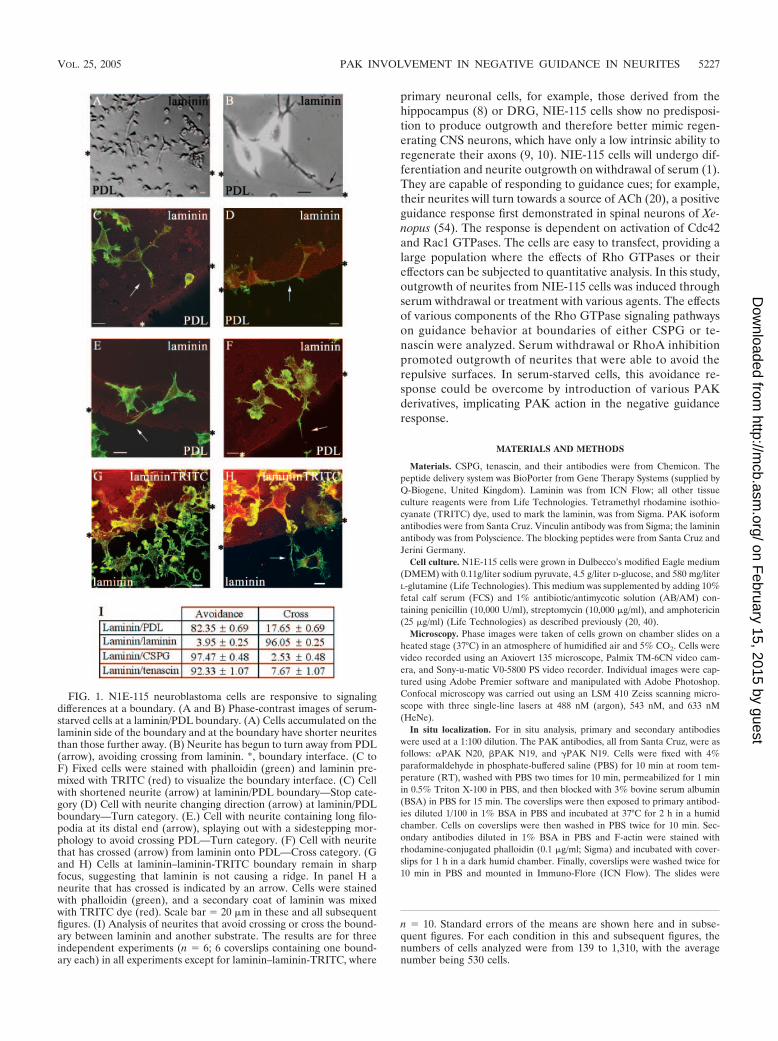
FIG. 1. N1E-115 neuroblastoma cells are responsive to signalingdifferences at a boundary. (A and B) Phase-contrast images of serum-starved cells at a laminin/PDL boundary. (A) Cells accumulated on thelaminin side of the boundary and at the boundary have shorter neuritesthan those further away. (B) Neurite has begun to turn away from PDL(arrow), avoiding crossing from laminin. *, boundary interface. (C toF) Fixed cells were stained with phalloidin (green) and laminin pre-mixed with TRITC (red) to visualize the boundary interface. (C) Cellwith shortened neurite (arrow) at laminin/PDL boundary—Stop cate-gory (D) Cell with neurite changing direction (arrow) at laminin/PDLboundary—Turn category. (E.) Cell with neurite containing long filo-podia at its distal end (arrow), splaying out with a sidestepping mor-phology to avoid crossing PDL—Turn category. (F) Cell with neuritethat has crossed (arrow) from laminin onto PDL—Cross category. (Gand H) Cells at laminin–laminin-TRITC boundary remain in sharpfocus, suggesting that laminin is not causing a ridge. In panel H aneurite that has crossed is indicated by an arrow. Cells were stainedwith phalloidin (green), and a secondary coat of laminin was mixedwith TRITC dye (red). Scale bar � 20 �m in these and all subsequentfigures. (I) Analysis of neurites that avoid crossing or cross the bound-ary between laminin and another substrate. The results are for threeindependent experiments (n � 6; 6 coverslips containing one bound-ary each) in all experiments except for laminin–laminin-TRITC, where
n � 10. Standard errors of the means are shown here and in subse-quent figures. For each condition in this and subsequent figures, thenumbers of cells analyzed were from 139 to 1,310, with the averagenumber being 530 cells.
VOL. 25, 2005 PAK INVOLVEMENT IN NEGATIVE GUIDANCE IN NEURITES 5227
on February 15, 2015 by guest
http://mcb.asm
.org/D
ownloaded from

allowed to set overnight before viewing with a Zeiss Axiovert microscope and aZeiss LSM 410 confocal microscope.
Cell guidance assays. (i) Permissive/attractive boundary. Coverslips were firstwashed and sterilized and then coated in one of the following ways. The cover-slips were covered with a solution of poly-D-lysine at 10 �g/ml and incubated for1 h at RT and then rinsed once with sterile water and allowed to air dry in alaminar flow hood before 30 �l of laminin (10 �g/ml) was placed centrally andthe coverslips incubated for 1 h at 37°C in a humid chamber to prevent evapo-ration and uneven application of the laminin. The coverslips were then rinsedtwice in sterile water before being dried in the hood.
(ii) Control boundary. The coverslips were coated with laminin (10 �g/ml) for1 h at RT and then rinsed in sterile water and left to dry. A second layer oflaminin was applied as a 30-�l drop mixed with 1 �g/ml of TRITC label.Coverslips were left for 1 h at 37°C in a humidity chamber and then rinsed twicewith sterile water and dried before use.
(iii) Attractive/repulsive boundary. Slides were coated with either tenascin at10 �g/ml or chondroitin sulfate proteoglycan (CSPG) at 5 �g/ml for 1 h at 37°Cin a humid chamber and then rinsed in sterile water and allowed to air dry beforea 30-�l drop of laminin (10 �g/ml) mixed with 1 �g/ml of TRITC label wasapplied to the center of the coverslip and left for 1 h at 37°C in a humiditychamber before two rinses in sterile water and drying before use. This coatingwas also done in reverse with the laminin applied first and then the tenascin orthe CSPG applied secondly. To ensure that the substrates were evenly applied andremained on the coverslips after attachment of cells, antibodies for tenascin, CSPG,or laminin were used to check the integrity of the substrates and to visualize theboundary on a sample of the coverslips; in this case the TRITC label was omitted.
Cells were then seeded onto the prepared coverslips at 2 � 105 cells in 1 ml ina 35-mm petri dish in DMEM, 10% FCS, and 1% AB/AM. Cells were allowed toattach, the medium was then removed, and the coverslips were rinsed in serum-free medium and incubated overnight (16 h) in serum-free medium. The cellswere fixed and stained to show the morphology of cells at the boundary.
Time-lapse analysis of neurite behavior. Cells were plated in Willco glass-bottom dishes coated with laminin–CSPG-TRITC or laminin–laminin-TRITC tocreate boundaries as described above. Cells were allowed to attach for 4 h. Afterwithdrawal of serum and addition of Y-27632 (final concentration, 10 �M) toinduce rapid outgrowth of neurites, the cells were placed in a mini-incubatormounted to a Bio-Rad Radiance 2000 confocal system. Cells were imaged over2.5 h. Images were captured at 45-s intervals using a Nikon 40� oil objective andwere processed using Adobe Photoshop.
Transient transfection. Cells were harvested by gentle pipetting to removeattached cells and plated out at 1.5 � 105 cells/ml in a 35-mm dish containingprepared coverslips and incubated overnight. The medium was then replacedwith 1 ml of serum-free DMEM only and the cells incubated for 1 h at 37°C. Twohundred microliters of DMEM was placed into a sterile 1.5-ml Eppendorf tube,and 1 �g of the plastid DNA (in eukaryotic expression vector PXJ40) was addedto each tube with 6 �l of Lipofectamine 2000 (GIBCO), followed by gentleinverting and then incubation at RT for 45 min to allow the lipids to package theDNA. At the end of the incubation the mix was added to the dish containing thecells, and they were then incubated for 4 h at 37°C. After incubation the trans-fection mix was then removed and the cells rinsed with 1 ml of DMEM–5%FCS–1% AB/AM; 1 ml of the same medium was added, and the cells incubatedfor a further 16 h.
Protein delivery system. The BioPorter reagent was prepared according to themanufacturer’s instructions. Cells were plated out at 1.5 � 105 cells in a 35-mmdish containing prepared coverslips and incubated overnight. The protein orpeptide of interest was diluted in 100 �l PBS to give a final concentration of 10�g/ml, and 1 �l of fluorescein isothiocyanate-labeled goat immunoglobulin G(IgG) was added to act as a marker. This mixture was added directly to the tubecontaining the BioPorter reagent, mixed by pipetting up and down two or threetimes, and then allowed to stand for 5 min. Cells were washed once with serum-free DMEM; 1 ml of serum-free DMEM was added to the tube containing thepeptide and BioPorter mix and vortexed gently before being added to the cells.The cells were incubated for 4 h at 37°C. After incubation the cells were thenincubated for a further 2 h in 5% FCS–DMEM or serum-free DMEM for cellsundergoing a substrate guidance assay. The cells were then fixed and stained.
Immunoblot analysis of PAK isoforms. N1E-115 cells were harvested aftergrowing in 10% FCS in DMEM. The supernatants of lysed, sonicated cells weresubjected to 10% sodium dodecyl sulfate-polyacrylamide gel electrophoresisanalysis. Gels containing separated proteins were blotted onto nitrocellulosefilters. The filters were blocked in 5% milk powder (Marvel)–PBS–0.1% Tween20, washed with PBS, and incubated with the relevant primary (PAK) antibody(1/1,000 dilution) in 1% milk–PBS–0.1% Tween 20 for 1 h at RT. The primaryantibody was washed off with PBS–0.1% Tween 20 (PBST), and secondary
antibody (1/1,000 dilution) added. Filters were incubated for 1 h, washed inPBST, blot dried, and then developed using ECL reagents (Amersham Pharma-cia Biotech). For immunoblots, primary and secondary antibodies were used ata 1/1,000 dilution.
Assay of Rac1-GTP levels. NIE-115 cells grown in 5% FCS on plastic, laminin,or CSPG were transfected with HA-RhoA N19, as described above. Cell extractsfrom mock- and RhoA N19-transfected cells were assayed for their Rac-GTPcontent using glutathione S-transferase (GST)/PAK-CRIB constructs and Rac1antibodies essentially as described previously (4).
FIG. 2. Transfection with RhoA N19 induces outgrowth of neuriteswith increased numbers crossing from laminin onto CSPG or tenascin.(A) RhoA N19-transfected cell on laminin, stained with phalloidin. (B)RhoA N19-transfected cell stained with HA antibody. (C) Neurite ofRhoA N19-transfected cell crossing from laminin onto CSPG, stainedwith phalloidin. (D) Analysis of increased neurite crossing from lami-nin onto CSPG by RhoA N19-transfected cells. Results are for threeindependent experiments (n � 6 for controls [serum starvation andmock transfection treatment]) (n � 21 for RhoA N19-transfectedcells). (E). RhoA N19-transfected N1E-115 cell stained with phalloidincrossing from laminin onto tenascin. (F) Analysis of increased neuritecrossing from laminin onto tenascin by RhoA N19-transfected cells.Results are for three independent experiments (n � 6 for control andRhoA N19-transfected cells).
5228 MARLER ET AL. MOL. CELL. BIOL.
on February 15, 2015 by guest
http://mcb.asm
.org/D
ownloaded from
Effect of Rac1 N17 expression on cell morphology. NIE-115 cells were grownin 10% FCS either on laminin or CSPG for 4 h overnight. Cells were thentransfected with HA/Rac1 N17 as described above. The morphologies of mock-and Rac1 N17-transfected cells were examined after staining with phalloidin orhemagglutinin (HA) antibody as described above. Cells were categorized intothose that were contracted (a), showing neurite outgrowth (b), flattened (c), orelongated (d).
Statistical analysis. Data are expressed as mean values from three indepen-dent experiments; error bars represent standard errors of the mean values. Then values refer to the numbers of coverslips, each containing a boundary, sub-jected to analysis. The neurites analyzed were all within a 40-�m strip along thelaminin side of the boundary in each coverslip, the average number analyzedbeing �50. Values from the control and experimental groups were comparedusing the Mann-Whitney U test with a P value of 0.05 considered significant.
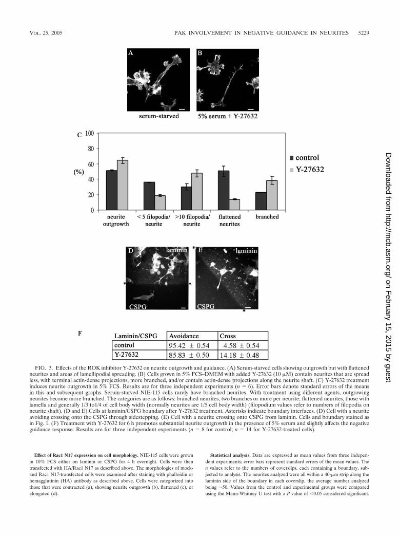
FIG. 3. Effects of the ROK inhibitor Y-27632 on neurite outgrowth and guidance. (A) Serum-starved cells showing outgrowth but with flattenedneurites and areas of lamellipodial spreading. (B) Cells grown in 5% FCS–DMEM with added Y-27632 (10 �M) contain neurites that are spreadless, with terminal actin-dense projections, more branched, and/or contain actin-dense projections along the neurite shaft. (C) Y-27632 treatmentinduces neurite outgrowth in 5% FCS. Results are for three independent experiments (n � 6). Error bars denote standard errors of the meansin this and subsequent graphs. Serum-starved NIE-115 cells rarely have branched neurites. With treatment using different agents, outgrowingneurites become more branched. The categories are as follows: branched neurites, two branches or more per neurite; flattened neurites, those withlamella and generally 1/3 to1/4 of cell body width (normally neurites are 1/5 cell body width) (filopodium values refer to numbers of filopodia onneurite shaft). (D and E) Cells at laminin/CSPG boundary after Y-27632 treatment. Asterisks indicate boundary interfaces. (D) Cell with a neuriteavoiding crossing onto the CSPG through sidestepping. (E) Cell with a neurite crossing onto CSPG from laminin. Cells and boundary stained asin Fig. 1. (F) Treatment with Y-27632 for 6 h promotes substantial neurite outgrowth in the presence of 5% serum and slightly affects the negativeguidance response. Results are for three independent experiments (n � 8 for control; n � 14 for Y-27632-treated cells).
VOL. 25, 2005 PAK INVOLVEMENT IN NEGATIVE GUIDANCE IN NEURITES 5229
on February 15, 2015 by guest
http://mcb.asm
.org/D
ownloaded from
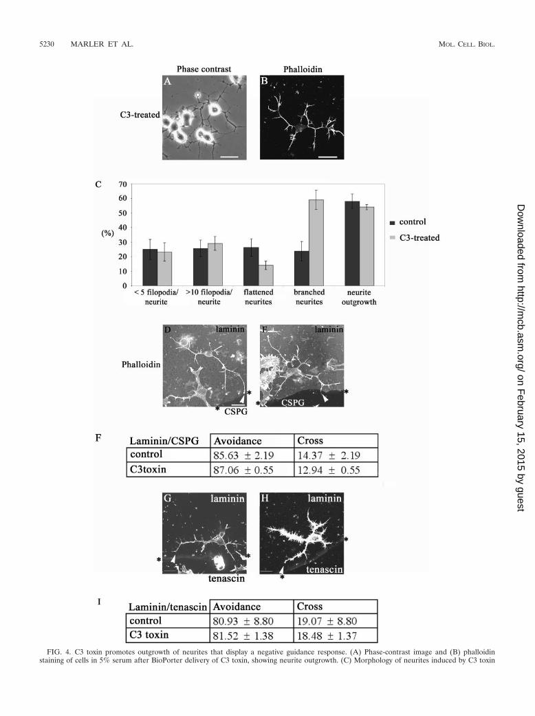
FIG. 4. C3 toxin promotes outgrowth of neurites that display a negative guidance response. (A) Phase-contrast image and (B) phalloidinstaining of cells in 5% serum after BioPorter delivery of C3 toxin, showing neurite outgrowth. (C) Morphology of neurites induced by C3 toxin
5230 MARLER ET AL. MOL. CELL. BIOL.
on February 15, 2015 by guest
http://mcb.asm
.org/D
ownloaded from
RESULTS
Response of N1E-115 neuroblastoma cells to negative guid-ance signals. We previously observed that N1E-115 neuroblas-toma cells directed their neurites towards a source of ACh,supplied as a gradient, only when first plated on laminin, butnot on culture dishes alone (20). This suggests that these cellsnot only respond to soluble guidance clues but also to thenature of the substrate.
To determine how neurite behavior was affected by favorableand nonfavorable substrates, cells were seeded onto coverslipswith discrete coatings of laminin and poly-D-lysine (PDL) andallowed to attach. The cells were then serum starved to causedifferentiation. Cells close to the boundary between the twosubstrates were viewed with an inverted microscope fitted witha CO2-controlled humidified chamber and a heated stage.Phase analysis showed that cells accumulated in a strip ofapproximately 300 �m on the laminin side of the boundary.Neurites in this strip tended to be shorter, suggesting thatgrowth may have terminated before the neurites reached fulllength, as serum-starved N1E-115 cells generally possess neu-rites in excess of two cell body lengths (Fig. 1A). In cells withneurites and growth cones at the boundary, the filopodia onthe growth cone were observed occasionally to make contactwith the other substrate. Phase analysis of cells with neurites inthis strip showed them to have specific morphologies, whichmay well reflect the avoidance response to PDL. Neurites closeto the boundary were often shorter than neurites further away,suggesting a premature termination (Fig. 1A). In other cases,filopodia often appeared to sense the boundary region, and onoccasions the growth cone splayed out with numerous projec-tions before the whole neurite grew and turned away from theboundary (Fig. 1B, arrow).
The assay relies on the ability of cells to produce outgrowthwhile undergoing guidance decision making over a time courseof around 18 h. Therefore, to study the morphology of the cellsat the boundary in more detail and in greater numbers, cellswere fixed and stained with phalloidin to visualize F-actin andan anti-laminin antibody was used to detect the laminin anddelineate the boundary. Neurites within a 40-�m strip alongthe laminin side of the boundary were analyzed; this wouldinclude neurites that had been able to sense the boundary andretracted. The fixed and stained cells at the boundary werecompared with phase images of the live cells to determine theircorresponding morphologies. Based on this comparison, theirmorphologies were then categorized. Neurites were scoredwhen greater than one cell body length as they generallystained positively for neurofilament; most serum-starved neu-rites were greater than two cell body lengths away from theboundary (Fig. 1A).
The categories were as follows. (i) Stop—the neurite had
stopped at or very close to the boundary (Fig. 1C). (ii) Turn—the neurite showed a marked deviation in direction, suggestingthat it had initiated a turn as seen in the phase analysis (Fig.1D) or the neurite had splayed to form numerous projectionsor a zigzag shape along the boundary, possibly sidesteppingbefore turning (Fig. 1E). (iii) Cross—the neurite had crossedfrom one substrate to the other (Fig. 1F).
The first two categories are indicative of cells that may beresponding to the (repulsive) signals from the other substrateand thus avoid crossing. The third category is indicative of cellsnot responsive to the signals from the substrate for variousreasons or of cells where the present and newly encounteredsubstrates evoke similar responses.
The first two categories were aggregated and scored as neu-rites avoiding crossing and compared with those that crossedthe boundary (Fig. 1I). A large majority (83%) of cells platedon the laminin substrate avoided crossing onto the PDL.
To determine whether the cells were responsive to signalingdifferences between different substrates at a boundary or mere-ly to an interface, experiments were repeated using “false”boundaries or two identical substrates. Cells encountering ei-ther a “false” boundary or a boundary between the same sub-strate should be subjected to identical signals and therefore beexpected not to show any avoidance behavior. False bound-aries were created by using a fine marker to mark a circle onthe coverslips. To demonstrate that laminin was not causing aridge when used to cover another substrate, coverslips werecoated with laminin and a central drop of laminin mixed withTRITC dye to visualize the boundary was then applied. Cellswere seeded and serum starved and analyzed as before (Fig.1G and H). At a laminin–laminin-TRITC boundary, cellsshowed very little avoidance behavior, with the majority cross-ing (Fig. 1I). Cells crossing or on the boundary remained insharp focus, indicating that the laminin did not cause a signif-icant ridge (Fig. 1G and H). N1E-115 cells thus have the abilityto respond to signals from differing substrates, and this resultsin the cells growing in a directed manner away from a lessfavorable substrate.
To ascertain if the N1E-115 cells would respond to signalsfrom more physiologically relevant substrates, especially thoseassociated with damaged areas of the nervous system, the guid-ance assay was repeated using a boundary between laminin (asthe permissive substrate) and CPSG or tenascin (as the repul-sive substrate). Cells showed a marked repulsion responsewhen encountering CSPG, with 97% of neurites avoidingcrossing onto CSPG. Only 2% crossed, as compared with about17% crossing from laminin onto PDL. Clearly, CPSG eliciteda greater repulsive response from N1E-115 cells than PDL,with the vast majority of the cells showing avoidance. As with
or control (serum starvation and BioPorter-delivered IgG). Results are for three independent experiments (n � 6 for control and n � 8 for C3toxin-treated cells). The categories of neurite morphology are as defined in the legend to Fig. 3. (D) Neurite of C3 toxin-treated cell is able to avoidcrossing from laminin onto the CSPG. The arrowhead points to the termini at branched neurite, with a rounded “club-like” appearance, suggestiveof neurite withdrawal and collapse. (E) As for panel D but with the neurite of C3 toxin-treated cell turning at a laminin/CSPG interface(arrowhead). (F) Avoidance of CSPG by neurites is retained in C3 toxin-treated cells. Results are for three independent experiments (n � 10 forcontrol, n � 19 for C3 toxin-treated cells). (G and H) C3 toxin-treated cells with neurites turning at laminin/tenascin boundary (arrowheads). (I)Avoidance of tenascin by neurites is retained in C3 toxin-treated cells compared with the control (serum starvation and BioPorter-delivered IgG).Results are for three independent experiments (n � 8 for control; n � 13 for C3 toxin-treated cells).
VOL. 25, 2005 PAK INVOLVEMENT IN NEGATIVE GUIDANCE IN NEURITES 5231
on February 15, 2015 by guest
http://mcb.asm
.org/D
ownloaded from
CPSG, a large majority of cells (92%) avoided crossing onto atenascin substrate (Fig. 1I).
RhoA N19 and neurite guidance. RhoA activation has beenimplicated in the regulation of negative guidance signals, in-volving ephrin A5-induced collapse of retinal growth cones(50). We then investigated the role of RhoA-mediated signal-ing in the negative guidance response using the dominant neg-FIG. 5. Rac1 activation is affected by RhoA N19 and is involved in
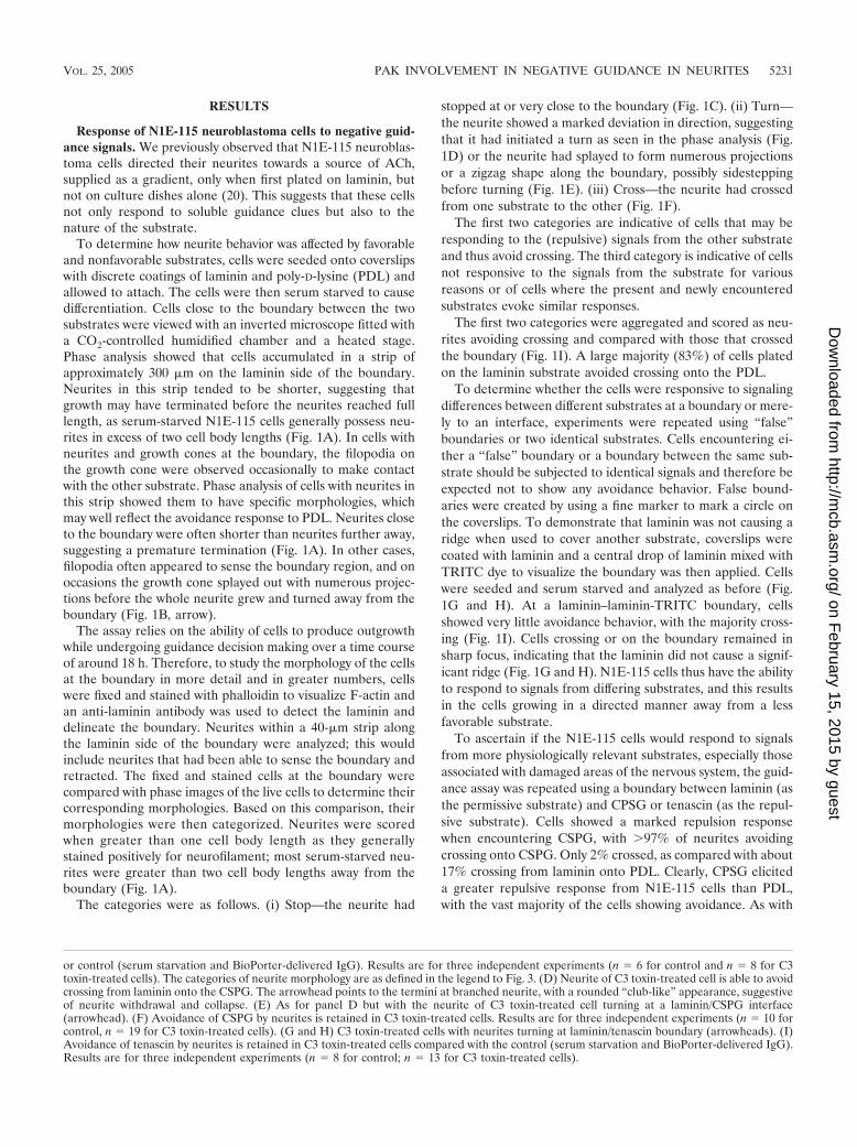
neurite outgrowth on CSPG. (A) NIE-115 cells grown in 5% FCS onplastic, laminin, or CSPG were transfected with HA-RhoA N19. Therelative content of Rac-GTP was analyzed using PAK-CRIB pull-downassays and Rac antibodies. In all cases, RhoA N19 decreased Rac-GTPcontent. The upper gel contains the GST/CRIB fragment; the lowergel is an immunoblot of Rac. The results shown are typical of fourindependent assays. (B) Effect of Rac1 N17 on cell morphology. NIE-115 cells grown in 5% FCS either on laminin or CSPG were trans-fected with HA/Rac1 N17. Cells were stained with phalloidin (red) oranti-HA (green). Expression of Rac1 N17 promotes neurite outgrowth
FIG. 6. Effects of membrane-targeted PAK on the negative guid-ance response. Neurite outgrowth in cells transfected with �PAK-CAAX (A and B) or �PAK-CAAX KD (C and D). Stained withphalloidin (A and C) or anti-HA (B and D). Neurites of cells trans-fected with �PAK-CAAX (E) or with �PAK-CAAX KD (F) crossinga laminin/CSPG boundary. HA-tagged cells are shown. (G) Neurites ofcells transfected with either PAK construct have an increased ability tocross from laminin onto CSPG. Results are for three independentexperiments (n � 6 for controls, n � 8 for �PAK-CAAX-transfectedcells, and n � 14 for �PAK-CAAX KD-transfected cells).
in cells grown on CSPG (see cell in bottom right panel). (C) Analysisof morphology of cells grown on laminin or CSPG. Note occurrence ofpredominantly flattened cells. (D) Analysis of effect of Rac1 N17 oncell morphology. Note enhanced neurite outgrowth on cells grown onCSPG but not on laminin. Error bars represent standard errors of themeans. **, P 0.01; ***, P 0.001.
5232 MARLER ET AL. MOL. CELL. BIOL.
on February 15, 2015 by guest
http://mcb.asm
.org/D
ownloaded from
ative RhoA N19 mutant. Neurite outgrowth was promoted incells transiently transfected with RhoA N19; the neurites werelong, straight, and predominantly unbranched (Fig. 2A and B).Cells plated on coverslips containing either a laminin/CSPG ora laminin/tenascin boundary were then transiently transfectedwith RhoA N19 and allowed to express for 18 h. The cells werefixed and stained for phalloidin and HA antibody to detect thetransfected cells; the laminin boundary was detected with lami-nin antibody (Fig. 2C).
Whereas the vast majority (98%) of the serum-starvedmock-transfected control cells contained neurites that did notcross from the laminin onto CSPG, only about half of cellstransfected with RhoA N19 avoided crossing (Fig. 2E). RhoAN19-transfected cells also showed a marked decrease in theiravoidance behavior towards tenascin, with less than half (43%)avoiding crossing (Fig. 2D and F).
ROK inhibition and neurite guidance. We next investigatedwhether this guidance response involved the Rho kinase ROK,which causes neurite collapse (14, 18). ROK activity is inhib-ited by the kinase inhibitor Y-27632 (18, 25), which is able toprevent lysophosphatidic acid-induced collapse of neurites orto induce neurite outgrowth in NIE-115 cells in the presence ofserum (14). Serum-starved cells contained flattened neuritesand areas of lamellipodial spreading. Treatment of cells withY-27632 (10 �M in 5% FCS) resulted in outgrowth of neuritesthat were more branched, with more filopodia on the neuriteshaft, and less flattened compared with serum-starved controls(Fig. 3A to C).
The effect of Y-27632 on the guidance of N1E-115 cellsat a laminin/CSPG boundary was then examined. Cells wereplated onto a laminin/CSPG boundary as described above.Y-27632 in 5% serum was added, and cells incubated for 6 hbefore fixing and staining. The results are shown in Fig. 3D,E, and F. In the case of serum-starved cells, 95% of theneurites were able to avoid crossing onto CSPG. Treatmentwith Y-27632 resulted in only a slight decrease in the num-ber of neurites that avoided crossing onto CSPG (86%).Since the ROK inhibitor Y-27632 caused only a minor disrup-tion in guidance when compared with RhoA N19, this may in-dicate that other Rho effectors or another pathway is involved.
Rho inhibition by C3 toxin and neurite guidance. To furtherunderstand the role of Rho signaling, we next used the Clos-tridium botulinum toxin C3, which specifically inhibits the ac-tion of Rho by ADP-ribosylation (41). C3 toxin in vivo is amore potent blocker of Rho function than RhoA N19 (45).Because C3 toxin is not cell permeable, the BioPorter systemwas used to provide its effective delivery into N1E-115 cells.After plating of the cells, 5 �g of C3 toxin and the BioPorterreagent were added. Six hours later, the cells were fixed andstained to determine the number of cells with neurites morethan one cell body length and the type of outgrowth produced(Fig. 4A to C). The effect on the cells was very similar to thatpreviously seen on C3 toxin injection (20). The morphologyproduced was that of multiple branched neurites, and the cellbody was more contracted compared with serum-starved con-trol cells.
There was no difference in the response to a laminin/CSPGboundary between the control serum-starved and C3 toxin-treated cells (Fig. 4D, E, and F). Nor was the ability of cells toguide at a laminin/tenascin boundary affected by the treatment
with C3 toxin (Fig. 4G, H, and I). These results with the C3toxin indicate that inhibition of Rho leads to outgrowth ofneurites that are able to avoid crossing on to repulsive sub-strates like CSPG and tenascin.
Rac, PAK, and neurite guidance. The exact role of RhoAN19 is uncertain; it may well affect activation of GTPases otherthan Rho. A Rac1-mediated collapse pathway has been impli-cated in other repulsive signaling, e.g., that involving Sema3A(16, 21, 49). Because its dominant negative N17 mutant blocksthe neurite outgrowth (20, 40) essential for our assay, Rac1involvement in guidance could not be investigated directly.Nevertheless, we were able to show that Rac1 activation wasaffected by RhoA N19. In cells plated either on plastic,laminin, or CSPG, transfection with RhoA N19 resulted indecreased levels of Rac1-GTP within the cell (Fig. 5A). Thissuggested that the enhancement by RhoA N19 of neuritecrossing onto CSPG (Fig. 2) could have occurred through itsinhibition of Rac1 pathways. We then examined whether Rac1N17 itself could promote neurite outgrowth of NIE-115 cellsgrown on CSPG. Many of the cells (�50%) plated on lamininor CSPG in 5% serum were flattened, with the rest equallydistributed between those that were contracted, elongated,or showing neurite outgrowth (Fig. 5B and C). Under theseconditions, a Rac- rather than Rho-type morphology predom-inated. Serum contains lysophosphatidic acid, which has beenshown recently to activate both Rho and Rac pathways sequen-tially, with activation of the latter leading to inhibition of theformer (48). On expressing Rac1 N17, the cells plated onlaminin (Fig. 5B and D) had an altered morphological distri-bution, with most (�60%) now being contracted (Rho-typemorphology) rather than flattened (12%). Expression of Rac1N17 by cells plated on CSPG also resulted in an altered mor-phological distribution, but with many now showing neuriteoutgrowth (�45%) rather than being flattened (15%). Thus,with cells plated on CSPG but not on laminin, Rac1 inhibitionpromoted neurite outgrowth (see example in Fig. 5B, lowerright panel).
Since Rac1 N17 is inimical to neurite outgrowth on laminin,we examined the role of its downstream effector PAK on thenegative guidance response. Transfection with �PAK doesnot lead to neurite outgrowth; PAK is probably subject tostringent intracellular regulation since autoactivated PAK(mutated in the p21-binding domain) evokes massive morpho-logical changes in cells (26). The latter observation precludedthe use of non-p21-binding mutants of PAK. However, the�PAK sequence modified to include the membrane-targetingsequence CAAX (26) promotes neurite outgrowth in PC12cells (6). We then examined the effects of �PAK-CAAX aswell as its kinase-dead derivative, �PAK-CAAXK298 (�PAK-CAAX KD). Cells transfected with �PAK-CAAX typicallypossess long neurites that are occasionally branched and ter-minate with a small growth cone (Fig. 6A and B). Cells trans-fected with �PAK-CAAX KD possess spindly neurites withnumerous filopodia along their length, and they have a taperedappearance (Fig. 6C and D). Cells were then plated onto alaminin/CSPG boundary, transiently transfected with �PAK-CAAX or �PAK-CAAX KD, and allowed to express for 18 h.The transfected cells were detected with an anti-HA antibody;the laminin boundary was detected as before, and the resultsare shown in Fig. 6E, F, and G.
VOL. 25, 2005 PAK INVOLVEMENT IN NEGATIVE GUIDANCE IN NEURITES 5233
on February 15, 2015 by guest
http://mcb.asm
.org/D
ownloaded from
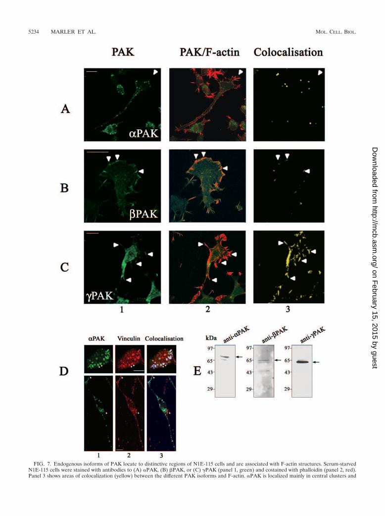
FIG. 7. Endogenous isoforms of PAK locate to distinctive regions of N1E-115 cells and are associated with F-actin structures. Serum-starvedN1E-115 cells were stained with antibodies to (A) �PAK, (B) �PAK, or (C) �PAK (panel 1, green) and costained with phalloidin (panel 2, red).Panel 3 shows areas of colocalization (yellow) between the different PAK isoforms and F-actin. �PAK is localized mainly in central clusters and
5234 MARLER ET AL. MOL. CELL. BIOL.
on February 15, 2015 by guest
http://mcb.asm
.org/D
ownloaded from

Cells transfected with �PAK-CAAX or its kinase-dead de-rivative displayed a marked change in their avoidance behav-ior. Whereas in control cells, only 2% of neurites crossed ontothe CSPG from laminin, in �PAK-CAAX- and �PAK-CAAXKD-transfected cells the corresponding numbers were 17%and 56%, respectively. A series of similar experiments withcells transfected with other Cdc42-binding effectors such asN-WASP and IRS-58 (2, 12) showed that they elicited no in-crease in the numbers crossing (data not shown). These resultsindicate that membrane-targeted PAK can overcome the neg-ative guidance response, with the kinase-dead form being moreeffective.
PAK isoforms in neurite outgrowth and actin structures.We then investigated which of the PAK isoforms might beinvolved in the negative guidance response. Expression of allthree endogenous PAK isoforms in N1E-115 cells was con-firmed by immunoblotting using isoform-specific antibodies.The antibodies for �PAK, �PAK, and � PAK separately pre-dominantly recognized 68-kDa, 65-kDa, and 62-kDa proteins,respectively, in cell extracts (Fig. 7E). These molecular massescorrespond to those of the different PAK isoforms. We alsoexamined the localization of endogenous PAK isoforms andtheir relationship to F-actin structures. Cells were plated ontolaminin-coated coverslips and serum starved to induce out-growth. Each PAK isoform showed a preferential distributionat distinct sites where they colocalized with F-actin. �PAK wasfound predominantly in the center of cells in small clusters andalong the neurite shaft but not associated with filopodia orlamellipodia (Fig. 7A). �PAK was found mainly in areas withlamellipodia and membrane ruffling; the staining in filopo-dium-like structures was more diffuse and not as enriched as inthe ruffling areas (Fig. 7B). �PAK was found mainly in areas offilopodium activity and some stabilized filopodia and not inareas of membrane ruffling (Fig. 7C).
The �PAK-containing clusters in cells growing in 10% FCSwere associated with vinculin, a focal complex marker (Fig.7D). Differentiation and neurite outgrowth through serumwithdrawal resulted in a substantial decrease in these clusters(Fig. 7D). The �PAK-containing clusters thus resemble RhoA-type focal adhesion complexes that are associated with stressfibers in fibroblasts.
Effects of PAK peptides on neurite outgrowth and morphol-ogy. We then used isoform-specific PAK peptides to investigatetheir effects on the outgrowth of neurites and the negative guid-ance response. The PAK peptides selected were small sequencesspanning the divergent N terminus of each isoform. They eachcontained a polyproline-rich segment essential for binding to Nck(53) and a small flanking region: �PAK, SNNGLDIQDKPPAPPMRNTS (�P2 peptide); �PAK, SDSLDNEEKPPAPPLRMNSNN (�P2 peptide); �PAK, SDNGELEDKPPAPVRMSSTI(�P2 peptide).
The individual peptides were delivered, using BioPorter withmarker IgG, to cells plated onto laminin-coated coverslips thatwere then incubated at 37°C for 6 h before staining with phal-loidin, all under serum-free conditions. Serum-starved controlcells were treated with BioPorter containing IgG. Staining forthe codelivered IgG marker identified the relevant cells. Noneof the PAK peptides blocked the neurite outgrowth caused byserum starvation, with the �P2 peptide slightly increasing thenumber of cells with neurites (Fig. 8A). The morphology of theneurites of peptide-containing cells was also compared withthat of control cells (Fig. 8B and C). �P2 peptide had no dis-cernible effect. �P2 peptide induced production of more filo-podia on the neurite shaft and on the growth cone, giving riseto a “hairy” appearance to the cells; more branched neuriteswere also seen. �P2 peptide-containing cells had neurites thatwere smoother or more flattened and with typically five or lessfilopodia per cell.
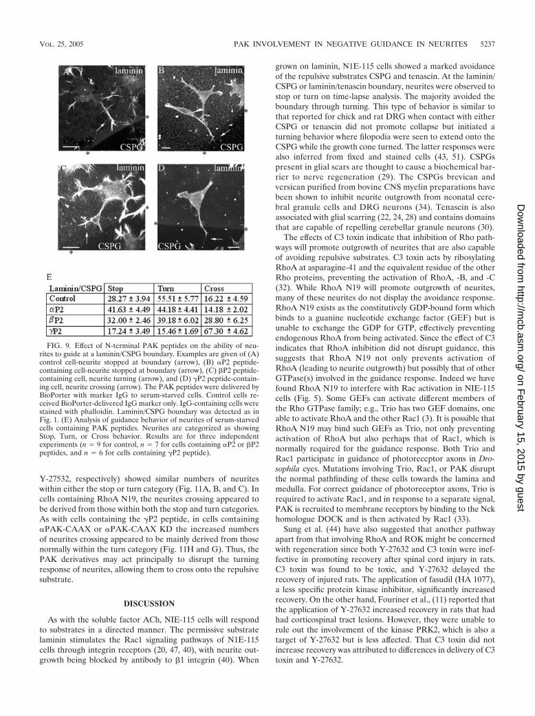
Effect of N-terminal PAK peptides on the guidance re-sponse. The peptides were then delivered to cells plated oncoverslips containing a laminin/CSPG boundary under serum-free conditions. After 6 h to induce neurite outgrowth, the cellswere fixed and stained. The neurites of the cells containing thePAK peptides all stained positively for F-actin and for neuro-filament. The morphology and response of these cells areshown in Fig. 9A to D. The response is quantified in Fig. 9E,where neurites that showed avoidance behavior were subcat-egorized as those showing “stop” or “turn” responses.
The neurites from cells containing the �P2 peptide showeda substantial change in their ability to respond to a laminin/CSPG boundary, with two-thirds now crossing onto the CSPG.The increase in numbers of neurites that crossed appearedlargely to be derived from those that normally would haveavoided crossing by turning away from the boundary. Thepresence of the �P2 peptide did not affect the numbers cross-ing, while that of the �P2 peptide elicited very much less of aneffect than the �P2 peptide.
As the �P2 peptide appeared to affect the crossing ability ofneurites that would normally have turned away from the re-pulsive substrate, we reexamined the effects of those agentsmost effective in disrupting guidance, this time subdividingthose neurites previously within the avoidance category intothe stop and turn categories. Figure 10 is a time-lapse analysisshowing neurites stopping at a laminin-CSPG boundary (Fig.10A) or crossing a laminin-laminin boundary (Fig. 10B) withina 2.5-h period. In this illustration of the stop category, neuriteoutgrowth has been accelerated by serum withdrawal com-bined with Y-27632 treatment to facilitate the analysis of neu-rite dynamics.
In our reexamination of the avoidance category, we notedthat cells that were not or were little affected in their negativeguidance response by the presence of inhibitors (C3 toxin and
along neurite shafts and absent from filopodia (arrowheads, A). �PAK occurs in lamellipodium/membrane ruffles (arrowheads, B) and �PAKmainly in filopodia (arrowheads, C). (D) Cells grown in 10% FCS were costained with antibodies to �PAK (upper panel 1, green) or vinculin(upper panel 2, red). Upper panel 3 shows colocalization (blue) of �PAK and vinculin in clusters resembling focal adhesions (two indicated byarrowhead). In the serum-starved cell (lower panels), clusters are smaller and centrally located; they appear in areas of flattening but not thoseenriched in filopodia or lamellipodia (arrowheads). (E) �-, �-, and �PAK expression in N1E-115 cells. Cellular proteins separated by sodiumdodecyl sulfate-polyacrylamide gel electrophoresis were subjected to Western blot analysis using antibodies to �PAK (62 kDa), �PAK (65 kDa),and �PAK (68 kDa).
VOL. 25, 2005 PAK INVOLVEMENT IN NEGATIVE GUIDANCE IN NEURITES 5235
on February 15, 2015 by guest
http://mcb.asm
.org/D
ownloaded from
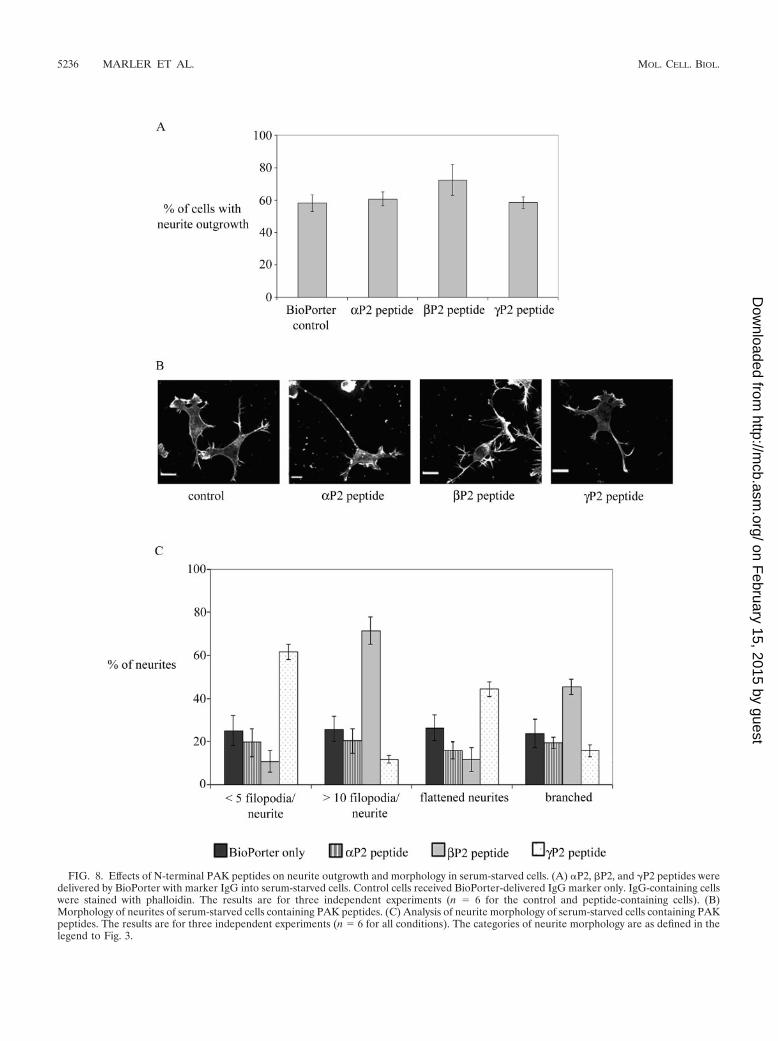
FIG. 8. Effects of N-terminal PAK peptides on neurite outgrowth and morphology in serum-starved cells. (A) �P2, �P2, and �P2 peptides weredelivered by BioPorter with marker IgG into serum-starved cells. Control cells received BioPorter-delivered IgG marker only. IgG-containing cellswere stained with phalloidin. The results are for three independent experiments (n � 6 for the control and peptide-containing cells). (B)Morphology of neurites of serum-starved cells containing PAK peptides. (C) Analysis of neurite morphology of serum-starved cells containing PAKpeptides. The results are for three independent experiments (n � 6 for all conditions). The categories of neurite morphology are as defined in thelegend to Fig. 3.
5236 MARLER ET AL. MOL. CELL. BIOL.
on February 15, 2015 by guest
http://mcb.asm
.org/D
ownloaded from
Y-27532, respectively) showed similar numbers of neuriteswithin either the stop or turn category (Fig. 11A, B, and C). Incells containing RhoA N19, the neurites crossing appeared tobe derived from those within both the stop and turn categories.As with cells containing the �P2 peptide, in cells containing�PAK-CAAX or �PAK-CAAX KD the increased numbersof neurites crossing appeared to be mainly derived from thosenormally within the turn category (Fig. 11H and G). Thus, thePAK derivatives may act principally to disrupt the turningresponse of neurites, allowing them to cross onto the repulsivesubstrate.
DISCUSSION
As with the soluble factor ACh, NIE-115 cells will respondto substrates in a directed manner. The permissive substratelaminin stimulates the Rac1 signaling pathways of N1E-115cells through integrin receptors (20, 47, 40), with neurite out-growth being blocked by antibody to �1 integrin (40). When
grown on laminin, N1E-115 cells showed a marked avoidanceof the repulsive substrates CSPG and tenascin. At the laminin/CSPG or laminin/tenascin boundary, neurites were observed tostop or turn on time-lapse analysis. The majority avoided theboundary through turning. This type of behavior is similar tothat reported for chick and rat DRG when contact with eitherCSPG or tenascin did not promote collapse but initiated aturning behavior where filopodia were seen to extend onto theCSPG while the growth cone turned. The latter responses werealso inferred from fixed and stained cells (43, 51). CSPGspresent in glial scars are thought to cause a biochemical bar-rier to nerve regeneration (29). The CSPGs brevican andversican purified from bovine CNS myelin preparations havebeen shown to inhibit neurite outgrowth from neonatal cere-bral granule cells and DRG neurons (34). Tenascin is alsoassociated with glial scarring (22, 24, 28) and contains domainsthat are capable of repelling cerebellar granule neurons (30).
The effects of C3 toxin indicate that inhibition of Rho path-ways will promote outgrowth of neurites that are also capableof avoiding repulsive substrates. C3 toxin acts by ribosylatingRhoA at asparagine-41 and the equivalent residue of the otherRho proteins, preventing the activation of RhoA, -B, and -C(32). While RhoA N19 will promote outgrowth of neurites,many of these neurites do not display the avoidance response.RhoA N19 exists as the constitutively GDP-bound form whichbinds to a guanine nucleotide exchange factor (GEF) but isunable to exchange the GDP for GTP, effectively preventingendogenous RhoA from being activated. Since the effect of C3indicates that RhoA inhibition did not disrupt guidance, thissuggests that RhoA N19 not only prevents activation ofRhoA (leading to neurite outgrowth) but possibly that of otherGTPase(s) involved in the guidance response. Indeed we havefound RhoA N19 to interfere with Rac activation in NIE-115cells (Fig. 5). Some GEFs can activate different members ofthe Rho GTPase family; e.g., Trio has two GEF domains, oneable to activate RhoA and the other Rac1 (3). It is possible thatRhoA N19 may bind such GEFs as Trio, not only preventingactivation of RhoA but also perhaps that of Rac1, which isnormally required for the guidance response. Both Trio andRac1 participate in guidance of photoreceptor axons in Dro-sophila eyes. Mutations involving Trio, Rac1, or PAK disruptthe normal pathfinding of these cells towards the lamina andmedulla. For correct guidance of photoreceptor axons, Trio isrequired to activate Rac1, and in response to a separate signal,PAK is recruited to membrane receptors by binding to the Nckhomologue DOCK and is then activated by Rac1 (33).
Sung et al. (44) have also suggested that another pathwayapart from that involving RhoA and ROK might be concernedwith regeneration since both Y-27632 and C3 toxin were inef-fective in promoting recovery after spinal cord injury in rats.C3 toxin was found to be toxic, and Y-27632 delayed therecovery of injured rats. The application of fasudil (HA 1077),a less specific protein kinase inhibitor, significantly increasedrecovery. On the other hand, Fouriner et al., (11) reported thatthe application of Y-27632 increased recovery in rats that hadhad corticospinal tract lesions. However, they were unable torule out the involvement of the kinase PRK2, which is also atarget of Y-27632 but is less affected. That C3 toxin did notincrease recovery was attributed to differences in delivery of C3toxin and Y-27632.
FIG. 9. Effect of N-terminal PAK peptides on the ability of neu-rites to guide at a laminin/CSPG boundary. Examples are given of (A)control cell-neurite stopped at boundary (arrow), (B) �P2 peptide-containing cell-neurite stopped at boundary (arrow), (C) �P2 peptide-containing cell, neurite turning (arrow), and (D) �P2 peptide-contain-ing cell, neurite crossing (arrow). The PAK peptides were delivered byBioPorter with marker IgG to serum-starved cells. Control cells re-ceived BioPorter-delivered IgG marker only. IgG-containing cells werestained with phalloidin. Laminin/CSPG boundary was detected as inFig. 1. (E) Analysis of guidance behavior of neurites of serum-starvedcells containing PAK peptides. Neurites are categorized as showingStop, Turn, or Cross behavior. Results are for three independentexperiments (n � 9 for control, n � 7 for cells containing �P2 or �P2peptides, and n � 6 for cells containing �P2 peptide).
VOL. 25, 2005 PAK INVOLVEMENT IN NEGATIVE GUIDANCE IN NEURITES 5237
on February 15, 2015 by guest
http://mcb.asm
.org/D
ownloaded from
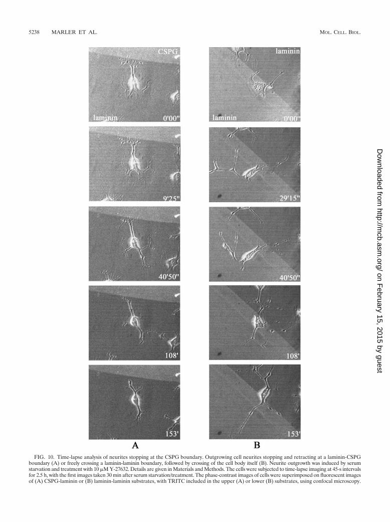
FIG. 10. Time-lapse analysis of neurites stopping at the CSPG boundary. Outgrowing cell neurites stopping and retracting at a laminin-CSPGboundary (A) or freely crossing a laminin-laminin boundary, followed by crossing of the cell body itself (B). Neurite outgrowth was induced by serumstarvation and treatment with 10 �M Y-27632. Details are given in Materials and Methods. The cells were subjected to time-lapse imaging at 45-s intervalsfor 2.5 h, with the first images taken 30 min after serum starvation/treatment. The phase-contrast images of cells were superimposed on fluorescent imagesof (A) CSPG-laminin or (B) laminin-laminin substrates, with TRITC included in the upper (A) or lower (B) substrates, using confocal microscopy.
5238 MARLER ET AL. MOL. CELL. BIOL.
on February 15, 2015 by guest
http://mcb.asm
.org/D
ownloaded from
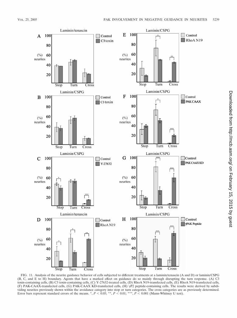
FIG. 11. Analysis of the neurite guidance behavior of cells subjected to different treatments at a laminin/tenascin (A and D) or laminin/CSPG(B, C, and E to H) boundary. Agents that have a marked effect on guidance do so mainly through disrupting the turn response. (A) C3toxin-containing cells, (B) C3 toxin-containing cells, (C) Y-27632-treated cells, (D) RhoA N19-transfected cells, (E) RhoA N19-transfected cells,(F) PAK-CAAX-transfected cells, (G) PAK-CAAX KD-transfected cells, (H) �P2 peptide-containing cells. The results were derived by subdi-viding neurites previously shown within the avoidance category into stop or turn categories. The cross categories are as previously determined.Error bars represent standard errors of the means. *, P 0.05; **, P 0.01; ***, P 0.001 (Mann-Whitney U test).
VOL. 25, 2005 PAK INVOLVEMENT IN NEGATIVE GUIDANCE IN NEURITES 5239
on February 15, 2015 by guest
http://mcb.asm
.org/D
ownloaded from

Concerning repulsive guidance behavior, Monnier et al. (31)reported that the inhibitory effect of CSPG on outgrowth inRGC could be blocked with Y-27632 or C3 toxin, implicat-ing a RhoA/ROK signaling pathway. These workers used astripe assay with alternating stripes of laminin and CSPGmixed with laminin, in which extending axons grew along thelaminin stripes and avoided contact with mixed CSPG- andlaminin-containing stripes. RGC previously treated with eitherY-27632 or C3 toxin showed no preference between the stripesand extended their axons in a more disorganized growing pat-tern crossing over various stripes. The disparity between theseand our findings may lie in differences in the assay and cell typeused. The boundary used by Monnier et al. (31) was lamininmixed with CSPG against laminin, while we used the boundarybetween laminin coated over CSPG against CSPG. Further, noextrinsic factor was required for RGC outgrowth.
Studies on negative guidance signaling in mammals haveimplicated Rac1. Rac1 is required for the collapse of neuritesevoked by Sema3A (16, 21); dominant negative Rac1 N17 butnot C3 toxin blocks the collapse (16). Since Rac1 inhibition (byRac1 N17) blocks neurite outgrowth in NIE-115 cells inducedby serum starvation (20, 40), Rac1 involvement in guidancesignaling could not be investigated directly in our study. In-stead, the downstream Rac effector �PAK was used. Because�PAK does not promote the outgrowth of neurites requiredfor the substrate boundary assay, we used the membrane-tar-geted �PAK-CAAX, which promotes neurite outgrowth inPC12 cells (6), as well as in NIE-115 cells. The membrane-targeted form of �PAK caused a significant disruption in neu-rite guidance at the laminin/CSPG boundary, with increasednumbers crossing. Kinase-inactive �PAK-CAAX KD was evenmore effective in this disruption. Using peptides correspondingto N-terminal amino acid residues 2 to 21 of the PAK isoforms,we found that only the �P2 peptide was able to simulate thedisrupting action of membrane-targeted �PAK-CAAX. The�P2 peptide, like the other peptides, did not affect the numbersof neurites whose outgrowth was induced by serum starvation.Time-lapse analysis of cells after delivery of the �P2 peptideshowed them to be deficient in producing filopodia, with somefilopodium-like structures turning out to be retraction fibersthat were left behind after the collapse of lamellipodia (datanot shown). Moreover, the neurites were more flattened thancontrols and also exhibited a reduction in the number of filo-podia. Given the sensory nature of filopodia, it is possible thattheir relative lack allows more of the filopodium-deficient neu-rites to extend into the hitherto repulsive substrate. Anotherplausible reason why the �P2 peptide but not the others sub-stantially disrupts guidance may lie in the location of �- but not�- or �PAK in filopodia.
A synthetic PAK peptide, 9-23, will prevent the second SH3domain of Nck from binding to PAK (53). It is possible that the�P2 peptide interferes with PAK activities at peripheral filo-podia by sequestering Nck, whose membrane-bound DOCKhomologue has been shown to participate in Drosophila pho-toreceptor axon axonal guidance (13, 33). The membrane-targeted �PAK-CAAX and �PAK-CAAX KD may disruptguidance through a similar sequestration of endogenous Ncklocated at the membrane. Nck binding to PAK is down-regu-lated through PAK kinase autophosphorylation of serine (53);a prolonged binding (sequestration) of Nck may explain why
the kinase-kinase-dead PAK is a more effective disrupter. Fur-ther experiments with other PAK forms, including those inca-pable of binding Nck, may show how PAK operates here.Regardless of how the PAK derivatives work and which iso-form is involved, our results strongly imply that PAK plays apart in the negative guidance response.
In conclusion, we show that NIE-115 neuroblastoma cellscan mimic neurons in their negative guidance response andprovide a facile model for studying this response. While inhi-bition of RhoA pathways will promote outgrowth of neurites,most of these will not cross onto unfavorable substrates. Therepulsive response involves another pathway, where PAK ap-pears to be crucially involved. To enhance regeneration andrepair requiring outgrowth of neurites over normally repulsivesubstrates like CSPG and tenascin expressed at pathologicalsites/scars, mere inhibition of RhoA pathways will not be suf-ficient. Modulating the activities of PAK may provide yet an-other (additional) therapeutic avenue for partial restoration offunction or combating further degeneration.
ACKNOWLEDGMENTS
We thank the GSK-Singapore Research Fund for their generoussupport.
We thank Ed Manser and Zhuo-Shen Zhao for the PAK peptidesand Bhav Mehta for help with the illustrations.
REFERENCES
1. Amano, T., E. Richelson, and M. Nirenberg. 1972. Neurotransmitter synthe-sis by neuroblastoma clones. Proc. Natl. Acad. Sci. USA 69:258–263.
2. Banzai, Y., H. Miki, H. Yamaguchi, and T. Takenawa. 2000. Essential role ofneural Wiskott-Aldrich syndrome protein in neurite extension in PC12 cellsand rat hippocampal primary culture cells. J. Biol. Chem. 275:11987–11992.
3. Bellanger, J. M., J. B. Lazaro, S. Diriong, A. Fernandez, N. Lamb, and A.Debant. 1998. The two guanine nucleotide exchange factor domains of Triolink the Rac1 and the RhoA pathways in vivo. Oncogene 16:147–152.
4. Benard, V., B. P. Bohl, and G. M. Bokoch. 1999. Characterization of Rac andCdc42 activation in chemoattractant-stimulated human neutrophils using anovel assay for active GTPases. J. Biol. Chem. 274:13198–13204.
5. Chiquet, M., and B. Wehrle-Haller. 1994. Tenascin-C in peripheral nervemorphogenesis. Perspect. Dev. Neurobiol. 2:67–74.
6. Daniels, R. H., P. S. Hall, and G. M. Bokoch. 1998. Membrane targeting ofp21-activated kinase 1 (PAK1) induces neurite outgrowth from PC12 cells.EMBO J. 17:754–764.
7. Davies, S. J., M. T. Fitch, S. P. Memberg, A. K. Hall, G. Raisman, and J.Silver. 1997. Regeneration of adult axons in white matter tracts of the centralnervous system. Nature 390:680–683.
8. Dotti, C. G., C. A. Sullivan, and G. A. Banker. 1988. The establishment ofpolarity by hippocampal neurons in culture. J. Neurosci. 8:1454–1468.
9. Fawcett, J. W. 1992. Intrinsic neuronal determinants of regeneration. TrendsNeurosci. 15:5–8.
10. Fawcett, J. W., and R. A. Asher. 1999. The glial scar and central nervoussystem repair. Brain Res. Bull. 49:377–391.
11. Fournier, A. E., B. T. Takizawa, and S. M. Strittmatter. 2003. Rho kinaseinhibition enhances axonal regeneration in the injured CNS. J. Neurosci. 23:1416–1423.
12. Govind, S., R. Kozma, C. Monfries, L. Lim, and S. Ahmed. 2001. Cdc42Hsfacilitates cytoskeletal reorganization and neurite outgrowth by localizing the58-kD insulin receptor substrate to filamentous actin. J. Cell Biol. 152:579–594.
13. Hing, H., J. Xiao, N. Harden, L. Lim, and S. L. Zipursky. 1999. Pak functionsdownstream of Dock to regulate photoreceptor axon guidance in Drosoph-ila. Cell 97:853–863.
14. Hirose, M., T. Ishizaki, N. Watanabe, M. Uehata, O. Kranenburg, W. H.Moolenaar, F. Matsumura, M. Maekawa, H. Bito, and S. Narumiya. 1998.Molecular dissection of the Rho-associated protein kinase (p160ROCK)-regulated neurite remodeling in neuroblastoma N1E-115 cells. J. Cell Biol.141:1625–1636.
15. Jalink, K., E. J. van Corven, T. Hengeveld, N. Morii, S. Narumiya, and W. H.Moolenaar. 1994. Inhibition of lysophosphatidate- and thrombin-inducedneurite retraction and neuronal cell rounding by ADP ribosylation of thesmall GTP-binding protein Rho. J. Cell Biol. 126:801–810.
16. Jin, Z., and S. M. Strittmatter. 1997. Rac1 mediates collapsin-1-inducedgrowth cone collapse. J. Neurosci. 17:6256–6263.
5240 MARLER ET AL. MOL. CELL. BIOL.
on February 15, 2015 by guest
http://mcb.asm
.org/D
ownloaded from
17. Jones, F. S., and P. L. Jones. 2000. The tenascin family of ECM glycopro-teins: structure, function, and regulation during embryonic development andtissue remodeling. Dev. Dyn. 218:235–259.
18. Katoh, H., J. Aoki, Y. Yamaguchi, Y. Kitano, A. Ichikawa, and M. Negishi.1998. Constitutively active G�12, G�13, and G�q induce Rho-dependentneurite retraction through different signaling pathways. J. Biol. Chem. 273:28700–28707.
19. Kozma, R., S. Ahmed, A. Best, and L. Lim. 1995. The Ras-related proteinCdc42Hs and bradykinin promote formation of peripheral actin microspikesand filopodia in Swiss 3T3 fibroblasts. Mol. Cell. Biol. 15:1942–1952.
20. Kozma, R., S. Sarner, S. Ahmed, and L. Lim. 1997. Rho family GTPases andneuronal growth cone remodelling: relationship between increased complex-ity induced by Cdc42Hs, Rac1, and acetylcholine and collapse induced byRhoA and lysophosphatidic acid. Mol. Cell. Biol. 17:1201–1211.
21. Kuhn, T. B., M. D. Brown, C. L. Wilcox, J. A. Raper, and J. R. Bamburg.1999. Myelin and collapsin-1 induce motor neuron growth cone collapsethrough different pathways: inhibition of collapse by opposing mutants ofRac1. J. Neurosci. 19:1965–1975.
22. Laywell, E. D., P. Friedman, K. Harrington, J. T. Robertson, and D. A.Steindler. 1996. Cell attachment to frozen sections of injured adult mousebrain: effects of tenascin antibody and lectin perturbation of wound-relatedextracellular matrix molecules. J. Neurosci. Methods 66:99–108.
23. Letourneau, P. C., M. L. Condic, and D. M. Snow. 1994. Interactions ofdeveloping neurons with the extracellular matrix. J. Neurosci. 14:915–928.
24. Lochter, A., L. Vaughan, A. Kaplony, A. Prochiantz, M. Schachner, and A.Faissner. 1991. J1/tenascin in substrate-bound and soluble form displayscontrary effects on neurite outgrowth. J. Cell Biol. 113:1159–1171.
25. Maekawa, M., T. Ishizaki, S. Boku, N. Watanabe, A. Fujita, A. Iwamatsu, T.Obinata, K. Ohashi, K. Mizuno, and S. Narumiya. 1999. Signaling from Rhoto the actin cytoskeleton through protein kinases ROCK and LIM-kinase.Science 285:895–898.
26. Manser, E., H. Y. Huang, T. H. Loo, X. Q. Chen, J. M. Dong, T. Leung, andL. Lim. 1997. Expression of constitutively active alpha-PAK reveals effects ofthe kinase on actin and focal complexes. Mol. Cell. Biol. 17:1129–1143.
27. Margolis, R. U., and R. K. Margolis. 1997. Chondroitin sulfate proteoglycansas mediators of axon growth and pathfinding. Cell Tissue Res. 290:343–348.
28. McKeon, R. J., R. C. Schreiber, J. S. Rudge, and J. Silver. 1991. Reductionof neurite outgrowth in a model of glial scarring following CNS injury iscorrelated with the expression of inhibitory molecules on reactive astrocytes.J. Neurosci. 11:3398–3411.
29. McKeon, R. J., M. J. Jurynec, and C. R. Buck. 1999. The chondroitin sulfateproteoglycans neurocan and phosphacan are expressed by reactive astrocytesin the chronic CNS glial scar. J. Neurosci. 19:10778–10788.
30. Meiners, S., M. L. Mercado, M. S. Nur-e-Kamal, and H. M. Geller. 1999.Tenascin-C contains domains that independently regulate neurite outgrowthand neurite guidance. J. Neurosci. 19:8443–8453.
31. Monnier, P. P., A. Sierra, J. M. Schwab, S. Henke-Fahle, and B. K. Mueller.2003. The Rho/ROCK pathway mediates neurite growth-inhibitory activityassociated with the chondroitin sulfate proteoglycans of the CNS glial scar.Mol. Cell. Neurosci. 22:319–330.
32. Nemoto, Y., T. Namba, S. Kozaki, and S. Narumiya. 1991. Clostridiumbotulinum C3 ADP-ribosyltransferase gene. Cloning, sequencing, and ex-pression of a functional protein in Escherichia coli. J. Biol. Chem. 266:19312–19319.
33. Newsome, T. P., S. Schmidt, G. Dietzl, K. Keleman, B. Asling, A. Debant,and B. J. Dickson. 2000. Trio combines with dock to regulate Pak activityduring photoreceptor axon pathfinding in Drosophila. Cell 101:283–294.
34. Niederost, B. P., D. R. Zimmermann, M. E. Schwab, and C. E. Bandtlow.1999. Bovine CNS myelin contains neurite growth-inhibitory activity associ-ated with chondroitin sulfate proteoglycans. J. Neurosci. 19:8979–8989.
35. Nobes, C. D., and A. Hall. 1995. Rho, rac, and cdc42 GTPases regulate the
assembly of multimolecular focal complexes associated with actin stressfibers, lamellipodia, and filopodia. Cell 81:53–62.
36. Postma, F. R., K. Jalink, T. Hengeveld, and W. H. Moolenaar. 1996. Sphin-gosine-1-phosphate rapidly induces Rho-dependent neurite retraction: ac-tion through a specific cell surface receptor. EMBO J. 15:2388–2392.
37. Reier, P. J., M. J. Perlow, and L. Guth. 1983. Development of embryonicspinal cord transplants in the rat. Brain Res. 312:201–219.
38. Ridley, A. J., and A. Hall. 1992. The small GTP-binding protein rho regulatesthe assembly of focal adhesions and actin stress fibers in response to growthfactors. Cell 70:389–399.
39. Ridley, A. J., H. F. Paterson, C. L. Johnston, D. Diekmann, and A. Hall.1992. The small GTP-binding protein rac regulates growth factor-inducedmembrane ruffling. Cell 70:401–410.
40. Sarner, S., R. Kozma, S. Ahmed, and L. Lim. 2000. Phosphatidylinositol3-kinase, Cdc42, and Rac1 act downstream of Ras in integrin-dependentneurite outgrowth in N1E-115 neuroblastoma cells. Mol. Cell. Biol. 20:158–172.
41. Sekine, A., M. Fujiwara, and S. Narumiya. 1989. Asparagine residue in therho gene product is the modification site for botulinum ADP-ribosyltrans-ferase. J. Biol. Chem. 264:8602–8605.
42. Silver, J., and J. H. Miller. 2004. Regeneration beyond the glial scar. Nat.Rev. Neurosci. 5:146–156.
43. Snow, D. M., N. Mullins, and D. L. Hynds. 2001. Nervous system-derivedchondroitin sulfate proteoglycans regulate growth cone morphology andinhibit neurite outgrowth: a light, epifluorescence, and electron microscopystudy. Microsc. Res. Tech. 54:273–286.
44. Sung, J. K., L. Miao, J. W. Calvert, L. Huang, H. H. Louis, and J. H. Zhang.2003. A possible role of RhoA/Rho-kinase in experimental spinal cord injuryin rat. Brain Res. 959:29–38.
45. Tashiro, A., A. Minden, and R. Yuste. 2000. Regulation of dendritic spinemorphology by the rho family of small GTPases: antagonistic roles of Racand Rho. Cereb. Cortex 10:927–938.
46. Tigyi, G., D. J. Fischer, A. Sebok, F. Marshall, D. L. Dyer, and R. Miledi.1996. Lysophosphatidic acid-induced neurite retraction in PC12 cells: neu-rite-protective effects of cyclic AMP signaling. J. Neurochem. 66:549–558.
47. van Leeuwen, F. N., H. E. Kain, R. A. van der Kammen, F. Michiels, O. W.Kranenburg, and J. G. Collard. 1997. The guanine nucleotide exchangefactor Tiam1 affects neuronal morphology; opposing roles for the smallGTPases Rac and Rho. J. Cell Biol. 139:797–807.
48. van Leeuwen, F. N., C. Olivo, S. Grivell, B. N. Giepmans, J. G. Collard, andW. H. Moolenaar. 2003. Rac activation by lysophosphatidic acid LPA1 re-ceptors through the guanine nucleotide exchange factor Tiam1. J. Biol.Chem. 278:400–406.
49. Vastrik, I., B. J. Eickholt, F. S. Walsh, A. Ridley, and P. Doherty. 1999.Sema3A-induced growth-cone collapse is mediated by Rac1 amino acids17–32. Curr. Biol. 9:991–998.
50. Wahl, S., H. Barth, T. Ciossek, K. Aktories, and B. K. Mueller. 2000.Ephrin-A5 induces collapse of growth cones by activating Rho and Rhokinase. J. Cell Biol. 149:263–270.
51. Williamson, T., P. R. Gordon-Weeks, M. Schachner, and J. Taylor. 1996.Microtubule reorganization is obligatory for growth cone turning. Proc. Natl.Acad. Sci. USA 93:15221–15226.
52. Wilson, M. T., and D. M. Snow. 2000. Chondroitin sulfate proteoglycanexpression pattern in hippocampal development: potential regulation ofaxon tract formation. J. Comp. Neurol. 424:532–546.
53. Zhao, Z. S., E. Manser, and L. Lim. 2000. Interaction between PAK andNck: a template for Nck targets and role of PAK autophosphorylation. Mol.Cell. Biol. 20:3906–3917.
54. Zheng, J. Q., M. Felder, J. A. Connor, and M. M. Poo. 1994. Turning of nervegrowth cones induced by neurotransmitters. Nature 368:140–144.
VOL. 25, 2005 PAK INVOLVEMENT IN NEGATIVE GUIDANCE IN NEURITES 5241
on February 15, 2015 by guest
http://mcb.asm
.org/D
ownloaded from
Copyright © 2022 FDOKUMEN