Inhibition of Apoptosis Through Localized Delivery of Rapamycin-Loaded Nanoparticles Prevented...
9
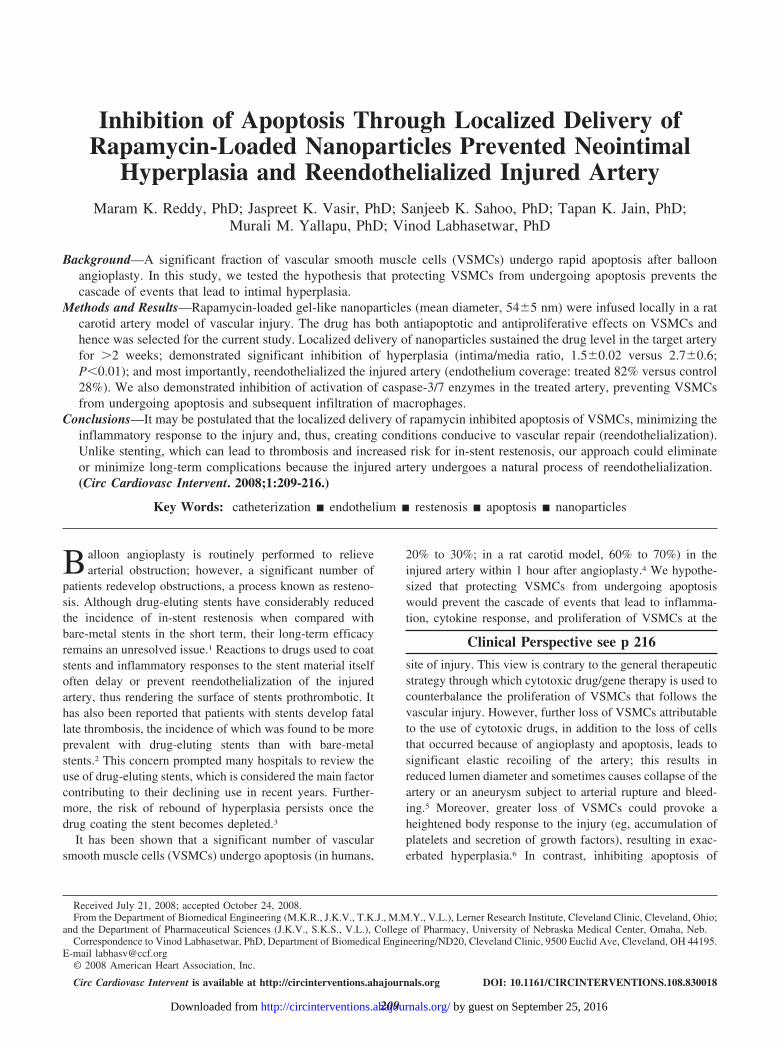
Inhibition of Apoptosis Through Localized Delivery of Rapamycin-Loaded Nanoparticles Prevented Neointimal Hyperplasia and Reendothelialized Injured Artery Maram K. Reddy, PhD; Jaspreet K. Vasir, PhD; Sanjeeb K. Sahoo, PhD; Tapan K. Jain, PhD; Murali M. Yallapu, PhD; Vinod Labhasetwar, PhD Background—A significant fraction of vascular smooth muscle cells (VSMCs) undergo rapid apoptosis after balloon angioplasty. In this study, we tested the hypothesis that protecting VSMCs from undergoing apoptosis prevents the cascade of events that lead to intimal hyperplasia. Methods and Results—Rapamycin-loaded gel-like nanoparticles (mean diameter, 545 nm) were infused locally in a rat carotid artery model of vascular injury. The drug has both antiapoptotic and antiproliferative effects on VSMCs and hence was selected for the current study. Localized delivery of nanoparticles sustained the drug level in the target artery for 2 weeks; demonstrated significant inhibition of hyperplasia (intima/media ratio, 1.50.02 versus 2.70.6; P0.01); and most importantly, reendothelialized the injured artery (endothelium coverage: treated 82% versus control 28%). We also demonstrated inhibition of activation of caspase-3/7 enzymes in the treated artery, preventing VSMCs from undergoing apoptosis and subsequent infiltration of macrophages. Conclusions—It may be postulated that the localized delivery of rapamycin inhibited apoptosis of VSMCs, minimizing the inflammatory response to the injury and, thus, creating conditions conducive to vascular repair (reendothelialization). Unlike stenting, which can lead to thrombosis and increased risk for in-stent restenosis, our approach could eliminate or minimize long-term complications because the injured artery undergoes a natural process of reendothelialization. (Circ Cardiovasc Intervent. 2008;1:209-216.) Key Words: catheterization endothelium restenosis apoptosis nanoparticles B alloon angioplasty is routinely performed to relieve arterial obstruction; however, a significant number of patients redevelop obstructions, a process known as resteno- sis. Although drug-eluting stents have considerably reduced the incidence of in-stent restenosis when compared with bare-metal stents in the short term, their long-term efficacy remains an unresolved issue. 1 Reactions to drugs used to coat stents and inflammatory responses to the stent material itself often delay or prevent reendothelialization of the injured artery, thus rendering the surface of stents prothrombotic. It has also been reported that patients with stents develop fatal late thrombosis, the incidence of which was found to be more prevalent with drug-eluting stents than with bare-metal stents. 2 This concern prompted many hospitals to review the use of drug-eluting stents, which is considered the main factor contributing to their declining use in recent years. Further- more, the risk of rebound of hyperplasia persists once the drug coating the stent becomes depleted. 3 It has been shown that a significant number of vascular smooth muscle cells (VSMCs) undergo apoptosis (in humans, 20% to 30%; in a rat carotid model, 60% to 70%) in the injured artery within 1 hour after angioplasty. 4 We hypothe- sized that protecting VSMCs from undergoing apoptosis would prevent the cascade of events that lead to inflamma- tion, cytokine response, and proliferation of VSMCs at the Clinical Perspective see p 216 site of injury. This view is contrary to the general therapeutic strategy through which cytotoxic drug/gene therapy is used to counterbalance the proliferation of VSMCs that follows the vascular injury. However, further loss of VSMCs attributable to the use of cytotoxic drugs, in addition to the loss of cells that occurred because of angioplasty and apoptosis, leads to significant elastic recoiling of the artery; this results in reduced lumen diameter and sometimes causes collapse of the artery or an aneurysm subject to arterial rupture and bleed- ing. 5 Moreover, greater loss of VSMCs could provoke a heightened body response to the injury (eg, accumulation of platelets and secretion of growth factors), resulting in exac- erbated hyperplasia. 6 In contrast, inhibiting apoptosis of Received July 21, 2008; accepted October 24, 2008. From the Department of Biomedical Engineering (M.K.R., J.K.V., T.K.J., M.M.Y., V.L.), Lerner Research Institute, Cleveland Clinic, Cleveland, Ohio; and the Department of Pharmaceutical Sciences (J.K.V., S.K.S., V.L.), College of Pharmacy, University of Nebraska Medical Center, Omaha, Neb. Correspondence to Vinod Labhasetwar, PhD, Department of Biomedical Engineering/ND20, Cleveland Clinic, 9500 Euclid Ave, Cleveland, OH 44195. E-mail [email protected] © 2008 American Heart Association, Inc. Circ Cardiovasc Intervent is available at http://circinterventions.ahajournals.org DOI: 10.1161/CIRCINTERVENTIONS.108.830018 209 by guest on September 25, 2016 http://circinterventions.ahajournals.org/ Downloaded from
Transcript of Inhibition of Apoptosis Through Localized Delivery of Rapamycin-Loaded Nanoparticles Prevented...
Inhibition of Apoptosis Through Localized Delivery ofRapamycin-Loaded Nanoparticles Prevented Neointimal
Hyperplasia and Reendothelialized Injured ArteryMaram K. Reddy, PhD; Jaspreet K. Vasir, PhD; Sanjeeb K. Sahoo, PhD; Tapan K. Jain, PhD;
Murali M. Yallapu, PhD; Vinod Labhasetwar, PhD
Background—A significant fraction of vascular smooth muscle cells (VSMCs) undergo rapid apoptosis after balloonangioplasty. In this study, we tested the hypothesis that protecting VSMCs from undergoing apoptosis prevents thecascade of events that lead to intimal hyperplasia.
Methods and Results—Rapamycin-loaded gel-like nanoparticles (mean diameter, 54�5 nm) were infused locally in a ratcarotid artery model of vascular injury. The drug has both antiapoptotic and antiproliferative effects on VSMCs andhence was selected for the current study. Localized delivery of nanoparticles sustained the drug level in the target arteryfor �2 weeks; demonstrated significant inhibition of hyperplasia (intima/media ratio, 1.5�0.02 versus 2.7�0.6;P�0.01); and most importantly, reendothelialized the injured artery (endothelium coverage: treated 82% versus control28%). We also demonstrated inhibition of activation of caspase-3/7 enzymes in the treated artery, preventing VSMCsfrom undergoing apoptosis and subsequent infiltration of macrophages.
Conclusions—It may be postulated that the localized delivery of rapamycin inhibited apoptosis of VSMCs, minimizing theinflammatory response to the injury and, thus, creating conditions conducive to vascular repair (reendothelialization).Unlike stenting, which can lead to thrombosis and increased risk for in-stent restenosis, our approach could eliminateor minimize long-term complications because the injured artery undergoes a natural process of reendothelialization.(Circ Cardiovasc Intervent. 2008;1:209-216.)
Key Words: catheterization � endothelium � restenosis � apoptosis � nanoparticles
Balloon angioplasty is routinely performed to relievearterial obstruction; however, a significant number of
patients redevelop obstructions, a process known as resteno-sis. Although drug-eluting stents have considerably reducedthe incidence of in-stent restenosis when compared withbare-metal stents in the short term, their long-term efficacyremains an unresolved issue.1 Reactions to drugs used to coatstents and inflammatory responses to the stent material itselfoften delay or prevent reendothelialization of the injuredartery, thus rendering the surface of stents prothrombotic. Ithas also been reported that patients with stents develop fatallate thrombosis, the incidence of which was found to be moreprevalent with drug-eluting stents than with bare-metalstents.2 This concern prompted many hospitals to review theuse of drug-eluting stents, which is considered the main factorcontributing to their declining use in recent years. Further-more, the risk of rebound of hyperplasia persists once thedrug coating the stent becomes depleted.3
It has been shown that a significant number of vascularsmooth muscle cells (VSMCs) undergo apoptosis (in humans,
20% to 30%; in a rat carotid model, 60% to 70%) in theinjured artery within 1 hour after angioplasty.4 We hypothe-sized that protecting VSMCs from undergoing apoptosiswould prevent the cascade of events that lead to inflamma-tion, cytokine response, and proliferation of VSMCs at the
Clinical Perspective see p 216
site of injury. This view is contrary to the general therapeuticstrategy through which cytotoxic drug/gene therapy is used tocounterbalance the proliferation of VSMCs that follows thevascular injury. However, further loss of VSMCs attributableto the use of cytotoxic drugs, in addition to the loss of cellsthat occurred because of angioplasty and apoptosis, leads tosignificant elastic recoiling of the artery; this results inreduced lumen diameter and sometimes causes collapse of theartery or an aneurysm subject to arterial rupture and bleed-ing.5 Moreover, greater loss of VSMCs could provoke aheightened body response to the injury (eg, accumulation ofplatelets and secretion of growth factors), resulting in exac-erbated hyperplasia.6 In contrast, inhibiting apoptosis of
Received July 21, 2008; accepted October 24, 2008.From the Department of Biomedical Engineering (M.K.R., J.K.V., T.K.J., M.M.Y., V.L.), Lerner Research Institute, Cleveland Clinic, Cleveland, Ohio;
and the Department of Pharmaceutical Sciences (J.K.V., S.K.S., V.L.), College of Pharmacy, University of Nebraska Medical Center, Omaha, Neb.Correspondence to Vinod Labhasetwar, PhD, Department of Biomedical Engineering/ND20, Cleveland Clinic, 9500 Euclid Ave, Cleveland, OH 44195.
E-mail [email protected]© 2008 American Heart Association, Inc.
Circ Cardiovasc Intervent is available at http://circinterventions.ahajournals.org DOI: 10.1161/CIRCINTERVENTIONS.108.830018
209 by guest on September 25, 2016http://circinterventions.ahajournals.org/Downloaded from
VSMCs to begin with could prevent this cascade of events,including the inflammatory response. This would not onlyinhibit hyperplasia but also may create conditions suitable forreendothelialization, a key factor for the long-term patency ofthe injured artery after an intervention.
We chose rapamycin to test our hypothesis because thisdrug is known to inhibit proliferation of VSMCs, primarily bycell-cycle arrest.7 More important, the drug has been shownto downregulate the genes responsible for induction ofapoptosis in VSMCs.8 Rapamycin, however, is not watersoluble and thus cannot be delivered intraluminally as asolution. We therefore developed water-dispersible gel-likenanoparticles (NPs) that were easily loaded with rapamycin.These loaded NPs were infused locally in the arterial wallimmediately after balloon angioplasty using an infusioncatheter. Because of their smaller size and gel-like flexiblestructure, our NPs were expected to result in efficient arterialdrug uptake and maintain the drug level in the target arterybecause of their sustained-release properties. We tested ourhypothesis in a rat carotid artery model of vascular injurybecause this model shows significant apoptosis of VSMCs(60% to 70%) after angioplasty.4 The results demonstratedsignificant inhibition of intimal hyperplasia and increasedreendothelialization of the injured artery.
MethodsSynthesis, Formulation, and Characterization ofRapamycin-Loaded NPsCross-linked gel-like polymeric NPs were synthesized throughrandom free-radical polymerization of N-isopropylacrylamide,N-vinyl pyrrolidone, and pegylated maleic polymers, as previouslydescribed.9 For rapamycin loading, 20 mg of lyophilized NPs wasdispersed in 2 mL of distilled water by vortexing for 2 minutes, towhich 250 �L of methanolic solution of rapamycin (Sigma, 4mg/mL) was added with constant stirring on a magnetic stir plate for2 hours to allow drug entrapment into NPs. The unentrappedrapamycin was separated by overnight dialysis of the dispersion ofNPs against 1 L of distilled water using a Spectrapore (SpectraporeInc) dialysis bag (MW cutoff size, 12 kDa). Drug-loaded NPs werethen lyophilized for 48 hours. Particle size distribution and zetapotential of NPs were determined using a ZetaPlus particle size andzeta analyzer (Brookhaven Instruments Corp). NPs were also viewedfor size using a transmission electron microscope (Philips/FEI Inc) afternegative staining with 2% (wt/vol) uranyl acetate solution. Drug releasefrom NPs in vitro was determined in phosphate-buffered saline(154 mmol/L, pH 7.4) containing 0.1% (wt/vol) Tween-80 at 37°C tomaintain a sink condition, as described in our previous study.9
Cell Culture and Antiproliferative Effect ofRapamycin-Loaded NPsHuman VSMCs (Cascade Biologics) at passage 3 to 4 were seededin 96-well plates (4000 cells per well); cells were allowed to attachfor 24 hours and were then treated with rapamycin. Medium in thewells was changed on day 2 and every alternate day thereafter withno further addition of drug. Inhibition of cell proliferation wasdetermined on day 8 using an MTS assay (CellTiter 96 AQueous,Promega). For cell-cycle analysis, cells cultured in T-75 cultureflasks were incubated with a dispersion of rapamycin-loaded NPs inthe growth medium (dose of rapamycin, 50 ng/mL). Two days aftertreatment, the cell monolayers were washed with phosphate-bufferedsaline, trypsinized, and resuspended in 1 mL of Telford reagent. Thecellular DNA content was analyzed by a fluorescence-activated-cellsorter FACStarPlus flow cytometer operating under Lysis II (BectonDickinson).
Rat Carotid Artery Model of Vascular Injury andMorphometric Analysis of Artery for Inhibitionof HyperplasiaMale Sprague-Dawley rats (240 to 260 g; Charles River Laborato-ries, Wilmington, Mass) were anesthetized with an intraperitonealinjection of a ketamine (100 mg/kg)/xylazine (10 mg/kg) mixture,then underwent carotid artery angioplasty using a 2F Fogarty ballooncatheter (Edwards Life Sciences). The balloon was inflated in thecarotid artery sufficiently to generate slight resistance and waswithdrawn 3 times consistently to produce endothelial denudation ofthe entire length of the left common carotid artery. On removal of theballoon catheter, a PE-10 catheter was inserted into the left commoncarotid artery to infuse a suspension of NPs (210 �L containing 60 �gof rapamycin-equivalent NPs) for �5 minutes at 2 atm of pressure (3infusions of 70 �L each, with a 1-minute period between infusions). Thedistal and proximal ends of the common carotid artery and internalcarotid artery were tied at the time of NP infusion. Five minutes afterinfusion, the ties were removed and blood flow was restored.
Three weeks after injury, the rats were euthanized and transcar-dially perfused through the left ventricle with 100 mL of heparinizedsaline followed by 4% paraformaldehyde, and arteries were collectedin 10% formalin. After 24 hours, arteries were cut into 3 pieces, eachpiece cut every 2 mm from the proximal to the distal end. Thesepieces of arteries were embedded in paraffin for sectioning (5-�mthickness), and duplicate slides were stained with hematoxylin andeosin. The stained sections were used for morphometric analysis ofmedial, intimal, and luminal areas using computer-assisted imageanalysis (Image-Pro Plus 6.1, Media Cybernetics Inc).
Quantification of Rapamycin Levels inArterial TissueTo determine arterial drug uptake and retention, carotid arteries fromboth sides (injured/treated and uninjured contralateral) were removed atdifferent time points after administration of NPs. Arterial samples wererinsed with saline, blotted dry with absorbent paper, and measured forwet weight. Next, the arteries were finely cut into small pieces andhomogenized in 2 mL of distilled water using a tissue homogenizer(BioSpec Products Inc) at 1000 rpm for 2 minutes. The homogenateswere then lyophilized for 48 hours. Drug from each dry tissue wasextracted by shaking samples with 1 mL of methanol at 37°C for 48hours at 150 rpm using an Environ orbital shaker (Fisher Scientific). Thesamples were centrifuged at 14 000 rpm for 10 minutes (Eppendorf_microcentrifuge, model 5417R, Brinkmann Instruments) to removecellular debris. The supernatants were analyzed by high-performanceliquid chromatography for rapamycin content. A standard plot wasprepared using arteries collected from animals that did not receiverapamycin treatment to determine the recovery efficiency of the drug.
ImmunohistochemistryAnimals were euthanized and arteries were collected postangioplastyat 1 hour for investigation of apoptosis and caspase activity, at 24hours for recruitment of macrophages, and at 3 weeks for evaluationof reendothelialization. Frozen sections of 5-�m thickness wereincubated with I-VIEW inhibitor to block endogenous peroxidaseactivity, washed with phosphate-buffered saline, and incubated withprimary antibody for 1 hour at room temperature. The followingprimary antibodies were used: monoclonal mouse CD31 antibody(1:100 dilution; DAKO, Carpenter, Calif), anticleaved Caspase-3/7(1:200 dilution; Cell Signaling Technology, Beverly, Mass), andmonoclonal CD68 antibody (1:50 dilution; Clone prostaglandin-M1,DAKO). Sections were then incubated with I-VIEW biotin andI-VIEW streptavidin-horseradish peroxidase. Sections were visual-ized using 3,3� diaminobenzidine chromogen and were counter-stained using I-VIEW copper.
Reendothelialization was calculated as the percentage of luminalsurface covered by CD31-positive cells 3 weeks after the angio-plasty. In addition, in a separate experiment, the extent of Evans bluedye leakage (blue staining) was used as an index for endothelial celllining integrity. The dye (0.5 mL of 0.5% solution, Sigma) wasinjected intravenously 30 minutes before euthanasia 3 weeks after
210 Circ Cardiovasc Intervent December 2008
by guest on September 25, 2016http://circinterventions.ahajournals.org/Downloaded from

angioplasty, and carotid arteries were harvested after transpericardialperfusion with heparinized saline (20 mL). The arteries were fixed inmethanol, carefully cut open vertically, and imaged using a flatbedscanner (HP Scanjet 3970, Hewlett-Packard Company).
For apoptotic cells, the terminal deoxynucleotidyl transferase-mediated dUTP nick end labeling (TUNEL) method was usedaccording to the manufacturer’s specifications (In Situ Cell DeathDetection Kit, Fluorescein, Roche Applied Sciences). Nuclei werecounterstained with propidium iodide. Sections were photographedon a Diaphot microscope (Nikon Instruments Inc) equipped with aphase-contrast and epifluorescence optics (�100) lens. The percent-ages of apoptotic nuclei were calculated by determining the numberof propidium iodide-stained nuclei that were also positive forTUNEL staining (n�9 sections per artery; n�5 animals per treat-ment). Approximately 100 nuclei were counted for each section.
Caspase-3/7 AssayCaspase-3/7 activity was determined in the tissue homogenates ofarteries treated with rapamycin-loaded NPs versus control arteries(those infused with NPs that carried no drug, called “void” NPs). Theanimals were euthanized 1 hour after angioplasty and arteries werehomogenized in a hypotonic cell lysis buffer. Caspase activity wasmeasured by the Apo-ONE Homogeneous Caspase-3/7 Assay System(Promega). The intensity of emitted fluorescence (relative fluorescenceunits) was determined at a wavelength of 521 nm using a FL�800Microplate Fluorescence Reader (BioTek Instruments Inc).
Statistical AnalysisAll the data are presented as mean�SE of means. The statisticalsignificance of differences among treatment groups was determinedby 1-way analysis of variance. Differences were considered signif-icant when P�0.05.
The authors had full access to and take full responsibility for theintegrity of the data. All authors have read and agree to themanuscript as written.
Results
Characterization of NPsGel-like NPs were almost spherical in shape, typically rang-ing in size from 46 to 60 nm with a mean diameter of 54�5nm (Figure 1A) and zeta potential (surface charge) of �8.45mV at pH 7. The drug loading in NPs was 4.2% (w/w) (ie,100 mg of formulation contained 4.2 mg of rapamycin) withan entrapment efficiency of 84% (ie, 84% of the added drugwas trapped in NPs). The release profile of rapamycin fromNPs under in vitro conditions demonstrated relatively rapidrelease during the initial stages (�20% cumulative release inthe first 24 hours) with more gradual release thereafter(�80% cumulative release in 28 days; Figure 1B).
Figure 1. A, Transmission electron micrograph of rapamycin-loaded NPs. Bar represents 100 nm. B, In vitro release profile of rapamy-cin from NPs in phosphate-buffered saline (pH 7.4, containing 0.1% Tween-80 at 37°C). Data shown as mean�SEM (n�3). C, Dose-response effect on inhibition of VSMC proliferation of rapamycin at 8 days after treatment either with rapamycin-loaded NPs (solid cir-cle) or drug in solution (open circle). *P�0.005 NPs versus solution (Sol) D, Cell-cycle analyses of VSMCs after (i) treatment withmedium, (ii) void NPs, (iii) rapamycin in solution, and (iv) rapamycin-loaded NPs. Cells were treated with rapamycin for 48 hours andcell-cycle perturbations were analyzed by flow cytometry.
Reddy et al Inhibition of Hyperplasia 211
by guest on September 25, 2016http://circinterventions.ahajournals.org/Downloaded from

Inhibition of VSMC Proliferation in VitroRapamycin-loaded NPs demonstrated greater inhibition ofVSMC proliferation than did rapamycin in solution during adose-response study conducted 8 days after treatment (Figure1C). The inhibitory effect of rapamycin was evident at as low as1 ng/mL concentration, which was the lowest dosage evaluatedin our study. Cells treated with void NPs showed almost similarcell growth as medium control, indicating their cytocompatibil-ity (data not shown). Flow cytometry data of cells treated withrapamycin-loaded NPs revealed that their antiproliferative effectarose primarily because of inhibition of cell cycle progression atthe G1 checkpoint. Approximately 75% of cells treated withrapamycin-loaded NPs were arrested in G0/G1 phase, whencompared with 63% in void NPs (Figure 1D).
Arterial Drug LevelsQuantitative analysis of the arterial segments for total druglevels demonstrated an initial drop during the first 24 hoursafter infusion, but levels were maintained thereafter for 2weeks before a slow decline (Figure 2). The drug was notdetectable in the contralateral carotid artery, indicating local-ized delivery of the drug.
Inhibition of Hyperplasia and ReendothelializationMorphometric analysis of the arterial sections from theanimals treated with drug-loaded NPs demonstrated signifi-cantly reduced hyperplasia than in control animals thatreceived void NPs (intima/media ratio, 1.5�0.02 versus
2.7�0.6; P�0.01; Figure 3A through 3C). The intraperito-neal administration of the same dose of drug-loaded NPsdemonstrated no effect on inhibition of hyperplasia, suggest-ing that the localized arterial drug delivery was necessary toachieve the therapeutic dose of the drug in the target artery(data not shown). Inhibition of hyperplasia in the treatedgroup also resulted in a significant increase in arterial lumen areathan in the control group (Figure 3D). Saline control and voidNPs demonstrated a comparable extent of hyperplasia, indicat-ing that NPs themselves do not cause any change in proliferativeresponse (data not shown). Therefore, all subsequent controlexperiments were carried out using void NPs. The group treatedwith rapamycin-NPs demonstrated significantly greater reendo-thelialization of the injured artery than control arteries (82%versus 28%; Figure 4A). This was further evident from reducedextravasation of Evans blue dye in rapamycin-treated arterieswhen compared with control arteries (Figure 4B and 4C).
Protection of VSMCs From ApoptosisHistochemical analysis of arterial sections using the TUNELassay demonstrated a significant reduction in the number ofapoptotic VSMCs (treated 24% versus control 70%) in themedial layer of the artery at 1 hour posttreatment withrapamycin-loaded NPs (Figure 5A and 5B). In addition, asignificant (P�0.01) reduction in the levels of active caspaseswas observed in the treated arteries when compared withcontrol arteries, which was further confirmed by immunohis-tochemical analysis of the arterial sections from the 2 groups(Figure 5C and 5D). Together, these results indicate thatrapamycin inhibited caspase activity in the injured artery andthus prevented VSMCs from undergoing apoptosis.
Inhibition of Infiltration of MacrophagesAnalysis of the artery sections 24 hours after the angioplastyprocedure indicates that rapamycin-loaded NPs reduced thenumber of macrophages infiltrating into the adventitial layersof the injured artery when compared with control (void) NPs(Figure 6). These results suggest that preventing apoptosis ofVSMCs also reduces the inflammatory reaction of the body inresponse to vascular injury, but this reduction in macrophagescould also be attributed to the antiinflammatory effect of therapamycin in the injured artery itself.10
Figure 2. Tissue concentration of rapamycin in injured artery atdifferent time points (1 hour, 1 day, 3 days, 7 days, 14 days,and 21 days) after localized delivery of drug-loaded NPs.Results are expressed as mean�SEM (n�3).
Figure 3. Inhibition of intimal hyperplasiawith rapamycin-loaded NPs in a ratcarotid artery model of vascular injury at3 weeks. A and B, Arterial sections of ani-mals treated with void NPs (left, control)and with rapamycin-loaded NPs (right,treated). C, Bar graphs represent themorphometric analysis of arterial sectionsfor the inhibition of hyperplasia (intima/media ratio) and (D) the lumen areas(mm2) of arteries in different treatmentgroups. Quantitative data derived from 3arterial sections at different levels fromeach animal in each group. *P�0.01 fortreated (n�6) versus control (n�3)groups.
212 Circ Cardiovasc Intervent December 2008
by guest on September 25, 2016http://circinterventions.ahajournals.org/Downloaded from

DiscussionDrug-eluting stents and catheter-based drug delivery are the 2main strategies used to inhibit the excessive VSMC migrationand proliferation that contribute to restenosis after balloonangioplasty.11,12 The advantage of drug-eluting stents is thatthey can provide localized drug therapy as well as a supportstructure for the injured artery to prevent the elastic recoilingthat often occurs within 24 hours after balloon angioplasty.13
However, several recent studies have reported that patientswith drug-eluting stents are vulnerable to developing late (upto 18 months) stent thrombosis, aneurysm formation, exten-sive inflammatory reaction (attributed to hypersensitivity to
the coated polymer),14 and are at risk for restenosis once the drugcoating is depleted.15 These results have thus renewed concernsabout the long-term efficacy of drug-eluting stents.2 The under-lying cause for such unintended effects is attributed to theinhibition or delay in reendothelialization of the stented artery.2
This delay could result from the drug used to coat the stent,which has direct contact with the endothelium, from the contin-ued inflammatory response to the stent material itself, or both.2
To avoid long-term complications after angioplasty, atherapeutic strategy that is focused on preventing hyperplasia,as well as promoting reendothelialization of the injuredartery, would be more effective. Toward this goal, we have
Figure 4. Reendothelialization of injured arteryafter treatment with rapamycin-loaded NPs. A,Quantification of reendothelialization. Data arederived from 3 arterial sections stained for CD-31at different levels from each animal in each group.*P�0.05 for treated (rapamycin-loaded NPs) ver-sus control (void NPs; n�3). Representative arter-ies from rats injected with Evans blue dye. VoidNPs (B) show greater blue staining thanrapamycin-loaded NPs (C), indicating less reen-dothelialization. Black arrows show the reducedextravasation of Evans blue dye due toreendothelialization.
Figure 5. A, Immunohistochemical analysis of arterial sections for apoptosis by TUNEL assay. i, ii, and iii show TUNEL staining; iv, v,and vi show nuclei counterstained with propidium iodide. i and iv represent the contralateral artery; ii and v represent the injured arterytreated with void NPs; and iii and vi represent the injured artery treated with rapamycin-loaded NPs. White arrows in injured/treatedarteries (ii versus iii) show the reduction in apoptosis with treatment. B, Quantitation of the number of apoptotic VSMCs in the mediallayer of artery at 1 hour posttreatment with rapamycin-loaded NPs. *P�0.05 for treated (rapamycin-loaded NPs) versus control (voidNPs). C, Immunohistochemical staining for activated caspase-3. Data are representative arterial sections from animals treated eitherwith void NPs (i) or rapamycin NPs (ii). Magnification �200. D, Caspase-3/7 activity as determined in the tissue homogenates of arter-ies. *P�0.05 for treated (rapamycin-loaded NPs) versus control (void NPs).
Reddy et al Inhibition of Hyperplasia 213
by guest on September 25, 2016http://circinterventions.ahajournals.org/Downloaded from

been investigating colloidal drug carrier systems that can beadministered intraluminally in the target artery using aninfusion catheter.16,17 The success of colloidal-based drugcarrier systems depends to a great degree on their ability tomaintain a therapeutic drug concentration in the target arteryto block proliferation of VSMCs until the injured artery hasbeen reendothelialized.
Particle size of the drug carrier is a critical determinantbecause size not only affects the efficiency of drug localiza-tion in the target artery but also its distribution in the arterialwall.18,19 We have previously shown that only a small fraction(�2% v/v) of arterial volume (extracellular fluid) can bereplaced with a suspension of drug carrier system.16 Hence, atherapeutic dose of the drug must be delivered in the dose ofthe carrier system that can be localized in the target artery. Inour study, we observed �9% efficiency of tissue uptake ofdrug (60 �g rapamycin injected, 5.3 �g localized, averagetissue weight of carotid artery segment 3.5 mg), which is�4-fold higher than that reported previously for other colloi-
dal drug delivery systems.20 This greater efficacy of drug uptakewith our gel-like NPs could be attributed to their size (smallerthan those used previously; 54 nm versus 160 nm)20 and gel-likeflexible structure that could penetrate the arterial wall moreeffectively than rigid and solid NPs. The smaller size of NPs isalso important because particles smaller than �100 nm candeposit in the vessel wall, whereas larger-sized particles accu-mulate primarily at the luminal surface of the artery.17,18
The localization of NPs could also influence drug retentionin the target artery. In our study, the rapamycin level in thetarget artery was 1.5 �g/mg at 1 hour postinfusion, and thelevel declined during the first 24 hours after infusion of NPsbut remained sustained thereafter for �2 weeks. It is possiblethat the NPs localized in the intimal layer were washed awaywith blood flow after the initial localization, resulting in theinitial decline in drug level. However, the NPs localized inthe arterial wall were retained and able to maintain the druglevel. In addition to direct localization of NPs into the arterialwall through the disrupted endothelium, a fraction of theseNPs could have been deposited in the arterial wall throughtheir transport through the vasa vasorum.21 These are smallcapillaries originating from the lumen of the artery with anetwork spreading to adventitia and media.22 Because ourNPs are small and flexible, they could have passed throughthese capillaries and been deposited in the media and adven-titia. This prolonged arterial drug retention could result fromthe sustained-release property of NPs in addition to thebinding of rapamycin to certain cellular proteins,23 thusslowing its rate of diffusion. We could not determine the drugretention without NPs because of the insoluble nature ofrapamycin, but others have shown that localized infusion ofdrug in solution results, within 24 hours, in rapid decline andcomplete loss of drug from the arterial wall.24
The studies with rapamycin-coated stents in a porcinecoronary model have demonstrated that the drug level of 95ng/mg of artery weight, observed at 3 days after the stentdeployment, was effective in inhibiting restenosis.25 The druglevels observed in our studies were above target level andhence can be considered as a local therapeutic dose to inhibitproliferation of VSMCs. It has been reported that particulatesystems infused intraluminally migrate away from the intimallayer toward the medial layer over time because of bloodpressure.26 Similarly, our NPs might have migrated, which isadvantageous because the drug would not be in direct contactwith the intimal layer to adversely influence the process ofreendothelialization of the injured artery. This fact is impor-tant because rapamycin has been reported to suppress theadhesion of endothelial progenitor cells and their differenti-ation into endothelium.27 Because we observed significantreendothelialization of the injured artery, the drug localized inthe artery was probably not interfering in the process,allowing vascular repair to occur by the natural mechanism.28
One of the main reasons that stented arteries do not undergoreendothelialization very well is because of the direct contactwith the intimal layer of the drug used to coat the stent,consequently either inhibiting the proliferation of endothelialcells or preventing adhesion and differentiation of circulatingendothelial progenitor cells to the injured endothelium.29
Figure 6. Immunohistochemical staining of arterial sections toassess the extent of infiltration of macrophages. Data are repre-sentative arterial sections from contralateral arteries (i and ii)and the injured arteries treated either with void NPs (iii and iv) orrapamycin-loaded NPs (v and vi). i, iii, and v represent macro-phages stained with fluoresceinisothiocyanate-conjugated anti-body; ii, iv, and vi represent counterstaining with propidiumiodide to visualize nuclei. Counterstaining was used to confirmthe presence of macrophages in the adventitial layer. Magnifica-tion �200. Ad indicates adventitia; L, lumen.
214 Circ Cardiovasc Intervent December 2008
by guest on September 25, 2016http://circinterventions.ahajournals.org/Downloaded from

In addition to the influence of drug carrier systems, theother important determinant is the mechanism of drug action.Most therapies are focused toward administering cytotoxicdrugs30 or genes31 to combat the proliferative response;however, this approach has raised many concerns, includingthe long-term patency of the injured artery.32 Our approachwas to protect VSMCs from undergoing apoptosis shortlyafter vascular injury and to prevent their proliferation. Thus,rapamycin seems to be the right choice of drug, not onlybecause of its antiproliferative and antiinflammatory activi-ties33 but also because it has been shown to downregulate theproapoptotic genes in VSMCs.8 Immunohistochemical anal-ysis of the arterial sections collected at 1 hour after infusionof rapamycin-loaded NPs demonstrated inhibition of apopto-sis in the treated artery (Figures 5A and 5B), thus supportingthe above mechanism of rapamycin. This antiapoptotic effectof rapamycin has been found to be caused by inhibition ofactivation of Caspase-3/7 in the treated artery, which is a keymediator of apoptosis of mammalian cells (Figure 5C and5D).8 Further, this reduction in the apoptotic cell populationin the treated artery also successfully inhibited the infiltrationof macrophages at the injured site (Figure 6), which is one ofthe well-known pharmacological effects of rapamycin.8 Thecell-cycle analysis of the rapamycin-loaded NP-treated cellsin vitro demonstrated inhibition of cell proliferation primarilyby cell-cycle arrest in the G0/G1 phase (Figure 1D).
Other pathways, which we have not investigated in this study,could be critical to the inhibition of hyperplasia. These includethe antiangiogenic effect of rapamycin or its effects on themammalian target of rapamycin, which directly or indirectlycontrols several cellular events.34 Inhibition of cell proliferationwith rapamycin is known to be mediated through its binding tothe cytosolic receptor FKBP12, and the resulting complexinhibits the protein kinase mammalian target of rapamycin,which is a critical regulator of VSMC migration and prolifera-tion.35,36 NPs could have facilitated the intracellular delivery ofrapamycin, thus allowing its interaction with the FKBP12receptor and resulting in inhibition of cell proliferation.
It has been shown in a porcine coronary model of resteno-sis that proliferation of VSMCs occurs primarily during thefirst 7 to 10 days after angioplasty.37 Therefore, it has beensuggested that suppressing this proliferative phase is criticalto the inhibition of restenosis. The prevention of hyperplasiaobserved in our studies could have been caused by the abilityof NPs to maintain a therapeutic dose of the drug in the targetartery to suppress this proliferative phase. Because of thecomplexity of the disease, it is a debatable question as towhich events are critical to inhibition of hyperplasia—initialevents such as platelet deposition and thrombosis, late eventssuch as VSMC migration and proliferation, or both.38 Perhapsour NPs worked early by protecting VSMCs from undergoingapoptosis, which minimized the body’s response to injury(infiltration of macrophages) and later by inhibiting theproliferation of VSMCs, which reduced intimal hyperplasia.By protecting VSMCs from undergoing apoptosis, the ther-apy diminished hostile conditions, such as the inflammatoryresponse and platelet aggregation, at the site of the arterialinjury, possibly creating favorable conditions under whichreendothelialization could occur.
Recently, Cyrus et al39 have demonstrated the efficacy ofrapamycin-loaded �v�3-targeted paramagnetic perfluorocar-bon NPs in inhibiting vascular stenosis in an atheroscleroticrabbit femoral artery injury model, suggesting the utility ofthe NP-based delivery system in a clinically relevant vascularcondition. The polymer system used in our NP formulationhas been extensively investigated for drug delivery applica-tions, with a commercial product introduced recently for thedelivery of paclitaxel in cancer treatment (Nanoxel, DaburPharma Ltd). The NPs used in our study are not biodegrad-able but could be made so easily by introducing disulfidelinkages in the polymer chains such that the smaller polymerfragments formed by degradation would be eliminated. Inaddition to coronary and carotid artery restenosis, our approachcould be effectively used to prevent the hyperplasia that isinduced after interventions involving renal or femoral arteries.
In conclusion, our study demonstrates that the apoptosis ofVSMCs that follows immediately after intraarterial ballooninjury is intricately involved in vascular hyperplasia and thatthe inhibition of this apoptotic process with locally deliveredrapamycin can abrogate the deleterious cascade of events,preventing neointima formation. Our system could also beused in other vascular conditions in which apoptosis ofVSMCs is implicated, such as in aneurysms, cerebrovascularocclusive diseases, and atherosclerosis.
AcknowledgmentsWe thank Melissa Jedlicka for proofreading the manuscript.
Sources of FundingThis work was supported by grants from Cleveland Clinic and theNebraska Research Initiative Program (to V.L.). S.K.S. was sup-ported by a postdoctoral and J.K.V. by a predoctoral fellowship fromthe American Heart Association, Heartland Affiliate.
DisclosuresV.L., M.R., and S.K. are coinventors on a pending U.S. patentapplication (20060134209 A1) that discloses the application of thecomposition of nanoparticles for vascular delivery of drugs. Thetechnology has been licensed to a company by UneMed Corporationof the University of Nebraska Medical Center, Omaha. V.L., M.R.,and S.K. have financially benefited from the licensing agreement.However, the work described herein was not supported by thecompany and has not influenced the results of the study.
References1. Nakazawa G, Finn AV, Virmani R. Drug-eluting stent pathology–should
we still be cautious? Nat Clin Pract Cardiovasc Med. 2008;5:1.2. Finn AV, Nakazawa G, Joner M, Kolodgie FD, Mont EK, Gold HK,
Virmani R. Vascular responses to drug eluting stents: importance ofdelayed healing. Arterioscler Thromb Vasc Biol. 2007;27:1500–1510.
3. Eisenberg MJ. Drug-eluting stents: some bare facts. Lancet. 2004;364:1466–1467.
4. Perlman H, Maillard L, Krasinski K, Walsh K. Evidence for the rapidonset of apoptosis in medial smooth muscle cells after balloon injury.Circulation. 1997;95:981–987.
5. Walsh K, Smith RC, Kim HS. Vascular cell apoptosis in remodeling,restenosis, and plaque rupture. Circ Res. 2000;87:184–188.
6. Wei GL, Krasinski K, Kearney M, Isner JM, Walsh K, Andres V.Temporally and spatially coordinated expression of cell cycle regulatoryfactors after angioplasty. Circ Res. 1997;80:418–426.
7. Ma WW, Jimeno A. Temsirolimus. Drugs Today (Barc). 2007;43:659–669.
8. Zohlnhofer D, Nuhrenberg TG, Neumann FJ, Richter T, May AE, SchmidtR, Denker K, Clauss MA, Schomig A, Baeuerle PA. Rapamycin effects
Reddy et al Inhibition of Hyperplasia 215
by guest on September 25, 2016http://circinterventions.ahajournals.org/Downloaded from
transcriptional programs in smooth muscle cells controlling proliferative andinflammatory properties. Mol Pharmacol. 2004;65:880–889.
9. Yallapu MM, Vasir JK, Jain TK, Vijayaraghavalu S, Labhasetwar V.Synthesis, characterization and antiproliferative activity of rapamycin-loaded poly (N-isopropylacrylamide)-based nanogels in vascular smoothmuscle cells. J Biomed Nanotechnol. 2008;4:16–24.
10. Ollivier V, Hammal S, Ameziane N, Labro MT, de Prost D. Modulationof tissue factor expression by rapamycin and FK-506 in lipopolysaccha-ride-stimulated human mononuclear cells and serum-stimulated aorticsmooth muscle cells. Thromb Haemost. 2005;94:46–52.
11. Baim DS. New devices for percutaneous coronary intervention are rapidlymaking bypass surgery obsolete. Curr Opin Cardiol. 2004;19:593–597.
12. Saia F, Degertekin M, Lemos PA, Serruys PW. Drug-eluting stents: fromrandomized trials to the real world. Minerva Cardioangiol. 2004;52:349–363.
13. Nelken N, Schneider PA. Advances in stent technology and drug-elutingstents. Surg Clin North Am. 2004;84:1203–1236.
14. Virmani R, Kolodgie FD, Farb A. Drug-eluting stents: are they reallysafe? Am Heart Hosp J. 2004;2:85–88.
15. Angiolillo DJ, Sabata M, Alfonso F, Macaya C. “Candy wrapper” effectafter drug-eluting stent implantation: deja vu or stumbling over the samestone again? Catheter Cardiovasc Interv. 2004;61:387–391.
16. Panyam J, Lof J, O’Leary E, Labhasetwar V. Efficiency of dispatch andinfiltrator cardiac infusion catheters in arterial localization of nanoparticles ina porcine coronary model of restenosis. J Drug Target. 2002;10:515–523.
17. Guzman LA, Labhasetwar V, Song C, Jang Y, Lincoff AM, Levy R,Topol EJ. Local intraluminal infusion of biodegradable polymeric nano-particles. A novel approach for prolonged drug delivery after balloonangioplasty. Circulation. 1996;94:1441–1448.
18. Westedt U, Barbu-Tudoran L, Schaper AK, Kalinowski M, Alfke H,Kissel T. Deposition of nanoparticles in the arterial vessel by porousballoon catheters: localization by confocal laser scanning microscopy andtransmission electron microscopy. AAPS PharmSci. 2002;4:E41.
19. Song C, Labhasetwar V, Cui X, Underwood T, Levy RJ. Arterial uptakeof biodegradable nanoparticles for intravascular local drug delivery:results with an acute dog model. J Control Release. 1998;54:201–211.
20. Fishbein I, Chorny M, Banai S, Levitzki A, Danenberg HD, Gao J, ChenX, Moerman E, Gati I, Goldwasser V, Golomb G. Formulation anddelivery mode affect disposition and activity of tyrphostin-loaded nano-particles in the rat carotid model. Arterioscler Thromb Vasc Biol. 2001;21:1434–1439.
21. Rome JJ, Shayani V, Flugelman MY, Newman KD, Farb A, Virmani R,Dichek DA. Anatomic barriers influence the distribution of in vivo genetransfer into the arterial wall. Modeling with microscopic tracer particlesand verification with a recombinant adenoviral vector. ArteriosclerThromb. 1994;14:148–161.
22. Gossl M, Beighley PE, Malyar NM, Ritman EL. Role of vasa vasorum intransendothelial solute transport in the coronary vessel wall: a study withcryostatic micro-CT. Am J Physiol. 2004;287:H2346–H2351.
23. Levin AD, Vukmirovic N, Hwang CW, Edelman ER. Specific binding tointracellular proteins determines arterial transport properties forrapamycin and paclitaxel. Proc Natl Acad Sci U S A. 2004;101:9463–9467.
24. Ferron GM, Jusko WJ. Species differences in sirolimus stability inhumans, rabbits, and rats. Drug Metab Dispos. 1998;26:83–84.
25. Suzuki T, Kopia G, Hayashi S, Bailey LR, Llanos G, Wilensky R,Klugherz BD, Papandreou G, Narayan P, Leon MB, Yeung AC, Tio F,Tsao PS, Falotico R, Carter AJ. Stent-based delivery of sirolimus reducesneointimal formation in a porcine coronary model. Circulation. 2001;104:1188–1193.
26. Westedt U, Barbu-Tudoran L, Schaper AK, Kalinowski M, Alfke H,Kissel T. Effects of different application parameters on penetration char-acteristics and arterial vessel wall integrity after local nanoparticledelivery using a porous balloon catheter. Eur J Pharm Biopharm. 2004;58:161–168.
27. Butzal M, Loges S, Schweizer M, Fischer U, Gehling UM, Hossfeld DK,Fiedler W. Rapamycin inhibits proliferation and differentiation of humanendothelial progenitor cells in vitro. Exp Cell Res. 2004;300:65–71.
28. Bauters C, Isner JM. The biology of restenosis. Prog Cardiovasc Dis.1997;40:107–116.
29. Cejna M, Virmani R, Jones R, Bergmeister H, Losert U, Xu Z, Yang P,Schoder M, Lammer J. Biocompatibility and performance of theWallstent and several covered stents in a sheep iliac artery model. J VascInterv Radiol. 2001;12:351–358.
30. Sriram V, Patterson C. Cell cycle in vasculoproliferative diseases: potentialinterventions and routes of delivery. Circulation. 2001;103:2414–2419.
31. Quarck R, Holvoet P. Gene therapy approaches for cardiovasculardiseases. Curr Gene Ther. 2004;4:207–223.
32. Beyar R. Novel approaches to reduce restenosis. Ann N Y Acad Sci.2004;1015:367–378.
33. Kulkarni P. Review: uveitis and immunosuppressive drugs. J OculPharmacol Ther. 2001;17:181–187.
34. Azzariti A, Porcelli L, Gatti G, Nicolin A, Paradiso A. Synergic antipro-liferative and antiangiogenic effects of EGFR and mTor inhibitors onpancreatic cancer cells. Biochem Pharmacol. 2008;75:1035–1044.
35. Poon M, Marx SO, Gallo R, Badimon JJ, Taubman MB, Marks AR.Rapamycin inhibits vascular smooth muscle cell migration. J Clin Invest.1996;98:2277–2283.
36. Marx SO, Jayaraman T, Go LO, Marks AR. Rapamycin-FKBP inhibitscell cycle regulators of proliferation in vascular smooth muscle cells. CircRes. 1995;76:412–417.
37. Gallo R, Padurean A, Toschi V, Bichler J, Fallon JT, Chesebro JH, Fuster V,Badimon JJ. Prolonged thrombin inhibition reduces restenosis after balloonangioplasty in porcine coronary arteries. Circulation. 1998;97:581–588.
38. Lincoff AM, Topol EJ, Ellis SG. Local drug delivery for the preventionof restenosis. Fact, fancy, and future. Circulation. 1994;90:2070–2084.
39. Cyrus T, Zhang H, Allen JS, Williams TA, Hu G, Caruthers SD, WicklineSA, Lanza GM. Intramural delivery of rapamycin with �v�3-targetedparamagnetic nanoparticles inhibits stenosis after balloon injury. Arte-rioscler Thromb Vasc Biol. 2008;28:820–826.
CLINICAL PERSPECTIVEVascular interventions, such as balloon angioplasty or stenting, to relieve arterial obstruction trigger a cascade of eventsthat lead to neointima formation and restenosis. One such early event is thought to be apoptosis of vascular smooth musclecells (VSMCs) at the site of vascular injury. To test the hypothesis that inhibiting apoptosis of VSMCs mitigates theinflammatory response and hyperplasia, we infused rapamycin-loaded gel-like nanoparticles locally in a rat carotid arterymodel of vascular injury. Rapamycin is known to down-regulate the genes responsible for induction of apoptosis inVSMCs. Our results demonstrated significant inhibition of hyperplasia, and most important, we observed reendotheli-alization of the injured artery. We showed inhibition of activation of Caspase-3/7 enzymes in the treated artery that couldhave prevented VSMCs from undergoing apoptosis, and thus inhibited the subsequent infiltration of macrophages. Thisresult suggests that apoptosis of VSMCs is intricately involved in vascular hyperplasia and that inhibition of apoptosis withlocally delivered rapamycin can abrogate the deleterious cascade of events, preventing neointima formation. In the clinicalscenario, one could infuse rapamycin-loaded nanoparticles locally immediately after balloon angioplasty using an infusioncatheter or could precondition patients with such a protective therapy before intervention. The treatment could alsominimize the elastic recoiling of the injured artery that usually follows within 24 hours after angioplasty, caused by the lossof VSMCs. Unlike stenting, which can lead to thrombosis and increased risk for in-stent restenosis, our approach couldeliminate or minimize such complications because of reendothelialization of the injured artery.
216 Circ Cardiovasc Intervent December 2008
by guest on September 25, 2016http://circinterventions.ahajournals.org/Downloaded from

Vinod LabhasetwarMaram K. Reddy, Jaspreet K. Vasir, Sanjeeb K. Sahoo, Tapan K. Jain, Murali M. Yallapu and
Prevented Neointimal Hyperplasia and Reendothelialized Injured ArteryInhibition of Apoptosis Through Localized Delivery of Rapamycin-Loaded Nanoparticles
Print ISSN: 1941-7640. Online ISSN: 1941-7632 Copyright © 2008 American Heart Association, Inc. All rights reserved.
Avenue, Dallas, TX 75231is published by the American Heart Association, 7272 GreenvilleCirculation: Cardiovascular Interventions
doi: 10.1161/CIRCINTERVENTIONS.108.8300182008;1:209-216Circ Cardiovasc Interv.
http://circinterventions.ahajournals.org/content/1/3/209World Wide Web at:
The online version of this article, along with updated information and services, is located on the
http://circinterventions.ahajournals.org//subscriptions/
is online at: Circulation: Cardiovascular Interventions Information about subscribing to Subscriptions:
http://www.lww.com/reprints Information about reprints can be found online at: Reprints:
document. Answer
Permissions and Rights Question andunder Services. Further information about this process is available in thepermission is being requested is located, click Request Permissions in the middle column of the Web pageClearance Center, not the Editorial Office. Once the online version of the published article for which
can be obtained via RightsLink, a service of the CopyrightCirculation: Cardiovascular Interventionsin Requests for permissions to reproduce figures, tables, or portions of articles originally publishedPermissions:
by guest on September 25, 2016http://circinterventions.ahajournals.org/Downloaded from