Effect of a Dipeptidyl Peptidase-IV Inhibitor, Des-Fluoro-Sitagliptin, on Neointimal Formation after...
11
Effect of a Dipeptidyl Peptidase-IV Inhibitor, Des-Fluoro- Sitagliptin, on Neointimal Formation after Balloon Injury in Rats Soo Lim 1,2 *, Sung Hee Choi 1,2 , Hayley Shin 3 , Bong Jun Cho 1 , Ho Seon Park 2 , Byung Yong Ahn 2 , Seon Mee Kang 1,2 , Ji Won Yoon 1,2 , Hak Chul Jang 1,2 , Young-Bum Kim 4 , Kyong Soo Park 2,5 1 Department of Internal Medicine, Seoul National University Bundang Hospital, Seongnam, Korea, 2 Department of Internal Medicine, Seoul National University College of Medicine, Seoul, Korea, 3 Johns Hopkins Bloomberg School of Public Health, Baltimore, Maryland, United States of America, 4 Division of Endocrinology, Diabetes and Metabolism, Department of Medicine, Beth Israel Deaconess Medical Center and Harvard Medical School, Boston, Massachusetts, United States of America, 5 Department of Molecular Medicine and Biopharmaceutical Sciences, Graduate School of Convergence Science and Technology and College of Medicine, Seoul National University, Seoul, Korea Abstract Background: Recently, it has been suggested that enhancement of incretin effect improves cardiac function. We investigated the effect of a DPP-IV inhibitor, des-fluoro-sitagliptin, in reducing occurrence of restenosis in carotid artery in response to balloon injury and the related mechanisms. Methods and Findings: Otsuka Long-Evans Tokushima Fatty rats were grouped into four: control (normal saline) and sitagliptin 100, 250 and 500 mg/kg per day (n = 10 per group). Sitagliptin or normal saline were given orally from 1 week before to 2 weeks after carotid injury. After 3 weeks of treatment, sitagliptin treatment caused a significant and dose- dependent reduction in intima-media ratio (IMR) in obese diabetic rats. This effect was accompanied by improved glucose homeostasis, decreased circulating levels of high-sensitivity C-reactive protein (hsCRP) and increased adiponectin level. Moreover, decreased IMR was correlated significantly with reduced hsCRP, tumor necrosis factor-a and monocyte chemoattractant protein-1 levels and plasminogen activator inhibitor-1 activity. In vitro evidence with vascular smooth muscle cells (VSMCs) demonstrated that proliferation and migration were decreased significantly after sitagliptin treatment. In addition, sitagliptin increased caspase-3 activity and decreased monocyte adhesion and NFkB activation in VSMCs. Conclusions: Sitagliptin has protective properties against restenosis after carotid injury and therapeutic implications for treating macrovascular complications of diabetes. Citation: Lim S, Choi SH, Shin H, Cho BJ, Park HS, et al. (2012) Effect of a Dipeptidyl Peptidase-IV Inhibitor, Des-Fluoro-Sitagliptin, on Neointimal Formation after Balloon Injury in Rats. PLoS ONE 7(4): e35007. doi:10.1371/journal.pone.0035007 Editor: Alice Y. W. Chang, Kaohsiung Chang Gung Memorial Hospital, Taiwan Received October 19, 2011; Accepted March 8, 2012; Published April 6, 2012 Copyright: ß 2012 Lim et al. This is an open-access article distributed under the terms of the Creative Commons Attribution License, which permits unrestricted use, distribution, and reproduction in any medium, provided the original author and source are credited. Funding: This work was supported by research grant from the American Diabetes Association (1-09-RA-87), the World Class University project of the Ministry of Education, Science and Technology and the Korea Science and Engineering Foundation (R31-2008-000-10103-0) and the investigator initiated studies programs of Merck. The funders had no role in study design, data collection and analysis, decision to publish, or preparation of the manuscript. Competing Interests: This work was partly supported by the investigator initiated studies programs of Merck. The sitagliptin used in this study is a Merck product. There are no patents, products in development or other marketed products to declare. This does not alter the authors’ adherence to all the PLoS ONE policies on sharing data and materials. * E-mail: [email protected] Introduction A novel approach in the treatment of type 2 diabetes is to harvest the actions of incretin hormones such as glucagon like peptide (GLP)-1 and glucose-dependent insulinotropic peptide (GIP). GLP-1 and GIP are gut peptides and are secreted in a nutrient-dependent manner, stimulating insulin secretion glucose- dependently. In rodents, these peptides promote pancreatic beta cell proliferation and inhibit apoptosis, leading to expansion of beta cell mass [1,2]. Further, GLP-1 improves glucose homeostasis via additional actions on both glucose sensors, inhibition of gastric emptying, food intake and glucagon secretion [3]. As such, GLP-1 administration potently stimulates insulin secretion and reduces blood glucose level in human subjects with type 2 diabetes [4]. Dipeptidyl peptidase IV (DPP-IV) is the enzyme responsible for the degradation of endogenous GLP-1 and GIP, rapidly cleaving and inactivating GLP-1 and GIP into inactive metabolites [5]. Hence, one recent glucose-lowering strategic approach is to enhance active incretin hormone levels and activity through the development of selective DPP-IV inhibitors [6,7]. The approach in both preclinical animal models of type 2 diabetes and in clinical studies of patients with type 2 diabetes has shown to increase levels of intact GLP-1 and GIP, ultimately leading to meaningful improvements in overall glucose homeostasis [8–10]. Cardiovascular disease is the main cause of death in type 2 diabetes. Almost 80% of diabetes patients die from cardiovascular diseases such as myocardial infarction or stroke. Primary coronary intervention with stent implantation is now widely performed to patients with symptomatic coronary artery diseases. Although the PLoS ONE | www.plosone.org 1 April 2012 | Volume 7 | Issue 4 | e35007
-
Upload
independent -
Category
Documents
-
view
2 -
download
0
Transcript of Effect of a Dipeptidyl Peptidase-IV Inhibitor, Des-Fluoro-Sitagliptin, on Neointimal Formation after...

Effect of a Dipeptidyl Peptidase-IV Inhibitor, Des-Fluoro-Sitagliptin, on Neointimal Formation after Balloon Injuryin RatsSoo Lim1,2*, Sung Hee Choi1,2, Hayley Shin3, Bong Jun Cho1, Ho Seon Park2, Byung Yong Ahn2, Seon
Mee Kang1,2, Ji Won Yoon1,2, Hak Chul Jang1,2, Young-Bum Kim4, Kyong Soo Park2,5
1 Department of Internal Medicine, Seoul National University Bundang Hospital, Seongnam, Korea, 2 Department of Internal Medicine, Seoul National University College
of Medicine, Seoul, Korea, 3 Johns Hopkins Bloomberg School of Public Health, Baltimore, Maryland, United States of America, 4 Division of Endocrinology, Diabetes and
Metabolism, Department of Medicine, Beth Israel Deaconess Medical Center and Harvard Medical School, Boston, Massachusetts, United States of America, 5 Department
of Molecular Medicine and Biopharmaceutical Sciences, Graduate School of Convergence Science and Technology and College of Medicine, Seoul National University,
Seoul, Korea
Abstract
Background: Recently, it has been suggested that enhancement of incretin effect improves cardiac function. Weinvestigated the effect of a DPP-IV inhibitor, des-fluoro-sitagliptin, in reducing occurrence of restenosis in carotid artery inresponse to balloon injury and the related mechanisms.
Methods and Findings: Otsuka Long-Evans Tokushima Fatty rats were grouped into four: control (normal saline) andsitagliptin 100, 250 and 500 mg/kg per day (n = 10 per group). Sitagliptin or normal saline were given orally from 1 weekbefore to 2 weeks after carotid injury. After 3 weeks of treatment, sitagliptin treatment caused a significant and dose-dependent reduction in intima-media ratio (IMR) in obese diabetic rats. This effect was accompanied by improved glucosehomeostasis, decreased circulating levels of high-sensitivity C-reactive protein (hsCRP) and increased adiponectin level.Moreover, decreased IMR was correlated significantly with reduced hsCRP, tumor necrosis factor-a and monocytechemoattractant protein-1 levels and plasminogen activator inhibitor-1 activity. In vitro evidence with vascular smoothmuscle cells (VSMCs) demonstrated that proliferation and migration were decreased significantly after sitagliptin treatment.In addition, sitagliptin increased caspase-3 activity and decreased monocyte adhesion and NFkB activation in VSMCs.
Conclusions: Sitagliptin has protective properties against restenosis after carotid injury and therapeutic implications fortreating macrovascular complications of diabetes.
Citation: Lim S, Choi SH, Shin H, Cho BJ, Park HS, et al. (2012) Effect of a Dipeptidyl Peptidase-IV Inhibitor, Des-Fluoro-Sitagliptin, on Neointimal Formation afterBalloon Injury in Rats. PLoS ONE 7(4): e35007. doi:10.1371/journal.pone.0035007
Editor: Alice Y. W. Chang, Kaohsiung Chang Gung Memorial Hospital, Taiwan
Received October 19, 2011; Accepted March 8, 2012; Published April 6, 2012
Copyright: � 2012 Lim et al. This is an open-access article distributed under the terms of the Creative Commons Attribution License, which permits unrestricteduse, distribution, and reproduction in any medium, provided the original author and source are credited.
Funding: This work was supported by research grant from the American Diabetes Association (1-09-RA-87), the World Class University project of the Ministry ofEducation, Science and Technology and the Korea Science and Engineering Foundation (R31-2008-000-10103-0) and the investigator initiated studies programs ofMerck. The funders had no role in study design, data collection and analysis, decision to publish, or preparation of the manuscript.
Competing Interests: This work was partly supported by the investigator initiated studies programs of Merck. The sitagliptin used in this study is a Merckproduct. There are no patents, products in development or other marketed products to declare. This does not alter the authors’ adherence to all the PLoS ONEpolicies on sharing data and materials.
* E-mail: [email protected]
Introduction
A novel approach in the treatment of type 2 diabetes is to
harvest the actions of incretin hormones such as glucagon like
peptide (GLP)-1 and glucose-dependent insulinotropic peptide
(GIP). GLP-1 and GIP are gut peptides and are secreted in a
nutrient-dependent manner, stimulating insulin secretion glucose-
dependently. In rodents, these peptides promote pancreatic beta
cell proliferation and inhibit apoptosis, leading to expansion of
beta cell mass [1,2]. Further, GLP-1 improves glucose homeostasis
via additional actions on both glucose sensors, inhibition of gastric
emptying, food intake and glucagon secretion [3]. As such, GLP-1
administration potently stimulates insulin secretion and reduces
blood glucose level in human subjects with type 2 diabetes [4].
Dipeptidyl peptidase IV (DPP-IV) is the enzyme responsible for
the degradation of endogenous GLP-1 and GIP, rapidly cleaving
and inactivating GLP-1 and GIP into inactive metabolites [5].
Hence, one recent glucose-lowering strategic approach is to
enhance active incretin hormone levels and activity through the
development of selective DPP-IV inhibitors [6,7]. The approach in
both preclinical animal models of type 2 diabetes and in clinical
studies of patients with type 2 diabetes has shown to increase levels
of intact GLP-1 and GIP, ultimately leading to meaningful
improvements in overall glucose homeostasis [8–10].
Cardiovascular disease is the main cause of death in type 2
diabetes. Almost 80% of diabetes patients die from cardiovascular
diseases such as myocardial infarction or stroke. Primary coronary
intervention with stent implantation is now widely performed to
patients with symptomatic coronary artery diseases. Although the
PLoS ONE | www.plosone.org 1 April 2012 | Volume 7 | Issue 4 | e35007

development of drug eluting stents have reduced the incidence of
restenosis after coronary intervention, restenosis of the intervened
vessel is still a critical issue of significant magnitude [11].
Interestingly, a recent paper proved that augmentation of GLP-
1 by inhibition of DPP-IV improved left ventricle performance in
response to stress in patients with coronary artery disease [12].
There is evidence suggesting effects of GLP-1 on various
cardiovascular risk factors [13]. However, whether DPP-IV
inhibition has a role in preventing restenosis after vascular injury
and more generally has potential anti-atherosclerotic properties
has not been adequately explored.
Plasma levels of matrix metalloproteinase (MMP) 2 and 9 are
increased in diabetic patients reflecting changes in extracellular
matrix (ECM) metabolism [14]. The expression and activity of
MMPs in diabetes thus far have been reported to correlate with
the macrovascular and microvascular complications seen in this
disease [15,16]. However, there have been no data available
about the role of DPP-IV inhibitor on MMPs in type 2 diabetic
patients.
Since the DPP-IV inhibitors have been shown to improve
glucose homeostasis, their profile relating to atherosclerosis should
also be elucidated. In relation to this, some of the cytokines
generated from fat cells, known as ‘‘adipocytokines’’, have been
reported to be associated with chronic diabetic complications
[17,18]. A central adipokine is adiponectin, the lower concentra-
tion of which is reported to increase risks of macrovascular
complications [17,19]. Until now, there have been limited reports
on adiponectin changes following treatment with DPP-IV
inhibitors. In addition, it has been reported that DPP-IV
interacting with caspase recruitment domain family, member 11
(CARD11 or CARMA1) leads to NFkB activation in T-cells [20].
NFkB is a common regulator that involves in the control of
proinflammatory genes and vascular smooth muscle cells prolif-
eration.
In the present study, we investigated the ability of a DPP-IV
inhibitor, des-fluoro-sitagliptin, in reducing occurrence of reste-
nosis after vascular balloon injury and the related mechanisms by
applying a model of obese animals with naturally developing type
2 diabetes and vascular cell lines.
Methods
Study Animals and Care for the AnimalsForty Otsuka Long-Evans Tokushima Fatty (OLETF) rats (5
week old, male), the animal model for obese, type 2 diabetes, were
donated by the Japanese Otsuka Pharmaceuticals. OLETF rats
naturally develop type 2 diabetes around 24 weeks of age and have
been applied in studies investigating glucose metabolism and
cardiovascular complications [21]. A DPP-IV inhibitor des-fluoro-
sitagliptin (Merck, NJ, USA) was administered to twenty-four
weeks old OLETF rats to test the hypothesis that sitagliptin can
reduce the restenosis in carotid artery after balloon injury. Rats
were grouped into four groups: 1) Control (saline), 2) Sita100
(sitagliptin 100 mg/kg per day), 3) Sita250 (sitagliptin 250 mg/kg
per day), and 4) Sita500 (sitagliptin 500 mg/kg per day) (n = 10
per group). Sitagliptin or normal saline were given orally for 1
week before and 2 weeks after carotid injury by using an oral
Zonde needle (Natsume, Tokyo, Japan).
Another 20 Long-Evans Tokushima Otsuka (LETO) rats, a
normal counterpart of the OLETF rat, were also donated by the
Otsuka Pharmaceutical Co. (Tokushima, Japan). Sitagliptin
(500 mg/day) or normal saline was administered to the rats (n
= 10 in each group) for 3 weeks to test the same hypothesis in this
normal control animal.
Animals were handled in compliance with the Guide for
Experimental Animal Research from the Laboratory for Exper-
imental Animal Research, Clinical Research Institute, Seoul
National University Hospital. Seoul National University Hospital
Ethics Committee for Animal Study approved this study (07177).
Animal Study(1) Active GLP-1 and glucagon levels in plasma of
study rats. After 3 weeks treatment of sitagliptin, plasma active
GLP-1 levels were measured by an enzyme immunoassay
(RENDO-85K, Linco Research, St. Charles, MO, USA). After
collecting blood samples, tube was inverted several times to mix,
and 10 mL of DPP-IV inhibitor (Millipore) per every ml of blood
collected was added immediately. Then the blood was centrifu-
gated for 10 minutes at 1000 6 g after clotting for 30 minutes.
Plasma glucagon level was measured by the same kit (RENDO-
85K, Linco Research).
(2) Rat carotid artery balloon denudation injury. A
previously well-established rat carotid artery balloon injury model
was used in this study [22]. Rats were anesthetized with a
combination anesthetic (ketamine, 70 mg/kg; xylazine, 7 mg/kg
IP; Yuhan Corp, Seoul, Korea). After the left external carotid
artery was exposed, heparin (35 IU) is administered systemically
via the external jugular vein. A 2F Fogarty embolectomy catheter
(Baxter Healthcare Corp, IL, USA) was introduced into an
external carotid arteriotomy incision, advanced to the common
carotid artery, and inflated with 0.2 mL of saline and withdrawn
10 times with rotation.
(3) Morphometric analysis. Two weeks after balloon
injury, rats were euthanized with a lethal dose of pentobarbital,
and carotid arteries were fixed by perfusion at 120 mmHg with
4% formaldehyde via an 18G intravenous cannula placed
retrograde in the abdominal aorta. Tissues were then embedded
in paraffin, and sections were stained with H&E. The extent of
neointimal formation in histologically stained sections was
quantified by computed planimetry. The cross-sectional areas of
the blood vessel layers, ie, the lumen, intimal, and medial areas,
are quantified in 3 different sections (proximal, middle, and distal)
using an Image-Pro Plus Analyzer Version 4.5 (Media Cybernet-
ics, MD, USA). The intima-media ratio (IMR) was calculated from
the mean of these determinations.
(4) Immunoblot analysis for DPP-IV and GLP-1receptors in carotid artery. Harvested vessel tissues were
homogenized with cell lysis buffer (Cell signaling, Beverly, MA,
USA) containing 20 mM Tris (pH 7.5), 150 mM NaCl, 1 mM
Na2EDTA, 1% Triton, 2.5 mM sodium pyrophate, 1 mM b-
glycerophosphate, 1 mM Na3VO4, 1 mg/mL leupeptin, and
1 mM PMSF for 30 minutes at 4uC and protein lysate
concentrations were measured by Bradford protein assay kit
(BioRad, Hercules, CA, USA). The same amounts of proteins
from whole cell lysates were subjected to sodium dodecyl sulfate
polyacrylamide gel electrophoresis (SDS-PAGE) and transfer onto
methanol-treated PVDF membranes (Millipore Co, Bedford, MA).
After blocking the membrane with Tris-buffered saline-Tween 20
(TBS-T, 0.1% Tween 20) containing 5% blocking buffer for 1
hour at room temperature, they were washed with TBS-T and
incubated primary antibodies, DPP-IV (Santa Cruz, CA), GLP-1-
receptor (Santa Cruz) and c-tubulin (Sigma, St Louis, MO) for 1
hour at room temperature or for overnight at 4uC. The
membranes were washed three times with TBS-T for 10 minutes,
and then incubated for 1 hour at room temperature with
horseradish peroxidase (HRP)-conjugated secondary antibodies.
Sitagliptin and Atherosclerosis
PLoS ONE | www.plosone.org 2 April 2012 | Volume 7 | Issue 4 | e35007

After extensive washing, the bands were detected by enhanced
chemiluminescence (ECL) reagent (Santa Cruz).
(5) Immunohistochemical staining for proliferation.To detect proliferating cells, immunohistochemical staining
against proliferating cell nuclear antigen (PCNA) were performed
on balloon injured arteries. Briefly, paraffin-embedded samples
were sectioned and treated with protease K for 4 minutes, and
then endogenous peroxidase was quenched with methanol/
peroxidase solution. Specimens were treated with 50 mmol/L
Tris HCl (pH 7.6) containing 0.15 mol/L NaCl and 0.1% Tween
20 for 5 minutes, and then incubated in 1:50 diluted anti-PC-10
antibody (Dako, Cincinnati, OH) for PCNA staining. Specimens
were then processed by incubation with 1:50 diluted 3,39-
diaminobenzidine tetrahydrochloride substrate solution (Dako)
and counterstained with Mayer hematoxylin (Dako). Proliferation
was defined as the percentage of PCNA positive cells versus total
nucleated cells in 4 different sectors per tissue section.
(6) TUNEL staining. Detection of apoptotic cells in vivo was
also performed using the TUNEL method with minor modifica-
tion [23]. Briefly, 5mm sections were deparaffinized and incubated
with proteinase K (Dako) (20 mg/mL) for 15 minutes at room
temperature. An apoptosis detection kit (Apoptag, Intergen
Company) was used with the chromogen DAB. Counterstaining
was done with Mayor hematoxylin (Dako). Apoptotic cells were
quantified by determining percentages of TUNEL-positive cells
versus total nucleated cell count in 4 different sectors per tissue
section.
(7) Immunofluorescence double staining. Immunofluo-
rescence double staining was performed to localize smooth muscle
cells and MMP2-expressing cells by the use of fluorescence-
conjugated antibodies in rat to clarify whether MMP2-positive
cells co-localize on proliferating smooth muscle cells using primary
antibody (rabbit anti-aSMA antibody, 1:50, Dako) and second
antibody (anti-rabbit antibody, 1:50, Dako) for smooth muscle cells
and primary antibody (mouse anti-MMP2 antibody, 1:250, Santa
Cruz) and second antibody (anti-mouse antibody, 1:50, Dako) for
MMP2. Immunofluorescence double staining was also performed
to localize aSMA (Dako) and MMP9 (Dako) to clarify whether
proliferating smooth muscle cells correspond to cells expressing
MMP9.
(8) Glucose metabolism, adiponectin and inflamma-tory status. Possible relevant factors affecting the degree of
neointimal formation, such as glucose homeostasis, adipocytokines
and inflammatory status were evaluated. For glucose homeostasis,
an intraperitoneal glucose tolerance test (IPGTT) was done at
baseline and 3 weeks after DPP-IV inhibitor treatment. After the
12 h fasting glucose concentration had been measured, each
animal was injected intraperitoneally with 1.5 g/kg of 50 M
glucose solution. Blood samples (about 10 mL) were collected from
an incision in the tail at 30, 60, 90 and 120 min after the glucose
load. Plasma glucose concentration was measured using reagent
strips read in a glucose meter (YSI 2300-STAT, Yellow Springs,
OH). The homeostasis model assessment of the insulin resistance
(HOMA-IR) and beta cell function (HOMA-B) were calculated
using fasting insulin and glucose levels. In addition, the area under
the curve for glucose (AUCglucose) was calculated using the
trapezoid rule for glucose data from 0 to 120 min. Rat adiponectin
and high-sensitivity C-reactive protein (hsCRP) were measured by
using ELISA kits developed by Adiopogen (Seoul, Korea) and BD
Biosciences Pharmingen (Heidelberg, Germany), respectively.
Monocyte chemoattractant protein-1 (MCP-1), TNFa and
plasminogen activator inhibitor-1 (PAI-1) activity were also
measured by a Multiplex kit (RADPK-81K, Linco).
Cell StudyRat aortic smooth muscle cells (RAoSMCs) were obtained from
Bio-Bud (Seoul, Korea) and cultured in Dulbecco’s modified
Eagle’s medium (Gibco BRL, Grand Island, NY) supplemented
with 10% fetal bovine serum (FBS) and 100 U/mL penicillin-
streptomycin. Primary cultures of human umbilical vein endothe-
lial cells (HUVECs; Cambrex, Walkersville, MD) were cultured in
endothelial cell growth medium (EGM-2; Cambrex) containing
2% FBS, 0.4% hydrocoritisone, 4% hFGF-B, 0.1% VEGF, 0.1%
R3-IGF, 0.1% ascorbic acid, 0.1% hEGF, 0.1% GA-1000, and
0.1% heparin, according to the manufacturer’s instruction. Cells
were grown in an atmosphere of 95% air and 5% CO2 at 37uC.
RAoSMCs were starved in DMEM supplemented without for 48
hours and used for the experiments. HUVECs were starved in
endothelial cell basal medium (EBM-2) (Cambrex) supplemented
with 0.2% FBS for 24 hours and used for the experiments.
Subcultured RAoSMCs from passages 4 to 11 and HUVECs from
passages 2 to 9 were used in this experiment.
(1) Cell proliferation assessed by MTT and thymidineincorporation assay. Cell proliferation was determined by a
modified 3-(4,5-dimethyl-thiazol-2-yl)-2,5-diphenyltetrazolium
bromide (MTT) assay. MTT (Sigma) (5 mg/mL) was dissolved
in PBS. RAoSMCs were grown in 48-well plates at a density of
26103 per well, and starved for 48 hours, and then placed in
DMEM supplemented with 0.5% FBS. The cells were exposed to
different doses of sitagliptin for 24 and 48 hours. The same
method was used for HUVECs. HUVECs were grown in 48-well
plates at a density of 26103 per well. After 24 hours of starvation,
EBM-2 was supplemented with 0.2% FBS. The cells were exposed
to the same dose of sitagliptin for 24 and 48 hours, after 0.5 mL of
MTT solution (5 mg/mL) in starvation medium was added to
each well, and the plates were incubated for 4 hours at 37uC in 5%
CO2/air. The medium was removed carefully, and the purple dye
was dissolved in 0.1 mL dimethyl sulfoxide. After 10-minute
incubation, 200 mL from each well was transferred to a 96-well
plate, and the absorbance was measured at 570 nm with a
spectrophotometer.
For the thymidine incorporation assay, VSMCs (56104 cells)
were seeded in 24-well plates in DMEM with 10% FBS. For
serum-induced or platelet-derived growth factor (PDGF) stimula-
tion, cells were incubated with 2% fetal bovine serum (FBS) or
10 ng/mL of PDGF (R&D Systems, Camarillo, USA) for 24 h,
respectively. Subsequently, [3H]-thymidine at 1 mCi per well was
incubated for a further 4 h. The incubation was terminated by 5%
trichloroacetic acid (TCA) for 20 min. The fixed cells were then
washed twice with 100% ethanol and treated with 0.3M NaOH in
2% Na2CO3. The tritium content of lysates was counted by
LS6500 Multipurpose Scintillation Counter (Beckman, CA).
(2) Cell migration assessed by wound healing assay.RAoSMCs were grown to confluence in 6-well plates and then
starved in DMEM with 0.5% FBS for 48 hours. Each well was
divided into a 263 grid. Using a 100–1000 mL pipette tip, a linear
wound was made in each hemisphere of the well. Immediately
after wounding, the medium was changed to starvation medium.
All reagents were mixed in starvation medium. Images were taken
of the intersections of the linear wound and each grid line, which
resulted in 3 fields per well. Cells were allowed to migrate for 24
hours at 37uC. Each field was measured at 24 hours.
(3) Monocyte adhesion assay. U937 cells (ATCC, Rock-
ville, MD) were washed 3 times with serum-free RPMI media and
then resuspended in this medium. U937 cells (1.256104) were
added to the HUVEC monolayers stimulated with TNFa (10 ng/
mL) for 18 hours and incubated for 30 minutes at 37uC in 5%
CO2. Unbound cells were removed by washing 3 times with PBS.
Sitagliptin and Atherosclerosis
PLoS ONE | www.plosone.org 3 April 2012 | Volume 7 | Issue 4 | e35007

EBM-2 media was then added and only the U937 cells adhering to
an endothelial cell were counted in 3–5 randomly selected fields of
view in each well by a phase-contrast microscopy.
(4) Measurement of caspase-3 activity. 16104 cells were
seeded into microplate and incubated in DMEM with 10% FBS,
then starved in DMEM with 0% FBS for 24 hr. For TNFastimulation, the cells were incubated with 10 ng/mL of TNFa(R&D systems, Camarillo). The cells were treated with 25, 50, 100,
and 200 mg/ml of sitagliptin for 24 h. Caspase-3 activity was
measured by using Caspase-Glo 3/7 assay kit (Promega, WI,
USA).
(5) MMP2 and MMP9 expression in HUVEC. Harvested
cells were solubilized in cell lysis buffer (Cell Signaling). Protein
lysate concentrations were measured using a Bradford protein
assay kit (BioRad). Primary antibodies for MMP2 and MMP9
(both from Santa Cruz) were applied.
(6) Mechanistic experiment with small interferingRNA (siRNA) against DPP-IV and CARD11. Recently,
Ohnuma et al. have shown that DPP-IV binding to CARD11
induces NFkB activation in T-cells [20]. To determine whether
both of DPP-IV and CARD11 can affect NFkB activation in
VSMCs, we examined the effect of DPP-IV and CARD11
knockdown in VSMCs. Presence of DPP-IV enzyme in VSMCs
was identified by RT-PCR. PDGF-BB and TNFa were used to
check their effects on DPP-IV expression. For knockdown of DPP-
IV, VSMCs were transfected with siDPP-IV (Dharmacon, 50nM)
using Lipofectamine RNAiMAX (Invitrogen). Thymidine incor-
poration assay was performed with siDPP-IV to confirm that
direct inhibition of DPP-IV was involved in attenuated cell
proliferation which was induced by FBS or PDGF-BB. Changes of
NFkB activation by siDPP-IV or sitagliptin were also evaluated in
EMSA study. The effect of sitagliptin on the expression of
CARD11 by RT-PCR in VSMCs was also explored. For
knockdown of CARD11, VSMCs were transfected with si-
CARD11 (Dharmacon, 50 nM) using Lipofectamine RNAiMAX.
Similarly, to investigate involvement of CARD11 in NFkB
pathway, changes of NFkB activation by siCARD11 was
determined by EMSA study. Detailed information of RT-PCR
and EMSA was described in the online supplement.
Statistical AnalysisResults are reported as means 6 SD. Mean values are
compared for the DPP-IV inhibitor treated group and the control
group by ANOVA with the post hoc test, and p , 0.05 is
considered statistically significant (SPSS 12.0, Chicago, IL).
Figure 1. Plasma active GLP-1 (A) and glucagon (B) levels after 3 week treatment of sitagliptin. Plasma active GLP-1 levels increased andglucagon levels decreased significantly in a dose-dependent manner (*p ,0.05 compared with control and {p ,0.05 compared with 100 mg/kg ofsitagliptin treatment). In vivo inhibition of neointimal formation after 3 weeks of treatment with sitagliptin. C. H&E stained sections of the four groups(control and sitagliptin groups: 100, 250 and 500 mg/kg). D. Intima-media ratios (IMRs) in the three groups (n = 10 in each group). Treatment withsitagliptin produced a lower IMR than controls in a dose-dependent manner (the lower IMR with the higher dose of sitagliptin, p , 0.05 betweencontrol and 250 mg/kg or 500 mg/kg of sitagliptin groups).doi:10.1371/journal.pone.0035007.g001
Sitagliptin and Atherosclerosis
PLoS ONE | www.plosone.org 4 April 2012 | Volume 7 | Issue 4 | e35007
Results
Animal Study(1) Biochemical parameters including plasma active
GLP-1 and glucagon levels, glucose metabolism, adipo-nectin and inflammatory status after 3 week treatmentof sitagliptin. Plasma active GLP-1 and glucagon levels were
measured at 4 hour post dosing after 3 week treatment of
sitagliptin. Plasma active GLP-1 levels increased and glucagon
levels decreased significantly in a dose-dependent manner (*p ,
0.05 compared with control and {p , 0.05 compared with
100 mg/kg of sitagliptin treatment) (Fig. 1A and 1B).
Three week treatment of sitagliptin reduced postprandial
glucose level significantly and dose dependently, although there
was borderline significance in reduction of fasting glucose level by
sitagliptin treatment (Table 1). There was no difference in
IPGTT findings at baseline (data not shown). However, after 3
weeks treatment of sitagliptin, glucose tolerance was improved in
these animals, as indicated by the significantly decreased
AUCglucose during OGTT in the DPP-IV inhibitor-treated rats
(500 mg/kg of sitagliptin) as compared to the control rats
(Table 1). HOMA-B, which is a surrogate marker of beta cell
function of pancreas, was increased significantly higher in high
dose treatment of sitagliptin than control. Furthermore, the
treatment with sitagliptin induced increasing pattern of plasma
concentration of adiponectin, which is known to be closely
associated with insulin sensitivity. Plasma levels of hsCRP and
MCP-1, well known inflammatory markers, were decreased by the
treatment of DPP-IV inhibitors although statistical significance
was only found between high dose of sitagliptin groups and control
rat (Table 1). Other inflammatory or thrombotic markers (TNFa,
IL6 or PAI-1) were also found to have decreasing patterns by
sitagliptin treatment although there was no statistical significance.
Furthermore, there were significant correlations between inflam-
matory or thrombotic markers and IMR (Fig. S1).
(2) In vivo inhibition of neointimal formation. To
determine whether sitagliptin treatment can alter neointimal
formation, we measured the IMR in H&E stained tissue sections of
injured arteries in OLETF rats. Two weeks after injury, sitagliptin
treated groups (sitagliptin 250 mg and 500 mg) showed a
significant reduction in neointimal formation versus the control
group (Fig. 1C). As shown in Fig. 1D, there was a dose-
dependent relationship in the reduction of IMR among the
sitagliptin treated groups (p , 0.05). A similar but slightly
attenuated result was found in LETO rats (Fig. S2).
(3) DPP-IV and GLP-1 receptor expression in thecarotid artery. The expression of DPP-IV and the GLP-1
receptor was confirmed by Western blotting of the harvested vessel
tissues (Fig. S3A). The expression of DPP-IV and the GLP-1
receptors did not differ significantly between the control and
sitagliptin groups (Fig. S3B and S3C).
(4) Inhibited proliferation and sustained apoptosis ofvascular smooth muscle cells after sitagliptin treatment.To determine whether sitagliptin can affect cell proliferation
directly, we measured cell proliferation by immunostaining for
PCNA in the 5 tissue sections of injured arteries. As shown in
Fig. 2A, cell proliferation was markedly lower in the sitagliptin-
treated groups than in the controls (open arrow). There was a
dose-dependently decreasing pattern in the proliferation index
among sitagliptin treated groups (Control, 34.2 6 4.1%; sitagliptin
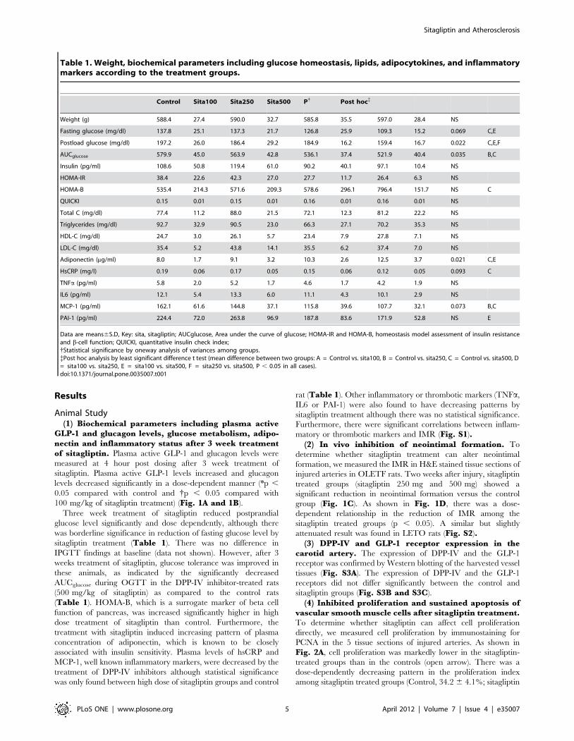
Table 1. Weight, biochemical parameters including glucose homeostasis, lipids, adipocytokines, and inflammatorymarkers according to the treatment groups.
Control Sita100 Sita250 Sita500 P{ Post hoc{
Weight (g) 588.4 27.4 590.0 32.7 585.8 35.5 597.0 28.4 NS
Fasting glucose (mg/dl) 137.8 25.1 137.3 21.7 126.8 25.9 109.3 15.2 0.069 C,E
Postload glucose (mg/dl) 197.2 26.0 186.4 29.2 184.9 16.2 159.4 16.7 0.022 C,E,F
AUCglucose 579.9 45.0 563.9 42.8 536.1 37.4 521.9 40.4 0.035 B,C
Insulin (pg/ml) 108.6 50.8 119.4 61.0 90.2 40.1 97.1 10.4 NS
HOMA-IR 38.4 22.6 42.3 27.0 27.7 11.7 26.4 6.3 NS
HOMA-B 535.4 214.3 571.6 209.3 578.6 296.1 796.4 151.7 NS C
QUICKI 0.15 0.01 0.15 0.01 0.16 0.01 0.16 0.01 NS
Total C (mg/dl) 77.4 11.2 88.0 21.5 72.1 12.3 81.2 22.2 NS
Triglycerides (mg/dl) 92.7 32.9 90.5 23.0 66.3 27.1 70.2 35.3 NS
HDL-C (mg/dl) 24.7 3.0 26.1 5.7 23.4 7.9 27.8 7.1 NS
LDL-C (mg/dl) 35.4 5.2 43.8 14.1 35.5 6.2 37.4 7.0 NS
Adiponectin (mg/ml) 8.0 1.7 9.1 3.2 10.3 2.6 12.5 3.7 0.021 C,E
HsCRP (mg/l) 0.19 0.06 0.17 0.05 0.15 0.06 0.12 0.05 0.093 C
TNFa (pg/ml) 5.8 2.0 5.2 1.7 4.6 1.7 4.2 1.9 NS
IL6 (pg/ml) 12.1 5.4 13.3 6.0 11.1 4.3 10.1 2.9 NS
MCP-1 (pg/ml) 162.1 61.6 144.8 37.1 115.8 39.6 107.7 32.1 0.073 B,C
PAI-1 (pg/ml) 224.4 72.0 263.8 96.9 187.8 83.6 171.9 52.8 NS E
Data are means6S.D, Key: sita, sitagliptin; AUCglucose, Area under the curve of glucose; HOMA-IR and HOMA-B, homeostasis model assessment of insulin resistanceand b-cell function; QUICKI, quantitative insulin check index;{Statistical significance by oneway analysis of variances among groups.{Post hoc analysis by least significant difference t test (mean difference between two groups: A = Control vs. sita100, B = Control vs. sita250, C = Control vs. sita500, D= sita100 vs. sita250, E = sita100 vs. sita500, F = sita250 vs. sita500, P , 0.05 in all cases).doi:10.1371/journal.pone.0035007.t001
Sitagliptin and Atherosclerosis
PLoS ONE | www.plosone.org 5 April 2012 | Volume 7 | Issue 4 | e35007

100 mg, 29.4 6 2.0%; sitagliptin 250 mg, 15.4 6 3.2%; sitagliptin
500 mg, 7.6 6 2.1%, P for trend , 0.01) (Fig. 2C).
To examine the effects of sitagliptin treatment on apoptosis
after balloon injury in vivo, we performed TUNEL staining in the
5 tissue sections of injured arteries (Fig. 2B). After three weeks
treatment of sitagliptin (at two weeks after balloon injury), the
apoptosis index was increased significantly in the sitagliptin
groups compared to the control group, which was in a dose-
dependent manner among sitagliptin treated groups (9.5 6 1.7%
in sitagliptin 100 mg, 18.4 6 2.9% in sitagliptin 250 mg and
22.4 6 2.2% in sitagliptin 500 mg compared to control, P for
trend , 0.01) (Fig. 2D). Using double staining for a-smooth
muscle actin and TUNEL, we identified the apoptotic cells as
smooth muscle cells; there were more vascular smooth muscle
cell apoptotic bodies in the sitagliptin treated group than in
control group (p , 0.01) (Fig. S4).
(5) Decrease in MMP2 and MMP9 expression insitagliptin treatment groups. To clarify whether proliferating
smooth muscle cells correspond to the cells expressing MMP2, we
performed double-staining with the anti-aSMA antibody (red) and
anti-MMP2 antibody (green) (Fig. 3A) or anti-MMP9 antibody
(green) (Fig. 3C) in injured arteries. Nucleus of cells was stained by
DAPI (blue). Fewer MMP2-expressing cells (Fig. 3B) and MMP9-
expressing cells (Fig. 3D) were observed in the sitagliptin treated-
groups than in the control groups, and the effects were dose-
dependent.
Figure 2. Effects of sitagliptin treatment or normal saline as a control on proliferation and apoptosis of vascular smooth musclecells. A. Cell proliferation measured by immunostaining for proliferating cell nuclear antigen (PCNA) was markedly lower in the sitagliptin-treatedgroups than in the controls (open arrow). B. TUNEL staining of the three groups (open arrow). C. The proliferation index was significantly lower in thesitagliptin-treated groups than in the control group. There was a dose-dependent pattern in the level of proliferation between sitagliptin-treatedgroups (*p , 0.05 vs. control and {p , 0.05 vs. sitagliptin 100 mg/kg). D. Apoptosis index (%) at 2 weeks after balloon injury. Apoptosis wassignificantly higher in the sitagliptin -treated groups than in the control group, and there was a dose-dependent pattern in the level of apoptosisbetween sitagliptin-treated groups (*p , 0.05 vs. control and {p , 0.05 vs. sitagliptin 100 mg/kg).doi:10.1371/journal.pone.0035007.g002
Sitagliptin and Atherosclerosis
PLoS ONE | www.plosone.org 6 April 2012 | Volume 7 | Issue 4 | e35007

Cell Study(1) Proliferation and migration of VSMCs and mono-
cyte adhesion in vitro after sitagliptin treatment. In MTT
assays, sitagliptin treatment inhibited PDGF-BB-induced cell
proliferation in RAoSMCs (Fig. 4A). This effect was initiated at
the dose of 50 mM and was increased dose dependently to be more
prominent at the dose of 200 mM. Similar finding was found in the
thymidine incorporation assay (Fig. S5). Sitagliptin treatment also
inhibited TNFa-directed migration of RAoSMCs in a dose-
dependent manner from 50 to 200 mg/mL (Fig. 4B, 4C). To test
whether sitagliptin can prevent TNFa-induced monocyte adhe-
sion, we performed the monocyte adhesion assay by using U937
cells. TNFa treatment significantly increased monocyte adhesion
in U937 cells, and these effects were blocked by treatment of
sitagliptin in a dose-dependent manner (Fig. 4D, 4E). This
Figure 3. Immunofluorescent double staining for MMP2 (A) and MMP9 (C) expressing cells in tissue sections of injured arteries.Double-staining was done with the anti-aSMA antibody (red) and anti-MMP2 or MMP9 antibody (green). Nucleus of cells was stained by DAPI (blue).MMP2 and MMP9 expressions were reduced by sitagliptin treatment (500 mg/kg) compared to control. Two scaled photos were displayed (6100 and6400). Effect of sitagliptin on MMP2 (B) and MMP9 (D) expression in tissue sections of injured arteries (*p , 0.01 compared with control and {p ,0.01 compared with 100 mg/kg of sitagliptin treatment).doi:10.1371/journal.pone.0035007.g003
Sitagliptin and Atherosclerosis
PLoS ONE | www.plosone.org 7 April 2012 | Volume 7 | Issue 4 | e35007

antiproliferative property of sitagliptin was not associated with
cytotoxicity, as shown by the calcein measurement and WST-8
assay (Fig. S6A and S6B).
(2) Change of caspase-3 activity after sitagliptintreatment. Apoptosis, a type of programmed cell death, is one
method of controlling immune responses such as cellular
homeostasis as well as a variety of physiological processes.
Caspases, a family of cysteine proteases, play a central role in
the apoptosis [24]. TNFa is secreted by VSMCs in the neointima
after a balloon injury as well as by macrophages in atherosclerotic
lesions [25]. Therefore, the effect of sitagliptin treatment on
caspase-3 activity was investigated in TNFa-stimulated VSMCs.
Sitagliptin treatment increased the level of caspase-3 activity in a
dose-dependent manner, reflecting apoptosis in RAoSMCs (Fig.S7).
(3) Effect of sitagliptin on MMP2 and MMP9expression in HUVECs and VSMCs. Increased proteolytic
activity in the vessel wall mediates the degradation of the ECM
surrounding smooth muscle cells in response to injury [26], a
necessary step to allow medial smooth muscle cells to migrate into
the intimal space. Matrix metalloproteinases (MMPs) such as
MMP2 (72 kDa) and MMP9 (92 kDa), have been implicated as
mediator of lesion development in response to vascular injury
[27,28]. The effect of sitagliptin on MMP2 and MMP9 expression
was examined in HUVECs and VSMCs. Expression of MMP2
and MMP9 decreased significantly in a dose-dependent manner
after treatment with sitagliptin compared with TNFa treatment (p
, 0.01) (Fig. S8 and Fig. S9).
(4) Effect of DPP-IV and CARD11 knockdown in VSMCs.DPP-IV was expressed in VSMCs and was knockdowned by
siDPP-IV (Fig. 5A). The expression of DPP-IV was not changed
by treatment of PDGF-BB or TNFa. FBS- or PDGF-BB-induced
cell proliferation was significantly attenuated by siDPP-IV
transfection (Fig. 5B). Attenuation of TNFa- or PDGF-BB-
induced NFkB activation by siDPP-IV transfection was also
confirmed in EMSA assay (Fig. 5C). TNFa-induced NFkB
activation was attenuated by sitagliptin treatment dose-depen-
dently (Fig. 5D). Expression of CARD11 was decreased dose-
dependently by sitagliptin treatment in VSMCs (Fig. 5E).
CARD11 was expressed in VSMCs and its expression was
knockdowned by siCard11 (Fig. 5F). TNFa-induced NFkB
activation was decreased by siCARD11 transfection in EMSA
assay (Fig. 5G).
Figure 4. Effects of sitagliptin on proliferation and migration of rat aortic smooth muscle cells (RAoSMC). A. In MTT viability assays, cellproliferation was significantly decreased by sitagliptin treatment (*p , 0.05 compared with PDGF-BB only). B. TNFa-directed migration withsitagliptin treatment. C. Dose-dependent inhibiting pattern of TNFa-directed migration from 50 to 200 mg/mL (*p , 0.05 compared with TNFatreatment). D. Effects of sitagliptin on TNFa-stimulated monocyte adhesion using U937 cells. Open arrows indicate adhered monocytes. E. Dose-dependent inhibiting pattern of TNFa-stimulated monocyte adhesion (*p , 0.05 compared with TNFa treatment).doi:10.1371/journal.pone.0035007.g004
Sitagliptin and Atherosclerosis
PLoS ONE | www.plosone.org 8 April 2012 | Volume 7 | Issue 4 | e35007

Discussion
The current study demonstrated that treatment with des-fluoro-
sitagliptin, a DPP-IV inhibitor, reduced restenosis in obese type 2
diabetic rats following balloon injury to the carotid artery. In vivo
and in vitro studies revealed that sitagliptin treatment suppressed
proliferation of VSMCs, promoted apoptosis and reduced
inflammatory process. Sitagliptin therapy also reduced hsCRP
and MCP-1 levels and increased adiponectin concentration,
indicating decrease of markers of inflammation and procoagula-
tion, and increase of insulin sensitivity. These findings suggest that
sitagliptin has protective role of development of macrovascular
complication, supporting an emerging role of sitagliptin in
modifying the elevated cardiovascular risks that are inherent in
obesity and type 2 diabetes.
Interestingly, it was recently demonstrated that GLP-1 amelio-
rates endothelial dysfunction in type 2 diabetes mellitus patients
with established coronary heart disease, suggesting a new
important cardioprotective role for GLP-1 [29,30]. In other study,
GLP-1 infusion significantly increased relative changes in brachial
artery diameter from baseline flow mediated dilatation (%) (3.1 6
0.6 vs. 6.6 6 1.0%, p , 0.05) in type 2 diabetes patients with
coronary artery disease [29], just as GLP-1 increased intracellular
cAMP (from 5.7 6 0.5 to 13.1 6 0.12 pmol/mg protein) in rat
cardiac myocytes [31]. Considering inhibition of DPP-IV has been
demonstrated to increase concentrations of intact GLP-1 2–3 fold
in patients with type 2 diabetes [3], it is conceivable that sitagliptin,
a potent and highly selective DPP-IV inhibitor, improves
endothelial dysfunction as a result.
In addition, it is well known that infiltration of inflammatory
cells occurs early after endothelial denudation [32–35]. Inhibition
of this process has been reported to be associated with a reduction
in medial VSMC proliferation [33]. Considering previous studies
showing linear relationship between tissue monocyte content and
neointimal area [35] and decreased neointimal thickening through
blocking early monocyte recruitment by anti-inflammatory drug
[36], inflammatory response related with monocyte infiltration
could be a causative factor in restenosis.
In the current study, sitagliptin treatment also reduced MMP2
and MMP9 expression in tissue sections in injured arteries. The
MMPs are a family of molecules that are associated with the
breakdown of constituents of the ECM. Both MMPs and their
tissue inhibitors are involved in the regulation of the ECM
metabolism [37]. ECM is a dynamic structure that requires
constant synthesis and degradation by MMPs [38]. Either MMP2
or MMP9 are synthesized and secreted locally in atherosclerotic
lesions, predominantly by monocyte-derived macrophages and
endothelial cells [39] and may participate in rupture of the
atherosclerotic plaque [40]. Type 2 diabetic patients are at high
risk for acute coronary events due to an increased propensity of
Figure 5. Mechanistic experiment with small interfering RNA (siRNA) against DPP-IV and CARD11. A. DPP-IV expression in VSMCs. TheDPP-IV expression was knockdowned by siDPP-IV while it was not changed by treatment of PDGF-BB or TNFa. B. FBS- or PDGF-BB-induced cellproliferation was significantly attenuated by siDPP-IV transfection. C. Attenuation of TNFa- or PDGF-BB-induced NFkB activation by siDPP-IVtransfection was also confirmed in EMSA assay. D. TNFa-induced NFkB activation was attenuated by sitagliptin treatment dose-dependently. E.Expression of CARD11 was decreased dose-dependently by sitagliptin treatment in VSMCs. F. CARD11 was expressed in VSMCs and its expression wasknockdowned by siCARD11. G. TNFa-induced NFkB activation was decreased by siCARD11 transfection in EMSA assay.doi:10.1371/journal.pone.0035007.g005
Sitagliptin and Atherosclerosis
PLoS ONE | www.plosone.org 9 April 2012 | Volume 7 | Issue 4 | e35007

their atherosclerotic plaques to ulceration and resultant overlying
thrombosis [41], and have increased level of MMP2 and MMP9
[14]. Thus, as shown in our study, the reduction of MMP2 and
MMP9 may be a possible mechanism in preventing macrovascular
complications in patients with type 2 diabetes by sitagliptin
treatment.
MCP-1, which is an important factor of monocyte recruitment,
has been shown to play a pivotal role in the development of
atherosclerosis and involves a sequence of events that include
monocyte attraction, tethering and rolling, and firm adhesion
[36,42]. Recently it has been described that blocking of the MCP-1
pathway results in reduced atherosclerosis and restenosis by
inhibition of monocyte adhesion to the vascular wall and to reduced
macrophage content in the atherosclerotic lesion [36]. In this study,
sitagliptin treatment significantly and dose-dependently reduced the
degree of monocyte adhesion and showed decreasing pattern of
serum concentration of MCP-1. These findings suggest another
mechanism by which sitagliptin has beneficial effect on counteract-
ing restenosis. In addition, such infiltrations of inflammatory cells
are known to promote an atherosclerotic milieu [32,43,44].
Whether sitagliptin has the ability to positively influence athero-
sclerotic plaque formation would warrant further investigation.
NFkB is a common regulator that involves in the control of
proinflammatory genes and VSMCs proliferation. It has been
reported that DPP-IV interacting with CARD11 or CARMA1
leads to NFkB activation in T-cells [20]. In the present study,
NFkB activation was reduced by DPP-IV or CARD11 Knock-
down, and attenuated dose-dependently by sitagliptin treatment in
VSMCs. Proliferation of VSMCs was inhibited by siDPP-IV
transfection and sitagliptin treatment. These results indicate that
gene expression of DPP-IV as well as enzyme activity of DPP-IV
plays a crucial role in NFkB activation. Thus, reduction of NFkB
activation by siDPP-IV and sitagliptin could be a major
mechanism in decrease of VSMCs proliferation.
Other possible relevant factors affecting the degree of
neointimal formation were considered in this study. Glucose
lowering effect by sitagliptin treatment might contribute to this
but sitagliptin were treated only for 3 weeks. Circulating levels
of adiponectin were increased significantly in sitagliptin
treatment in a dose-dependent manner. Low adiponectin level
is a risk factor for the subsequent development of cardiovascular
diseases [17,19]. Further, high dose of sitagliptin induced
decreasing pattern of hsCRP and PAI-1 activity in this study
although statistical significance was not obtained. This finding is
in line with a recent study describing that GLP-1 treatment
attenuated mRNA expression by TNFa and induction of PAI-1
protein [45]. In the latter study, GLP-1 also inhibited the effect
of TNFa on a reporter gene construct harboring the proximal
PAI-1 promoter.
In conclusion, this study, in addition to glucose lowering effects,
demonstrates that sitagliptin, a DPP-IV inhibitor, has protective
properties against restenosis after carotid injury in an animal
model of type 2 diabetes and vascular cell lines. These finding raise
the possibility that sitagliptin could offer a novel agent for the
treatment of macrovascular-related complications in patients with
type 2 diabetes.
Supporting Information
Figure S1 Correlations between intima-media ratio(IMR) and hsCRP, TNFa and MCP-1 levels and PAI-1activity. There were positive correlations between IMR and each
factor (p , 0.05 except IMR vs. PAI-1 activity).
(TIF)
Figure S2 In vivo inhibition of neointimal formationafter 3 weeks of treatment with des-fluoro-sitagliptin inLETO rats. A, H&E-stained sections of the control and
sitagliptin (500 mg/kg) groups. B, Intima-media ratios (IMRs) in
the two groups (n = 10 in each group). The IMR was calculated
from the mean areas of the intima and media. Treatment with
sitagliptin produced a lower IMR than in controls (p , 0.05
between the control and 500 mg/kg sitagliptin-treated groups).
(TIF)
Figure S3 A. Western blot of DPP-IV and GLP-1 receptorin the injured carotid arteries of control and sitagliptintreated rats. Representative three samples were displayed.
Quantification of Western blot images of DPP-IV (B) and GLP-1
receptor (C).
(TIF)
Figure S4 Double staining of a-smooth muscle actin(aSMA) and TUNEL in the injured carotid vessel wall.Apoptotic cells were smooth muscle cells; there were more
vascular smooth muscle cell apoptotic bodies in the sitagliptin
treated group (18.7%) than in the control group (5.2%) (p , 0.01)
(Arrows indicate vascular smooth muscle cell apoptotic bodies).
(TIF)
Figure S5 Thymidine incorporation assay to checkeffect of sitagliptin on FBS- or PDGF-induced cellproliferation. There were dose-dependent decreasing patterns
of thymidine uptake by sitagliptin treatment (*p , 0.05 compared
with FBS or PDGF-BB treatment only).
(TIF)
Figure S6 Effect of sitagliptin on cell survival. A. Calcein-
acetoxymethyl ester (calcein-AM) cell viability assay kit was used
(Biotium, Hayward, CA, USA). Cells were washed with PBS and
incubated with 2 mM calcein AM for 30 min. The fluorescence
was measured using 485 nm excitation wavelength and 530 nm
emission wavelength with a Victor 3 instrument (Perkin-Elmer,
Boston, MA, USA). B. Cell viability was also measured with Cell
Counting Kit-8 (CCK-8, Dojindo, Japan). Absorbance was
measured at 450 nm (VersaMax; Molecular Devices, Sunnyvale,
CA, USA). Cell Counting Kit-8 (CCK-8) allows convenient assays
by utilizing Dojindo’s highly water-soluble tetrazolium salt. WST-
8 [2-(2-methoxy-4-nitrophenyl)-3-(4-nitrophenyl)-5-(2,4-disulfo-
phenyl)-2H-tetrazolium, monosodium salt] produces a water-
soluble formazan dye upon reduction in the presence of an
electron carrier. WST-8 is reduced by dehydrogenases in cells to
give a yellow-colored product (formazan), which is soluble in the
tissue culture medium. The amount of the formazan dye
generated by the activity of dehydrogenases in cells is directly
proportional to the number of living cells.
(TIF)
Figure S7 Induction of apoptosis shown by the activa-tion of caspase-3 with sitagliptin treatment in VSMCs.There was a dose-dependent increasing pattern of caspase-3
activity (*p , 0.05 compared with TNFa treatment only).
(TIF)
Figure S8 Effects of sitagliptin on MMP2 and MMP9expression levels in human umbilical vein endothelialcells (A). Expressions of MMP2 (B) and MMP9 (C) decreased
significantly with the treatment of sitagliptin compared to TNFatreatment in a dose-dependent manner (*p , 0.05 compared with
TNFa treatment).
(TIF)
Sitagliptin and Atherosclerosis
PLoS ONE | www.plosone.org 10 April 2012 | Volume 7 | Issue 4 | e35007

Figure S9 Effects of sitagliptin on MMP2 and MMP9expression levels in vascular smooth muscle cells (A).Expressions of MMP2 (B) and MMP9 (C) decreased significantly
with the treatment of sitagliptin compared to TNFa treatment in a
dose-dependent manner (*p , 0.05 compared with TNFatreatment).
(TIF)
Acknowledgments
We thank the Otsuka Pharmaceuticals for donating OLETF rats.
Author Contributions
Conceived and designed the experiments: SL SMK JWY. Performed the
experiments: BJC BYA HSP SL. Analyzed the data: HS SHC YBK HCJ
KSP. Contributed reagents/materials/analysis tools: SMK JWY HS.
Wrote the paper: SL HS.
References
1. Buteau J, Foisy S, Joly E, Prentki M (2003) Glucagon-like peptide 1 inducespancreatic beta-cell proliferation via transactivation of the epidermal growth
factor receptor. Diabetes 52: 124–32.
2. Buteau J, El-Assaad W, Rhodes CJ, Rosenberg L, Joly E, et al. (2004) Glucagon-
like peptide-1 prevents beta cell glucolipotoxicity. Diabetologia 47: 806–15.
3. Drucker DJ (2003) Enhancing incretin action for the treatment of type 2diabetes. Diabetes Care 26: 2929–40.
4. Klonoff DC, Buse JB, Nielsen LL, Guan X, Bowlus CL, et al. (2008) Exenatideeffects on diabetes, obesity, cardiovascular risk factors and hepatic biomarkers in
patients with type 2 diabetes treated for at least 3 years. Curr Med Res Opin 24:275–86.
5. Lambeir AM, Durinx C, Scharpe S, De M, I (2003) Dipeptidyl-peptidase IV
from bench to bedside: an update on structural properties, functions, and clinical
aspects of the enzyme DPP IV. Crit Rev Clin Lab Sci 40: 209–94.
6. Holst JJ (2003) Implementation of GLP-1 based therapy of type 2 diabetesmellitus using DPP-IV inhibitors. Adv Exp Med Biol 524: 263–79.
7. Drucker DJ (2003) Therapeutic potential of dipeptidyl peptidase IV inhibitors
for the treatment of type 2 diabetes. Expert Opin Investig Drugs 12: 87–100.
8. Ristic S, Byiers S, Foley J, Holmes D (2005) Improved glycaemic control with
dipeptidyl peptidase-4 inhibition in patients with type 2 diabetes: vildagliptin(LAF237) dose response. Diabetes Obes Metab 7: 692–8.
9. Ahren B, Landin-Olsson M, Jansson PA, Svensson M, Holmes D, et al. (2004)
Inhibition of dipeptidyl peptidase-4 reduces glycemia, sustains insulin levels, and
reduces glucagon levels in type 2 diabetes. J Clin Endocrinol Metab 89:2078–84.
10. Mari A, Sallas WM, He YL, Watson C, Ligueros-Saylan M, et al. (2005)
Vildagliptin, a dipeptidyl peptidase-IV inhibitor, improves model-assessed beta-cell function in patients with type 2 diabetes. J Clin Endocrinol Metab 90:
4888–94.
11. Rodriguez AE, Maree AO, Mieres J, Berrocal D, Grinfeld L, et al. (2007) Late
loss of early benefit from drug-eluting stents when compared with bare-metalstents and coronary artery bypass surgery: 3 years follow-up of the ERACI III
registry. Eur Heart J 28: 2118–25.
12. Read PA, Khan FZ, Heck PM, Hoole SP, Dutka DP (2010) DPP-4 Inhibition by
Sitagliptin Improves the Myocardial Response to Dobutamine Stress andMitigates Stunning in a Pilot Study of Patients with Coronary Artery Disease.
Circ Cardiovasc Imaging.
13. Yoon JS, Lee HW (2011) Understanding the cardiovascular effects of incretin.Diabetes Metab J 35: 437–43.
14. Derosa G, D’Angelo A, Tinelli C, Devangelio E, Consoli A, et al. (2007)Evaluation of metalloproteinase 2 and 9 levels and their inhibitors in diabetic
and healthy subjects. Diabetes Metab 33: 129–34.
15. Marx N, Froehlich J, Siam L, Ittner J, Wierse G, et al. (2003) Antidiabetic PPARgamma-activator rosiglitazone reduces MMP-9 serum levels in type 2 diabetic
patients with coronary artery disease. Arterioscler Thromb Vasc Biol 23: 283–8.
16. Maxwell PR, Timms PM, Chandran S, Gordon D (2001) Peripheral blood level
alterations of TIMP-1, MMP-2 and MMP-9 in patients with type 1 diabetes.Diabet Med 18: 777–80.
17. Schulze MB, Shai I, Rimm EB, Li T, Rifai N, et al. (2005) Adiponectin and
future coronary heart disease events among men with type 2 diabetes. Diabetes
54: 534–9.
18. Song HK, Lee MH, Kim BK, Park YG, Ko GJ, et al. (2008) Visfatin: a newplayer in mesangial cell physiology and diabetic nephropathy. Am J Physiol
Renal Physiol 295: F1485–F1494.
19. Pischon T, Girman CJ, Hotamisligil GS, Rifai N, Hu FB, et al. (2004) Plasma
adiponectin levels and risk of myocardial infarction in men. JAMA 291: 1730–7.
20. Ohnuma K, Uchiyama M, Yamochi T, Nishibashi K, Hosono O, et al. (2007)Caveolin-1 triggers T-cell activation via CD26 in association with CARMA1.
J Biol Chem 282: 10117–31.
21. Yu Y, Ohmori K, Chen Y, Sato C, Kiyomoto H, et al. (2004) Effects of
pravastatin on progression of glucose intolerance and cardiovascular remodelingin a type II diabetes model. J Am Coll Cardiol 44: 904–13.
22. Clowes AW, Reidy MA, Clowes MM (1983) Kinetics of cellular proliferation
after arterial injury. I. Smooth muscle growth in the absence of endothelium.Lab Invest 49: 327–33.
23. Ansari B, Coates PJ, Greenstein BD, Hall PA (1993) In situ end-labelling detectsDNA strand breaks in apoptosis and other physiological and pathological states.
J Pathol 170: 1–8.24. Lavrik IN, Golks A, Krammer PH (2005) Caspases: pharmacological
manipulation of cell death. J Clin Invest 115: 2665–72.
25. Tanaka H, Sukhova G, Schwartz D, Libby P (1996) Proliferating arterial smoothmuscle cells after balloon injury express TNF-alpha but not interleukin-1 or
basic fibroblast growth factor. Arterioscler Thromb Vasc Biol 16: 12–8.26. Newby AC, Zaltsman AB (2000) Molecular mechanisms in intimal hyperplasia.
J Pathol 190: 300–9.27. Bendeck MP, Zempo N, Clowes AW, Galardy RE, Reidy MA (1994) Smooth
muscle cell migration and matrix metalloproteinase expression after arterial
injury in the rat. Circ Res 75: 539–45.28. Galis ZS, Muszynski M, Sukhova GK, Simon-Morrissey E, Unemori EN, et al.
(1994) Cytokine-stimulated human vascular smooth muscle cells synthesize acomplement of enzymes required for extracellular matrix digestion. Circ Res 75:
181–9.
29. Nystrom T, Gutniak MK, Zhang Q, Zhang F, Holst JJ, et al. (2004) Effects ofglucagon-like peptide-1 on endothelial function in type 2 diabetes patients with
stable coronary artery disease. Am J Physiol Endocrinol Metab 287:E1209–E1215.
30. Nystrom T, Gonon AT, Sjoholm A, Pernow J (2005) Glucagon-like peptide-1relaxes rat conduit arteries via an endothelium-independent mechanism. Regul
Pept 125: 173–7.
31. Vila Petroff MG, Egan JM, Wang X, Sollott SJ (2001) Glucagon-like peptide-1increases cAMP but fails to augment contraction in adult rat cardiac myocytes.
Circ Res 89: 445–52.32. Moreno PR, Bernardi VH, Lopez-Cuellar J, Newell JB, McMellon C, et al.
(1996) Macrophage infiltration predicts restenosis after coronary intervention in
patients with unstable angina. Circulation 94: 3098–102.33. Welt FG, Edelman ER, Simon DI, Rogers C (2000) Neutrophil, not
macrophage, infiltration precedes neointimal thickening in balloon-injuredarteries. Arterioscler Thromb Vasc Biol 20: 2553–8.
34. Moreno PR, Fallon JT, Murcia AM, Leon MN, Simosa H, et al. (1999) Tissue
characteristics of restenosis after percutaneous transluminal coronary angioplas-ty in diabetic patients. J Am Coll Cardiol 34: 1045–9.
35. Rogers C, Welt FG, Karnovsky MJ, Edelman ER (1996) Monocyte recruitmentand neointimal hyperplasia in rabbits. Coupled inhibitory effects of heparin.
Arterioscler Thromb Vasc Biol 16: 1312–8.36. Mori E, Komori K, Yamaoka T, Tanii M, Kataoka C, et al. (2002) Essential role
of monocyte chemoattractant protein-1 in development of restenotic changes
(neointimal hyperplasia and constrictive remodeling) after balloon angioplasty inhypercholesterolemic rabbits. Circulation 105: 2905–10.
37. Li-Saw-Hee FL, Edmunds E, Blann AD, Beevers DG, Lip GY (2000) Matrixmetalloproteinase-9 and tissue inhibitor metalloproteinase-1 levels in essential
hypertension. Relationship to left ventricular mass and anti-hypertensive
therapy. Int J Cardiol 75: 43–7.38. Nagase H, Woessner JF, Jr. (1999) Matrix metalloproteinases. J Biol Chem 274:
21491–4.39. Falk E, Shah PK, Fuster V (1995) Coronary plaque disruption. Circulation 92:
657–71.40. Galis ZS, Sukhova GK, Lark MW, Libby P (1994) Increased expression of
matrix metalloproteinases and matrix degrading activity in vulnerable regions of
human atherosclerotic plaques. J Clin Invest 94: 2493–503.41. Cooper ME, Bonnet F, Oldfield M, Jandeleit-Dahm K (2001) Mechanisms of
diabetic vasculopathy: an overview. Am J Hypertens 14: 475–86.42. Egashira K, Zhao Q, Kataoka C, Ohtani K, Usui M, et al. (2002) Importance of
monocyte chemoattractant protein-1 pathway in neointimal hyperplasia after
periarterial injury in mice and monkeys. Circ Res 90: 1167–72.43. Pasceri V, Willerson JT, Yeh ET (2000) Direct proinflammatory effect of C-
reactive protein on human endothelial cells. Circulation 102: 2165–8.44. Ferns GA, Avades TY (2000) The mechanisms of coronary restenosis: insights
from experimental models. Int J Exp Pathol 81: 63–88.45. Liu H, Hu Y, Simpson RW, Dear AE (2008) Glucagon-like peptide-1 attenuates
tumour necrosis factor-alpha-mediated induction of plasmogen activator
inhibitor-1 expression. J Endocrinol 196: 57–65.
Sitagliptin and Atherosclerosis
PLoS ONE | www.plosone.org 11 April 2012 | Volume 7 | Issue 4 | e35007


![Molecular Imaging of Murine Intestinal Inflammation With 2-Deoxy-2-[ 18F]Fluoro- d-Glucose and Positron Emission Tomography](https://static.fdokumen.com/doc/165x107/6344fff26cfb3d4064097a1a/molecular-imaging-of-murine-intestinal-inflammation-with-2-deoxy-2-18ffluoro-.jpg)