Solution structure of an arabinonucleic acid (ANA)/RNA duplex in a chimeric hairpin: comparison with...
10
4284–4293 Nucleic Acids Research, 2001, Vol. 29, No. 21 © 2001 Oxford University Press Solution structure of an arabinonucleic acid (ANA)/RNA duplex in a chimeric hairpin: comparison with 2′-fluoro-ANA/RNA and DNA/RNA hybrids Alexei Yu. Denisov 1 , Anne M. Noronha 2 , Christopher J. Wilds 2 , Jean-François Trempe 1 , Richard T. Pon 3 , Kalle Gehring 1,2 and Masad J. Damha 2, * 1 Department of Biochemistry and Montreal Joint Centre for Structural Biology, McGill University, Montreal, QC H3G 1Y6, Canada, 2 Department of Chemistry, Otto Maas Chemistry Building, McGill University, 801 Sherbrooke Street West, Montreal, QC H3A 2K6, Canada and 3 University Core DNA Services, University of Calgary, Calgary, AB T2N 4N1, Canada Received July 26, 2001; Revised and Accepted September 17, 2001 ABSTRACT Hybrids of RNA and arabinonucleic acid (ANA) as well as the 2′-fluoro-ANA analog (2′F-ANA) were recently shown to be substrates of the enzyme RNase H. Although RNase H binds to double- stranded RNA, no cleavage occurs with such duplexes. Therefore, knowledge of the structure of ANA/RNA hybrids may prove helpful in the design of future antisense oligonucleotide analogs. In this study, we have determined the NMR solution struc- tures of ANA/RNA and DNA/RNA hairpin duplexes and compared them to the recently published struc- ture of a 2′F-ANA/RNA hairpin duplex. We demon- strate here that the sugars of RNA nucleotides of the ANA/RNA hairpin stem adopt the C3′ -endo (north, A-form) conformation, whereas those of the ANA strand adopt a ‘rigid’ O4′-endo (east) sugar pucker. The DNA strand of the DNA/RNA hairpin stem is flexible, but the average DNA/RNA hairpin structural parameters are close to the ANA/RNA and 2′F-ANA/ RNA hairpin parameters. The minor groove width of ANA/RNA, 2′F-ANA/RNA and DNA/RNA helices is 9.0 ± 0.5 Å, a value that is intermediate between that of A- and B-form duplexes. These results rationalize the ability of ANA/RNA and 2′F-ANA/RNA hybrids to elicit RNase H activity. INTRODUCTION Over the past few years, synthetically modified oligodeoxy- nucleotides (ODN) have been widely used for artificially regu- lating gene expression through the targeting of RNA (1,2). Substitutions at the 2′-position of ribose yield nuclease- resistant RNA analogs with increased binding affinity towards target RNAs (3). This is because alkylation of the ribose 2′-position generally leads to structures that are conformationally biased or conformationally locked in the north or C3′-endo sugar pucker (3,4). However, preorganization of the ODN into a pure north conformation (A-DNA-like) produces an ODN/RNA duplex not generally recognized by RNase H, a critical step in the mechanism of action of antisense compounds (5). Such a lack of recognition has been generally solved by the use of the so-called ‘gapmer’ technology, where 2′-deoxynucleotide ‘gaps’ are flanked at either end of the ODN with north-biased nucleotide units (e.g. RNA-DNA-RNA chimeras) (6–8). With this modifi- cation, the ‘gap’ of the resulting chimeric duplex has a hybrid- like conformation which is then efficiently recognized by the enzyme RNase H. Recently, we showed that the stereochemistry at the 2′-position of the sugar of antisense oligonucleotides is a key determinant in the target RNA binding affinity and the activation of RNase H (9,10). We demonstrated that both arabinonucleic acid (ANA) and the corresponding 2′-fluoro-arabinonucleic acid analog (2′F-ANA; Fig. 1) form hybrids with RNA, and that they are able to induce RNase H degradation of the target RNA. Since RNA/RNA duplexes are not substrates of RNase H (11), these findings provided an important advancement towards understanding the catalytic mechanism and substrate selectivity of RNase H. Relatively little is known about the structure of duplexes with ANA modifications. Previous studies have been mostly restricted to DNA duplexes containing one or two neighboring ANA or 2′F-ANA residues (12–20). X-ray crystallographic data revealed the adoption of an O4′-endo (east) sugar pucker by 2′F-ANA residues (20), a conformation that lies halfway between the C2′-endo (south, B-DNA) and C3′-endo (north, A-RNA) puckers (Fig. 2). This is consistent with the results of more recent molecular dynamics (MD) calculations of the conformation of 2′F-ANA-containing duplexes (21). Modeling studies of two other ANA analogs, namely [3.3.0]-bicyclo-ANA (bc-ANA) and [3.2.0]-bicyclo-ANA were also suggestive of the adoption of east-type (O4′-endo) sugar puckers (22). Recently, Egli and co-workers (23) crystallized a DNA dodecamer duplex containing bc-ANA modifications and *To whom correspondence should be addressed. Tel: +1 514 398 7552; Fax: +1 514 398 3797; Email: [email protected]
-
Upload
independent -
Category
Documents
-
view
2 -
download
0
Transcript of Solution structure of an arabinonucleic acid (ANA)/RNA duplex in a chimeric hairpin: comparison with...

4284–4293 Nucleic Acids Research, 2001, Vol. 29, No. 21 © 2001 Oxford University Press
Solution structure of an arabinonucleic acid(ANA)/RNA duplex in a chimeric hairpin:comparison with 2′-fluoro-ANA/RNA and DNA/RNAhybridsAlexei Yu. Denisov1, Anne M. Noronha2, Christopher J. Wilds2, Jean-François Trempe1,Richard T. Pon3, Kalle Gehring1,2 and Masad J. Damha2,*
1Department of Biochemistry and Montreal Joint Centre for Structural Biology, McGill University, Montreal,QC H3G 1Y6, Canada, 2Department of Chemistry, Otto Maas Chemistry Building, McGill University, 801 SherbrookeStreet West, Montreal, QC H3A 2K6, Canada and 3University Core DNA Services, University of Calgary, Calgary,AB T2N 4N1, Canada
Received July 26, 2001; Revised and Accepted September 17, 2001
ABSTRACT
Hybrids of RNA and arabinonucleic acid (ANA) aswell as the 2′-fluoro-ANA analog (2′F-ANA) wererecently shown to be substrates of the enzymeRNase H. Although RNase H binds to double-stranded RNA, no cleavage occurs with suchduplexes. Therefore, knowledge of the structure ofANA/RNA hybrids may prove helpful in the design offuture antisense oligonucleotide analogs. In thisstudy, we have determined the NMR solution struc-tures of ANA/RNA and DNA/RNA hairpin duplexesand compared them to the recently published struc-ture of a 2′F-ANA/RNA hairpin duplex. We demon-strate here that the sugars of RNA nucleotides of theANA/RNA hairpin stem adopt the C3′-endo (north,A-form) conformation, whereas those of the ANAstrand adopt a ‘rigid’ O4′-endo (east) sugar pucker.The DNA strand of the DNA/RNA hairpin stem isflexible, but the average DNA/RNA hairpin structuralparameters are close to the ANA/RNA and 2′F-ANA/RNA hairpin parameters. The minor groove width ofANA/RNA, 2′F-ANA/RNA and DNA/RNA helices is9.0 ± 0.5 Å, a value that is intermediate between thatof A- and B-form duplexes. These results rationalizethe ability of ANA/RNA and 2′F-ANA/RNA hybrids toelicit RNase H activity.
INTRODUCTION
Over the past few years, synthetically modified oligodeoxy-nucleotides (ODN) have been widely used for artificially regu-lating gene expression through the targeting of RNA (1,2).Substitutions at the 2′-position of ribose yield nuclease-resistant RNA analogs with increased binding affinity towardstarget RNAs (3). This is because alkylation of the ribose2′-position generally leads to structures that are conformationally
biased or conformationally locked in the north or C3′-endosugar pucker (3,4). However, preorganization of the ODN intoa pure north conformation (A-DNA-like) produces an ODN/RNAduplex not generally recognized by RNase H, a critical step inthe mechanism of action of antisense compounds (5). Such alack of recognition has been generally solved by the use of theso-called ‘gapmer’ technology, where 2′-deoxynucleotide ‘gaps’are flanked at either end of the ODN with north-biased nucleotideunits (e.g. RNA-DNA-RNA chimeras) (6–8). With this modifi-cation, the ‘gap’ of the resulting chimeric duplex has a hybrid-like conformation which is then efficiently recognized by theenzyme RNase H.
Recently, we showed that the stereochemistry at the 2′-positionof the sugar of antisense oligonucleotides is a key determinantin the target RNA binding affinity and the activation of RNaseH (9,10). We demonstrated that both arabinonucleic acid(ANA) and the corresponding 2′-fluoro-arabinonucleic acidanalog (2′F-ANA; Fig. 1) form hybrids with RNA, and thatthey are able to induce RNase H degradation of the targetRNA. Since RNA/RNA duplexes are not substrates of RNaseH (11), these findings provided an important advancementtowards understanding the catalytic mechanism and substrateselectivity of RNase H.
Relatively little is known about the structure of duplexeswith ANA modifications. Previous studies have been mostlyrestricted to DNA duplexes containing one or two neighboringANA or 2′F-ANA residues (12–20). X-ray crystallographicdata revealed the adoption of an O4′-endo (east) sugar puckerby 2′F-ANA residues (20), a conformation that lies halfwaybetween the C2′-endo (south, B-DNA) and C3′-endo (north,A-RNA) puckers (Fig. 2). This is consistent with the results ofmore recent molecular dynamics (MD) calculations of theconformation of 2′F-ANA-containing duplexes (21). Modelingstudies of two other ANA analogs, namely [3.3.0]-bicyclo-ANA(bc-ANA) and [3.2.0]-bicyclo-ANA were also suggestive ofthe adoption of east-type (O4′-endo) sugar puckers (22).Recently, Egli and co-workers (23) crystallized a DNAdodecamer duplex containing bc-ANA modifications and
*To whom correspondence should be addressed. Tel: +1 514 398 7552; Fax: +1 514 398 3797; Email: [email protected]

Nucleic Acids Research, 2001, Vol. 29, No. 21 4285
found that the bc-ANA residues display sugar puckers that falluniformly within the O4′-endo range. They also concluded thatsteric constraints within the bicyclic ANA framework prohibitedadoption of either the C2′-endo or C3′-endo conformations.Our recently published NMR structure of a 2′F-ANA/RNAhairpin duplex also revealed an O4′-endo pucker for the 2′F-ANAresidues (24). This is in contrast to NMR and MD studies ofmonomeric nucleoside units (e.g. araF-A,T), which identifiedthe C2′-endo/C1′-exo puckers as the most stable conformations(25,26).
While 2′-fluoroarabino and bc-arabinonucleotides exhibit apreference for the east pucker, arabinonucleotides (β-2′-OH)adopt a variety of conformations. The actual conformation
appears to depend on the environment (crystal, solution) andother factors. For example, computational simulations haveshown that the arabinose (2′OH) sugars of ANA/RNA hybridsprefer the south C2′-endo pucker due to an intranucleotideO2′-H···O5′ hydrogen bond (21). This feature is also observedin the crystal structure of arabinocytidine (ara-C) (27). Crystalstructures of Z-DNA duplexes that contain ANA residues alsoshow a preference for the south conformational rangeC2′-endo/C3′-exo (17,18). Other studies on duplex structurescontaining ANA residues have detected the south C2′-endo(12,15,16), C1′-exo (13) and north C4′-exo (14) conformations(Fig. 2). Conformational variations have also been noted at themonomeric nucleoside level (27–30).
To date, no NMR structures of ANA (2′OH)/RNA hybridshave been reported, although the conformation of such hybridshas been predicted on the basis of molecular modeling studies(21). This fact, combined with the knowledge that ANA/RNAhybrids are thermodynamically less stable than DNA/RNA(10,31) and 2′F-ANA/RNA hybrids (9), prompted us to carryout an NMR-based structure study of a chimeric hairpincontaining an ANA/RNA stem (Fig. 1). The results suggeststriking similarities in the global and local structure of ANA/RNA, 2′F-ANA/RNA and DNA/RNA hybrids.
MATERIALS AND METHODS
Sample preparation
Chimeric hairpin oligonucleotides were synthesized asdescribed previously (9,10,31). The samples at 1 mM concen-tration were dissolved in 0.4 ml of 100% D2O or 9:1 H2O/D2O(v/v) (for imino-proton spectra). The solution contains 0.1 mMEDTA sodium salt, and the pH was adjusted to 7.0 by addingNaOH.
The hairpin melting temperatures (Tm) determined at concen-trations of ∼1 OD/ml were confirmed qualitatively byrecording proton NMR spectra at different temperatures. Asobserved for unimolecular transitions in similar all-RNA orall-DNA hairpin studies (32,33), the Tm values were inde-pendent of oligonucleotide concentration. The Tm values forthe ANA/RNA hairpin (1) and for the DNA/RNA hairpin (2)are 44 and 60°C, respectively. These are less than the Tm of67°C for the previously studied 2′F-ANA/RNA hairpin duplex(3) (24).
NMR spectroscopy
The NMR spectra were recorded on a Bruker DRX-500spectrometer equipped with a 1H/13C/31P triple resonance (x, y, z)gradient probe operating at 500.13 MHz of proton resonance.The proton chemical shifts were measured relative to internalDSS, and the phosphorus resonances were indirectly refer-enced to 85% H3PO4 by multiplying the DSS proton resonanceabsolute frequency by 0.404807356 (24). All 2D spectra werecollected in the phase-sensitive mode via the TPPI method andpresaturation. The 3-9-19 pulse sequence (34) was used tosuppress the water signal for samples in 9:1 H2O/D2O.
NOESY experiments in D2O were performed at 7 and 15°C[hairpin (1)] and at 20°C [hairpin (2)] using mixing times tm of50, 70, 200 and 400 ms. The final spectral size was 2K × 2Kpoints after Fourier transformation. The volumes of crosspeaksof NOESY spectra were calculated using XWINNMR
Figure 1. Chemical structures of the hairpins. The first four RNA nucleotides,r(GGAC), are base paired to the last four ANA, DNA or 2′F-ANA nucleotides.The loop, d(TTCG), is of constant base and sugar composition.
Figure 2. Schematic of the pseudorotation phase angle (P) cycle with thepositions of selected pucker types indicated. The P angles of arabino-residuesin average structure of ANA/RNA hairpin (1) (+) are compared with the Pangles of arabinonucleotides in X-ray and NMR structures of B- and Z-DNAduplexes (*) (12–18). In the O4′-endo conformation, the 2′-OH of arabinose ispseudoaxial and gauche to the ring oxygen O4′.

4286 Nucleic Acids Research, 2001, Vol. 29, No. 21
(Bruker). NOESY experiments in 9:1 H2O/D2O (v/v) wereperformed at 15°C with a mixing time of 180 ms. DQF-COSYspectra were collected with and without phosphorus decoupling(final data size of 8K × 2K points). Proton MLEV-17 TOCSYexperiments were performed with a mixing time of 84 ms.H,C-correlation HMQC spectra were recorded using GARPheteronuclear decoupling (1JCH = 180 Hz). Inverse(H,P)-HetCOSY and HetTOCSY spectra were collected withfinal spectral sizes of 2K × 1K data points. In 2D NMRexperiments, relaxation delays were 1.5–2.5 s, the number of t1experiments was 256 or 512, and the number of scans was32–128 for each FID. Correlation times (τc = 2.0 ns) weredetermined using the H5-H6 NOE of deoxyribo, ribo and arabino-cytidine residues of the various hairpins as reference.
Structural modeling
The starting coordinates of the hairpins were generated usingSybyl 6.5 software (Tripos Inc.) from both A- and B-type DNAstructures. The X-PLOR 3.843 package (35) with standardnucleic acid force field (parallhdg.dna) was used for hairpinmolecular modeling. Partial charges for arabinonucleotideswere set to be the same as for ribonucleotides.
At the first stage of refinement, 10 ‘randomized’ structures(five with canonical A-type and five with B-type of hairpinstem) were generated by 1–2 ps MD at 300 K without experi-mental constraints. Subsequent stages of simulated annealingwith NOE-distance and torsion angle constraints were similarto those described previously for hairpins (32,33,36,37). TheX-PLOR refinement involved an initial energy minimizationand MD simulation of 14 ps (2 ps of heating from 300 to 1000 K,5 ps at 1000 K, 4 ps of cooling back to 300 K and then 3 ps at300 K). A distance-dependent dielectric constant was used tomimic the solvent. During the last 2 ps, the atomic coordinateswere saved at 200 fs interval, and 10 conformers collected.These conformers were then averaged and energy minimized.Atomic coordinates of the hairpin structures have been depositedin the PDB under accession numbers 1HO6 for the ANA/RNAhairpin (1) and 1HOQ for the DNA/RNA hairpin (2).
Distance restraints were derived from NOESY spectra atdifferent mixing times by crosspeak volume integration, usingthe r–6 distance relationship (38) and average crosspeak volumevalues for calibration for H5-H6 in cytidines (r = 2.46 Å) and forMe-H6 in thymidines (r = 2.70 Å). The distance constraints inthe hairpin stems were given 10% of lower and 15% of upperbounds. Due to loop mobility, the NOEs for loops were classi-fied as strong, medium, weak and absent with correspondingdistance ranges of 1.8–3.2, 2.0–5.0, 3.0–7.0 and >4.0 Å (33).No constraints were applied for loop NOEs involvingexchangeable protons. Sugar puckering was determined by thePSEUROT 3B program (39) from vicinal JHH couplingconstants. The five ν0–ν4 torsion angles for hairpin sugars wereconstrained (with ±10° bounds) according to sugar conforma-tions determined from J-couplings; in the case of stem DNAnucleotides [hairpin (2)] torsion angles were constrainedaccording to intrasugar NOE distances. Backbone torsionangle constraints were set as described in the text. Duringrefinement, the NOE distance and hydrogen bond forceconstants were gradually built up to final values of 30–40 kcal/(mol Å2) and the backbone torsion angle constants to 60 kcal/(mol rad2). Calculation of helical parameters was carried outwith CURVES 5.2 program (40).
RESULTS
Resonance assignments
The assignment of oligonucleotide non-exchangable protonresonances in NOESY spectra of hairpins (1) and (2) wascarried out using the H1′(i)-H6/H8(i)-H1′(i-1) pathway in thestandard manner employed for right-handed DNA duplexes(Fig. 3A) (38). The DQF-COSY, TOCSY and (H,C)-HMQCspectra based on different 13C chemical shifts for C2′/C3′, C4′and C5′ were also used to assign the sugar protons. All assign-ments were further corroborated by the H6/H8(i)-H2′, H2″, H3′,H4′(i,i-1) pathways. The adenine H2 resonances were identifiedby inversion–recovery experiments and confirmed by NOESYintranucleotide H2-H1′ and interstrand H2(A3)-H1′(C11)crosspeaks. It should be mentioned that some sugar crosspeaksfor the dG9 nucleotide in hairpin (2) were broadened at roomtemperature. The assignment of stem imino-protons was madefrom NOESY spectra in H2O/D2O using the crosspeaks ofNH(G) with NH2(C) or NH(T,U10) with H2(A3) of comple-mentary base pairs. Signals of NH2(C) were easily identifiedfrom their strong crosspeaks with H5(C) of the same nucleotide.Phosphorus signals were determined from strong H3′-P cross-peaks in (H,P)-HetCOSY spectra.
Proton and phosphorus chemical shifts of harpins (1) and (2)are provided in the Supplementary Material. The proton chemicalshifts for loop nucleotides are close to the all-DNA (33) or2′F-ANA/RNA (24) hairpins, both having flexible loopconformations. The set of sequential NOE contacts betweenloop residues C4, T5 and T6 was complicated by medium–weak H2′(T5)-H5(C7) crosspeaks in both hairpins (1) and (2).No strong or medium sequential NOEs were found between T6and C7, or between C7 and G8. As a result, we were unable tobuild a single loop conformation.
Sugar ring conformation
Sugar conformations were determined from 3JHH data obtainedfrom DQF-COSY experiments (Fig. 3D and Table 1).Coupling constants J1′2′ and J1′2′′ were determined from H1′-H2″ or H1′-H2′ crosspeak splitting, whereas J2′3′, J2′′3′ and J3′4′were extracted from sums of coupling constants as previouslydescribed (38,41,42). The phase angles of pseudorotation (P)and mole fractions of conformers (f) were determined using thePSEUROT program (39) with fixed puckering amplitude of37°. The ranges for pseudorotation parameters were calculatedfrom the uncertainty of measured values of J-couplings andare given in Table 1.
The data show that, in both hybrids, the ribose sugars adoptthe north (mostly C3′-endo) pucker, whereas the arabino-nucleotide (2′OH) residues prefer the east (mostly O4′-endo)sugar pucker (Fig. 2). We conclude that ANA and 2′F-ANAadopt the same pucker when hybridized to complementaryRNA (24). Consistent with the O4′-endo conformation is thevery strong H1′-H4′ NOE crosspeaks of the arabinose residues(d = 2.5 Å) (Fig. 3B). Only the sugar moiety of terminal arabino-nucleotide residue (C12) exists as a 1:1 mixture of east andprobably north conformers. The coupling constant PSEUROTand H1′-H4′ NOE crosspeak analysis for sugar conformationsin the DNA loops revealed a mostly north pucker for T5 loopresidue probably stacked with neighboring C4 ribonucleotide(33) and south puckers for the remaining loop nucleotides.

Nucleic Acids Research, 2001, Vol. 29, No. 21 4287
Nevertheless, the PSEUROT analysis of sugar puckering forG9-C12 DNA nucleotides in DNA/RNA hairpin is at variancewith measured values of H1′-H4′ NOE distances (d ≈ 2.8 Å)due to the presence of multiple conformational states for stemDNA sugars (see Discussion).
Backbone β, γ , ε and glycosidic χ torsion angles
The β torsion angles were constrained using the informationabout H5′/H5″-P and H4′-P crosspeaks in H,P-HetCOSYspectra. Except for G9 nucleotides, the β angles were found tobe in the trans conformation (180 ± 45°) as determined by thesymmetry and low intensity of the H5′/H5″-P crosspeaks anddetectable 4JH4′-P W-pathway coupling constants (29,38,43). Inthe case of G9 [hairpins (1) and (2)] very strong coupling wasdetected between the 5′-phosphate and one of the 5′-protons,and therefore we did not constrain the value of β(G9).
The γ angles were constrained using the sums of JH4′H5′ andJH4′H5′′, which were available from phosphorus decoupled
DQF-COSY spectra (Table 1), and the NOE H1′/H6/H8-H4′crosspeak linewidths (43). The sums of these couplingconstants in most nucleotides were <5 Hz, which is consistentwith a γ angle range of 60 ± 20°. For T6, C7, G9 and araC11nucleotides the linewidths of H4′ lie in the range 10–14 Hzand, hence, in these cases, the range of γ angles can beconstrained to 60 ± 40°. For G8 nucleotide, the sum of JH4′H5′and JH4′H5′′ coupling constants (8 Hz) and large H4′ linewidths(>15 Hz) did not allow constraint of the γ(G8) angle in the flexibleloop of both hairpins (1) and (2).
The ε angles have been estimated from the vicinal 3JH3′-Pcoupling constants (Table 1). These values of couplingconstants were determined from comparison of DQF-COSYcrosspeak splittings in spectra with and without phosphorusdecoupling. The 3JH3′-P coupling constants lie in the range of8–10 Hz for nucleotides in RNA strands and ε angles wereconstrained to 240 ± 50° from the Karplus equation (43) inaccordance with data for DNA/RNA duplexes (44–48). In
Figure 3. Expanded plots of NOESY spectra at 500 MHz. (A) ANA/RNA hairpin (1), 7°C, mixing time tm = 200 ms, the assignment of oligonucleotide protonsare shown by solid lines and nucleotide name with number; I-IV-H5-H6(C4,7,11,12) crosspeaks, respectively. (B and C) ANA/RNA hairpin (1), 7°C, and DNA/RNA hairpin (2), 20°C, tm = 50 ms in both cases, H1′-H4′ crosspeaks are labeled by nucleotide name with number, H1′-H2′ (RNA) crosspeaks (*) and H1′-H2″(ANA) crosspeaks (**). (D) Expanded plots of DQF-COSY spectrum of ANA/RNA hairpin (1) at 500 MHz, 15°C, H1′-H2″ (ANA) crosspeaks are marked bynucleotide name with number.

4288 Nucleic Acids Research, 2001, Vol. 29, No. 21
contrast, the small 3JH3′-P values observed for the stem arabinoand deoxyribonucleotides (3–4 Hz) suggest an ε value of 170 ±50°. The ε angles for the flexible loop nucleotides wereconstrained in the range of both BI- and BII-type phosphateconformations, i.e. 240 ± 120° (49).
Glycosidic χ angles were not constrained in both hairpins (1)and (2); rather, they were fixed indirectly by intranucleotidearomatic–sugar distance constraints. Most nucleotides are inanti-conformation except for G8 nucleotides where NOESYcrosspeak volumes correspond to syn/anti conformationalexchange with an H8-H1′ NOE distance of 2.7–2.8 Å.Aromatic–sugar intranucleotide distance constraints for G8were omitted during structure refinement calculations.
Structure refinement and analysis
In order to determine hairpin structures that are consistent withthe experimental data, NMR-restrained MD calculations wereperformed. The structural statistics of X-PLOR refinement arepresented in Table 2, and superimposition of final individualstructures for ANA/RNA hairpin (1) is shown in Figure 4A.Ten A- and B-type starting structures (an overall r.m.s.d. of 1.8 Åfor stem heavy atoms) with randomly organized loop structures
for both hairpins (1) and (2) were refined by X-PLOR tor.m.s.d. of 0.3–0.8 Å for stem nucleotides. As expected from2′F-ANA/RNA (24) and all-DNA hairpin (33) studies, theloops in both hairpins are flexible and do not significantlyinfluence stem structures. The stacking between stem C4 andloop T5 bases was confirmed in both hairpin structures leadingto a smaller r.m.s.d. for T5 (1.1–1.7 Å) in comparison withother loop nucleotides (up to 4.4 Å).
The final individual structures of hairpins (1), (2) and previ-ously published (24) hairpin (3) were averaged and energyminimized for the purpose of comparison of average structuralparameters for ANA/RNA, DNA/RNA and 2′F-ANA/RNAhairpin stems. The view of stem superimposition for thesehairpin structures is shown in Figure 4B. The r.m.s.d. are 0.92 Åbetween stem heavy atoms of ANA/RNA and DNA/RNAhairpins, and 1.05 Å between ANA/RNA and 2′F-ANA/RNAhairpins. Backbone torsion angles in hairpin stems (Fig. 5) areclose and reflect the strong similarity of the hairpin structures.The phase angles of sugar puckers for four G9-C12 arabino-nucleotides in average structure of hairpin (1) are in the range89–96° (Fig. 2).
Table 1. Coupling constants (Hz) in hairpins (1) and (2) and calculated sugar puckersa
aError ±0.5 Hz for J1′2′ and J1′2′′ and ±1 Hz for other coupling constants; puckers were calculated by the PSEUROT program (39); n.d., not determined.bThe measured sum of J1′2′ + J1′2′′ (12.5 Hz) can be used for determination of the fsouth = 39–54% values (42).
Residue Hairpin J1′2′ J1′2′′ J2′3′ J2′′3′ J3′4′ J4′5′ + J4′5′′ JH3′-P f1 (%) P1 P2
G1 (1) 2 – 4.5 – 8 7 9.5 0 – 26–58
(2) <2 – 5.5 – 7.5 6.5 9 0 – 21–49
G2 (1) <2 – 4 – 10 <4 7.5 0 – 24–45
(2) <2 – 5 – 9 <4 9 0 – 28–49
A3 (1) <2 – 4 – 10 <4 8.5 0 – 24–45
(2) <2 – 5 – 8.5 4 9.5 0 – 22–48
C4 (1) 2 – 4.5 – 9.5 <4 9 0 – 30–58
(2) 2 – 4.5 – 9.5 <4 8 0 – 30–58
T5 (1) 4.2 7.4 7 5 5 <4 5 30–43 156–170 2–37
(2) 4.5 7.8 7 5 5 <4 5.5 28–42 150–165 2–42
T6 (1) 10.2 5.2 7 <2 <2 n.d. 5.5 99–100 132–154 –
(2) 9.8 5.5 7.5 <2 <2 n.d. 6 98–100 130–149 –
C7 (1) 9.8 6.0 6 <2 2 n.d. 6 96–100 151–188 –
(2) 9.8 5.7 6.5 <2 <2 n.d. 6.5 96–100 141–171 –
G8 (1) 9.5 5.3 7.5 <2 3 n.d. 4 95–100 122–143 –
(2) 6.3 7.5 6 4.5 4.5 8 3.5 44–61 158–198 0–48
G9 (1) – 4.3 – <2 5 n.d. 4 82–100 91–115 –
(2) n.d.b n.d.b n.d. n.d. n.d. n.d. 4 – – –
U10 (1) – 4.5 – <2 5 5 4 80–100 90–115 –
T10 (2) 4.5 8.0 8 5 5 <4 4 21–40 147–161 10–49
C11 (1) – 4.5 – <2 5.5 n.d. 4 87–100 83–106 –
(2) 4.5 7.8 7.5 5 5 4 3 26–45 150–165 4–45
C12 (1) – 5.1 – 3.1 6 4 – 46–64 93–122 –
(2) 5.5 7.0 7 5.5 4.5 <4 – 33–50 142–190 0–40
Nucleic Acids Research, 2001, Vol. 29, No. 21 4289
DISCUSSION
There is considerable interest in the contribution made by the2′-hydroxyl group of ribose residues to the overall stability ofnucleic acids (50). In addition to stabilizing the C3′-endoconformation of the ribose, the 2′-hydroxyl seems to play asignificant role in stabilizing A-helices through interactionswith water molecules in the minor groove (50,51). This featuremay be responsible for the very different thermodynamicstability of DNA and RNA duplexes.
Unlike RNA, ANA and its 2′-fluoro derivative (2′F-ANA)form hybrids with RNA that are capable of activating RNaseH, resulting in cleavage of the RNA strand (9,10). In this work,we determined the solution NMR structure of a hybrid duplexformed between ANA and a complementary RNA strand. Wehoped these studies would provide a basis for the recognitionand cleavage of ANA/RNA hybrids by RNase H, and explainthe lower thermal stability of ANA/RNA hybrids relative toRNA/RNA duplexes (10,31). The loss of stability on invertingthe configuration at C2′ is significant (10). This destabilizationmight well arise from the fact that, in arabinonucleosides, the2′-hydroxyl group is cis with respect to the heterocyclic base,leading to changes about the N-glycosidic bond conformationwhich, in turn, would result in local deformation (unstacking)of the base pairs (17,52). In fact, slight steric contacts havebeen noted between arabino C2′-OMe groups and C6/C8carbons of the flanking aromatic bases in a hybrid duplex (21).Steric effects can be probed by replacing the 2′-OH group witha smaller substituent (fluorine). We showed that 2′-F substitu-tion results in significantly more stabilization than observed
with the ANA (2′-OH) analog (9,25). The relative order ofstability of hybrid duplexes is: 2′F-ANA/RNA > RNA/RNA >DNA/RNA > thioate-DNA/RNA > ANA/RNA. This is anunusual finding in that the 2′F-ANA/RNA hybrid is signifi-cantly more stable than the DNA/RNA hybrid and thereforesteric effects cannot fully account for the observed trends.Consistent with this notion is the fact that our solved ANA/RNA structure showed no evidence of severe steric interac-tions between the flanking bases and the arabinose 2′-OHgroups (Fig. 2).
Other possible explanations may be considered. The 2′-OHgroups in RNA duplexes are prominent in the minor groove ofthe helix, where they propagate stable and conserved networksof water molecules (51). In addition, the 2′-OH groups lock theribose-phosphate backbone in a conformation (C3′-endo) thatallows water molecules in the major groove to bridge adjacentphosphates, while mediating hydrogen bonding to adjacentsugars (O4′). In this work we show that inversion of stereo-chemistry at C2′ is accompanied by a conformational change inthe ribose sugar from the north (C3′-endo) to an east (O4′-endo)pucker. This pucker, combined with the β orientation of theC2′-substituents, orients the arabinose 2′-OH groups into themajor groove. We therefore propose that the low stabilityobserved for duplexes containing ANA strands, e.g. Tm ofANA/ANA < ANA/RNA < RNA/RNA, may be attributed atleast in part to the differential hydration pattern of thesehelices. The differential stability observed for ANA (O4′-endo)/RNA versus 2′F-ANA (O4′-endo)/RNA hybrids may be due todifferent extents of hydration of the 2′-hydroxy versus 2′-fluo-rine groups. In fact, Egli and co-workers (20) have suggestedthat the increased stability of 2′F-ANA/RNA hybrids relativeto DNA/RNA hybrids originates not only from the conforma-tional preorganization of the sugar moieties, but also fromclathrate-like ordered water molecules around the fluorineatoms. A potential hydrogen bonding interaction between the‘up’ 2′-fluorine atom and the base protons (H6 or H8) mayprovide additional preorganization to 2′F-ANA relative toANA strands (24,25).
The ANA/RNA hybrid mimics quite closely the structure2′F-ANA/RNA hybrids and average structure of the DNA/RNA hybrids. The minor groove widths and principal helicalparameters (such as X-displacement and inclination presentedin Table 3) for all three hairpin stems are close to one anotherand between A- and B-type DNA parameters. Similar structuralparameters were observed in DNA/RNA duplexes earlier(44–48). The precise conformation adopted by deoxyriboses inDNA/RNA duplexes is somewhat controversial. Both possibil-ities—a mixture of south (C2′-endo) and north (C3′-endo)puckers (44,47,53) as well as a more east (O4′-endo) sugarpucker—have been proposed (45,46). NMR studies suggestthat the deoxyribose sugars are very flexible and that thecomplete set of NMR experimental parameters do not fit asingle east pucker mode, or an equilibrium between south andnorth conformers (44,45,47,48). These findings are consistentwith the results obtained in this study. PSEUROT analysis ofour DNA/RNA hybrid (2) is consistent with of a north–southequilibrium for the deoxy G9-C12 residues with somepredominance of north form typical for DNA (Py-rich)/RNA(Pu-rich) hybrids (47,54); however, the NOE data clearlyindicate that the O4′-endo conformer is populated as well.Based on our work on the 2′F-ANA/RNA duplex, we recently
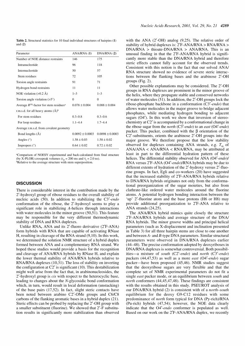
Table 2. Structural statistics for 10 final individual structures of hairpins (1)and (2)
aComparison of NOESY experimental and back-calculated from final structure(by X-PLOR) crosspeak volumes (tm = 200 ms and τc = 2.0 ns).bRelative to the average structure with stem superposition.
Parameter ANA/RNA (1) DNA/RNA (2)
Number of NOE distance restraints 146 175
Intranucleotide 96 118
Internucleotide 50 57
Stem residues 72 105
Torsion angle restraints 91 71
Hydrogen bond restraints 11 11
NOE violation (>0.2 Å) 1–3 1–3
Torsion angle violation (>5°) 0 0
Average R1/6-factor for stem residuesa 0.078 ± 0.004 0.088 ± 0.004
r.m.s.d. for all heavy atoms (Å)b
For stem residues 0.3–0.8 0.3–0.6
For loop residues 1.1–4.4 1.7–4.4
Average r.m.s.d. from covalent geometry
Bond lengths (Å) 0.0092 ± 0.0003 0.0098 ± 0.0003
Angles (°) 1.58 ± 0.03 1.58 ± 0.02
Impropers (°) 0.64 ± 0.02 0.72 ± 0.02

4290 Nucleic Acids Research, 2001, Vol. 29, No. 21
suggested that the DNA strands in DNA/RNA hybrids shouldbe modelled as a mixture of three conformers; C2′-endo, C3′-endoand O4′-endo (24). In the case of ANA/RNA and 2′F-ANA/RNA hybrids, both H1′-H4′ NOE distances (d = 2.35–2.5 Å)and J-coupling data imply the existence of a single O4′-endosugar pucker for the antisense strand. We note that a 20%O4′-endo is necessary to yield 2.8 Å average H1′-H4′ NOEdistance as observed in the DNA strand of our DNA/RNAhybrids (Fig. 3C) (24). Assuming a three-conformer equilibriummodel, the remaining 80% would be a mixture of C2′-endo andC3′-endo, in order to account for the observed scalarcouplings. For comparison, a pure south–north equilibrium (asseen for the loop DNA residues) would yield H1′-H4′ NOEdistances of 3.0–3.3 Å.
Reid and co-workers (45,46) have shown that DNA/RNAhybrid duplexes have a minor groove width that is intermediatebetween A- and B-form duplexes. The calculated values for theminor groove widths of ANA/RNA, 2′F-ANA/RNA and theDNA/RNA studied here are ∼9.0 ± 0.5 Å. Since ANA/RNA,2′F-ANA/RNA and the native DNA/RNA hybrids are
substrates of RNase H, this supports the hypothesis madeearlier by Reid and co-workers (46) that the uniqueness of theminor groove width in hybrids is highly important for RNase Hrecognition and cleavage of the RNA strand.
While DNA/RNA and ANA/RNA (and 2′F-ANA/RNA)hybrids have many conformational and biochemical propertiesin common, there are some differences when examined insufficient detail. DNA/RNA hybrids are generally (but notalways) better substrates for RNase H than ANA/RNA (10)and 2′F-ANA/RNA hybrids (M.J.Damha, M.A.Parniak,K.Viazovkina, C.J.Wilds, C.-N.Lok and K.-C.Min, unpublishedresults). Combined analysis of NOE and J-coupling data haveindicated that the arabinose sugars (2′OH or 2′F) are conforma-tionally ‘rigid’ and adopt almost exclusively the O4′-endopucker. This contrasts with the ‘dynamic’ sugar conformationobserved in the DNA strands of DNA/RNA hybrid duplexes,where a mixture of conformers likely co-exist (O4′, C2′ andC3′-endo) (24). The energy barrier for interconversionbetween ANA, and particularly 2′F-ANA, sugar conformers, ishigher due to the large gauche effect from the 2′-substituent
Figure 4. Spatial structures of the hairpins. (A) Stereoview of the superimposition of 10 final individual structures (sugar-phosphate heavy atoms only; red) andaverage minimized structure (blue) for ANA/RNA hairpin (1). (B) View of the stem superimposition of average minimized structures of ANA/DNA (1) (green),DNA/RNA (2) (blue) and 2′F-ANA/RNA (3) (red) hairpins.

Nucleic Acids Research, 2001, Vol. 29, No. 21 4291
(OH or F) and the furanose ring oxygen (O4′) (25). Theseobservations hint at a correlation between dynamics of theantisense strand and the efficiency with which RNase H cancleave the RNA complement. We therefore propose that thegreater conformational freedom (‘flexibility’) of 2′-deoxyri-bose relative to the arabinose sugars account, at least in part,for the more rapid rate of cleavage of DNA/RNA hybrids rela-tive to ANA/RNA and 2′F-ANA/RNA hybrids. Although wehave no formal proof of this hypothesis, we note that theconformationally locked bc-ANA analog (O4′-endo) forms ahybrid with RNA that does not activate RNase H (23). Aninduced-fit model based on the flexibility of deoxyribose andon the hydration patterns of the duplexes was also proposed(55).
We conclude that for an antisense oligonucleotidecompound to elicit RNase H activity, it must fulfill thefollowing criteria: (i) adopt an east DNA-like conformation(O4′-endo); (ii) possess phosphodiester linkages (PNA and
methylphosphonate-DNA do not elicit RNase H activity);(iii) display sugar mobility (flexibility), although somepreorganization is desirable (e.g. 2′F-ANA) to increasebinding affinity towards the target RNA; (iv) form hybridduplexes with a proper characteristic pattern of hydration(55,56), and with a minor groove width that is intermediatebetween those of pure A- and B-form duplexes (46). Thisknowledge may prove helpful in the design of future antisensemodifications.
CONCLUSIONS
Structural parameters of the ANA/RNA hybrid, especiallyminor groove widths, were found to be similar to structuralparameters in 2′F-ANA/RNA and average structural para-meters in the native DNA/RNA hybrids. The structural similari-ties between DNA/RNA hybrids and the present ANA/RNAhybrid suggest that the overall structure assumed by these
Figure 5. Backbone torsion angles (α, β, γ, δ, ε, ζ), glycosidic torsion angles (χ) and pseudorotation phase angles (P) (in degrees) for stem residues in averageminimized structures. Open circles, ANA/RNA; filled squares, DNA/RNA; open diamonds, 2′F-ANA/RNA.

4292 Nucleic Acids Research, 2001, Vol. 29, No. 21
duplexes may in fact be an important factor responsible forRNase H recognition and cleavage of hybrid duplexes (46,57).
Contrary to what was recently proposed through computa-tional studies (21), we find that the ANA residues of the hybridassume conformations in the O4′-endo (east) range. The sameis observed for the arabinose sugars of 2′F-ANA/RNA hybridduplexes. This contrasts strongly with the ‘dynamic’ sugarconformation in the DNA strands of DNA/RNA hybrids,where a mixture of conformers is likely to co-exist (O4′ + C2′+ C3′-endo) (24). Since all these hybrids are substrates ofRNase H, our findings support the hypothesis that the geometry ofthe antisense strand that is processed by RNase H falls withinthe O4′-endo range (23,24). The rather low stability observedfor duplexes containing ANA strands, e.g. Tm of ANA/ANA< ANA/RNA < RNA/RNA, may be attributed to the differenthydration pattern of these helices. The differential stabilityobserved for ANA/RNA versus 2′F-ANA/RNA hybrids maybe due to different extents of hydration of the 2′-hydroxyversus 2′-fluorine groups. Differential rates of RNase Hcleavage as observed for ANA/RNA versus DNA/RNAhybrids (10) are likely related to differences in the bindingaffinity and sugar flexibility associated with ANA versus DNAstrands.
SUPPLEMENTARY MATERIAL
Supplementary Material is available at NAR Online.
ACKNOWLEDGEMENTS
This work was supported by grants from the Natural Sciencesand Engineering Research Council of Canada (M.J.D.) and theCanadian Institutes of Health Research (K.G. and M.J.D.).
REFERENCES
1. Uhlmann,E. and Peyman,A. (1990) Chem. Rev., 90, 543–584.2. Knorre,D.G., Vlassov,V.V., Zarytova,V.F., Lebedev,A.V. and
Fedorova,O.S. (1994) Design and Targeted Reactions of OligonucleotideDerivatives. CRC Press, Boca Raton, FL.
3. Manoharan,M. (1999) 2′-Carbohydrate modifications in antisenseoligonucleotide therapy: importance of conformation, configuration andconjugation. Biochim. Biophys. Acta, 1489, 117–130.
4. Wengel,J. (1999) Synthesis of 3′-C- and 4′-C-branchedoligodeoxynucleotides and the development of locked nucleic acid(LNA). Acc. Chem. Res., 32, 301–310.
5. Walder,R.Y. and Walder,J.A. (1988) Role of RNase H in hybrid-arrestedtranslation by antisense oligonucleotides. Proc. Natl Acad. Sci. USA, 85,5011–5015.
6. Inoue,H., Hayase,Y., Iwai,S. and Ohtsuka,E. (1987) Sequence-dependenthydrolysis of RNA using modified oligonucleotide splints and RNase H.FEBS Lett., 215, 327–330.
7. Monia,B.P., Lesnik,E.A., Gonzalez,C., Lima,W.F., McGee,D.,Guinosso,C.J., Kawasaki,A.M., Cook,P.D. and Freier,S.M. (1993)Evaluation of 2′-modified oligonucleotides containing 2′-deoxy gaps asantisense inhibitors of gene expression. J. Biol. Chem., 268,14514–14522.
8. Zhou,W. and Agrawal,S. (1998) Mixed-backbone oligonucleotides assecond-generation antisense agents with reduced phosphorothioate-related side effects. Bioorg. Med. Chem. Lett., 8, 3269–3274.
9. Damha,M.J., Wilds,C.J., Noronha,A., Brukner,I., Borkow,G., Arion,D.and Parniak,M.A. (1998) Hybrid of RNA and arabinonucleic acids(ANA and 2′F-ANA) are substrates of ribonuclease H. J. Am. Chem. Soc.,120, 12976–12977.
10. Noronha,A.M., Wilds,C.J., Lok,C.N., Viazovkina,K., Arion,D.,Parniak,M.A. and Damha,M.J. (2000) Synthesis and biophysicalproperties of arabinonucleic acids (ANA): circular dichroic spectra,melting temperatures, and ribonuclease H susceptibility of ANA–RNAhybrid duplexes. Biochemistry, 39, 7050–7062.
11. Lima,W.F. and Crooke,S.T. (1997) Binding affinity and specificity ofEscherichia coli RNase H1: impact on the kinetics of catalysis ofantisense oligonucleotide–RNA hybrids. Biochemistry, 36, 390–398.
12. Pierters,J.M., de Vroom,E., van der Marel,G.A., van Boom,J.H.,Koning,T.M.G., Kaptein,R. and Altona,C. (1990) Hairpin structures inDNA containing arabinofuranosylcytosine. A combination of nuclearmagnetic resonance and molecular dynamics. Biochemistry, 29, 788–799.
13. Gao,Y.G., van der Marel,G.A., van Boom,J.H. and Wang,A.H. (1991)Molecular structure of a DNA decamer containing anticancer nucleosidearabinosylcytosine: conformational perturbation by arabinosylcytosine inB-DNA. Biochemistry, 30, 9922–9931.
14. Schweitzer,B.I., Mikita,T., Kellogg,G.W., Gardner,K.H. andBeardsley,G.P. (1994) Solution structure of a DNA dodecamer containingthe anti-neoplastic agent arabinocytosine: combined use of NMR,restrained molecular dynamics, and full relaxation matrix refinement.Biochemistry, 33, 11460–11475.
15. Gotfredsen,C.H., Spielmann,H.P., Wengel,J. and Jacobsen,J.P. (1996)Structure of a DNA duplex containing a single 2′-O-methyl-β-D-araT:combined use of NMR, restrained molecular dynamics, and full relaxationmatrix refinement. Bioconjug. Chem., 7, 680–688.
16. Gmeiner,W.H., Konerding,D. and James,T.L. (1999) Effect of Cytarabineon the NMR structure of a model Okazaki fragment from the SV40genome. Biochemistry, 38, 1166–1175.
17. Teng,M.K., Liaw,Y.C., van der Marel,G.A., van Boom,J.H. andWang,A.H. (1989) Effects of the O2′ hydroxyl group on Z-DNAconformation: structure of Z-RNA and (araC)-[Z-DNA]. Biochemistry,28, 4923–4928.
18. Zhang,H., van der Marel,G.A., van Boom,J.H. and Wang,A.H. (1992)Conformational perturbation of the anticancer nucleotidearabinosylcytosine on Z-DNA: molecular structure of (araC-dG)3 at 1.3 Åresolution. Biopolymers, 32, 1559–1569.
Table 3. Rangesa of minor groove widths and global helical parameters for stem base pairs in average minimized structures of thehairpins and in canonical A- and B-types of DNA
aAverage values for these ranges with corresponding deviations for the set of stem base pairs are shown. Helical parameters werecalculated by CURVES program (40).
Structure Minor groove width(Å)
X-displacement (Å) Inclination (°) Rise (Å) Helical twist (°)
ANA/RNA (1) 9.0 ± 0.2 –2.5 ± 0.4 5.5 ± 2.5 2.8 ± 0.2 33.7 ± 4.5
DNA/RNA (2) 9.2 ± 0.2 –3.3 ± 0.5 10.5 ± 1.5 2.7 ± 0.2 35.0 ± 4.2
2′F-ANA/RNA (3) 8.8 ± 0.3 –3.0 ± 0.2 2.9 ± 1.3 3.1 ± 0.2 34.0 ± 2.3
A-type DNA 11.1 –5.4 19.3 2.6 32.7
B-type DNA 5.9 –0.7 –6.0 3.4 36.1

Nucleic Acids Research, 2001, Vol. 29, No. 21 4293
19. Ikeda,H., Fernandez,R., Wilk,A., Barchi,J.J., Huang,X. and Marquez,V.E.(1998) The effect of two antipodal fluorine-induced sugar puckers on theconformation and stability of the Dickerson–Drew dodecamer duplex[d(CGCGAATTCGCG)]2. Nucleic Acids Res., 26, 2237–2244.
20. Berger,I., Tereshko,V., Ikeda,H., Marques,V.E. and Egli,M. (1998)Crystal structures of B-DNA with incorporated 2′-deoxy-2′-fluoro-arabino-furanosyl thymines: implications of conformationalpreorganization for duplex stability. Nucleic Acids Res., 26, 2473–2480.
21. Venkateswarlu,D. and Ferguson,D.M. (1999) Effect of C2′-substitutionon arabinonucleic acid structure and conformation. J. Am. Chem. Soc.,121, 5609–5610.
22. Christensen,N.K., Petersen,M., Nielsen,P., Jacobsen,J.P., Olsen,C.E. andWengel,J. (1998) A novel class of oligonucleotide analogues containing2′-O,3′-C-linked [3.2.0]bicycloarabinonucleoside monomers: synthesis,thermal affinity studies, and molecular modeling. J. Am. Chem. Soc., 120,5458–5463.
23. Minasov,G., Teplova,M., Nielsen,P., Wengel,J. and Egli,M. (2000)Structure basis of cleavage by RNase H of hybrids of arabinonucleic acidsand RNA. Biochemistry, 39, 3525–3532.
24. Trempe,J.-F., Wilds,C.J., Denisov,A.Y., Pon,R.T., Damha,M.J. andGehring,K. (2001) NMR solution structure of an oligonucleotide hairpinwith a 2′F-ANA/RNA stem: implications for RNase H specificity towardDNA/RNA hybrid duplexes. J. Am. Chem. Soc., 123, 4896–4903.
25. Wilds,C.J. and Damha,M.J. (2000) 2′-Deoxy-2′-fluoro-β-D-arabinonucleotides and oligonucleotides (2′F-ANA): synthesis andphysicochemical studies. Nucleic Acids Res., 28, 3625–3635.
26. Barchi,J.J., Jeong,L.S., Siddiqui,M.A. and Marquez,V.E. (1997)Conformational analysis of the complete series of 2′ and 3′monofluorinated dideoxyuridines. J. Biochem. Biophys. Methods, 34,11–29.
27. Chwang,A.K. and Sundaralingam,M. (1973) Intramolecular hydrogenbonding in 1-β-D-arabinofuranosylcytosine (ara-C). Nature New Biol.,243, 78–79.
28. Ekiel,I., Remin,M., Darzynkiewich,E. and Shugar,D. (1979) Correlationsof conformational parameters and equilibrium conformational states in avariety of β-D-arabinonucleosides and their analogues. Biochim. Biophys.Acta, 562, 177–191.
29. Saenger,W. (1984) Principles of Nucleic Acids Structure. Springer-Verlag, New York, NY.
30. Ford,H., Dai,F., Mu,L., Siddiqui,M.A., Nickaus,M.C., Anderson,L.,Marquez,V.E. and Barchi,J.J. (2000) Adenosine deaminase prefers adistinct sugar ring conformation for binding and catalysis: kinetic andstructural studies. Biochemistry, 39, 2581–2592.
31. Guinnares,P.A. and Damha,M.J. (1994) Hybridization properties ofoligoarabinonucleotides. Can. J. Chem., 72, 909–918.
32. Varani,G., Cheong,C. and Tinoco,I. (1991) Structure of an unusuallystable RNA hairpin. Biochemistry, 30, 3280–3289.
33. James,J.K. and Tinoco,I. (1993) The solution structure of a d[C(TTCG)G]DNA hairpin and comparison to the unusually stable RNA analogue.Nucleic Acids Res., 21, 3287–3293.
34. Piotto,M., Saudek,V. and Sklenar,V. (1992) Gradient-tailored excitationfor single-quantum NMR spectroscopy of aqueous solutions. J. Biomol.NMR, 2, 661–666.
35. Brünger,A.T. (1992) X-PLOR 3.1, A System for X-ray Crystallographyand NMR. Yale University Press, New Haven, CT.
36. Klinck,R., Sprules,T. and Gehring,K. (1997) Structural characterization ofthree RNA hexanucleotide loops from the internal ribosome entry site ofpolioviruses. Nucleic Acids Res., 25, 2129–2137.
37. Kim,C.H., Kao,C.C. and Tinoco,I. (2000) RNA motifs that determinespecificity between a viral replicase and its promoter. Nature Struct. Biol.,7, 415–422.
38. Wüthrich,K. (1986) NMR of Protein and Nucleic Acids. John Wiley &Sons, New York, NY.
39. de Leeuw,F.A.A.M. and Altona,C. (1983) Quantum Chemistry ProgramExchange, no. 463: PSEUROT 3B. Indiana University, Bloomington, IN.
40. Lavery,R. and Sklenar,H. (1997) Curves 5.2, Helical Analysis of IrregularNucleic Acids. Laboratory de Biochimie Theorique, CNRS URA 77,Paris.
41. van Wijk,J., Huckriede,B.D., Ippel,J.H. and Altona,C. (1992) Furanosesugar conformations in DNA from NMR coupling constants. MethodsEnzymol., 211, 286–306.
42. Rinkel,L.J. and Altona,C. (1987) Conformational analysis of thedeoxyribofuranose ring in DNA by means of sums of proton–protoncoupling constants: a graphical method. J. Biomol. Struct. Dyn., 4,621–649.
43. Kim,S.G., Lin,L.J. and Reid,B.R. (1992) Determination of nucleic acidbackbone conformation by 1H NMR. Biochemistry, 31, 3564–3574.
44. Gonzalez,C., Stec,W., Reynolds,M.A. and James,T.L. (1995) Structureand dynamics of a DNA–RNA hybrid duplex with a chiralphosphorothionate moiety: NMR and molecular dynamics withconventional and time-averaged restraints. Biochemistry, 34, 4969–4982.
45. Fedoroff,O.Y., Ge,Y. and Reid,B.R. (1997) Solution structure ofr(gaggacug):d(CAGTCCTC) hybrid: implications for the initiation ofHIV-1 (+)-strand synthesis. J. Mol. Biol., 269, 225–239.
46. Fedoroff,O.Y., Salazar,M. and Reid,B.R. (1993) Structure of aDNA–RNA hybrid duplex. Why RNase H does not cleave pure RNA.J. Mol. Biol., 233, 509–523.
47. Gyi,J.I., Lane,A.N., Conn,G.L. and Brown,T. (1998) Solution structuresof DNA–RNA hybrids with purine-rich and pyrimidine-rich strands:comparison with the homologous DNA and RNA duplexes. Biochemistry,37, 73–80.
48. Bachelin,M., Hessler,G., Kurtz,G., Hacia,J.G., Dervan,P.B. andKessler,H. (1998) Structure of a stereoregular phosphorothionateDNA/RNA duplex. Nature Struct. Biol., 5, 271–275.
49. Gorenstein,D.G., Meadows,R.P., Metz,J.T., Nikonowicz,E. and Post,C.B.(1990) 31P and 1H 2-dimensional NMR and NOESY-distance restrainedmolecular dynamics methodologies for defining sequence-specificvariations in duplex oligonucleotides. A comparison of NOESY two-spinand relaxation rate matrix analyses. Adv. Biophys. Chem., 1, 47–124.
50. Wang,A.H., Fujii,S., van Boom,J.H., van der Marel,G.A., vanBoeckel,S.A. and Rich,A. (1982) Molecular structure ofr(GCG)d(TATACGC): a DNA–RNA hybrid helix joined to double helicalDNA. Nature, 299, 601–604.
51. Egli,M., Portmann,S. and Usman,N. (1996) RNA hydration: a detailedlook. Biochemistry, 35, 8489–8494.
52. Mikita,T. and Beardsley,G.P. (1988) Functional consequences of thearabinosylcytosine structural lesion in DNA. Biochemistry, 27,4698–4705.
53. Lane,A.N., Ebel,S. and Brown,T. (1993) NMR assignments and solutionconformation of the DNA.RNA hybrid duplexd(GTGAACTT).r(AAGUUCAC). Eur. J. Biochem., 215, 297–306.
54. Xiong,Y. and Sundaralingam,M. (2000) Crystal structure oa aDNA–RNA hybrid duplex with a polypurine RNA r(gaagaagag) with acomplementary polypyrimidine DNA d(CTCTTCTTC). Nucleic AcidsRes., 28, 2171–2176.
55. Szyperski,T., Gotte,M., Billeter,M., Perola,E., Cellai,L., Heumann,H. andWüthrich,K. (1999) NMR structure of the chimeric hybrid duplexr(gcaguggc).r(gcca)d(CTGC) comprising the tRNA–DNA junctionformed during initiation of HIV-1 reverse transcription. J. Biomol. NMR,13, 343–355.
56. Hsu,S.T., Chou,M.T. and Cheng,J.W. (2000) The solution structure of[d(CGC)r(aaa)d(TTTGCG)]2: hybrid junctions flanked by DNA duplexes.Nucleic Acids Res., 28, 1322–1331.
57. Nakamura,H., Oda,Y., Iwai,S., Inore,H., Ohtsuka,E., Kanaya,S.,Kimura,S., Katsuda,C., Katayannagi,K., Morikawa,K., Miyashiro,H. andIkehara,M. (1991) How does RNase recognize a DNA–RNA hybrid?Proc. Natl Acad. Sci. USA, 88, 11535–11539.