![Synthesis, Structural Characterization and Antibacterial Activity of Novel 7β-{[3-(substituted phenyl)-2-propenoyl]amino}-3-[(2,5-dihydro-6-hydroxy- 2-methyl)-5-oxo-cis-triazin-3-yl]-thiomethyl-cefalosporins](https://static.fdokumen.com/doc/165x107/631d3fce93f371de1901d7a5/synthesis-structural-characterization-and-antibacterial-activity-of-novel-7v-3-substituted.jpg)
Anti-inflammatory activity of a naphthyridine derivative...
8
Anti-inflammatory activity of a naphthyridine derivative (7-chloro-6-fluoro-N- (2-hydroxy-3-oxo-1-phenyl-3-(phenylamino)propyl)-4-oxo-1-(prop-2-yn-1-yl)- 1,4-dihydro-1,8-naphthyridine-3-carboxamide) possessing in vitro anticancer potential Alka Madaan a, ⁎, Vivek Kumar b , Ritu Verma a , Anu T. Singh a , S.K. Jain c , Manu Jaggi a a Cell Biology Lab, Dabur Research Foundation, 22, Site IV, Sahibabad, Ghaziabad, Uttar Pradesh-201010, India b Chemical Research Lab, Dabur Research Foundation, 22, Site IV, Sahibabad, Ghaziabad, Uttar Pradesh-201010, India c Department of Biotechnology, Jamia Hamdard, New Delhi-110062, India abstract article info Article history: Received 27 October 2012 Received in revised form 14 January 2013 Accepted 14 January 2013 Available online 29 January 2013 Keywords: 1,8-Naphthyridine-3-carboxamide Anticancer Anti-inflammatory Dendritic cells Cytokines We have previously synthesized a series of 1,8-naphthyridine-3-carboxamide derivatives to identify poten- tial anti-cancer/anti-inflammatory compounds. Three derivatives, 7-chloro-N-(3-(cyclopentylamino)-3- oxo-1-phenylpropyl)-6-fluoro-4-oxo-1-(prop-2-yn-1-yl)-1,4-dihydro-1,8-naphthyridine-3-carboxamide (C-22), 7-chloro-N-(2-hydroxy-3-oxo-1-phenyl-3-(phenylamino)propyl)-4-oxo-1-(prop-2-yn-1-yl)-1,4- dihydro-1,8-naphthyridine-3-carboxamide (C-31) and 7-chloro-6-fluoro-N-(2-hydroxy-3-oxo-1-phenyl- 3-(phenylamino)propyl)-4-oxo-1-(prop-2-yn-1-yl)-1,4-dihydro-1,8-naphthyridine-3-carboxamide (C-34) dem- onstrated high cytotoxicity against a number of cancer cell lines and inhibited secretion of IL-1-β and IL-6. In the present study, C-22, C-31 and C-34 were assessed for modulation of pro-inflammatory cytokines, TNF-α and IL-8, chemokine RANTES and NO produced by lipopolysaccharide (LPS)-treated mouse Dendritic cells (DCs). Among the 3 compounds, C-34 showed the most potent inhibition of inflammatory markers in DC model at 0.2 and 2 μM. C-34 also significantly downregulated the secretion of TNF-α, IL-1-β and IL-6 by murine splenocytes and THP-1 cells against LPS induced levels. In vitro effects of C-34 on bone marrow toxicity were assessed in CFU-GM assay. Human CFU-GM population was comparatively more sensitive to C-34 (0.1–10 μM) than murine CFU-GM. IC 50 values for murine and human CFU-GM were not attained. C-34 was further examined for in vivo suppression of LPS induced cytokines in a mice model. At doses ranging from 1.25 to 5 mg/kg, C-34 led to significant inhibition of TNF-α, IL-1-β, IL-6 and MIP-1-α. At the highest dose of 5 mg/kg, C-34 also protected LPS-treated mice against endotoxin-induced lethality. In conclusion, C-34 demonstrates anti-inflammatory activity in vitro and in vivo in ad- dition to cytotoxic properties. This finding suggests its potential for further development as a synthetic naphthyridine derivative with dual anti-cancer and anti-inflammatory (cytokine inhibition) properties. © 2013 Elsevier B.V. All rights reserved. 1. Introduction Inflammation has been identified as one of the key hallmarks in cancer development [1]. It has been well established that the devel- opment of most of, if not all, cancers from inflammation might be a process mediated by crosstalk among inflammatory cells, cancer cells and inflammatory cytokines [2]. Increased infiltration and re- cruitment of immune cells to the developing tumor site results in ex- cessive secretion of pro-inflammatory mediators. This favors enhanced angiogenesis, tumor growth and progression [3–6]. Cyto- kines and chemokines represent promising target for anticancer therapeutic interventions [7,8]. Therefore, compounds possessing anticancer activities along with additional anti-inflammatory poten- tial may present as attractive candidates in cancer therapeutics. Compounds belonging to naphthyridine class have been reported to possess anticancer and anti-inflammatory activities. SNS-595, an anticancer naphthyridine analog which acts by induc- ing DNA damage, is presently in phase III clinical trial [9]. Synthesis and structure–activity relationships of naphthyridine derivatives with modifications at N-1, C-5, C-6, and C-7 positions as antitumor agents have been reported [10,11]. 1,8-Naphthyridine derivatives have demonstrated an anti-inflammatory activity by inhibition of reactive oxygen species (ROS) and myeloperoxidase by human polymorphonuclear cells (PMNs) [12]. 9-Substituted derivatives of 1,8-naphthyridine have shown anti-inflammatory activity in carrageenin-induced paw edema model [13]. Earlier, we have reported a series of 1,8-naphthyridine-3- carboxamide derivatives by introduction of different cyclic as well International Immunopharmacology 15 (2013) 606–613 ⁎ Corresponding author. Tel.: +91 120 3378558; fax: +91 120 4152279. E-mail address: [email protected] (A. Madaan). 1567-5769/$ – see front matter © 2013 Elsevier B.V. All rights reserved. http://dx.doi.org/10.1016/j.intimp.2013.01.011 Contents lists available at SciVerse ScienceDirect International Immunopharmacology journal homepage: www.elsevier.com/locate/intimp
-
Upload
dabarresearch -
Category
Documents
-
view
1 -
download
0
Transcript of Anti-inflammatory activity of a naphthyridine derivative...

International Immunopharmacology 15 (2013) 606–613
Contents lists available at SciVerse ScienceDirect
International Immunopharmacology
j ourna l homepage: www.e lsev ie r .com/ locate / in t imp
Anti-inflammatory activity of a naphthyridine derivative (7-chloro-6-fluoro-N-(2-hydroxy-3-oxo-1-phenyl-3-(phenylamino)propyl)-4-oxo-1-(prop-2-yn-1-yl)-1,4-dihydro-1,8-naphthyridine-3-carboxamide) possessing in vitroanticancer potential
Alka Madaan a,⁎, Vivek Kumar b, Ritu Verma a, Anu T. Singh a, S.K. Jain c, Manu Jaggi a
a Cell Biology Lab, Dabur Research Foundation, 22, Site IV, Sahibabad, Ghaziabad, Uttar Pradesh-201010, Indiab Chemical Research Lab, Dabur Research Foundation, 22, Site IV, Sahibabad, Ghaziabad, Uttar Pradesh-201010, Indiac Department of Biotechnology, Jamia Hamdard, New Delhi-110062, India
⁎ Corresponding author. Tel.: +91 120 3378558; fax:E-mail address: [email protected] (A. M
1567-5769/$ – see front matter © 2013 Elsevier B.V. Allhttp://dx.doi.org/10.1016/j.intimp.2013.01.011
a b s t r a c t
a r t i c l e i n f oArticle history:Received 27 October 2012Received in revised form 14 January 2013Accepted 14 January 2013Available online 29 January 2013
Keywords:1,8-Naphthyridine-3-carboxamideAnticancerAnti-inflammatoryDendritic cellsCytokines
We have previously synthesized a series of 1,8-naphthyridine-3-carboxamide derivatives to identify poten-tial anti-cancer/anti-inflammatory compounds. Three derivatives, 7-chloro-N-(3-(cyclopentylamino)-3-oxo-1-phenylpropyl)-6-fluoro-4-oxo-1-(prop-2-yn-1-yl)-1,4-dihydro-1,8-naphthyridine-3-carboxamide(C-22), 7-chloro-N-(2-hydroxy-3-oxo-1-phenyl-3-(phenylamino)propyl)-4-oxo-1-(prop-2-yn-1-yl)-1,4-dihydro-1,8-naphthyridine-3-carboxamide (C-31) and 7-chloro-6-fluoro-N-(2-hydroxy-3-oxo-1-phenyl-3-(phenylamino)propyl)-4-oxo-1-(prop-2-yn-1-yl)-1,4-dihydro-1,8-naphthyridine-3-carboxamide (C-34) dem-onstrated high cytotoxicity against a number of cancer cell lines and inhibited secretion of IL-1-β and IL-6. In thepresent study, C-22, C-31 and C-34 were assessed for modulation of pro-inflammatory cytokines, TNF-α and IL-8,chemokine RANTES and NO produced by lipopolysaccharide (LPS)-treated mouse Dendritic cells (DCs). Amongthe 3 compounds, C-34 showed the most potent inhibition of inflammatory markers in DC model at 0.2 and2 μM. C-34 also significantly downregulated the secretion of TNF-α, IL-1-β and IL-6 by murine splenocytes andTHP-1 cells against LPS induced levels. In vitro effects of C-34 on bone marrow toxicity were assessed in CFU-GMassay. Human CFU-GM population was comparatively more sensitive to C-34 (0.1–10 μM) than murine CFU-GM.IC50 values for murine and human CFU-GM were not attained. C-34 was further examined for in vivo suppressionof LPS induced cytokines in a mice model. At doses ranging from 1.25 to 5 mg/kg, C-34 led to significant inhibitionof TNF-α, IL-1-β, IL-6 and MIP-1-α. At the highest dose of 5 mg/kg, C-34 also protected LPS-treated mice againstendotoxin-induced lethality. In conclusion, C-34 demonstrates anti-inflammatory activity in vitro and in vivo in ad-dition to cytotoxic properties. This finding suggests its potential for further development as a syntheticnaphthyridine derivative with dual anti-cancer and anti-inflammatory (cytokine inhibition) properties.
© 2013 Elsevier B.V. All rights reserved.
1. Introduction
Inflammation has been identified as one of the key hallmarks incancer development [1]. It has been well established that the devel-opment of most of, if not all, cancers from inflammation might be aprocess mediated by crosstalk among inflammatory cells, cancercells and inflammatory cytokines [2]. Increased infiltration and re-cruitment of immune cells to the developing tumor site results in ex-cessive secretion of pro-inflammatory mediators. This favorsenhanced angiogenesis, tumor growth and progression [3–6]. Cyto-kines and chemokines represent promising target for anticancertherapeutic interventions [7,8]. Therefore, compounds possessing
+91 120 4152279.adaan).
rights reserved.
anticancer activities along with additional anti-inflammatory poten-tial may present as attractive candidates in cancer therapeutics.
Compounds belonging to naphthyridine class have beenreported to possess anticancer and anti-inflammatory activities.SNS-595, an anticancer naphthyridine analog which acts by induc-ing DNA damage, is presently in phase III clinical trial [9]. Synthesisand structure–activity relationships of naphthyridine derivativeswith modifications at N-1, C-5, C-6, and C-7 positions as antitumoragents have been reported [10,11]. 1,8-Naphthyridine derivativeshave demonstrated an anti-inflammatory activity by inhibition ofreactive oxygen species (ROS) and myeloperoxidase by humanpolymorphonuclear cells (PMNs) [12]. 9-Substituted derivativesof 1,8-naphthyridine have shown anti-inflammatory activity incarrageenin-induced paw edema model [13].
Earlier, we have reported a series of 1,8-naphthyridine-3-carboxamide derivatives by introduction of different cyclic as well

607A. Madaan et al. / International Immunopharmacology 15 (2013) 606–613
as open amino-acids at the C-3 position with in vitro cytotoxic activity[14–16]. From these studies, C-22, C-31 and C-34 [14] were shortlistedon the basis of potent cytotoxic activity in human cancer cell lines andinhibition of IL-1-β and IL-6 secretion. Here, inhibitory effects of C-22,C-31 and C-34 were further studied on the production of other relevantpro-inflammatory markers, TNF-α, IL-8, RANTES and NO fromLPS-treated DCs. Anti-inflammatory activity of C-34 was furtherassessed in other immune cell populations; murine splenocytes andTHP-1 cells by inhibition of LPS-induced TNF-α, IL-1-β and IL-6. Possibletoxic effects of C-34 on bone-marrow cells were estimated in vitro inmurine and human CFU-GM assays. In vivo anti-inflammatory activityof C-34 was evaluated by examining its effects on secretion of cytokines(TNF-α, IL-1-β, IL-6 and MIP-1-α) and inhibition of endotoxic shockusing LPS model of inflammation in mice.
2. Materials and methods
2.1. Chemicals and reagents
Recombinant mouse and human granulocyte-macrophage colonystimulating factor (rmGMCSF and rhGMCSF) and cytokine-specificELISA kits for murine TNF-α, IL-1-β, IL-6, IL-8, RANTES andMIP-1-α (Quantikine) were purchased from R&D Systems, Minneap-olis, MN. Methylcellulose and Human Bone Marrow MononuclearCells (Stem Cell Technologies), Lipopolysaccharide (LPS, fromEscherichia coli serotype 0127:B8), Thalidomide, Curcumin, PMA(Phorbol 12-myristate 13-acetate), MTT (3-(4,5-dimethylthiazol-2-yl)-2,5-diphenyltetrazolium bromide), Bovine Serum Albumin(BSA), L-glutamine, Sulphanilamide, NED (N-(1-naphthyl) ethylene-diamine dihydrochloride), DMSO and DNAase I from Sigma, IMDM,RPMI 1640 (Invitrogen), FBS and Penicillin–streptomycin (Hyclone)were used in the study.
2.2. Animals
Specific pathogen-free male C57BL/6 mice (20 to 25 g, 8–10 weeks)and female BALB/c mice (18 to 20 g, 8–10 weeks) used in the studywere obtained from National Centre for Laboratory Animal Sciences
C-227-chloro-N-(3-(cyclopentylamino)-3-oxo-1-
phenylpropyl)-6-fluoro-4-oxo-1-(prop-2-yn-1-yl)-1,4-dihydro-1,8-naphthyridine-3-carboxamide
C-347-chloro-6-fluoro-N-(2-hydro
(phenylamino)propyl)-4-oxo-dihydro-1,8-naphthyridin
Fig. 1. Chemical structures of 1,8-naphthyridine-3-
(NCLAS), and National Institute of Nutrition (NIN), Hyderabad, India.Mice were kept in the in-house animal facility maintained at 22±3 °Cand 55±15% relative humidity with 12 h light–dark cycle. They weregiven autoclaved pelleted feed and filtered drinking water ad libitum.All experiments employing the mice were performed under the proto-cols approved by the Institutional Animal Ethics Committee (IAEC) ofDabur Research Foundation.
2.3. 1,8-Naphthyridine-3-carboxamide derivatives
C-22, C-31 and C-34 (structures are shown in Fig. 1) were chemi-cally synthesized as described previously [14]. For purity, elementalanalyses (C, H, N) were undertaken using an elemental analyzer andwere observed within 0.4% of the calculated values.
2.4. Murine DCs culture from bone marrow
DCs were generated from murine bone marrow, using a modifiedmethod [17]. Femurs were excised from C57BL/6 mice and bone mar-rowwas harvested by gently flushing the femur with RPMI 1640 medi-umusing a 23-gauge needle. Cellswere cultured (2×10⁶ cells/10 ml) in90-mm culture Petri dishes with growth medium (RPMI 1640 mediumsupplemented with 10% FBS, 20 mM penicillin–streptomycin and20 ng/ml rmGMCSF) for 6 days at 37 °C in a humidified 5% CO2 atmo-sphere. On day 3, cells were replenished with additional 10 ml ofgrowth medium containing rmGMCSF. Semi-adherent, immature DCswere harvested on day 6 by gentle pipetting and used in experiments.Viable cells were counted using trypan blue exclusion method.
2.5. Measurement of TNF-α, IL-8, RANTES and NO in DCs
C-22, C-31 and C-34 were dissolved in sterile DMSO at 10 mM anddiluted in serum free RPMI 1640 medium. Effects of C-22, C-31 andC-34 on viability of DCs were assessed by MTT assay. DCs (50,000cells/well) were plated in 96-well culture plate and treated with C-22,C-31 and C-34 at 0.2, 2 and 5 μM. The cytotoxic effects on DCs were de-termined after 48 h of incubation by addition ofMTT (0.5 mg/ml). After3 h, cells were centrifuged at 250 g for 8 min and cell pellets were
C-317-chloro-N-(2-hydroxy-3-oxo-1-phenyl-3-
(phenylamino)propyl)-4-oxo-1-(prop-2-yn-1-yl)-1,4-dihydro-1,8-naphthyridine-3-carboxamide
xy-3-oxo-1-phenyl-3-1-(prop-2-yn-1-yl)-1,4-e-3-carboxamide
carboxamide derivatives; C-22, C-31 and C-34.
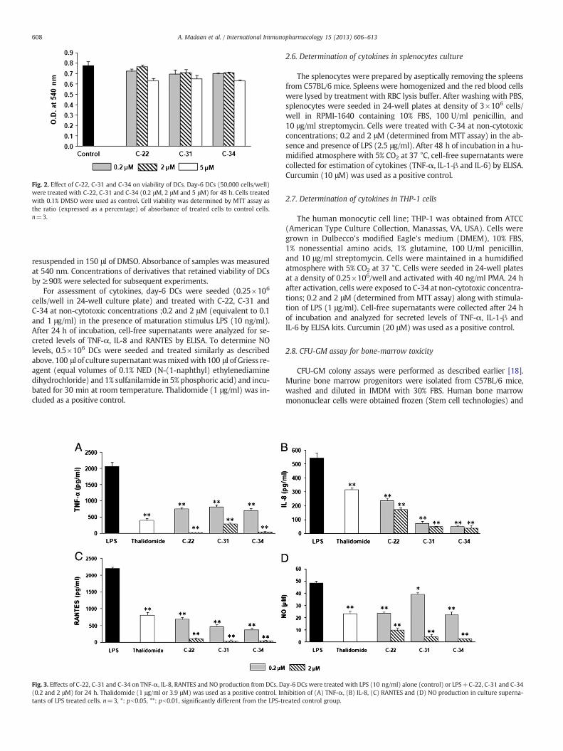
Fig. 2. Effect of C-22, C-31 and C-34 on viability of DCs. Day-6 DCs (50,000 cells/well)were treated with C-22, C-31 and C-34 (0.2 μM, 2 μM and 5 μM) for 48 h. Cells treatedwith 0.1% DMSO were used as control. Cell viability was determined by MTT assay asthe ratio (expressed as a percentage) of absorbance of treated cells to control cells.n=3.
608 A. Madaan et al. / International Immunopharmacology 15 (2013) 606–613
resuspended in 150 μl of DMSO. Absorbance of samples was measuredat 540 nm. Concentrations of derivatives that retained viability of DCsby ≥90% were selected for subsequent experiments.
For assessment of cytokines, day-6 DCs were seeded (0.25×10⁶
cells/well in 24-well culture plate) and treated with C-22, C-31 andC-34 at non-cytotoxic concentrations ;0.2 and 2 μM (equivalent to 0.1and 1 μg/ml) in the presence of maturation stimulus LPS (10 ng/ml).After 24 h of incubation, cell-free supernatants were analyzed for se-creted levels of TNF-α, IL-8 and RANTES by ELISA. To determine NOlevels, 0.5×10⁶ DCs were seeded and treated similarly as describedabove. 100 μl of culture supernatantwasmixedwith 100 μl of Griess re-agent (equal volumes of 0.1% NED (N-(1-naphthyl) ethylenediaminedihydrochloride) and 1% sulfanilamide in 5% phosphoric acid) and incu-bated for 30 min at room temperature. Thalidomide (1 μg/ml) was in-cluded as a positive control.
Fig. 3. Effects of C-22, C-31 and C-34 on TNF-α, IL-8, RANTES and NO production from DCs. D(0.2 and 2 μM) for 24 h. Thalidomide (1 μg/ml or 3.9 μM) was used as a positive control. Intants of LPS treated cells. n=3, *: pb0.05, **: pb0.01, significantly different from the LPS-tr
2.6. Determination of cytokines in splenocytes culture
The splenocytes were prepared by aseptically removing the spleensfrom C57BL/6 mice. Spleens were homogenized and the red blood cellswere lysed by treatment with RBC lysis buffer. After washing with PBS,splenocytes were seeded in 24-well plates at density of 3×10⁶ cells/well in RPMI-1640 containing 10% FBS, 100 U/ml penicillin, and10 μg/ml streptomycin. Cells were treated with C-34 at non-cytotoxicconcentrations; 0.2 and 2 μM (determined from MTT assay) in the ab-sence and presence of LPS (2.5 μg/ml). After 48 h of incubation in a hu-midified atmosphere with 5% CO2 at 37 °C, cell-free supernatants werecollected for estimation of cytokines (TNF-α, IL-1-β and IL-6) by ELISA.Curcumin (10 μM) was used as a positive control.
2.7. Determination of cytokines in THP-1 cells
The human monocytic cell line; THP-1 was obtained from ATCC(American Type Culture Collection, Manassas, VA, USA). Cells weregrown in Dulbecco's modified Eagle's medium (DMEM), 10% FBS,1% nonessential amino acids, 1% glutamine, 100 U/ml penicillin,and 10 μg/ml streptomycin. Cells were maintained in a humidifiedatmosphere with 5% CO2 at 37 °C. Cells were seeded in 24-well platesat a density of 0.25×10⁶/well and activated with 40 ng/ml PMA. 24 hafter activation, cells were exposed to C-34 at non-cytotoxic concentra-tions; 0.2 and 2 μM (determined from MTT assay) along with stimula-tion of LPS (1 μg/ml). Cell-free supernatants were collected after 24 hof incubation and analyzed for secreted levels of TNF-α, IL-1-β andIL-6 by ELISA kits. Curcumin (20 μM) was used as a positive control.
2.8. CFU-GM assay for bone-marrow toxicity
CFU-GM colony assays were performed as described earlier [18].Murine bone marrow progenitors were isolated from C57BL/6 mice,washed and diluted in IMDM with 30% FBS. Human bone marrowmononuclear cells were obtained frozen (Stem cell technologies) and
ay-6 DCs were treated with LPS (10 ng/ml) alone (control) or LPS+C-22, C-31 and C-34hibition of (A) TNF-α, (B) IL-8, (C) RANTES and (D) NO production in culture superna-eated control group.

Fig. 4. Effects of C-34 on TNF-α, IL-1-β and IL-6 production from murine splenocytes.Splenocytes were treated with LPS (2.5 μg/ml) alone (control) or LPS+C-34 (0.2 and2 μM) for 48 h. Curcumin (10 μM) was used as a positive control. Inhibition of (A)TNF-α, (B) IL-1-β, and (C) IL-6 production in culture supernatants of LPS treatedcells. n=3, **: pb0.01, significantly different from the LPS-treated control group.
Fig. 5. Effects of C-34 on TNF-α, IL-1-β and IL-6 production from human monocytesTHP-1. THP-1 cells were treated with LPS (1 μg/ml) alone (control) or LPS+C-34 (0.2and 2 μM) for 24 h. Curcumin (20 μM) was used as a positive control. Inhibition of (A)TNF-α, (B) IL-1-β, and (C) IL-6 production in culture supernatants of LPS treated cells.n=3, **: pb0.01, significantly different from the LPS-treated control group.
609A. Madaan et al. / International Immunopharmacology 15 (2013) 606–613
thawed before using. While processing, cells were treatedwith DNAse I(0.1 mg/ml) to prevent cell clumping in CFU assays. For murineCFU-GM assay, progenitor cells were seeded in the growth medium(1% methylcellulose in IMDM, 30% FBS, 1% BSA, 2 mM L-glutamineand 10 ng/ml rmGMCSF) at a density of 40,000 cells/35 mm Petridish/ml and incubated at 37 °C in humidified 5% CO2 for 7 days.Human bone marrow mononuclear cells were seeded at a density of70,000 cells/dish in the growthmedium containing rhGMCSF and incu-bated for 14 days. Inhibitory effects on the development of murine andhumanCFU-GMcolonieswere observed by continuous exposure of pro-genitor cells to C-34 (0.1–10 μM). CFU-GM colonieswere counted usingan inverted microscope.
2.9. In vivo cytokine inhibition
C-34 was dissolved in sterile DMSO at 10 mM and subsequently di-luted in pyrogen-free saline to the desired concentration. 2 h prior toLPS challenge, C-34 (in saline with a final concentration of DMSO not
exceeding 1% v/v) was administered by intraperitoneal (i.p.) route toBALB/c mice (n=5) at doses of 1.25, 2.5 and 5 mg/kg. Negative controlanimals received injections of saline with 1% DMSO (v/v) as vehicle.Endotoxic shock was induced in mice by i.p. injection of 5 mg/kg LPS(E. coli) dissolved in saline solution. After 1.5 h, mice were bled fromthe retro-orbital plexus and levels of pro-inflammatory cytokinesTNF-α, IL-1-β, IL-6 and MIP-1-α were quantified in serum by ELISA.Thalidomide was used as a positive control (10 mg/kg).
2.10. Protection against LPS-induced death
For the protection studies, BALB/c mice (n=5) were administeredi.p. with a single dose of C-34 (5 mg/kg), 2 h before LPS injection.

610 A. Madaan et al. / International Immunopharmacology 15 (2013) 606–613
Mice were challenged with a lethal dose of LPS (7.5 mg/kg). Survivalwas monitored for 96 h after LPS injection.
2.11. Statistical analysis
Experimental values were expressed as arithmetic mean±SEM.Statistical differences between control and treatment groups weredetermined using one-way ANOVA with Dunnett's multiple compar-ison post test. The survival rate of mice was analyzed by log-ranktest (Kaplan–Meier analysis).
3. Results
3.1. In vitro anti-inflammatory activity
3.1.1. Inhibition of TNF-α, IL-8, RANTES and NO secretion in DCsTo identify non-cytotoxic concentrations of C-22, C-31 and C-34,
effects on cellular viability were determined after 48 h of treatment.There was no considerable loss in viability of DCs upon treatmentwith C-22, C-31 and C-34. At 0.2 and 2 μM, cell viability was foundto be ≥90% (Fig. 2). Hence, anti-inflammatory effect of C-22, C-31and C-34 was evaluated at non-cytotoxic concentrations; 0.2 and2 μM by determining TNF-α, IL-8, RANTES and NO secretion inLPS-treated (10 ng/ml) DCs. After 24 h of treatment, C-22, C-31 andC-34 demonstrated significant inhibition of pro-inflammatory markers,when added simultaneously with LPS (Fig. 3). Among the 3 compoundstested, C-34 demonstrated the best activity in DC based model by maxi-mally down-regulating all the inflammatory markers. Therefore, C-34was selected for further evaluation of anti-inflammatory activity. Thalido-mide was used as a positive control (1 μg/ml) and also led to significantinhibition of LPS-stimulated production of TNF-α, IL-8, RANTES and NOsecretions.
Fig. 6. Effects of C-34 on in vitro CFU-GM colony formation. Murine bone marrow and humaexposure to C-34 (0.1–10 μM) for 7 and 14 days respectively. n=3. (A) Morphology of murand (iii) multi-centric colonies. Colonies were counted using inverted microscope at magninies. Colony scoring was performed according to CFU-atlas ECVAM INVITTOX no. 101. (B) Inhtrol (0.1% DMSO treated cells).
3.1.2. Inhibition of cytokines in murine splencoytesTreatment of murine splenocyte cultures with LPS (2.5 μg/ml) en-
hanced levels of inflammatory cytokines. C-34 exhibited significant re-duction of LPS-stimulated cytokines in murine splenocytes after a timeperiod of 48 h. At non-cytotoxic concentrations 0.2 and 2 μM, levels ofTNF-α, IL-1-β and IL-6 were significantly reduced in comparison withLPS (Fig. 4). Curcumin, which was used as a positive control at 10 μMshowed significant inhibition of all the cytokines.
3.1.3. Inhibition of cytokines in THP-1 cellsAnti-inflammatory activity of C-34 was tested in human monocytic
cells (THP-1) at non-cytotoxic concentrations; 0.2 and 2 μM. LPS at1 μg/ml stimulated levels of cytokines secreted by THP-1 cells. After24 h of treatment, C-34 was able to significantly downregulateLPS-stimulated secretion of TNF-α, IL-1-β and IL-6 at 0.2 and 2 μM(Fig. 5). Curcumin 20 μM (positive control) also resulted in declinedlevels of LPS-induced cytokines.
3.2. Inhibitory effects of C-34 on in vitro murine and human CFU-GM
Fig. 6a represents the characteristic morphology of murine andhuman CFU-GM colonies on 7 and 14 days respectively. Culture ofbone-marrow precursors in the presence of GMCSF resulted in theformation of 3 types of CFU-GM colonies with variable morphology.Compact colonies (with a central dense nucleus and a peripheralhalo), diffuse and spread colonies (without apparent nucleus) andmulticentric colonies (with two or more dense nuclei and commonperipheral halo, growing at the same depth in the plate) were ob-served in both murine and human assays.
To evaluate the in vitro toxic effects on bone marrow, murine andhuman bone-marrow cells were cultured along with C-34 (0.1 μM–
10 μM) in the presence of GMCSF for 7 and 14 days respectively. In
n bone marrow cells were cultured in presence of GMCSF (10 ng/ml) with continuousine and human CFU-GM colonies. (i) Compact colonies (ii) diffuse and spread coloniesfication of 40×. Aggregates containing 50 or more cells were counted as CFU-GM colo-ibition of CFU-GM colonies formation by C-34 was determined in comparison with con-

Fig. 7. Effects of C-34 on LPS induced pro-inflammatory cytokines in mice serum. Female BALB/c mice were injected i.p. with LPS (5 mg/kg) alone (control) or LPS+C-34 at 1.25, 2.5and 5 mg/kg (2 h before LPS). Thalidomide (10 mg/kg) was used as a positive control. Inhibition of TNF-α (A), IL-1β (B), IL-6 (C) and MIP-1-α (D) in sera of mice treated with LPS.n=5, **: pb0.01, significantly different from the LPS-treated control group.
611A. Madaan et al. / International Immunopharmacology 15 (2013) 606–613
this study, human CFU-GM colonies showed higher sensitivity toC-34 as compared to mouse CFU-GM colonies (Fig. 6b). After contin-uous in vitro exposure for 7 and 14 days, C-34 at 2.5 μM led to 12%and 20% inhibitions of murine and human CFU-GM respectively. At10 μM, murine and human CFU-GM colonies were inhibited by amaximum of 30% and 37% respectively. However, IC50 for murineand human CFU-GM colony formation could not be achieved withinthe indicated dose range.
3.3. In vivo cytokine inhibition by C-34
For evaluation of in vivo anti-inflammatory activity of C-34, BALB/cmicewere treatedwith LPS to induce inflammation. Secretion of key in-flammatory markers; TNF-α, IL-1-β, IL-6 and MIP-1-α in serum wassubstantially increased after LPS injection (5 mg/kg), with optimumlevels reaching after 90 min. In this model, pre-administration ofBALB/c mice with C-34 (at doses of 1.25, 2.5 and 5 mg/kg) 2 h beforeLPS challenge resulted in significant inhibition of stimulated levels ofpro-inflammatory cytokines TNF-α, IL-1-β and IL-6 and chemokineMIP-1-α in a dose-dependent manner (Fig. 7). Thalidomide(10 mg/kg) as a positive control significantly inhibited TNF-α,IL-1-β, IL-6 andMIP-1-α levels as compared to control (LPS induced)group.
3.4. Protection from LPS-mediated endotoxic shock by C-34
The protective effect of C-34 was evaluated in the mouse model ofendotoxic shock. BALB/c mice were pretreated (i.p.) for 2 h with asingle dose of C-34 (5 mg/kg), followed by a lethal dose of LPS(7.5 mg/kg). The effect of C-34 on preventing endotoxin lethalitywas assessed by determining the survival rate of LPS injected mice.
C-34 showed protective effect against inflammation by prolongingthe survival in LPS treated mice (Fig. 8). At 72 h, C-34 protected themice (40%) treated with a lethal dose of LPS against death. At a latertime point of 96 h, survival rate was 20% in C-34 pre-treated miceas compared to total death observed in LPS treated group.
4. Discussion
The molecular link between cancer and inflammation is wellestablished [19,20]. Inflammatory responses mediate tumor develop-ment at various stages, such as initiation, promotion,malignant conver-sion, invasion and metastasis [21]. The inflammatory milieu in tumormicroenvironment is primarily occupied by a wide population of resi-dent and infiltrated immune cells; macrophages, neutrophils, DCs, NKcells and lymphocytes [22]. Excessive production of inflammatory com-ponents (cytokines, chemokines, prostaglandins; PGs, reactive oxygenspecies and NO) by immune and cancer cells plays a critical role in sup-pressing the immune-surveillance. This results in acceleration of tumorgrowth through activation of multiple signaling pathways [23]. Due tothis paradoxical behavior, inflammatory components are emerging asattractive targets in an anticancer therapy and led to the search foragents that suppress inflammatory signaling pathways [24]. Com-pounds possessing an anti-inflammatory potential in conjunction withanti-cancer activity are of special interest in cancer therapy. Curcumin,a major constituent of Curcuma longa has chemopreventive activityalong with inhibition of NF-κB, COX-2 and inflammatory cytokines;TNF-α, IL-1-β and IL-6 [25]. Flavanoids, such as luteolin have beenreported for cancer prevention and anti-inflammatory activity byblocking TNF-α and IL-6 secretions through inhibition of NF-κB andmitogen-activated protein kinase (MAPK) family members ERK, p38,and JNK in macrophages [26]. Production of inflammatory mediators;

Fig. 8. Effect of C-34 on survival of mice after LPS administration. Female BALB/c micewere injected i.p. with LPS (7.5 mg/kg) after 2 h in the absence (closed circle) or pres-ence of C-34 at 5 mg/kg (open circle). Occurrence of death was recorded during 96 h.Results are expressed as percentage of mice surviving at 96 h after LPS administration.n=5. Data are presented as Kaplan–Meier plot, p=0.08 (log-rank test).
612 A. Madaan et al. / International Immunopharmacology 15 (2013) 606–613
CCL-2 and IL-6 in the immune cells is inhibited by Yondelis, anovel antitumor agent [27]. Resveratrol, a polyphenolic antioxidantdemonstrated an antitumor potential and simultaneous alleviation ofNF-κB and COX activities [28].
Recently, compounds belonging to naphthyridine class have beenexplored in the areas of cancer chemotherapy and inflammation.Synthetic derivatives of naphthyridine are reported to possess both an-ticancer [10,11] and anti-inflammatory potential [13,29–32]. Theanti-inflammatory activity of substituted 1,8-naphthyridine derivativeshas been determined in vivo by inhibition of carrageenin-induced pawedema in rats. Also, 1,8-naphthyrdines demonstrated in vitro inhibitionof reactive oxygen species (ROS) and myeloperoxidase activity inhuman PMNs [12].
In attempts to find new anticancer/anti-inflammatory compoundsin the naphthyridine group, we reported synthesis of various3-substituted 1,8-naphthyridine derivatives [14–16]. In vitro cytotoxic-ity of these compounds was assessed by inhibitory effects on prolifera-tion of human cancer cell lines by MTT assay. Considering the pivotalrole of immune cells and cytokines in cancer-related inflammation,we investigated the inhibitory potential of 1,8-naphthyridine deriva-tives on the cytokine arm. From these studies, C-22, C-31 and C-34were identified based on the IC50 values in a panel of human cancercell lines of various types [14] and further explored for theiranti-inflammatory activity. These compounds were not only most
Murine splenocytes
Human monocytes
TNF-α, IL-1-β, ILIL-8, RANTES
TNF-α, IL-1-
TNF-α, IL-1-
Cellular proliferation of human caPA1 (ovary), DU145 (prostate), KB (oral),
C-34 BMDC
Fig. 9. Schematic representation of anticancer
potent cytotoxic agents but also displayed strong inhibition of IL-1-βand IL-6 in the preliminary studies.
In the present study, C-22, C-31 and C-34 were further evaluatedfor inhibition of other critical pro-inflammatory mediators such asTNF-α, IL-8, RANTES and NO secreted by LPS-stimulated DCs.Among the 3 compounds tested in DC based model, C-34 displayeda maximum inhibition of all the inflammatory markers secretedinto extracellular medium against LPS-stimulation. In addition,C-34 was previously reported as the most potent cytotoxic com-pound, which has shown high and broad spectrum of cytotoxicitywith IC50 of 0.5, 0.6, 1.1 and 1.4 μM against ovarian, prostate,oral and colon cancer cell lines respectively along with good safetyindex in normal cells [14]. Therefore the anti-inflammatory activityof C-34 was further examined in other key immune cell populations;murine splenocytes and human monocytes (THP-1). Significantreduction in the secreted levels of inflammatory cytokines TNF-α,IL-1-β and IL-6 was observed in splenocytes and THP-1 cells upontreatment with C-34. This is noteworthy that C-34 demonstratedin vitro anti-inflammatory activity via cytokine inhibition in DCs,splenocytes and THP-1 cells at 0.2 and 2 μM, within the same con-centration range in which it has shown cytotoxic potential inhuman cancer cell lines. Moreover, 0.2 and 2 μM were identified asnon-cytotoxic concentrations for C-34, indicating that in vitroanti-inflammatory activity was not associated with any cytotoxic ef-fects in immune cells.
Hematological toxicity (bone marrow suppression) is a frequentlyencountered side-effect of cytotoxic chemotherapy for cancer.Clonogenic assays have been widely employed to investigate the he-matological adverse effects of various anticancer agents [33,34]. Invitro CFU-GM assays are based on differentiation of murine andhuman bone-marrow precursors in the presence of GMCSF to discretecolonies representing granulocyte-macrophage population and serveas predictive tools for the assessment of bone-marrow toxic effects[35]. Here, we investigated the bone-marrow toxicity associatedwith C-34 using in vitro murine and human CFU-GM assays. At con-centrations ≤2.5 μM, which encompass both in vitro cytotoxic effectsas well as anti-inflammatory potential of C-34, a maximum of 20% in-hibition of CFU-GM was observed. Even at the highest concentrationtested; 10 μM, IC50 values were not achieved for either murine orhuman CFU-GM. This finding suggests that C-34 might not exert dras-tic inhibitory effects on bone-marrow development, which needs tobe confirmed further in an in vivo model.
In vivo cytokine inhibitory activity substantiated in vitro data byrevealing that i.p. administration of C-34 at 1.25–5 mg/kg resultedin a significant inhibition of TNF-α, IL-1-β, IL-6 and MIP-1-α levelsin serum against LPS-induced inflammation in a mice model. C-34at the highest dose tested; 5 mg/kg also led to considerable protec-tion of mice against LPS induced endotoxic shock at lethal dose byprolonging the survival.
Anti-cancer activity
Anti-inflammatory activity
-6, Nitric Oxide
β, IL-6
β, IL-6
ncer cells SW620 (colon)
and anti-inflammatory activities of C-34.

613A. Madaan et al. / International Immunopharmacology 15 (2013) 606–613
Fig. 9 summarizes the mode of action of C-34 as an anticancer andanti-inflammatory agent. C-34 shows cancer-preventive effects pri-marily by killing the tumor cells, owing to its cytotoxic nature. Re-sults obtained in the present study strongly suggest that theanticancer compound C-34 exerts its anti-inflammatory effects byinhibiting cytokine production. C-34 potentially inhibits productionof cytokines in immune cells (DCs, splenocytes, monocytes) as wellas in a mice model against LPS induced inflammation. TNF-α, IL-8,IL-1-β and IL-6 are excessively produced in various cancers and con-tribute towards tumor-associated inflammation [5]. Chemokinessuch as RANTES and MIP-1-α are also known to play a critical rolein tumor promotion by encouraging cellular migration due to theirchemo-attractant properties [36]. In addition, NO is massively pro-duced by NO synthase (iNOS) during inflammation and carcinogen-esis [37]. C-34 may also have possible inhibitory effects on thecascade involving MAPK and NF-κB, the major pathways activatedin response to cytokines-mediated inflammation [38].
The ability of C-34 to target the cytokines-mediated inflamma-tion can be compared with other synthetic compounds for whichdual anticancer and anti-inflammatory potentials are reported. Forinstance, anticancer chalcone derivatives also exhibited inhibitionof cytokines such as TNF-α, IL-6 and NO secreted by human mono-cytes (THP-1) and murine macrophages (RAW264.7) [39,40]. 17-O-acetylacuminolide (a terpenoid) and Suberoylanilide hydroxamicacid (SAHA) suppress carcinogenesis and additionally inhibit inflam-matory cytokines in RAW264.7 cells and BALB/c mice againstLPS-inflammation [41,42]. C-34 follows a similar mechanism of ac-tion by targeting the cytokines activity in addition to anticancerpotential.
Because inflammation and associated signaling pathways have anintricate association with carcinogenesis, anti-inflammatory role ofC-34 may further contribute towards anticancer potential. C-34endowed with cytotoxic activity in cancer cells, in combinationwith anti-inflammatory properties may serve as a new syntheticnaphthyridine derivative for further development of promisingtherapeutic agent in cancer.
References
[1] Mantovani A. Cancer: inflaming metastasis. Nature 2009;457(7225):36–7.[2] Coussens LM, Werb Z. Inflammation and cancer. Nature 2002;420(6917):860–7.[3] Ben-Baruch A. Inflammation-associated immune suppression in cancer: the roles
played by cytokines, chemokines and additional mediators. Semin Cancer Biol2006;16(1):38–52.
[4] Germano G, Allavena P, Mantovani A. Cytokines as a key component ofcancer-related inflammation. Cytokine 2008;43(3):374–9.
[5] Lin WW, Karin M. A cytokine-mediated link between innate immunity, inflamma-tion, and cancer. J Clin Invest 2007;117(5):1175–83.
[6] Hadden JW. Immunodeficiency and cancer: prospects for correction. IntImmunopharmacol 2003;3(8):1061–71.
[7] Dinarello CA. The paradox of pro-inflammatory cytokines in cancer. Cancer Me-tastasis Rev 2006;25(3):307–13.
[8] Lu H, Ouyang W, Huang C. Inflammation, a key event in cancer development. MolCancer Res 2006;4(4):221–33.
[9] Abbas JA, Stuart RK. Vosaroxin: a novel antineoplastic quinolone. Expert OpinInvestig Drugs 2012;21(8):1223–33.
[10] Tomita K, Tsuzuki Y, Shibamori K, TashimaM, Kajikawa F, Sato Y, et al. Synthesis andstructure–activity relationships of novel 7-substituted 1,4-dihydro-4-oxo-1-(2-thiazolyl)-1,8-naphthyridine-3-carboxylic acids as antitumor agents. Part 1. J MedChem 2002;45(25):5564–75.
[11] Tsuzuki Y, Tomita K, Shibamori K, Sato Y, Kashimoto S, Chiba K. Synthesis andstructure–activity relationships of novel 7-substituted 1,4-dihydro-4-oxo-1-(2-thiazolyl)-1,8-naphthyridine-3-carboxylic acids as antitumor agents. Part 2. JMed Chem 2004;47(8):2097–109.
[12] Dianzani C, Collino M, Gallicchio M, Di Braccio M, Roma G, Fantozzi R. Effects ofanti-inflammatory [1, 2, 4] triazolo[4, 3-a] [1, 8]naphthyridine derivatives onhuman stimulated PMN and endothelial cells: an in vitro study. J Inflamm(Lond) 2006;3:4.
[13] Roma G, Di Braccio M, Grossi G, Piras D, Ballabeni V, Tognolini M, et al.1,8-Naphthyridines VIII. Novel 5-aminoimidazo[1,2-a] [1,8]naphthyridine-6-carboxamide and 5-amino[1,2,4]triazolo[4,3-a] [1,8]naphthyridine-6-carboxamidederivatives showing potent analgesic or anti-inflammatory activity, respectively,and completely devoid of acute gastrolesivity. Eur J Med Chem 2010;45(1):352–66.
[14] Kumar V, Jaggi M, Singh AT, Madaan A, Sanna V, Singh P, et al. 1,8-Naphthyridine-3-carboxamide derivatives with anticancer and anti-inflammatory activity. Eur JMed Chem 2009;44(8):3356–62.
[15] Kumar V, Madaan A, Sanna VK, Vishnoi M, Joshi N, Singh AT, et al. Anticancer andimmunomodulatory activities of novel 1,8-naphthyridine derivatives. J EnzymeInhib Med Chem 2009;24(5):1169–78.
[16] Srivastava SK, Jaggi M, Singh AT, Madan A, Rani N, Vishnoi M, et al. Anticancer andanti-inflammatory activities of 1,8-naphthyridine-3-carboxamide derivatives.Bioorg Med Chem Lett 2007;17(23):6660–4.
[17] Lutz MB, Kukutsch N, Ogilvie AL, Rössner S, Koch F, Romani N, et al. An advancedculture method for generating large quantities of highly pure dendritic cells frommouse bone marrow. J Immunol Methods 1999;223(1):77–92.
[18] Pessina A, Albella B, Bueren J, Brantom P, Casati S, Gribaldo L, et al. Prevalidationof a model for predicting acute neutropenia by colony forming unitgranulocyte/macrophage (CFU-GM) assay. Toxicol In Vitro 2001;15(6):729–40.
[19] Balkwill F, Mantovani A. Inflammation and cancer: back to Virchow? Lancet2001;357(9255):539–45.
[20] Kundu JK, Surh YJ. Inflammation: gearing the journey to cancer. Mutat Res2008;659(1–2):15–30.
[21] Grivennikov SI, Greten FR, Karin M. Immunity, inflammation, and cancer. Cell2010;140(6):883–99.
[22] de Visser KE, Eichten A, Coussens LM. Paradoxical roles of the immune systemduring cancer development. Nat Rev Cancer 2006;6(1):24–37.
[23] Dunn GP, Bruce AT, Ikeda H, Old LJ, Schreiber RD. Cancer immunoediting: fromimmunosurveillance to tumor escape. Nat Immunol 2002;3(11):991–8.
[24] Rayburn ER, Ezell SJ, Zhang R. Anti-inflammatory agents for cancer therapy. MolCell Pharmacol 2009;1(1):29–43.
[25] Jurenka JS. Anti-inflammatory properties of curcumin, a major constituent ofCurcuma longa: a review of preclinical and clinical research. Altern Med Rev2009;14(2):141–53.
[26] Lin Y, Shi R, Wang X, Shen HM. Luteolin, a flavonoid with potential for cancer pre-vention and therapy. Curr Cancer Drug Targets 2008;8(7):634–46.
[27] Allavena P, Signorelli M, Chieppa M, Erba E, Bianchi G, Marchesi F, et al.Anti-inflammatory properties of the novel antitumor agent Yondelis (Trabectedin):inhibition of macrophage differentiation and cytokine production. Cancer Res2005;65(7):2964–71.
[28] Udenigwe CC, Ramprasath VR, Aluko RE, Jones PJ. Potential of resveratrol in anti-cancer and anti-inflammatory therapy. Nutr Rev 2008;66(8):445–54.
[29] Roma G, Di Braccio M, Grossi G, Mattioli F, Ghia M. 1,8-Naphthyridines IV.9-substituted N, N-dialkyl-5-(alkylamino or cycloalkylamino) [1,2,4]triazolo[4,3-a][1,8]naphthyridine-6-carboxamides, new compounds with anti-aggressive and po-tent anti-inflammatory activities. Eur J Med Chem 2000;35(11):1021–35.
[30] Grossi G,Di BraccioM, RomaG, Ballabeni V, TognoliniM, Barocelli E. 1,8-NaphthyridinesV. novel N-substituted 5-amino-N, N-diethyl-9-isopropyl [1,2,4]triazolo[4,3-a] [1,8]naphthyridine-6-carboxamides, as potent anti-inflammatory and/or analgesic agentscompletely devoid of acute gastrolesivity. Eur J Med Chem 2005;40(2):155–65.
[31] Di Braccio M, Grossi G, Roma G, Piras D, Mattioli F, Gosmar M. 1,8-NaphthyridinesVI. Synthesis and anti-inflammatory activity of 5-(alkylamino)-N, N-diethyl[1,2,4]triazolo[4,3-a][1,8]naphthyridine-6-carboxamides with a new substitution pat-tern on the triazole ring. Eur J Med Chem 2008;43(3):584–94.
[32] Roma G, Grossi G, Di Braccio M, Piras D, Ballabeni V, Tognolini M, et al.1,8-Naphthyridines VII. New substituted 5-amino[1,2,4]triazolo[4,3-a][1,8]naphthyridine-6-carboxamides and their isosteric analogues, exhibiting nota-ble anti-inflammatory and/or analgesic activities, but no acute gastrolesivity.Eur J Med Chem 2008;43(8):1665–80.
[33] Parchment RE, Huang M, Erickson-Miller CL. Roles for in vitro myelotoxicity testsin preclinical drug development and clinical trial planning. Toxicol Pathol1993;21(2):241–50.
[34] Du DL, Volpe DA, Grieshaber CK, Murphy Jr MJ. Effects of L-phenylalaninemustardand L-buthionine sulfoximine on murine and human hematopoietic cells in vitro.Cancer Res 1990;50(13):4038–43.
[35] Pessina A, Albella B, Bayo M, Bueren J, Brantom P, Casati S, et al. Application of theCFU-GM assay to predict acute drug-induced neutropenia: an international blindtrial to validate a prediction model for the maximum tolerated dose (MTD) ofmyelosuppressive xenobiotics. Toxicol Sci 2003;75(2):355–67.
[36] Allavena P, Germano G, Marchesi F, Mantovani A. Chemokines in cancer relatedinflammation. Exp Cell Res 2011;317(5):664–73.
[37] Maeda H, Akaike T. Nitric oxide and oxygen radicals in infection, inflammation,and cancer. Biochemistry (Mosc) 1998;63(7):854–65.
[38] Karin M. Mitogen activated protein kinases as targets for development of novelanti-inflammatory drugs. Ann Rheum Dis 2004;63(2):ii62–4.
[39] Bandgar BP, Gawande SS, Bodade RG, Totre JV, Khobragade CN. Synthesis and biolog-ical evaluation of simple methoxylated chalcones as anticancer, anti-inflammatoryand antioxidant agents. Bioorg Med Chem 2010;18(3):1364–70.
[40] Cheng JH, Hung CF, Yang SC, Wang JP, Won SJ, Lin CN. Synthesis and cytotoxic,anti-inflammatory, and anti-oxidant activities of 2′,5′-dialkoxylchalcones as can-cer chemopreventive agents. Bioorg Med Chem 2008;16(15):7270–6.
[41] Achoui M, Appleton D, Abdulla MA, Awang K, Mohd MA, Mustafa MR. In vitro andin vivo anti-inflammatory activity of 17-O-acetylacuminolide through the inhibi-tion of cytokines, NF-κB translocation and IKKβ activity. PLoS One 2010;5(12):e15105.
[42] Leoni F, Zaliani A, Bertolini G, Porro G, Pagani P, Pozzi P, et al. The antitumor histonedeacetylase inhibitor suberoylanilide hydroxamic acid exhibits antiinflammatoryproperties via suppression of cytokines. Proc Natl Acad Sci U S A 2002;99(5):2995–3000.


![4-[2-(4-Chlorophenyl)hydrazinylidene]-3-methyl-5-oxo-4,5-dihydro-1 H -pyrazole-1-carbothioamide](https://static.fdokumen.com/doc/165x107/634485f36cfb3d4064093fa9/4-2-4-chlorophenylhydrazinylidene-3-methyl-5-oxo-45-dihydro-1-h-pyrazole-1-carbothioamide.jpg)