Vertex Deletion Parameterized by Elimination Distance ... - arXiv

Genetic Deletion or Pharmacological Inhibition ofDipeptidyl Peptidase-4 Improves CardiovascularOutcomes After Myocardial Infarction in MiceMeghan Sauve,1 Kiwon Ban,2 M. Abdul Momen,2 Yu-Qing Zhou,3 R. Mark Henkelman,3
Mansoor Husain,2 and Daniel J. Drucker1
OBJECTIVE—Glucagon-like peptide-1 (7-36)amide (GLP-1) iscleaved by dipeptidyl peptidase-4 (DPP-4) to GLP-1 (9-36)amide.We examined whether chemical inhibition or genetic eliminationof DPP-4 activity affects cardiovascular function in normoglyce-mic and diabetic mice after experimental myocardial infarction.
RESEARCH DESIGN AND METHODS—Cardiac structureand function was assessed by hemodynamic monitoring andechocardiography in DPP-4 knockout (Dpp4�/�) mice versuswild-type (Dpp4�/�) littermate controls and after left anteriordescending (LAD) coronary artery ligation–induced myocardialinfarction (MI). Effects of sustained DPP-4 inhibition with sita-gliptin versus treatment with metformin were ascertained afterexperimental MI in a high-fat diet–streptozotocin model of mu-rine diabetes. Functional recovery from ischemia-reperfusion(I/R) injury was measured in isolated hearts from Dpp4�/�
versus Dpp4�/� littermates and from normoglycemic wild-type(WT) mice treated with sitagliptin or metformin. Cardioprotec-tive signaling in the murine heart was examined by RT-PCR andWestern blot analyses.
RESULTS—Dpp4�/� mice exhibited normal indexes of cardiacstructure and function. Survival post-MI was modestly improvedin normoglycemic Dpp4�/� mice. Increased cardiac expressionof phosphorylated AKT (pAKT), pGSK3�, and atrial natriureticpeptide (ANP) was detected in the nonischemic Dpp4�/� heart,and HO-1, ANP, and pGSK3� proteins were induced in nonisch-emic hearts from diabetic mice treated with sitagliptin or met-formin. Sitagliptin and metformin treatment of wild-type diabeticmice reduced mortality after myocardial infarction. Sitagliptinimproved functional recovery after I/R injury ex vivo in WT micewith similar protection from I/R injury also manifest in heartsfrom Dpp4�/� versus Dpp4�/� mice.
CONCLUSIONS—Genetic disruption or chemical inhibition ofDPP-4 does not impair cardiovascular function in the normogly-cemic or diabetic mouse heart. Diabetes 59:1063–1073, 2010
Type 2 diabetes is associated with an increasedrisk of cardiovascular disease, hence there isconsiderable interest in strategies that reducecardiovascular morbidity and mortality in dia-
betic subjects. Although aggressive treatment of bloodpressure and dyslipidemia reduces cardiovascular eventsin both nondiabetic and diabetic patients, whether reduc-tion in blood glucose alone reduces cardiac events insubjects with established diabetes remains controversial(1). Moreover, pharmacotherapy of diabetes using agentswith unique antidiabetic mechanisms may be associatedwith differential and occasionally unexpected adverseeffects on cardiovascular outcomes, independent of effectson glucose control (2,3). Therefore, a detailed understand-ing of the unique cardiovascular benefits and risks of eachantidiabetic drug used to treat diabetes seems prudent.
The two most recently approved drug categories for thetreatment of type 2 diabetes, GLP-1 receptor (GLP-1R)agonists and dipeptidyl peptidase-4 (DPP-4) inhibitors,exert their antidiabetic actions largely through potentia-tion of GLP-1R activation (4). Because these agents haveonly been used clinically for several years, there are scantdata on cardiovascular outcomes associated with theseincretin-based therapies. GLP-1 improves endothelial func-tion in subjects with type 2 diabetes (5), and transientGLP-1 administration improves cardiovascular outcomesin subjects with myocardial infarction (MI) or congestiveheart failure (CHF) (6,7). Moreover, preclinical data dem-onstrate that GLP-1 is cardioprotective when administeredbefore the induction of ischemia (8–10). Furthermore,therapy with the GLP-1R agonists exenatide and liraglutideis associated with blood pressure reduction in the majorityof treated subjects (11,12). Hence, although limited, theavailable data support the hypothesis that antidiabetictherapy with GLP-1 may be associated with beneficialeffects on cardiovascular outcomes. Nevertheless, asGLP-1R agonists may produce weight loss (13), the extentto which the salutary effects of GLP-1R activation on thecardiovascular system in vivo reflect the beneficial conse-quences of weight loss remains unclear.
In contrast, much less is known about the cardiovascu-lar biology of DPP-4. Unlike therapy with GLP-1R agonists,the use of sitagliptin, saxagliptin, or vildagliptin has notbeen associated with weight loss or sustained improve-ment in lipid profiles (4). Moreover, inhibition of DPP-4enzyme activity modulates the activity of cardioactivepeptides such as brain natriuretic peptide, neuropeptide Y,and stromal cell–derived factor-1 (SDF-1) (14) via non–GLP-1 mechanisms of action. More recently, GLP-1 (9-36),a peptide metabolite derived from native GLP-1 (7-
From the 1Samuel Lunenfeld Research Institute, Mt. Sinai Hospital, Universityof Toronto, Toronto, Ontario, Canada; the 2Toronto General Hospital, theHeart & Stroke Richard Lewar Centre of Excellence in CardiovascularResearch, and the Departments of Medicine and Physiology, University ofToronto, Toronto, Ontario, Canada; and the 3Mouse Imaging Centre, Hos-pital for Sick Children, University of Toronto, Toronto, Ontario, Canada.
Corresponding author: Daniel J. Drucker, [email protected] 29 June 2009 and accepted 6 January 2010. Published ahead of
print at http://diabetes.diabetesjournals.org on 22 January 2010. DOI:10.2337/db09-0955.
M.H. and D.J.D. contributed equally to this work.© 2010 by the American Diabetes Association. Readers may use this article as
long as the work is properly cited, the use is educational and not for profit,and the work is not altered. See http://creativecommons.org/licenses/by-nc-nd/3.0/ for details.
The costs of publication of this article were defrayed in part by the payment of pagecharges. This article must therefore be hereby marked “advertisement” in accordancewith 18 U.S.C. Section 1734 solely to indicate this fact.
ORIGINAL ARTICLE
diabetes.diabetesjournals.org DIABETES, VOL. 59, APRIL 2010 1063

36)amide after cleavage by DPP-4, has been shown toexert cardioprotective actions in rodents (15,16) and im-prove cardiovascular function in dogs with CHF (17).Accordingly, to delineate the importance of DPP-4 forcardiovascular biology in vivo, we studied cardiovascularfunction in Dpp4�/� mice in vivo, in isolated perfusedDpp4�/� and Dpp4�/� hearts ex vivo, and in wild-typediabetic mice subjected to experimental MI and treatedwith the DPP-4 inhibitor sitagliptin.
RESEARCH DESIGN AND METHODS
Animal models and drug treatments. Experimental procedures adhered to
approved protocols of the University Health Network and Mt. Sinai Hospital
Animal Care Committees. Mice were housed under pathogen-free conditions
in micro-isolator cages and maintained on a 12-h light (0700)/dark (1900) cycle
with access to standard rodent food and water ad libitum, except where
noted. All experiments used age- and sex-matched littermates. Dpp4�/� mice
were inbred on the C57BL/6 background (18). Experimental animals were
derived by crosses between heterozygous Dpp4�/� mice to generate Dpp4�/�
and Dpp4�/� littermate mice. All genotypes were confirmed by PCR analyses
of tail DNA.
C57BL/6 mice, 4 weeks old, were purchased from Taconic (Germantown,
NY) and housed as described above, but placed on a high-fat diet (HFD: 45%
kcal from fat; Research Diets, New Brunswick, NJ). After 5 weeks of HFD,
mice were fasted for 5 h and then injected with a single dose of streptozotocin
(STZ; Sigma) (75 mg/kg i.p.) as a freshly prepared solution in 0.1 mmol/l
sodium citrate, pH 5.5. Mice were then randomized to treatment with either
HFD alone, HFD plus a DPP-4 inhibitor (sitagliptin, 250 mg � kg�1� day�1) (19),
or HFD plus metformin (450 mg � kg�1� day�1). The dose of metformin was
chosen based on related studies (20) and following pilot studies demonstrat-
ing optimal antidiabetic actions without significant effects on food intake or
body weight. This dose of sitagliptin does not affect food intake, yet results in
90% inhibition of DPP-4 (19,21). Sitagliptin and metformin were supplied by
Merck Research Labs (Rahway, NJ).
For RNA and protein analyses by real-time quantitative PCR and Western
blot, respectively, heart tissue was obtained from separate groups of normo-
glycemic Dpp4�/� or Dpp4�/� mice fed regular food, or wild-type C57BL/6
mice exposed to HFD for 4 weeks, given a single dose of STZ (75 mg/kg), and
then treated with either HFD alone, or HFD plus 1) metformin, 2) sitagliptin,
or 3) the GLP-1R agonist liraglutide (Novo Nordisk, Novo Alle, Bagsvaerd,
Denmark) (10), 75 �g/kg i.p. twice daily for an additional 7 days. All animals
were killed by exposure to carbon dioxide.
Isolated heart preparations were from 12-week-old male Dpp4�/� and
Dpp4�/� littermates or separate groups of wild-type C57BL/6 mice. Only
isolated mouse hearts exhibiting a heart rate �350 bpm were used. Wild-type
C57BL/6 mice were treated with an intraperitoneal injection of either sitaglip-
tin (20 mg/kg) or metformin (125 �g/kg) or saline at 24 h and 1 h before heart
excision.
Metabolic measurements. Oral glucose tolerance tests were performed in
sitagliptin- or metformin-treated wild-type C57BL/6 mice and untreated con-
trols. Mice were fasted for 16 h and administered glucose (1.5 mg/g) via oral
gavage. A1C and blood glucose levels were measured on whole blood using
the DCA 2000� Analyzer and a Glucometer (both Bayer, Toronto, ON,
Canada). GLP-1 (7-36)amide levels were measured by a Meso Scale Discovery
A
Age
(wks) 0 12 16
LAD ligation Endpoint: Infarct size
Dpp4+/+ and Dpp4-/- mice
Dpp4+/+ &-/- Sham (26)
Dpp4-/- LAD (31)
Dpp4+/+ LAD (26)
Normal chow diet (7% fat)
B
0.00.51.01.52.02.53.03.54.04.55.05.5
+/+ -/--/- +/+
males femalesDpp4 genotype
HW
(m
g)/
BW
(g
)D
0 5 10 15 20 25 3050
60
70
80
90
100
*
Days post myocardial infarction
Perc
en
t su
rviv
al
0
1
2
3
4
5
6
7
+/+ +/+ -/- -/-
-/-
LADDpp4 genotype
**(9)
(8) (12)
(15)
LAD ShamSham
HW
(m
g)/
BW
(g)
E
+/+0
5
10
15
20
Dpp4 genotype
(10)
(20)
Perc
en
t in
farc
t (%
)
C
FIG. 1. Dpp4�/� mice exhibit increased survival after myocardial ischemic injury. A: Heart weight (HW)–to–body weight (BW) ratios in12-week-old Dpp4�/� versus Dpp4�/� mice not exposed to LAD ligation. B: Experimental scheme for analysis of normoglycemic Dpp4�/� andDpp4�/� mice on a regular diet. C: MI was induced by permanent coronary artery ligation (LAD) and survival was assessed in male and female12-week-old Dpp4�/� and Dpp4�/� mice over the subsequent 4 weeks. D: Hypertrophy is observed in Dpp4�/� and Dpp4�/� mice after MI. E: Infarctsize was determined through quantitative histological analysis of hearts from male and female Dpp4�/� and Dpp4�/� mice. *P < 0.05; numbersin brackets correspond to number of male and female mice (combined) per treatment.
DPP-4 AND CARDIOVASCULAR OUTCOMES
1064 DIABETES, VOL. 59, APRIL 2010 diabetes.diabetesjournals.org
(MSD) metabolic sandwich immunoassay (MSD, Gaithersburg, MD) in plasma
from mice fasted for 5 h.
Coronary artery ligation. Experimental MI was induced by left anterior
descending (LAD) artery ligation as described (22). Briefly, 12-week-old male
and female Dpp4�/� and Dpp4�/� mice were anesthetized using 1% isoflurane,
intubated, and ventilated with room air using a positive-pressure respirator
(model 680; Harvard, South Natick, MA). Left thoracotomy was performed via
the fourth intercostal space; the lungs were retracted to expose the heart, and
the pericardium was opened. The LAD was ligated with an 8–0 silk suture
near its origin between the pulmonary outflow tract and the edge of the left
atrium. Acute myocardial ischemia was considered successful when the
anterior wall of the left ventricle (LV) turned pale. The lungs were inflated by
an increase in positive end-expiratory pressure, and the thoracotomy was
closed. Animals were kept on the ventilator until awake. Sham operation
differed only in that the 7–0 suture was passed under the coronary artery and
then removed.
Ultrasound biomicroscopy in mice after LAD artery ligation. Male
C57BL/6 mice, 10 weeks old, from Taconic (Germantown, NY) were housed
under pathogen-free conditions in micro-isolator cages and maintained on a
12-h light (0700)/dark (1900) cycle. At 11 weeks of age, mice were placed on
either a control diet (Research Diets) or a diet containing sitagliptin (250 mg �
kg�1� day�1) or metformin (450 mg � kg�1
� day�1) for 1 week before LAD
ligation. High-frequency ultrasound imaging was carried out on day 4 post-MI.
Mice were killed on day 5 after coronary artery ligation, and hearts were
collected for RNA and protein analyses.
Isolated heart preparations. After administration of heparin (1,000 IU/kg
s.c.) and sodium pentobarbital (200 mg/kg i.p.), hearts were excised, cannu-
lated through the aorta, and perfused at 80 mmHg in a Langendorff apparatus
with gassed (95% O2, 5% CO2) Krebs-Hensleit buffer (mmol/l: 118 NaCl, 4.7 KCl,
11 glucose, 1.2 MgSO4, 25 NaHCO3, 1.2 KH2PO4, and 2.5 CaCl2) maintained at
37°C (Harvard Apparatus, Holliston, MA) as described (15). For measurement
of isovolumetric pressures, a small plastic balloon was inflated in the LV via
a small left atrial incision. Both end systolic (LVESP) and end diastolic
(LVEDP) pressures were monitored, the latter maintained between 4 and 8
mmHg throughout the experiment. LV developed pressure (LVDP) was
calculated as LVESP � LVEDP.
Ischemia reperfusion protocol. Hearts underwent 20- and 40-min equilib-
rium and perfusion phases, respectively, during which hemodynamic param-
eters were recorded. Global ischemia and reperfusion phases were produced
by clamping and restoring inflow for 30 and 40 min, respectively. For direct
infusion of sitagliptin (5 �mol/l) (versus PBS), the agent was added to the
perfusion buffer for the final 20 min of the perfusion phase. Recovery of LVDP
was measured at the end of reperfusion and expressed as % of LVDP at the end
of perfusion (i.e., before ischemia) (15).
Histology. Animals were anesthetized using 3% isoflurane/97% air. The chest
was opened to expose the heart, where an apical injection of KCl (1 mol/l) was
used to arrest the heart in diastole. The heart was then perfusion-fixed with 4%
buffered formalin at physiological pressure. Hearts were postfixed in formalin,
embedded in paraffin, sectioned at 6 �m, and stained with hematoxylin and
eosin (H&E) or Masson’s Trichrome. Cardiac morphometry was performed
with LEICA QWin V3 software (2003) using digital planimetry of images
obtained from mid-ventricular cross-sections. Infarcted LV area was calcu-
lated as a % of total LV area.
Quantitative RT-PCR. Total RNA was prepared from mouse heart using
Tri-Reagent (Sigma-Aldrich). First-strand cDNA synthesis used random hex-
amers and the Superscript II (Invitrogen) system. Real-time PCR analysis was
carried out using Taqman gene expression assays and Universal PCR master
mix (Applied Biosystems), using the ABI prism 7900 Sequence Detection
System. Primers used included mouse Akt1 (Mm00437443_m1), Mmp9
(Mm00442991_m1), Ho1 (Mm00516005_m1), Ppar� (Mm00440939_m1), Gsk3�
(Mm00444911_m1), PI3K (Mm01282781_m1), �-actin (Mm00607939_s1) (Ap-
plied Biosystems), and Gapdh (Mm99999915-g1). Relative mRNA transcript
levels were quantified with the 2���CT method, using �-actin as an internal
control.
Western blots. Extracts from whole hearts were prepared as described (23).
After SDS-PAGE, proteins were electrotransferred onto a Hybond-C nitrocel-
lulose membrane (Amersham, Piscataway, NJ). Blots were incubated over
night at 4°C with 1o antibody (Ab). Horseradish peroxidase–conjugated 2o Ab
and enhanced chemiluminescence (Amersham, Piscataway, NJ) were used to
detect proteins. Primary Abs include pGSK3� (Ser9) 1:2,000 (Cell Signaling,
Danvers, MA), atrial natriuretic peptide (ANP) 1:500 (Santa Cruz, Santa Cruz,
CA), HO-1 1:5,000 (Stressgen, Ann Arbor, MI), pAKT (Ser 473) and total AKT
1:1,000 (Cell Signaling), and HSP90 1:2,000 (BD Biosciences, San Jose, CA).
Statistical analysis. Data are presented as mean � SE except where noted.
Analyses were performed using Prism software (Version 4.02; GraphPad
Software, San Diego, CA). Differences in the number of surviving animals
were analyzed using Kaplan-Meier Survival analyses. The remaining results
were analyzed using ANOVA followed by Bonferroni’s post hoc tests. P � 0.05
was considered statistically significant.
RESULTS
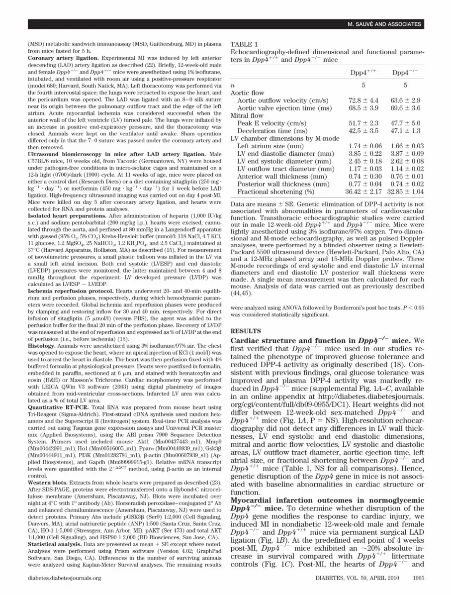
Cardiac structure and function in Dpp4�/� mice. Wefirst verified that Dpp4�/� mice used in our studies re-tained the phenotype of improved glucose tolerance andreduced DPP-4 activity as originally described (18). Con-sistent with previous findings, oral glucose tolerance wasimproved and plasma DPP-4 activity was markedly re-duced in Dpp4�/� mice (supplemental Fig. 1A–C, availablein an online appendix at http://diabetes.diabetesjournals.org/cgi/content/full/db09-0955/DC1). Heart weights did notdiffer between 12-week-old sex-matched Dpp4�/� andDpp4�/� mice (Fig. 1A, P NS). High-resolution echocar-diography did not detect any differences in LV wall thick-nesses, LV end systolic and end diastolic dimensions,mitral and aortic flow velocities, LV systolic and diastolicareas, LV outflow tract diameter, aortic ejection time, leftatrial size, or fractional shortening between Dpp4�/� andDpp4�/� mice (Table 1, NS for all comparisons). Hence,genetic disruption of the Dpp4 gene in mice is not associ-ated with baseline abnormalities in cardiac structure orfunction.Myocardial infarction outcomes in normoglycemicDpp4�/� mice. To determine whether disruption of theDpp4 gene modifies the response to cardiac injury, weinduced MI in nondiabetic 12-week-old male and femaleDpp4�/� and Dpp4�/� mice via permanent surgical LADligation (Fig. 1B). At the predefined end point of 4 weekspost-MI, Dpp4�/� mice exhibited an 20% absolute in-crease in survival compared with Dpp4�/� littermatecontrols (Fig. 1C). Post-MI, the hearts of Dpp4�/� and
TABLE 1Echocardiography-defined dimensional and functional parame-ters in Dpp4�/� and Dpp4�/� mice
Dpp4�/� Dpp4�/�
n 5 5Aortic flow
Aortic outflow velocity (cm/s) 72.8 � 4.4 63.6 � 2.9Aortic valve ejection time (ms) 68.5 � 3.9 69.6 � 3.6
Mitral flowPeak E velocity (cm/s) 51.7 � 2.3 47.7 � 5.0Deceleration time (ms) 42.5 � 3.5 47.1 � 1.3
LV chamber dimensions by M-modeLeft atrium size (mm) 1.74 � 0.06 1.66 � 0.03LV end diastolic diameter (mm) 3.85 � 0.22 3.87 � 0.09LV end systolic diameter (mm) 2.45 � 0.18 2.62 � 0.08LV outflow tract diameter (mm) 1.17 � 0.03 1.14 � 0.02Anterior wall thickness (mm) 0.74 � 0.30 0.76 � 0.01Posterior wall thickness (mm) 0.77 � 0.04 0.74 � 0.02Fractional shortening (%) 36.42 � 2.17 32.85 � 1.04
Data are means � SE. Genetic elimination of DPP-4 activity is notassociated with abnormalities in parameters of cardiovascularfunction. Transthoracic echocardiographic studies were carriedout in male 12-week-old Dpp4�/� and Dpp4�/� mice. Mice werelightly anesthetized using 3% isoflurane/97% oxygen. Two-dimen-sional and M-mode echocardiography, as well as pulsed Doppleranalyses, were performed by a blinded observer using a Hewlett-Packard 5500 ultrasound device (Hewlett-Packard, Palo Alto, CA)and a 12-MHz phased array and 15-MHz Doppler probes. ThreeM-mode recordings of end systolic and end diastolic LV internaldiameters and end diastolic LV posterior wall thickness weremade. A single mean measurement was then calculated for eachmouse. Analysis of data was carried out as previously described(44,45).
M. SAUVE AND ASSOCIATES
diabetes.diabetesjournals.org DIABETES, VOL. 59, APRIL 2010 1065
Dpp4�/� mice underwent similar compensatory hypertro-phy (Fig. 1D). Although infarct size was reduced inDpp4�/� mice, this difference was not statistically signifi-cant (Fig. 1E).
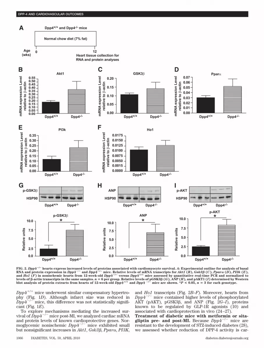
To explore mechanisms mediating the increased sur-vival of Dpp4�/� mice post-MI, we analyzed cardiac mRNAand protein levels of known cardioprotective genes. Nor-moglycemic nonischemic Dpp4�/� mice exhibited smallbut nonsignificant increases in Akt1, Gsk3�, Ppar�, PI3K,
and Ho1 transcripts (Fig. 2B–F). Moreover, hearts fromDpp4�/� mice contained higher levels of phosphorylatedAKT (pAKT), pGSK3�, and ANP (Fig. 2G–I), proteinsknown to be regulated by GLP-1R agonists (10) andassociated with cardioprotection in vivo (24–27).Treatment of diabetic mice with metformin or sita-gliptin pre- and post-MI. Because Dpp4�/� mice areresistant to the development of STZ-induced diabetes (28),we assessed whether reduction of DPP-4 activity is car-
Akt1
PI3k Ho1
mR
NA
ex
pre
ss
ion
Le
ve
l
rela
tiv
e t
o β
-acti
nm
RN
A e
xp
ressio
n L
evel
rela
tive t
o β
-acti
n
mR
NA
exp
ressio
n L
evel
rela
tive t
o β
-acti
n
mR
NA
ex
pre
ss
ion
Le
ve
l
rela
tiv
e t
o β
-acti
n
mR
NA
ex
pre
ss
ion
Le
ve
l
rela
tiv
e t
o β
-acti
n
GSK3β PparαCB
E F
IG H
** *
D
0
A
Age
(wks) 12
Dpp4+/+ and Dpp4-/- mice
Dpp4-/-Dpp4+/+ Dpp4-/-Dpp4+/+
Dpp4-/-Dpp4+/+
Dpp4-/-Dpp4+/+
Dpp4-/-Dpp4+/+ Dpp4-/-Dpp4+/+ Dpp4-/-Dpp4+/+
Dpp4-/-Dpp4+/+ Dpp4-/-Dpp4+/+
p-GSK3β
p-GSK3β
HSP90
Rela
tive u
nit
s
ANP
ANP
HSP90
p-AKT
p-AKT
HSP90
Dpp4-/-Dpp4+/+
Dpp4-/-Dpp4+/+
Normal chow diet (7% fat)
Heart tissue collection for
RNA and protein analyses
0.000.050.100.150.200.250.300.350.400.450.500.55
0.00
0.01
0.02
0.03
0.04
0.05
0.06
0.07
0.0000
0.0015
0.0050
0.0075
0.0100
0.0125
0.0150
0.0175
0.00
0.0
2.5
5.0
7.5
10.0
Rela
tive u
nit
s
0.0
2.5
5.0
7.5
10.0
Rela
tive u
nit
s
0.0
2.5
5.0
7.5
10.0
0.05
0.10
0.15
0.20
0.25
0.30
0.35
0.00
0.05
0.10
0.15
0.20
FIG. 2. Dpp4�/� hearts express increased levels of proteins associated with cardiomyocyte survival. A: Experimental outline for analysis of basalRNA and protein expression in Dpp4�/� and Dpp4�/� mice. Relative levels of mRNA transcripts for Akt1 (B), Gsk3� (C), Ppar� (D), PI3k (E),and Ho1 (F) in nonischemic hearts from 12-week-old Dpp4�/� versus Dpp4�/� mice assessed by quantitative real-time PCR and normalized tolevels of �-actin transcripts in the same samples. n � 6 per group. Relative levels of pGSK3� (G), ANP (H), and pAKT1 (I) determined by Westernblot analysis of protein extracts from hearts of 12-week-old Dpp4�/� and Dpp4 �/� mice are shown. *P < 0.05, n � 3 for each genotype.
DPP-4 AND CARDIOVASCULAR OUTCOMES
1066 DIABETES, VOL. 59, APRIL 2010 diabetes.diabetesjournals.org

dioprotective in diabetic Dpp4�/� mice. Wild-type micewere placed on an HFD for 4 weeks, rendered diabeticwith STZ, and maintained for an additional 12 weeks onHFD alone or on HFD plus either sitagliptin or metformin(Fig. 3A). After 8 weeks on drug treatment or HFD alone,mice were subjected to LAD ligation and observed for anadditional 4 weeks (Fig. 3A). Random blood glucose (Fig.3B), levels of A1C (Fig. 3C), and oral glucose tolerance
(Fig. 3D–E) were improved to a similar extent with bodyweights remaining comparable (Fig. 3F) in mice treatedwith sitagliptin or metformin. Moreover, plasma levels ofactive GLP-1 (7-36)amide were increased to a similarextent in sitagliptin- versus metformin-treated mice (Fig.3G).
Cumulative survival assessed up to 4 weeks after LADligation was improved in mice treated with either sita-
* *
AWild-type C57BL/6 mice
High Fat Diet
(45%)
No treatment
1. High Fat Diet (45%)
2. High Fat Diet + Sitagliptin (250 mg/kg)
3. High Fat Diet + Metformin (450 mg/kg)
4STZ
75 mg/kg
8 15
35HFD/STZ
STZ injection
Day
OGTT AUC
SitagliptinMetformin
HFD/STZSitagliptinMetformin
HFD/STZSitagliptinMetformin
HFD/STZ Sitagliptin Metformin
HFD/STZ Sitagliptin Metformin
HFD/STZ
Sitaglip
tin
Met
form
in
Blo
od
glu
co
se
(mm
ol/
L)
Blo
od
glu
co
se
(mm
ol/L
)B
W (
g)
GL
P-1
(7-3
6)
am
ide (
pg
/mL
)
GLP-1(7-36) amide
30
30 40 50 60
25
20
20
15
10
10
5
0
34
32
30
28
26
24
22
30
404000
3000
2000
1000
0
20
10
0
0
60Time (min)
Weeks
80 100 120 14020 400
OGTT
HbA1c
Hb
A1
c(%
)A
UC
(g
luc
os
e)
HbA1c
LAD
Ligation
Endpoint
Infarct size
16 20
B
ED
C
0123456789 ***
***
***
***
***
***
0 2 4 6 8 10
1011
12 14 16
F
0123456789
G
Age
(wks)
FIG. 3. Sitagliptin and metformin reduce blood glucose levels and increase plasma GLP-1 (7-36)amide levels in diabetic mice. Male C57BL/6 mice were placedon HFD (45% fat) for 4 weeks (A). At the start of week 5, mice were injected with a single dose of STZ (75 mg/kg) and then randomized into three treatmentgroups: 1) HFD/STZ alone, 2) HFD/STZ � sitagliptin (250 mg/kg), or 3) HFD/STZ � metformin (450 mg/kg) for an additional 8 weeks. At week 12, miceunderwent LAD ligation or control sham surgery. At week 16, surviving mice were killed and infarct size was measured. Levels of random fed blood glucose(B) and A1C (C) were significantly reduced in mice treated with sitagliptin or metformin. Oral glucose tolerance (D and E) was significantly improved insitagliptin-treated (n � 29) or metformin-treated (n � 23) mice compared with HFD alone (n � 23). F: Body weight (BW) in mice treated with HFD/STZ alone,sitagliptin, or metformin (n � 22–23 per treatment). Plasma active GLP-1 is increased in mice treated with sitagliptin or metformin (n � 14 per treatment)(G). *P < 0.05, *** P < 0.001 vs. the untreated HFD/STZ group. AUC, area under the curve.
M. SAUVE AND ASSOCIATES
diabetes.diabetesjournals.org DIABETES, VOL. 59, APRIL 2010 1067
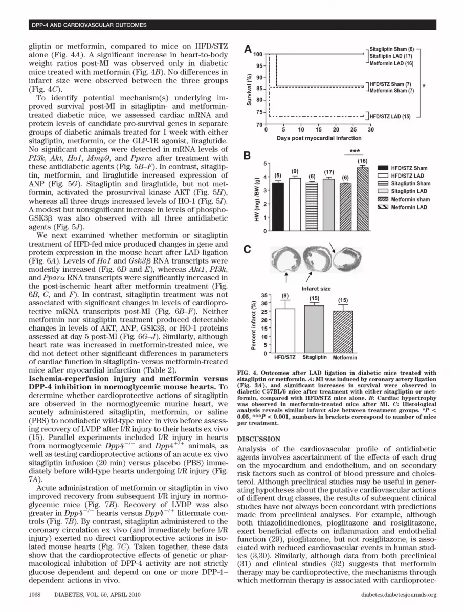
gliptin or metformin, compared to mice on HFD/STZalone (Fig. 4A). A significant increase in heart-to-bodyweight ratios post-MI was observed only in diabeticmice treated with metformin (Fig. 4B). No differences ininfarct size were observed between the three groups(Fig. 4C).
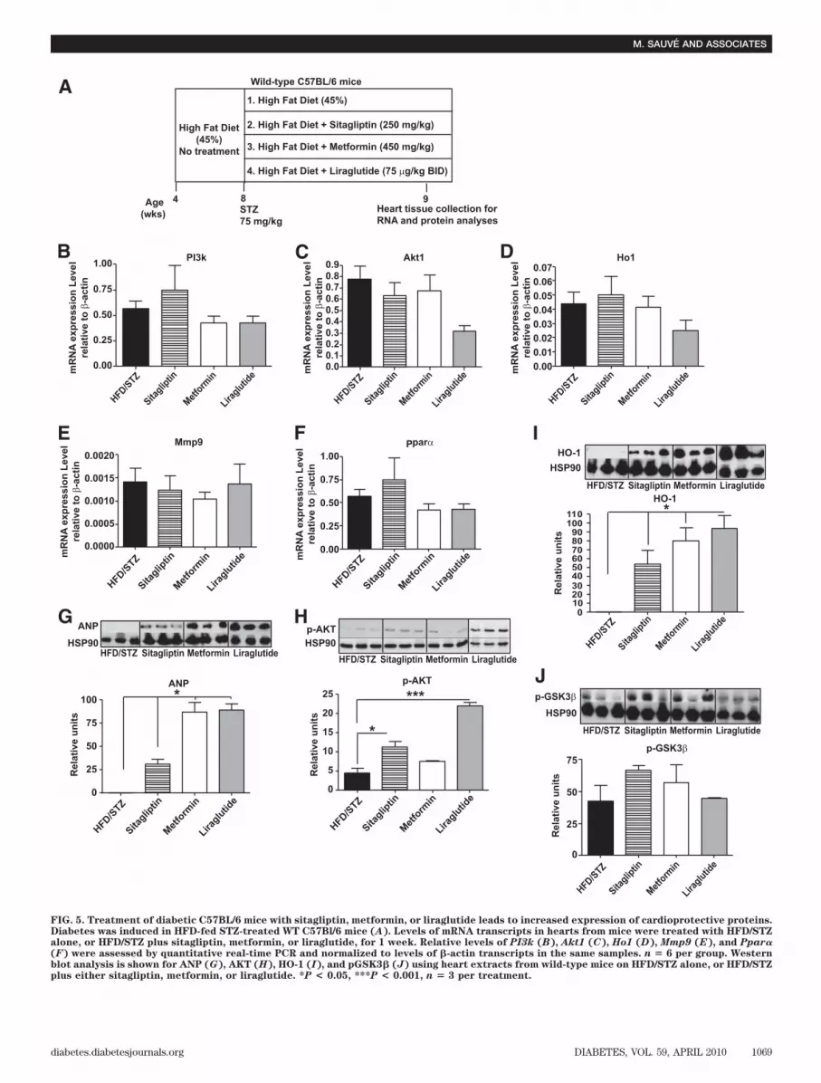
To identify potential mechanism(s) underlying im-proved survival post-MI in sitagliptin- and metformin-treated diabetic mice, we assessed cardiac mRNA andprotein levels of candidate pro-survival genes in separategroups of diabetic animals treated for 1 week with eithersitagliptin, metformin, or the GLP-1R agonist, liraglutide.No significant changes were detected in mRNA levels ofPI3k, Akt, Ho1, Mmp9, and Ppar� after treatment withthese antidiabetic agents (Fig. 5B–F). In contrast, sitaglip-tin, metformin, and liraglutide increased expression ofANP (Fig. 5G). Sitagliptin and liraglutide, but not met-formin, activated the prosurvival kinase AKT (Fig. 5H),whereas all three drugs increased levels of HO-1 (Fig. 5I).A modest but nonsignificant increase in levels of phospho-GSK3� was also observed with all three antidiabeticagents (Fig. 5J).
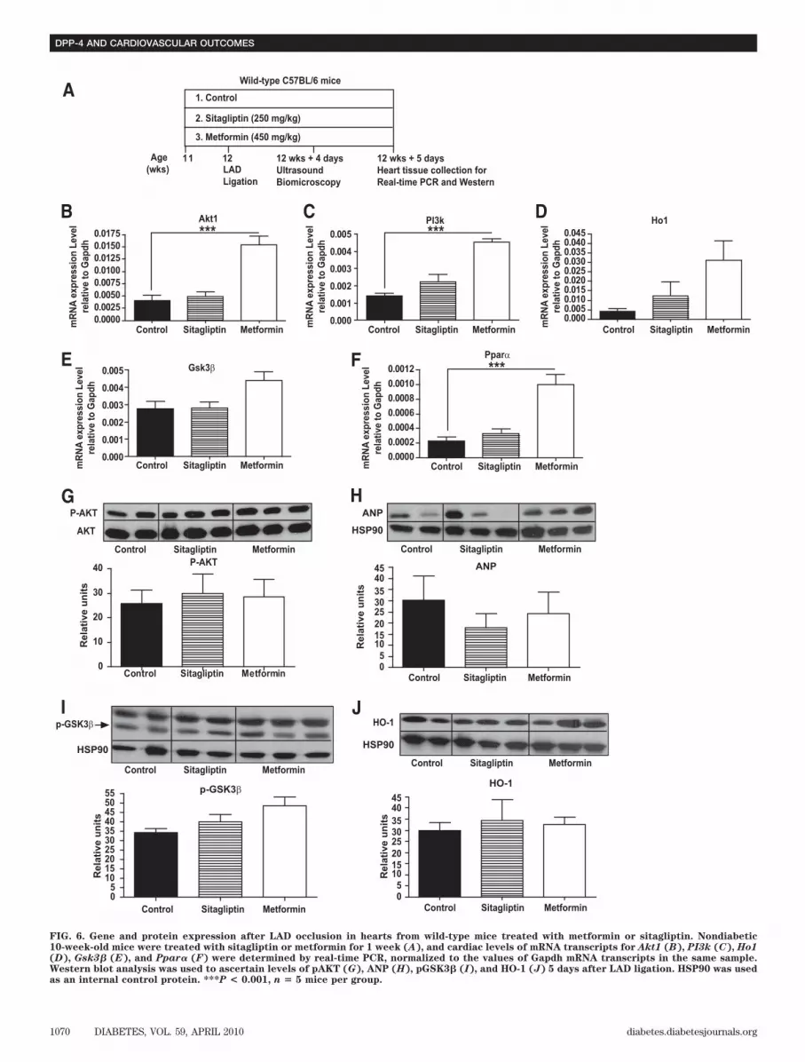
We next examined whether metformin or sitagliptintreatment of HFD-fed mice produced changes in gene andprotein expression in the mouse heart after LAD ligation(Fig. 6A). Levels of Ho1 and Gsk3� RNA transcripts weremodestly increased (Fig. 6D and E), whereas Akt1, PI3k,and Ppar� RNA transcripts were significantly increased inthe post-ischemic heart after metformin treatment (Fig.6B, C, and F). In contrast, sitagliptin treatment was notassociated with significant changes in levels of cardiopro-tective mRNA transcripts post-MI (Fig. 6B–F). Neithermetformin nor sitagliptin treatment produced detectablechanges in levels of AKT, ANP, GSK3�, or HO-1 proteinsassessed at day 5 post-MI (Fig. 6G–J). Similarly, althoughheart rate was increased in metformin-treated mice, wedid not detect other significant differences in parametersof cardiac function in sitagliptin- versus metformin-treatedmice after myocardial infarction (Table 2).Ischemia-reperfusion injury and metformin versusDPP-4 inhibition in normoglycemic mouse hearts. Todetermine whether cardioprotective actions of sitagliptinare observed in the normoglycemic murine heart, weacutely administered sitagliptin, metformin, or saline(PBS) to nondiabetic wild-type mice in vivo before assess-ing recovery of LVDP after I/R injury to their hearts ex vivo(15). Parallel experiments included I/R injury in heartsfrom normoglycemic Dpp4�/� and Dpp4�/� animals, aswell as testing cardioprotective actions of an acute ex vivositagliptin infusion (20 min) versus placebo (PBS) imme-diately before wild-type hearts undergoing I/R injury (Fig.7A).
Acute administration of metformin or sitagliptin in vivoimproved recovery from subsequent I/R injury in normo-glycemic mice (Fig. 7B). Recovery of LVDP was alsogreater in Dpp4�/� hearts versus Dpp4�/� littermate con-trols (Fig. 7B). By contrast, sitagliptin administered to thecoronary circulation ex vivo (and immediately before I/Rinjury) exerted no direct cardioprotective actions in iso-lated mouse hearts (Fig. 7C). Taken together, these datashow that the cardioprotective effects of genetic or phar-macological inhibition of DPP-4 activity are not strictlyglucose dependent and depend on one or more DPP-4–dependent actions in vivo.
DISCUSSION
Analysis of the cardiovascular profile of antidiabeticagents involves ascertainment of the effects of each drugon the myocardium and endothelium, and on secondaryrisk factors such as control of blood pressure and choles-terol. Although preclinical studies may be useful in gener-ating hypotheses about the putative cardiovascular actionsof different drug classes, the results of subsequent clinicalstudies have not always been concordant with predictionsmade from preclinical analyses. For example, althoughboth thiazolidinediones, pioglitazone and rosiglitazone,exert beneficial effects on inflammation and endothelialfunction (29), pioglitazone, but not rosiglitazone, is asso-ciated with reduced cardiovascular events in human stud-ies (3,30). Similarly, although data from both preclinical(31) and clinical studies (32) suggests that metformintherapy may be cardioprotective, the mechanisms throughwhich metformin therapy is associated with cardioprotec-
0 5 10 15 20 25 30
100Sitagliptin Sham (6)
Sitagliptin Sham
Sitafliptin LAD (17)
Sitagliptin LAD
Metformin LAD (16)
Metformin LAD
Metformin Sham (7)
Metformin sham
HFD/STZ Sham (7)
HFD/STZ Sham
HFD/STZ LAD (15)
HFD/STZ LAD
Sitagliptin MetforminHFD/STZ
95
90
85
80
75
70
*
Days post myocardial infarction
Su
rviv
al (%
)
HW
(m
g)
/BW
(g
)P
erc
en
t in
farc
t (%
)
A
C
B
0
5
10
15
20
25
30
35
0
1
2
3
4
5
***
(5)(9)
(9)
(6)(17)
(6)
(16)
(15)
Infarct size
(15)
FIG. 4. Outcomes after LAD ligation in diabetic mice treated withsitagliptin or metformin. A: MI was induced by coronary artery ligation(Fig. 3A), and significant increases in survival were observed indiabetic C57BL/6 mice after treatment with either sitagliptin or met-formin, compared with HFD/STZ mice alone. B: Cardiac hypertrophywas observed in metformin-treated mice after MI. C: Histologicalanalysis reveals similar infarct size between treatment groups. *P <
0.05, ***P < 0.001, numbers in brackets correspond to number of miceper treatment.
DPP-4 AND CARDIOVASCULAR OUTCOMES
1068 DIABETES, VOL. 59, APRIL 2010 diabetes.diabetesjournals.org
E
D
F
CB
0.00
0.25
0.50
0.75
1.00
0.00.10.20.3
0.40.50.6
0.70.80.9
A
4 8 9
Wild-type C57BL/6 mice
High Fat Diet
(45%)
No treatment
1. High Fat Diet (45%)
2. High Fat Diet + Sitagliptin (250 mg/kg)
3. High Fat Diet + Metformin (450 mg/kg)
4. High Fat Diet + Liraglutide (75 µg/kg BID)
STZ
75 mg/kg
Heart tissue collection for
RNA and protein analyses
Age
(wks)
Akt1PI3k
Mmp9 parα
Ho1
HFD/STZ
Sitaglip
tin
Met
form
in
Liraglu
tide
HFD
/STZ
Sita
gliptin
Met
form
inLir
aglu
tide
HFD
/STZ
Sita
gliptin
Met
form
inLir
aglu
tide
HFD/STZ
Sitaglip
tinM
etfo
rmin
Liraglu
tide
HFD/STZ
Sitaglip
tin
Met
form
inLira
glutid
e
HFD/STZ
Sita
gliptin
Met
form
inLira
glutid
e
HFD/STZ
Sitaglip
tin
Met
form
inLira
glutid
e
HFD
/STZ
Sita
gliptin
Met
form
inLir
aglu
tide
HFD
/STZ
Sita
gliptin
Met
form
inLir
aglu
tide
mR
NA
ex
pre
ss
ion
Le
ve
l
rela
tiv
e t
o β
-ac
tin
0.00
0102030405060708090
100110
0.25
0.50
0.75
1.00
mR
NA
ex
pre
ss
ion
Le
ve
l
rela
tiv
e t
o β
-ac
tin
Re
lati
ve
un
its
Re
lati
ve
un
itsR
ela
tiv
e u
nit
s
Re
lati
ve
un
its
mR
NA
ex
pre
ss
ion
Le
ve
l
rela
tiv
e t
o β
-ac
tin
0.00
0.01
0.02
0.03
0.04
0.05
0.06
0.07
mR
NA
ex
pre
ss
ion
Le
ve
l
rela
tiv
e t
o β
-ac
tin
0.0000
0.0005
0.0010
0.0015
0.0020
mR
NA
ex
pre
ss
ion
Le
ve
l
rela
tiv
e t
o β
-ac
tin
ANP
ANP
0
25
50
75
100 *
G H
p-AKT
p-AKT
0
5
10
15
20
25
*
***
HFD/STZ Sitagliptin Metformin Liraglutide
HFD/STZ Sitagliptin Metformin Liraglutide
HFD/STZ Sitagliptin Metformin LiraglutideHFD/STZ Sitagliptin Metformin Liraglutide
HO-1
HO-1
HSP90
HSP90HSP90
HSP90
p-GSK3β
p-GSK3β
I
*
J
0
25
50
75
P
FIG. 5. Treatment of diabetic C57BL/6 mice with sitagliptin, metformin, or liraglutide leads to increased expression of cardioprotective proteins.Diabetes was induced in HFD-fed STZ-treated WT C57Bl/6 mice (A). Levels of mRNA transcripts in hearts from mice were treated with HFD/STZalone, or HFD/STZ plus sitagliptin, metformin, or liraglutide, for 1 week. Relative levels of PI3k (B), Akt1 (C), Ho1 (D), Mmp9 (E), and Ppar�(F) were assessed by quantitative real-time PCR and normalized to levels of �-actin transcripts in the same samples. n � 6 per group. Westernblot analysis is shown for ANP (G), AKT (H), HO-1 (I), and pGSK3� (J) using heart extracts from wild-type mice on HFD/STZ alone, or HFD/STZplus either sitagliptin, metformin, or liraglutide. *P < 0.05, ***P < 0.001, n � 3 per treatment.
M. SAUVE AND ASSOCIATES
diabetes.diabetesjournals.org DIABETES, VOL. 59, APRIL 2010 1069
0.000
0.001
0.002
0.003
0.004
0.005
0.000
0.001
0.002
0.003
0.004
0.005
0.0000
0.0025
0.0050
0.0075
0.0100
0.0125
0.0150
0.0175 ***
***
***
Gsk3β
Pparα
0.0000.0050.0100.0150.0200.0250.0300.0350.0400.045
0.0000
0.0002
0.0004
0.0006
0.0008
0.0010
0.0012
B C D
FE
A
11 12 wks + 4 days
Ultrasound
Biomicroscopy
12 wks + 5 days
Heart tissue collection for
Real-time PCR and Western
12
AKT
P-AKT
P-AKT
Control Sitagliptin Metformin0
10
20
30
40
05
10152025303540455055
05
HO-1
G H
I J
Wild-type C57BL/6 mice
1. Control
2. Sitagliptin (250 mg/kg)
3. Metformin (450 mg/kg)
Control Sitagliptin Metformin Control Sitagliptin Metformin
Control Sitagliptin Metformin
Control Sitagliptin Metformin Control Sitagliptin Metformin
Control Sitagliptin MetforminControl Sitagliptin Metformin
Control Sitagliptin Metformin Control Sitagliptin Metformin
Control Sitagliptin Metformin
Control Sitagliptin Metformin
Control Sitagliptin Metformin
Age
(wks) LAD
Ligation
Akt1 PI3k Ho1
mR
NA
ex
pre
ss
ion
Le
ve
l
rela
tiv
e t
o G
ap
dh
mR
NA
ex
pre
ss
ion
Le
ve
l
rela
tiv
e t
o G
ap
dh
mR
NA
ex
pre
ss
ion
Le
ve
l
rela
tiv
e t
o G
ap
dh
mR
NA
ex
pre
ss
ion
Le
ve
l
rela
tiv
e t
o G
ap
dh
mR
NA
ex
pre
ss
ion
Le
ve
l
rela
tiv
e t
o G
ap
dh
ANP
ANP
HSP90
HSP90
Rela
tive u
nit
s
Rela
tive u
nit
s
Rela
tive u
nit
s
101520
25
3530
4045
05
Rela
tive u
nit
s
101520
25
3530
4045
p-GSK3β
p-GSK3β
HO-1
HSP90
FIG. 6. Gene and protein expression after LAD occlusion in hearts from wild-type mice treated with metformin or sitagliptin. Nondiabetic10-week-old mice were treated with sitagliptin or metformin for 1 week (A), and cardiac levels of mRNA transcripts for Akt1 (B), PI3k (C), Ho1(D), Gsk3� (E), and Ppar� (F) were determined by real-time PCR, normalized to the values of Gapdh mRNA transcripts in the same sample.Western blot analysis was used to ascertain levels of pAKT (G), ANP (H), pGSK3� (I), and HO-1 (J) 5 days after LAD ligation. HSP90 was usedas an internal control protein. ***P < 0.001, n � 5 mice per group.
DPP-4 AND CARDIOVASCULAR OUTCOMES
1070 DIABETES, VOL. 59, APRIL 2010 diabetes.diabetesjournals.org

tion remain poorly understood. Moreover, sulfonylureashave been associated with increased rates of death fromcardiovascular disease in some (33) but not all studies(32).
We have now investigated the cardiovascular conse-quences arising from genetic elimination or pharmacolog-ical inhibition of DPP-4 activity. DPP-4 has three majorfunctions; adenosine deaminase binding, peptidase activ-ity, and extracellular matrix binding, all of which poten-tially influence the activity of the immune and/orendocrine systems (34). Although DPP-4 cleaves and inac-tivates several cardioactive peptides, including neuropep-tide Y, brain natriuretic peptide, SDF-1, and GLP-1, thereis little information on the cardiovascular consequencesof reduced or absent DPP-4 activity. NormoglycemicDpp4�/� mice exhibit normal cardiac structure and func-tion in the basal state, yet increased survival after exper-imental MI. Whether the increased survival after LADligation is directly due to loss of DPP-4 activity per se incardiomyocytes or blood vessels, or indirectly due to thesubsequent upregulation of cardioprotective moleculessuch as GLP-1 (18) or SDF-1 (35), cannot be inferred fromthe present study. Zaruba et al. (35) also observed modestimprovements in survival after experimental MI inDpp4�/� mice or in WT mice treated with a DPP-4inhibitor, and more robust improvements in survival wereobserved after administration of G-CSF, findings attrib-uted to SDF-1–dependent mobilization of cardiac stemcells. Our studies extend their observations through exam-ination of the cardiovascular effects of DPP-4 inhibition indiabetic mice and by demonstrating that direct sitagliptinadministration into the circulation of the ischemic mouseheart is not directly cardioprotective ex vivo, suggestingthat acute reduction of cardiac DPP-4 activity is notsufficient to produce cardioprotection.
Our findings demonstrating that both sitagliptin and theGLP-1R agonist liraglutide upregulated levels of cardiopro-tective proteins in the nonischemic myocardium suggest apossible role for GLP-1 in the context of enhanced survival
after DPP-4 inhibition and experimental MI. Nevertheless,we did not observe a sustained induction of cardioprotec-tive proteins in sitagliptin-treated murine hearts when thesame proteins were examined after MI. Moreover, we didnot detect significant changes in infarct size or cardiacfunction after MI that might directly account for theimproved survival seen with genetic or chemical reductionof DPP-4 activity. Hence, the precise mechanisms mediat-ing the improvements in survival observed after pharma-cological treatment with sitagliptin in diabetic mice orgenetic reduction of DPP-4 activity in normoglycemicDpp4�/� mice require further investigation, ideallythrough examination of whether DPP-4 inhibitors exertcardioprotective actions in Glp1r�/� mice.
Western blot analysis of proteins in nonischemic heartsdemonstrated that both sitagliptin and metformin therapyinduced an overlapping set of cardioprotective proteins.Metformin is thought to exert its cardioprotective actionsthrough distinct mechanisms requiring activation of AMPkinase and endothelial nitric oxide (31). Intriguingly, ad-ministration of metformin has also been associated withreduction of DPP-4 activity (36) and increased circulatinglevels of GLP-1 in both rodent (37) and clinical studies(38), and we detected increased levels of GLP-1 in bothmetformin- and sitagliptin-treated mice. Accordingly, theextent to which therapy with sitagliptin and metforminproduces an overlapping spectrum of actions reflectingsimilarities in their mechanism(s) of action through en-hanced levels of GLP-1 requires further clarification.
Both metformin and sitagliptin significantly increasedsurvival in diabetic mice, possibly due to a comparablereduction in blood glucose achieved with either agent.Hyperglycemia is a risk factor for a poor outcome after MIin humans (39), and there is considerable interest indetermining whether intensive glucose control safely andconsistently improves outcomes post-MI (40). Similarly,hyperglycemia is known to be associated with reducedsurvival and impaired LV function in mice after MI (41–43), and it seems likely that reduction in the severity of
TABLE 2Ultrasound biomicroscopy-defined cardiac hemodynamic, functional, and dimensional parameters in mice treated with either control,sitagliptin, or metformin on day 4 post-LAD ligation
Control Sitagliptin Metformin
n 6 6 5Aortic flow
Heart rate (bpm) 441 � 17 423 � 19 526 � 23*VTImax (cm) 2.64 � 0.14 2.40 � 0.08 2.39 � 0.30Aortic orifice diameter (mm) 1.04 � 0.04 1.06 � 0.03 1.04 � 0.03LV stroke volume (�l) 22.38 � 1.24 21.10 � 1.33 20.34 � 3.07Cardiac output (ml/min) 9.82 � 0.46 8.95 � 0.71 10.70 � 1.66
Mitral flowHeart rate (bpm) 447 � 13 433 � 16 792 � 33Peak E velocity (cm/s) 67.6 � 3.0 62.6 � 2.4 53.4 � 6.2Peak A velocity (cm/s) 48.0 � 49 36.0 � 40 54.6 � 73Peak E/A ratio 1.47 � 0.15 1.89 � 0.29 0.99 � 0.08
LV chamber dimensions by M-modeHeart rate (bpm) 493 � 35 458 � 15 541 � 18LV end diastolic diameter (mm) 4.26 � 0.15 4.28 � 0.19 4.43 � 0.16LV end systolic diameter (mm) 3.54 � 0.27 3.68 � 0.21 3.89 � 0.22LV fractional shortening (%) 17.3 � 3.5 14.2 � 1.2 12.4 � 2.4
Data are means � SE. The 12-week-old C57Bl/6 mice treated with control, sitagliptin, or metformin for 1 week before experimental cardiacischemic injury were subjected to high-frequency ultrasound imaging. VTImax, velocity-time integral of Doppler flow waveform by tracing themaximal velocity. In mitral inflow, the peak E velocity represents the maximal velocity of the early diastolic wave caused by active leftventricular relaxation. The peak A velocity represents the maximal velocity caused by atrial contraction in late diastole. Acquisition of imagesand analysis of data were carried out as previously described (44,45). *P � 0.05 vs. control.
M. SAUVE AND ASSOCIATES
diabetes.diabetesjournals.org DIABETES, VOL. 59, APRIL 2010 1071

hyperglycemia contributes to improved survival, perhapsindependent of the antidiabetic mechanisms unique toeach agent under study.
In contrast, the increased survival observed in normo-glycemic Dpp4�/� mice after MI supports the concept thatreduction of DPP-4 activity may be cardioprotective in theabsence of hyperglycemia (35). Similarly, our observationsdemonstrating that genetic or chemical inhibition of DPP-4is associated with enhanced recovery of LVDP in thenormoglycemic ischemic murine heart ex vivo suggest thatDPP-4 modifies cardiovascular outcomes independent of
glucoregulation and provide a useful model for futurestudies. Given the increasing interest in using strategiesbased on DPP-4 inhibition for the treatment of diabetes, amore detailed understanding of the role of DPP-4 in thenormal and diabetic cardiovascular system is clearlywarranted.
ACKNOWLEDGMENTS
M.S. was supported by a BBDC–Novo Nordisk studentshipaward and a Government of Ontario/Heart and Stroke Foun-dation of Ontario (OGSST) Award. D.J.D. was supported bya Chair in Regulatory Peptides from the Canada ResearchChairs Program. D.J.D. has served as an advisor or consul-tant within the past 12 months to Amylin Pharmaceuticals,Arena Pharmaceuticals, Arisaph Pharmaceuticals, Eli Lilly,GlaxoSmithKline, Glenmark Pharmaceuticals, Hoffman La-Roche, Isis Pharmaceuticals, Merck Research Laboratories,Metabolex, Novartis Pharmaceuticals, Novo Nordisk, Pheno-mix, and Transition Pharmaceuticals. D.J.D. receives partialoperating grant support for DPP-4–related research fromMerck Frosst. M.H. has served as a consultant within the past12 months to sanofi-aventis and Merck. M.H. was supportedby a Career Investigator Award (CI-5503) of the Heart &Stroke Foundation of Ontario (HSFO). This work was sup-ported in part by operating grants IRO-80668 from theCanadian Institutes of Health Research (to D.J.D.) and NA-5926 from the HSFO (to M.H. and D.J.D.) in conjunction withgrant support from Merck Frosst Inc. No other potentialconflicts of interest relevant to this article were reported.
REFERENCES
1. Buse JB, Ginsberg HN, Bakris GL, Clark NG, Costa F, Eckel R, Fonseca V,
Gerstein HC, Grundy S, Nesto RW, Pignone MP, Plutzky J, Porte D, Redberg R,
Stitzel KF, Stone NJ, American Heart Association, American Diabetes Associa-
tion. Primary prevention of cardiovascular diseases in people with diabetes
mellitus: a scientific statement from the American Heart Association and the
American Diabetes Association. Diabetes Care 2007;30:162–172
2. Action to Control Cardiovascular Risk in Diabetes Study Group, Gerstein
HC, Miller ME, Byington RP, Goff DC Jr, Bigger JT, Buse JB, Cushman WC,
Genuth S, Ismail-Beigi F, Grimm RH Jr, Probstfield JL, Simons-Morton DG,
Friedewald WT. Effects of intensive glucose lowering in type 2 diabetes.
N Engl J Med 2008;358:2545–2559
3. Nissen SE, Wolski K. Effect of rosiglitazone on the risk of myocardial
infarction and death from cardiovascular causes. N Engl J Med 2007;356:
2457–2471
4. Drucker DJ, Nauck MA. The incretin system: glucagon-like peptide-1
receptor agonists and dipeptidyl peptidase-4 inhibitors in type 2 diabetes.
Lancet 2006;368:1696–1705
5. Basu A, Charkoudian N, Schrage W, Rizza RA, Basu R, Joyner MJ.
Beneficial effects of GLP-1 on endothelial function in humans: dampening
by glyburide but not by glimepiride. Am J Physiol Endocrinol Metab
2007;293:E1289–E1295
6. Nikolaidis LA, Mankad S, Sokos GG, Miske G, Shah A, Elahi D, Shannon
RP. Effects of glucagon-like peptide-1 in patients with acute myocardial
infarction and left ventricular dysfunction after successful reperfusion.
Circulation 2004;109:962–965
7. Sokos GG, Nikolaidis LA, Mankad S, Elahi D, Shannon RP. Glucagon-like
peptide-1 infusion improves left ventricular ejection fraction and functional status
in patients with chronic heart failure. J Card Fail 2006;12:694–699
8. Bose AK, Mocanu MM, Carr RD, Brand CL, Yellon DM. Glucagon-like
peptide 1 can directly protect the heart against ischemia/reperfusion
injury. Diabetes 2005;54:146–151
9. Bose AK, Mocanu MM, Carr RD, Yellon DM. Myocardial ischaemia-
reperfusion injury is attenuated by intact glucagon like peptide-1 (GLP-1)
in the in vitro rat heart and may involve the p70s6K pathway. Cardiovasc
Drugs Ther 2007;21:253–256
10. Noyan-Ashraf MH, Momen MA, Ban K, Sadi AM, Zhou YQ, Riazi AM, Baggio
LL, Henkelman RM, Husain M, Drucker DJ. GLP-1R agonist liraglutide
activates cytoprotective pathways and improves outcomes after experi-
mental myocardial infarction in mice. Diabetes 2009;58:975–983
11. Garber A, Henry R, Ratner R, Garcia-Hernandez PA, Rodriguez-Pattzi H,
20 min
Equilibrate
40 min
Perfusion
40 min
Reperfusion
30 min
Ischemia
(C) Pre-treatment (20 min)A
*
*
0 20 40 60 80 100 120
0 20 40 60 80 100 120
0
20
40
60
80
100
120
LV
DP
(m
m H
g)
0
20
40
60
80
100
120
LV
DP
(m
m H
g)
Time (min)
Time (min)
PBS infusion (5)
Sitagliptin infusion (5)
PBS IP (5)
Metformin IP (6)
Sitagliptin IP (6)
Dpp 4+/+ (5)
Dpp 4-/- (6)
B
C
FIG. 7. Functional recovery after I/R injury in the murine heart. A:Experimental protocol for I/R injury in isolated mouse hearts. B:Intraperitoneal (IP) injections of sitagliptin and metformin in 12-week-old wild-type mice in vivo reduced I/R injury of their hearts exvivo. The 12-week-old Dpp4�/� mice display significantly greater im-provement in functional recovery after I/R compared with Dpp4�/�
littermate controls. C: Direct 20-min infusion of sitagliptin started(arrow) before ischemia did not improve functional recovery after I/Rinjury in the isolated heart from WT mice. *P < 0.05, numbers inbrackets correspond to number of mice per treatment.
DPP-4 AND CARDIOVASCULAR OUTCOMES
1072 DIABETES, VOL. 59, APRIL 2010 diabetes.diabetesjournals.org

Olvera-Alvarez I, Hale PM, Zdravkovic M, Bode B, LEAD-3 (Mono) Study
Group. Liraglutide versus glimepiride monotherapy for type 2 diabetes
(LEAD-3 Mono): a randomised, 52-week, phase III, double-blind, parallel-
treatment trial. Lancet 2009;373:473–481
12. Moretto TJ, Milton DR, Ridge TD, Macconell LA, Okerson T, Wolka AM,
Brodows RG. Efficacy and tolerability of exenatide monotherapy over 24
weeks in antidiabetic drug-naive patients with type 2 diabetes: a random-
ized, double-blind, placebo-controlled, parallel-group study. Clin Ther
2008;30:1448–1460
13. Lovshin JA, Drucker DJ. Incretin-based therapies for type 2 diabetes
mellitus. Nat Rev Endocrinol 2009;5:262–269
14. Drucker DJ. Dipeptidyl peptidase-4 inhibition and the treatment of type 2
diabetes: preclinical biology and mechanisms of action. Diabetes Care
2007;30:1335–1343
15. Ban K, Noyan-Ashraf MH, Hoefer J, Bolz SS, Drucker DJ, Husain M.
Cardioprotective and vasodilatory actions of glucagon-like peptide 1
receptor are mediated through both glucagon-like peptide 1 receptor-
dependent and -independent pathways. Circulation 2008;117:2340–2350
16. Sonne DP, Engstrøm T, Treiman M. Protective effects of GLP-1 analogues
exendin-4 and GLP-1(9–36) amide against ischemia-reperfusion injury in
rat heart. Regul Pept 2008;146:243–249
17. Nikolaidis LA, Elahi D, Shen YT, Shannon RP. Active metabolite of GLP-1
mediates myocardial glucose uptake and improves left ventricular perfor-
mance in conscious dogs with dilated cardiomyopathy. Am J Physiol Heart
Circ Physiol 2005;289:H2401–H2408
18. Marguet D, Baggio L, Kobayashi T, Bernard AM, Pierres M, Nielsen PF,
Ribel U, Watanabe T, Drucker DJ, Wagtmann N. Enhanced insulin secre-
tion and improved glucose tolerance in mice lacking CD26. Proc Natl Acad
Sci U S A 2000;97:6874–6879
19. Lamont BJ, Drucker DJ. Differential antidiabetic efficacy of incretin
agonists versus DPP-4 inhibition in high fat fed mice. Diabetes 2008;57:
190–198
20. Hull RL, Shen ZP, Watts MR, Kodama K, Carr DB, Utzschneider KM, Zraika
S, Wang F, Kahn SE. Long-term treatment with rosiglitazone and met-
formin reduces the extent of, but does not prevent, islet amyloid deposi-
tion in mice expressing the gene for human islet amyloid polypeptide.
Diabetes 2005;54:2235–2244
21. Maida A, Hansotia T, Longuet C, Seino Y, Drucker DJ. Differential
importance of glucose-dependent insulinotropic polypeptide vs glucagon-
like peptide 1 receptor signaling for beta cell survival in mice. Gastroen-
terology 2009;137:2146–2157
22. Ohta K, Nakajima T, Cheah AY, Zaidi SH, Kaviani N, Dawood F, You XM,
Liu P, Husain M, Rabinovitch M. Elafin-overexpressing mice have im-
proved cardiac function after myocardial infarction. Am J Physiol Heart
Circ Physiol 2004;287:H286–H292
23. Koehler JA, Drucker DJ. Activation of glucagon-like peptide-1 receptor
signaling does not modify the growth or apoptosis of human pancreatic
cancer cells. Diabetes 2006;55:1369–1379
24. Shiraishi I, Melendez J, Ahn Y, Skavdahl M, Murphy E, Welch S, Schaefer
E, Walsh K, Rosenzweig A, Torella D, Nurzynska D, Kajstura J, Leri A,
Anversa P, Sussman MA. Nuclear targeting of Akt enhances kinase activity
and survival of cardiomyocytes. Circ Res 2004;94:884–891
25. Yet SF, Tian R, Layne MD, Wang ZY, Maemura K, Solovyeva M, Ith B, Melo
LG, Zhang L, Ingwall JS, Dzau VJ, Lee ME, Perrella MA. Cardiac-specific
expression of heme oxygenase-1 protects against ischemia and reperfusion
injury in transgenic mice. Circ Res 2001;89:168–173
26. Heineke J, Molkentin JD. Regulation of cardiac hypertrophy by intracellu-
lar signalling pathways. Nat Rev Mol Cell Biol 2006;7:589–600
27. Juhaszova M, Zorov DB, Kim SH, Pepe S, Fu Q, Fishbein KW, Ziman BD,
Wang S, Ytrehus K, Antos CL, Olson EN, Sollott SJ. Glycogen synthase
kinase-3beta mediates convergence of protection signaling to inhibit the
mitochondrial permeability transition pore. J Clin Invest 2004;113:1535–
1549
28. Conarello SL, Li Z, Ronan J, Roy RS, Zhu L, Jiang G, Liu F, Woods J,
Zycband E, Moller DE, Thornberry NA, Zhang BB. Mice lacking dipeptidyl
peptidase IV are protected against obesity and insulin resistance. Proc Natl
Acad Sci U S A 2003;100:6825–6830
29. Parulkar AA, Pendergrass ML, Granda-Ayala R, Lee TR, Fonseca VA.
Nonhypoglycemic effects of thiazolidinediones. Ann Intern Med 2001;134:
61–71
30. Lincoff AM, Wolski K, Nicholls SJ, Nissen SE. Pioglitazone and risk of
cardiovascular events in patients with type 2 diabetes mellitus: a meta-
analysis of randomized trials. JAMA 2007;298:1180–1188
31. Calvert JW, Gundewar S, Jha S, Greer JJ, Bestermann WH, Tian R, Lefer
DJ. Acute metformin therapy confers cardioprotection against myocardial
infarction via AMPK-eNOS-mediated signaling. Diabetes 2008;57:696–705
32. Holman RR, Paul SK, Bethel MA, Matthews DR, Neil HA. 10-year follow-up
of intensive glucose control in type 2 diabetes. N Engl J Med 2008;359:
1577–1589
33. Simpson SH, Majumdar SR, Tsuyuki RT, Eurich DT, Johnson JA. Dose-
response relation between sulfonylurea drugs and mortality in type 2
diabetes mellitus: a population-based cohort study. CMAJ 2006;174:169–
174
34. Aytac U, Dang NH. CD26/dipeptidyl peptidase IV: a regulator of immune
function and a potential molecular target for therapy. Curr Drug Targets
Immune Endocr Metabol Disord 2004;4:11–18
35. Zaruba MM, Theiss HD, Vallaster M, Mehl U, Brunner S, David R, Fischer
R, Krieg L, Hirsch E, Huber B, Nathan P, Israel L, Imhof A, Herbach N,
Assmann G, Wanke R, Mueller-Hoecker J, Steinbeck G, Franz WM. Synergy
between CD26/DPP-IV inhibition and G-CSF improves cardiac function
after acute myocardial infarction. Cell Stem Cell 2009;4:313–323
36. Green BD, Irwin N, Duffy NA, Gault VA, O’harte FP, Flatt PR. Inhibition of
dipeptidyl peptidase-IV activity by metformin enhances the antidiabetic
effects of glucagon-like peptide-1. Eur J Pharmacol 2006;547:192–199
37. Yasuda N, Inoue T, Nagakura T, Yamazaki K, Kira K, Saeki T, Tanaka I.
Enhanced secretion of glucagon-like peptide 1 by biguanide compounds.
Biochem Biophys Res Commun 2002;298:779–784
38. Mannucci E, Tesi F, Bardini G, Ognibene A, Petracca MG, Ciani S, Pezzatini
A, Brogi M, Dicembrini I, Cremasco F, Messeri G, Rotella CM. Effects of
metformin on glucagon-like peptide-1 levels in obese patients with and
without type 2 diabetes. Diabetes Nutr Metab 2004;17:336–342
39. Verges B, Zeller M, Dentan G, Beer JC, Laurent Y, Janin-Manificat L, Makki
H, Wolf JE, Cottin Y. Impact of fasting glycemia on short-term prognosis
after acute myocardial infarction. J Clin Endocrinol Metab 2007;92:2136–
2140
40. Weston C, Walker L, Birkhead J, National Audit of Myocardial Infarction
Project, National Institute for Clinical Outcomes Research. Early impact of
insulin treatment on mortality for hyperglycaemic patients without known
diabetes who present with an acute coronary syndrome. Heart 2007;93:
1542–1546
41. Shiomi T, Tsutsui H, Ikeuchi M, Matsusaka H, Hayashidani S, Suematsu N,
Wen J, Kubota T, Takeshita A. Streptozotocin-induced hyperglycemia
exacerbates left ventricular remodeling and failure after experimental
myocardial infarction. J Am Coll Cardiol 2003;42:165–172
42. Greer JJ, Ware DP, Lefer DJ. Myocardial infarction and heart failure in the
db/db diabetic mouse. Am J Physiol Heart Circ Physiol 2006;290:H146–
H153
43. Liu X, Wei J, Peng DH, Layne MD, Yet SF. Absence of heme oxygenase-1
exacerbates myocardial ischemia/reperfusion injury in diabetic mice.
Diabetes 2005;54:778–784
44. Zhou YQ, Foster FS, Nieman BJ, Davidson L, Chen XJ, Henkelman RM.
Comprehensive transthoracic cardiac imaging in mice using ultrasound
biomicroscopy with anatomical confirmation by magnetic resonance im-
aging. Physiol Genomics 2004;18:232–244
45. Zhou YQ, Zhu Y, Bishop J, Davidson L, Henkelman RM, Bruneau BG,
Foster FS. Abnormal cardiac inflow patterns during postnatal development
in a mouse model of Holt-Oram syndrome. Am J Physiol Heart Circ Physiol
2005;289:H992–H1001
M. SAUVE AND ASSOCIATES
diabetes.diabetesjournals.org DIABETES, VOL. 59, APRIL 2010 1073
Copyright © 2022 FDOKUMEN