Abnormal cortical activation during response inhibition in 22q11.2 deletion syndrome
10
Abnormal Cortical Activation During Response Inhibition in 22q11.2 Deletion Syndrome Doron Gothelf, 1,2 * Fumiko Hoeft, 3 Christine Hinard, 4 Joachim F. Hallmayer, 3 John Van Dover Stoecker, 3 Stylianos E. Antonarakis, 5 Michael A. Morris, 4 and Allan L. Reiss 3 1 The Behavioral Neurogenetics Center, Child Psychiatry Department, Schneider Children’s Medical Center of Israel, Petah Tiqwa, Israel 2 Sackler Faculty of Medicine, Tel Aviv University, Tel Aviv, Israel 3 Center for Interdisciplinary Brain Sciences Research (CIBSR), Department of Psychiatry and Behavioral Sciences, Stanford University School of Medicine, Stanford, California 4 Service of Medical Genetics, Geneva University Hospitals, Geneva, Switzerland 5 Department of Genetic Medicine and Development, University of Geneva Medical School, Geneva, Switzerland Abstract: 22q11.2 deletion syndrome (22q11.2DS) is a well-known genetic risk factor for schizophrenia. The catechol-O-methyltransferase (COMT) gene falls within the 22q11.2 minimal critical region of the deletion. Brain activity, as measured by functional magnetic resonance imaging (fMRI) during a Go/NoGo, response inhibition task was assessed in adolescents with 22q11.2DS (n ¼ 13), typically developing (TD) controls (n ¼ 14), and controls with developmental disability (DD, n ¼ 9). Subjects with 22q11.2DS were also genotyped for the COMT Met/Val polymorphism. Groups did not differ on task performance. However, compared to both control groups, the 22q11.2DS group showed greater brain activation within left parietal regions. Comparison of brain activation between 22q11.2DS Met and Val subgroups revealed significantly increased activation (Met>Val) in the cingulate but not the dorsolateral prefrontal cortex. These preliminary findings suggest that adolescents with 22q11.2DS compensate for executive dysfunction via recruitment of parietal regions. Fur- ther, the COMT Met subgroup of 22q11.2DS may recruit additional cingulate activation for tasks requiring attention and inhibition. 22q11.2DS is a unique model for learning about the deleterious effects of decreased dosage of the COMT gene on brain function. Hum Brain Mapp 28:533–542, 2007. V V C 2007 Wiley-Liss, Inc. Key words: velocardiofacial syndrome; schizophrenia; functional MRI; imaging genetics; COMT; parie- tal lobe; cingulate INTRODUCTION The 22q11.2 deletion syndrome (22q11.2DS), also known as DiGeorge/velocardiofacial syndrome is caused by a microdeletion on chromosome 22 and occurs in at least 1:5000 live births [Botto et al., 2003]. The syndrome has more than 180 possible physical manifestations, the most prominent of which are congenital anomalies of the palate, heart and great vessels and typical facial features (the velocardiofacial manifestations), hypocalcaemia, and T- cells deficiency (the DiGeorge manifestations) [Kirkpatrick and DiGeorge, 1968; Shprintzen, 2000]. In addition to the Contract grant sponsor: NARSAD Young Investigator Award; Contract grant sponsor: NIH; Contract grant numbers: HD31715, MH50047, MH19908; Contract grant sponsor: ‘‘Child Care Founda- tion,’’ Swiss National Science Foundation, European Union. *Correspondence to: Dr. Doron Gothelf, Child Psychiatry Depart- ment, Schneider Children’s Medical Center of Israel, 14 Kaplan St., Petah Tiqwa 49202, Israel. E-mail: [email protected] Received for publication 12 December 2006; Revised 28 February 2007; Accepted 5 March 2007 DOI: 10.1002/hbm.20405 Published online 10 April 2007 in Wiley InterScience (www. interscience.wiley.com). V V C 2007 Wiley-Liss, Inc. r Human Brain Mapping 28:533–542 (2007) r
Transcript of Abnormal cortical activation during response inhibition in 22q11.2 deletion syndrome

Abnormal Cortical Activation During ResponseInhibition in 22q11.2 Deletion Syndrome
Doron Gothelf,1,2* Fumiko Hoeft,3 Christine Hinard,4 Joachim F. Hallmayer,3
John Van Dover Stoecker,3 Stylianos E. Antonarakis,5
Michael A. Morris,4 and Allan L. Reiss3
1The Behavioral Neurogenetics Center, Child Psychiatry Department, Schneider Children’s MedicalCenter of Israel, Petah Tiqwa, Israel
2Sackler Faculty of Medicine, Tel Aviv University, Tel Aviv, Israel3Center for Interdisciplinary Brain Sciences Research (CIBSR), Department of Psychiatry and Behavioral
Sciences, Stanford University School of Medicine, Stanford, California4Service of Medical Genetics, Geneva University Hospitals, Geneva, Switzerland
5Department of GeneticMedicine andDevelopment, University of GenevaMedical School, Geneva, Switzerland
Abstract: 22q11.2 deletion syndrome (22q11.2DS) is a well-known genetic risk factor for schizophrenia. Thecatechol-O-methyltransferase (COMT) gene falls within the 22q11.2 minimal critical region of the deletion.Brain activity, as measured by functional magnetic resonance imaging (fMRI) during a Go/NoGo, responseinhibition task was assessed in adolescents with 22q11.2DS (n ¼ 13), typically developing (TD) controls (n ¼14), and controls with developmental disability (DD, n ¼ 9). Subjects with 22q11.2DS were also genotyped forthe COMT Met/Val polymorphism. Groups did not differ on task performance. However, compared to bothcontrol groups, the 22q11.2DS group showed greater brain activation within left parietal regions. Comparisonof brain activation between 22q11.2DS Met and Val subgroups revealed significantly increased activation(Met>Val) in the cingulate but not the dorsolateral prefrontal cortex. These preliminary findings suggest thatadolescents with 22q11.2DS compensate for executive dysfunction via recruitment of parietal regions. Fur-ther, the COMT Met subgroup of 22q11.2DS may recruit additional cingulate activation for tasks requiringattention and inhibition. 22q11.2DS is a unique model for learning about the deleterious effects of decreaseddosage of the COMT gene on brain function.Hum Brain Mapp 28:533–542, 2007. VVC 2007Wiley-Liss, Inc.
Key words: velocardiofacial syndrome; schizophrenia; functional MRI; imaging genetics; COMT; parie-tal lobe; cingulate
INTRODUCTION
The 22q11.2 deletion syndrome (22q11.2DS), also knownas DiGeorge/velocardiofacial syndrome is caused by amicrodeletion on chromosome 22 and occurs in at least1:5000 live births [Botto et al., 2003]. The syndrome hasmore than 180 possible physical manifestations, the mostprominent of which are congenital anomalies of the palate,heart and great vessels and typical facial features (thevelocardiofacial manifestations), hypocalcaemia, and T-cells deficiency (the DiGeorge manifestations) [Kirkpatrickand DiGeorge, 1968; Shprintzen, 2000]. In addition to the
Contract grant sponsor: NARSAD Young Investigator Award;Contract grant sponsor: NIH; Contract grant numbers: HD31715,MH50047, MH19908; Contract grant sponsor: ‘‘Child Care Founda-tion,’’ Swiss National Science Foundation, European Union.
*Correspondence to: Dr. Doron Gothelf, Child Psychiatry Depart-ment, Schneider Children’s Medical Center of Israel, 14 KaplanSt., Petah Tiqwa 49202, Israel. E-mail: [email protected]
Received for publication 12 December 2006; Revised 28 February2007; Accepted 5 March 2007
DOI: 10.1002/hbm.20405Published online 10 April 2007 in Wiley InterScience (www.interscience.wiley.com).
VVC 2007 Wiley-Liss, Inc.
r Human Brain Mapping 28:533–542 (2007) r
physical phenotype, there is a high rate of psychiatric mor-bidity in subjects with 22q11.2DS and most subjects with22q11.2DS suffer from learning disabilities [Feinstein et al.,2002; Gothelf et al., 2004; Murphy et al., 1999; Swillenet al., 1997]. There is an overall delay in cognitive develop-ment in subjects with 22q11.2DS with an average overallIQ in the range of 70–85 [Swillen et al., 1997].The 22q11.2DS is a promising model to learn about risk
factors for the evolution of schizophrenia. This is becauseup to one third of individuals with this condition developschizophrenia or related psychotic disorders, making22q11.2DS the most common known risk factor for devel-opment of psychosis [Gothelf et al., 2005; Murphy et al.,1999]. In addition, the psychiatric characteristics and cogni-tive deficits of 22q11.2DS schizophrenia are very similar toschizophrenia in non-22q11.2DS [Bassett et al., 2003; vanAmelsvoort et al., 2004].Similar to subjects with idiopathic schizophrenia, sub-
jects with 22q11.2DS are especially weak in areas of execu-tive functioning, maintenance of attention, and visuo-spa-tial processing [Bish et al., 2005; Sharma and Antonova2003; Sobin et al., 2004; Woodin et al., 2001]. Also, similarto subjects with idiopathic schizophrenia, children with22q11.2DS show deficient startle inhibition as measured bythe prepulse inhibition [Sobin et al., 2005]. Several studieshave investigated cognition in subjects with 22q11.2DS.Relative to individuals with 22q11.2DS who do not mani-fest schizophrenia, persons with 22q11.2DS and schizo-phrenia demonstrate worse performance in core cognitivedomains typically associated with idiopathic schizophreniaincluding attention, spatial working memory, visual recog-nition, and social cognition [Chow et al., 2006; van Amels-voort et al., 2004].The purpose of the present study was to elucidate the
abnormal neural processing underlying executive dys-function and cognitive disinhibition in individuals with22q11.2DS [Bish et al., 2005; Sobin et al., 2004]. These defi-cits are of special importance, as they also seem to be cog-nitive endophenotypes of schizophrenia [Gottesman andGould 2003]. For the study described here, we chose to usea cognitive task with low-demand, a Go/NoGo task, soas to be able to match subjects with 22q11.2DS to controlson task performance. Obtaining appropriate levels of com-pliance from children with developmental disability andmental retardation for functional magnetic resonance(fMRI) studies is a complex endeavor. This may explainwhy there are no published fMRI studies evaluating execu-tive function in 22q11.2DS to date. Though subject groupswere matched in terms of task performance, we predictedthat subjects with 22q11.2DS would demonstrate higheractivation of brain regions typically involved in the Go/NoGo task, reflecting reduced efficiency of neural pro-cessing.The second purpose of the study was to compare activa-
tion of the prefrontal cortex (PFC) during performance ofthe Go/NoGo task between the 22q11.2DS catechol-O-methyltransferase (COMT) Met and Val subgroups. The
COMT gene, one of the major genes hypothesized to medi-ate prefrontal cognitive deficits in schizophrenia [Eganet al., 2001], is within the 22q11.2 critical deletion region[Maynard et al., 2003]. Since all subjects with 22q11.2DSare hemizygous for COMT, genotypic variation for thisgene affords the opportunity to learn about the effect ofextreme COMT deficiency on PFC activation. Based on ourprevious findings of association between the COMTMet allele, cognitive decline, and the emergence of psy-chotic symptoms [Gothelf et al., 2005], we predicted that22q11.2DS Met hemizygotes would require greater brainactivation than Val hemizygotes, within the cingulate anddorsolateral PFC (DLPFC), to achieve comparable levels oftask performance. We limited the Met/Val comparison tothese two PFC regions of interest (ROIs) as previous stud-ies with non-22q11.2DS subjects have shown that theCOMT Met/Val genotype affects brain activation in thesebrain areas during executive function tasks [Blasi et al.,2005; Egan et al., 2001].
SUBJECTS AND METHODS
Participants
Thirty-six subjects comprising three groups participatedin the study. These included 13 subjects with 22q11.2DS,14 typically developing (TD) subjects, and 9 subjects withidiopathic developmental disabilities (DD). Twenty sub-jects with 22q11.2DS were initially scanned, but sevenwere excluded because of severe motion. Subjects wererecruited as part of the Stanford Center for Interdiscipli-nary Brain Sciences Research (CIBSR) longitudinal neuroi-maging studies of 22q11.2DS and other neurogenetic/neu-rodevelopmental conditions. The TD and DD controlgroups were recruited by advertisement on our website(http://cibsr.stanford.edu), local newspapers, online par-ent groups, and from local schools. The presence of the22q11.2 microdeletion was confirmed by fluorescence insitu hybridization (FISH) with probes D0832 (COMT) andN48C12 (D22S264) [Karayiorgou et al., 1995]. Controlswere not tested for the presence of chromosomal or meta-bolic abnormalities.As can be seen in Table I, the three groups were similar
in age, ethnicity, handedness, and gender distribution. Onanalysis of variance (ANOVA), the three groups signifi-cantly differed in full scale IQ (FSIQ). On Sheffe posthocpairwise comparisons, the FSIQ of the TD group (mean 6
SD, 122.2 6 12.8) was significantly higher (P < 0.001) thanthe 22q11.2DS (74.5 6 18.9) and DD (61.2 6 10.1) groups.There was no significant difference in FSIQ between the22q11.2DS and DD groups (P ¼ 0.13). All subjects under-went fMRI while performing the Go/NoGo task. Threeparticipants were treated at the time of the study with ris-peridone (two from the 22q11.2DS group and one from theDD group) and two subjects from the DD group weretreated with stimulants. Three subjects from the 22q11.2DSgroup had a psychotic disorder (two schizoaffective disor-
r Gothelf et al. r
r 534 r

der and one schizophrenia). Written informed consent wasobtained from the subjects after the nature of the experi-mental procedures was explained. In case of minors, assentwas obtained from the subjects and written consent fromtheir parents. The study was approved by the StanfordUniversity Institutional Review Board.
Genotyping
Blood was drawn and genotypes determined only forthe 22q11.2DS group and their available parents. DNAwas extracted from blood leukocytes by standard techni-ques (Puregene, Gentra, MN). The Val108/158Met poly-morphism (rs165688) was genotyped by PCR and restric-tion digestion according to standard techniques and wasdetermined by NlaIII digestion (NlaIII+ ¼ Met, NlaIII� ¼Val) [Lachman et al., 1996].
Neuropsychological Assessment
General cognitive level was assessed with the WechslerIntelligence Scale for Children, 3rd edition (WISC III)[Wechsler, 1991] for subjects 17 years and younger and theWechsler Adult Intelligence Scale, 3rd edition (WAIS III)[Wechsler 1997] for subjects older than 17 years.
Task
The experimental task consisted of two alternating con-ditions: Go and Go/NoGo as described [Hoeft et al., 2007,same issue]. Throughout both conditions, subjects vieweda series of letters once every 2 s and responded with a keypress, using the forefinger of the right hand, to every letterexcept the letter ‘‘X,’’ to which they were instructed towithhold response. In the Go (control) condition, subjectswere presented a random sequence of letters other thanthe letter ‘‘X.’’ In the Go/NoGo (experimental) condition,
subjects were presented with the letter ‘‘X’’ 50% of thetime. The entire task lasted a total of 372 s and consistedof 12 alternating 26-s periods of Go and Go/NoGo condi-tions flanked at the beginning and end by 30-s rest periodsduring which the subject passively viewed a blank screen.The beginning of each period consisted of a 2-s instructionalerting the subject to the present task condition. The hitrates, false alarm rates, and reaction times (RT) to Go trialsduring each condition were compared between groupsusing analyses of variance.
Image Acquisition
All imaging and imaging-related procedures were con-ducted at the Lucas Center for Magnetic Resonance Spec-troscopy and Imaging, Stanford University, on a 1.5T Gen-eral Electric Signa whole-body MR system (GE MedicalSystems, Milwaukee, WI). A total of 186 whole-brain vol-umes were collected on 18 axial-oblique slices (6 mm thick,1 mm skip) prescribed parallel to the intercommissural(AC-PC) line, using a T2*-weighted gradient echo spiralpulse sequence [Glover and Lai 1998] sensitive to bloodoxygen level-dependent (BOLD) contrast with the follow-ing acquisition parameters: echo time (TE) ¼ 30 ms, repeti-tion time (TR) ¼ 2000 ms, flip angle ¼ 89, FOV ¼ 24 � 24,acquisition matrix ¼ 64 � 64, voxel size ¼ 3.75 � 3.75 � 6mm. Participants’ heads were immobilized using a cus-tom-built head stabilizer to ensure minimal head move-ment during the scan. All subjects participated in a mockscan to familiarize them with the scanning procedures.
fMRI Preprocessing
fMRI image processing and statistical analyses were per-formed using Statistical Parametric Mapping software(SPM2) (Wellcome Department of Cognitive Neurology,
TABLE I. Demographic characteristics and behavioral performance during
the Go/NoGo and Go conditions in the 22q11.2DS, TD, and DD groups
22q11.2DS(n ¼ 13)
TD(n ¼ 14)
DD(n ¼ 9)
Statistics(df ¼ 2, 33)
Age 17.8 (3.5) 17.2 (3.4) 18.1 (4.8) F ¼ 0.2, P ¼ 0.86Males/females 8/5 7/7 4/5 w2 ¼ 0.7, P ¼ 0.71Full-scale IQa 74.5 (18.9) 122.2 (12.8) 61.2 (10.1) F ¼ 57.2, P < 0.001EthnicityCaucasian 11 12 6 w2 ¼ 1.7, P ¼ 0.78Asian 1 1Hispanic 1 1Mixed race 1 2
HandednessRight/left 12/1 14/0 8/1 w2 ¼ 1.5, P ¼ 0.48
Hit rate 86.8 (23.6) 96.3 (5.1) 89.5 (18.7) F ¼ 1.8, P ¼ 0.17False alarms rate 13.2 (12.1) 8.4 (5.1) 15.3 (14.7) F ¼ 1.3, P ¼ 0.29RT to Go trials 504.4 (84.4) 504.4 (111.7) 584.8 (115.4) F ¼ 1.9, P ¼ 0.17RT to NoGo errors 411.8 (127.3) 417.7 (139.9) 532.8 (210.2) F ¼ 1.4, P ¼ 0.26
aOn Scheffe posthoc pairwise comparisons, TD group had significantly higher full-scale IQ than22q11.2DS and DD groups.
r Response Inhibition in 22q11.2DS r
r 535 r

London, UK). After scanning, images were reconstructedand realigned to the reference functional volume. Sessionswere then normalized using the mean functional volumeresampled to 2 � 2 � 2 mm voxels in Montreal Neurologi-cal Institute (MNI) stereotaxic space and smoothed inthree-dimensional space with a Gaussian filter (4 mm fullwidth half-maximum, FWHM).
Data Analysis
Behavioral scores on the Go/NoGo task between22q11.2DS, TD, and DD groups were compared by usingANOVAs with Scheffe posthoc pairwise comparisons. Be-havioral scores between 22q11.2DS COMT genotype sub-groups were compared using Mann-Whitney U test.Each subject’s fMRI data were high pass filtered at
120 s, globally scaled, and analyzed using a fixed effectsmodel to reveal those areas that showed greater activation
during the Go/NoGo than the Go task conditions (Go/NoGo contrast). A whole brain analysis using one-samplet-test combining all subjects was performed to examineregions that showed a significant effect for the Go/NoGocontrast in the three groups. We then performed groupanalyses using a random effects model. Our assumptionwas that 22q11.2DS subjects would show less efficientbrain activation while performing the Go/NoGo task,beyond that associated with general cognitive impairment.We therefore directly compared the 22q11.2DS with TDand DD groups. Further, we included four nuisance varia-bles: IQ, d0, antipsychotics, and stimulants intake. Signifi-cant clusters of activation were determined by using thejoint-expected probability distribution with height (P <0.01) and extent (P < 0.01) thresholds corrected at thewhole-brain level [Poline et al., 1997]. Confirmatory ANOVAscomparing the extracted contrast estimates among thethree groups were also performed.
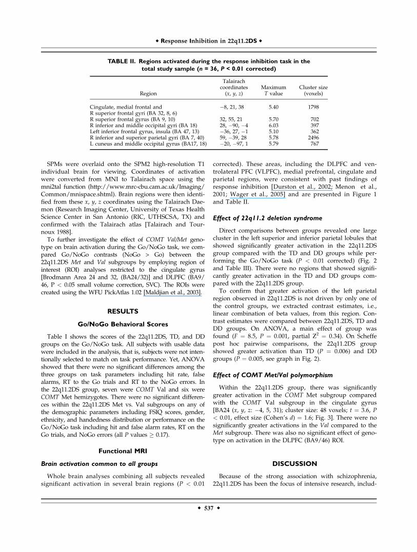
Figure 1.
Brain activation patterns across 22q11.2DS,
TD and DD groups.
r Gothelf et al. r
r 536 r
SPMs were overlaid onto the SPM2 high-resolution T1individual brain for viewing. Coordinates of activationwere converted from MNI to Talairach space using themni2tal function (http://www.mrc-cbu.cam.ac.uk/Imaging/Common/mnispace.shtml). Brain regions were then identi-fied from these x, y, z coordinates using the Talairach Dae-mon (Research Imaging Center, University of Texas HealthScience Center in San Antonio (RIC, UTHSCSA, TX) andconfirmed with the Talairach atlas [Talairach and Tour-noux 1988].To further investigate the effect of COMT Val/Met geno-
type on brain activation during the Go/NoGo task, we com-pared Go/NoGo contrasts (NoGo > Go) between the22q11.2DS Met and Val subgroups by employing region ofinterest (ROI) analyses restricted to the cingulate gyrus[Brodmann Area 24 and 32, (BA24/32)] and DLPFC (BA9/46, P < 0.05 small volume correction, SVC). The ROIs werecreated using the WFU PickAtlas 1.02 [Maldjian et al., 2003].
RESULTS
Go/NoGo Behavioral Scores
Table I shows the scores of the 22q11.2DS, TD, and DDgroups on the Go/NoGo task. All subjects with usable datawere included in the analysis, that is, subjects were not inten-tionally selected to match on task performance. Yet, ANOVAshowed that there were no significant differences among thethree groups on task parameters including hit rate, falsealarms, RT to the Go trials and RT to the NoGo errors. Inthe 22q11.2DS group, seven were COMT Val and six wereCOMT Met hemizygotes. There were no significant differen-ces within the 22q11.2DS Met vs. Val subgroups on any ofthe demographic parameters including FSIQ scores, gender,ethnicity, and handedness distribution or performance on theGo/NoGo task including hit and false alarm rates, RT on theGo trials, and NoGo errors (all P values � 0.17).
Functional MRI
Brain activation common to all groups
Whole brain analyses combining all subjects revealedsignificant activation in several brain regions (P < 0.01
corrected). These areas, including the DLPFC and ven-trolateral PFC (VLPFC), medial prefrontal, cingulate andparietal regions, were consistent with past findings ofresponse inhibition [Durston et al., 2002; Menon et al.,2001; Wager et al., 2005] and are presented in Figure 1and Table II.
Effect of 22q11.2 deletion syndrome
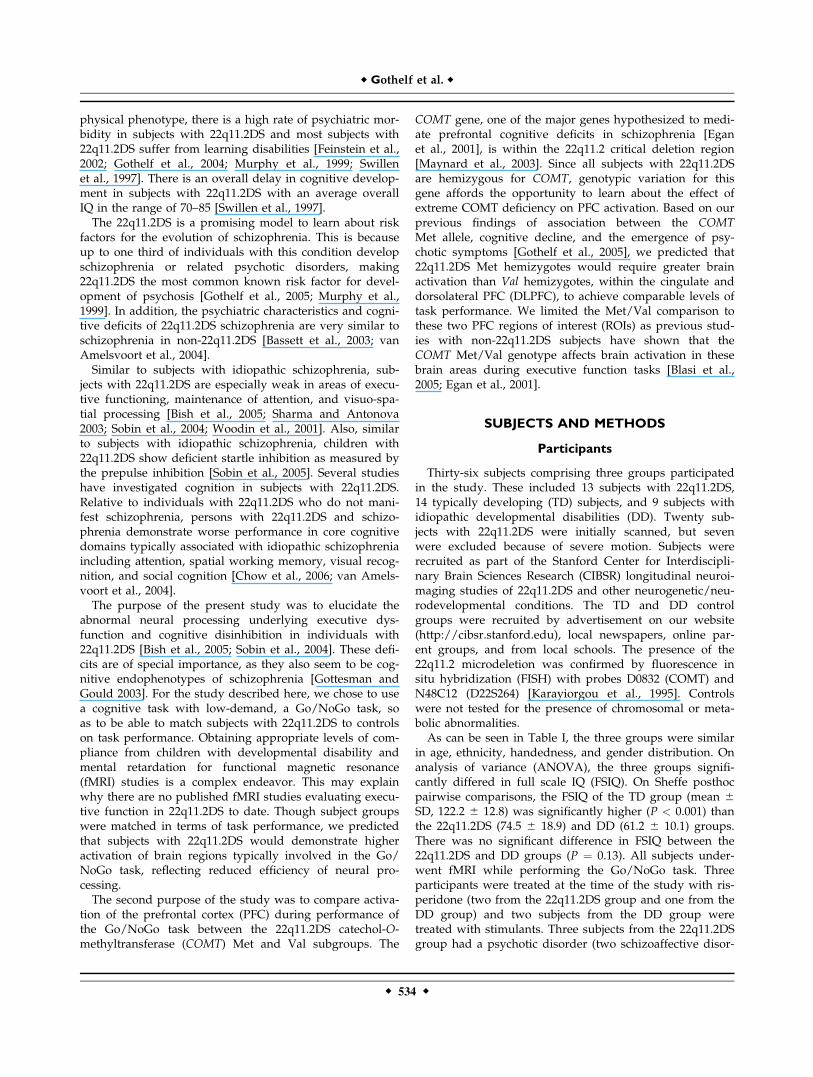
Direct comparisons between groups revealed one largecluster in the left superior and inferior parietal lobules thatshowed significantly greater activation in the 22q11.2DSgroup compared with the TD and DD groups while per-forming the Go/NoGo task (P < 0.01 corrected) (Fig. 2and Table III). There were no regions that showed signifi-cantly greater activation in the TD and DD groups com-pared with the 22q11.2DS group.To confirm that greater activation of the left parietal
region observed in 22q11.2DS is not driven by only one ofthe control groups, we extracted contrast estimates, i.e.,linear combination of beta values, from this region. Con-trast estimates were compared between 22q11.2DS, TD andDD groups. On ANOVA, a main effect of group wasfound (F ¼ 8.5, P ¼ 0.001, partial Z2 ¼ 0.34). On Scheffepost hoc pairwise comparisons, the 22q11.2DS groupshowed greater activation than TD (P ¼ 0.006) and DDgroups (P ¼ 0.005, see graph in Fig. 2).
Effect of COMT Met/Val polymorphism
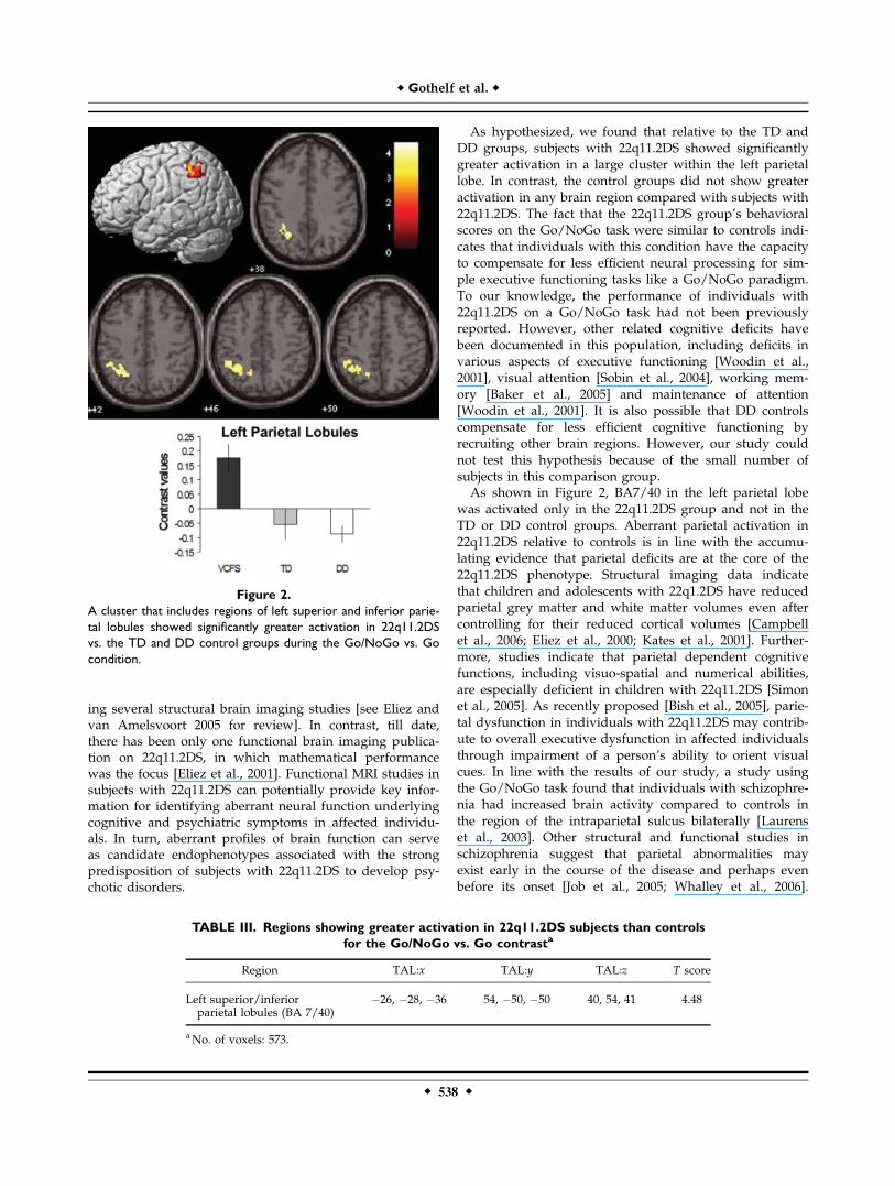
Within the 22q11.2DS group, there was significantlygreater activation in the COMT Met subgroup comparedwith the COMT Val subgroup in the cingulate gyrus[BA24 (x, y, z: �4, 5, 31); cluster size: 48 voxels; t ¼ 3.6, P< 0.01, effect size (Cohen’s d) ¼ 1.6; Fig. 3]. There were nosignificantly greater activations in the Val compared to theMet subgroup. There was also no significant effect of geno-type on activation in the DLPFC (BA9/46) ROI.
DISCUSSION
Because of the strong association with schizophrenia,22q11.2DS has been the focus of intensive research, includ-
TABLE II. Regions activated during the response inhibition task in the
total study sample (n = 36, P < 0.01 corrected)
Region
Talairachcoordinates(x, y, z)
MaximumT value
Cluster size(voxels)
Cingulate, medial frontal andR superior frontal gyri (BA 32, 8, 6)
�8, 21, 38 5.40 1798
R superior frontal gyrus (BA 9, 10) 32, 55, 21 5.70 702R inferior and middle occipital gyri (BA 18) 28, �90, �4 6.03 397Left inferior frontal gyrus, insula (BA 47, 13) �36, 27, �1 5.10 362R inferior and superior parietal gyri (BA 7, 40) 59, �39, 28 5.78 2496L cuneus and middle occipital gyrus (BA17, 18) �20, �97, 1 5.79 767
r Response Inhibition in 22q11.2DS r
r 537 r
ing several structural brain imaging studies [see Eliez andvan Amelsvoort 2005 for review]. In contrast, till date,there has been only one functional brain imaging publica-tion on 22q11.2DS, in which mathematical performancewas the focus [Eliez et al., 2001]. Functional MRI studies insubjects with 22q11.2DS can potentially provide key infor-mation for identifying aberrant neural function underlyingcognitive and psychiatric symptoms in affected individu-als. In turn, aberrant profiles of brain function can serveas candidate endophenotypes associated with the strongpredisposition of subjects with 22q11.2DS to develop psy-chotic disorders.
As hypothesized, we found that relative to the TD andDD groups, subjects with 22q11.2DS showed significantlygreater activation in a large cluster within the left parietallobe. In contrast, the control groups did not show greateractivation in any brain region compared with subjects with22q11.2DS. The fact that the 22q11.2DS group’s behavioralscores on the Go/NoGo task were similar to controls indi-cates that individuals with this condition have the capacityto compensate for less efficient neural processing for sim-ple executive functioning tasks like a Go/NoGo paradigm.To our knowledge, the performance of individuals with22q11.2DS on a Go/NoGo task had not been previouslyreported. However, other related cognitive deficits havebeen documented in this population, including deficits invarious aspects of executive functioning [Woodin et al.,2001], visual attention [Sobin et al., 2004], working mem-ory [Baker et al., 2005] and maintenance of attention[Woodin et al., 2001]. It is also possible that DD controlscompensate for less efficient cognitive functioning byrecruiting other brain regions. However, our study couldnot test this hypothesis because of the small number ofsubjects in this comparison group.As shown in Figure 2, BA7/40 in the left parietal lobe
was activated only in the 22q11.2DS group and not in theTD or DD control groups. Aberrant parietal activation in22q11.2DS relative to controls is in line with the accumu-lating evidence that parietal deficits are at the core of the22q11.2DS phenotype. Structural imaging data indicatethat children and adolescents with 22q1.2DS have reducedparietal grey matter and white matter volumes even aftercontrolling for their reduced cortical volumes [Campbellet al., 2006; Eliez et al., 2000; Kates et al., 2001]. Further-more, studies indicate that parietal dependent cognitivefunctions, including visuo-spatial and numerical abilities,are especially deficient in children with 22q11.2DS [Simonet al., 2005]. As recently proposed [Bish et al., 2005], parie-tal dysfunction in individuals with 22q11.2DS may contrib-ute to overall executive dysfunction in affected individualsthrough impairment of a person’s ability to orient visualcues. In line with the results of our study, a study usingthe Go/NoGo task found that individuals with schizophre-nia had increased brain activity compared to controls inthe region of the intraparietal sulcus bilaterally [Laurenset al., 2003]. Other structural and functional studies inschizophrenia suggest that parietal abnormalities mayexist early in the course of the disease and perhaps evenbefore its onset [Job et al., 2005; Whalley et al., 2006].
TABLE III. Regions showing greater activation in 22q11.2DS subjects than controls
for the Go/NoGo vs. Go contrasta
Region TAL:x TAL:y TAL:z T score
Left superior/inferiorparietal lobules (BA 7/40)
�26, �28, �36 54, �50, �50 40, 54, 41 4.48
aNo. of voxels: 573.
Figure 2.
A cluster that includes regions of left superior and inferior parie-
tal lobules showed significantly greater activation in 22q11.2DS
vs. the TD and DD control groups during the Go/NoGo vs. Go
condition.
r Gothelf et al. r
r 538 r

Taken together, parietal dysfunction is a candidate endo-phenotype that may be associated with the development ofpsychosis in individuals with 22q11.2DS. It should be notedthat while there is certainly some overlap between the neu-roanatomical and cognitive deficits associated with22q11.2DS and those associated with idiopathic schizophre-nia, the patterns are not identical. For example, in compari-son to typically developing controls, frontal lobe volume in22q11.2DS tends to be preserved while it is decreased involume in idiopathic schizophrenia [Eliez et al., 2000]. Sim-ilarly, though verbal memory is impaired in patients withidiopathic schizophrenia, there are no differences in thiscognitive function between 22q11.2DS subjects with andwithout schizophrenia [van Amelsvoort et al., 2004].To test the second hypothesis regarding differences
between COMT Met and Val hemizygotes within the22q11.2DS group, we chose to focus on two key prefrontalregions for executive control, the DLPFC and the cingulate.The cingulate (BA24/32) ROI was chosen as healthy sub-jects were observed to show a robust effect of COMT Met/Val polymorphism on brain activation within this regionduring performance of an attentional control task [Blasiet al., 2005]. This attentional control task is similar to theGo/NoGo task used in the present study with respect toreliance on attention and conflict monitoring. The locationof the cluster in the cingulate gyrus of 22q11.2DS subjects
that showed greater activation in the Met compared to theVal subgroup was overlapping with the cluster identifiedin the Blasi et al. [2005] study. As shown in Figure 3, thecluster of activation in BA24 occurred only in the22q11.2DS Met and not in the 22q11.2DS Val carriers, sug-gesting that greater cingulate recruitment is required inMet hemizygotes when performing a response inhibitiontask. Although the DLPFC was activated during the Go/NoGo performance in the 22q11.2DS group, the COMT ge-notype did not affect activation of this region. Similarly, inBlasi et al. [2005], though the DLPFC and additional corti-cal regions were activated during the attentional controltask, COMT genotype was observed to affect degree ofactivation only in the cingulate gyrus. Together, these find-ings suggest that the COMT Met/Val polymorphism par-ticularly affects attentional control and response inhibitionvia the cingulate gyrus as opposed to the DLPFC.The segment of the cingulate gyrus that was signifi-
cantly more activated on the Go/NoGo task in Met vs. Valhemizygote 22q11.2DS subjects receives abundant dopami-nergic projections [Seamans 2004]. In the Blasi et al. [2005]study, Val/Val subjects showed greater (less efficient) cin-gulate gyrus activation than Met/Met subjects. In the pres-ent study, 22q11.2DS Met hemizygotes showed greater cin-gulate gyrus activation than Val hemizygote subjects. Theapparent opposing effect of COMT Met/Val polymor-phism in 22q11.2DS and non-22q11.2DS subjects can bestbe explained according to the known inverted-U shaperelation that exists between prefrontal cortical dopaminelevels and cognitive functioning [Goldman-Rakic et al.,2000]. Consequently, too little or too much prefrontal D1
receptor stimulation is associated with less optimal cogni-tive function. Non-22q11.2DS hemizygotes for Val have toomuch COMT activity and thus have less than optimal pre-frontal dopamine levels; whereas 22q11.2DS Met hemizy-gotes are putatively in extreme shortage of COMT activityand hence are thought to have too high prefrontal dopa-mine levels. In a recent longitudinal study of group of chil-dren with 22q11.2DS followed to adolescence we foundevidence supporting the effect of the inverted-U shapemodel on cognitive function in 22q11.2DS. During adoles-cence, the subgroup of 22q11.2DS children with theMet allele declined more in VIQ and expressive languageabilities and developed more severe psychotic symptomsthan the subgroup with the Val allele [Gothelf et al., 2005].In the present study, functional imaging data were
acquired from most subjects at the second time point of ourlongitudinal study. Of note, other cross-sectional studies in22q11.2DS have presented conflicting results regarding theeffect of COMT Met/Val genotype on cognitive functioning[Bearden et al., 2004; Glaser et al., 2006]. In our cohort, theeffect of the COMT Met/Val polymorphism was onlyrevealed by longitudinal analysis [Gothelf et al., 2005]. Inother words, though there was no significant difference in IQbetween the COMT Met and Val carriers at Time 2, thedecline in VIQ among carriers of the COMT Met subgroupwas significant compared to the COMT Val subgroup.
Figure 3.
Effect of COMT genotype on cingulate gyrus (BA24/32) activation
during the Go/NoGo vs. Go condition in subjects with
22q11.2DS. A cluster within the cingulate gyrus (BA24) with
greater activation in the COMT Met vs. Val subgroups is shown.
r Response Inhibition in 22q11.2DS r
r 539 r
Abnormal activation of the cingulate gyrus, during per-formance of response conflict and response inhibitiontasks, has been observed in individuals with idiopathicschizophrenia [Kerns et al., 2005; Snitz et al., 2005]. Also,activation of the cingulate in individuals with schizophre-nia appears to be sensitive to changes in dopamine levels[Dolan et al., 1995]. Thus, abnormal activation of the cingu-late gyrus in 22q11.2DS COMT Met carriers may representa biological marker for increased risk for psychosis.In healthy subjects, the COMT genotype affects PFC res-
ponse to medication-induced increase in brain dopaminelevels. Mattay et al. [2003] observed that administration ofamphetamine improved working memory performanceof Val/Val healthy subjects while causing deterioration ofcognitive performance in Met/Met subjects. These effectswere also noted on brain activity during a task where pre-frontal activation was observed to be significantly greaterin the Met/Met than in the Val/Val group. It could behypothesized that like healthy subjects treated with am-phetamine, the Met subgroup of 22q11.2DS subjects alsoexperience a hyperdopaminergic condition, that is, bothare on the down slope of the inverted-U shape of dopa-mine level in relation to cognitive functioning. Thus, inboth situations we observe less efficient cognitive function-ing and greater cingulate activation. The lack of COMTVal158Met effect on DLPFC activation in the present studyis likely to be related to the task chosen, that is, responseinhibition, which is not a DLPFC-specific cognitive task.The data presented here provide further support for the
hypothesis that COMT genotype influences neural functionin 22q11.2DS. However, other (non-imaging) studies did notfind a clear association between COMT genotype and eitherneuropsychiatric [Bassett et al., 2007] or cognitive deficits[Glaser et al., 2006] in affected individuals. Taken as a whole,these findings are likely to reflect differences in studyingbehavior at more than one level of scientific inquiry (i.e.fMRI vs. IQ) as well as potential interactive effects betweenthe COMT gene and other genes from the 22q11.2 deletionregion. In particular, it is likely that halpoinsufficeincy formore than one gene contributes to cognitive and neuro-psychiatric outcome in persons with 22q11.2DS. One majorcandidate for such interaction is the proline-dehydrogenase(PRODH) gene. The PRODH gene is within the 22q11.2 dele-tion region and codes for the proline dehydrogenaseenzyme. In mice knocked-out for PRODH there is an upre-gulation of COMT expression, indicating a homeostaticresponse to enhanced dopaminergic signaling in the frontalcortex that emerges secondary to PRODH deficiency [Pater-lini et al., 2005]. In line with these animal findings, a recentstudy reported that within the overall 22q11.2DS population,those subjects with hyperprolinemia and the COMTMet allele were 3 times more likely to have a psychotic disor-der [Raux et al., 2007].In summary, we found abnormal brain activation in pa-
rietal regions in subjects with 22q11.2DS performing aresponse inhibition task. The role of the COMT genotypeon cognitive performance and brain activation requires
further study with a larger sample of subjects and withother cognitive tasks assessing prefrontal functioning. Inparticular, the effect of COMT genotype on DLPFC func-tion in 22q11.2DS should be evaluated using more DLPFC-specific experiments such as N-Back working memorytasks. Future longitudinal studies should also evaluatewhether abnormal brain activations (e.g., in parietal re-gions) predict the later development of psychotic disordersin 22q11.2DS.
ACKNOWLEDGMENTS
We thank Eugene Gu, Noopur Jain, and Ira Patnaik fordata processing and Lin Xiaoyan for DNA extraction.
REFERENCES
Baker K, Baldeweg T, Sivagnanasundaram S, Scambler P, Skuse D(2005): COMT Val108/158 Met modifies mismatch negativityand cognitive function in 22q11 deletion syndrome. Biol Psy-chiatry 58:23–31.
Bassett AS, Caluseriu O, Weksberg R, Young DA, Chow EW (2007):Catechol-O-methyl transferase and expression of schizophreniain 73 adults with 22q11 deletion syndrome. Biol Psychiatry 2007in press.
Bassett AS, Chow EW, AbdelMalik P, Gheorghiu M, Husted J,Weksberg R (2003): The schizophrenia phenotype in 22q11 de-letion syndrome. Am J Psychiatry 160:1580–1586.
Bearden CE, Jawad AF, Lynch DR, Sokol S, Kanes SJ, McDonald-McGinn DM, Saitta SC, Harris SE, Moss E, Wang PP, Zackai E,Emanuel BS, Simon TJ. (2004): Effects of a functional COMTpolymorphism on prefrontal cognitive function in patients with22q11.2 deletion syndrome. Am J Psychiatry 161:1700–1702.
Bish JP, Ferrante SM, McDonald-McGinn D, Zackai E, Simon TJ(2005): Maladaptive conflict monitoring as evidence for execu-tive dysfunction in children with chromosome 22q11.2 deletionsyndrome. Dev Sci 8:36–43.
Blasi G, Mattay VS, Bertolino A, Elvevag B, Callicott JH, Das S,Kolachana BS, Egan MF, Goldberg TE, Weinberger DR (2005):Effect of catechol-O-methyltransferase val158met genotype onattentional control. J Neurosci 25:5038–5045.
Botto LD, May K, Fernhoff PM, Correa A, Coleman K, RasmussenSA, Merritt RK, O’Leary LA, Wong LY, Elixson EM and others(2003): A population-based study of the 22q11.2 deletion: Phe-notype, incidence, and contribution to major birth defects inthe population. Pediatrics 112 (1 Part 1):101–107.
Campbell LE, Daly E, Toal F, Stevens A, Azuma R, Catani M, NgV, van Amelsvoort T, Chitnis X, Cutter W, Murphy DG, Mur-phy KC. (2006): Brain and behaviour in children with 22q11.2deletion syndrome: A volumetric and voxel-based morphome-try MRI study. Brain 129:1218–1228.
Chow EW, Watson M, Young DA, Bassett AS (2006): Neurocogni-tive profile in 22q11 deletion syndrome and schizophrenia.Schizophr Res 87:270–278.
Dolan RJ, Fletcher P, Frith CD, Friston KJ, Frackowiak RS, GrasbyPM (1995): Dopaminergic modulation of impaired cognitiveactivation in the anterior cingulate cortex in schizophrenia. Na-ture 378(6553):180–182.
Durston S, Thomas KM, Worden MS, Yang Y, Casey BJ (2002):The effect of preceding context on inhibition: an event-relatedfMRI study. Neuroimage 16:449–453.
r Gothelf et al. r
r 540 r
Egan MF, Goldberg TE, Kolachana BS, Callicott JH, MazzantiCM, Straub RE, Goldman D, Weinberger DR (2001): Effect ofCOMT Val108/158 Met genotype on frontal lobe functionand risk for schizophrenia. Proc Natl Acad Sci USA 98:6917–6922.
Eliez S, Blasey CM, Menon V, White CD, Schmitt JE, Reiss AL(2001): Functional brain imaging study of mathematical reason-ing abilities in velocardiofacial syndrome (del22q11.2). GenetMed 3:49–55.
Eliez S, Schmitt JE, White CD, Reiss AL (2000): Children and ado-lescents with velocardiofacial syndrome: A volumetric MRI study.Am J Psychiatry 157:409–415.
Eliez S, van Amelsvoort T (2005): Neuroimaging in velocardiofa-cial syndrome. In: Murphy KC, Scambler PJ, Eds. Velo-Cardio-Facial Syndrome: A model for understanding MicrodeletionDisorders. Cambridge: Cambridge University Press.
Feinstein C, Eliez S, Blasey C, Reiss AL (2002): Psychiatric disor-ders and behavioral problems in children with velocardiofacialsyndrome: Usefulness as phenotypic indicators of schizophre-nia risk. Biol Psychiatry 51:312–318.
Glaser B, Debbane M, Hinard C, Morris MA, Dahoun SP, Anto-narakis SE, Eliez S (2006): No evidence for an effect ofCOMT Val158Met genotype on executive function in patientswith 22q11 deletion syndrome. Am J Psychiatry 163:537–539.
Glover GH, Lai S (1998): Self-navigated spiral fMRI: interleavedversus single-shot. Magn Reson Med 39:361–368.
Goldman-Rakic PS, Muly EC, 3rd, Williams GV (2000): D(1) recep-tors in prefrontal cells and circuits. Brain Res Brain Res Rev31:295–301.
Gothelf D, Eliez S, Thompson T, Hinard C, Penniman L, Fein-stein C, Kwon H, Jin S, Jo B, Antonarakis SE, Morris MA,Reiss AL (2005): COMT genotype predicts longitudinal cog-nitive decline and psychosis in 22q11.2 deletion syndrome.Nat Neurosci 8:1500–1502.
Gothelf D, Presburger G, Zohar AH, Burg M, Nahmani A, Fryd-man M, Shohat M, Inbar D, Aviram-Goldring A, Yeshaya J,Steinberg T, Finkelstein Y, Frisch A, Weizman A, Apter A.(2004): Obsessive-compulsive disorder in patients with velocar-diofacial (22q11 deletion) syndrome. Am J Med Genet B Neu-ropsychiatry Genet 126:99–105.
Gottesman Gould TD (2003): The endophenotype concept in psy-chiatry: Etymology and strategic intentions. Am J Psychiatry 160:636–645.
Hoeft F, Hernandez A, Parthasarathy S, Watson CL, Hall SS, ReissAL (2007): Fronto-striatal dysfunction and potential compensa-tory mechanisms in male adolescents with fragile X syndrome.Hum Brain Mapp, in press
Job DE, Whalley HC, Johnstone EC, Lawrie SM (2005): Grey mat-ter changes over time in high risk subjects developing schizo-phrenia. Neuroimage 25:1023–1030.
Karayiorgou M, Morris MA, Morrow B, Shprintzen RJ, GoldbergR, Borrow J, Gos A, Nestadt G, Wolyniec PS, Lasseter VK,Eisen H, Childs B, Kazazian HH, Kucherlopati R, AntonarakisSE, Pulver AE, Housman DE. (1995): Schizophrenia susceptibil-ity associated with interstitial deletions of chromosome 22q11.Proc Natl Acad Sci USA 92:7612–7616.
Kates WR, Burnette CP, Jabs EW, Rutberg J, Murphy AM, Gra-dos M, Geraghty M, Kaufmann WE, Pearlson GD (2001): Re-gional cortical white matter reductions in velocardiofacial syn-drome: a volumetric MRI analysis. Biol Psychiatry 49:677–684.
Kerns JG, Cohen JD, MacDonald AW III, Johnson MK, StengerVA, Aizenstein H, Carter CS (2005): Decreased conflict- and
error-related activity in the anterior cingulate cortex in subjectswith schizophrenia. Am J Psychiatry 162:1833–1839.
Kirkpatrick JA Jr, DiGeorge AM (1968): Congenital absence ofthe thymus. Am J Roentgenol Radium Ther Nucl Med103:32–37.
Lachman HM, Papolos DF, Saito T, Yu YM, Szumlanski CL, Wein-shilboum RM (1996): Human catechol-O-methyltransferasepharmacogenetics: Description of a functional polymorphismand its potential application to neuropsychiatric disorders. Phar-macogenetics 6:243–250.
Laurens KR, Ngan ET, Bates AT, Kiehl KA, Liddle PF (2003): Ros-tral anterior cingulate cortex dysfunction during error process-ing in schizophrenia. Brain 126 (Part 3):610–622.
Maldjian JA, Laurienti PJ, Kraft RA, Burdette JH (2003): An auto-mated method for neuroanatomic and cytoarchitectonic atlas-based interrogation of fMRI data sets. Neuroimage 19:1233–1239.
Mattay VS, Goldberg TE, Fera F, Hariri AR, Tessitore A, Egan MF,Kolachana B, Callicott JH, Weinberger DR (2003): Catechol-O-methyltransferase val 158-met and individual variation in thebrain response to amphetamine. Proc Natl Acad Sci USA100(10):6186–6191.
Maynard TM, Haskell GT, Peters AZ, Sikich L, Lieberman JA,LaMantia AS (2003): A comprehensive analysis of 22q11 geneexpression in the developing and adult brain. Proc Natl AcadSci USA 100:14433–14438.
Menon V, Adleman NE, White CD, Glover GH, Reiss AL (2001):Error-related brain activation during a Go/NoGo response in-hibition task. Hum Brain Mapp 12:131–143.
Murphy KC, Jones LA, Owen MJ (1999): High rates of schizophre-nia in adults with velo-cardio-facial syndrome. Arch Gen Psy-chiatry 56:940–945.
Paterlini M, Zakharenko SS, Lai WS, Qin J, Zhang H, Mukai J,Westphal KG, Olivier B, Sulzer D, Pavlidis P, Siegelbaum SA,Karayiorgou M, Gogos JA. (2005): Transcriptional and behav-ioral interaction between 22q11.2 orthologs modulates schizo-phrenia-related phenotypes in mice. Nat Neurosci 8:1586–1594.
Poline JB, Worsley KJ, Evans AC, Friston KJ (1997): Combiningspatial extent and peak intensity to test for activations in func-tional imaging. Neuroimage 5(2):83–96.
Raux G, Bumsel E, Hecketsweiler B, van Amelsvoort T, ZinkstokJ, Manouvrier-Hanu S, Fantini C, Breviere GM, Di Rosa G, Pus-torino G. (2007): Involvement of hyperprolinemia in cognitiveand psychiatric features of the 22q11 deletion syndrome. HumMol Genet 16:83–91.
Seamans JK, Yang CR. (2004): The principal features and mecha-nisms of dopamine modulation in the prefrontal cortex. ProgNeurobiol 74:1–57.
Sharma T, Antonova L (2003): Cognitive function in schizophrenia.Deficits, functional consequences, and future treatment. Psy-chiatr Clin North Am 26:25–40.
Shprintzen RJ (2000): Velo-cardio-facial syndrome: a distinctive be-havioral phenotype. Ment Retard Dev Disabil Res Rev 6:142–147.
Simon TJ, Bearden CE, Mc-Ginn DM, Zackai E (2005): Visuospatialand numerical cognitive deficits in children with chromosome22q11.2 deletion syndrome. Cortex 41:145–155.
Snitz BE, MacDonald A III, Cohen JD, Cho RY, Becker T, CarterCS (2005): Lateral and medial hypofrontality in first-episodeschizophrenia: Functional activity in a medication-naive stateand effects of short-term atypical antipsychotic treatment. AmJ Psychiatry 162:2322–2329.
r Response Inhibition in 22q11.2DS r
r 541 r
Sobin C, Kiley-Brabeck K, Daniels S, Blundell M, Anyane-YeboaK, Karayiorgou M (2004): Networks of attention in childrenwith the 22q11 deletion syndrome. Dev Neuropsychol 26:611–626.
Sobin C, Kiley-Brabeck K, Karayiorgou M (2005): Lower prepulseinhibition in children with the 22q11 deletion syndrome. Am JPsychiatry 162:1090–1099.
Swillen A, Devriendt K, Legius E, Eyskens B, Dumoulin M, Gewil-lig M, Fryns JP (1997): Intelligence and psychosocial adjust-ment in velocardiofacial syndrome: A study of 37 children andadolescents with VCFS. J Med Genet 34:453–458.
Talairach J, Tournoux P (1988). Co-Planar Stereotaxic Atlas of theHuman Brain. New York: Thieme.
van Amelsvoort T, Henry J, Morris R, Owen M, Linszen D, Mur-phy K, Murphy D (2004): Cognitive deficits associated withschizophrenia in velo-cardio-facial syndrome. Schizophr Res 70:223–232.
Wager TD, Sylvester CY, Lacey SC, Nee DE, Franklin M, Jonides J(2005): Common and unique components of response inhibitionrevealed by fMRI. Neuroimage 27:323–340.
Wechsler D (1991). Manual for the Wechsler Intelligence Scale forChildren, 3rd ed. San Antonio, TX: The Psychological Corpora-tion.
Wechsler D (1997). Wechsler Adult Intelligence Scale, 3rd ed.Administration and Scoring Manual. San Antonio, TX: ThePsychological Corporation.
Whalley HC, Simonotto E, Moorhead W, McIntosh A, Marshall I,Ebmeier KP, Owens DG, Goddard NH, Johnstone EC, LawrieSM (2006): Functional imaging as a predictor of schizophrenia.Biol Psychiatry 60(5):454–462.
Woodin M, Wang PP, Aleman D, McDonald-McGinn D, Zackai E,Moss E (2001): Neuropsychological profile of children and ado-lescents with the 22q11.2 microdeletion. Genet Med 3:34–39.
r Gothelf et al. r
r 542 r