Magnetic resonance imaging of abnormal ventricular septal ...
Upload
khangminh22Category
view
7download
0
EDUCATIONAL REVIEW Open Access
Thoracic abnormal air collections inpatients in the intensive care unit:radiograph findings correlated with CTMasafumi Sakai1,2* , Takashi Hiyama3, Hirofumi Kuno3, Kensaku Mori2, Tsukasa Saida2, Toshitaka Ishiguro2,Hiroaki Takahashi4, Ken Koyama1 and Manabu Minami2
Abstract
An abnormal collection of air in the thorax is one of the most common life-threatening events that occurs in theintensive care unit. Patient management differs depending on the location of the air collection; therefore, detectingabnormal air collection and identifying its exact location on supine chest radiographs is essential for early treatmentand positive patient outcomes. Thoracic abnormal air collects in multiple thoracic spaces, including the pleuralcavity, chest wall, mediastinum, pericardium, and lung. Pneumothorax in the supine position shows differentradiographic findings depending on the location. Many conditions, such as skin folds, interlobar fissure, bullae inthe apices, and air collection in the intrathoracic extrapleural space, mimic pneumothorax on radiographs.Additionally, pneumopericardium may resemble pneumomediastinum and needs to be differentiated. Further,some conditions such as inferior pulmonary ligament air collection versus a pneumatocele or pneumothorax in theposteromedial space require a differential diagnosis based on radiographs. Computed tomography (CT) is requiredto localize the air and delineate potential etiologies when a diagnosis by radiography is difficult. The purposes ofthis article are to review the anatomy of the potential spaces in the chest where abnormal air can collect, explaincharacteristic radiographic findings of the abnormal air collection in supine patients with illustrations and correlatedCT images, and describe the distinguishing features of conditions that require a differential diagnosis. Sincemanagement differs based on the location of the air collection, radiologists should try to accurately detect andidentify the location of air collection on supine radiographs.
Keywords: Radiograph, Computed tomography, Air collection, Thorax, Intensive care unit
Key points
� Management differs based on the location ofthoracic abnormal air collection.
� Identifying abnormal air collection on radiographs isessential for early treatment.
� Air collection is classified into the pleural cavity,chest wall, etc.
� Radiographic findings vary depending on thelocation of the lesion.
� Understanding CT anatomy enables locatingabnormal thoracic air on radiographs.
BackgroundA portable chest radiograph is the most commonly usedradiographic examination in the intensive care unit(ICU) [1]. Despite its diagnostic limitations [2, 3], chestradiographs often reveal critical abnormalities that mayremain unnoticed clinically. One of the life-threateningevents in the ICU is the development of abnormal aircollection in the thorax caused by interstitial or cysticlung disease, infectious diseases, trauma, positive pres-sure ventilation, and other complications associated withmedical interventions [4–11].The management of abnormal air collections in the
thorax depends on its location. Some abnormal air
© The Author(s). 2020 Open Access This article is distributed under the terms of the Creative Commons Attribution 4.0International License (http://creativecommons.org/licenses/by/4.0/), which permits unrestricted use, distribution, andreproduction in any medium, provided you give appropriate credit to the original author(s) and the source, provide a link tothe Creative Commons license, and indicate if changes were made.
* Correspondence: [email protected] of Diagnostic and Interventional Radiology, Ibaraki PrefecturalCentral Hospital, Kasama, Japan2Department of Diagnostic and Interventional Radiology, University ofTsukuba Hospital, Amakubo 2-1-1, Tsukuba, Ibaraki 305-8576, JapanFull list of author information is available at the end of the article
Insights into ImagingSakai et al. Insights into Imaging (2020) 11:35 https://doi.org/10.1186/s13244-020-0838-z
collections are managed through careful observation,and other air collections require surgical intervention.Pneumothorax, especially tension pneumothorax, istreated by drainage [12]. Subcutaneous emphysema, aircollection in the intrathoracic extrapleural space, andpneumomediastinum are usually not considered urgentconditions and often only require clinical observation[13–15]. However, in the case of abnormal air collectioncaused by tracheal/esophageal rupture, emergent surgi-cal intervention may be required. Pneumopericardium istreated by emergent pericardiocentesis in a case of car-diac tamponade [9]. The main treatment goal of pul-monary interstitial emphysema (PIE) is to achieveadequate oxygenation with a lower mean and peak air-way pressure of ventilation [16]. Consequently, the de-tection and accurate localization of abnormal aircollection is essential for early and optimal treatmentand favorable patient outcomes.Abnormal air collection can be more accurately evalu-
ated by computed tomography (CT) than by chest radiog-raphy [3]. Nevertheless, CT is not easily conducted in anICU setting because patients are critically ill and oftensupported by many devices. Lung ultrasound is readilyavailable and may effectively and accurately detectpneumothorax as the absence of lung sliding and comet-tail artifact, but its outcomes are contingent on the opera-tor’s skill. Additionally, it is often difficult to observe dee-per regions of the chest and obtain a complete view [17].Consequently, portable chest radiography with a patientin the supine position is the fundamental imaging examin-ation that is conducted in the ICU. However, air collectioncan be easily overlooked on supine radiographs if a clin-ician does not have a comprehensive understanding of thethree-dimensional anatomy of the chest and the predictedsites of abnormal air collection.
The purposes of this article are to review the anatomyof potential spaces in the chest where air can collect, ex-plain the characteristic radiographic findings of abnor-mal air collection in supine patients using illustrationsand correlated CT images, and describe the distinguish-ing features of conditions that require a differential diag-nosis. We classify the chest into the following fivelocations based on its anatomy (Table 1): the pleuralcavity, chest wall, mediastinum, pericardium, and lung.We also described diseases of abnormal air collections ineach of these spaces (Table 1).
Abnormal air collection in the pleural cavityThe pleural cavity and chest wall are anatomically classifiedinto several layers (Fig. 1), and abnormal air predominantlycollects in each anatomical space (Fig. 1). The pleural spaceis the area between the visceral and the parietal pleurae (Fig.1). The visceral pleura covers the lung and folds back on it-self at the root of the lung (pulmonary hilum) to become theparietal pleura (Fig. 1). The pleural space normally contains asmall amount of fluid (between 1 and 5 mL) [18], and thepleural space is not visualized on radiographs or CT imageswithout pleural effusion or pneumothorax. The pleural spaceis primarily classified as anteromedial, subpulmonary, apico-lateral, and posteromedial [19, 20].
PneumothoraxPneumothorax is a disease characterized by the collection ofair in one or several spaces of the pleural cavity [21]. Here,we describe pneumothorax in each space; tension pneumo-thorax, which is an emergent condition; and conditions thatrequire a differential diagnosis of pneumothorax. The mainsymptoms of pneumothorax are sudden chest pain, dyspnea,and dry cough. Pneumothorax is caused by the rupture of abulla, trauma, and iatrogenic complications after an
Table 1 Classification of thoracic abnormal air collection based on anatomy
Location Anatomical structure and space Disease of abnormal air collection
I. Pleural cavity Anteromedial space Pneumothorax in the anteromedial space (I-1)
Subpulmonary space Pneumothorax in the subpulmonary space (I-2)
Apicolateral space Pneumothorax in the apicolateral space (I-3)
Posteromedial space Pneumothorax in the posteromedial space (I-4)
(Emergency condition) Tension pneumothorax (I-5)
(Differential diagnosis) Mimics of pneumothorax (I-6)
II. Chest wall Subcutaneous space Subcutaneous emphysema (II-1)
Intrathoracic extrapleural space Air collection in the intrathoracic extrapleural space (II-2)
III. Mediastinum Mediastinum Pneumomediastinum (III-1)
Inferior pulmonary ligament Air collection in the inferior pulmonary ligament (III-2)
IV. Pericardium Pericardium Pneumopericardium (IV-1)
V. Lung Interstitium Pulmonary interstitial emphysema (V-1)
Parenchyma Pneumatocele (V-2)
The numbers in the parentheses correspond to the numbers in the text and figures
Sakai et al. Insights into Imaging (2020) 11:35 Page 2 of 17

intervention, such as thoracentesis and positive pressure ven-tilation [4]. In the ICU, pneumothorax is commonlycaused by barotrauma associated with ventilation [4].Pneumothorax occurs in nearly 20% of patients with thor-acic trauma [4]. It is treated with thoracic drainage. Basedon the British Thoracic Society Guidelines 2010, a largepneumothorax is defined on erect chest radiographs as anair space that is more than 2 cm from the pleural surfaceto the lung edge at the level of the hilum [22]. A largepneumothorax usually requires thoracentesis, and emer-gency thoracentesis is necessary to treat tension pneumo-thorax [22]. Conversely, patients with a smallpneumothorax and without significant breathlessness areusually managed through clinical observation [22]. Airmainly collects in the apicolateral space in erect patients.Saeki [23] reported that 62 of 73 (84.9%) pneumothoraceswere detected in the apicolateral space on erect radio-graphs. A radiographic sign of pneumothorax in erect pa-tients is characterized by the visualization of a thinvisceral pleural line in the apicolateral space with no vas-cular markings beyond that line [19]. Since there are fewoverlapping structures in the apicolateral space, such as themediastinum, pneumothorax can be easily identified in anerect radiograph. In supine patients, the spaces where air col-lects are different because air in the pleural cavity is influ-enced by gravity, lung recoil due to adhesion, and theanatomy of the pleural cavity [19, 24]. Tocino et al. [20] re-ported that the air collection space was anteromedial in 44%,subpulmonary in 29%, apicolateral in 13%, and posterome-dial in 13% of 68 cases of pneumothorax in ICU supine pa-tients [20].
Pneumothorax in the anteromedial space (I-1)In supine patients, the anteromedial space is the leastdependent pleural space [19, 20, 24]. Therefore,pneumothorax in the anteromedial space is mostcommonly observed in supine patients [20]. The
anteromedial space is divided by the hilum into thesuprahilar space and the infrahilar space.Radiographic signs of pneumothorax in the supra-
hilar anteromedial space include a sharp delineationof the superior vena cava (SVC) and the azygos veinon the right, sharp delineation of the left subclavianartery and the left superior intercostal vein on theleft, and sharp delineation of the anterior junctionline and the superior pulmonary vein on the af-fected side (Fig. 2) [19, 25]. The anterior junctionline is a normal anatomical structure that is ob-served on chest radiography and is caused by thevisceral and parietal pleurae of both lungs meetinganteriorly at the midline [26].Radiographic signs of pneumothorax in the infra-
hilar anteromedial space include the sharp delinea-tion of the heart border, inferior vena cava (IVC),pericardial fat pad, and deep anterior cardiophre-nic sulcus (Fig. 3) [19]. The pericardial fat padsimulates a tumor or segmental lung collapse onradiography, which is easily differentiated with CT.The deep anterior cardiophrenic sulcus sign repre-sents abnormal deepening and lucency of the an-terior cardiophrenic sulcus due to air collection(Fig. 3). CT can clearly reveal air collection in theanterior cardiophrenic sulcus (Fig. 3). When theanteromedial space is large, the affected lung fieldmay appear hyperlucent as compared with the op-posite side and may also appear as a rounded oroval-shaped area of increased lucency (i.e., the“black oval”).
Pneumothorax in the subpulmonary space (I-2)The subpulmonary space is the second most commonspace of air collection in supine patients with pneumo-thorax [20]. Unfortunately, there is a lack of knowledgeregarding the radiographic anatomy of this space, and
Fig. 1 Anatomical scheme of the pleural cavity and the chest wall and disease of abnormal air collection in each anatomical space. Axial chest CTof a 50-year-old man with chronic empyema shows fluid collection in the pleural cavity and thickened pleura. The CT shows the visceral pleura(white arrowheads), pleural cavity (PC), parietal pleura (black arrowheads), extrapleural fat (EF), innermost intercostal muscle and endothoracicfascia (white asterisks), and intercostal fat (IF). The innermost intercostal muscle is normally defective in the dorsal mediastinal side of thechest wall
Sakai et al. Insights into Imaging (2020) 11:35 Page 3 of 17

this may result in the high failure rate associated withthe detection of subpulmonary pneumothorax [19].Radiographic signs of pneumothorax in the subpul-
monary space include a hyperlucent upper quadrant ofthe abdomen, the visualization of the inferior surface ofthe lung, a sharply outlined diaphragm, the sharp delin-eation of the IVC on the right lung, the “deep sulcus”sign, and the “double diaphragm” sign (Fig. 4) [19, 27,28]. In addition, the diaphragmatic dome becomesslightly elevated as the lung collapses.The “deep sulcus” sign appears on radiographs as an ab-
normal deepening and lucency of the lateral costophrenicsulcus that extends toward the hypochondrium (Fig. 4)[19, 27, 28]. This sign is caused by air that collects in thelateral subpulmonary pleural space [19, 27, 28].The “double diaphragm” sign is caused by the aerated
lung that outlines the diaphragmatic dome and air in the
pleural cavity that outlines the anterior costophrenicangle [27, 29]. Sagittal CT reveals air collection in theanterior costophrenic angle.Pneumomediastinum and pneumoperitoneum may
mimic subpulmonary pneumothorax. Unlike subpul-monary pneumothorax, the central portion of thediaphragm in these conditions may be outlined byair (i.e., the “continuous diaphragm” sign).
Pneumothorax in the apicolateral space (I-3)The apicolateral space is a pleural recess where air ana-tomically does not easily accumulate in supine patients[20]. It is preceded by air collection in the anteromedialand subpulmonary space (Fig. 5) [19, 20].A radiographic sign of pneumothorax in the apico-
lateral space is a thin visceral pleural line with novascular markings beyond the line (Fig. 5) [19].
Fig. 2 Pneumothorax in the suprahilar anteromedial space of an 83-year-old man with enteritis. a A supine radiograph shows sharp delineationof the superior vena cava (SVC) (arrowhead), right brachiocephalic vein (white arrow), and anterior junction line (black arrow). b Axial CT image atthe level of the SVC shows air collection in the pleural cavity on the right sides of the SVC (arrowhead) and the anterior junction line(black arrow)
Fig. 3 Pneumothorax in the infrahilar anteromedial space of a 39-year-old woman due to traffic accident-related trauma. a A supine radiographshows a sharp delineation of the left heart border (arrow) and a deep anterior cardiophrenic sulcus (asterisk). b An axial CT of the lung baseshows substantial air collection in the pleural cavity at the anterior cardiophrenic sulcus (asterisk) and on the left side of the heart border (arrow)
Sakai et al. Insights into Imaging (2020) 11:35 Page 4 of 17

Vascular markings beyond the line are often observedwhen lung recoil differs in the presence of parenchy-mal diseases [19]. Visualization of the pleural line re-quires an aerated lung and air in the pleural cavity;therefore, the pleural line may not be visible in casesof pulmonary parenchymal diseases, such as pneumo-nia and acute respiratory distress syndrome (ARDS),pleural effusion, and focal pleural adhesions [19].
Pneumothorax in the posteromedial space (I-4)Pneumothorax in the posteromedial space occurs inthe presence of lower lobe collapse or parenchymaldiseases [19, 20]. Radiographic findings include alucent triangle with its vertex in the hilum and aV-shaped base that delineates the costovertebralsulcus (Fig. 6) [19, 20]. The medial surface of thetriangle is the paraspinal line and descending
Fig. 4 Pneumothorax in the subpulmonary space of a 36-year-old woman with interstitial pneumonia. a A supine radiograph shows ahyperlucent upper quadrant of the abdomen (black asterisk). The inferior surface of the lung (arrowheads), a sharply outlined diaphragm (blackarrows), the sharp delineation of the inferior vena cava (IVC) (white arrow), and the “deep sulcus” sign (white asterisk) are also visible. b A coronalCT image clearly depicts the inferior surface of the lung (arrowheads), a sharply outlined diaphragm (black arrows), the sharp delineation of theIVC, and the “deep sulcus” sign (white asterisk)
Fig. 5 Pneumothorax in the apicolateral space of a 77-year-old man. Esophageal perforation caused by endoscopic submucosal dissection foresophageal cancer resulted in pneumomediastinum, pneumothorax, and subcutaneous emphysema. a A supine radiograph shows the visceralpleural line (black arrows) in the apicolateral space. The black arrowheads indicate the parietal pleura. Pneumothorax in the anteromedial space(black asterisk) and subpulmonary space (white asterisk), pneumomediastinum, and subcutaneous emphysema (red oval) are also visible. Novascular markings exist beyond the visceral pleural line. Subcutaneous emphysema in the pectoralis major muscle shows a linear disposition thatfollows the direction of the muscle fibers. b A coronal CT image reveals the visceral pleural line (black arrow) and the lack of any structures in thepleural cavity. The parietal pleura (arrowhead), pneumothorax in the anteromedial space (black asterisk), and pneumothorax in the subpulmonaryspace (white asterisk) are also observed. c A coronal CT image shows a linear disposition of subcutaneous emphysema that follows the directionof the muscle fibers of the pectoralis major muscle (red ovals)
Sakai et al. Insights into Imaging (2020) 11:35 Page 5 of 17

aorta, and the lateral surface of the triangle is themedial surface of the collapsed lower lobe that isdisplaced from the midline [19, 20]. CT reveals acollapsed lower lobe that is anchored to the medi-astinum by the inferior pulmonary ligament (IPL)and air collection in the posteromedial space (Fig. 7) [19].Pneumomediastinum may mimic pneumothorax inthe posteromedial space. The former often con-tinues into the retroperitoneum or extends acrossthe midline, whereas the latter remains above thediaphragm, ends at the costovertebral sulcus, anddoes not extend across the midline [19].
Tension pneumothorax (I-5)Tension pneumothorax is characterized by an ab-normal increase in the pressure of the involvedthoracic cavity. A one-way valve between the
involved lung and the pleura leads to the continu-ous leakage of air into the pleural cavity and causesthe accumulation of air within the pleural cavity[30]. Next, the ipsilateral lung collapses, the medias-tinum is displaced away from the affected side, andthe ipsilateral diaphragm is displaced downwards (es-pecially in positive pressure ventilation) and may in-vert downwards [30]. The cardiac output is reducedbecause of the impaired venous return [30], and cardiac ar-rest eventually ensues [30]. Tension pneumothorax is a life-threatening event that occurs in the ICU; thus, it should beidentified immediately, especially in patients who are treatedwith positive pressure ventilation. Immediate decompressionof the thorax is mandatory in such cases [12].Radiographic findings of the lung in tension pneumo-
thorax are the same as those of the lung in a simplepneumothorax. In addition, mediastinal displacement,
Fig. 6 Pneumothorax in the posteromedial space of an 82-year-old man with chronic obstructive pulmonary disease. a A supine radiographshows a lucent triangle (red dotted line) with a partially collapsed left lower lobe (black asterisk). The lucent triangle corresponds to the aircollection space with its vertex in the hilum and a V-shaped base that delineates the costovertebral sulcus between the paraspinal line/descending aorta and the medial surface of the partially collapsed lower lobe. b An axial CT image of the lung bases shows pneumothorax in theposteromedial space (white asterisk). The left inferior pulmonary ligament (arrow), which is the linear structure between the mediastinum and theleft lower lobe (black asterisk), are clearly visible
Fig. 7 Tension pneumothorax due to traffic trauma in an 18-year-old man. a A supine radiograph shows left pneumothorax with a rightwardmediastinal shift (black arrowheads) and partly inverted left hemidiaphragm (black arrows). The left lung is collapsed with a contusion (asterisk). bA coronal CT image clearly depicts a rightward mediastinal shift (black arrowheads) and an inverted left hemidiaphragm (black arrows)
Sakai et al. Insights into Imaging (2020) 11:35 Page 6 of 17

diaphragmatic inversion, increased intercostal space, andtotal or subtotal lung collapse reflect the expansion ofthe affected hemithorax (Fig. 7) [19, 31]. A more import-ant sign of tension pneumothorax is the flattening of theheart border, SVC, and IVC [19]. This sign reflects im-paired venous return [19].
Mimics of pneumothorax (I-6)The radiographic findings of many situations, suchas skin folds, interlobar fissures, bullae in the api-ces, and air collection in intrathoracic extrapleuralspace [4, 12], mimic pneumothorax.
Skin foldsWhen a portable chest radiography is conducted, the x-ray cassette is positioned behind the patient, and skinfolds may occur between the chest wall and the cassette[4]. Skin folds are likely to occur in elderly and cachecticpatients who have sagging skin.In contrast to the pleural line in pneumothorax, skin
folds manifest as a broad radiopaque band that may ex-tend beyond the parietal pleural line or midline with vas-cular markings beyond the lines of the folds (Fig. 8) (4, 12,18, 31). Skin folds usually run in pairs or three at a time.Additionally, skin folds may be multiple or bilateral ormay disappear suddenly [19]. A negative Mach effect en-hancing the skin folds may be seen [19, 32]. Clothing orbed sheets may also produce a similar artifact [12].
Interlobar fissuresIn the presence of interlobar pleural effusion or thickeningof interlobar or accessory fissures, the interlobar fissuresmay resemble the visceral pleura seen in pneumothorax.Moreover, if a normal interlobar fissure is parallel to thex-ray beam, it may be visible and mimic the visceral pleurathat is observed in pneumothorax on radiographs (Fig. 9).However, unlike pneumothorax, vascular markings arealso visible outside of the fissure (Fig. 9).
BullaeBullae, especially giant bullae, in the lung periphery canmimic pneumothorax (Fig. 10) [4]. A bulla is a well-defined airspace by a thin wall measuring more than 1cm in diameter [21]. It is within the lung parenchymaand results from the destruction of the alveolar tissue.Additionally, bullae are often accompanied by emphyse-matous changes [21, 33]. Giant bullae occupy more than30% of a hemithorax [34] and may be misdiagnosed as apneumothorax even though the management of thesetwo conditions differs [34]. Most patients with giant bul-lae with bullous emphysema are cigarette smokers, andusually present with progressive dyspnea over several
months [33]. Conversely, patients with pneumothoraxusually present with sudden dyspnea and chest pain.Radiographic findings that are suggestive of bullae in-
clude the lack of a lung edge, round nature of the bullaeconvex to the lung, and presence of multiple bullae else-where [4]. Unlike the pleural line, the line demarcating abulla is usually more horizontal [33].
Abnormal air collection in the chest wallSubcutaneous emphysema and air collection in the intra-thoracic extrapleural space are diseases that are associatedwith air collection in the chest wall. Since the intrathoracicextrapleural space and the pleural cavity are anatomicallyclose, air collection in the intrathoracic extrapleural spaceresembles pneumothorax on radiographs.
Fig. 8 A skin fold mimicking pneumothorax on a radiograph in a 66-year-old woman with glioblastoma. The skin fold appears as a broad radiopaqueband (black arrows) that is adjacent to a radiolucent band (white arrows),which reflects a negative Mach band effect in the left lung field on a supineradiograph. It extends beyond the pleural cavity. Vascular markings (blackarrowhead) are visible beyond its edge
Sakai et al. Insights into Imaging (2020) 11:35 Page 7 of 17

Subcutaneous emphysema (II-1)Subcutaneous emphysema occurs when air accumulatesat the level of subcutaneous fatty tissue and superficiallyto the deep fascia that covers the skeletal muscle planes(Fig. 1) [5]. The most common clinical symptom of subcuta-neous emphysema is swelling around the neck accompaniedby pain in the chest [35]. Crepitus can be typically felt by phys-ical examinations. Trauma and iatrogenic complication (aftera surgical procedure or insertion of a chest tube, etc.) are itscommon causes. Infectious diseases can also cause it [5]. Ab-normal air comes from outside or inside of the body(pneumothorax, pneumomediastinum, PIE, retroperitonealgas, etc.) or from gas-forming microbes (necrotizing fasciitis,Fournier gangrene, etc.) [5, 35]. Subcutaneous emphysema
often causes minimal symptoms, is not critical, and does notrequire a specific treatment [35]. Accordingly, treatment is tar-geted to the underlying cause of the condition.Subcutaneous emphysema appears as multiple lucencies
in the subcutaneous tissue on radiographs. When air in-volves deeper tissue muscles, a characteristic radiographicfinding is observed as linear lucencies with a linear dispos-ition that follow the direction of the fascial planes and/ormuscle fibers (Fig. 5) [5]. Multiple lucencies in the soft tis-sue can mimic alveolar infiltration. By contrast, true pul-monary parenchymal diseases or pneumothorax may beobscured by subcutaneous emphysema. Superposition ofexternal structures (e.g., long hair) showing linear radio-lucency may resemble subcutaneous emphysema [5].
Fig. 9 The major fissure mimicking pneumothorax on a radiograph in a 71-year-old man. He underwent partial resection of the right S6 for lung metastasis ofesophageal cancer. a The major fissure appears as a thick linear opacity (black arrows) in the right lung field on a supine radiograph. It resembles the visceral pleurain pneumothorax. Unlike pneumothorax, vascular markings (black arrowhead) are visible outside of the thickened fissure. b An axial CT image shows the majorfissure extending in the anteroposterior direction with a slight oblique angle (black arrows). Some volume loss in the right lower lobe caused the fissure to bedisplaced posteriorly; therefore, it seems to be visible on the anteroposterior radiograph
Fig. 10 A giant bulla mimicking pneumothorax on a radiograph in a 67-year-old man. He has chronic obstructive pulmonary disease and acigarette smoking habit. a A giant bulla appears as an oval radiolucency (black arrows) in the right lung apex on an erect radiograph. It resemblespneumothorax in the apicolateral space. Bullous emphysema exists in both lungs, and a large bulla is observed in the left lung apex (arrowhead).b A coronal CT image reveals a giant bulla (black arrows) in the right lung apex. A large bulla (black arrowhead) also exists in the left lung apexin addition to bullous changes in both lungs
Sakai et al. Insights into Imaging (2020) 11:35 Page 8 of 17
Air collection in the intrathoracic extrapleural space (II-2)The extrapleural space is anatomically part of the chestwall and lies between the parietal pleura and the innersurface of the ribs. It contains extrapleural fat, endothor-acic fascia, and the innermost intercostal muscle (Fig. 1)[13, 36]). The normal extrapleural space is not distin-guishable as separate structures on CT images [13, 36],and it can extend into the mediastinum [13]. Air collec-tion in the extrapleural space is caused by barotrauma,disruption of the tracheobronchial and esophagus, andthe extension of air from other space, such as the neckand retroperitoneum [13]. Although this condition isusually managed with close observation, other treat-ments may be needed in cases of tracheobronchial treeor esophageal lesions [13].Air collection in the extrapleural space appears on ra-
diographs as a lucency, such as visible fascial websbounded by the pleural line (i.e., consisting of the par-ietal and visceral pleura) (Fig. 11) [6, 13]. Air collectionin the extrapleural space resembles pneumothorax, andthe differential diagnosis is very important to avoid un-necessary thoracentesis (Fig. 11) [6, 13]. Some radio-graphic findings can help their differentiation. With aircollection in the extrapleural space, visible fascial websare detected outside of the pleural line and air distribu-tion does not change with patients’ positions (Fig. 11) [6,
13]. The pleural line is wavier in air collections in theextrapleural space than the typical visceral line inpneumothorax. CT allows for the clear visualization offascial webs outside of the pleural line.
Abnormal air collection in the mediastinumPneumomediastinum and IPL air collection are diseasesin which air collects in the mediastinum. Figure 12shows the CT anatomy of the mediastinum and the peri-cardium and diseases of abnormal air collection in eachanatomical space. The mediastinum and the pericardiumare anatomically close; thus, pneumomediastinum re-sembles pneumopericardium on radiographs. Intravascu-lar air is the abnormal collection of air in the cardiacchamber and great vessels, and it is mainly caused byiatrogenic complications, including intravenous injec-tions, central venous catheters, lung biopsies, andmarker placements. Although intravascular air can bedetected by CT, it may be difficult to identify on radio-graphs unless it is present in large amounts.
Pneumomediastinum (III-1)Pneumomediastinum represents air in the mediastinumexcept for the lumens of the esophagus and airway [6–8]. Pneumomediastinum has many causes. Air comesfrom the alveoli, tracheobronchial trees, and esophagus,
Fig. 11 Air collection in the intrathoracic extrapleural space of a 64-year-old woman. She has Sjögren’s syndrome and interstitial pneumonia. a Asupine radiograph shows a linear opacity (black arrows) in the left peripheral lung field that resembles pneumothorax. Fascial webs (blackarrowheads) are visible outside of the linear opacity; this finding proves that air does not exist in the pleural cavity but collects in theintrathoracic extrapleural space. The linear opacity is composed of both the visceral and parietal pleura. Pneumomediastinum and subcutaneousemphysema are also visible. b A coronal CT image clearly depicts fascial webs (black arrowheads) in the intrathoracic extrapleural space. Themixture of the visceral and parietal pleura (black arrows) is slightly wavier than the typical visceral line in pneumothorax
Sakai et al. Insights into Imaging (2020) 11:35 Page 9 of 17

or from the passage of abnormal extraluminal gas intothe thorax from the neck, retroperitoneum, or chest wall[6–8]. Pneumomediastinum can cause pneumothorax,pneumopericardium, pneumoperitoneum, and pneumor-etroperitoneum [7, 8]. The treatment of pneumomedias-tinum differs depending on its cause; therefore, it isnecessary to detect the exact cause using CT (especiallyin cases of tracheal/esophageal rupture).Pneumomediastinum appears as radiolucent lines or
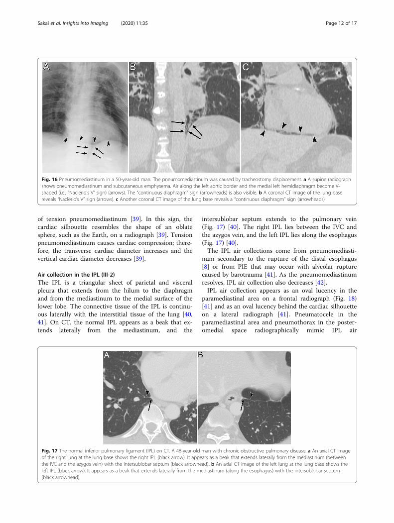
bubbles outlining the mediastinal structures that are notnormally visible or as an outwardly elevated mediastinalpleura [6–8]. In particular, the mediastinal air may out-line the medial border of the SVC, right innominate ar-tery, left common carotid artery, and left subclavianartery [8]. The radiographic signs of pneumomediasti-num include the “tubular artery” sign, “ring around theartery” sign, “double bronchial wall” sign, “spinnaker
sail” sign, “continuous diaphragm” sign, and “Naclerio’sV” sign [6–8, 37]. The “tubular artery” sign is thevisualization of both sides of a vessel in a pneumome-diastinum or aerated lung (Fig. 13) [8]. “A ring aroundthe artery” sign is seen on lateral chest radiographs andappears as a lucent ring that surrounds the right pul-monary artery (Fig. 14) [6–8, 37]. The “double bronchialwall” sign is the visualization of both sides of the bron-chial wall due to air in the bronchus and air surroundingthe wall (Fig. 13) [8]. The “spinnaker sail” sign is de-scribed as thymus elevation by pneumomediastinum(Fig. 15) [7] and is usually found in children [7]. The“continuous diaphragm” sign is a classic sign of pneu-momediastinum that is observed on frontal radiographswhen gas in the mediastinum separates the pericardiumand superior surface of the diaphragm across the midline(Fig. 16) [6–8]. “Naclerio’s V” sign was first reported by
Fig. 12 Anatomy of the pericardium and related diseases of abnormal air collection at each anatomical space. Axial chest CT of a 47-year-oldman with chronic kidney disease shows fluid collection in the pericardial cavity. The CT shows the pericardial fat (PF), parietal pericardium (whitearrowheads), pericardial cavity (PC), visceral pericardium (i.e., epicardium) (black arrowheads), epicardial fat (EF), myocardium (white asterisks), andcardiac chamber (CC)
Fig. 13 Pneumomediastinum due to chronic cough and vomiting in a 22-year-old man. a A supine radiograph shows the “tubular artery” sign(black arrows) and the “double bronchial wall” sign (black arrowheads). Subcutaneous emphysema is also evident (white arrow). b A coronalcervical CT image shows the “tubular artery” sign (black arrows). Both sides of the left internal jugular vein are visible due topneumomediastinum. Subcutaneous emphysema is also visible (white arrow). c The coronal CT image at the carina level shows the “doublebronchial wall” sign (black arrowheads). Both sides of the left bronchial wall are visible because of air in the bronchus and pneumomediastinumsurrounding the wall
Sakai et al. Insights into Imaging (2020) 11:35 Page 10 of 17

Naclerio [38] as a radiographic sign of spontaneousesophageal rupture. This sign is described as V-shapedair collection that consists of air along the left aorticborder and medial left hemidiaphragm (Fig. 16) [6, 7].These signs can be clearly detected on CT.Pneumothorax in the anteromedial space or pneumo-
pericardium may exhibit similar to the radiographicfindings of patients with pneumomediastinum. Unlikepneumothorax, pneumomediastinum can spread to thesoft tissue of the neck and face and to the retroperiton-eal space, outlines the mediastinal structures, and willnot change in distribution when the patient’s positionchanges [6]. Intra-aortic balloon pumping may also re-semble pneumomediastinum. An oval lucency along the
aorta reflecting air within the balloon accompanied by acatheter caudally is a characteristic imaging finding.Tension pneumomediastinum is a rare life-threatening
condition [14, 39] that is associated with mechanicalventilation. Increased intramediastinal pressure com-presses the great vessels and reduces venous return,stroke volume, and cardiac output [14, 39]. In cases ofcardiorespiratory embarrassment, mediastinal decom-pression (e.g., percutaneous mediastinal drainage) is ne-cessary for treatment [14].Although it is not difficult to detect pneumomediasti-
num on radiographs, it is difficult to diagnose tensionpneumomediastinum on radiographs. The “Earth-Heart”sign was recently reported to be useful for the diagnosis
Fig. 14 Pneumomediastinum of a 43-year-old man with acute myelogenous leukemia. a Erect lateral radiograph shows the “ring around theartery” sign. b A sagittal CT image also shows a lucent ring (arrows) surrounding the right pulmonary artery (asterisk), a feature called the “ringaround the artery” sign
Fig. 15 Pneumomediastinum and pneumopericardium in a 3-day-old girl with pulmonary atresia. Pneumomediastinum is caused by theobstruction and check valve effect of the tracheal tube with pulmonary secretions. a A supine radiograph shows the “spinnaker sail” sign withthymus elevation (asterisks) by pneumomediastinum (arrows). Pneumopericardium (arrowheads) is also visible. b A coronal CT image revealspneumomediastinum (arrows) surrounding the thymus (asterisks) and pneumopericardium (arrowheads)
Sakai et al. Insights into Imaging (2020) 11:35 Page 11 of 17
of tension pneumomediastinum [39]. In this sign, thecardiac silhouette resembles the shape of an oblatesphere, such as the Earth, on a radiograph [39]. Tensionpneumomediastinum causes cardiac compression; there-fore, the transverse cardiac diameter increases and thevertical cardiac diameter decreases [39].
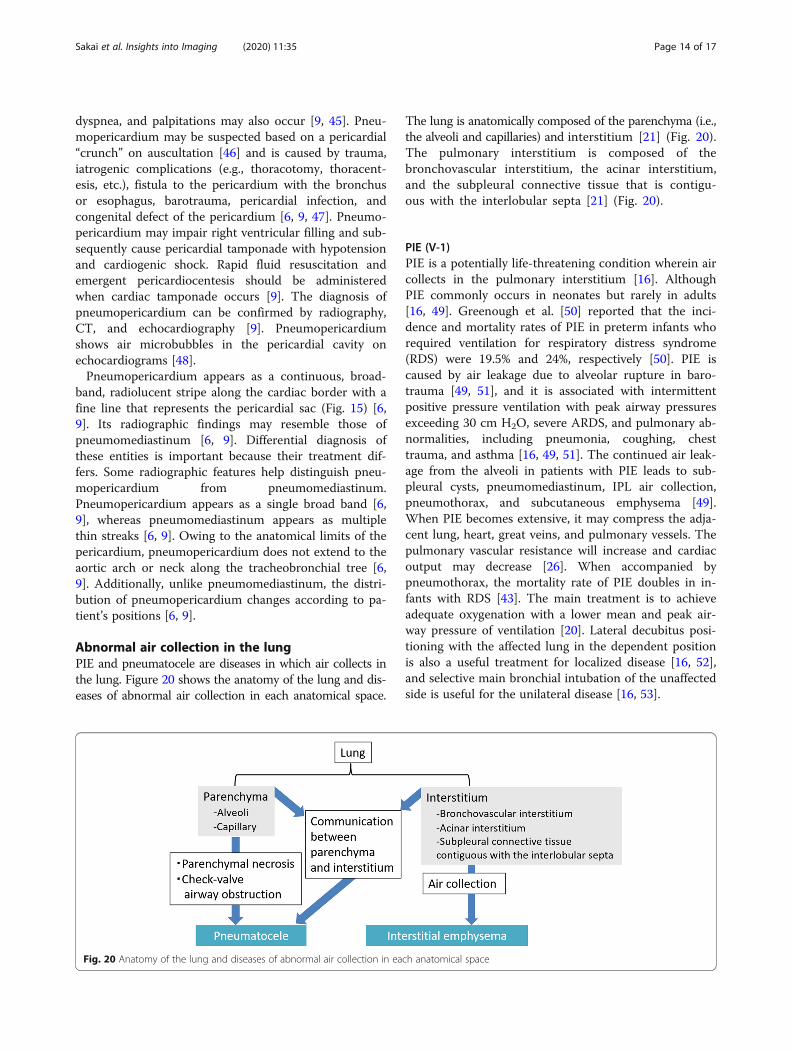
Air collection in the IPL (III-2)The IPL is a triangular sheet of parietal and visceralpleura that extends from the hilum to the diaphragmand from the mediastinum to the medial surface of thelower lobe. The connective tissue of the IPL is continu-ous laterally with the interstitial tissue of the lung [40,41]. On CT, the normal IPL appears as a beak that ex-tends laterally from the mediastinum, and the
intersublobar septum extends to the pulmonary vein(Fig. 17) [40]. The right IPL lies between the IVC andthe azygos vein, and the left IPL lies along the esophagus(Fig. 17) [40].The IPL air collections come from pneumomediasti-
num secondary to the rupture of the distal esophagus[8] or from PIE that may occur with alveolar rupturecaused by barotrauma [41]. As the pneumomediastinumresolves, IPL air collection also decreases [42].IPL air collection appears as an oval lucency in the
paramediastinal area on a frontal radiograph (Fig. 18)[41] and as an oval lucency behind the cardiac silhouetteon a lateral radiograph [41]. Pneumatocele in theparamediastinal area and pneumothorax in the poster-omedial space radiographically mimic IPL air
Fig. 16 Pneumomediastinum in a 50-year-old man. The pneumomediastinum was caused by tracheostomy displacement. a A supine radiographshows pneumomediastinum and subcutaneous emphysema. Air along the left aortic border and the medial left hemidiaphragm become V-shaped (i.e., “Naclerio’s V” sign) (arrows). The “continuous diaphragm” sign (arrowheads) is also visible. b A coronal CT image of the lung basereveals “Naclerio’s V” sign (arrows). c Another coronal CT image of the lung base reveals a “continuous diaphragm” sign (arrowheads)
Fig. 17 The normal inferior pulmonary ligament (IPL) on CT. A 48-year-old man with chronic obstructive pulmonary disease. a An axial CT imageof the right lung at the lung base shows the right IPL (black arrow). It appears as a beak that extends laterally from the mediastinum (betweenthe IVC and the azygos vein) with the intersublobar septum (black arrowhead). b An axial CT image of the left lung at the lung base shows theleft IPL (black arrow). It appears as a beak that extends laterally from the mediastinum (along the esophagus) with the intersublobar septum(black arrowhead)
Sakai et al. Insights into Imaging (2020) 11:35 Page 12 of 17

collection (Fig. 19 )[43]. In contrast to IPL air collec-tion, a pneumatocele and pneumothorax may extendover the hilum or may be posterior to the IPL [43].IPL air collection is usually accompanied by pneumo-mediastinum or PIE (Fig. 19) [41], and CT is usefulfor distinguishing between these diseases. In cases ofa pneumatocele or pneumothorax, a normal IPL maybe identified by CT (Fig. 19).
Abnormal air collection in the pericardium[pneumopericardium (IV-1)]Pneumopericardium is a rare but potentially life-threatening condition in which air enters the pericardialcavity. The visceral pericardium is the membranous layer
that covers the myocardium and epicardial fat. It also re-flects back near the origin of the major vessels and iscontinuous with the parietal pericardium (Fig. 12). Thepericardial cavity is the space between both pericardia(Fig. 12), and each pericardium is observed as a thin lin-ear structure on CT. Each pericardium is often not visu-alized over much of the left ventricle where a smallamount of pericardial and epicardial fat exists and be-cause motion artifacts of the left ventricle degrade imagequality [44]. The pericardial cavity normally contains asmall amount of fluid (between 15 and 50 mL) [44].Pneumopericardium more commonly occurs in thepediatric population [45]. The main clinical symptom ofpneumopericardium is chest pain, and radiating pain,
Fig. 18 Air collection in the inferior pulmonary ligament (IPL) of an 8-year-old boy with traumatic tracheal injury. a A supine radiograph shows anoval lucency (black arrows) in the left paramediastinal area, which reflects air collection in the left IPL. b An axial CT image reveals air collection inleft IPL (black arrows) because air collects along the mediastinum at the level of the esophagus (E). Pneumomediastinum (white asterisk),intrathoracic extrapleural space air collection (black arrowheads), and subcutaneous emphysema (white arrow) are also evident
Fig. 19 A pneumatocele mimicking inferior pulmonary ligament (IPL) air collection of a 61-year-old man. He has right main bronchial stricturetreated under positive pressure ventilation. a A supine radiograph shows an oval lucency (black arrows), which represents a left lower lobepneumatocele, in the left paramediastinal area. A pneumothorax (white asterisk) is also visible, but the pneumomediastinum is not present. b Anaxial CT image reveals a left lower lobe pneumatocele (black arrows), while the normal left IPL (black arrowhead) lies along the esophagus. Apneumothorax (white asterisk) is also visible. The pneumatocele and pneumothorax disappeared on the supine radiograph 3 months later
Sakai et al. Insights into Imaging (2020) 11:35 Page 13 of 17
dyspnea, and palpitations may also occur [9, 45]. Pneu-mopericardium may be suspected based on a pericardial“crunch” on auscultation [46] and is caused by trauma,iatrogenic complications (e.g., thoracotomy, thoracent-esis, etc.), fistula to the pericardium with the bronchusor esophagus, barotrauma, pericardial infection, andcongenital defect of the pericardium [6, 9, 47]. Pneumo-pericardium may impair right ventricular filling and sub-sequently cause pericardial tamponade with hypotensionand cardiogenic shock. Rapid fluid resuscitation andemergent pericardiocentesis should be administeredwhen cardiac tamponade occurs [9]. The diagnosis ofpneumopericardium can be confirmed by radiography,CT, and echocardiography [9]. Pneumopericardiumshows air microbubbles in the pericardial cavity onechocardiograms [48].Pneumopericardium appears as a continuous, broad-
band, radiolucent stripe along the cardiac border with afine line that represents the pericardial sac (Fig. 15) [6,9]. Its radiographic findings may resemble those ofpneumomediastinum [6, 9]. Differential diagnosis ofthese entities is important because their treatment dif-fers. Some radiographic features help distinguish pneu-mopericardium from pneumomediastinum.Pneumopericardium appears as a single broad band [6,9], whereas pneumomediastinum appears as multiplethin streaks [6, 9]. Owing to the anatomical limits of thepericardium, pneumopericardium does not extend to theaortic arch or neck along the tracheobronchial tree [6,9]. Additionally, unlike pneumomediastinum, the distri-bution of pneumopericardium changes according to pa-tient’s positions [6, 9].
Abnormal air collection in the lungPIE and pneumatocele are diseases in which air collects inthe lung. Figure 20 shows the anatomy of the lung and dis-eases of abnormal air collection in each anatomical space.
The lung is anatomically composed of the parenchyma (i.e.,the alveoli and capillaries) and interstitium [21] (Fig. 20).The pulmonary interstitium is composed of thebronchovascular interstitium, the acinar interstitium,and the subpleural connective tissue that is contigu-ous with the interlobular septa [21] (Fig. 20).
PIE (V-1)PIE is a potentially life-threatening condition wherein aircollects in the pulmonary interstitium [16]. AlthoughPIE commonly occurs in neonates but rarely in adults[16, 49]. Greenough et al. [50] reported that the inci-dence and mortality rates of PIE in preterm infants whorequired ventilation for respiratory distress syndrome(RDS) were 19.5% and 24%, respectively [50]. PIE iscaused by air leakage due to alveolar rupture in baro-trauma [49, 51], and it is associated with intermittentpositive pressure ventilation with peak airway pressuresexceeding 30 cm H2O, severe ARDS, and pulmonary ab-normalities, including pneumonia, coughing, chesttrauma, and asthma [16, 49, 51]. The continued air leak-age from the alveoli in patients with PIE leads to sub-pleural cysts, pneumomediastinum, IPL air collection,pneumothorax, and subcutaneous emphysema [49].When PIE becomes extensive, it may compress the adja-cent lung, heart, great veins, and pulmonary vessels. Thepulmonary vascular resistance will increase and cardiacoutput may decrease [26]. When accompanied bypneumothorax, the mortality rate of PIE doubles in in-fants with RDS [43]. The main treatment is to achieveadequate oxygenation with a lower mean and peak air-way pressure of ventilation [20]. Lateral decubitus posi-tioning with the affected lung in the dependent positionis also a useful treatment for localized disease [16, 52],and selective main bronchial intubation of the unaffectedside is useful for the unilateral disease [16, 53].
Fig. 20 Anatomy of the lung and diseases of abnormal air collection in each anatomical space
Sakai et al. Insights into Imaging (2020) 11:35 Page 14 of 17

Fig. 22 Decision trees based on the location of air collection. a Abnormal air collection along the chest wall. b Abnormal air collection aroundthe suprahilar mediastinum. c Abnormal air collection around the infrahilar mediastinum. d Abnormal air collection in the cardiac shadow
Fig. 21 Pulmonary interstitial emphysema. a A supine radiograph of a 6-day-old girl with an extremely low birth weight shows generalizedirregular radiolucent mottling and perivascular air (black arrow) in the left lung. b An axial chest CT image of a different patient (a 20-month-oldgirl with pneumonia) shows a subpleural cyst (asterisk), air within the interlobular septa (arrowheads), perivascular and peribronchial emphysema(white arrows), and pneumothorax
Sakai et al. Insights into Imaging (2020) 11:35 Page 15 of 17

PIE appears radiographically as lucent streaks that radiatefrom the hila to the periphery of the lung. This finding re-flects peribronchovascular air and air within the interlobularsepta that resembles an air bronchogram (Fig. 21) [16, 49,51]. The lucent streaks of PIE tend to be more numerousand more irregular on a radiograph than those of an airbronchogram [49]. Irregular radiolucent mottling in the lungreflects subpleural cysts and parenchymal cysts (Fig. 21) [16,49, 51]. On a CT image, PIE is visualized as perivascular andperibronchial emphysema, air within the interlobular septa,and subpleural cysts (Fig. 21) [16, 49, 51]. Rapid changes inthe distribution and extent of air are characteristic of PIE[36], and it may disappear within 1 to 2 days [49].
Pneumatocele (V-2)A pneumatocele is a thin-walled, air-filled space in thelungs [21]. The causes of pneumatocele include severepneumonia, thoracic trauma, hydrocarbon ingestion, andpositive pressure ventilation [10, 11]. Three theories havebeen proposed to explain the formation of a pneumatocele:(a) pulmonary overinflation caused by check valve airwayobstruction, (b) drainage of necrotic lung parenchyma withsubsequent enlargement caused by check valve airway ob-struction, and (c) focal air collection in pulmonary intersti-tial tissue caused by a direct communication between thepulmonary interstitium and parenchyma due to inflamma-tion and necrosis of the airway wall [10]. A pneumatocele isusually transient and generally resolves spontaneously [54];however, it is still associated with some complications. Spe-cifically, a pneumatocele may be accompanied by infection[11, 54], and the development of the air-fluid level withinthe cyst may be observed in an infected pneumatocele [11].Additionally, a pneumatocele may rupture through thepleural surface and result in pneumothorax [11, 54] or in-crease in size and compress the adjacent area and impairthe cardiorespiratory system [11, 54].A pneumatocele appears as a thin-walled, well-defined
round air space in the lungs on radiographs and CT images[10, 11, 54, 55] (Fig. 19). It usually does not contain any fluid[55]. Pneumatoceles are transient and only exist for a limitedamount of time (Fig. 19). At most, it takes weeks to monthsfor a pneumatocele to disappear [54, 55].
Diagnostic approach to abnormal air collectionbased on the locationDecision treesWe presented four decision trees for the location of aircollections along the chest wall (Fig. 22a), around thesuprahilar mediastinum (Fig. 22b), around the infrahilarmediastinum (Fig. 22c), and in the cardiac shadow (Fig.22d). A differential diagnosis can be made when one ofthe four decision trees is selected depending on the loca-tion of air collection on a radiograph.
ConclusionA comprehensive understanding of three-dimensionalchest anatomy based on CT enables the prediction ofsites of air collection on radiographs in supine patients.The management of patients differs based on the loca-tion of the air collection; thus, radiologists should at-tempt to detect an abnormal air collection andaccurately identify its location on radiographs.
AbbreviationsARDS: Acute respiratory distress syndrome; CT: Computed tomography;ICU: Intensive care unit; IPL: Inferior pulmonary ligament; IVC: Inferior venacava; PIE: Pulmonary interstitial emphysema; RDS: Respiratory distresssyndrome; SVC: Superior vena cava
Authors’ contributionsAll authors contributed to the collection of cases and drafting of themanuscript. All authors read and approved the final manuscript.
FundingNot applicable.
Availability of data and materialsNot applicable.
Ethics approval and consent to participateNot applicable.
Consent for publicationNot applicable.
Competing interestsThe authors declare that they have no competing interests.
Author details1Department of Diagnostic and Interventional Radiology, Ibaraki PrefecturalCentral Hospital, Kasama, Japan. 2Department of Diagnostic andInterventional Radiology, University of Tsukuba Hospital, Amakubo 2-1-1,Tsukuba, Ibaraki 305-8576, Japan. 3Department of Diagnostic Radiology,National Cancer Center Hospital East, Kashiwa, Japan. 4Department ofDiagnostic Radiology, Mayo Clinic Minnesota, 200 First St. SW, Rochester, MN55905, USA.
Received: 1 November 2019 Accepted: 23 January 2020
References1. Rubinowitz AN, Siegel MD, Tocino I (2007) Thoracic imaging in the ICU. Crit
Care Clin 23:539–5732. Lichtenstein D, Goldstein I, Mourgeon E, Cluzel P, Grenier P, Rouby JJ (2004)
Comparative diagnostic performances of auscultation, chest radiography,and lung ultrasonography in acute respiratory distress syndrome.Anesthesiology 100:9–15
3. Tagliabue M, Casella TC, Zincone GE, Fumagalli R, Salvini E (1994) CT andchest radiography in the evaluation of adult respiratory distress syndrome.Acta Radiol 35:230–234
4. Rankine JJ, Thomas AN, Fluechter D (2000) Diagnosis of pneumothorax incritically ill adults. Postgrad Med J 76:399–404
5. Marti De Gracia M, Gutierrez FG, Martinez M, Dueñas VP (2009)Subcutaneous emphysema: diagnostic clue in the emergency room. EmergRadiol 16:343–348
6. Bejvan SM, Godwin JD (1996) Pneumomediastinum: old signs and newsigns. AJR Am J Roentgenol 166:1041–1048
7. Matsusako M (2014) Pneumomediastinum and pneumomediastinitis.NICHIDOKU-IHO 59:77–91
8. Zylak CM, Standen JR, Barnes GR, Zylak CJ (2000) Pneumomediastinumrevisited. Radiographics 20:1043–1057
Sakai et al. Insights into Imaging (2020) 11:35 Page 16 of 17

9. Brander L, Ramsay D, Dreier D, Peter M, Graeni R (2002) Continuous lefthemidiaphragm sign revisited: a case of spontaneous pneumopericardiumand literature review. Heart 88:e5
10. Quigley MJ, Fraser RS (1988) Pulmonary pneumatocele: pathology andpathogenesis. AJR Am J Roentgenol 150:1275–1277
11. Dibardino DJ, Espada R, Seu P, Goss JA (2003) Management of complicatedpneumatocele. J Thorac Cardiovasc Surg 126:859–861
12. O'connor AR, Morgan WE (2005) Radiological review of pneumothorax. BMJ330:1493–1497
13. Santamarina MG, Beddings I, Lermanda Holmgren GV, Opazo Sanchez H,Volpacchio MM (2017) Multidetector CT for Evaluation of the ExtrapleuralSpace. Radiographics 37:1352–1370
14. Clancy DJ, Lane AS, Flynn PW, Seppelt IM (2017) Tension pneumomediastinum: aliteral form of chest tightness. J Intensive Care Soc 18:52–56
15. Paramasivam E, Bodenham A (2008) Air leaks, pneumothorax, and chestdrains. Continuing education in anaesthesia Critical Care & Pain 8:204–209
16. Sherren PB, Jovaisa T (2011) Pulmonary interstitial emphysema presenting ina woman on the intensive care unit: case report and review of literature. JMed Case Rep 5:236
17. Staub LJ, Biscaro RRM, Kaszubowski E, Maurici R (2018) Chestultrasonography for the emergency diagnosis of traumatic pneumothoraxand haemothorax: a systematic review and meta-analysis. Injury 49:457–466
18. Muller NL (1993) Imaging of the pleura. Radiology 186:297–30919. Tocino IM (1985) Pneumothorax in the supine patient: radiographic
anatomy. Radiographics 5:557–58620. Tocino IM, Miller MH, Fairfax WR (1985) Distribution of pneumothorax in the
supine and semirecumbent critically ill adult. AJR Am J Roentgenol 144:901–905
21. Hansell DM, Bankier AA, MacMahon H, McLoud TC, Müller NL, Remy J (2008)Fleischner Society: glossary of terms for thoracic imaging. Radiology 246:697–722
22. Macduff A, Arnold A, Harvey J, BTS Pleural Disease Guideline Group. (2010)Management of spontaneous pneumothorax: British Thoracic SocietyPleural Disease Guideline 2010. Thorax 65 Suppl 2:ii18-ii31
23. Saeki M (1988) Clinical study for findings of pneumothoraces on the plainchest film. Nippon Igaku Hoshasen Gakkai Zasshi 48:1371–1380
24. Moskowitz PS, Griscom NT (1976) The medial pneumothorax. Radiology 120:143–14725. Ball JB Jr, Proto AV (1982) The variable appearance of the left superior
intercostal vein. Radiology 144:445–45226. Markowitz RI (1988) The anterior junction line: a radiographic sign of
bilateral pneumothorax in neonates. Radiology 167:717–71927. Kong A (2003) The deep sulcus sign. Radiology 228:415–41628. Gordon R (1980) The deep sulcus sign. Radiology 136:25–2729. Ziter FM Jr, Westcott JL (1981) Supine subpulmonary pneumothorax. AJR
Am J Roentgenol 137:699–70130. Watts BL, Howell MA (2001) Tension pneumothorax: a difficult diagnosis.
Emerg Med J 18:319–32031. Gobien RP, Reines HD, Schabel SI (1982) Localized tension pneumothorax:
unrecognized form of barotrauma in adult respiratory distress syndrome.Radiology 142:15–19
32. Fisher JK (1978) Skin fold versus pneumothorax. AJR Am J Roentgenol 130:791–792
33. Waseem M, Jones J, Brutus S, Munyak J, Kapoor R, Gernsheimer J (2005)Giant bulla mimicking pneumothorax. J Emerg Med 29:155–158
34. Im Y, Farooqi S, Mora A Jr (2016) Vanishing lung syndrome. Proc (Bayl UnivMed Cent) 29:399–401
35. Aghajanzadeh M, Dehnadi A, Ebrahimi H et al (2015) Classification andmanagement of subcutaneous emphysema: a 10-year experience. Indian JSurg 77:673–677
36. Valente T, Tortora G, Bocchini G, Rea G, Marino M, Muto M (2017) MDCTand US of intrathoracic extrapleural space soft tissue-containing lesions: USextrapleural fat sign and MDCT fat ghost ribs sign. Radiol Med 122:479–486
37. Agarwal PP (2006) The ring-around-the-artery sign. Radiology 241:943–94438. Naclerio EA (1957) The V sign in the diagnosis of spontaneous rupture of
the esophagus (an early roentgen clue). Am J Surg 93:291–29839. Obeso Carillo GA, Barge Caballero G, Canizares Carretero MA (2014) The
Earth-Heart sign: a new diagnostic finding in a patient with tensionpneumomediastinum. Lancet 383:486
40. Godwin JD, Vock P, Osborne DR (1983) CT of the pulmonary ligament. AJRAm J Roentgenol 141:231–236
41. Volberg FM Jr, Everett CJ, Brill PW (1979) Radiologic features of inferiorpulmonary ligament air collections in neonates with respiratory distress.Radiology 130:357–360
42. Yoon H-K (2016) Interpretation of neonatal chest radiography. J Korean SocRadiol 74:279–290
43. Friedman PJ (1985) Adult pulmonary ligament pneumatocele: a loculatedpneumothorax. Radiology 155:575–576
44. Breen JF (2001) Imaging of the pericardium. J Thorac Imaging 16:47–5445. Shakya D, Pathak MR (2014) Pneumopericardium. J Nepal Paediatr Soc 34:
163–16546. Dudzinski DM, Mak GS, Hung JW (2012) Pericardial diseases. Curr Probl
Cardiol 37:75–11847. Inaba H, Ohta S, Nishimura T, Itoh Y, Ishida I (1997) A case of congenital
complete defect of the left pericardium complicated with spontaneouspneumothorax. Jpn J Chest Surg 11:823–828
48. Abrahan Iv LL, Obillos SMO, Aherrera JM et al (2017) A Rare Case ofPneumopericardium in the Setting of Tuberculous Constrictive Pericarditis.Case Rep Cardiol 2017:4257452
49. Westcott JL, Cole SR (1974) Interstitial pulmonary emphysema in childrenand adults: roentgenographic features. Radiology 111:367–378
50. Kemper AC, Steinberg KP, Stern EJ (1999) Pulmonary interstitial emphysema:CT findings. AJR Am J Roentgenol 172:1642
51. Greenough A, Dixon AK, Roberton NR (1984) Pulmonary interstitialemphysema. Arch Dis Child 59:1046–1051
52. Schwartz AN, Graham CB (1986) Neonatal tension pulmonary interstitialemphysema in bronchopulmonary dysplasia: treatment with lateraldecubitus positioning. Radiology 161:351–354
53. Chalak LF, Kaiser JR, Arrington RW (2007) Resolution of pulmonary interstitialemphysema following selective left main stem intubation in a prematurenewborn: an old procedure revisited. Paediatr Anaesth 17:183–186
54. Al-Saleh S, Grasemann H, Cox P (2008) Necrotizing pneumonia complicatedby early and late pneumatoceles. Can Respir J 15:129–132
55. Flaherty RA, Keegan JM, Sturtevant HN (1960) Post-pneumonic pulmonarypneumatoceles. Radiology 74:50–53
Publisher’s NoteSpringer Nature remains neutral with regard to jurisdictional claims inpublished maps and institutional affiliations.
Sakai et al. Insights into Imaging (2020) 11:35 Page 17 of 17
Copyright © 2022 FDOKUMEN