ESTI2012 Congress guIdE - European Society of Thoracic ...
104
www.esti2012.org European Society of Thoracic Imaging ESTI2012 CONGRESS GUIDE 20 TH ANNIVERSARY MEETING 22-24 June 2012 | Westminster, Central London
-
Upload
khangminh22 -
Category
Documents
-
view
0 -
download
0
Transcript of ESTI2012 Congress guIdE - European Society of Thoracic ...

www.esti2012.org
European Society of Thoracic Imaging
ESTI2012 Congress guIdE
20Th AnnIVERSARY MEETIng 22-24 June 2012 | Westminster, Central London


02 10
03 12
04 13
05 14
06 15
07 16
08 17 09
EST
I2012
Co
ng
ress g
uid
e
22-24 June 2012 | Westminster, Central London 1
Contents
Welcome
esti Memories
Friday Progamme
saturday Progamme
sunday Progamme
Committees
Faculty
Venue Floor Plan
sponsors
Local info: Cafés & Restaurants
Local info: things to do
getting Around: Area Map
getting Around: Bus & Water
getting Around: tube
Abstracts
iMPRinteditor: Ruth Warne British institute of Radiology, 131-151 great titchfield street, London W1W 5BB
scientific content: Sujal DesaiKing’s College Hospital, denmark Hill, London, se5 9Rs
Please note that abstracts are printed as initially submitted.

EST
I2012
Co
ng
ress g
uid
e
22-24 June 2012 | Westminster, Central London2
Dear Friends,
It is my great pleasure to welcome you all to the European Society of Thoracic Imaging (ESTI) meeting in London. As you will be aware this is the 20th Anniversary Congress and ESTI continues to grow in strength and popularity. The programme for the 2012 meeting is exciting, reflecting many of the current trends and controversies in thoracic imaging. I am thankful to my Programme Committee for putting together this fantastic programme, which I am sure you will find both interesting and stimulating. I am grateful to my faculty for their contributions to this congress; this year it is also my pleasure to welcome two other societies – The Society of Thoracic Radiology (from
North America) and the British Society of Thoracic Imaging – with whom we are running joint sessions on lung cancer screening and pulmonary vascular disease. For the anniversary meeting ESTI has had an unprecedented number of abstract submissions, prompting an increase in the numbers of Scientific Sessions. Delegate numbers are high, almost certainly reflecting the interest in thoracic imaging around the globe!
In addition to joining us in celebrating ESTI’s 20th year, I am hoping that the central location of this glorious venue will encourage you to explore some of the sights and sounds of London.
sujal DesaiPresident, european society of thoracic imaging 2012
Welcome to ESTI2012

EST
I2012
Co
ng
ress g
uid
e
22-24 June 2012 | Westminster, Central London 3
During the European Congress of Radiology in 1991, a handful of thoracic radiologists, who were involved in shaping the Educational sessions, were stimulated to create a European Society and decided to meet the next year to go a step further. Max Coulomb hosted an informal two days seminar in Grenoble (France) in Spring 1992, gathering less than twenty, mostly academic, radiologists, with an exclusive or strong involvement in pulmonary radiology. The brainstorming was enthusiastic and fertile. As it happens usually in European gatherings, where English is used as the ‘lingua franca’, their strong command of the language powered the British colleagues. No wonder that the statutes, that were rather rapidly outlined during the meeting,
were inspired, or better, copied from existing models in the UK. An interesting debate was the name to be given to the new society. The proposal that ‘imaging’ had more political and lobbying power than the restrictive term of ‘radiology’ was rapidly clear to all participants. Another issue was about other components of the name, such as ‘respiratory’, ‘lung’ and ‘pulmonary’. Finally the term of ‘thoracic’ was adopted, again with the idea that it encompasses it all, and nothing that could be politically relevant in the future, was omitted…At the end of the discussion, we formulated the name, European Society of Thoracic Imaging… At the beginning of ESTI, diverging trends were identified in the management of the Society: some concentrated on fostering science and education within a rather small entity, others pushed in the direction of creating an open and large society, that would be politically recognized as a competitive partner within a larger european radiological organization. These questions have been settled since. Personally I keep excellent memories from these intense years in my professional career. I considered that organization of the founding meeting was nothing more than a preliminary trial. It was a great honour for me, when ESTI allowed me again to organize the fifth annual congress in 1997 – this time in Brussels.
Among many fond memories of ESTI since it first began is the founding meeting hosted by Philippe Grenier in a mountain hotel near Grenoble. ESTI owes a lot to Philippe’s organising skill, drive and generosity. The founding members, many of whom had never met each other prior to the meeting, were in many instances unaware of how to set up an organisation such as ESTI. To our astonishment and delight, Robert Dondelinger not only placed a draft constitution on the table, but announced that he would take the financial risk of organising the first ESTI conference. After that, Philippe volunteered Paris as the venue for the second meeting asking (telling!) Jacques Remy and Martine Remy-Jardin to organise the conference, which they did and made enough profit for ESTI to ensure the subsequent meetings were less of a financial risk for subsequent Presidents. I have never attended a one
day committee meeting which was so successful - it set up ESTI and put it on the firm foundations from which it has since prospered. All of our thanks should go to Robert and Philippe, and all the original founder members (almost all whom subsequently became Presidents of ESTI) for their enthusiasm, energy and unfailing spirit of cooperation.
ESTI is a super organisation, though I guess I am pretty biased having been involved since the start and having a very vested interest. There has always been some excellent ‘science’ but the main virtue is the relatively small size and the fact that members get to know one another very quickly forming firm friendships
which are a wonderful way of learning a lot more from a lecture and, of course, providing ‘cross-cultural pollination’ (important for any ‘flower’!!) – the best way for friendships to flourish. ESTI grew out of an idea hatched over a few drinks by Philippe and me in Bern in 1990 at a Fleischner meeting and then pursued by Philippe with an inaugural meeting in Grenoble hosted by Max Coulomb in 1992… Personally, I have made many friends and learnt a great deal – and not just about Radiology! These friendships (always including wives) have extended into my retirement with regular social gatherings in various countries throughout Europe. One such group is the ‘Faakers’ that meets every September, which I anticipate with relish and from which I return immensely contented from a combination of excellent food, wine and exercise, usually at altitude.
sujal DesaiPresident, european society of thoracic imaging 2012
ESTI Memories
Christopher Flower
Founding Member Honorary esTI Fellow
Robert Dondelinger
Founding Member esTI President (1993 & 1997)
Peter Armstrong
Founding Member esTI President (1996)

EST
I2012
Pro
gr
aM
Me
22-24 June 2012 | Westminster, Central London4
FRIDAy22nD
TImE SESSIon TITlE / lECTuRE moDERAToRS & SPEAkERS Room
08:00-10:15 Registration Dean’s Yard Entrance
08:15-09:45
SCIENTIFIC SESSION I: Interstitial lung disease
Moderators: Nicola Sverzellati & Nicholas Screaton
Assembly Hall
SCIENTIFIC SESSION II: Pulmonary vascular disease
Moderators: Christoph Engelke & Susan Copley
Harvey Goodwin Suite
SCIENTIFIC SESSION III: Lung cancer/pulmonary nodules
Moderators: Simon Padley & Nevzat Karabulut
Bishop Partridge Hall
B R E A k ( R e F R e s H M e n t s i n t H e H o A R e M e M o R i A L H A L L )
10:15-10:30 Opening ceremony Sujal Desai Assembly Hall
10:30-12:30 Lung cancerModerators: José Vilar & Christian Herold
Assembly Hall
10:30-11:00 Pathology of lung cancer: implications of re-classification Andrew Nicholson Harvey Goodwin Suite (“Overflow” room with live feed)
11:00-11:30 The revised TNM staging: a guide for radiologists Anna Larici
11:30-12:00 Non-solid & part-solid nodules – pratical management Denise Aberle
12:00-12:30 Selection criteria for lung cancer surgery Simon Jordan
B R E A k ( R e F R e s H M e n t s i n t H e H o A R e M e M o R i A L H A L L )
PARALLEL LuNCHTIME SYMPOSIA
13:00-14:00Siemens lunchtime symposium: CT/MRI of the thorax: quo vadis?
Moderator: Joachim Wildberger
Assembly Hall
Computer-aided diagnosis: why, how and when? Marco Das
MRI of the thorax: why, how and when? Juergen Biederer
Wrap Up: cross-sectional imaging of the chest Joachim Wildberger
13:00-14:00Bracco lunchtime symposium: contrast management strategies at low kVp
Moderator: Christoph Becker
Harvey Goodwin Suite
Strategies for radiation dose reduction in vascular CT Christian Loewe
Strategies for radiation dose reduction in cardiac CT Francesca Pugliese
B R E A k ( R e F R e s H M e n t s i n t H e H o A R e M e M o R i A L H A L L )
14:30-15:30Fungal infections and haematopoietic stem cell transplantation (HSCT)
Moderator: Cornelia Schaefer-Prokop
Assembly Hall
14:30-14:50Fungal infection after HSCT: the challenge for haemato-oncologists
Antonio Pagliuca Harvey Goodwin Suite (“Overflow” room with live feed)14:50-15:10 Capabilities and limitations of imaging in fungal infection Tomás Franquet
15:10-15:30 Fungal infection after HSCT: an integrated approach Mansour Ceesay
B R E A k ( R e F R e s H M e n t s i n t H e H o A R e M e M o R i A L H A L L )
16:00-18:00ESTI meets the British Society of Thoracic Imaging
Pulmonary vascular diseaseModerators: Hans-ulrich Kauczor & John Reynolds
Assembly Hall
16:00-16:30 CT signs of pulmonary hypertension Nicholas Screaton Harvey Goodwin Suite (“Overflow” room with live feed)
16:30-17:00 MR imaging in pulmonary hypertension Sanjeev Bhalla
17:00-17:30 Thromboembolic disease - imaging algorithms in pregnancy Fergus Gleeson
17:30-18:00 Thromboembolic disease - latest CT imaging Martine Remy-Jardin
18:15 WELCOME RECEPTION (Bishop Partridge Hall & Assembly Hall)

EST
I2012
Pro
gr
aM
Me
22-24 June 2012 | Westminster, Central London 5
SATuRDAy23rD
TImE SESSIon TITlE / lECTuRE moDERAToRS & SPEAkERS Room
08:00-10:00 Registration Dean’s Yard Entrance
08:15-09:45
SCIENTIFIC SESSION I: COPD (Chronic Obstructive Pulmonary Disease)
Moderators: Ioannis Vlahos & Nigel Howarth
Assembly Hall
SCIENTIFIC SESSION II: Airways diseases / Interventional procedures
Moderators: Anand Devaraj & Sanjeev Bhalla
Harvey Goodwin Suite
SCIENTIFIC SESSION III: Dose and technique optimization
Moderators: Karl-Friederich Kreitner & Joachim Wildberger
Bishop Partridge Hall
B R E A k ( R e F R e s H M e n t s i n t H e H o A R e M e M o R i A L H A L L )
10:00-10:30 Presidential Award & Honorary Lecture Moderator: Sujal Desai Assembly Hall
B R E A k ( R e F R e s H M e n t s i n t H e H o A R e M e M o R i A L H A L L )
PARALLEL EDuCATION SESSIONS
11:00-13:00 Basic HRCT course Moderator: Katharina Marten
Assembly Hall
11:00-11:30 Reticular and nodular patterns Nicola Sverzellati
11:30-12:00 Ground-glass opacification Cornelia Schaefer-Prokop
12:00-12:30 An approach to fibrosing lung disease Susan Copley
12:30-13:00 Small airways disease Johny Verschakelen 11:00-13:00 Advanced HRCT course Moderator: Juergen Biederer Harvey Goodwin
Suite11:00-11:30 Radiation dose: what every radiologist (& clinician) needs to know Denis Tack
11:30-12:00Optimal CT protocols & reconstruction techniques for airway imaging
François Laurent
12:00-12:30 Pulmonary MRI - what’s new? Hans-Ulrich Kauczor
12:30-13:00Functional respiratory imaging: current applications and future perspectives
Alex Bankier
B R E A k ( R e F R e s H M e n t s i n t H e H o A R e M e M o R i A L H A L L )
PARALLEL LuNCHTIME SYMPOSIA
13:30-14:30GE lunchtime symposium: The radiologist and the heart
Moderator: Carl Roobottom Assembly Hall
13:30-13:50 Dose considerations in cardiac CT: how low can we go? Carl Roobottom
13:50-14:10 Imaging after coronary intervention: how good is CT? Smita Patel
14:10-14:30 CT coronary angiography: a cardiologist's perspective Gareth Morgan-Hughes
13:30-14:30Toshiba lunchtime symposium: The physical & clinical impact of 320-row CT and AIDR 3D on lung diagnoses
Moderator: Mathias Prokop Harvey Goodwin Suite
13:30-13:50 Lung nodules segmentation with 320-row CT Bram van Ginneken
13:50-14:10 Ultra low dose chest with AIDR 3D Trond Aalokken
14:10-14:30 Ultra low dose lung screening Alain Blum
B R E A k ( R e F R e s H M e n t s i n t H e H o A R e M e M o R i A L H A L L )
14:45-16:15 Interstitial lung diseaseModerators: Susan Copley & Maria-Luigia Storto
Assembly Hall
14:45-15:15 ATS/ERS reclassification: all change or no change? David Hansell Harvey Goodwin Suite (“Overflow” room with live feed)
15:15-15:45 Modern staging for management and prognostication in ILD Athol Wells
15:45-16:15 HRCT Techniques in ILD in 2012: optimising the Image Philippe Grenier
B R E A k ( R e F R e s H M e n t s i n t H e H o A R e M e M o R i A L H A L L )
16:45-18:30 Film panelModerators: Nigel Howarth and Katerina Malagarir
Assembly Hall
TEAM 1 José Vilar (Captain), François Laurent, Ineke Hartmann, Ioannis Vlahos
TEAM 2 Peter Vock (Captain), Simon Padley, Catherine Beigelman, Emmanuel Coche
19:30 FACuLTY DINNER
Harvey Goodwin Suite (“Overflow” room with live feed)

EST
I2012
Pro
gr
aM
Me
22-24 June 2012 | Westminster, Central London6
SunDAy24TH
TImE SESSIon TITlE / lECTuRE moDERAToRS & SPEAkERS Room
08:00-08:30 Registration Dean’s Yard Entrance
08:30-10:301st ESTI-STR collaborative plenary session Lung cancer screening
Moderators: Stefan Diederich & Gerald Abbott
Assembly Hall
08:30-09:00 Implications of the NLST for the rest of the world Denise Aberle
Harvey Goodwin Suite (“Overflow” room with live feed)
09:00-09:30 UK lung cancer screening trial design features Anand Devaraj
09:30-10:00 Lung cancer screening in Europe: what next? Mathias Prokop
10:00-10:30Incidental nodule detection and management outside screening trials
Melissa Rosado de Christenson
B R E A k ( R e F R e s H M e n t s i n t H e H o A R e M e M o R i A L H A L L )
11:00-11:20 Presentation of poster & oral paper prizes Moderator: José Vilar Assembly Hall
11:20-12:50 Thoracic intervention Moderator: Christoph Engelke Assembly Hall
11:20-11:50Pulmonary radiofrequency ablation and microwave ablation: current status and trends
Benoît Ghaye Harvey Goodwin Suite (“Overflow” room with live feed)11:50-12:20 Bronchial arterial intervention Katerina Malagari
12:20-12:50 Brochoscopic valves – who, how, when and why? Gilbert Ferretti
M e e t i n g C L o s e
12:50 ESTI Annual General Meeting

EST
I2012
Co
ng
ress g
uid
e
22-24 June 2012 | Westminster, Central London 7
CoMMITTees
Susan CopleyConsultant radiologist and reader in radiology, Imperial College London
Susan Copley was appointed Consultant Radiologist at Imperial College NHS Trust (Hammersmith and Charing Cross Hospitals) London in 2001 and Reader in Thoracic Imaging, Imperial College, London in 2008. She is an author of over 50 peer reviewed papers, 12 book chapters and 2 textbooks. She is an Editorial Board Member for Clinical Radiology and reviews for several European and North American Journals. She is the Educational Lead, British Society of Thoracic Imaging.
Fergus GleesonConsultant radiologist, oxford radcliffe Hospitals nHs Trust, oxford, UK
Fergus Gleeson was appointed as a Consultant Radiologist in Oxford in 1991, having trained in Cambridge, London and Los Angeles. He is now the Professor of Radiology, Oxford University, and leads a team of scientists and radiologists investigating pleural disease, and novel imaging technologies and data analysis.
David HansellConsultant radiologist, royal Brompton Hospital, London, UK
David Hansell was appointed Consultant Radiologist at the Royal Brompton Hospital in 1989 and Professor of Thoracic Imaging, National Heart and Lung Institute, Imperial College School of Medicine, London in 1998. His primary specialty is diagnostic imaging of the lungs and he has a particular interest in high resolution computed tomography of diffuse lung diseases. He is a past President (2005) of the European Society of Thoracic Imaging and is President-Elect of the Fleischner Society.
Simon PadleyConsultant radiologist, royal Brompton Hospital, London, UK
Dr Padley specializes in thoracic & vascular imaging and intervention & general body imaging and has been an invited speaker on thoracic imaging at the European Congress of Radiology Annual Meeting and European Society of Thoracic Imaging Annual Meetings. He is the organizer and lead on courses on both cardiac and thoracic imaging. Dr Padley is an assistant editor of Clinical Radiology and peer reviewer for a number of journals. He has co-authored more over 30 book chapters and 90 peer reviewed papers.
Juergen BiedererConsultant radiologist, University Hospital schleswig-Holstein, Kiel, germany
Prof. Dr. Juergen Biederer is Associate Director of the Department of Diagnostic Radiology and Associate Professor of the Medical Faculty in Kiel. His specific expertise is advanced diagnostic imaging strategies for thoracic diseases with multi-slice detector CT and magnetic resonance imaging. His international clinical and experimental research collaborations comprise institutions in Denver, CO, Edinburgh, UK, Cambridge, UK and Heidelberg, DE. Prof. Biederer is an active member of the German and European Societies of Radiology (NDRG, DRG, ESR), European Society of Thoracic Imaging (ESTI) as well as the International Society of Magnetic Resonance in Medicine (ISMRM).
Emmanuel CocheProfessor of cardiothoracic imaging, Cliniques Universitaires st-Luc, Brussels, Belgium
Emmanuel Coche is Professor of cardio-thoracic imaging at Cliniques Universitaires St-Luc in Brussels. His main research activities are in the field of thoracic vessels, cardiac disorders and CT development He is a board member of several scientific associations (ESTI, SIT, SRBR/KBVR, ESR, SFR) and reviewer for several scientific journals such as European Radiology, European Journal of Radiology, Insights into Imaging , Journal of Radiology, Revue de pneumologie. Professor Coche has published more than 100 peer-reviewed papers and written several chapter books.
Cornelia Schaefer-Prokop associate Professor of radiology, Meander Medical Centre, amersfoort, The netherlands
Cornelia has worked as radiologist in Hannover, Germany (1993-98), AKH Vienna, Austria (1998-2004), AMC Amsterdam (2005-2209) and since 2009 in Meander Medical Centre Amersfoort, research affiliations with AMC Amsterdam and Radboud University, Nijmegen. Her main research interests are in digital radiography, CAD and CT of PE and interstitial lung diseases. She is an editorial board member of European Radiology, Journal of Thoracic Imaging and Insights into Imaging and is also a member of DRG, NVvR, ESR, RSNA, ESTI, and the Fleischner Society. In addition, she is the author of 110 peer reviewed publications and editor of two books.
nick ScreatonConsultant radiologist and Clinical Director, Papworth Hospital, Cambridge, UK
Dr Nick Screaton is a Consultant Cardiothoracic Radiologist and Radiology Director at Papworth Hospital, Cambridge. He trained in Cambridge, Oxford and London and underwent Fellowship Cardiothoracic Radiology training in Cambridge and Vancouver. He is currently British Society of Thoracic Imaging President. He has published over 50 papers in peer reviewed journals on a range of cardiothoracic topics, contributed to various national guideline groups, published several chapters and has given over 25 invited national and international lectures.
LoCAL oRgAnising CommITTEE
PRogRAMMe CommITTEE

EST
I2012
Co
ng
ress g
uid
e
22-24 June 2012 | Westminster, Central London8
FaCULTy & MoDeraTors At ESTI2012
Trond Aalokken oslo, norway
Gerald AbbottBoston, usA
Denise AberleLos Angeles, usA
Alex BankierBoston, usA
Christoph BeckerMunich, germany
Catherine Beigelman-AubryLausanne, switzerland
Sanjeev Bhallast Louis, usA
Juergen BiedererKiel, germany
Alain Blumnancy, France
mansour CeesayLondon, uK
Emmanuel CocheBrussels, Belgium
Susan CopleyLondon, uK
marco DasAachen, germany
Sujal DesaiLondon, uK
Anand DevarajLondon, uK
Stefan Diederichdusseldorf, germany
Christoph Engelkegoettingen, germany
Gilbert Ferrettigrenoble, France
Tomás FranquetBarcelona, spain
Benoît GhayeBrussels, Belgium
Fergus Gleesonoxford, uK
Philippe GrenierParis, France
David HansellLondon, uK
Ieneke HartmannRotterdam, the netherlands
Christian HeroldVienna, Austria
nigel HowarthChêne-Bougeries, switzerland
Simon JordanLondon, uK
nevzat karabulutdenizli, turkey
Hans-ulrich kauczorHeidelberg, germany
karl-Freiderich kreitnerMainz, germany
Anna lariciRome, italy
François laurentBordeaux, France
Christian loeweVienna, Austria
katerina malagariAthens, greece
katharina martengoettingen, germany
Gareth morgan-HughesPlymouth, uK
Andrew nicholsonLondon, uK
Simon PadleyLondon, uK
Antonio PagliucaLondon, uK
Smita PatelMichigan, usA
mathias Prokopnijmegen, the netherlands
Francesca PuglieseLondon, uK
martine Remy-JardinLille, France
John ReynoldsBirmingham, uK
Carl RoobottomPlymouth, uK
melissa Rosado de ChristensonKansas, usA
Cornelia Schaefer-ProkopAmersfoot, the netherlands
nicholas ScreatonCambridge, uK
maria-luigia StortoPrinceton, usA
nicola SverzellatiParma, italy
Denis TackBraine-L’Alleud, Belgium
Bram van Ginnekennijmegen, the netherlands
Johny VerschakelenLeuven, Belgium
José VilarValencia, spain
Ioannis VlahosLondon, uK
Peter VockBern, switzerland
Athol WellsLondon, uK
Joachim WildbergerMaastricht, the netherlands
EST
I2012
Co
ng
ress g
uid
e
22-24 June 2012 | Westminster, Central London 9
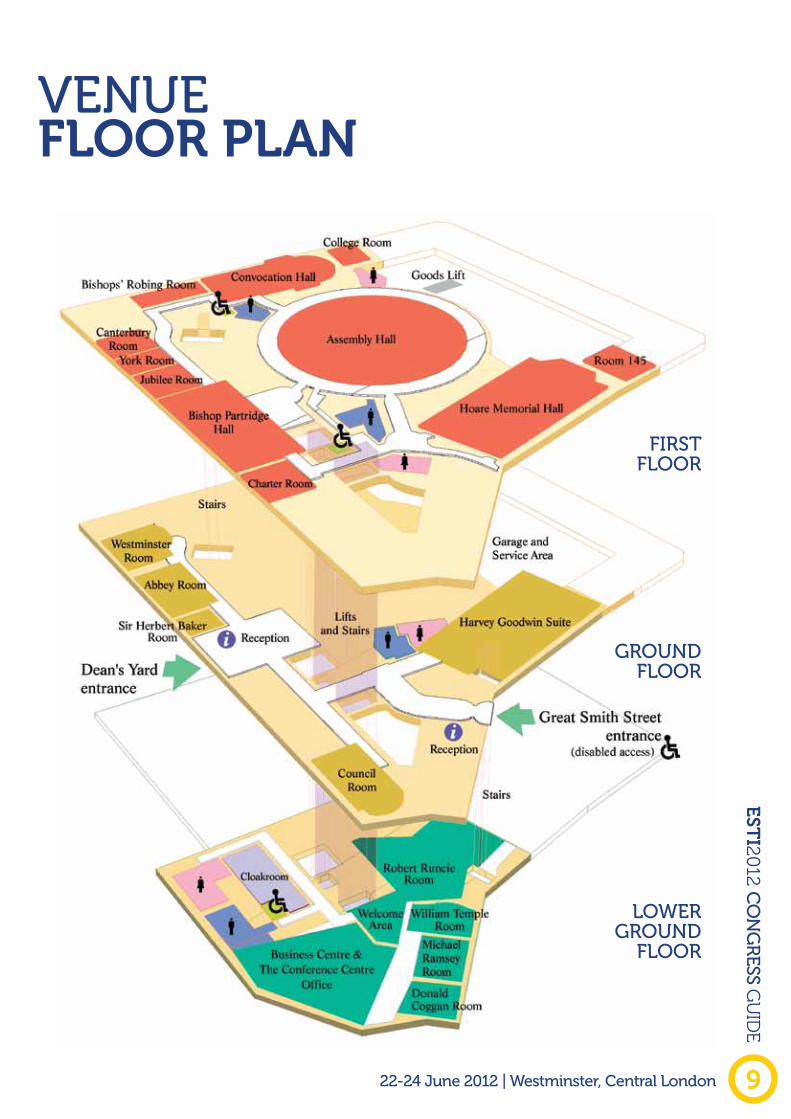
VenUeFlooR PlAn
FIrsT FLoor
groUnD FLoor
LoWer groUnD
FLoor

EST
I2012
Co
ng
ress g
uid
e
22-24 June 2012 | Westminster, Central London10
sPonsors
Bracco Imaging S.p.A., part of the Bracco Group, is one of the world’s leading companies in the diagnostic imaging business. Headquartered in Milan, Italy, Bracco Imaging develops, manufactures and markets diagnostic imaging agents and solutions that meet medical needs and facilitate clinical solutions.
Bracco Imaging offers a product and solution portfolio for all key diagnostic imaging modalities: X-Ray Imaging (including Computed Tomography-CT, Interventional Radiology, and Cardiac Catheterization), Magnetic Resonance Imaging, Contrast Enhanced Ultrasound, Nuclear Medicine through radioactive tracers, and Gastrointestinal Endoscopy.
The Company operates in over 90 markets worldwide, either directly or indirectly, through subsidiaries, joint ventures, licenses and distribution partnership agreements.
Lena Rasch, + 41 91 985 3030Bracco, Bracco Suisse SA, Via Cantonale Centro Galleria 2, 6928 Manno, Switzerland
Boehringer Ingelheim Ltd has kindly supported the meeting by the provision of lanyards.
Covidien is a $12 billion global healthcare products leader dedicated to innovation and long-term growth. Covidien creates innovative medical solutions for better patient outcomes and delivers value through clinical leadership and excellence. Covidien sells and develops energy-based devices, including RF and MW ablation devices and, because we are especially knowledgeable about those technologies, we can give impartial advice to the physician wishing to treat inoperable tumours in lung and other organs.
Richard Waddington, Field Sales Manager, Interventional Oncology, UK and Ireland, [email protected]
Covidien uK, 4500 Parkway, Whiteley, Hampshire PO15 7NY
GE Healthcare Systems provides transformational medical technologies and services that are shaping a new age of patient care. Our broad expertise in medical imaging, information technologies, medical diagnostics, patient monitoring systems and ultrasound solutions help our customers to deliver better care to for millions of patients everyday – from wellness screening to advanced diagnostics to life-saving treatment. Visit us at www.gehealthcare.com/uken/index.html
Louisa Mayo, CT Clinical Education Specialist, + 44 783 140 1850 Jackie Bye, Advanced Applications Specialist, + 44 783 140 1839 Jane Hickey, CT Modality Manager UK & Ireland, + 44 776 863 6846
GE Healthcare, 71 Great North Road, Hatfield, Herts, AL9 5EN

EST
I2012
Co
ng
ress g
uid
e
22-24 June 2012 | Westminster, Central London 11
Following our Made for Life commitment, patients are the primary focus of Toshiba’s innovations. Whether giving surgical teams better patient access to perform complex procedures or, giving patients the option to complete needed MR exams when contrast is contra-indicated, this commitment to help improve patient care spans across all of our modalities. Like the world’s first Dynamic Volume CT scanner - the Aquilion ONE - that is both the latest step in the natural progression of multislice imaging and a quantum leap that will carry CT imaging into the future.
Jacqueline de Graaf, +31 (0)79 368 9222 / +31 (0) 6 211 73 265Toshiba Medical Systems, Zilverstraat 1, 2718 RP, ZOETERMEER, The Netherlands
The Siemens Healthcare Sector is one of the world’s largest suppliers to the healthcare industry and a trendsetter in medical imaging, laboratory diagnostics, medical information technology and hearing aids. Siemens offers its customers products and solutions for the entire range of patient care from a single source – from prevention and early detection to diagnosis, and on to treatment and aftercare. By optimizing clinical workflows for the most common diseases, Siemens also makes healthcare faster, better and more cost-effective.
Andreas Rumpp, +49 9191 18-9822Siemens AG, Healthcare Sector, Imaging & Therapy Division Siemensstr. 1, 91301 Forchheim, Germany
Gilead Sciences is a biopharmaceutical company that discovers, develops and commercialises innovative therapeutics in areas of unmet medical need.
The company’s mission is to advance the care of patients suffering from life-threatening diseases worldwide.
Headquartered in Foster City, California, Gilead has operations in North America, Europe and Australia. Based in Cambridge for more than a decade, Gilead Sciences Ltd has grown to more than 200 employees across medical science, marketing, sales and finance, supported by major regional manufacturing and distribution operations in the Republic of Ireland.
Ayesha Chibb, +44 (0) 7796 177 059Gilead Sciences Ltd. Granta Park, Abington, Cambridge, United Kingdom, CB21 6GT
At Pfizer, we apply science and our global resources to improve health and well-being at every stage of life. We strive to set the standard for quality, safety and value in the discovery, development and manufacturing of medicines for people and animals. Our diversified global health care portfolio includes human and animal biologic and small molecule medicines and vaccines, as well as nutritional products and many of the world’s best-known consumer products.
Every day, Pfizer colleagues work to advance wellness, prevention, treatments and cures that challenge the most feared diseases of our time. Consistent with our responsibility as the world’s leading biopharmaceutical company, we also collaborate with health care providers, governments and local communities to support and expand access to reliable, affordable health care around the world. For more than 150 years, Pfizer has worked to make a difference for all who rely on us. In the UK, Pfizer has its business headquarters in Surrey and is a major supplier of medicines to the NHS. To learn more about our commitments, please visit us at www.pfizer.co.uk
Shelagh Gordon, + 44 7812 0404946Pfizer Ltd, Walton Oaks, Dorking Road, Walton-on-the-hill, Tadworth, Surrey, KT20 7NS

EST
I2012
Co
ng
ress g
uid
e
22-24 June 2012 | Westminster, Central London12
LoCAL inFo: CAFES & RESTAuRAnTS
eAteRies
eVening DInInG
There are many sandwich bars and delis in the nearby area. Look out for popular chains such as Pret A Manger, Eat, Costa and Café Nero along Victoria Street.
London has a large variety of restaurants, most of which just a short cab ride away. Visit www.toptable.com or www.squaremeal.co.uk for more information.
We recommend:
Roux at Parliament Square British Cuisine11 great george street, London, sW1P 3Ad+44 (0)20 7334 3737
Roux at Parliament Square, an elegant restaurant in Westminster, is an exclusive dining destination - popular with London’s politicians, socialites and high-powered business barons who love to linger over lunch. The brain-child of Michelin starred chef Michel Roux Junior – of Le Gavroche in Mayfair – Roux at Parliament Square boasts not only a luxurious and iconic location, but a light and inspired modern European menu that compliments the tasteful interior. With period features and modern furniture, the dining room at Roux at Parliament Square is a light-filled space perfect for business dining, and the restaurant’s chic bar is suitable for pre-dinner drinks and discreet cocktail dates.
The Cinnamon Club Indian Cuisinethe old Westminster Library, 30-32 great smith street, London, sW1P 3Bu +44 (0)20 7222 2555
The Cinnamon Club in the heart of Westminster offers a unique dining experience. Set in the historic Grade II listed former Westminster Library, the restaurant presents constantly evolving menus and a carefully matched wine list - all designed to reflect an ethos of innovation and creativity in one of the most stunning dining rooms in the country.
Atrium SW1 Modern european Cuisine4 Millbank, London sW1P 3JA+44 (0)20 7222 2211
Located beneath a striking glass atrium, it mixes luxury with a touch of boho chic, while the food is defined as ‘old school… with a hint of molecular’. Eat casually from a brasserie-style menu of small plates & populist standbys (think pork hock terrine with black pudding, rabbit ragoût or beetroot risotto), or sample the full works in the swanky Atrium Floor. ‘Day-caught fish’ & specials from the ‘vegetable allotment’ sit alongside the likes of duck leg with kohlrabi, pickled cherries & bitter leaves or venison with swede, kale, orange & juniper.
The mango Tree Thai Cuisine46 grosvenor Place, London, sW1X 7eQ+44 (0)20 7823 1888
The Mango Tree is a highly renowned Thai restaurant in Belgravia, South West London. Part of parent company ‘Coca’ in Asia, The Mango Tree became the flagship Thai restau-rant in Europe. Our work has paid off, with The Mango Tree winning numerous awards, commending our food creations and polished service. This has been the result of speedy growth which has continued unabated since 2001.

EST
I2012
Co
ng
ress g
uid
e
22-24 June 2012 | Westminster, Central London 13
LoCAL inFo: THInGS To Do
neARBy PLACes oF inteRest
London is brimming with things to do, we recommend: Madame TussaudsThe London DungeonTower of London
Natural History MuseumWellcome Collection Check out www.visitlondon.co.uk for further ideas.
Westminster Abbey 20 dean’s yard, Westminster Abbey, London, sW1P 3PA
Kings, queens, statesmen and soldiers; poets, priests, heroes and villains - the Abbey is a must-see living pageant of British history. Every year Westminster Abbey welcomes over one million visitors who want to explore this wonderful 700-year-old building. Thousands more join us for worship at our daily services. The Abbey is in the heart of London. Once inside audio guides are available in eight languages or there is the highly-popular verger-led tour.
Photo: Jean-Michel BAUD
Churchill War Rooms Clive steps, King Charles street, London, sW1A 2AQ020 7930 6961
In 1940, shortly after becoming Prime Minister, Churchill stood in the War Cabinet Room and declared: ‘This is the room from which I will direct the war’. Today, you can step back in time to explore the secret headquarters where Churchill and his staff changed the course of history.
london Duck Tours duck stop, Chicheley street, London, se1 7Py (dePARtuRe Point)020 7928 3132
London Duck Tours offer a unique view of the city’s most iconic landmarks from both land and water on vehicles used for the D-Day landings in 1944.The tour lasts about 75 minutes, with approximately 30 minutes on the river and is enhanced by an entertaining, action packed commentary by our tour guides who provide an alternative and insightful view of London’s history, plus fascinating facts and figures about the original D-Day vehicles themselves.
london Eye edF energy London eye, Riverside Building, County Hall, Westminster Bridge Road, London, se1 7PBBook at www.londoneye.com for discounted tickets.
The height of the London Eye is 135m (equivalent to 64 red telephone boxes piled on top of each other) making it the fourth tallest structure in London after the BT Tower, Tower 42 and One Canada Square in Canary Wharf. Not one for vertigo sufferers!

BU
CK
ING
HA
M P
AL
AC
E R
D
BIRDCAGE WALK
GREAT GEORGE ST
AB
ING
DO
N S
T M
ILB
AN
K
HORSEFERRY RD
OLD PYE ST
GR
EA
T
SM
ITH
ST M
AR
SH
HA
M S
T
GREAT COLLE GE ST
TU
FT
ON
ST
GREAT PETER ST
DEANS
YA
RD
A202 VAUXHALL BRIDGE RD
RO
CH
EST
ER
RO
W
JOH
N I
SLIP
ST
RE
GE
NC
Y S
T
V ICTORIA
ST
A302
BROAD SANCTUARY
GR
OSV
ENO
R PL
Westminster
St James’sPark
Pimlico
BuckinghamPalace
WestminsterAbbey
Palace ofWestminster
Houseof Lords
GrovesnorHotel
Victoria
H
ESTI2012
P
P
211
3
11
EST
I2012
Co
ng
ress g
uid
e
22-24 June 2012 | Westminster, Central London14
getting ARound: AREA mAP

BU
CK
ING
HA
M P
AL
AC
E R
D
BIRDCAGE WALK
GREAT GEORGE ST
AB
ING
DO
N S
T M
ILB
AN
K
HORSEFERRY RD
OLD PYE ST
GR
EA
T
SM
ITH
ST M
AR
SH
HA
M S
T
GREAT COLLE GE ST
TU
FT
ON
ST
GREAT PETER ST
DEANS
YA
RD
A202 VAUXHALL BRIDGE RD
RO
CH
EST
ER
RO
W
JOH
N I
SLIP
ST
RE
GE
NC
Y S
T
V ICTORIA
ST
A302
BROAD SANCTUARY
GR
OSV
ENO
R PL
Westminster
St James’sPark
Pimlico
BuckinghamPalace
WestminsterAbbey
Palace ofWestminster
Houseof Lords
GrovesnorHotel
Victoria
H
ESTI2012
P
P
211
3
11
EST
I2012
Co
ng
ress g
uid
e
22-24 June 2012 | Westminster, Central London 15
getting ARound: BuS & WATERLoCAL BuS InForMaTIon
tRAVeLLing By WATER
PARKing
City Cruises is the largest operator of boat tours on the Thames. Enjoy sightseeing with multilingual commentary on wheelchair-accessible boats with open-air decks and hop-on hop-o� services, operating between Westminster, London Eye, Tower Pier and Greenwich.
Daily until 4 November(unless shown otherwise).
A – Daily 16 July to 9 September only
B – Daily until 15 July and 10 September to 4 November only.
Westminster – London Eye – Tower – Greenwich
• Book online to get a discount• Child fares apply to children aged 5-16. Children aged under 5 travel free• The Family River Red Rover applies for two adults and up to three children• A River Red Rover o�ers unlimited hop-on, hop-o� travel all day• Groups of 20 or more receive a discount • Single tickets are also available• Fares from London Eye are the same as those from Westminster
Fares Adult single
Adult return
Child return
Senior return
Westminster – Tower £9.50 £12.50 £6.25 £8.75Westminster – Greenwich £12 £15.50 £7.75 £10.85Tower – Greenwich £9.50 £12.50 £6.25 £8.75River Red Rover – £19 £9.50 £13.30
Eastbound A B A A A A AWestminster 0915 0945 1015 1045
Then at these minutes past each hour
15 45
Until
1615 1645 1645 1715 1745 1815 1845 1915 1945 2030 2100London Eye 0925 0955 1025 1055 25 55 1625 1655 1655 1725 1755 1825 1855 1925 1955 2040 2110Tower 0955 1025 1055 1125 55 25 1655 1715 1725 1745 1815 1845 1915 1945 2015 2100 2130Greenwich 1025 1055 1125 1155 25 55 1725 – 1755 – – – – – – – –
Westbound B A A A A A AGreenwich – – 1025 1055
Then at these minutes past each hour
25 55
Until
1725 1755 1755 – 1825 – – – –Tower 1005 1035 1105 1135 05 35 1805 1835 1835 1850 1905 1955 2025 2110 –Westminster 1045 1115 1145 1215 45 15 1845 1900 1915 1945 1930 2030 2100 2135 –London Eye 1055 1125 1155 1225 55 25 1855 – 1925 1955 – 2040 2110 – –
City Cruises 020 7740 0400 citycruises.com
Transport for London (www.tfl.org.uk) provides a large network of buses which can take you all over the city. Travelling by public transport in London is cheaper if you purchase an Oyster card; these can be purchased at any London Underground station. If you are travelling by bus, you will be able to use your Oyster card. Please note: if you prefer not to get one of these cards, you must purchase your bus ticket at one of the machines at the bus stop. PAYMENT IS NOT ACCEPTED ON BUSES.
Bus routes:211 Hammersmith – Victoria – Waterloo 11 Fulham Broadway – Victoria – Liverpool street 3 Crystal Palace – Brixton – oxford Circus
other routes through Westminster:12 dulwich – oxford Circus24 Hampstead Heath – Pimlico 53 Plumstead – Whitehall87 Aldwych – Wandsworth88 Camden town – Clapham Common148 Camberwell green – White City159 Paddington Basin – streatham453 deptford Bridge – Marylebone
Local car parks are indicated on the map with P

EST
I2012
Co
ng
ress g
uid
e
22-24 June 2012 | Westminster, Central London16
getting ARound: TuBE
european society of thoracic imaging
ESTI2012 aBsTraCTs
EST
I2012
Co
ng
ress g
uid
e
22-24 June 2012 | Westminster, Central London 17
oraL & PosTer PresenTaTIons

21
22
23
24
25
26
27
28
29
30
31
32
33
34
35
37
36
38
39
40
41
42
43
44
45
46
EST
I2012
aB
sTr
aC
T C
on
ten
ts
22-24 June 2012 | Westminster, Central London18
oRAl ABstRACt Contents interstitial lung disease
o1: HRCt findings of collagen vascular disease-related interstitial pneumonia (CVd-iP): Comparative study among individual underlying diseaseso2: Pulmonary changes in lung donors treated with a new hemodynamic stabilizing drug regime assessed with High Resolution Computed tomography (HRCt) in an experimental pig modelo3: idiopathic Pulmonary Fibrosis: HRCt prognostic evaluation following the Ats/eRs/JRs/ALAt guidelines 2011 o4: Prognostic value and clinical applicability of traction bronchiectasis versus honeycombing in fibrotic lung disease o5: Pulmonary sarcoidosis: integrated composite physiological index and computed tomography patterns for prognostic staging
o6: Pulmonary sarcoidosis: relationship between HRCt patterns and physiological profileso7: uiP without honeycombing: key Ct features
o8: Perfusion Parametric Map for the Characterization of interstitial Lung disease
Pulmonary vascular disease
o9: Cardiac MRi assessment of right atrial volumes in pulmonary hypertensiono10: dual energy gemstone spectral imaging (gsi) lung perfusion MdCt in acute pulmonary embolism: blood volume changes assessment and correlation with clinical symptoms
o11: Ct Features in Porto-Pulmonary Hypertension: Relationships with echocardiography and Right-Heart Cardiac Catheterisation datao12: Frequency and origin of lung perfusion defects on dual-energy Computed tomography (deCt) in an unselected patient group
o13: Cardiac MRi of left atrial volume as a diagnostic tool in pulmonary hypertensiono14: time-resolved 3d MR angiography pulmonary transit times are inversely proportional to cardiac index in patients with pulmonary hypertension
o15: stunned lung’: a novel observation of resolving pulmonary embolism on lung perfusion Cto16: imaging V/Q in Chronic thromboembolic Pulmonary Hypertension with 3He and 1H MRi
o17: Computed tomographic (Ct) pulmonary vascular dimensions in adults with sickle cell disease: relationships with lung function indiceso18: Assessment of operability by means of CtPA and perfusion sPeCt in patients with chronic thromboembolic pulmonary hypertension
Lung cancer/pulmonary nodules
o19: Histopathologic scoring for Prognosis Prediction in solitary Pulmonary nodular Lung Adenocarcinoma: Correlation with imaging Biomarkers study Resultso20: impact of Localization and tumor size in 18-F-Fdg-Pet and Ct Lung Cancer tumor delineationo21: BoLd - predictive value for dignity evaluation in pulmonary tumours
o22: Combining visual perfusion - and quantitative diffusion-weighted MR imaging leads to a high accuracy for preoperative diagnosis of pulmonary noduleso23: Angiogenesis in non-small cell lung cancer: early assessment of therapeutic response to antiangiogenic chemotherapy with perfusion multidetector-row Ct (MdCt)
o24: Adenocarcinoma of the Lung Association with epidermal growth Factor Receptor (egFR): Ct Predictor of Response to egFR-tyrosine Kinase inhibitorso25: outcome of patients with non-small Cell Lung Cancer (nsCLC) who underwent Ct perfusion examination before surgery
o26: subtle mediastinal pleural thickening as a predictor of malignancyo27: Histologic subtyping of Lung Adenocarcinoma Based on new iAsLC/Ats/eRs Classification: Correlation with functional and metabolic imaging biomarkerso28: the impact of numbers of readers and methods of arbitration on pulmonary nodule detection in the context of lung cancer screening with Ct
Chronic obstructive pulmonary disease
o29: Can Ct help recognize a link between left atrial volume, impacting left ventricular preload, and the severity of emphysema?o30: Pseudo-embolic perfusion defects in CoPd: evaluation with dual-energy Ct angiography (deCt) in 170 patients
o31: Krypton ventilation imaging using dual-energy Ct in CoPd patients: initial experienceo32: Quantitative Ct Assessment Between Mass in intercostal Muscle and emphysema score in Patients with Chronic obstructive Pulmonary disease
o33: Visual assessment of CoPd-related morphologic changes on computed tomographyo34: Feasibility of Ct pulmonary segmentation techniques in evaluation of total lobar perfusion in patients with emphysema
o35: Comparing Hyperpolarised 3He MRi Ventilated Lung Volumes with spirometry in CoPdo36: Quantitative Ct air trapping is associated with lung function decline in heavy smokers, independent of Ct emphysema, age and smoking characteristicso37: Validation of an Automatic Lung segmentation Method for MRi-based Lung Perfusion studies
o38: the incidence of Pe in acute exacerbation of CoPd
Airways diseases/interventional procedures
o39: Predictors of bleeding during percutaneous Ct-guided transthoracic biopsy of pulmonary noduleso40: Rebiopsy for Mutational Analysis of Resistant non-small Cell Lung Cancers to Previously Chemotherapy: Adequacy and Complicationso41: Radiofrequency Ablation for Lung Metastases: the early King’s College Hospital experience
o42: Anthracofibrosis: Comparison with endobronchial tB and Fibrotic Bronchial stenosis on Cto43: serial Ct Findings of Mycobacterium avium-intracellulare complex (MAC) Lung disease with Antibiotic therapy
o44: High-resolution Ct Findings of non-tuberculous Mycobacterium: diagnostic Accuracy in Adults with Cystic Fibrosiso45: Percutaneous microwave ablation (MWA) of pulmonary malignancies using a high energy antennae system
o46: tracheomalacia in Adults with Cystic Fibrosis: determination of Prevalence and severity with dynamic Cine Cto47: standardization of a chest-Ct protocol for multi-center trial in cystic fibrosis (cf) infants
o48: stenting as an effective treatment of superior vena cava syndrome: review of 217 cases. single centre experience
dose and technique optimisation
o49: thoracic Ct with a dose of 0.1msv: is it feasible?o50: digital tomosynthesis as a diagnostic tool to exclude pseudolesions in patients with suspected thoracic lesions on chest x-ray radiography
o51: influence of Contrast Media on Computational Airway Analysis on MdCto52: techniques for Very Low dose thoracic digital tomosynthesis
o53: interscan Variability of Quantitative Computed tomography Air trapping Measures in Low-dose Chest Cto54: Bone suppression imaging improves observer performance for the detection of lung nodules in chest radiographs
o55: Radiation dose reduction to the breast: Comparison of bismuth shielding and use of a low kilovoltage in thoracic Cto56: the effect of iterative reconstruction versus filtered back projection on lung nodule volumetry at different Ct parameterso57: spectral optimization of chest Ct angiography: experience in 80 patients
o58: Can iterative reconstruction restore image quality at 60% dose reduction? Clinical experience in 50 patients with simultaneous availability of low-dose and standard-dose images from dual-source datasets

47 60
61
62
63
64
65
66
67
68
69
70
71
72
73
48
49
50
51
52
53
54
55
56
57
58
59
EST
I2012
aB
sTr
aC
T C
on
ten
ts
22-24 June 2012 | Westminster, Central London 19
PoSTER ABstRACt Contents P1: Prediction of the lung adenocarcinoma metastatic spread according to initial MdCt examinationP2: Mechanical injuries during Pulmonary surgery revealed by alveolar-capillary membrane permeabilityP3: imaging in 100 patients of pulmonary Hydatid disease; A review of unusual imaging appearances
P4: Frequency and progression rate of coronary artery calcification on low dose ungated MdCt for lung cancer screening P5: Congenital abnormalities of the neonatal thorax: a pictorial review.P6: imaging role in pulmonary tuberculosis
P7: Computed tomography for pulmonary embolism: scan assessment of a one-year cohort and estimated cancer risk associated with diagnostic irradiationP8: tracheobronchial lesions - A pictorial reviewP9: Variation in the Lingular Arteries
P10: MdCt findings of unusual thoracic Hydatid Cysts: a report of 10 cases with a review of current literatureP11: Hand-held ultrasonography device: is really a good tool for chest physicians?
P12: the Fleischner society’s recommendations for the follow up of Ct detected pulmonary nodules: A program to optimize their implementation.P13: Ct manifestations of fat embolism following lower limb intramedullary nail fixationP14: edge Factor: the importance of looking at the edges of chest radiographs
P15: Chest HRCt findings in patients with fu0ngal infection: Characteristic findings and pitfalls.P16: Artifacts on chest radiographs: pictorial reviewP17: Pulmonary Ct Findings of Visceral Larva Migrans due to Ascaris suum
P18: Ct findings in people who were environmentally exposed to asbestosP19: Parenchymal and pleural abnormalities in 2490 asbestos-exposed workers in Brazil: diagnostic accuracy of chest radiograph compared with HRCtP20: Pulmonary aspergilloma: imaging findings with pathologic correlation
P21: Reliability Analysis of Visual Ranking of Coronary Artery Calcification on Low-dose Ct of the thorax for Lung Cancer screening: Comparison with eCg-gated Calcium scoring CtP22: Malignant Pleural Mesothelioma: a Pictorial Review
P23: Ct findings of lung sequestration in adults: report of 8 patientsP24: organizing pneumonia: typical and atypical Ct manifestations of a great mimicker in chest radiologyP25: diffusion-weighted magnetic resonance for directing accurate shooting in ct-guided cutting needle biopsy of lung lesions
P26: Role of MRi in the evaluation of lung lesions suspicious for malignancy- experience of an oncology centerP27: thoracic Arterial injury - imaging and endovascular ManagementP28: guns and daggers: A Pictorial Review of the imaging Features and the Management of Penetrating thoracic injury Presenting to a Level 1 trauma Centre.
P29: Pulmonary Hypertension. experience and lessons from a satellite uK centreP30: new points of view of MRi in the evaluation of mediastinal lesions suspicious for malignancy
P31: thin-section Computed tomography Findings in Pseudomonas aeruginosa Pulmonary infectionP32: Proportion of lung emphysema in lung cancer patients and related factors using multi-slice thoracic computed tomography
P33: thoracic manifestation of myeloperoxidase-antineutrophil cytoplasmic antibody (MPo-AnCA)-related disease: Ct findings in 149 patientsP34: Reliability and Validity of soft Copy images based on Flat-panel detector in Pneumoconiosis Classification using the iLo 2000 guidelines: Comparison with the Analog RadiographP35: imaging Features of Pleural tuberculosis
P36: usefulness of diffuse high-attenuation within mediastinal lymph nodes on nonenhanced Ct scan for predicting benignancyP37: Vats in the management of complications of chest injuriesP38: Radiofrequency Ablation in the Lung: post-ablation Ct imaging spectrum, helping the early diagnosis of recurrence
P39: Lung Perfusion defects on dual-energy Computed tomography (deCt): Review of Morphology and differential diagnosisP40: Ct evaluation of pre and post-teVAR (thoracic endovascular Aortic Repair).What the radiologist needs to know!
P41: Agreement between preoperative tumor measurement on Ct and size of resected specimen in non-small-cell lung cancerP42: the role of initial chest radiographs in patient with influenza A (H1n1) infectionP43: Ct findings of influenza A (H1n1) pneumonia in adults and prognostic correlation
P44: Community-Acquired Acinetobacter baumannii Pneumonia: Radiologic and Clinical CharacteristicsP45: Pulmonary Carcinoid tumors: Computed tomography Features
P46: Pulmonary sequestrationP47: the role of MdCt in the detection and characterisation of hypervascular mediastinal lesionsP48: Radiation dose reduction by limiting scan volume in Ct follow-up of incidental pulmonary nodules
P49: Comparison of ultra low dose MdCt angiography protocol for thoracic aorta evaluation using low tube voltage and low concentration contrast media versus standard protocol with 100 Kvp and high iodine concentration contrast media: preliminary experienceP50: Cardiac Ct beyond the coronaries. “Major” extra-cardiac findings – possible explanation of symptoms or need for further investigation
P51: Complications after lung transplantation - Pictorial review in a time analysisP52: Mucin Producing Pulmonary tumors on Chest Ct and Pet/Ct: Primary vs MetastaticP53: Pulmonary inflammatory myofibroblastic (iMt) of 5 cases: Ct and Fdg-Pet findings and review of the literature
P54: Lung neovascularity: a new Ct sign in evalutation of grading of pulmonary arterial hypertension. Preliminary study of 198 patientsP55: evaluation of Quality of Life in Patients with Primary and Metastatic Lung Cancer following Radiofrequency AblationP56: diverse presentation of aberrant left brachiocephalic vein: 10 years experience.
P57: improvement of medical tactics of complicated echinococcosis of the right lung and liverP58: Pulmonary Vasculitis: Ct Features and Correlation with Clinical, Laboratory, and Histopathologic FindingsP59: endoscopic correction of tracheal stenosis
P60: dual-energy Ct for pulmonary functional imaging: assessment of pulmonary perfusion and ventilation abnormalities in patients with suspected pulmonary embolismP61: emergency computed tomography in chest wounds
P62: Radiological features of Legionella Pneumophila Pneumonia P63: Prognostic value of CtCA for predicting major adverse cardiac events in a recently developed non-invasive angiography centreP64: Chest imaging findings in hospitalized children with H1n1 influenza
P65: Can multi detector computer tomography (MdCt) be helpfull in the diagnosis of bronchial complications after lung transplantation?P66 :imaging guided transthoracic Biopsy: tips and tricks
P67: A form for visual scoring of early interstitial lung abnormalities in smokersP68: imaging of HiV-related lung pathologyP69: use of digital tomosynthesis in pulmonary mycobacterial disease: a preliminary experience
P70: Beyond a glimpse of Pulmonary embolismP71: eCg-gated High Resolution Ct of Chest: its efficacy in motion artefact reductionP72: Comparison of dual energy subtraction and electronic bone suppression combined with computer-aided detection software on chest radiographs: effect on human observers’ performance in nodule detection

74
75
76
77
78
79
80
81
82
83
84
85
86
87
88
89
90
91
92
93
94
95
96
97
98
99
100
EST
I2012
aB
sTr
aC
T C
on
ten
ts
22-24 June 2012 | Westminster, Central London20P73: introducing Pulmonary Radiological intervention as a new service for the treatment of massive haemoptysis: A structured Appraisal P74: Chest Ct findings and clinical findings as risk and predictive factors for prognosis in acute pulmonary embolism
P75: Muscle metastasis as initial manifestation of lung cancer: a seven year reviewP76: Chest Ct finding in 409 patients with noncardiac chestP77: Ct findings in differential diagnosis between tuberculous Pleurisy and Malignant effusion
P78: techniques for Very Low dose thoracic digital tomosynthesisP79: Radiation dose reduction and image quality improvement of chest Ct using iterative reconstruction in the follow-up of thoracic malignancy in patients with breast cancer
P80: Ct of the bronchial artery: anatomy, variants and abnormal conditions P81: esophageal Cancer staging essentials: the new tnM staging system (7th edition) and Clinicoradiologic implicationsP82: Pictorial essay of chest wall tumors without calcification nor fatP83: Coexisted, lung lesions in patients with diffuse interstitial lung disease: common and uncommon Ct findings
P84: emphysema in asymptomatic smokers: quantitative Ct evaluation in correlation with pulmonary function testsP85: diagnosis of Pulmonary embolism by 64-dedector MdCt combined with doppler ultrasonography and indirect CtV of the Leg: A different Protocol
P86: Fissures integrity analysis with MdCt: an interobserver agreement study among radiologists and pneumologistsP87: Ct Protocols in interstitial Lung diseases—A survey Among Members of the european society of thoracic imaging
P88: diffuse Alveolar Hemorrhage: initial and Follow-up HRCt FeaturesP89: Ct Findings of influenza A (H1n1) Pneumonia in Adults: Pattern Analysis and Prognostic ComparisonsP90: Chronic eosinophilic pneumonia after radiation therapy for breast cancer
P91: 18F-Fdg Pet Finding of Pulmonary ActinomycosisP92: Clinical and imaging experience of Chest wall disordersP93: Kartagener syndrome: the utility of Multidetector Row Computed tomography Prior to Lung transplantation
P94: 1H and 31P MR spectroscopy for myocardium of human and rat model: feasibility study at 3t MR scannerP95: Ct features of lung disease in AidsP96: imaging pulmonary disease in HiV infection: a diagnostic paradigm
P97: in vitro sequential alterations of the apparent diffusion coefficients values of pleural effusions in diffusion weighted MR imagingP98: Pulmonary arterial hypertension associated congenital heart defect (PAH-CHd): Ct features of patients with and without eisenmenger syndrome
P99: Missed lung cancer at chest radiography: prevalence and radiographic lesion characteristicsP100: Radiation-induced lung disease (RiLd) after 3d Conformal (3d-CRt) and stereotactic Body Radiotherapy (sBRt) in patients treated for non-small cell lung cancer (nsCLC): correlation with dosimetric parameters and pulmonary function tests (PFts)P101: Computed tomography and magnetic resonance imaging findings of cases with mesothelioma
P102: Automatic classification of pulmonary function in CoPd patients using trachea analysis in chest Ct scansP103: evaluation of an Automated image Quantification system of interstitial Lung disease in Ct
P104: interstitial lung disease progression in Ct: registration algorithm evaluationP105: Bone metastasis: detection by 18f-fluorodeoxyglucose (Fdg) positron emission tomography (Pet)/ computed tomography (Ct) in lung cancer patientsP106: neuroendocrine tumors of the lung revisited
P107: Pleuropulmonary tuberculosis in edP108: imaging pulmonary embolism in pregnancy: What is the value of routine bilateral leg doppler ultrasound in women without symptoms of deep venous thrombosis?
P109: thoracic manifestations of tropical diseasesP110: Prior Pulmonary tuberculosis: the Prevalence of Airflow obstruction and Comparison with high-resolution Ct FindingsP111: Classic signs in thoracic imaging
P112: Chest computed tomography: did you look at the breasts?P113: the additional information by 18F-Fdg Pet/Ct in indeterminate pulmonary nodules in a subset of patients with history of cancerP114: silent brain metastases in nsCLC patients undergoing radical radiotherapy and surgery: is routine screening likely to be worthwhile?
P115: the many faces of pulmonary metastatic diseaseP116: imaging of drug-induced Lung disease P117: extension of pulmonary fibrosing diseases: a comparison of quantification scoring systems
P118: Reproducibility of semiautomated airway measurements on 64-detector row Ct at inspiratory and expiratory scans: impact of two reconstruction algorithmsP119: geometric predictors of ischemic mitral regurgitation in cardiac magnetic resonance
P120: Koroleva i.M., sokolina i.A., Kailash septic pulmonary embolism in patients with inflammatory diseases of maxillofacial region. 1st MsMu sechenov, Moscow, RussiaP121: MR evaluation of pectus excavatum: Better alternative to Ct?
P122: the diagnostic yield and contributing factors of Ct pulmonary angiography: a retrospective studyP123: imaging of pulmonary tuberculosis: A new view in the 21st centuryP124: the cervico-thoraco-brachial outlet radiologic anatomy
P125: Appearances of empyema on Computed tomography – Analysis of the Mist 2 CohortP126: extracardiac complications after fontan procedure
P127: usefulness of bone window in oncologic patients examined with MdCt and Pet-CtP128: detection of Bicuspid Aortic Valves on standard non-eCg-gated Chest Ct: diagnostic utility of the Mercedes-benz, Maltese-cross and Linear-line Aortic Valve signs P129: imaging in pulmonary thrombo-embolic disease
P130: Bronchoscopic emphysema treatment: utility of thoracic CtP131: MdCt findings of the upper and the lower airway diseases in the elderlyP132: Cardiac output determination by dynamic contrast-enhanced computed tomography
P133: the performance of Positron emission tomography in evaluation and staging of small cell lung cancerP134: imaging features of chest involvement in Behçet disease
P135: surgical correction of functional univentricular heart diseases: spectrum of imaging findingsP136: Longitudinal outcomes in patients with solitary pulmonary nodules with low or absent uptake on 18F-Fdg Pet-CtP137: imaging features of pulmonary tuberculosis
P138: Pleural plaques as a biomarker of asbestos exposure: a prospective pilot study based on computed tomographic (Ct) observations P139: the setting up of a new pleural ultrasound service at King’s College Hospital.
P140: An analysis of the sample adequacy and complication rate of percutaneous, Ct-guided core biopsies in 161 patients undergoing pulmonary lesion sampling over a five year period

FRidAy 22nd June 2012, 08:15-09:45 AsseMBLy HALL
EST
I2012
or
aL A
Bst
RA
Ct
s
22-24 June 2012 | Westminster, Central London 21
InTERSTITIAl lunG DISEASE
o1: HRCT FInDInGS oF CollAGEn VASCulAR DISEASE-RElATED InTERSTITIAl PnEumonIA (CVD-IP): ComPARATIVE STuDy AmonG InDIVIDuAl unDERlyInG DISEASES.nobuyuki Tanaka1, yoshie Kunihiro1, Tsuneo Matsumoto2, naofumi Matsunaga1
1dept. of Radiology, yamaguchi university graduate school of Medicine, ube, yamaguchi, Japan, 2dept. of Laboratory Medicine, national Hospital organization,yamaguchi - ube Medical Center, ube, yamaguchi, Japan
oBJECTIVES
To identify the differences in HRCT findings of interstitial pneumonia (IP) associated with CVDs, including rheuma-toid arthritis (RA), systemic sclerosis (SSc), dermatomyositis/polymyositis (DM/PM), mixed connective tissue disease (MCTD), Sjogren syndrome (SjS) and systemic lupus erythe-matosus (SLE) using data from a large number of patients.
mATERIAlS AnD mETHoDS
This study retrospectively reviewed the CT scans of 157 patients with CVD (48 RA, 42 SSc, 40 DM/PM, 12 MCTD, 8 SjS, and 7 SLE) between 2001 and 2011. HRCT findings were assessed for airspace consolidation, ground-glass opacity (GGO), nodules, and honeycombing, along with their extent and distribution. The enlargement of pulmo-nary artery (PA) and lymph node (LN) and esophageal dilatation were also assessed. One CT pattern for IP was determined for each patient based on the classifications of idiopathic interstitial pneumonia.
RESulTS
There was no significant difference in the frequency of GGO. However, the extent of GGO was significantly higher in SSc. GGO was significantly predominant in RA and SSC, while consolidation was predominant in DM/PM. Honeycombing was more frequent in RA, while LN enlargement and esophageal dilatation were more frequent in SSc. Concerning the CT pattern, NSIP pattern was the most frequent (68.2%), followed by OP (15.3%) and UIP (12.3%) patterns. There was no significant differ-ence in the CT pattern among CVDs (p=0.075), however, OP pattern was more frequent in DM/PM (35.0%) and UIP pattern in RA (18.8%).
ConCluSIonS
There were some differences in HRCT features of IP among CVDs.
o2: PulmonARy CHAnGES In lunG DonoRS TREATED WITH A nEW HEmoDynAmIC STABIlIzInG DRuG REGImE ASSESSED WITH HIGH RESoluTIon ComPuTED TomoGRAPHy (HRCT) In An ExPERImEnTAl PIG moDEl
gracijela Bozovic1, stig steen2, Trygve sjöberg2, Cornelia schaefer-Prokop3, Johny Verschakelen4, Qiu Ming Liao2, roger siemund1, Isabella Björkman-Burtscher1
1Center for Medical imaging and Physiology, skåne university Hospital Lund, Lund, sweden, 2Cardiothoracic surgery, skåne university Hospital Lund, Lund, sweden, 3university Medical Centre utrecht, utrecht, the netherlands, 4university Hospitals, Leuven, Belgium
oBJECTIVES
To assess the incidence of lung oedema in a pig model treated with a new hemodynamic stabilizing drug regime and evaluate high resolution computed tomography (HRCT) for pre-transplant work-up.
mATERIAlS AnD mETHoDS
Eleven pigs were decapitated (DC) assuring brain death, attached to a ventilator and treated with a new drug regime to optimize circulation thereby preventing lung oedema and donor rejection. Eleven non-decapitated pigs (N-DC) were attached to a venti-lator and supported with conventional treatment. All were monitored for 24 h and thereafter examined with chest HRCT. Images were analysed by two radiol-ogists using a semi quantitative score (evaluating e.g. consolidation, ground glass opacities (GGO), mosai-cism) finalised with an overall conclusion regarding presence of oedema, infection or airway pathology. Severity was estimated in a subjective scale.
RESulTS
After 24-hour monitoring there were no signifi-cant differences between the groups regarding mean arterial pressure (MAP), median partial pressure, arte-rial oxygen/fraction of inspired oxygen (PaO2/FiO2), amount of infused liquid or urine production. HRCT showed consolidation in 6/11 (DC) and 9/11 (N-DC) and GGO in 6/11 (DC) and 7/11 (N-DC). The overall conclusion appraised presence of oedema in 2/11 (DC), old or recent infection in 6/11 (DC) and 7/11 (N-DC) and hypersensitive pneumonitis in 1/11 (N-DC). Pres-ence of infection is consistent with normal prevalence in swedish domestic pigs.
ConCluSIonS
HRCT allows evaluation of pre-transplant lungs and nuanced interpretation of clinical findings. After 24 hours the frequency of lung oedema is not significantly increased in brain dead pigs treated with a new drug regime compared to controls.
o3: IDIoPATHIC PulmonARy FIBRoSIS: HRCT PRoGnoSTIC EVAluATIon FolloWInG THE ATS/ERS/JRS/AlAT GuIDElInES 2011Chiara romei1 ,3, Fabio Falaschi1, Laura Tavanti2, Paola sbragia3, annalisa De Liperi1, Ferruccio

EST
I2012
or
aL A
Bst
RA
Ct
s
22-24 June 2012 | Westminster, Central London22
aquilini2, antonio Palla2
12nd Radiology unit, university Hospital, Pisa,, italy, 21st Pneumology unit, university Hospital, Pisa, italy, 31st Radiology unit, university Hospital, Pisa, italy
oBJECTIVES
To determine if HRCT criteria for Usual Interstitial Pneumonia (UIP) pattern recommended by ATS/ERS/JRS/ALAT guidelines 2011 were accurate to predict disease progression and prognosis.
mATERIAlS AnD mETHoDS
Two radiologists assessed baseline HRCT in 70 patients with Idiopathic Pulmonary Fibrosis (IPF) and distributed them into three groups (UIP type= group 1, possible UIP=group 2, inconsistence UIP=group 3) on the basis of 2011 guidelines.
The different abnormalities (honeycombing, reticula-tion, ground-glass, bronchiectasis) were visually scored at baseline and during the follow-up (total HRCT 174, mean follow-up 1386 days, DS 915). Overall CT score and fibrotic score (honeycombing plus reticulation) were calculated.
The progression of the abnormalities and the corre-lation with mortality rate were assessed (Kaplan-Mayer survival estimates).
RESulTS
The interreader agreement was substantial or almost perfect (k=0.73-0.85).
The mortality rate was significantly greater in group 1 (44 patients, 18 died) versus group 2 and 3 (13 patients, 1 died each).
In group 1 baseline honeycombing rate greater than 20%, fibrotic score greater than 30, overall CT score greater than 45 and bronchiectasis in more than 4 lobes significantly predicted mortality risk.
During follow-up a significant increment of fibrotic score and honeycombing rate was demonstrated in group 1 and 3 but not in group 2. Honeycombing progression was quantified in 3 points/year for UIP type.
ConCluSIonS
In our study HRCT criteria for UIP pattern on the basis of 2011 guidelines showed high accuracy in the risk stratification of patients with IPF.
o4: PRoGnoSTIC VAluE AnD ClInICAl APPlICABIlITy oF TRACTIon BRonCHIECTASIS VERSuS HonEyComBInG In FIBRoTIC lunG DISEASE simon WalshRoyal Brompton Hospital, London, uK
oBJECTIVESHoneycombing has been shown in several studies to
be an important predictor of mortality in fibrotic lung disease. Recent studies suggest that traction bronchiec-tasis may also have significant prognostic value in several
fibrotic lung diseases. The aim of this study is to compare the prognostic strength and observer agreement for honeycombing and traction bronchiectasis in a large cohort of patients with a variety of fibrotic lung diseases.
mATERIAlS AnD mETHoDS
HRCT scans of 464 patients with various fibrotic lung diseases (including UIP, NSIP, chronic hypersensi-tivity pneumonitis) were scored by two observers on the extent of abnormal lung and the proportional contribu-tion of ground-glass opacification, fine and coarse retic-ulation and honeycombing. Finally, a score for severity of traction bronchiectasis was assigned. Continuous traction bronchiectasis and honeycomb scores were also converted to simple absent/present scores. Using death as the primary outcome measure, variables were analyzed by Cox proportional hazards model.
RESulTS
On bivariate analysis with total disease extent, coarse reticulation (HR1.02 p<0.0001 95%CI 1.01-1.03), honeycombing (HR1.01 p<0.008 95%CI 1.00-1.06) and traction bronchiectasis (HR 1.05 p<0.001 95%CI 1.01-1.07) were independently predictive of mortality. Interobserver agreement for the absent/present trac-tion bronchiectasis and honeycombing scores were κw=0.63 and κw=0.52 respectively. On bivariate analysis with total disease extent, the presence of traction bron-chiectasis (HR 3.89 p<0.0001 95%CI 1.81-8.34) was a stronger predictor of mortality than the presence of honeycombing (HR 1.74 p<0.0001 95%CI 1.29-2.36)
ConCluSIonS
Agreement on the presence of traction bronchiectasis is superior and is a more powerful predictor of mortality than the presence of honeycombing in patients with fibrotic lung disease.
o5: PulmonARy SARCoIDoSIS: InTEGRATED ComPoSITE PHySIoloGICAl InDEx AnD ComPuTED TomoGRAPHy PATTERnS FoR PRoGnoSTIC STAGInGsimon Walsh1, nicola sverzellati2, Lucio Calandriello3, greg Keir1, athol Wells1, David Hansell1
1Royal Brompton Hospital, London, uK, 2department of Clinical sciences, Radiology section, universityof Parma, Parma, italy, 3department of Bioimaging and Radiological sciences, institute of Radiology, Catholic universitys, Rome, italy
oBJECTIVES
To derive and test a staging algorithm for deter-mining prognosis in pulmonary sarcoidosis.
mATERIAlS AnD mETHoDSThe prognostic value of high resolution computed
tomography (HRCT) patterns, the main pulmonary artery diameter to ascending aorta diameter ratio (MPAd/

EST
I2012
or
aL A
Bst
RA
Ct
s
22-24 June 2012 | Westminster, Central London 23
AAd) and pulmonary function tests (PFTs), including the composite physiological index (CPI) was determined in patients with pulmonary sarcoidosis (Group A, n=251). Prognostic physiologic and HRCT variables were inte-grated to form a clinical staging algorithm predictive of mortality. This algorithm was tested in a separate cohort of patients with sarcoidosis (Group B, n=252
RESulTS
In Group A, HRCT variables associated with increased mortality were reticulation (HR 1.02, p=0.0009, 95% CI 1.00-1.04) and MPAd/AAd (HR 1.96, p=0.003, 95% CI 1.25-3.07). The CPI was the strongest predictor of mortality (HR 1.04. p<0.0001, 95% CI 1.02-1.06). An optimal CPI threshold of 40 units was identified (HR 4.24 p<0.0001, 95% CI 2.84-6.33). The CPI40, MPAd/AAd, and a absence/presence score of reticulation were combined. This staging model was strikingly more predic-tive of mortality than any individual variable alone (HR 5.19 p<0.0001, 95%CI 2.68-10.08) in Group B (n=252).
ConCluSIonS
A clear prognostic separation of patients with pulmo-nary sarcoidosis is provided by a simple staging system integrating the CPI and two HRCT variables.
o6: PulmonARy SARCoIDoSIS: RElATIonSHIP BETWEEn HRCT PATTERnS AnD PHySIoloGICAl PRoFIlESsimon Walsh1, nicola sverzellati2, Lucio Calandriello3, athol Wells1, David Hansell1
1Royal Brompton Hospital, London, uK, 2department of Clinical sciences, Radiology section, universityof Parma, Parma, uK, 3department of Bioimaging and Radiological sciences, institute of Radiology, Catholic universitys, Rome, italy
oBJECTIVES
To determine whether there are relationships between individual HRCT patterns and specific physiological profiles in a large cohort of patients with pulmonary sarcoidosis
mATERIAlS AnD mETHoDS
503 patients pulmonary sarcoidosis were divided into 4 categories based upon pulmonary function: 1) normal, 2) obstructive defect, 3) restrictive defect and 4) mixed defect as defined by the American Thoracic Society. HRCTs for each patient were scored on extent of total disease, reticu-lation, perihilar disease, nodular opacities, airspace disease, ground glass opacification and lobular airtrapping. Using the normal lung function category as the base value, multi-nomial regression analysis was used to identify individual HRCT patterns, which were predictive of physiologic cate-gory, and this analysis was used to determine the likelihood of each pattern, expressed as the relative risk (RR).RESulTS
On multivariate analysis, increasing extent of perihilar disease (RR=1.27, p=0.02), lobular airtrapping (RR=1.11, p=0.009) and decreasing extent of ground glass opacifi-
cation (RR=0.96, p=0.02) were associated with obstruc-tive pulmonary physiology. Increasing extent of reticula-tion (RR=1.02, p=0.007) and perihilar disease (RR=1.29, p=0.02) were associated with restrictive pulmonary physi-ology. Only perihilar disease was associated with increased risk of mixed pulmonary physiology (RR=1.34, p=0.005).
ConCluSIonS
Relationships between HRCT patterns and pulmo-nary function profiles are largely predictable, for example lobular airtrapping and an obstructive deficit. However, the association of increasing perihilar disease with both obstruction and restriction is intriguing and merits further study.
o7:uIP WITHouT HonEyComBInG: kEy CT FEATuRESJames gruden, Prasad Panse, Clinton WellnitzMayo Clinic Arizona, Phoenix, AZ, usA
oBJECTIVES
Honeycombing is often required for the CT diagnosis of usual interstitial pneumonitis (UIP). We assessed CT findings in patients with open biopsy-proven UIP who lacked honeycombing to identify findings that could enable specific diagnosis. We assessed serial scans to determine potentially predictable changes that could add to diagnostic confidence.
mATERIAlS AnD mETHoDS
We reviewed the electronic pathology records at our institution for UIP diagnoses from 2000-2009. Patients with CT within 3 months of biopsy were eligible. We excluded patients with honeycombing and those with acute diffuse ground glass attenuation. Patients with connective tissue disease were also excluded. 28 patients met the criteria. Three thoracic radiologists reviewed the CT images and recorded findings by consensus. Specific findings included reticulation, traction bronchiolectasis, lobular distortion, and intralobular lines. The readers also noted whether the findings were homogeneous (uniform appearance) or heterogeneous (variable find-ings between areas). Serial scans were assessed when available.
RESulTS
Most patients (n=20) had heterogeneous, peripheral reticulation with traction bronchiolectasis and lobular distortion involving all lobes. 8 had reticulation and distor-tion without traction bronchiolectasis, also heterogeneous. Serial scans in 20 patients showed evolution beginning with reticulation, progressing to reticulation with traction bronchiolectasis, and finally to honeycombing. Intralobular lines and distorion increased over time.
ConCluSIonS
CT findings of peripheral, nonsegmental, hetero-geneous reticulation with mild distortion with or without traction bronchiolectasis may be diagnostic of

FRidAy 22nd June 2012, 08:15-09:45 HARVey goodWin suite
EST
I2012
or
aL A
Bst
RA
Ct
s
22-24 June 2012 | Westminster, Central London24
UIP without honeycombing. Upper lobe involvement and heterogeneity of findings are key features. Serial progression is predictable.
o8: PERFuSIon PARAmETRIC mAP FoR THE CHARACTERIzATIon oF InTERSTITIAl lunG DISEASEJung Won Moon, Chae Jin Jeong, Jae-Hun Kim, Julius Chung, Man Pyo Chung, Joungho Han, Ho yun Lee, Kyung soo Lee, Chin a yisamsung medical center, seoul, Republic of Korea
oBJECTIVES
To evaluate the value of perfusion parametric map in the assessment of disease activity in patients with inter-stitial lung diseases (ILDs).
mATERIAlS AnD mETHoDS
We included 21 patients with biopsy-proven ILD and who underwent thoracic dynamic contrast enhanced-MRI (DCE-MRI) and CT. The diagnosis were UIP/ IPF (n = 10), NSIP (n = 5), COP (n = 4), DIP (n = 1), and RB-ILD (n = 1). The extents of lesion were followed with chest CT or radiography. Volume transfer constant (Ktrans) and volume of extravascular extracellular space per unit volume of tissue (ve) were quantified. Percentage change of Ktrans and ve compared to those of normal lungs were displayed as perfusion parametric map and compared between UIP/IPF and non-UIP/IPF and between lesions with progressive fibrosis and improvement.
RESulTS
The Ktrans% was more decreased in UIP/IPF compared to that of non-UIP/IPF (0.873 and 0.937, respectively; P = 0.017). The Ktrans% and ve% were more decreased in progressive fibrosis or stable lesions compared to lesions with improvement (Ktrans%, 0.881 and 0.954; ve%, -.122 and 0.420; P = 0.020 and 0.004, respectively). In patients with non-UIP/IPF (n= 11), lesions with risk of fibrotic progression (n = 2) showed signifi-cantly lower Ktrans% than lesions with improvement (n = 5) (0.864 and 0.962, respectively; P = 0.011).
ConCluSIonS
We quantified perfusion parameters from DCE-MRI in patients with ILDs. Perfusion parametric map delin-eates significant kinetic differences between UIP/IPF and the other ILDs and can be a non-invasive imaging biomarker for predicting prognosis in patients with ILDs.
PulmonARy VASCulAR DISEASE
o9: CARDIAC mRI ASSESSmEnT oF RIGHT ATRIAl VolumES In PulmonARy HyPERTEnSIonThomas sproson, andrew swift, smitha rajaram, David Capener, samuel Janoff, David Kiely, robin Condliffe, Charles elliot, Jim Wilduniversity of sheffield, sheffield, south yorkshire, uK
oBJECTIVES
The right atrium (RA) is rarely measured in the management of pulmonary hypertension (PH). The aim of this study was to evaluate cardiac magnetic resonance imaging (MRI) as a method to both quickly approximate and accurately measure RA volume.
mATERIAlS AnD mETHoDS
Patients who attended the pulmonary vascular clinic between April and September 2010 were included.
Standard 2D balanced steady-state free precession cardiac MRI sequences of the right ventricular long-axis and axial 4-chamber views were used to approximate RA end-systolic and end-diastolic volume. Volume estimations were made using single and bi-plane area-length formulae. These rapid estimations were compared with accurate RA volumes calculated using Simpson’s numerical integration from the summation of RA area tracings in the 4-chamber stack sequence.
Intraobserver and interobserver validation was performed on a random selection of 10 patients.
RESulTS
In total 71 patients with pulmonary hypertension were included: mean age was 65 ± 15.5 years and 48 (67.6%) were female.
Approximated volumes significantly underestimated RA volume when compared with accurate measure-ments. Underestimations ranged from 18.5 - 39.2ml, equating to a 26.3% - 46.9% deficit (P < 0.001 in all instances).
Inter- and intraobserver analysis showed a high degree of agreement. Intraobserver analysis ranged from -3.8% to 3.8% excluding one method of RA volume approximation, which showed an intraobserver error of 8.4%.
ConCluSIonS
The right atrium does not conform to a standard geometric form and as such cannot be approximated quickly. Cardiac MRI is a reproducible method for assessing RA volumes.
o10: DuAl EnERGy GEmSTonE SPECTRAl ImAGInG (GSI) lunG PERFuSIon mDCT In ACuTE PulmonARy EmBolISm: BlooD VolumE CHAnGES ASSESSmEnT AnD CoRRElATIon WITH ClInICAl SymPTomS.
EST
I2012
or
aL A
Bst
RA
Ct
s
22-24 June 2012 | Westminster, Central London 25
andrea Daniele annoni, alberto Formenti, enrica nobili, Francesca Besana, giovanni Ballerini, Mauro PepiCentro cardiologico Monzino, Milan, italy
oBJECTIVES
Aim of the study is to report feasibility and useful-ness of single tube Dual Energy Spectral CT with rapid kVp switching imaging as surrogate of perfusion to evaluate the changes of pulmonary blood flow in symptomatic pa tients with suspected pulmonary embolism (PE) as support to conventional CT Pulmo-nary Angiography (CTPA).
mATERIAlS AnD mETHoDS
CTPA was performed in 86 consecutive patients (36 male, mean age: 74.8±10.9 yy) with suspected PE (64-slice Discovery CT 750HD, GE) using Dual Energy Gemston Spectral Imaging (GSI). Dual Energy images were processed at workstation with MD review Spec-tral imaging software (Iodine-water).
CTPA images and GSI iodine maps were assessed for presence of PE and iodine contrast. The image quality was evaluated using a semiquantitative score (0: no artifacts, 1: minimal artifacts, 2: substantial artifacts, 3: not evaluable).CTPA and GSI images were compared and related to clinical symptoms of patients
RESulTS
CTPA detected PE in 33 out of 86 pts. Perfusion defects were detected in 111 out of 1548 lung segments. Mean image quality score was 1.4±0.8.
In comparison to CT angiography GSI showed in a patient based model sensibility, specificity, negative predictive value, positive predictive value and accuracy of: 73.3%, 87.5%, 85.9%, 75.8%, 82.5% respectively, while in a segment based model sensibility, specificity, negative predictive value, positive predictive value and accuracy were respectively 48.9%, 96.4%, 91.6%, 70.2% and 89.4%
In comparison with CTPA, all patients classified as posi-tive on GSI iodine maps were symptomatics for dyspnea.
ConCluSIonS
Dual Energy GSI lung perfusion imaging is feasible and is a valid support to conventional CT angiography to assess presence of PE, specially in symptomatics patients that could have small vessels clots not detect-able with standard images only.
o11: CT FEATuRES In PoRTo-PulmonARy HyPERTEnSIon: RElATIonSHIPS WITH ECHoCARDIoGRAPHy AnD RIGHT-HEART CARDIAC CATHETERISATIon DATAanand Devaraj1,3, Diana Bosanac2, robert Loveridge2, Julia Wendon2,4, Will Bernal2,4, Chris Willars2,4, georg auzinger2,4, sujal Desai2,4
1st george’s Hospital, London, uK, 2King’s College Hospital, London, uK, 3st george’s, university of
London, London, uK, 4King’s College London, London, uK
oBJECTIVES
To evaluate the reliability of CT signs of pulmonary hypertension (PH) in patients with porto-pulmonary hypertension.
mATERIAlS AnD mETHoDS
Patients with chronic liver disease who had thoracic computed tomography (CT) and right heart catheterisa-tion were retrospectively identified. CT signs evaluated were main pulmonary artery/ascending aorta diameter ratio (PA/AA), average left and right main pulmonary artery (PA) diameter, segmental artery-bronchus ratio (ABR) and the presence of serpiginous vessels (neovascularity). CT measurements were correlated with mean pulmonary artery pressure (mPAP) and pulmonary vascular resistance index (PVRi). The predictive value of CT measurements for identifying PH and raised PVRi were evaluated by ROC curve analysis. The reliability of CT was also
compared with echocardiography-derived right ventricular systolic pressure (RVSP) in predicting PH.
RESulTS
49 patients (median age=54 years (range=23-72)) were studied. There were moderately strong correla-tions between mPAP and PA/AA ratio (Spearman’s correlation coefficient (rs)=0.41, p=0.004), right and left main PA diameter (rs=0.42, p=0.003) and segmental ABR (rs=0.43,p=0.02). The PA/AA ratio best predicted PH (area under curve (AUC)=0.73, p=0.002), in contrast to echocardiogram-derived RVSP (AUC=0.62, p=0.2). PVRi correlated most strongly with the mean right and left main PA diameter (rs=0.48, p=0.0005), while the right and left main PA diameter (AUC=0.85, p<0.0001) and segmental ABR (AUC=0.9, p<0.0001) both most strongly predicted raised PVRi. Neovascularity was 36% sensitive and 95% specific for detecting raised PVRi.
ConCluSIonS
The main PA/AA ratio is a reliable sign of PH in patients with porto-pulmonary hypertension. However, enlarge-ment of the more peripheral arteries is more closely asso-ciated with raised PVRi.
o12: FREquEnCy AnD oRIGIn oF lunG PERFuSIon DEFECTS on DuAl-EnERGy ComPuTED TomoGRAPHy (DECT) In An unSElECTED PATIEnT GRouP.a.e. odink1, M.J.P rossius1, W. Vermeule1, C.M. schaefer-Prokop3, I.J.C. Hartmann2
1erasmus Medical Center, Rotterdam, the netherlands, 2Maasstad Hospital, Rotterdam, the netherlands, 3Meander Medical Center, Rotterdam, the netherlands
oBJECTIVES
Lung perfusion defects seen on DECT are not neces-

EST
I2012
or
aL A
Bst
RA
Ct
s
22-24 June 2012 | Westminster, Central London26
sarily caused by acute pulmonary embolism (APE) and therefore may cause interpretation difficulties. Purpose of the study was to assess frequency and underlying cause of perfusion defects on DECT in an unselected patient group.
mATERIAlS AnD mETHoDS
The study group consisted of 200 consecutive patients who underwent DECT for suspected APE between May 2009 and February 2011. State of the art protocols were used with a second generation DECT scanner. All scans were retrospectively revised by two radiologists with respect to the presence, morphology and location of perfusion defects and to the presence and type of artefacts. Findings were correlated with the findings at lung window settings and the CTA findings.
RESulTS
In 189 (94%) of the included 200 patients perfu-sion maps were available for evaluation. Twenty three patients (12%) were positive for APE, of whom 14 had wedge shaped perfusion. In 140 of the 175 patients (80%) underlying lung diseases were identified causing perfusion defects. These included emphysema (18%), pleural fluid with compression atelectasis and/or pneu-monic consolidations (62%). 162 DECT perfusion maps (93%) showed some type of perfusion defects caused by artefacts such as beam hardening or pulsa-tion artefacts, none of them were found to interfere with the diagnosis.
ConCluSIonS
In an unselected study group, the majority of patients show perfusion defects on DECT which are not caused by APE and correct interpretation requires close correla-tion with lung parenchymal findings and CTA findings.
o13: CARDIAC mRI oF lEFT ATRIAl VolumE AS A DIAGnoSTIC Tool In PulmonARy HyPERTEnSIonThomas sproson, andrew swift, smitha rajaram, David Capener, samuel Janoff, David Kiely, robin Condliffe, Charles elliot, Jim Wilduniversity of sheffield, sheffield, south yorkshire, uK
oBJECTIVES
The aim was to use cardiac magnetic resonance imaging (MRI) techniques to rapidly estimate left atrial (LA) volume. Estimations of LA volume could prove to be a possible surrogate for non-invasive identification of patients with a raised pulmonary capillary wedge pressure (PCWP).
mATERIAlS AnD mETHoDS
Patients attending the pulmonary vascular clinic between April and September 2010, who had under-gone right heart catheterisation and cardiac MRI within a 48-hour period, were included.
Systolic and diastolic volume estimations were made
using 2D balanced steady-state free precession cardiac MRI sequences of the 2, 3 and 4 chamber views. Single and bi-plane area-length calculations were used. Esti-mated volumes were compared with volumes calcu-lated using Simpson’s numeric integral from LA trac-ings in the 4-chamber stack sequence.
Intraobserver and interobserver variability analysis was performed on a random selection of 10 patients.
RESulTS
Volume estimations using all methods, with one exception, were shown to accurately reflect true LA volume within 0.6 - 5.9 ml of true volume. Estimations using the 3-chamber view significantly underestimated end-systolic and end-diastolic volume by 24.5ml and 24.1ml respectively.
An estimated LA end-systolic volume of ≥48.3 ml was found to have a high sensitivity (90%) and speci-ficity (79%) for identifying patients with previously unknown raised PCWP.
Intraobserver analysis reflected a high-degree of reproducibility (-4.99% to 0.81%). Interobserver anal-ysis is on-going.
ConCluSIonS
Rapid LA volume estimations using cardiac MRI are a useful non-invasive approach for detecting patients with left heart disease in pulmonary hypertension.
o14: TImE-RESolVED 3D mR AnGIoGRAPHy PulmonARy TRAnSIT TImES ARE InVERSEly PRoPoRTIonAl To CARDIAC InDEx In PATIEnTS WITH PulmonARy HyPERTEnSIonandrew swift1, adam Telfer1, smitha rajaram1, robin Condliffe2, Helen Marshall1, Dave Capener1, Judith Hurdman2, Charlie elliot2, David Kiely2, Jim Wild1
1university of sheffield, sheffield, uK, 2sheffield Pulmonary Vascular Clinic, sheffield, uK
oBJECTIVES
This study aims to define the relationship between time-resolved 3D MR pulmonary angiography contrast transit times and invasive haemodynamic indices meas-ured at right heart catheterisation (RHC) in patients with pulmonary hypertension (PH).
mATERIAlS AnD mETHoDS
59 patients with PH and seven patients with ‘No PH’ underwent MR imaging at 1.5T using a time-resolved contrast enhanced 3D spoiled gradient echo sequence, RHC was performed within 48 hours of the MRI scan. In addition, a prospective validation cohort (n=40) were studied. Pulmonary artery rise time (PART) and pulmonary artery time to peak (PATTP) values were generated from ROI’s placed in the main pulmonary artery (PA). The study was performed following local institutional review board approval.

EST
I2012
or
aL A
Bst
RA
Ct
s
22-24 June 2012 | Westminster, Central London 27
RESulTSBased on a simple fluid dynamics model, regression
curve fitting identified significant inverse proportional relationships between cardiac index (CI) and time-resolved 3D MR angiography measurements, PART (R2=0.56) and PATTP (R2=0.58). This strong inverse linear relationship between CI and time-resolved 3D MR angiography measurements was confirmed in the prospective validation cohort. Direct proportional relationships were not found to be as strong in corre-lation between 3D MR perfusion contrast transit times and invasive catheter measurements of pulmonary vascular resistance (PVR), and the pulmonary vascular driving pressure.
ConCluSIonS
CI and MR angiography contrast transit times meas-ured at the pulmonary artery show a strong inverse linear relation.
o15: ‘STunnED lunG’: A noVEl oBSERVATIon oF RESolVInG PulmonARy EmBolISm on lunG PERFuSIon CT.John reid, saeed Mirsadraee, John T Murchison, Martin Connell, Michelle C Williams, nik Hirani, edwin van Beekuniversity of edinburgh, edinburgh, uK
oBJECTIVES
Pulmonary emboli (PE) frequently resolve in an incon-sistent manner. A pilot study was carried out to assess CT perfusion patterns of PE resolution and to potentially identify viable parenchyma exhibiting delayed perfusion similar to the phenomenon of ‘stunned’ myocardium.
mATERIAlS AnD mETHoDS
Following institutional approval, a pilot study of 12 patients (6M:6F, mean 62 years) with CTPA diagnosis of major PE were prospectively evaluated by CT lung perfusion after an average of 4 months. Thrombus load (modified Miller score MMS) and RV:LV ratio were determined. Perfusion maps were visually scored and correlated with residual endoluminal filling defects. Defects were further assessed by generating time/perfusion curves using regions of interest.
RESulTS
The mean initial thrombus load was MMS 15 and mean RV:LV ratio was 1.36:1. The mean residual thrombus load was MMS 2 (range 0-7) with no post treatment RV dilatation. Only 4 (33%) patients were clear of residual thrombus, and only 2 patients (16%) had no residual perfusion defects after a minimum of three months treatment. Perfusion defects ranged from 10% to 80% of the affected lung. CT perfu-sion defects matched areas showing residual embolic material in only 3 patients. In 4 (33%) patients perfu-sion defects showed delayed contrast enhancement.
ConCluSIonSMost patients have perfusion defects at three
months, often substantial and these may not correspond with the areas of residual endoluminal material. Some defects exhibit delayed enhancement (which we have termed ‘stunned’ lung) suggesting they may potentially respond to revascularisation techniques.
o16: ImAGInG V/q In CHRonIC THRomBoEmBolIC PulmonARy HyPERTEnSIon WITH 3HE AnD 1H mRIHelen Marshall1, David g Kiely2, David Capener1, Martin H Deppe1, Juan Parra-robles1, andrew J swift1, smitha rajaram1, Judith Hurdman2, robin Condliffe2, Charles a elliot2, Jim M Wild1
1unit of Academic Radiology, university of sheffield, sheffield, south yorkshire, uK, 2Pulmonary Vascular disease unit, university of sheffield, sheffield, south yorkshire, uK
oBJECTIVES
To image V/Q matching in Chronic Thromboembolic Pulmonary Hypertension (CTEPH) using 3He ventilation and 1H perfusion MRI.
mATERIAlS AnD mETHoDS
Six CTEPH patients were scanned using a 1.5T whole body MRI system. Hyperpolarised 3He ventilation images and contrast-enhanced 1H perfusion images were acquired at breath-hold with full lung coverage. Perfusion images were registered to the ventilation images, and qualitative V/Q ratio and V-Q intersection maps (showing lung tissue which is both perfused and ventilated) were generated. One patient was scanned both pre and post pulmonary endarterectomy. MR angiography and perfusion scintigraphy data were also acquired from each patient.
RESulTS
Ventilation defects spatially-matched to perfu-sion defects were seen in most patients, although many un-perfused regions remained ventilated. In the patient scanned pre and post pulmonary endarterec-tomy, the lungs were generally well ventilated and the perfused lung volume was greatly increased by the endarterectomy. MRA images also showed improved vessel patency post-endarterectomy.
The MR images have substantially higher spatial resolution than ventilation-perfusion scintigraphy, and therefore may be able to provide further infor-mation about the disease process. The method can be sensitive to mis-registration near the diaphragm if the lungs are at different levels of inflation in the 3He and 1H datasets.
ConCluSIonS3He ventilation and 1H perfusion MRI provide good
visualisation of ventilation and perfusion distributions in CTEPH patients. The non-ionising nature of V/Q

EST
I2012
or
aL A
Bst
RA
Ct
s
22-24 June 2012 | Westminster, Central London28
MRI makes it particularly suitable for repeat studies monitoring patient response to interventions.
o17: ComPuTED TomoGRAPHIC (CT) PulmonARy VASCulAR DImEnSIonS In ADulTS WITH SICklE CEll DISEASE: RElATIonSHIPS WITH lunG FunCTIon InDICES.alan Lunt1, sujal Desai2, David Hansell4, swee-Lay Thein3, gerrard rafferty1, anne greenough1
1school of Medicine, King’s College London, London, uK, 2department of Radiology, King’s College Hospital nHs Foundation trust, London, uK, 3division of gene and Cell Based therapy, King’s College London, London, uK, 4department of Radiology, Royal Brompton Hospital, London, London, uK
oBJECTIVES
In patients with sickle cell disease (SCD), vascular injury is known to occur. The aim of the present study was to determine the relationships between CT-derived pulmonary vascular dimensions and pulmonary function in adults with SCD.
mATERIAlS AnD mETHoDS
Volumetric CT examinations were performed in all patients and the following were measured: i) ratio of diameter of the main pulmonary artery diameter to the ascending aorta (MPA/Ao) and ii) the ratios of short-axis diameters of up to four segmental pulmonary arterial (A) and bronchial (B) branches in both upper and lower lobes. Spirometric (FEV1, VC) and plethysmographic (TLC, RV) volumes were measured. Transfer factor for carbon monoxide (TLCOc, KCOc) was assessed using the single-breath method. Thirty-five subjects with SCD were assessed (median age 42 (17-73) yrs; 31% male).
RESulTS
The mean MPA/Ao was 0.94. The mean segmental A/B ratio was 1.44 and was increased above 1.25 in 23 patients. Lung function tests demonstrated restrictive (37.1%), obstructive (17.1%), and mixed (5.7%) patterns. Mean A/B ratios correlated significantly with FEV1 (r = -0.5443, p = 0.0007), RV:TLC ratio (r = 0.5760, p = 0.0003), VC (r = -0.3834, p = 0.0230), and MMEF25/75 (r = -0.5883, p = 0.0002), but not with TLC, RV, FEV:VC, TLCOc or KCOc. MPA/Ao correlated with VC (r = -0.340, p = 0.046), but not with any other indices.
ConCluSIonSVascular abnormalities on CT, particularly an increase
the segmental A/B ratio, are prevalent in patients with SCD and correlate with indices of airflow obstruction.
o18: ASSESSmEnT oF oPERABIlITy By mEAnS oF CTPA AnD PERFuSIon SPECT In PATIEnTS WITH CHRonIC
THRomBoEmBolIC PulmonARy HyPERTEnSIonaleksandar grgic, Florian Miodek, Hans-Joachim schaefers, arno Buecker, Carl-Martin Kirsch, Heinrike Wilkenssaarland university Medical Center, Homburg, germany
oBJECTIVES
The objective of this study was to evaluate the value of rigidly registered CTPA and perfusion SPECT in differ-entiating between operable and non-operable patients with chronic thromboembolic pulmonary hypertension (CTEPH).
mATERIAlS AnD mETHoDS
49 patients with CTEPH were evaluated for pulmo-nary thrombendarterectomy(PEA) by interdisciplinary expert board, whose statement served as the gold standard. For the evaluation of SPECT a lobe based perfusion score was visually assessed by score of 0[lack of perfusion] to 1[normal perfusion]),after which the percentage of vascular obstruction(PVO) was calculated:PVO=[1-Perfusion score]x100. By CTPA,the vascular obstruction index(OI) of central,peripheral,and global PA-bed and diameters of large vessels (pulmo-nary artery(PA), aorta(Ao) and PA/Ao was determined).The accuracy of the alignment was determined by a fusion score(FS) ranging from 1(no alignment) to 5(exact alignment).In angiography PA-pressure(PAP), pulmonary vascular resistance(PVR) and PA wedge pressure(PAWP) were determined.
RESulTS
29 patients were considered surgically amenable, and 20 patients were considered inoperable. Mean PAP, PVR and PAWP were 48±11 mmHg, 868±461dynes*sec*cm-5, and 11±5mmHg, respec-tively. In all patients accurate registration was reached (FS=4.1±0.7). PVO and central OI separated PEA-amenable patients(p≤0.001) resulting in the area under the curve of 0.828(cut-off for PVO:37,8% with a sensi-tivity of 82% and specificity of 79%) and 0.755(cut-off for central OI:29% with a sensitivity and specificity of 86,2% and 79%)for operability. Hemodynamic values were not able to differentiate between operable and non-operable patients.
ConCluSIonS
An accurate interpretation of rigidly registered CTPA and perfusion SPECT may contribute to stratification of operability in patients with CTEPH.

FRidAy 22nd June 2012, 08:15-09:45 BisHoP PARtRidge HALL
EST
I2012
or
aL A
Bst
RA
Ct
s
22-24 June 2012 | Westminster, Central London 29
lunG CAnCER/PulmonARy noDulES
o19: HISToPATHoloGIC SCoRInG FoR PRoGnoSIS PREDICTIon In SolITARy PulmonARy noDulAR lunG ADEnoCARCInomA: CoRRElATIon WITH ImAGInG BIomARkERS STuDy RESulTSJung Won Moon, Ho yun Lee, Ji yun Jeong, Kyung soo Lee, Byung-Tae Kim, Jhingook Kim, young Mog shimsamsung Medical Center, seoul, Republic of Korea
oBJECTIVES
To evaluate the usefulness of histopathologic scoring for prognosis prediction in patients with soli-tary pulmonary nodular (SPN) lung adenocarcinomas and to correlate the histopathologic scoring with the results of thin-section CT and 18fluorine fluorodeoxy-glucose (FDG) PET/CT.
mATERIAlS AnD mETHoDS
Our institutional review board approved this retro-spective study with a waiver of informed consent. A total of 161 patients with SPN adenocarcinoma under-went PET/CT and CT. Correlations between histo-pathologic scores estimated by using two predominant histologic subtypes and mass within the nodule at CT or maximum standardized uptake values (SUVmax) at PET/CT were assessed. Disease-free survival (DFS) was estimated using the Kaplan-Meier method, and the log-rank test was used to evaluate differences in each histopathologic subtype.
RESulTS
In 148 (92%) patients, tumors had mixed subtypes. The most frequently observed histologic subtypes eval-uated in consideration of the two most predominant histologic subtypes in each tumor, in decreasing order, were acinar in 51% of patients, lepidic in 16%, solid in 9%, and papillary in 8%. DFSs at 5 years were higher than 90% for the group of patients with lepidic growth pattern, and 50% for the micropapillary pattern. The Pathologic score proved to be a significant predictor of DFS (P < .001). Both SUVmax and mass within the nodule were closely correlated with pathologic score.
ConCluSIonSPathologic scoring appears to help predict prog-
nosis in patients with SPN lung adenocarcinoma and shows close correlation with imaging biomarker studies, namely, the mass in the nodule and SUVmax at PET/CT.
o20: ImPACT oF loCAlIzATIon AnD TumoR SIzE In 18-F-FDG-PET AnD CT lunG CAnCER TumoR DElInEATIonaleksandar grgic1, yoo-Jin Kim1, andrea schaefer1, sebastian Mai1, Jan-Martin Kuhnigk2, Hans-Joachim schaefers1, rainer Bohle1, Carl-Martin Kirsch1
1saarland university Medical Center, Homburg, germany. 2Fraunhofer-MeVis, Bremen, germany
oBJECTIVES
The objectives of this prospective study were to investigate the relationship between CT- and F-18-FDG-PET-based tumor volumes in non-small cell lung cancer(NSCLC) and impact of localization and tumor size in the delineation of tumors on PET-images.
mATERIAlS AnD mETHoDS
15 patients with NSCLC and tumor volumes >3mL were included. The fresh lung specimens were lami-nated in standardized manner after curative lobectomy for NSCLC and digital macro-photographs of all tumor slices for volume determination were created. The refer-ence volumes of the tumors(TV) were compared to the delineated tumor volumes on CT-images and tumor uptake on corresponding PET-images. Maximum diam-eters of the delineated volumes were compared with the corresponding histopathology references. Correla-tions between the anatomic volumes and differences between delineations including localization and tumor size were included in the analysis.
RESulTS
The TVs in the pathology ranged between 3mL and 177mL. All volumes of the CT and PET deline-ated tumors significantly correlated with TV(r>0,9; p<0,001). CT-delineation resulted in a larger overes-timation of volumes(+60%±89%) compared to PET-images(+49%±64%). The mean difference of tumor diameters in PET and CT compared to pathology was 3,1mm (range:0,2mm to 7mm for PET) and 4,9mm (range:1mm to 19mm for CT). Tumor size had no impact on the delineation of the tumors. Finally, centrally located tumors showed significantly higher differences(p<0,01) from pathology in terms of meas-ured diameters and volumes in both PET and CT compared to the peripheral tumors.
ConCluSIonS
Volumes based on PET images were smaller than those based on CT images. Tumor size has no impact on tumor delineation. The dependence of the results on the tumor localization indicates that a further development of the algorithms for the PET- and CT-based delineation is warranted.
o21: BolD - PREDICTIVE VAluE FoR DIGnITy EVAluATIon In PulmonARy TumouRSPaul Flechsig1, oliver sedlaczek1, sebastian Ley1, reinmuth nils2, Klingmüller Ursula3, Heussel Claus Peter4, Kauczor Hans-Ulrich1
1university Hospital Heidelberg, diagnostic and interventional Radiology, Heidelberg, germany, 2thoraxklinik Heidelberg, department of internal Medicine, oncology, germany, 3german Cancer

EST
I2012
or
aL A
Bst
RA
Ct
s
22-24 June 2012 | Westminster, Central London30
Research Center, Heidelberg, germany, 4thoraxklinik Heidelberg, department of diagnostic and interventional Radiology, germany
oBJECTIVES
In patients with breast and prostate cancer severe hypoxia in tumours is correlated with higher ratios of relax-ation in blood oxygenation level dependent (BOLD) MRI.
BOLD contrast has not been used in patients with lung tumours since moving artefacts are a limiting factor.
To evaluate the use of BOLD in lung tumours a BOLD sequence was implemented in a standard thorax MRI protocol.
mATERIAlS AnD mETHoDS
50 patients (31 male, median age 62 ± 5 years) with suspicious lung masses above 2cm (median size 2,6 ± 1,3cm) were examined.
Histological analyses were available (biopsy, resected tissue). MRI measurements were performed on a 1.5 T Scanner (Avanto, Siemens). Besides BOLD, DWI and DCE measurements were acquired.
BOLD sequence consisted of 150 3D datasets of the whole lung (4 mm, isovoxel). Patients were supplied with room air or oxygen (15L/min) via mask.
For postprocessing and evaluation SPM 8 Standard methods were used.
RESulTS
38 patients showed significant changes in BOLD signal (p<0.05) under varying oxygen conditions.
20 histologies revealed benign diagnoses with a significant BOLD activation being detectable in 16. Of the patients with malignant histologies 22 out of 30 had significant changes in BOLD signal.
Amplitudes of BOLD signal tended to be smaller in patients with malignant diagnoses compared to patients with benignant findings (p<0.08).
ConCluSIonS
The BOLD technique is applicable for thorax MRI, however the vascular reactivity as shown by BOLD reflects the dignity of lesions insufficiently. Identification of typical signal changes might be helpful predicting dignity.
o22: ComBInInG VISuAl PERFuSIon - AnD quAnTITATIVE DIFFuSIon-WEIGHTED mR ImAGInG lEADS To A HIGH ACCuRACy FoR PREoPERATIVE DIAGnoSIS oF PulmonARy noDulESJohan Coolen, Frederik De Keyzer, Walter De Wever, Paul De Leyn, Johan Vansteenkiste, eric Verbeken, Kristiaan nackaerts, steven Dymarkowski, Johny VerschakelenAZ gasthuisberg, LeuVen, Belgium
oBJECTIVES
To evaluate the diagnostic accuracy of dynamic contrast enhanced (DCE) magnetic resonance (MR)
and diffusion weighted imaging (DWI) sequences for the preoperative diagnosis of preoperative pulmonary nodules.
mATERIAlS AnD mETHoDS
Sixty-three consecutive preoperative patients aged 62.5 ± 12.4 years (42 males and 21 females) and staged on PET/CT as N0M0 were included in this study. We performed an additional MR including diffusion- and perfusion-weighted imaging one day before the surgical procedure. PET/CT staging occurred in the clinical routine patient diagnosis. All MR images were interpreted independently by 2 experienced radiolo-gists, and were evaluated in consensus afterwards. Histopathology of the surgical specific served as standard of reference.
RESulTS
Seventy lung lesions in this cohort - 16 benign and 54 maligant lung nodules - had a diameter that varied from 3 to 62 mm (mean 16.2 mm). The sensitivity, specificity and accuracy of PET/CT is 87.0%, 31.3% and 74.3%. Using the conventional MR sequences with visual interpretation of DCE-MR curves gives 96.3%, 68.8% and 90.0%. This can be augmented by additional interpretation of the quantitative ADC values (with a cut-off value of 1.52 mm²/s of ADCav-erage and 1.3 mm²/s of ADChigh) leading to 96.3 %, 100% and 97.1% respectively.
ConCluSIonS
Visual DCE-MR based curve interpretation can be used as a first separation of benign from malignant lesions, while further quantitative DWI-based interpre-tation can lead to a higher accuracy.
o23: AnGIoGEnESIS In non-SmAll CEll lunG CAnCER: EARly ASSESSmEnT oF THERAPEuTIC RESPonSE To AnTIAnGIoGEnIC CHEmoTHERAPy WITH PERFuSIon mulTIDETECToR-RoW CT (mDCT)nunzia Tacelli, Teresa santangelo, arnaud scherpereel, Jacques remy, Martine remy-JardinCHRu de Lille - Calmette Hospital, Lille, France
oBJECTIVES
To evaluate temporal changes in CT perfusion parameters under antiangiogenic drugs.
mATERIAlS AnD mETHoDS
40 patients with non-small cell lung carcinoma, treated by chemotherapy (platinum-based and third generation drugs) alone (Group 2; n=23) or combined with bevacizumab (Group 1; n=17) underwent quan-titative CT perfusion examinations with 64-slice MDCT before (T0: n=40) and after chemotherapy (T1: n=40; T2: n=34; T3: n=26) with an interval of 3 weeks between therapeutic sessions. The CT parameters eval-

EST
I2012
or
aL A
Bst
RA
Ct
s
22-24 June 2012 | Westminster, Central London 31
uated included: (a) RECIST measurements; (b) tumor blood volume (BV) and capillary permeability (CP) indexed to tumor volume. The relationship between therapeutic response according to RECIST criteria and changes in CT functional parameters was investigated.
RESulTS
Comparing T0 and T1, and T0 and T2: (a) the median values of BV and CP were significantly reduced in Group 1 (BV at T1 vs T0: p=0.0395; CP at T1 vs T0: p=0.0150) (BV at T2 vs T0: p=0.0043; CP at T2 vs T0: p<.0001), (b) functional parameters did not change in Group 2. In Group 1 the median values of BV at T1 were significantly reduced in patients classified as having partial response at T2 compared with patients classified as having stable disease at T2 on the basis of RECIST criteria (p=0.0128).
ConCluSIonS
Perfusion CT demonstrates restoration of the neovessel walls after the first therapeutic session of antiangiogenic chemotherapy, enabling assessment of therapeutic response before changes in RECIST criteria.
o24: ADEnoCARCInomA oF THE lunG ASSoCIATIon WITH EPIDERmAl GRoWTH FACToR RECEPToR (EGFR): CT PREDICToR oF RESPonSE To EGFR-TyRoSInE kInASE InHIBIToRSMi young Kim1, Chang-Min Choi2, Jae Cheol Lee3
1department of Radiology and Research institute of Radiology, university of ulsan College of Medicine, Asan Medical Center, seoul, Republic of Korea, 2department of Pulmonary and Critical Care Medicine, university of ulsan College of Medicine, Asan Medical Center, seoul, Republic of Korea, 3department of oncology, university of ulsan College of Medicine, Asan Medical Center, seoul, Republic of Korea
oBJECTIVES
To evaluation of initial CT predictor of response to Epidermal Growth Factor Receptor (EGFR)-Tyrosine Kinase Inhibitors (TKI) of inoperable adenocarcinoma patients
mATERIAlS AnD mETHoDS
130 patients with adenocarcinoma with EGFR mutation from Nov. 2004 to Apr. 2010 in single tertiary center were enrolled, retrospectively. A chest radiolo-gist performed analysis according to RECIST criteria on CT image within 1 week of tissue confirm. We analyze target lesion as following: size (nodule <3cm, mass, consolidation with GGO), character (solid, necrosis, cavitations, GGO, consolidation), axial location, pres-ence of pleural effusion or pleural metastasis, and lung metastasis (miliary, scattered, lymphangitic). Statis-tically, predictor of TKI response on initial CT the PR (partial response) to the SD and PD (stable and progres-
sive disease) was measured with Cox PH model.
RESulTSAll patients underwent TKI therapy after identify
of mutation in exon of the EGFR gene (19del; n=77, L858R; n= 43, G719A; n= 10). There was PR (n=103), SD (n=22) and PD (n=5) after TKI. Nodule (p=0.011), consolidation with GGOs (p=0.019), central loca-tion (p=0.02), consolidation (p=0.008), pleural effu-sion (p=0.04), scattered (p=0.057) and, lymphangitic metastasis (p=0.043) showed good response to EGFR-TKI. Deletion in exon 19 showed favorable response (p=0.011).
ConCluSIonS
Smaller nodule size (less than 3cm), presence of consolidation, central location, pleural effusion, scat-tered lung or lymphangitic metastasis, and deletion in exon 19 can be predictor of higher response to EGFR-TKI.
o25: ouTComE oF PATIEnTS WITH non-SmAll CEll lunG CAnCER (nSClC) WHo unDERWEnT CT PERFuSIon ExAmInATIon BEFoRE SuRGERyLucio Calandriello, anna rita Larici, annemilia del Ciello, Michele amato, giulia Carnassale, Lorenzo BonomoCatholic university, Rome, italy, italy
oBJECTIVES
Assessing the outcome of patients with histologi-cally proven resectable Non-Small Cell Lung Cancer (NSCLC) who underwent CT Perfusion (CTP) examina-tion before surgery.
mATERIAlS AnD mETHoDS
20 surgery-eligible patients with NSCLC (stage IA – IIB) underwent CTP examination using 64 row CT and contrast medium administration (50 ml, @ 5 ml/sec) before surgery. Diameter, volume, blood flow (BF) and peak enhancement intensity (PEI) were calculated for each lesion. No patient underwent postoperative adjuvant therapy. All patients underwent a 22 months follow-up; two groups were identified: disease-free patients and progressive disease patients. The differ-ence of the considered parameters between the two groups was calculated (t test, p<0,05).
RESulTS
During the follow-up, 13 patients were disease-free and 7 patients showed progressive disease (3 with local progression, 2 with distant metastases and 2 with both of them). Mean value of diameter, volume, BF and PEI were respectively 4.4 cm, 44.6 cc, 121 ml/min/ml and 105.8 HU in disease-free patients and 5.4 cm, 63.6 cc, 50 ml/min/ml and 54.4 HU in progressive disease patients. A statistically significant difference

EST
I2012
or
aL A
Bst
RA
Ct
s
22-24 June 2012 | Westminster, Central London32
was obtained only between the mean value of BF and PEI (BF: p= 0.0017; PEI: p= 0.0094).
ConCluSIonS
BF and PEI were significantly lower in patients with progressive disease; this is probably due to a bigger extension of necrotic areas and might be considered as a predictive factor for the outcome of surgery-eligible NSCLC patients.
o26: SuBTlE mEDIASTInAl PlEuRAl THICkEnInG AS A PREDICToR oF mAlIGnAnCyThayahlan Iyngkaran, amit roy, steve ellisBarts Health, London, uK
oBJECTIVES
To determine the negative and positive predictive value (NPV,PPV) for malignancy of finding subtle thick-ening of the mediastinal pleura in the presentation of a simple unilateral pleural effusion
mATERIAlS AnD mETHoDS
From a list of 1204 pleural histology and cytology investigations performed over a 5 year period 99 patients, 45yrs or over, were identified where the pres-entation was with a simple unilateral pleural effusion on a contemporaneous CT scan performed prior to any pleural intervention.
The CT scans were scored according to the pres-ence of mediastinal pleural thickening of 4mm or less, presence of other significant pleural thickening, lung mass, lymphadenopathy, hemi-thorax volume loss and presence of calcified pleural plaques.
RESulTS
77 of the selected cases had no other signs of malig-nancy in the chest, malignant type pleural thickening, parenchymal nodules/masses or lymphadenopathy. 39/77 had malignancy of which 26 had subtle medi-astinal pleural thickening (sMPT), PPV 67%, NPP 60%. Including the co-existence of volume loss or calcified pleural plaques reduced the PPV.
ConCluSIonS
Mediastinal pleural thickening as a sign of malig-nancy is established in the literature but considers only significant thickening of 1cm or greater. This study shows that subtle 4mm or less mediastinal pleural thick-ening is also a significant indicator of malignancy and should prompt thorough investigation of an otherwise simple unilateral pleural effusion.
o27: HISToloGIC SuBTyPInG oF lunG ADEnoCARCInomA BASED on nEW IASlC/ATS/ERS ClASSIFICATIon: CoRRElATIon WITH FunCTIonAl AnD mETABolIC ImAGInG BIomARkERS
geewon Lee, Ho yun Lee, Ji yun Jeong, Chin a yi, Byung-Tae Kim, Hee Kang, o Jung Kwon, young Mog shim, Joungho Han, Kyung soo Leesamsung Medical Center, seoul, Republic of Korea
oBJECTIVES
A lung adenocarcinoma is a heterogeneous tumor, and it usually consists of variable combinations of two or more histologic subtypes. The purpose of this study was to correlate the results of subtyping and grading of lung adenocarcinoma based on new IASLC/ATS/ERS Classification with the maximal standardized uptake value (SUVmax) on PET/CT and the apparent diffusion coefficient (ADC) values on diffusion-weighted MRI (DWI).
mATERIAlS AnD mETHoDS
Forty-three patients, who had preoperative PET/CT and DWI and who had undergone subsequent complete resection for invasive lung adenocarcinoma, were enrolled in this study. The SUVmax and mean ADC of the tumors were measured and correlated with the histology (six subtypes and three degrees of tumor differentiation) of lung adenocarcinoma.
RESulTS
Tumors with a solid growth pattern had higher SUVmax and lower mean ADC than those without (P = .03 and P = .003, respectively). A significant differ-ence in SUVmax was found among six major subtypes, whereas a significant difference in ADC values was observed among three different tumor differentia-tions. The SUVmax of solid predominant adenocarci-noma was significantly greater (14.3 ± 6.4) than that of lepidic predominant adenocarcinoma (2.0 ± 0.4). Mean ADC values were 1926 ± 457 X 10-6 mm2/s in well-differentiated tumors, 1332 ± 245 10-6 mm2/s in moderately-differentiated tumors, and 1000 ± 171 10-6 mm2/s in poorly-differentiated tumors.
ConCluSIonS
Lung adenocarcinomas of different histological subtypes or different differentiations have significantly different 18F-FDG uptakes on PET/CT and ADC values on DWI. 18F-FDG uptake more faithfully reflects histo-logic subtypes, whereas ADC values reflect tumor differ-entiation.
o28: THE ImPACT oF numBERS oF READERS AnD mETHoDS oF ARBITRATIon on PulmonARy noDulE DETECTIon In THE ConTExT oF lunG CAnCER SCREEnInG WITH CTarjun nair1, sujal Desai3, susan Copley4, anthony edey5, simon Walsh2, graham robinson6, John Field7, David Baldwin8, rozemarijn Vliegenthart9, Matthijs oudkerk9, Bram van ginneken11, Pim de Jong10, Mathias Prokop11, David Hansell1, anand Devaraj2
1Royal Brompton Hospital, London, uK, 2st georges

sAtuRdAy 23Rd June 2012, 8:15-09:45 AsseMBLy HALL
EST
I2012
or
aL A
Bst
RA
Ct
s
22-24 June 2012 | Westminster, Central London 33
Hospital, London, uK, 3King’s College Hospital, London, uK, 4imperial College nHs trust, London, uK, 5north Bristol nHs trust, Bristol, uK, 6Royal united Hospital, Bath, uK, 7the university of Liverpool Cancer Research Centre, Liverpool, uK, 8nottingham university Hospitals, nottingham, uK, 9university Medical Center groningen, groningen, the netherlands, 10university Medical Center utrecht, utrecht, the netherlands, 11Radboud university nijmegen Medical Centre, nijmegen, the netherlands
oBJECTIVES
To determine whether a) increasing numbers of readers and b) methods of arbitration significantly influence nodule detection.
mATERIAlS AnD mETHoDS
85 CTs performed as part of the NELSON lung cancer screening trial were read by five experienced thoracic radiologists twice. During the first reading, radiologists classified all opacities as positive (nodules>3mm) or negative (non-nodular opacities and nodules ≤3mm). In the second reading, each radiologist categorised the opacities identified by the other radiologists. Readers’ final scores were combined to simulate double- and triple-reading. For double-reading, if there was disa-greement a third independent reader provided arbitra-tion. For triple-reading, ≥ 2 radiologists in agreement constituted a positive reading. The reference standard was agreement by ≥ 4 radiologists.
RESulTS
531 opacities were identified. 186 met reference standard criteria for nodules. Mean (and range) single reader sensitivity and specificity were 64.5% (51-87%) and 84.6% (67-92%) respectively. Nodule detection was variably impacted by double reading without arbitration: sensitivity and specificity increased in 8 (p<0.05) and 16 pairs (p<0.005) respectively, did not change in 10 and 3 pairs respectively, and decreased in 2 (p<0.0001) and 1 pairs (p=0.03) respectively. Triple-reading or double-reading with independent arbitra-tion significantly increased sensitivity in 30/30 triple-reader combinations (p<0.005) and 9/10 pairs (p<0.05) respectively, but significantly decreased specificity in 30/30 triple-reader combinations (p<0.001) and 10/10 pairs respectively (p<0.0001).
ConCluSIonS
Double-reading does not invariably improve nodule detection accuracy for experienced thoracic radiolo-gists. However improved sensitivity is achieved by triple-reading or double-reading with independent arbitrati
CHRonIC oBSTRuCTIVE PulmonARy DISEASE
o29: CAn CT HElP RECoGnIzE A lInk BETWEEn lEFT ATRIAl VolumE, ImPACTInG lEFT VEnTRICulAR PREloAD, AnD THE SEVERITy oF EmPHySEmA?Lucie Cassagnes, emanuela algerie, Jean-Baptiste Faivre, Teresa santangelo, Jacques remy, Martine remy-JardinCHRu de Lille - Calmette Hospital, Lille, France
oBJECTIVES
To evaluate left atrial (LA) volume in smokers according to the severity of emphysema and total lung volume (TLV).
mATERIAlS AnD mETHoDS112 consecutive smokers, free of clinical cardio-
vascular disease, underwent a high-pitch and high-temporal resolution CT angiographic examination of the chest with dual-source, single-energy CT enabling quantification of emphysema, lung volumes and LA volume measurements normalized to body surface area.
RESulTS
The CT phenotypes of the population were as follows: emphysema-predominant (Group1; n=57); airway-predominant (Group2; n=30); a mixed pattern (Group3; n=15), and absence of CT abnor-malities (Group4; n=19). A negative correlation was found between the indexed LA volume and the percentage of emphysema: (a) in the overall study group (p=0.032; r= -0.19); (b) in Group1 (p=0.0163; r=-0.32); and (c) in Groups 1 and 3 when analyzed together (p=0.0492; r= -0.23). A negative correlation was also found between the indexed LA volume and TLV in the overall study group (p=0.039; r= - 0.19) and in Group1 (p=0.048; r= - 0.26).
ConCluSIonS
The LA volume, impacting left ventricular preload, is significantly reduced in patients with severe emphysema.
o30: PSEuDo-EmBolIC PERFuSIon DEFECTS In CoPD: EVAluATIon WITH DuAl-EnERGy CT AnGIoGRAPHy (DECT) In 170 PATIEnTSFrançois Pontana, Chloé Chalayer, Jean-Baptiste Faivre, Colm Murphy, Martine remy-Jardin, Jacques remyCHRu de Lille - Calmette Hospital, Lille, France
oBJECTIVES
To investigate the frequency of pseudo-embolic perfusion defects secondary to the underlying bron-chopulmonary disease in COPD patients.

EST
I2012
or
aL A
Bst
RA
Ct
s
22-24 June 2012 | Westminster, Central London34
mATERIAlS AnD mETHoDS170 patients with stable COPD and no history
of acute and/or chronic pulmonary vascular disease underwent DECT with reconstruction of diagnostic and perfusion scans. This volumetric acquisition at deep inspiration was completed by sequential expira-tory scans. Two radiologists evaluated, by consensus, (1) the presence of pseudo-embolic perfusion defects (i.e. triangular, pleural-based and sharply marginated hypoattenuated areas) on lung perfusion scans; (2) with systematic depiction of morphological changes in the corresponding anatomical zones on inspiratory and expiratory diagnostic images.
RESulTS
A total of 143 pseudo-embolic perfusion defects were depicted in 47 patients (27.6%) with (a) a predominant distribution in the lower lobes (89/143; 62.2%); (b) observed with the concurrent presence of small-airways disease alone (120/143; 83.9%), emphysematous lesions alone (11/143; 7.7%), a variable association of small-airways disease and emphysema (n=9; 6.3%) or the exclusive finding of central-airways abnormalities (n=3; 2.1%) in the corresponding areas. The CT features of small-airways disease depicted in the areas of pseudo-embolic perfu-sion defects included air trapping (81/120; 67.5%), focal hypoattenuated area (67/120; 55.8%), mucoid impactions (59/120; 49.2%), tree-in-bud (18/120; 15%) and/or bronchiolectasis (8/120; 6.7%).
ConCluSIonS
Pseudo-embolic perfusion defects were identified in 27.6% of COPD patients, mainly seen in association with CT features of small airways disease.
o31: kRyPTon VEnTIlATIon ImAGInG uSInG DuAl-EnERGy CT In CoPD PATIEnTS: InITIAl ExPERIEnCEMartine remy-Jardin1, suonita Khung1, Lidwine Wemeau1, Jean-François Cazaubon2, Jacques remy1
1CHRu de Lille - Calmette Hospital, Lille, France, 2Air Liquide-France, Paris, France
oBJECTIVESTo evaluate the level of lung enhancement achiev-
able after inhalation of stable krypton whose atomic number (36) and lack of anesthetic properties make it eligible for ventilation imaging using dual-energy CT.
mATERIAlS AnD mETHoDS
32 patients with severe emphysema were included in this study aimed at comparing the attenuation within normal lung (presumed to be normally venti-lated) with that of emphysema (presumed to be poorly or not ventilated) after inhalation of a mixture of krypton (80%) and oxygen (20%). They underwent a dual-source-dual-energy chest CT with reconstruction of diagnostic and ventilation images. The statistical
analysis was defined on the following assumption: the level of attenuation within the emphysematous lung was expected to be reduced in comparison to the level of attenuation within the normal lung; when present, this result was defined as a “success”.
RESulTS
All studies were successfully performed, without adverse effects. Analysis of ventilation images showed differences in lung attenuation between normal lung and emphysematous areas in 28 patients (87.5%; 95% CI= [71%-96.5%]), thus demonstrating the feasibility of lung ventilation with krypton. The maximal level of attenuation within normal lung was 18.5 HU. Krypton attenuation difference between normal and emphysematous lung was statistically significant with a median value of 51.8% (p<0.001). The mean (±SD) DLP value of this protocol was 387.1 ± 32.15 mGy.cm.
ConCluSIonS
Dual-energy CT of the lungs after inhalation of stable krypton was found to be technically feasible and well tolerated by all patients.
o32: quAnTITATIVE CT ASSESSmEnT BETWEEn mASS In InTERCoSTAl muSClE AnD EmPHySEmA SCoRE In PATIEnTS WITH CHRonIC oBSTRuCTIVE PulmonARy DISEASEMi Jung Park, Kyung nyeo Jeon, Hyeo young Choi, Jae Min Jo, Ji eun Kimdepartment of Radiology, gyeongsang national university Hospital, the College of Medicine, Jinju City, gyeongnam-do, Republic of Korea
oBJECTIVES
It is known the association of mass of intercostal muscles and severity and exacerbation of chronic obstructive pulmonary disease (COPD). We retro-spectively investigated whether COPD patients have the association between the mass of intercostal muscle and emphysema score through quantitative CT assessment.
mATERIAlS AnD mETHoDS
Sixty-four patients underwent pulmonary function tests, BMI and quantitative CT assessment (Age; 69.9 ± 9.8 years, Male; 47, BMI; 22.8 ± 4.3 kg/m2). These patients were grouped into mild (n = 23), moderate (n = 25), and severe (n = 16) COPD according to GOLD spirometric criteria. Axial and coronal reformatted CT images were obtained to assess the cross-sectional areas (CSA) of threshold between -29 and +150 HU in the pectoralis, and intercostal muscles. Also we used in-house software to measure the emphysema score by CT parameters, including volume fraction of emphysema (V950), and mean lung density (MLD).

EST
I2012
or
aL A
Bst
RA
Ct
s
22-24 June 2012 | Westminster, Central London 35
RESulTSSevere COPD group had significantly the highest
V950, and the lowest intercostal/pectoralis, inter-costals/BMI ratio, and MLD compared to mild and moderate COPD groups. There were no statistical differences of these CT parameters between mild and moderate COPD groups. The MLD density correlated to the intercostal/pectoralis ratio (r = 0.471, p < 0.05). The FEV1correlated to the intercostal/BMI ratio (r = 0.317, p < 0.05).
ConCluSIonS
The mass of intercostal muscles are relatively preserved in the patients with mild and moderate COPD but decreased in those with severe COPD. Also the mass of intercostals muscles correlates to the emphysema score estimated by CT and spirometry.
o33: VISuAl ASSESSmEnT oF CoPD-RElATED moRPHoloGIC CHAnGES on ComPuTED TomoGRAPHyHester gietema1, onno Mets1, Pim de Jong1, Jan-Willem Lammers1, Matthijs oudkerk2, Mathias Prokop3, Cornelia schaefer-Prokop4
1university Medical Center utrecht, utrecht, the netherlands, 2university Medical Centre groningen, groningen, the netherlands, 3Radboud university, nijmegen, the netherlands, 4Meander Medical Center, Amersfoort, the netherlands
oBJECTIVES
Chronic obstructive pulmonary disease (COPD) -related changes can be assessed on chest computed tomography (CT), but their prevalence and contribution to airflow limitation is not known. It is hypothesized that morphologic changes can already be detected in smoking controls without lung function impairment.
Aim of this study was to assess prevalence and severity of lung parenchyma and airway changes in a cohort of COPD-patients and smoking controls without lung function impairment.
mATERIAlS AnD mETHoDS
We included 248 subjects: 50 heavy smoking controls taken from a lung cancer screening trial and 198 patients with known COPD, equally distributed over all 4 GOLD classes (Global initiative on COPD) (48 GOLD 4). All subjects underwent inspiratory and expiratory chest CT and pulmonary function testing within a few days (2.2+/-8.9). Two observers indepen-dently assessed all CT scans for presence of: emphy-sema, airway pathology and interstitial lung disease. Prevalence of abnormalities was compared between the groups and κ-values were calculated to assess inter-observer agreement.
RESulTS
Bronchial wall thickening was the most frequent abnormality, increasing from 78% in controls to 98% in
GOLD 4 (p<0.001). Tree-in-bud was the least reported abnormality and exclusively seen in COPD subjects. Three controls (6%) showed no abnormalities at all. All observed morphologic changes, except for presence of mucus plugging, increased with increasing COPD severity (p<0.001). Interobserver agreement ranged from good for emphysema (κ = 0.74) poor for ground glass nodules (κ = -0.09).
ConCluSIonS
COPD-related changes can already be detected in heavy smokers without airflow limitation, but preva-lence increases with disease severity.
o34: FEASIBIlITy oF CT PulmonARy SEGmEnTATIon TECHnIquES In EVAluATIon oF ToTAl loBAR PERFuSIon In PATIEnTS WITH EmPHySEmA.saeed Mirsadraee, Martin Connell, John Murchison, John reid, William Macnee, edwin van Beekuniversity of edinburgh, edinburgh, uK
oBJECTIVES
Disorders of pulmonary vasculature can be the cause or the complication of pulmonary emphysema. Dynamic contrast enhanced CT perfusion (DCE-CTP) is used to evaluate pulmonary perfusion. We hypoth-esise that lobar quantification of perfusion may be more reproducible than using multiple regions of interest. This study evaluated the feasibility of post-processing segmentation techniques for the evalua-tion of lobar pulmonary perfusion.
mATERIAlS AnD mETHoDS
Five patients underwent 320-DCE-CTP (emphy-sema=3, no emphysema=2). Emphysema analysis and lobar segmentation of inspiratory HRCT images was performed (Apollo software). HRCT images were registered to a reference frame in the dynamic perfu-sion CT images and the lobar segmentation mask transferred from HRCT to perfusion images using the same warp. Average perfusion curves and mean perfu-sion (ml/min/100ml) per lobe was calculated from the perfusion images. The percentage of lung pixels with attenuation below specific thresholds was used as an emphysema index. Pearson correlation coefficients were calculated to determine correlation between lobar perfusion and emphysema index values.
RESulTS
Lobar perfusion was successfully quantified using segmentation of DCE-CTP datasets. In this small cohort, severity of lobar emphysema negatively correlated with calculated lobar perfusion values (Pearson Correlation value>0.92; p-value<0.05). The correlation was not significant in non-emphy-sematous patients (Pearson Correlation value<0.2; p-value>0.75).

EST
I2012
or
aL A
Bst
RA
Ct
s
22-24 June 2012 | Westminster, Central London36
ConCluSIonSEvaluation of total lobar perfusion is feasible. This tech-
nique may be advantageous compared to regional assess-ment techniques in evaluation of conditions with diffuse pulmonary perfusion abnormalities such as emphysema.
o35: ComPARInG HyPERPolARISED 3HE mRI VEnTIlATED lunG VolumES WITH SPIRomETRy In CoPDsamuel Janoff1, Helen Marshall1, Martin Deppe1, Cath Billings2, Juan Parra-robles1, sue Hillis2, sam Miller3, David Lipson4, Jim Wild1, rod Lawson2
1Academic Radiology, university of sheffield, sheffield, uK, 2Respiratory Medicine, sheffield teaching Hospitals nHs trust, sheffield, uK, 3glaxosmithKline, stockley Park, uK, 4glaxosmithKline, King of Prussia, usA
oBJECTIVES
To quantify changes in lung function in response to bronchodilator in COPD using hyperpolarised 3He MRI.
mATERIAlS AnD mETHoDS
10 patients with moderate to severe COPD (per NICE) were scanned before and after bronchodilator at Functional Residual Capacity +1L. Lung volume (LV) was defined by region growing algorithms (Slicer 3D, Harvard) on conventional 1H MRI. Ventilated volume (VV) was defined by manual threshold on 3He MRI. Percentage ventilation (PV) was defined as VV÷LV. Lungs were segmented into 8 regions with large airways removed.
RESulTS
Using Wilcoxon Signed Ranks, FEV1 percentage predicted showed significant increase treatment (p<0.02). Visual ventilation change, including recruit-ment of newly ventilated areas, were seen after broncho-dilator in many patients indicating FEV1 is significantly affected by geographical variation of lung recruitment, as opposed to global change in gas flow. Global PV had Pearson’s correlation 0.802 with FEV1% and -0.728 with Residual Volume (p<0.01).
Regional analysis of MRI showed varied patterns of change in different lung areas. There was no constant direction or magnitude of change in each region across all patients therefore no test showed a significant pattern in any one region.
ConCluSIonS
Like spirometry, global MRI measures simplify the lung to one unit. Regional analysis illustrates each patients unique response to bronchodilator and explains better changes in lung function.
o36: quAnTITATIVE CT AIR TRAPPInG IS ASSoCIATED WITH lunG FunCTIon DEClInE In HEAVy SmokERS,
InDEPEnDEnT oF CT EmPHySEmA, AGE AnD SmokInG CHARACTERISTICSonno Mets1, Pieter Zanen1, Ivana Isgum1, Mathias Prokop3,1, Jan-Willem Lammers1, Matthijs oudkerk2, Pim de Jong1
1university Medical Center utrecht, utrecht, the netherlands, 2university Medical Center groningen, groningen, the netherlands, 3Radboud university nijmegen Medical Centre, nijmegen, the netherlands
oBJECTIVES
Chronic obstructive pulmonary disease (COPD) is characterized by an excessive decline in lung func-tion. It would be advantageous when cross-sectional computed tomography (CT) can estimate lung func-tion decline, especially in mild stages or subjects at risk without disease. We evaluated the association between lung function decline and quantitative CT measures of air trapping and emphysema in a cohort of male heavy smokers.
mATERIAlS AnD mETHoDS
We included 442 lung cancer screening participants who underwent paired inspiratory and expiratory CT and repeated lung function testing over a period of three years. FEV1 and FEV1/FVC were analyzed with a linear mixed model analysis. Quantitative CT air trap-ping (Expiratory/Inspiratory-ratio of mean lung density) was the main explanatory factor, and we adjusted for CT emphysema (percentage of voxels <-950 Hounsfield Units), age, height, smoking status, packyears smoked and observation time.
RESulTS
Study population characteristics were a mean age of 62.9 years, 52% current smokers, 40.6 packyears, 82% without or mild COPD. After adjustment, more exten-sive CT air trapping was significantly associated with an increasingly lower FEV1 and FEV1/FVC (both p<0.001). For a 1% higher CT air trapping value the FEV1 dropped by 33 ml (roughly the annual decline in healthy male subjects) and the FEV1/FVC dropped by 0.58% (roughly three times the annual decline in healthy male subjects). Current smokers showed an accelerated FEV1 decline over time, compared to non-smokers.
ConCluSIonS
Expiratory CT air trapping is independently associ-ated with accelerated loss of lung function in current and former heavy smokers.
o37: VAlIDATIon oF An AuTomATIC lunG SEGmEnTATIon mETHoD FoR mRI-BASED lunG PERFuSIon STuDIESPeter Kohlmann1, Bertram Jobst2,3, oliver sedlaczek2,3, angela o. anjorin2,3, sebastian Ley4, Jan strehlow1, stefan Krass1, Heinz-otto Peitgen1, Hans-Ulrich Kauczor2,3, Mark o. Wielpütz2,5
1Fraunhofer MeVis, Bremen, germany, 2university
EST
I2012
or
aL A
Bst
RA
Ct
s
22-24 June 2012 | Westminster, Central London 37
Hospital of Heidelberg, Heidelberg, germany, 3Heidelberg thoracic imaging Platform (HtiP), Heidelberg, germany, 4university of toronto, toronto, Canada, 5Junior Research group on Airway imaging, Heidelberg, germany
oBJECTIVES
Quantitative lung perfusion analysis of the whole lung based on dynamic contrast-enhanced MRI requires the segmentation of the lungs which is a tedious and time-consuming task if performed manually. The purpose of this study is to validate results from a novel fully-automatic method against results from manual lung segmentation.
mATERIAlS AnD mETHoDS
N=20 examinations of COPD patients covering the whole lung in inspiratory breath-hold comprised morphological T1-wheighted sequences in transversal and coronal plane (256x224 and 512x512 matrix, 4mm slice thickness) and a dynamic perfusion scan (256x256 matrix, 5mm slice thickness). The novel image-processing pipeline automatically segmented both lungs in morphological scans and transferred resulting contour masks to the perfusion scan via non-linear image registration. A chest radiologist performed lung segmentation manually in the perfusion scans. Lung volume, volume overlap based on the Dice coefficient (VO%) and volume difference (VD%) were computed for corresponding masks.
RESulTS
Automatic lung segmentation was successful in all cases. Mean lung volume was 6110±1640mL (range 3024-8633mL) in automatically vs. 6224±1662mL (range 3060-8803mL) in manually generated masks. Mean VO% was 92.9±1.1% (range 91.4-94.6%) for the left and 93.6±1.1% (range 90.2-95.3%) for the right lung. Mean VD% was 3.3±2.5% (range 0.3-8.3%) for the left and 2.7±2.3% (range 0.1-7.7%) for the right lung. Manual segmentation on average took 17min whereas automatic segmentation was computed in approx. 1min on a modern personal computer.
ConCluSIonS
Fully-automatic lung segmentation for perfusion analysis transferred from morphologic sequences is highly valuable and will help to introduce user-inde-pendency into MRI-based lung perfusion analysis and to accelerate the overall analysis workflow.
o38: THE InCIDEnCE oF PE In ACuTE ExACERBATIon oF CoPDsteve ellis, Lieske KuitertBarts Health, London, uK
oBJECTIVES
A prospective study to determine the incidence of
pulmonary embolus as a cause of breathlessness in acute exacerbation of COPD not readily attributable to infection.
mATERIAlS AnD mETHoDS
45 COPD patients were recruited (69%male, mean age 65.6 years. 67% GOLD stage 3/4); 43 had V/Q scans of which 37 also had CTPA within 24 hours of presentation with acute breathlessness. All had clinical assessment of likelihood of DVT or PE, no symptoms of infection and no CXR changes other than those expected in COPD.
RESulTS
93% had a low clinical probability score for DVT or PE, 4.7% intermediate and 2.3% high risk. 38/43 (90%) VQ scans were reported as normal/ low proba-bility of PE, 3 were intermediate and 1 high probability. None of the 37 patients that had CTPA, including the intermediate/high probability cases from V/Q scanning, had PEs.
ConCluSIonS
Our findings suggest that routine screening for PEs in COPD patients presenting with acute attacks of breathlessness not readily attributable to infection is unnecessary.
The study was funded by a grant from the British Lung Foundation.

sAtuRdAy 23Rd June 2012, 08:15-09:45 HARVey goodWin suite
EST
I2012
or
aL A
Bst
RA
Ct
s
22-24 June 2012 | Westminster, Central London38
AIRWAyS DISEASES/InTERVEnTIonAl PRoCEDuRES
o39: PREDICToRS oF BlEEDInG DuRInG PERCuTAnEouS CT-GuIDED TRAnSTHoRACIC BIoPSy oF PulmonARy noDulESTomas Jurencak1, Casper Mihl1, Ulrich Lalji1, Patty nelemans1, georgi nalbantov2, Joachim Wildberger1, Marco Das1
1Maastricht university medical centre, Maastricht, the netherlands, 2Maastro clinic, Maastricht, the netherlands
oBJECTIVES
To determine factors which predict bleeding in patients undergoing transthoracic needle lung biopsy.
mATERIAlS AnD mETHoDS
121 consecutive patients (45 female, age=64±14 years) who underwent a transthoracic coaxial cutting needle biopsy (18G) were chosen retrospectively. Coag-ulation markers such as thrombocytes count, aPTT (acti-vated partial thromboplastin time), PT (prothrombin time), INR (international normalized ratio) were recorded if examined within a month before or after the proce-dure. Other variables were lesion size, attenuation, loca-tion of a lesion, position of a patient, number of needle passes, procedure time and gender. Data of all patients was evaluated concerning bleeding as the primary aim, secondary pneumothorax. Binary logistic regression analysis and T-test comparison were performed.
RESulTS
Intraparenchymal bleeding occurred in 60 subjects (50 %), (10 patients with hemoptoe, three with hematothorax). No further intervention due to the bleeding was required. Pneumothorax was present in 62 subjects (51 %) with the need to place a drain in 9 individuals. Associated with bleeding from univar-iate logistic regression appeared to be the lesion size (p≤0.001, OR=0.96) and thrombocytes count (p=0.049, OR=1.00). All other variables did not prove to determine bleeding significantly. Comparison of coagulation markers by independent samples t-test between groups with and without bleeding provides a significant difference in thrombocytes count (p=0.04).
ConCluSIonS
Intraparenchymal bleeding occurs frequently in patients undergoing a transthoracic punction, but its risk for further intervention is very low. Values of coag-ulation markers did not prove to determine strongly occurrence of bleeding.
o40: REBIoPSy FoR muTATIonAl AnAlySIS oF RESISTAnT non-SmAll CEll lunG CAnCERS To PREVIouSly
CHEmoTHERAPy: ADEquACy AnD ComPlICATIonSgeewon Lee, Ho yun Lee, Hyun Jung yoon, Kyung soo Lee, Hee Kang, Inyoung song, eun Ju Lee, Myung-Ju ahn, Keunchil Park, Tae sung Kim, Myung Jin Chung, Chin a yisamsung Medical Center, seoul, Republic of Korea
oBJECTIVES
To evaluate the feasibility and safety of rebiopsy for mutational analysis in patients with non-small cell lung cancer (NSCLC) who have a resistance history to previous chemotherapy.
mATERIAlS AnD mETHoDS
This prospective study was approved by our institu-tional review board and written informed consent was obtained from all patients. Of 126 patients referred for rebiopsy with a resistant NSCLC to conventional chem-otherapy or epidermal growth factor receptor (EGFR)-tyrosine kinase inhibitors, 94 patients (M:F = 31:63; 57±10.3 years) were selected for rebiopsy. 32 patients were excluded for several reasons after strict review of the chest CT. Percutaneous transthoracic lung biopsy was performed under C-arm cone-beam CT guidance. The technical success rates for the rebiopsy and the adequacy rates of specimens for mutation analysis were evaluated. Any biopsy-related complications were recorded.
RESulTS
The technical success rate for biopsy was 100%. In 75 (80%) of 94 patients, specimens were adequate for mutation analysis. Of 75 specimens, 35 were tested for EGFR mutation, 34 for anaplastic lymphoma kinase (ALK) gene rearrangement, and six for both. The results were positive for EGFR mutation in 20, for T790M in five, and for ALK rearrangement in 11. Postprocedural complications occurred in 13 (14%) of 94 patients.
ConCluSIonS
When performed by employing rigorous CT criteria, rebiopsies for the mutational analysis of NSCLCs treated previously with chemotherapy are feasible in all patients and are adequate in approximately four fifths of referred patients for gene analysis, with acceptable rates of complications.
o41: RADIoFREquEnCy ABlATIon FoR lunG mETASTASES: THE EARly kInG’S CollEGE HoSPITAl ExPERIEnCErumana Hossain, Phillip Lung, Michael Marrinan, Donald Whittaker, ranjit Deshpande, Praveen Peddu, sujal DesaiKing’s College Hospital, London, uK
oBJECTIVESTo review the preliminary experience of radi-
ofrequency ablation (RFA) of lung metastases at our institution.

EST
I2012
or
aL A
Bst
RA
Ct
s
22-24 June 2012 | Westminster, Central London 39
mATERIAlS AnD mETHoDSPatients referred for RFA of lung metastases between
September 2008 and February 2012 were identi-fied from hospital records. Clinical and imaging data were reviewed; the number, size and depth of lesions (perpendicular to the pleural surface) on computed tomographic (CT) images was recorded. LeVeenTM CoAccess (Boston Scientific) and CovidienTM Cool-tipTM needle systems were used. All procedures were performed under general anaesthesia. Immediate post-procedure CT appearances and complications were noted.
RESulTS
Twenty-four patients (males=16; mean age=60.0±11.0 yrs) were referred for RFA; 40 nodules were treated at 32 treatment episodes. The majority of patients (21/24 [83%]) had colorectal metastases. The mean diameter of nodules was 10.0±5.0 mm and the mean depth from the pleural surface was 1.2 ±1.1 cm. In 18/40 (45%), two RFA burns were applied per treatment episode.
Immediately following RFA, there was peri-tumoural ground-glass opacification with complete obscuration of the nodule in 27/40 (68%). Post-RFA pneumothoraces occurred in 16/32 (50%) episodes with 3/16 (19%) requiring drainage. All patients were asymptomatic at 48-hours post procedure. There were no deaths.
ConCluSIonS
Radiofrequency ablation is an interventional tech-nique which has an acknowledged and evolving role in the management of patients with lung metastases. Our preliminary experience suggests that RFA is asso-ciated with acceptable technical success and compli-cation rates.
o42: AnTHRACoFIBRoSIS: ComPARISon WITH EnDoBRonCHIAl TB AnD FIBRoTIC BRonCHIAl STEnoSIS on CTJeong Joo Woo, yun Jung Kim, Byung Hoon Leeeulji Hospital, eulji university, seoul, Republic of Korea
oBJECTIVES
The purposes of our study were to differentiate anthracofibrosis from endobronchial TB and fibrotic bronchial stenosis on CT findings and to assess the major causative factors of pigmentation in anthracofibrosis by analyzing CT and demographics of clinical findings.
mATERIAlS AnD mETHoDS
We reviewed 58 patients (mean age: 74.0 years) with anthracofibrosis, 27 patients (mean age: 43.9 years) with acute endobronchial TB that was confirmed by pathology, and 31 patients (mean age: 53.6 years) with fibrotic bronchial stenosis. We also did chart reviews to find the age, gender, history of smoking, occupation, and TB medication and then analyzed
the data. Two radiologists reviewed the CT findings, including the pattern of stenosis, pulmonary anthra-cosis, lymphadenopathy with/without calcification, location and number of bronchial stenosis, old TB scar and active pulmonary TB findings.
RESulTS
Pulmonary anthracosis was seen only in 12 cases in the anthracofibrosis group, but in none in the other groups. A larger number of calcified lymph nodes were observed in the group with anthracofibrosis compared to the other two groups. Anthracofibrosis frequently involved a large number of lobar and segmental bronchi, while endobronchial TB and fibrotic stenosis showed more involvement of trachea and main bron-chus. The pulmonary TB scar and fibrotic bronchial stenosis were located in the same lobe in the group with fibrotic bronchial stenosis, suggesting that fibrotic bronchial stenosis is highly related to the sequelae of endobronchial TB.
ConCluSIonS
Anthracofibrosis exhibited the several characteristic CT findings, which are helpful to differentiate anthra-cofibrosis from endobronchial TB and fibrotic stenosis on imaging study. Anthracofibrosis had a predominance of older females, who lived in a rural area and had more chance to be exposed to the biomass such as wood smoke. Therefore, the exposure to biomass such as wood smoke seems to be a significant etiological factor of anthracofibrosis than endobronchial TB.
o43: SERIAl CT FInDInGS oF myCoBACTERIum AVIum-InTRACEllulARE ComPlEx (mAC) lunG DISEASE WITH AnTIBIoTIC THERAPyHee Kang3, Kyung soo Lee1, Won-Jung Koh2, Hyun su Kim1, Kyeongman Jeon2, Tae sung Kim1, geewon Lee1
1department of Radiology and Center for imaging science, samsung Medical Center, sungkyunkwan university school of Medicine, seoul, Republic of Korea, 2division of Pulmonary and Critical Care Medicine, department of Medicine, samsung Medical Center, sungkyunkwan university school of Medicine, seoul, Republic of Korea, 3department of Radiology Kosin university gospel Hospital, Kosin university College of Medicine, Busan, Republic of Korea
oBJECTIVES
To present the serial CT findings of Mycobacterium avium-intracellulare complex (MAC) lung disease with antibiotic therapy
mATERIAlS AnD mETHoDS
We reviewed retrospectively the serial chest CT scans of 96 patients with nodular bronchiectatic form of Mycobacterium avium-intracellulare complex (MAC) lung disease. The organisms were identified

EST
I2012
or
aL A
Bst
RA
Ct
s
22-24 June 2012 | Westminster, Central London40
to be M. avium in 54 patients and M. intracellulare in 42 patients. Patients were treated with calrithro-mycin-containing combination antibiotics regimen, and sputum examinations were performed regularly until the end of treatment. We review the CT scans obtained at the beginning of antibiotic therapy and the time of 12-month antibiotic therapy. Two chest radi-ologists independently evaluated CT scans.
RESulTS
Negative sputum conversion was accomplished in all patients with MAC lung disease who underwent antibiotic therapy. The CT findings of MAC lung disease were cellular bronchiolitis (100%), bronchiectasis with or without mucus plugging (100%), nodules (81%), consolidation (81%) and small cavity (24%). There are no significant differences of initial CT findings between M. avium and M. intracellulare. All patients showed decrease in overall CT score at the time of 12-month therapy. Improvement was noticeable in bronchiolitis and consolidation in MAC lung disease. There were also no significant differences of the CT score changes between M. avium and M. intracellulare.
ConCluSIonS
Serial CT scans show improvement in most patients with nodular bronchiectatic form of MAC lung disease who demonstrate negative sputum conversion with antibiotic treatment in 12-month therapy.
o44: HIGH-RESoluTIon CT FInDInGS oF non-TuBERCulouS myCoBACTERIum: DIAGnoSTIC ACCuRACy In ADulTS WITH CySTIC FIBRoSISsinead Mcevoy1, Lisa Lavelle1, aoife Kilcoyne1, Colin McCarthy1, Pim deJong2, Martine Loeve3, Harm Tiddens3, edward McKone1, Charles gallagher1, Jonathan Dodd1
1st Vincent’s univerity Hospital, dublin, ireland, 2university Medical Center utrecht, utrecht, the netherlands, 3erasmus MC-sophia Children’s Hospital, Rotterdam, the netherlands
oBJECTIVES
Adults with cystic fibrosis (CF) have a high prevalence of nontuberculous mycobacterium (NTM) with impli-cations for treatment and prognosis. A specific high-resolution CT (HRCT) imaging phenotype has not been described. Our objective is to determine the diagnostic accuracy of HRCT for the detection of NTM in adult CF.
mATERIAlS AnD mETHoDS
Twenty seven CF patients with sputum-culture proven NTM (NTM+) underwent HRCT. An age, gender and spiro-metrically matched group of 27 CF patients without NTM (NTM-) were included as controls. Images were randomly and blindly analyzed by two readers in consensus and scored using a modified Bhalla scoring system.
RESulTSSignificant differences were seen between NTM
(+) and NTM (-) patients in severity of bronchiectasis subscore (45% vs. 35%, P = 0.029), collapse/consoli-dation subscore (33% vs. 15%, P = 0.002) tree-in-bud/centrilobular nodules subscore (43% vs. 25%, P = 0.002) and total CT score (56% vs 46%, P = 0.002). Binary logistic regression revealed BMI, peribronchial thickening, collapse/consolidation, and tree-in-bud/centrilobular nodules to be predictors of NTM status (R2 = 0.43). Receiver-operator curve analysis of the regression model showed an area under the curve of 0.89, P < 0.0001.
ConCluSIonS
In adults with CF, increasing extent of collapse/consolidation and tree-in-bud/centrilobular nodules on HRCT is highly predictive of NTM (+).
o45: PERCuTAnEouS mICRoWAVE ABlATIon (mWA) oF PulmonARy mAlIGnAnCIES uSInG A HIGH EnERGy AnTEnnAE SySTEmMW Little, DyF Chung, P Boardman, FV gleeson, eM andersondepartment of radiology, Churchill Hospital, oxford, uK
oBJECTIVES
Evaluation of the technical success, safety and imaging follow-up of malignant pulmonary nodules treated with high energy percutaneous MWA.
mATERIAlS AnD mETHoDS
Between July 2010 and September 2011, 24 patients, 13 male, mean age 68 years (30-87), with 30 pulmo-nary malignancies of median diameter 21mm (8-57mm) underwent computed tomography (CT)-guided MWA. A 16G microwave needle antenna (Microsulis, Ports-mouth. UK) was used with power up to 180W. Technical success was defined as needle placement in the intended lesion without death or serious injury. Adequacy of abla-tion was assessed at 24 hours on contrast-enhanced CT. Circumferential solid or ground glass opacification > 4mm was deemed adequate, and >5mm was deemed ideal. Local tumour recurrence was assessed at 1, 3 and 6 months post-ablation.
RESulTS
MWA was technically successful in 93% (n=28). Mean ablation duration was 3.4 minutes (1-9 minutes). 13 (54%) patients developed a pneumoth-arax post procedure, 3 (13%) required a chest drain. 30-day mortality rate was 0%. 19 (79%) patients were discharged within 24-hours. The mean hospital stay was 1.4 days (1-7 days). 23 (77%) lesions were surrounded by ≥ 5mm ground glass or solid opaci-fication post procedure. At a median follow up of 5-months, local recurrence was identified in 3 lesions

EST
I2012
or
aL A
Bst
RA
Ct
s
22-24 June 2012 | Westminster, Central London 41
(local recurrence rate of 14%). Recurrence data is missing on two lesions as the patients died before 3-month imaging follow up.
ConCluSIonS
MWA using a high power antenna of pulmonary malignancies is a safe, successful technique that allows shorter ablation times and potential treatment of larger lesions compared to radiofrequency ablation (RFA). The complication rates are comparable to RFA.
o46: TRACHEomAlACIA In ADulTS WITH CySTIC FIBRoSIS: DETERmInATIon oF PREVAlEnCE AnD SEVERITy WITH DynAmIC CInE CT.Lisa Lavelle1, shaunagh McDermott2, Jonathan Dodd1
1st Vincent’s university Hospital, dublin 4, ireland, 2Massachusetts general Hospital, Boston, usA
oBJECTIVES
To determine the prevalence and severity of trache-omalacia in adults with cystic fibrosis (CF) by using dynamic cine multidetector computed tomography (CT) and to correlate these findings with pulmonary function test (PFT) results and the severity of paren-chymal lung disease.
mATERIAlS AnD mETHoDS
Forty patients with CF prospectively underwent PFTs, standard thin-section CT, and two dynamic cine multidetector CT acquisitions. Ten control subjects underwent dynamic cine multidetector CT. After standard thin-section CT was completed, dynamic cine multidetector CT was performed during a forced expiratory maneuver and during coughing. Dynamic cine multidetector CT images in nine patients were excluded. Maximal inspiratory, dynamic expiratory, and end-expiratory tracheal luminal areas were compared and correlated with PFT results and severity of disease.
RESulTS
Mean predicted FEV1 was 70.6% +/- 20.7, and mean Bhalla CT score was 41.8% +/- 13.6. In patients with CF, dynamic cine mean tracheal cross-sectional area reduction was 51.7% +/-18.4 (range, 9%-89%) for forced expiratory maneuvers and 68.8% +/- 11.7 (range, 18%-88%) for coughing (P = .001). Tracheo-malacia was demonstrated in 24 (69%) patients and no control subjects during forced expiratory maneu-vers (P = .001) and in 10 (29%) patients and one (10%) control subject during coughing. For end-expi-ration images, mean tracheal luminal reduction was 16.1% +/- 14.0% (range, 0.0%-53.0%), with one patient demonstrating tracheal luminal reduction of more than 50%.
ConCluSIonSTracheomalacia depicted at dynamic cine multide-
tector CT is a highly prevalent finding in adults with CF. We have not demonstrated a correlation between tracheal cross-sectional luminal reduction and either predicted FEV1 or CT Bhalla score.
o47: STAnDARDIzATIon oF A CHEST-CT PRoToCol FoR mulTI-CEnTER TRIAl In CySTIC FIBRoSIS (CF) InFAnTSPierluigi Ciet1,2, Karla gonzalez graniel1,2, stephen stick5,6, Marleen de Bruijne3,4, Harm Tiddens1,2, Marcel van straten1
1erasmus MC- department of Radiology, Rotterdam, the netherlands, 2erasmus MC - sophia Children’s Hospital-department of Pulmonology and Allergology, Rotterdam, the netherlands, 3erasmus MC - department of informatics, Rotterdam, the netherlands, 4university of Copenhagen - department of Computer science, Copenhagen, denmark, 5Princess Margaret Hospital for Children - department of Respiratory Medicine, Perth, Australia, 6on behalf of CoMBAt CF study team, Australia, Australia
oBJECTIVES
Objective was to standardize the scan protocols for CT scanners of participating centers in a multi-center study (clinicaltrials.gov NCT01270074) for the primary prevention of radiologically-defined bron-chiectasis in newly diagnosed infants with CF. The purpose was to ensure maximum image quality at the minimum radiation dose.
mATERIAlS AnD mETHoDS
Three different sized custom-made phantoms (QRM, Germany) were used to assess the scanners’ performance of automatic exposure control (AEC). CT dose-index and dose-length-product were recorded. The phantoms contained various inserts for assess-ment of the slice-sensitivity-profile, in-plane spatial resolution, noise, and the Hounsfield Unit (HU) scale. Scans were made for several dose levels and recon-struction kernels. Images were analyzed with custom-made software (MatLab, USA) to obtain the standard deviation of the noise, point-spread-function (PSF) and slice thickness.
RESulTS
Eight different scanners with 64 slices or more from 4 manufacturers were assessed. Despite differences in AEC’s performance, we obtained approximately the same dose level at each center by recommending site and age specific AEC reference levels. This resulted in a virtually constant image noise level for all phantoms sizes and scanners. Although the naming of kernels differed strongly between manufacturers, the PSF’s full-width at half-maximum was made equal. For example, the Siemens B70 kernel corresponded to the GE LUNG kernel. Small variations in the HU scale were present between scanners and kernels.

sAtuRdAy 23Rd June 2012, 08:15-09:45 BisHoP PARtRidge HALL
EST
I2012
or
aL A
Bst
RA
Ct
s
22-24 June 2012 | Westminster, Central London42
ConCluSIonSObjective measurements on CT images allowed
for matching of scan protocols on CT scanners from different manufacturers. All state-of-the-art scanners performed equally well for a given dose level.
o48: STEnTInG AS An EFFECTIVE TREATmEnT oF SuPERIoR VEnA CAVA SynDRomE: REVIEW oF 217 CASES. SInGlE CEnTRE ExPERIEnCEslavka Kudrnova, Balazs nemes, Kalman Hüttlsemmelweis university Heart Centre, Budapest, Hungary
oBJECTIVES
VCSS constitutes a severe clinical manifestation of central venous outflow obstruction mostly caused by external compression by malignancies or intralu-minal thrombus formation due to indwelling medical devices. This potentially fatal clinical entity with increasing prevalence requires prompt management to restore upper body venous outflow thus relieving symptoms and enabling diagnostic and therapeutical procedures ameliorating and prolonging patients’ life. VCS stenting represents an effective, low-morbidity/mortality alternative to insufficient medical treatment, irradiation, chemotherapy or high-risk open surgery. We evaluate its technical success, clinical effective-ness, primary and secondary patency following the procedur in a large group of patients.
mATERIAlS AnD mETHoDS
217 patients (age range 23-87 years) under-went endovascular treatment for SVCS in our center between November 2002 and December 2011 (malig-nant etiology in 203 cases, benign in 14). SVC was primarily stented in 205, balloon dilated in 1, interven-tional attempt was unsuccessful in 11 cases. Patients were invited back upon recurrence of symptoms.
RESulTSAll successful interventions lead to immediate relief
of symptoms. No peri-procedural mortality occurred. 1 SVC rupture, 1 stent fracture, 2 hemopericardium, 1 pericardial puncture were solved percutaneously. Reocclusion occurred in 12 cases (including 5 and 3 times in a single patient), 2 days-2.5 months after the procedure, thrombolysis reintervention. Restenosis was reported in 10 cases (including 5, 3 and 2 times in a single patient), 2 weeks-9 months following the proce-dure, re-PTA).
ConCluSIonSStenting has in our center proved to be a highly tech-
nically successful, clinically effective, low-morbidity, low-mortality, low-complication, low-restenosis rate treat-ment option for VCSS.
DoSE AnD TECHnIquE oPTImISATIon
o49: THoRACIC CT WITH A DoSE oF 0.1mSV: IS IT FEASIBlE?angeliki neroladaki, Botsikas Diomidis, Boudabbous sana, Becker Christoph, Montet XavierCantonal Hospital of geneva, geneva, switzerland
oBJECTIVES
The purpose of this study was to investigate the image quality of pulmonary CT with a radiation dose similar to conventional chest radiography by using iterative reconstruction.
mATERIAlS AnD mETHoDS
The CT data of 43consecutive patients undergoing non-enhanced thoracic CT(GE 750HD)were included in this study. The protocol consisted of a standard dose CT and ultra-low dose acquisition (100kV, 10mAs). The ultra-low dose(ULD)CT was reconstructed with filtered back projection(FBP), with adaptive statistical iterative reconstruction(ASIR) and with model based iterative reconstruction(MBIR). The image quality of the reconstruction was compared by means of a 5 point scale(1 = High quality, 5 = non diagnostic), taking into account normal anatomy and pathologic features, eg,pulmonary nodules.
RESulTS
The dose length product(DLP)for ULD CT and full dose CT was 7.59+/-1.85 and 347+/-247 mGy.cm, respectively. The quality of the CT was graded 1.2+/-0.5 for full dose CT, 3.5+/-0.8 for ULD CT reconstructed with a FBPalgo-rithm, 3.1+/-0.8 for ULD CT reconstructed with an ASIR algorithm and 1.6+/-0.7 for ULD CT reconstructed with a MBIRalgorithm (p < 0.001).
163micro-nodules were seen on full dose CT.77, 114 and 159 micronodules were seen on FBP, ASIR and MBIR,respectively.
27 nodules were seen on standard dose CT. 16, 22 and 27 nodules were seen on FBP, ASIR and MBIR, respectively.
ConCluSIonS
ULD CT reconstructed with an MBIR algorithm provides pulmonary images of diagnostic quality and allows detecting the same number of micro-nodules and nodules as standard dose CT.
o50: DIGITAl TomoSynTHESIS AS A DIAGnoSTIC Tool To ExCluDE PSEuDolESIonS In PATIEnTS WITH SuSPECTED THoRACIC lESIonS on CHEST x-RAy RADIoGRAPHyemilio Quaia, elisa Baratella, Federica Casagrande, Vincenzo Cioffi, Maria assunta Covadepartment of Radiology, university of trieste, trieste, italy

EST
I2012
or
aL A
Bst
RA
Ct
s
22-24 June 2012 | Westminster, Central London 43
oBJECTIVESTo assess the capability of digital tomosynthesis
(DT) as a diagnostic tool in excluding pseudolesion(s) in patients with suspected thoracic lesions on chest x-ray radiography (CXR).
mATERIAlS AnD mETHoDS
359 patients (212 male, 147 female; age, 71.19±11.9 years) with suspected thoracic lesion(s) on CXR underwent DT. Two independent readers (experience, 3 and 20 years) prospectively analyzed in consensus CXR and DT images and proposed a confidence score for each lesion: 1/2=defi-nitely/probably benign or calcified or extra-pulmonary lesion or pseudolesion; 3=doubtful lesion; 4/5=probably/definitely non-calcified pulmonary lesion. According to the reader score after DT image analysis patients did (scores 3-5) or did not undergo chest CT (scores 1-2). In patients who did not undergo chest CT the DT findings had to be confirmed by 6-12 months imaging follow-up.
RESulTS
DT identified 173 thoracic lesions, 125 pulmonary and 48 extra-pulmonary, in 173 patients while the remaining 186 patients revealed pseudolesions - vascular kinkings (n=55 patients), accessory fissures (n=20), cardiac calcifications (n=8), prominent cardiac auricula (n=10) or mediastinal contour (n=12), or composite areas of increased opacity (n=81) -. DT and CXR differed in accuracy (90% vs. 43% - reader 1- and 92% vs. 49% - reader 2) and confidence (Az=0.944 vs. 0.788 - reader 1 and 0.997 vs. 0.840 - reader 2, p< 0.05). Only 81/359 patients underwent CT since DT confirmed the presence of true pulmonary non-calcified lesion(s).
ConCluSIonS
DT allowed to rule-out all pseudolesions initially consid-ered as potential thoracic lesions on the preliminary on-site assessment of CXR and allowed to exclude the presence of pulmonary lesions requiring CT assessment in about 75% of the patients. The combination CXR-DT could be used as a “pre-screening” method for lung lesions.
o51: InFluEnCE oF ConTRAST mEDIA on ComPuTATIonAl AIRWAy AnAlySIS on mDCTMark o. Wielpütz1,2, Christian Hintze3, Betram Jobst1,2, Monika eichinger2,4, Hans-Ulrich Kauczor1,2, Michael Puderbach2,5, oliver Weinheimer6
1university Hospital of Heidelberg, Heidelberg, germany, 2Heidelberg thoracic imaging Platform, Heidelberg, germany, 3university Hospital of schleswig-Holstein, Kiel, germany, 4german Cancer Research Center (dkfz), Heidelberg, germany, 5thoraxklinik at university of Heidelberg, Heidelberg, germany, 6university Hospital of Mainz, Mainz, germany
oBJECTIVES
Computational airway analysis on multi-detector computed tomography (MDCT) plays an increasingly
important role in assessing airway disease. The influence of contrast media on the integral-based method to measure airway dimensions remains unclear. Thus, healthy domestic pigs were contrast-perfused during continuous MDCT and airways were evaluated by dedicated software.
mATERIAlS AnD mETHoDS
MDCT (120kV, 300mAs, 2.4mm slice thickness) was performed in n=7 intubated pigs in inspiratory breath hold over a period of 60s during contrast media infu-sion (120ml Imeron 300®, 5ml/min). Identical slices in non-enhanced, pulmonary arterial, systemic arterial and venous phase were subjected to in-house YACTA software. Total diameter (TD), lumen area (LA), wall area (WA) and wall thickness (WT) were measured for n=10 main bronchi (MB) and n=6 segmental bronchi (SB) running almost perpendicular to image plane.
RESulTS
Fourfold measurement of MB and SB on non-enhanced slices did not show significant differences of any parameter. TD, LA and WA differed significantly among the four phases. Precisely, in MB mean TD (14.3 vs. 13.9mm) and WA (45.7 vs. 41.8mm2) decreased from non-enhanced to pulmonary arterial phase (p<0.05). In SB, TD (6.7 vs. 6.1mm), LA (21.1 vs. 17.0mm2) and WA (15.0 vs. 12.3mm2) decreased also (p<0.05). All param-eters were restored to initial values at venous phase.
ConCluSIonS
Contrast media significantly altered airway dimen-sions, which is likely to be an artifact of computational analysis. Further optimization is needed to obtain comparable results in non-enhanced and enhanced MDCT, to allow analysis of contrast uptake of the bron-chial wall in airway disease.
o52: TECHnIquES FoR VERy loW DoSE THoRACIC DIGITAl TomoSynTHESISJohn M. sabol, Beth Heckelge Healthcare, Waukesha, Wi, usA
oBJECTIVES
Digital tomosynthesis (DTS) is a technique that enables low-dose acquisition of tomographic images using radiographic systems. The advantages of tomo-graphic imaging in combination with accessibility and workflow advantages of traditional radiography have led to increasing utilization of DTS for many indica-tions. Among others, for thoracic imaging, DTS has been reported to be advantageous for the detection of lung nodules, monitoring CF, and in infectious diseases. As DTS requires the acquisition of 60 individual projec-tion radiographs, care must be used to ensure that the dose to the patient is ALARA.
mATERIAlS AnD mETHoDS
We used technique information from clinical PA chest radiographs to simulate the effective dose (ED) of

EST
I2012
or
aL A
Bst
RA
Ct
s
22-24 June 2012 | Westminster, Central London44
DTS using Monte Carlo methods. Techniques ranging from 90kVp without filtration to 150kVp with 0.3mm additional Cu filtration were simulated.
RESulTS
As expected, for equivalent detector exposure, the ED was found to be dependent upon beam quality and selected dose ratio of the DTS scan relative to a PA acquisition. Notable was the wide range of calculated ED, from 0.047 - 0.183mSv.
ConCluSIonS
Thus, with appropriate choice of acquisition tech-nique, a complete set of thoracic DTS slice images can be acquired with considerably less than the ED of a standard 2-view exam. However, with poor choice of technique, the ED can be considerably increased. In particular, the use of additional filtration can ensure that the acquisition employs exposures longer than the minimum the x-ray generator can deliver. The ED for different technique factors and those that result in minimal dose to the patient will be presented.
o53: InTERSCAn VARIABIlITy oF quAnTITATIVE ComPuTED TomoGRAPHy AIR TRAPPInG mEASuRES In loW-DoSE CHEST CTonno Mets1, Ivana Isgum1, Christian Mol1, Hester gietema1, Pieter Zanen1, Matthijs oudkerk2, Mathias Prokop3,1, Pim de Jong1
1university Medical Center utrecht, utrecht, the netherlands, 2university Medical Center groningen, groningen, the netherlands, 3Radboud university nijmegen Medical Centre, nijmegen, the netherlands
oBJECTIVES
Quantitative computed tomography (CT) is increas-ingly used and may play a role in monitoring disease progression and evaluation of therapy. However, the extent of interscan variability in quantification of CT air trapping is unknown. Therefore, we aimed to determine the interscan variability in quantitative CT measures of air trapping in low-dose chest CTs of heavy smokers.
mATERIAlS AnD mETHoDS
We evaluated 45 lung cancer screening participants who were scanned twice within 3 months. Inspiratory and expiratory low-dose CT was obtained after breath hold instructions. CT air trapping was defined as the expiratory to inspiratory ratio of mean lung density (E/I-ratioMLD) and as the percentage of voxels in expiratory CT with an attenuation below -856HU (EXP-856). Inter-scan variability was determined using limits of agree-ment defined as 1.96 times the standard deviation of the mean difference. The effect of both lung volume correction and breath hold reproducibility on interscan variability was determined.
RESulTSUncorrected CT air trapping measurements showed
wide limits of agreement of -9.8% – 8.0% (E/I-rati-oMLD) and -15.0% – 11.7% (EXP-856). Statistical volume correction did not significantly improve these limits for E/I-ratioMLD (-7.5% – 5.8%, p=0.17) and EXP-856 (-12.5% – 8.8%, p=0.12). Optimal breath hold reproducibility significantly narrowed the limits of agreement for EXP-856 (-10.7% – 7.5%, p=0.002), but not for E/I-ratioMLD (-9.2% – 7.9%, p=0.75).
ConCluSIonS
Quantitative CT air trapping measures on low-dose CT imaging show substantial interscan variability. Lung volume correction or breath hold reproducibility have only little influence on this variability.
o54: BonE SuPPRESSIon ImAGInG ImPRoVES oBSERVER PERFoRmAnCE FoR THE DETECTIon oF lunG noDulES In CHEST RADIoGRAPHSsteven schalekamp1, Bram van ginneken1, Mechli Imhof-Tas1, emmeline Koedam1, Liesbeth Peters-Bax1, Lorentz Quekel2, Miranda snoeren1, nico Karssemeijer1, Cornelia schaefer-Prokop1,2
1Radboud university nijmegen Medical Centre, nijmegen, the netherlands, 2Meander Medical Centre, Amersfoort, the netherlands
oBJECTIVES
To assess observer performance in detecting lung nodules in chest x-rays using bone suppressed imaging.
mATERIAlS AnD mETHoDS
Posteroanterior and lateral digital chest radiographs of 108 patients with a solitary CT-proven nodule and 192 controls were read by 5 certified radiologists. Commercially available software (Softview 2.4, Rive-rain Medical Group, Miamisburg, Ohio) was used to generate bone suppressed images (BSI). We conducted a sequential reader study, in which radiologists marked and scored suspicious regions first on the standard radiographs alone and subsequently with the use BSI. Receiver operating characteristics (ROC) were used for statistical analysis: partial area under the curve (pAUC), based on the specificity normally applied in clinical routine, served as figure of merit (interval 0 - 0.1).
RESulTS
Average nodule size was 17.5mm (median 17mm), with a malignancy rate of 83%. Use of BSI significantly improved detection performance compared to chest radiographs alone (pAUC 0.053 vs. pAUC=0.059, p=0.04). Operating at a specificity of 95%, lung nodule detection sensitivity increased from 56.8% to 63.2% with the aid of BSI. For the individual readers, the sensitivities were 51%-56%, 73%-72%, 55%-68%, 49%-56%, and 56%-64%, without and with BSI, respectively.

EST
I2012
or
aL A
Bst
RA
Ct
s
22-24 June 2012 | Westminster, Central London 45
ConCluSIonSBone suppression imaging significantly improves the
radiologist’s performance for the detection of pulmo-nary nodules in radiographs.
o55: RADIATIon DoSE REDuCTIon To THE BREAST: ComPARISon oF BISmuTH SHIElDInG AnD uSE oF A loW kIloVolTAGE In THoRACIC CTMarie-Pierre revel1, Benjamin Fedida2, Isabelle Fitton2, etienne audureau1, Corinne Bordonne1, Pascal rousset1
1Hotel-dieu Hospital, Paris, France, 2Pompidou Hospital, Paris, France
oBJECTIVES
To compare breast dose and image noise using low kilovoltage or bismuth shielding for CT acquisitions
mATERIAlS AnD mETHoDS
Phantom study with prosthetic breasts of different thicknesses.
64-detector CT acquisitions with effective mAs kept constant, performed at 120 and 100 kVp, with and without bismuth shielding (AttenuRad, PMA). Dose profiles were measured using Optically Stimulated Lumi-nescence Dosimeters (OSLD) placed superficially and deep to the prosthetic breasts, and also cranially and caudally to them. Measurements of the CT number standard deviation at 6 different positions on the CT images were performed for noise estimates.
RESulTS
Globally, breast dose was reduced by 40% and noise was increased by 22% at 100kVp, compared to 44% and 21% respectively with shielding. There was no increase of dose cranially or caudally to the shield.
Using shielding, dose was reduced by 55% (versus 42% at 100kVp) and noise increased by 28% (versus 24%), for the smallest breast thickness. For intermediate breast thickness, dose was reduced by 42% (versus 40% at 100 kVp) and noise increased by 23% (versus 27%). For large breast thickness, dose was reduced by 28% (versus 38% at 100kV) and noise increased by 14% (versus 17%).
ConCluSIonS
Effects on dose and noise depend on breast thick-ness. Bismuth shielding appears to be a better option for small breast thicknesses.
o56: THE EFFECT oF ITERATIVE REConSTRuCTIon VERSuS FIlTERED BACk PRoJECTIon on lunG noDulE VolumETRy AT DIFFEREnT CT PARAmETERSMartin J. Willemink1, Tim Leiner1, ricardo P.J. Budde1,2, Freek P.L. de Kort1, rozemarijn Vliegenthart3, Peter M.a. van ooijen3, Matthijs oudkerk3, Pim a. de Jong1
1utrecht university Medical Center, department of Radiology, utrecht, the netherlands, 2gelre Hospital, department of Radiology, Apeldoorn, the netherlands, 3university of groningen / university Medical Center groningen, Center for Medical imaging – north east netherlands (CMinen), department of Radiology, groningen, the netherlands
oBJECTIVES
Since iterative reconstruction (IR) has the potential to reduce chest computed tomography (CT) radiation dose, it can be of great interest for indeterminate lung nodules follow-up imaging. Since the effects of IR on nodule volume compared to standard filtered-back projection (FBP) are unknown, we compared nodule volumes meas-ured using IR and FBP at various CT-parameters.
mATERIAlS AnD mETHoDS
We scanned an anthropomorphic chest phantom with a 256-slice CT-scanner using different tube-volt-ages (80, 100 and 120-kVp) and tube current-time products (25, 50 and 100-mAs). Reconstruction of raw data was performed by FBP and IR (Philips Health-care, iDose-levels 2, 4 or 6). Five spherical nodules were inserted within the phantom, these nodules had a radiodensity of 100-HU and different diameters (3, 5, 8, 10 and 12-mm). Two observers measured the nodule volumes with semi-automatic software at thin-slice reconstructions.
RESulTS
FBP and IR measurements of very small nodules (volume 14.1-mm3, diameter 3-mm) showed over-estimations up to 160%. Volumes of larger nodules (volume≥65.4-mm3, diameter≥5-mm) were under-estimated by CT, however only minor errors occurred (within 25%) and these errors remained small with reduced kVp and mAs, even without using IR.
ConCluSIonS
Our phantom study showed no differences between measured volumes of lung nodules≥5-mm between FBP and IR also at reduced kVp and mAs. Therefore, this study suggests that FBP protocols can be converted safely to IR and that kVp and mAs can be reduced for follow-up imaging of lung nodules.
o57: SPECTRAl oPTImIzATIon oF CHEST CT AnGIoGRAPHy: ExPERIEnCE In 80 PATIEnTSMarie-aurélie Delesalle, Jean-Baptiste Faivre, nunzia Tacelli, Valérie Delannoy, Martine remy-Jardin, Jacques remyCHRu de Lille - Calmette Hospital, Lille, France
oBJECTIVES
To determine the energy levels providing adequate contrast-enhancement and artifact-free depiction of thoracic vessels.

EST
I2012
or
aL A
Bst
RA
Ct
s
22-24 June 2012 | Westminster, Central London46
mATERIAlS AnD mETHoDS80 patients underwent a dual-source-dual-energy
CT angiography of the chest after administration of 120 mL of a 17% iodinated contrast medium. From each data set, 8 series of images were reconstructed: (a) 6 monochromatic series, from 50 to 100 keV; and (b) 2 polychromatic series acquired at 80 and 140 kV. On each series, quantitative and qualitative analysis of 5 anatomical compartments was obtained, including systemic veins, pulmonary arteries, pulmonary veins; and aorta. For each anatomical compartment, the monochromatic series providing the best image quality was searched for and compared with images acquired at 80 kVp and 140 kVp.
RESulTS
For the aorta, the pulmonary arteries and veins, the reconstruction at 60 keV (a) was found to provide adequate attenuation in 90% of patients, with the highest CNR and the lowest levels of subjective and objective noise; (b) without significant differences when compared with images at 80 kVp. For the supe-rior vena cava and brachiocephalic veins (a) the recon-struction at 100 keV (a) enabled artefact-free analysis of the perivascular anatomical zone; (b) without signifi-cant differences when compared to images at 140 kVp.
ConCluSIonS
An optimal analysis of systemic and pulmonary circu-lations on mediastinal images of chest CT angiograms can be achieved by the reading of two reconstructions at 60 keV and 100 keV.
o58: CAn ITERATIVE REConSTRuCTIon RESToRE ImAGE quAlITy AT 60% DoSE REDuCTIon? ClInICAl ExPERIEnCE In 50 PATIEnTS WITH SImulTAnEouS AVAIlABIlITy oF loW-DoSE AnD STAnDARD-DoSE ImAGES FRom DuAl-SouRCE DATASETS.François Pontana1, Dominique Moureau1, Jean-Baptiste Faivre1, Thomas Flohr2, Martine remy-Jardin1, Jacques remy11CHRu de Lille - Calmette Hospital, Lille, France, 2siemens Healthcare, Forchheim, germany
oBJECTIVES
To compare image quality of low-dose images reconstructed with a raw-data-based iterative recon-struction technique (SAFIRE) with standard-dose filtered back projection (FBP) CT.
mATERIAlS AnD mETHoDS
50 consecutive dual-source chest CT datasets, acquired in the conditions of routine clinical practice (120 kVp; 110 mAs) with (a) both tubes set at similar energy, (b) the total reference mAs split up in a way that 40% of the reference mAs was applied to tube A (i.e., 44 mAs); (c) while 60% of the reference mAs was
applied to tube B (i.e., 66 mAs). Two series of images were generated: (a) full-dose images (generated from both systems) reconstructed with FBP (Group 1); and (b) low-dose images (generated from tube A) recon-structed with SAFIRE (Group 2).
RESulTS
On Group 2 images, there was: (a) a significant reduction in the objective image noise measured at the level of the trachea on mediastinal (16.04 ±5.66 vs 17.66 ±5.84) (p=0.0284) and lung images (29.77±6.79 vs 37.96 ± 9.03) (p<0.0001); (b) a similar visual perception of noise on mediastinal (p=1) and lung images (p=1), mainly rated as minimal; and (d) a similar overall image quality, rated as excellent in 66% (33/50) of examinations, without loss of diagnostic information as assessed by the comparative analysis of individual CT features of lung infiltration (98.4 %; 95% CI=[96.9%-99.9%]).
ConCluSIonS
Despite 60% dose reduction, the raw-data-based iterative reconstruction technique allowed better objec-tive and similar subjective image quality of low-dose images compared to full-dose FBP images.

EST
I2012
PosT
er A
Bst
RA
Ct
s
22-24 June 2012 | Westminster, Central London 47
P1: PREDICTIon oF THE lunG ADEnoCARCInomA mETASTATIC SPREAD ACCoRDInG To InITIAl mDCT ExAmInATIonTamara Milosavljevic1, aleksandar Ivkovic2
1Radiology ZC Vranje, Vranje, serbia, 2Center of Radiology KC nis, nis, serbia
oBJECTIVES
Adenocarcinoma of the lung gives a wide specter of different metastatic spread. Often we can find them before primary neoplasm. Therapy and prognosis depends not only on possibility of medicament or surgical treatment but also on location of neoplasm.
Aim of the study is to show spread direction of lung adenocarcinoma according to localization of primary neoplasm followed by computerized tomography.
mATERIAlS AnD mETHoDS
This is study of 4356 patients with 14528 CT exami-nations. All patients were with adenocarcinoma of the lung. All examination were performed on 16 or 64 MDCT, using virtual bronchoscopy were it was possible. Metastatic spread was followed by conventional radiog-raphy, ultrasound, scintigraphy, MDCT and MRI according on location. Male patients were 2821 (64.76%), female 1535 (35.24%). Middle age of patients was 68.3 years.
RESulTS
We divided results according to lobar anatomy parts of the first sign of neoplasm on initial CT examination, with hilar presentation of neoplasm like separate entity. Neoplasm of both upper lung lobes were spread dominantly on supra renal gland (67.14%), after that on liver and then on bones. Neoplasm of middle right lobe was spread in both hilar regions. Basal tumors are mostly spread in bones (34.73%) and after that in liver. Hilar neoplasm spread to brain mostly (76.36%). Second group were patients were we find metastatic spread before primary neoplasm and on second examination lung adenocarcinoma.
ConCluSIonS
Spread prediction of lung neoplasm is very impor-tant for therapy and prognosis. CT is golden standard for evaluation.
P2: mECHAnICAl InJuRIES DuRInG PulmonARy SuRGERy REVEAlED By AlVEolAR-CAPIllARy mEmBRAnE PERmEABIlITyshah-Hwa Chou1,3, yu-Wen Chen2, Meei-Feng Huang1
1department of surgery, Kaohsiung Medical university Hospital, Kaohsiung, taiwan, 2department of nuclear Medicine, Kaohsiung Medical university Hospital, Kaohsiung, taiwan, 3department of Respiratory therapy, Kaohsiung Medical university, Kaohsiung, taiwan
oBJECTIVESComplications sometimes occur after pulmonary
resection and related to many factors. This study is to verify whether mechanical injuries, caused by manipula-tion during operation be one of those factors.
mATERIAlS AnD mETHoDS
Ten cases of lobectomy and eight cases of wedge resec-tion were collected. 99m Tc-DTPA radioaerosol studies were performed on the day before surgery and the 3rd or 4th or 5th day after surgery depending on the patients’ recovery. At that time, the patients should be in a stable condition with chest radiography of clear lung field.
The postoperative alveolar-capillary membrane permeability of each patient was compared with his own preoperative data by paired or t-test.
RESulTS
The permeability after lobectomy and wedge resec-tion was significantly increased on the operated side but not on the contralateral side. Also the degree of increase in the lobectomy group was similar to that in the wedge resection group.
ConCluSIonS
The increase of permeability was the same in both groups showing it was not attributed to the stretching effect of the remaining lung. It may be caused by mechanical injuries during surgical procedure, which could be a possible factor inducing postoperative pulmonary complications.
P3: ImAGInG In 100 PATIEnTS oF PulmonARy HyDATID DISEASE; A REVIEW oF unuSuAl ImAGInG APPEARAnCESamirali Khodayari namini, Mehrdad Bakhshayeshkaramshahid Beheshti university of medical science, tehran, iran
oBJECTIVES
To evaluate the chest radiography and CT scan char-acteristics of pulmonary hydatid disease (PHD).
mATERIAlS AnD mETHoDS
One hundred (59 male and 41 female, age ranged 9 to 80 years) patients with surgically proven pulmo-nary hydatid cysts were enrolled for study. In this study we reviewed clinical findings and imaging of patients including postroanterior and lateral chest roentgeno-grams and conventional CT of the chest. Only 82 patients had CT scan in their files, but all patients had chest radiography. The radiological features (localiza-tion, diameter, architecture, density and other radiolog-ical signs and appearances) were determined.
RESulTS
On CT of 82 patient,a total of 112 cysts were detected and in 5 CT scans no cyst was detected. The

EST
I2012
PosT
er A
Bst
RA
Ct
s
22-24 June 2012 | Westminster, Central London48
most frequent site of involvement was RLL(29.6%).15 hydatid cysts appeared as sold mass on CT.57 cysts were ruptured cysts and 25 patients with ruptured cysts had hemoptysis (43.9%).38% of cysts had thin wall and 62% had thick wall. 64 cysts appears round shape (55.7%).Single cysts were seen in63 patients while multiple cysts were seen in 37.19% of cysts were infected. At last other imaging findings were medias-tinal shift, altelectasis, infiltration, pericystic lung reac-tion, chest wall involvement and rib destruction.
ConCluSIonS
CXR is helpful for diagnosis of intact cysts but it is impossible to define entire morphology of the compli-cated cysts. CT imaging recognizes certain details of the lesions and discovers others that are not visible by radiog-raphy. In conclusion, CT scan should be done to elucidate cystic nature of the lung masses and for accurate locali-zation in the preoperative period. In endemic regions like Iran, atypical imaging presentation of complicated pulmonary hydatid disease such as solid masses should be considered in differential diagnosis of pulmonary lesions
P4: FREquEnCy AnD PRoGRESSIon RATE oF CoRonARy ARTERy CAlCIFICATIon on loW DoSE unGATED mDCT FoR lunG CAnCER SCREEnInG Jeong geun yi, Jeong Hee Park, Jin Ho HwangKonkuk university, seoul, Republic of Korea
oBJECTIVES
The purpose of our study was to determine the frequency and progression rate of coronary artery calci-fication (CAC) in healthy people undergoing low dose ungated multi detector computed tomography (MDCT) for lung cancer screening
mATERIAlS AnD mETHoDS
426 participants underwent low dose chest CT. CAC and Agatston scores were evaluated via visual assessment and workstation. All participants underwent baseline CT and follow-up CT (mean follow-up period; 14 mo.)
RESulTS
At baseline, 84 participants (M:F = 77:7, mean age = 60) had CAC. Frequency of positive CAC were 9/163 (5.5 %) for younger than 50 years, 23/131 (17.6 %) for ages of 50-59 years, 45/114 (39.5 %) for ages of 60-69, 7/18 (38.9 %) for older than 70 years. With workstation, a total of 56 participants were proven positive with CAC. Status of CAC at follow-up CT in 84 participants with initial positive CAC was progressive in 24 (28.5 %), stable in 60 participants (71.5 %). For three participants without CAC at baseline CT, new CAC were found on follow-up.
ConCluSIonS
Ungated low dose chest MDCT for lung cancer screening is useful, not only for detection of early lung
cancer but also for detection of frequency and progres-sion rate of CAC.
P5: ConGEnITAl ABnoRmAlITIES oF THE nEonATAl THoRAx: A PICToRIAl REVIEW.annamaria Deganello, Maria sellars, Pamela allenKing’s College Hospital, London, uK
oBJECTIVES
There are a number of congenital abnormalities that can affect the neonatal thorax involving the respira-tory, gastrointestinal and cardiovascular systems. These abnormalities are often extremely difficult to diagnose clinically and therefore the role of imaging remains vital.
They are important causes of morbidity in newborns and with the advancement of neonatal surgery, imaging both antenatally and postnatally can assist in earlier accurate diagnoses to allow the evaluation of thera-peutic options and planning of surgery.
The radiological findings are described and the perti-nent radiological features are highlighted to increase confidence in reporting of such conditions.
mATERIAlS AnD mETHoDS
We present a pictorial review covering a range of congenital thoracic abnormalities seen in our everyday practise including diaphragmatic hernias, atresias and various cystic abnormalities but excluding cardiovas-cular abnormalities. The anatomy and embryology are illustrated where relevant and we include examples of images from modalities including plain film, ultrasound, CT scans and MRI scans including antenatal images.
P6: ImAGInG RolE In PulmonARy TuBERCuloSISDulce antunes, Paula Campos, Isabel TávoraHospital st Maria, Lisboa, Portugal
Tuberculosis is a worldwide problem that is still a serious
health threat, with about 9 million of new cases/year and close association with HIV epidemics. In our country inci-dence is still high and crescent number of multi-drug-resistant (MDR) TB cases is noted. Based on the revision of the imaging workup from patients with confirmed pulmonary TB evaluated in our department we describe the imaging contribute in the diagnosis of pulmonary TB and the imaging findings that can be associated with this pathology and its’ complications. Clinical and imaging findings suggestive of pulmonary TB are sufficient for case definition, being confirmation dependent on sputum culture. Chest radiograph is widely accepted as a screening and diagnosis tool, being sufficient for therapeutic deci-sion in the great majority of patients. Chest CT, with higher sensitivity for the diagnosis, can evidence faint signs of the disease, being particularly helpful in the immunosupressed patient. Beyond diagnosis, radiological patterns can give information about activity and extension of the disease,

EST
I2012
PosT
er A
Bst
RA
Ct
s
22-24 June 2012 | Westminster, Central London 49
primary or post-primary forms, hematogeneous or bron-chogenic spread, response to therapy and complications assessment and therapy planning. Imaging findings of MDR TB do not basically differ from those of drug-sensitive TB. In conclusion radiology can be crucial in pulmonary TB diagnosis and management of complications. Although radiograph is sufficient for the adequate evaluation of great majority of the cases, a normal examination doesn’t exclude TB and, namely in imunossupressed patients, CT can be essential for the imaging characterization.
P7: ComPuTED TomoGRAPHy FoR PulmonARy EmBolISm: SCAn ASSESSmEnT oF A onE-yEAR CoHoRT AnD ESTImATED CAnCER RISk ASSoCIATED WITH DIAGnoSTIC IRRADIATIon.Tilo niemann1,3, Isabella Zbinden1, Hans W roser2, Jens Bremerich1, Martine remy-Jardin3, georg Bongartz1
1university Hospital Basel, department of Radiology and nuclear Medicine, Basel, switzerland, 2university Hospital Basel, department of Radiology and nuclear Medicine, Radiological Physics, Basel, switzerland, 3university Lille nord de France, Hospital Calmette, department of thoracic imaging, Lille, France
oBJECTIVES
The objective of this study is to demonstrate a method to evaluate the lifetime attributable risk of cancer inci-dence and mortality due to a single diagnostic irradia-tion in a one-year cohort of consecutive chest CT scans for suspicion of pulmonary embolism.
mATERIAlS AnD mETHoDS
A one-year cohort of consecutive chest CT for suspi-cion of pulmonary embolism (691 patients) was analysed retrospectively.
Normalized patient specific estimations of the radia-tion doses received by individual organs were correlated with age and sex specific mean predicted cancer inci-dence and age and sex specific predicted cancer mortality based on the BEIR VII results. Additional correlation was provided for natural occurring risks in Switzerland.
RESulTS
Lifetime attributable risks of cancer incidence/mortality after one chest CT scan were calculated for cancer of the stomach, colon, liver, lung, breast, uterus, ovaries, bladder, thyroid and for leukemia.
Lifetime attributable risk remains very low (<0.035 %) for all age and sex categories, but being higher than the natural occurring risk for some cancer sites in the young age groups, e.g. lung and breast cancer (0.009-0.034% vs. <0.005%).
ConCluSIonS
Using the method presented in this work, the attrib-utable risks of cancer incidence and cancer mortality for
a single chest CT for pulmonary embolism are very low for all age groups and sex, but being above natural risk estimates for young patients. Hence the risk for radia-tion induced organ cancers must be outweighed with the potential benefit or a treatment and the potential risks of a missed and therefore untreated pulmonary embolism.
P8: TRACHEoBRonCHIAl lESIonS - A PICToRIAl REVIEWHelen Massey, Ladli Chandratreyasouthmead Hospital, Bristol, uK
oBJECTIVES
To present both the common and more unusual causes of tracheobronchial lesions in the form of a pictorial review.
mATERIAlS AnD mETHoDS
The tracheobronchial tree can be reviewed on every CT of the thorax. We have collected interesting cases from the last few years presenting to our institution.
RESulTS
We present a series of 20 CT images from our insti-tution with a focus on methods to differentiate between appearances. Topics covered include congenital lesions such as tracheal bronchus, bronchogenic cyst, bron-chocoele, and tracheobronchomalacia; acquired lesions including broncholithiasis, tracheal stenosis, tracheal diver-ticulum, tracheobronchomegaly, calcification secondary to warfarin, bronchiectasis and tracheobronchopathia osteo-chondroplastica; and tumours such as carcinoid, adenoma, carcinoma, secondary tracheal malignancy.
ConCluSIonS
CT provides a noninvasive and accurate way of visu-alizing tracheobronchial anatomy and pathology. Multi-planar reformats can complement conventional axial CT to demonstrate various disorders and provide essential complimentary information to help plan and guide any necessary bronchoscopic or surgical intervention.
P9: VARIATIon In THE lInGulAR ARTERIES Mi-Jin Kang1, Taekyung Kang2
1department of Radiology, inje university, sanggye Paik Hospital, seoul, Republic of Korea, 2departement of emergency Medicine, inje university, sanggye Paik Hospital, seoul, Republic of Korea
oBJECTIVES
To describe anatomical variation in the lingular artery (Li-A) in terms of its origin and to determine the frequency of each variation.
mATERIAlS AnD mETHoDS
The study group consisted of 156 consecutive patients who underwent contrast-enhanced chest CT

EST
I2012
PosT
er A
Bst
RA
Ct
s
22-24 June 2012 | Westminster, Central London50
scan using 64-channel MDCT with 1-mm collimation. Anatomical variation in the LiA was classified according to the origin, numbers of branch and the presence or absence of the common trunk.
RESulTS
LiA were originated from pars interlobaris(ILA), left upper lobar artery(LULA) or anteromedial basal segmental artery(AMB). Most commonly LiA was originated for one proximal artery(n=94, 60.3%) : ILA(n=74), LULA(n=17), AMB(n=3). Some LiA had sepa-rate two(n=57, 36.5%) or three(n=5, 3.2%) origins, i.e.originated from both sides of ILA and LULA(n=21, 13.5%), ILA and AMB (n=25, 16%), or LULA and AMB(n=11, 7.1%) and furthermore it originated from all three sides of ILA, LULA and AMB(n=5, 3.2%).
The numbers of branch were most three(n=123, 78.8%), two(n=29, 18.6%), or four(n=4, 2.6%). And usually LiA showed common trunk at its origin(n=90, 57.7%).
ConCluSIonS
There is significant anatomic variation in the origin, branches and common trunk of the LiA. Knowledge of this variation and notification to surgeon can reduce post-op complication.
P10: mDCT FInDInGS oF unuSuAl THoRACIC HyDATID CySTS: A REPoRT oF 10 CASES WITH A REVIEW oF CuRREnT lITERATuREKoray Hekimoglu1, Dalokay Kilic2, alper Findikcioglu2, Mehmet Coskun1
1Baskent university, school of Medicine, department of Radiology, Ankara, turkey, 2Baskent university, school of Medicine, department of thoracic surgery, Ankara, turkey
oBJECTIVES
Hydatid disease is a worldwide zoonosis caused by the Echinococcus tapeworm. Although it can involve almost every part of the body, lung infestation is the second most common place following the hepatic involvement. Multidedector Computed Tomography (MDCT) reveals multiplanar imaging capability with thin reformatted slices for detecting unusual lesions and their close relations with nearby organs. In this study, we evaluated ten unusual hydatid cases of chest with MDCT.
mATERIAlS AnD mETHoDS
We present 10 patients [6 men and 4 woman and mean age was 47] with diagnosis of hydatid cyst of the thorax. The lesions were detected in lung paren-chyma, mediastinum, chest wall, cardiac, endo-bronchial, pulmonary artery, and diaphragma. All Patients evaluated with 16 slices MDCT. Diagnosis of hydatid cyst was confirmed histopathologically after surgery.
RESulTSAccording to results of MDCT, hydatid cysts were
detected on chest wall in three of ten patients. In a patient chest wall was involved presenting as a dumb-bell tumor. Two lesions were evaluated in pulmonary.So, one of them on pulmonary artery and the other one located on airways. Three other cases were determined on diaphragma, mediastinum and heart respectively.
ConCluSIonS
Hydatid cyst can present with a constellation of symptoms, signs, and radiologic appearances. Although it is common in the lung parenchyma, it can be located in any part of the thorax and may cause difficulties in atypical locations, especially when it is complicated. Precise diagnosis and definitive location of the cysts is very important and usually determines the type of surgery.
P11: HAnD-HElD ulTRASonoGRAPHy DEVICE: IS REAlly A GooD Tool FoR CHEST PHySICIAnS?sevda sener Comert1, Benan Caglayan1, ali Fidan1, Coskun Dogan2
1dr.Lutfi Kirdar Kartal training and Research Hospital, department of Pulmonary diseases, istanbul, turkey, 2Kafkas university, department of Pulmonary diseases, Kars, turkey
oBJECTIVES
During the recent years,the use of ultrasonography becomes widespread increasingly in the diagnosis of the pulmonary and pleural diseases.The hand-held ultrasonography that has been become available recently,found an area of application particularly in cardiology.The goal of our study is to investigate the availability of the hand-held ultrasonography at the bedside monitoring in the pulmonary diseases.
mATERIAlS AnD mETHoDS
A hundred successive patients,hospitalized in pulmonary diseases clinic or in other clinics,having indication for ultrasonography by chest physicians were included.According to lesions being observed in chest x-rays of cases,there were pre-diagnoses such as pleural fluid,pericardial fluid,pulmonary mass or pneu-monia.Thoracic ultrasonography was performed by General Electric(GE) Vscan hand-held ultrasonography with using 1.7 to 3.8-MHz sector probe.When indi-cated thoracentesis or needle aspiration biopsy from the mass were performed.In cases where hand-held US is not sufficient or in cases which lesion cannot be detected,ultrasonography procedure was repeated with GE Logic 7 ultrasound and 3.5-MHz convex probe.
RESulTS
Hundred cases with mean age of 64.0±16.4 years in which 44 of them were females(44%) and 56 of them(56%) were males were included.There were pleural

EST
I2012
PosT
er A
Bst
RA
Ct
s
22-24 June 2012 | Westminster, Central London 51
effusions in 80% of cases.While pericardial effusion was detected in 8 cases,masses were seen in 14 cases and consolidations were seen in 6 cases.56 cases having pleural fluid,thoracentesis under the guidance of hand-held US was performed at bedside and pleurocan was inserted into the four cases.Transthoracic needle aspira-tion biopsy was performed in companion of the hand-held US to the 4(28.6%) of 14 cases in which ultrasono-graphic findings consistent with mass in the lung.
ConCluSIonS
The use of hand-held US is an effective method for the detection of thoracic pathologies, primarily for the detection of pleural fluid,at the cases in which mobiliza-tion is difficult, hospitalized particularly in the emergency room and in the intensive care unit.In the presence of the pleural fluid,the success of the thoracentesis regard-less the amount of the fluid is high and the method is secure.During the thoracic intervention for the mass and nodular lesions compact instruments should be used in conditions where hand-held US is not sufficient.
P12: THE FlEISCHnER SoCIETy’S RECommEnDATIonS FoR THE FolloW uP oF CT DETECTED PulmonARy noDulES: A PRoGRAm To oPTImIzE THEIR ImPlEmEnTATIon.Joy Borgaonkar, Daria Manos, robert Miller, Jonathan galedalhousie university, Halifax, nova scotia, Canada
oBJECTIVES
Lung cancer is the most common cause of cancer death in Canada. When found at an early stage, it can often be completely cured. The Fleischner recom-mendations for the follow up of CT detected pulmo-nary nodules are based on nodule size and the patient’s risk status (high or low risk of developing lung cancer). Observation: Risk status was rarely known by the reporting radiologist therefore the guidelines could not be utilized accurately.
Objective: Develop a program which allows the recommendations to be applied.
mATERIAlS AnD mETHoDS
A questionnaire to determine risk status was admin-istered to patients having thoracic CT scans. When follow up was indicated, the patient was automati-cally booked for the follow up appointment. Reports of ~1500 thoracic CT scans done before and after imple-mentation of the program were reviewed to assess the program’s impact.
RESulTS
Prior to implementation of the program, when pulmo-nary nodules were detected, 2 options for follow up were given in 21.4% of cases. Single recommendations were provided that were often not in accordance with the Fleis-cher Society’s recommendations. Following implementa-
tion, a single follow up recommendation in accordance with the guidelines was provided in 95% of cases. Patients returned for follow up at the recommended interval 74.3% of the time versus 54.8% in the pre program group. The patients lost to follow up decreased from 26.7% to 15.5%.
ConCluSIonS
The program has resulted in statistically signifi-cant improvements. It was established using existing resources and has been positively received.
P13: CT mAnIFESTATIonS oF FAT EmBolISm FolloWInG loWER lImB InTRAmEDullARy nAIl FIxATIonKatrina newbiginRoyal Brisbane Hospital, Brisbane, Queensland, Australia
oBJECTIVES
To compare the computer tomography manifestations of fat embolism in the acute and subacute post operative settings of lower limb intramedullary nail fixation.
mATERIAlS AnD mETHoDS
Retrospective analysis of twenty patients referred for CT chest with clinical features of fat embolism and no other explanation for hypoxia. Imaging was preformed in either the acute (24hrs) or subacute (48-72hrs) setting following either tibial or femoral nail fixation.
RESulTS
Femoral nail fixation and imaging performed in the subacute setting were more likely to demonstrate the classically described features of fat embolism of ground glass change with septal lines. However this analysis demonstrated a separate pattern occurring with tibial nail fixation and in the those imaged in the immediate post operative period. In this scenario, the majority of demonstrated both subpleural lines (92%) and subpleural nodules (58%).
ConCluSIonS
The classically described radiological pattern of fat embolism occurred with femoral nail fixation and in the subacute post operative period. A uniquely separate pattern of subpleural lines and subpleural nodules occurred with both tibial nail fixation and in the acute post operative period. These two separate patterns may reflect the differ-ences in higher versus lower embolic burden of disease.
P14: EDGE FACToR: THE ImPoRTAnCE oF lookInG AT THE EDGES oF CHEST RADIoGRAPHSsweni shah, nabil el saeity, nagabathula rameshMidland Regional Hospital, Portlaoise, ireland
To show the importance of reviewing the “corners”/
edges on chest radiographs

EST
I2012
PosT
er A
Bst
RA
Ct
s
22-24 June 2012 | Westminster, Central London52
As students and registrars we are often taught the importance of looking at the corners on a chest. The adage still holds good particularly in the film viewing part of the final fellowship examination. In these days of hectic daily work schedule and increasing workload we tend to rush through reporting films overlooking the so called review areas {lateral ends of clavicles, proximal humerus, below or above the diaphragm etc on a chest film. This is especially true with the accident and emer-gency, GP and out patients referrals.
The pictorial essay aims to depict these areas and tips to improve detection.
P15: CHEST HRCT FInDInGS In PATIEnTS WITH Fu0nGAl InFECTIon: CHARACTERISTIC FInDInGS AnD PITFAllS.nobuyuki Tanaka, yoshie Kunihiro, naofumi Matsunagadeptartment of Radiology, yamaguchi university graduate school of Medicine, ube, yamaguchi, Japan
Fungal infection occurs mainly in immunocompro-
mised patients and is often a life-threatening illness. An early and precise diagnosis is essential. Chest radiographic features are usually nonspecific and HRCT is supposed to be superior to chest radiography in terms of early detec-tion. The purpose of this exhibit is to show the typical chest HRCT findings of fungal infection useful for making an early diagnosis and to determine the differential diagnosis from other entities occurring in the immunocompromised patients. In addition, another purpose of this exhibit is to point out the atypical HRCT findings, including pulmonary hemorrhage and microvascular thrombosis, caused by the angio-invasive nature of fungus. This exhibit will illustrate the typical chest HRCT findings in patients with invasive aspergillosis, candidiasis, cryptococcosis, mucormycosis, and pneumocystis pneumonia. We evaluated the frequency of ground-glass opacity (GGO), airspace consolidation, nodules, CT-halo sign, reticular opacities, and so on. As a result, HRCT findings mainly included multiple nodules and airspace consolidation. They also include extensive GGO in cases of pneumocystis infections. Pulmonary hemorrhage or extensive microvascular thrombosis may cause extensive GGO. Although HRCT findings of pulmonary fungal infec-tion were diverse, some characteristic HRCT findings will be seen which are useful in discriminating fungal infections from other lung diseases occurring in the immunocompro-mised patients.
P16: ARTIFACTS on CHEST RADIoGRAPHS: PICToRIAl REVIEWsweni shah, nabil el saeity, nagabathula rameshMidland Regional Hospital, Portlaoise, ireland
The purpose of this poster presentation is to high-
light the artifacts seen on chest radiographs and the
potential problems that may occur. Chest radiographs remains the most requested
radiological investigation. Although most patients are examined in a radiology gown, artifacts are often seen on chest radiographs. These artifacts can range from medical equipment { tubes and lines}, medical dressing, ECG leads to skin lesions, body jewellery as well as several hardware and software faults. These artifacts can mimic lesions or obscure pulmonary lesions. Several of these artifacts can be avoided by proper examination and routine quality assurance and maintenance of the equipment
Reporting radiologists should be aware of and recognise the artifacts that cause potential misleading diagnosis. Every attempt should be made to avoid these artifacts.
P17: PulmonARy CT FInDInGS oF VISCERAl lARVA mIGRAnS DuE To ASCARIS SuumKoichi Honda1, Fumito okada1, yumiko ando2, asami ono3, shunro Matsumoto1, Hiromu Mori1
1department of Radiology, oita university Faculty of Medicine, oita, Japan, 2nishibeppu national hospital, oita, Japan, 3oita Prefectural hospital, oita, Japan
oBJECTIVES
To retrospectively evaluate the CT findings of pulmo-nary involvement in patients with visceral larva migrans due to Ascaris suum.
mATERIAlS AnD mETHoDS
Institutional review board approval was obtained, and informed consent was waived. Chest CT scans obtained between January 1994 and December 2007 in 35 patients with Ascaris suum were retrospectively evaluated by three chest radiologists. In 4 patients who underwent surgical or transbronchial biopsy, comparisons of the CT images with the actual speci-mens were performed.
RESulTS
On CT scans, abnormal findings were seen in 30 patients. The most common abnormality consisted of nodules (n=20) in which the majority had a halo of ground-glass attenuation (n=18), followed by ground-glass attenuation (n=19), and interlobular septal thickening (n=15). These abnormalities were predominantly seen in the peripheral lung (n=25). Of the 7 patients who underwent follow-up CT scans, nodules (n=6) and ground-glass attenuation (n=5) had migrated in 4 patients. Pathologically, these find-ings corresponded to marked eosinophilic infiltration into the interstitium.
ConCluSIonS
These CT findings are considered to be suggestive of thoracic involvement in patients with visceral larva migrans due to Ascaris suum.

EST
I2012
PosT
er A
Bst
RA
Ct
s
22-24 June 2012 | Westminster, Central London 53
P18: CT FInDInGS In PEoPlE WHo WERE EnVIRonmEnTAlly ExPoSED To ASBESToSJeung sook Kim1, yookyung Kim2, Jai soung Park3
1dongguk university ilsan Hospital, goyang-si, gyeonggi-do, Republic of Korea, 2ewha Womans university Mokdong Hospital, seoul, Republic of Korea, 3soonchunhang university Buchenon Hospital, Buchenon-si, gyeonggi-do, Republic of Korea
oBJECTIVES
To evaluate the CT findings of lung and pleural lesions in people who were environmentally exposed to asbestos and to analyze the frequency of specific CT findings and investigate the correlation of these find-ings with pulmonary fibrosis.
mATERIAlS AnD mETHoDS
We reviewed CT images of 85 participants who had abnormal CXR &/or clinical symptoms among 215 residents (over 40 years old, more than 1 year residence during mining period and living within 1km from mines). Chest CT were evaluated by consensus of 3 radiologists and parenchymal abnormalities were reviewed: subpleural dot-like or branching opacities, subpleural curvilinear line (SCL), parenchymal bands (PB), intralobular interstitial thickening (IIT), inter-lobular septal thickening (IST), honeycombing (HC), traction bronchiectasis (TB), GGO, emphysema, dila-tation of intralobular bronchiole, bronchiolectasis within consolidation, consolidation, centrilobular opacities (CLOs) and lung nodule or mass. Pleural plaque and pleural mass were evaluated.
RESulTS
In total 85 people (M:F=48:37), mean residency time was 52.6 years. Pulmonary fibrosis was found in 65%.The CT findings were follows; CLOs (86%), subpleural dot-like or branching opacities (77%), IST (59%), IIT (47%), PB (40%), emphysema (21), SCL (18%), GGO (18%), TB (15%), dilatation of intralobular bronchiole (12%), HC (8%), consolida-tion (1%) and bronchiolectasis within consolidation (2%). CLOs were evenly distributed and emphysema was common in upper lung. Other findings were frequently seen in lower lung. Pleural plaque was observed in 94%. A lung nodule and a pleural mass were detected in female non-smokers with previous occupation history.
ConCluSIonS
Pleural plaques are found in 94 % and lung fibrosis is found in 65 %. There is no significant difference of the incidence of pleural plaque and lung fibrosis according to sex, age, occupation, and smoking history. CT findings related to lung fibrosis are subpleural dot-like or branching opacities, IST, IIT, PB, SCL, GGO, TB, CLO, dilatation of intralobular bronchiole and HC.
P19: PAREnCHymAl AnD PlEuRAl ABnoRmAlITIES In 2490 ASBESToS-ExPoSED WoRkERS In BRAzIl: DIAGnoSTIC ACCuRACy oF CHEST RADIoGRAPH ComPARED WITH HRCTgustavo Meirelles1,2, Luiz eduardo nery2, Lara Maris napolis2, C. Isabela silva6, nestor Muller5, Mário Terra Filho4, ericson Bagatin3
1Fleury, sao Paulo, Brazil, 2unifesp, sao Paulo, Brazil, 3unicamp, Campinas, Brazil, 4usP, sao Paulo, Brazil, 5university of British Columbia, Vancouver, Canada, 6Clínica delfin, salvador, Brazil
oBJECTIVES
To evaluate the accuracy of chest radiography (CR) compared to high-resolution CT (HRCT) in detecting parenchymal and pleural abnormalities in 2490 asbestos-exposed workers in Brazil.
mATERIAlS AnD mETHoDS
To evaluate the accuracy of chest radiography (CR) compared to high-resolution CT (HRCT) in detecting parenchymal and pleural abnormalities in 2490 asbestos-exposed workers in Brazil.
RESulTS
On CR: profusion was 0 (N=2243), 1 (N=213),2 (N=30) and 3 (N=4); and there were 406 subjects with pleural plaques (PP). On HRCT we found 152 interstitial parenchymal abnormalities on HRCT (113 with mild and 39 were moderate/severe involvement); and PP were observed in 662 subjects. CR, compared to HHCT, had 66% sensitivity, 94% specificity and 92% accuracy in detecting parenchymal abnormalities; and 54% sensi-tivity, 97% specificity, and 86.5% accuracy for the detection of PP.
ConCluSIonS
CR is considerably less sensitive and accurate than HRCT in detecting lung and pleural abnormalities in asbestos-exposed subjects. Ours results indicate that HRCT should be included in the diagnostic evaluation of these subjects.
P20: PulmonARy ASPERGIllomA: ImAGInG FInDInGS WITH PATHoloGIC CoRRElATIonKemal odev1, soner Demirtas2, orhan ozbek1, olgun K. aribas3, ahmet Kucukapan1, Ibrahim guler1
1Konya university Meram school of Medicine, department of Radiology, Konya, turkey, 2Konya university Meram school of Medicine, department of Chest disease, Konya, turkey, 3Konya university Meram school of Medicine, department of thoracic surgery, Konya, turkey
oBJECTIVES:
This study illustrates the radiologic findings of pulmo-
EST
I2012
PosT
er A
Bst
RA
Ct
s
22-24 June 2012 | Westminster, Central London54
nary aspergillomas and shows the correlation between the imaging and pathologic findings.
mATERIAlS AnD mETHoDS:Between 1997 and 2012 the chest radiographs and
CT scans of 65 patients with pulmonary aspergilloma were reviewed. Fifty-five patients were men and ten women, with an age range of 35 to 80 years. All 62 patients had a chest radiographs. Spiral chest CT scans were performed in 30 patients and multislice chest CT scans in 33. Underlying lung disease was present in the 63 patients and two patients had squamous lung cancers associated with aspergilloma. Patients follow-up ranged from two to 13 years.
RESulTS:Twelve patients(18.5%) were free of symptoms,
demonstrating radiological chest lesion only. Thirty-one patients (47.7%) were complaining of chronic cough. Twenty of the 63 patients presenting hemoptysis had a history of recurrent hemoptysis in the months before operation. Of the 63 patients with aspergilloma 58 had tuberculosis, 1 sarcoidosis, 1 romatoid arthritis, 1 astma and bronchiectasis, 2 Behçet’s disease and 2 had squamous cell carcinoma. In 43 patients, the diag-nosis of aspergilloma was based on radiologic examina-tions. Sputum culture and/or serodiagnosis revealed A funigatus in 20 and equivocal in 32 and negative in 11 patients.
Twenty of 58 tuberculosis patients had surgical resecting of the aspergilloma because of persistent or massive hemoptysis.
ConCluSIonS:The diagnosis of an aspergilloma is usually made
by a combination or radiologic and serologic findings. CT is often necessary to demonstrate the fungus ball in patients with clinically suspected aspergilloma.
P21: RElIABIlITy AnAlySIS oF VISuAl RAnkInG oF CoRonARy ARTERy CAlCIFICATIon on loW-DoSE CT oF THE THoRAx FoR lunG CAnCER SCREEnInG: ComPARISon WITH ECG-GATED CAlCIum SCoRInG CTyon Mi sung, yoon Kyung Kim, young nam Park, so Hyun Cho, eun young Kim, Hye-young Choigil Hospital, gachon university of Medicine and science, incheon, Republic of Korea
oBJECTIVES
Coronary artery calcification (CAC) is frequently detected on low-dose CT of the thorax (LDCT). The aim of our study was to evaluate the reliability of visual ranking of CAC on LDCT compared to Agatston score on ECG-gated calcium scoring CT.
mATERIAlS AnD mETHoDS
The subjects were 576 patients consecutively regis-
tered for health screening and undergoing both LDCT and ECG-gated calcium scoring CT. We excluded the subjects who had calcium score of zero. The final study cohort was 117 patients who had CAC (97 men; mean age, 53.4±8.5). Agatston score on ECG-gated calcium scoring CT was used as gold standard (mean score, 166.0; range, 0.4-3719.3). Two board-certified radi-ologists and two radiology residents participated in an observer performance study. Visual ranking of CAC was performed into four categories (1-10, 11-100, 101-400, 401 or higher), as for coronary artery disease (CAD) risk stratification.
RESulTS
The degree of reliability on visual ranking of CAC on LDCT compared to ECG-gated calcium scoring CT was excellent for board-certified radiologists (kappa=0.765 and 0.776, respectively) and good for radiology resi-dents (kappa=0.671 and 0.666, respectively). Both board-certified radiologists correctly categorized CAC score ranks in 73 of 117 patients (62.4%), whereas in 52 for both radiology residents (44.4%).
ConCluSIonS
Visual ranking of CAC on LDCT is reliable for predic-tion of categorization of Agatston score ranks. LDCT is useful for CAD risk stratification as well as for lung cancer screening.
P22: mAlIGnAnT PlEuRAl mESoTHElIomA: A PICToRIAl REVIEWJeung sook Kimdongguk university ilsan Hospital, goyang-si, gyeonggi-do, Republic of Korea
Diffuse malignant mesothelioma is an uncommon
neoplasm derived from mesothelial cells of the pleura, pericardium, peritoneum or tunica vaginalis of the testis. Malignant pleural mesothelioma (MPM) develops in the parietal pleura which spread to the visceral pleura, chest wall, mediastinum, diaphragm and abdomen. It is a relatively rare but highly malignant, increasingly recog-nized pleural neoplasm which is strongly associated with asbestos exposure. More than 50-90 % of MPM patient has history of asbestos exposure, but only 10 % of asbestos exposures acquire MPM. There is a latency period of 35 to 40 years between time of exposure and development of mesothelioma. The overall incidence is much higher in men than women with older age.
Malignant pleural mesothelioma is an uncommon but fatal neoplasm derived from mesothelial cells of the pleura. Although the use of asbestos has been banned by law in many countries, the incidence rate of MPM will increase progressively. Therefore early diag-nosis, localization and precise staging for appropriate treatment are important.
The purpose of this exhibit is to show the imaging findings of asbestos-related MPM, and furthermore, to describe its clinical features and differential diagnosis.

EST
I2012
PosT
er A
Bst
RA
Ct
s
22-24 June 2012 | Westminster, Central London 55
P23: CT FInDInGS oF lunG SEquESTRATIon In ADulTS: REPoRT oF 8 PATIEnTS alexandra osadchy1,2, rivka Zissin1,2
1Meir Medical Center, Kfar saba, israel, 2sackler Faculty of Medicine tel-Aviv university, tel-Aviv, israel
oBJECTIVES
To assess the CT findings in adults with lung sequestration.
mATERIAlS AnD mETHoDSEight patients, 4 men and 4 women, aged 40-66
years, diagnosed with lung sequestration, during an 8 years period, were retrospectively reviewed. Four patients underwent thoracic spiral CT for various respiratory causes. In the other four patients a sequestration was incidentally found on a contrast-enhanced abdominal CT.
RESulTS
The sequestration was right-sided in 4 cases and left-sided in 4. A typical anomalous artery arising from the descending aorta was seen in all cases. Pulmonary consolidation was seen in seven of them, associated with peri-lesion hyperlucency in six. In one patient the sequestrated segment showed focal hypelucency only.
ConCluSIonS
As CT is often used to evaluate various clinical condi-tions in adults, it may be the first imaging modality to discover a pulmonary sequestration, often clinically unsuspected. Radiologists must be aware of the charac-teristic imaging findings of the pathognomonic anoma-lous arterial supply and peri-lesion hyperlucency, as they may be the first to diagnose it.
P24: oRGAnIzInG PnEumonIA: TyPICAl AnD ATyPICAl CT mAnIFESTATIonS oF A GREAT mImICkER In CHEST RADIoloGyM.J.P rossius1, C. schaefer-Prokop3, M.a. den Bakker2, I.J.C. Hartmann2
1erasmus MC, Rotterdam, the netherlands, 2Maasstad Hospital, Rotterdam, the netherlands, 3Meander MC, Amersfoort, the netherlands
lEARnInG oBJECTIVES
To present an overview of the spectrum of typical and atypical CT findings in organizing pneumonia (OP) and to discuss the most important differential diagnoses.
BACkGRounD
OP is a nonneoplastic, noninfectious lung disease that may occur without any identifiable cause (crypto-genic OP) but is much more frequently seen secondarily to a multitude of disorders of various origins. Due to the broad range of causes and imaging findings, there is a long list of differential diagnoses. OP is mostly a diag-nosis of exclusion based on imaging and clinical findings.
Especially cases with atypical imaging findings, however, require more invasive diagnostics and histology remains the gold standard.
ImAGInG FInDInGS
The classic CT features of OP consolidations with air bronchogram that are sharply demarcated by lobular septa and are peripheral and/or peribroncho-vascular in distribution. Mostly consolidations are associated with ground glass to a variable degree. Less frequently but quite typical findings refer to the reversed Halo sign, the perilobular pattern and the Atoll sign. Nodular opacities, diffuse tree-in-bud or exclusively ground glass can also be found in OP, though findings may be so atypical or non-specific that only histology reveals the diagnosis.
The exhibit provides an overview of the spectrum of CT imaging findings, including classic patterns and more atypical but pathology proven findings. Imaging features of differential diagnosis will be presented side-to-side.
ConCluSIon
OP is one of the most challenging morphological mimickers in chest radiology. Being familiar with typical and atypical features provides the base for successful differential diagnosis.
P25: DIFFuSIon-WEIGHTED mAGnETIC RESonAnCE FoR DIRECTInG ACCuRATE SHooTInG In CT-GuIDED CuTTInG nEEDlE BIoPSy oF lunG lESIonSMarcos Duarte, almir Bitencourt, Paula Barbosa, Chiang Tyng, rubens Chojniak, eduardo Lima, Jefferson grossHospital AC Camargo, são Paulo, sP, Brazil
oBJECTIVES
To assess the use of Magnetic Resonance Imaging (MRI) with Diffusion-Weighted Imaging (DWI) on the evaluation of lung lesions and its impact on Computed Tomography (CT)-guided biopsy.
mATERIAl AnD mETHoDSProspective study of patients with pulmonary lesions
suspicious for malignancy and referred for transthoracic CT-guided biopsy. We selected lesions that could provide a discordant diagnosis depending on the site of the biopsy, such as heterogeneous masses, with necrosis, associated with atelectasis or consolidations, infiltrating chest wall or mediastinum, near the pulmonary hilum large vessels or lesions whose result on transbronchial biopsy was either negative or divergent from the clinical suspicion. MRI scan of the lungs with DWI was performed before the procedure in order to choose the most appropriate area to be biopsied.
RESulTSTen patients were included. Biopsies were targeted
to the area of higher signal on DWI sequence and

EST
I2012
PosT
er A
Bst
RA
Ct
s
22-24 June 2012 | Westminster, Central London56
lowest ADC (Aparent Diffusion Coeficient) value. All of them had adequate material allowing a specific histopathologic diagnosis, including primary lung adenocarcinoma, small cell carcinoma, sarcoma and metastasis from melanoma and colorectar carcer. In only one case the lesion had low signal on DWI and the histopathologic diagnosis showed an indolent carcinoid tumor.
ConCluSIonSMRI of the lungs with DWI has been an extremely
useful tool in guiding biopsies of lung lesions enabling the collection of adequate material for specific histopathologic diagnosis.
P26: RolE oF mRI In THE EVAluATIon oF lunG lESIonS SuSPICIouS FoR mAlIGnAnCy- ExPERIEnCE oF An onColoGy CEnTER Marcos Duarte, almir Bitencourt, Cícera Fabrícia gonçalves, Paula Barbosa, Chiang Tyng, rubens Chojniak, eduardo Lima, Jefferson grossHospital AC Camargo, são Paulo, sP, Brazil
oBJECTIVES
To describe relevant aspects of Magnetic Reso-nance Imaging (MRI) of the chest in the evaluation of lung lesions suspicious for malignancy.
mATERIAlS AnD mETHoDSObservational study of patients with lung lesions
suspicious for malignancy and referred to chest MRI for characterization, staging and treatment planning. All lesions were confirmed histologically by transtho-racic biopsy or surgical resection.
RESulTS12 patients were included. From 9 male patients,
histologic evaluation showed 2 cases of small cell lung cancer, a typical case of a carcinoid tumor, a high-grade sarcoma and three cases of metastasis of melanoma, sarcoma and colorectal carcer. All 3 female patients has primary lung adenocarcinomas. Relevant aspects of MRI in the characterization and assessment of the extent of pulmonary lesions were compared with Computed Tomography (CT) scans and Positron Emission Tomography (PET) / CT, when available. MRI had good results for the evaluation of the extent of the lesion and invasion of adjacent structures like the chest wall and mediastinum. The contribution of this method in the decision-making process was also assessed.
ConCluSIonSMRI scans showed to be a useful tool in appro-
priate characterization and evaluation of extent of lung lesions and should be considered in evaluating patients with suspected malignancy.
P27: THoRACIC ARTERIAl InJuRy - ImAGInG AnD EnDoVASCulAR mAnAGEmEnTraymond ramnarine, Thoraya ammar, anoushka Ljutikov, Lisa Meacock, Hasti robbie, Mark Chopra, Dean HuangKing’s College Hospital, London, uK
lEARnInG oBJECTIVES
In this review we provide a pictorial demonstration of the variety of arterial injuries to both the great vessels and other vessels within the thorax and the diversity of their endovascular management. We also discuss some pitfalls in diagnosis and treatment.
BACkGRounD
Multiple arterial structures reside within the thorax. Both the great vessels (the aorta and its branches and the pulmonary arteries) and other vessels within the chest such as the internal thoracic artery, intercostals vessels, and the radicular arteries can be subject to injury post trauma and during surgery or intervention.
ClInICAl FInDInGS/PRoCEDuRE
In this review we cover the imaging findings and management in cases of:•Dissectionandtransectionofthethoracicaorta.•Pseudoaneurysms of the internal mammary artery,
pulmonary arteries and subclavian artery.•Acutebleedfromanintercostalartery.Management using stent grafts, covered stents, coils and other embolic agents are discussed.
ConCluSIon
The advent of multi-detector computed tomog-raphy (MDCT) and CT angiography has enabled accu-rate identification and localisation of arterial injury within the chest, which has proven invaluable in plan-ning and guiding endovascular intervention. Endovas-cular management of these injuries in our centre is considered the first line, life saving management in the vast majority of cases
P28: GunS AnD DAGGERS: A PICToRIAl REVIEW oF THE ImAGInG FEATuRES AnD THE mAnAGEmEnT oF PEnETRATInG THoRACIC InJuRy PRESEnTInG To A lEVEl 1 TRAumA CEnTRE.anoushka Ljutikov, Thoraya ammar, Hasti robbie, Mark Chopra, gibran yousuf, Lisa MeacockKing’s College Hospital, London, uK
lEARnInG oBJECTIVES
We provide a pictorial review of the broad spec-trum of injuries sustained in patients with penetrating thoracic trauma.
The value of multidetector computed tomography (MDCT) in the initial assessment and the optimal CT protocols and techniques are discussed.

EST
I2012
PosT
er A
Bst
RA
Ct
s
22-24 June 2012 | Westminster, Central London 57
Specific emphasis is made on mechanism of injury, penetration velocity and relevant anatomy.
BACkGRounD
There is an increasing prevalence of penetrating chest injury associated with knife and gun-crime. Thoracic injuries are the most lethal of penetrating injuries due to the vital vascular structures housed within the thoracic cavity(1). Although the majority of penetrating chest trauma seen in our level 1 trauma centre is the result of knife stabbings or gunshot wounds, industrial accidents and sporting activities are also contributory.
ImAGInG FInDInGS
The range of injuries demonstrated in this review are:•Chestwall Rib and sternal fractures Surgical emphysema Intercostal / Internal mammary vessel injury •PleuralCavity Pneumothorax/ Penumomediastinum Haemothorax / Haemo-pneumothorax Broncho-Pleural fistula•Lungandairways Pulmonary contusion Tracheal laceration•HeartandMajorVessels Atrial/ ventricular laceration Pneumopericardium Haemopericardium•DiaphragmaticRupture ConCluSIon
Diagnostic imaging plays a vital role in the identifi-cation and assessment of potentially lethal penetrating thracic injuries. This allows the early performance of life saving interventions Having robust MDCT protocls is a mandatory pre-requisit in this pathway.
P29: PulmonARy HyPERTEnSIon. ExPERIEnCE AnD lESSonS FRom A SATEllITE uk CEnTREgraham robinson1, gerry Coghlan2, Joanna Pepke-Zaba3, Jacob easaw1, Jay suntharalingam1
1Royal united Hospital, Bath, uK, 2Royal Free Hospital, London, uK, 3Papworth nHs trust, Papworth, uK
oBJECTIVES
To develop and deliver a dedicated satellite Pulmo-nary Hypertension (PH) service in Bath serving the South West UK with support from the Nationally Commis-sioned Group hospitals of the Royal Free and Papworth.
mATERIAlS AnD mETHoDS
In 2005 Bath developed a satellite PH service with support from Papworth and the Royal Free. Since 2007 Bath have seen and fully investigated new referrals
seeing patients from Bath, Gloucester, Cheltenham, Bristol, Swindon, Yeovil and Taunton.
RESulTS
Since 2005 Bath have seen over 250 new refer-rals. 50% have been outside the catchment area. Using European Society Cardiology guidelines 29% of patients have been diagnosed with PAH, 14% with chronic thromboembolic disease (CTEPH), 10% PH associated with lung disease, 16% PH associated left heart disease. In 31% PH was excluded. Before 2007 no patients were referred for pulmonary endarterec-tomy; since 2007 34 patients have been diagnosed with CTEPH, all referred to Papworth’s MDT. 24/34 (71%) had operable disease. 17/24 (71%) have under-gone / awaiting surgery. As a result of this PH service 250,000 patient miles have been saved.
ConCluSIonS
Pulmonary Hypertension was under-diagnosed regionally and there is now significantly greater awareness. Patients are now increasingly referred for pulmonary endarterectomy and started on appro-priate vasoactive therapy. The CT radiologist may be first to suggest the diagnosis of PH. Some of the main CT learning points are briefly discussed.
P30: nEW PoInTS oF VIEW oF mRI In THE EVAluATIon oF mEDIASTInAl lESIonS SuSPICIouS FoR mAlIGnAnCyMarcos Duarte1, Pablo santana2, Cícera Fabrícia gonçalves1, andressa Miranda2, almir Bitencourt1, rubens Chojniak1, antônio Carlos Portugal gomes2
1Hospital AC Camargo, são Paulo, sP, Brazil, 2Hospital Beneficência Portuguesa, são Paulo, sP, Brazil
oBJECTIVES
To evaluate the role of Magnetic Resonance Imaging (MRI) of the chest in the evaluation of medi-astinal lesions.
mATERIAlS AnD mETHoDS: Observational study of patients with mediastinal
lesions that performed chest MRI for characteriza-tion, staging and treatment planning. All lesions were confirmed histologically by transthoracic biopsy or surgical resection.
RESulTS12 patients were included. The histologic evalua-
tion showed different pathologies: ectopic hartoma-tous thymoma, thymoma, goiter, lymphangioma, rhabdomyosarcoma, germ cell tumor, gangli-omeuroma, malignant peripheral nerve sheath tumor, chondrosarcoma, ganglionar tuberculosis and chronic inflammatory process with abscess. Relevant aspects of MRI in anatomical details, characterization and evaluation of the extent of mediastinal lesions were compared with computed tomography (CT),

EST
I2012
PosT
er A
Bst
RA
Ct
s
22-24 June 2012 | Westminster, Central London58
with the advantage of not exposing the patient to radiation or the risk inherent in the use of iodinated contrast, and to Positron Emission Tomography (PET) / CT, when available. MRI had good results for evalu-ating the extent of the lesion and invasion of adja-cent structures such as lung, pleura, pericardium, great vessels, diaphragm and chest wall.ConCluSIonS
MRI scans showed to be a useful and safe tool in appropriate characterization and evaluation of extent of mediastinal lesions. This method can contribute in the decision making process and should be considered in the evaluation of patients with suspected mediastinal malignancies.
P31: THIn-SECTIon ComPuTED TomoGRAPHy FInDInGS In PSEuDomonAS AERuGInoSA PulmonARy InFECTIonFumito okada, yumiko ando, asami ono, Tomoko nakayama, Haruka sato, ayaka Kira, Koichi Honda, Hiromu Morioita university Faculty of Medicine, yufu, Japan
oBJECTIVES
The aim of this study was to assess clinical and pulmonary thin-section computed tomography (CT) findings in patients with acute Pseudomonas aerugi-nosa (PA) pulmonary infection.
mATERIAlS AnD mETHoDSWe retrospectively identified 44 patients with
acute PA pneumonia who had undergone chest thin-section CT examinations between January 2004 and December 2010. A diagnosis of pneumonia was established when clinical features existed and fiber-optic bronchoscopy cultured specimens yielded a sufficient concentration of organisms (>105 cfu/mL). We excluded nine patients with concurrent infec-tions. The final study group comprised 35 patients (21 men, 14 women; ages 30–89 years, mean 66.9 years) with PA pneumonia. The patients’ clinical findings were assessed. Parenchymal abnormalities, enlarged lymph nodes, and pleural effusion were evaluated on thin-section CT.
RESulTSUnderlying diseases included malignancy (n = 13),
a smoking habit (n = 11), and cardiac disease (n = 8). CT scans of all patients revealed abnormal find-ings, including ground-glass opacity (n = 34), bron-chial wall thickening (n = 31), consolidation (n = 23), and cavities (n = 5). Pleural effusion was found in 15 patients.
ConCluSIonSPulmonary infection caused by PA was observed in
patients with underlying diseases such as malignancy or a smoking habit. The CT findings in patients with
PA consisted mainly of GGA and bronchial wall thick-ening, and cavities were found significantly more frequently than with other common pathogens.
P32: PRoPoRTIon oF lunG EmPHySEmA In lunG CAnCER PATIEnTS AnD RElATED FACToRS uSInG mulTI-SlICE THoRACIC ComPuTED TomoGRAPHyaziza Ikhsan1, elisna syahruddin2, yopi simargi3, Muchtaruddin Mansyur4
1instalation of Radiology, Persahabatan Hospital, Jakarta, indonesia, 2department of Pulmonology and Respiratory Medicine. Faculty of Medicine, universitas indonesia–Persahabatan Hospital, Jakarta, indonesia, 3department of Radiology. Faculty of Medicine, universitas indonesia, Jakarta, indonesia, 4department of Community Medicine. Faculty of Medicine, Jakarta, indonesia
oBJECTIVES
Lung cancer and Emphysema are tobacco related diseases. However, we have no data about the corre-lation of both in patients. This study was performed to know the proportion of lung emphysema in lung cancer patients without history of chronic obstructive pulmonary disease.
mATERIAlS AnD mETHoDS
In this retrospective study, Thoracic CT scan was analyzed from 102 patients who were diagnosed with lung cancer without history of chronic obstruc-tive pulmonary disease CT taken with slice thickness of 0.6 mm or 0.75-mm through the chest. Twelve lung fields were evaluated based on Hunsaker’s criteria. Visual scoring was used to assess the extent and severity of emphysema. CT scores ranged from 0 to 144. Association was analyzed between sex, age, histology and location of lung cancer lesion with emphysema, type and severity of emphysema.
RESulTS
Eighty four of 102 (84%) patients had emphy-sema, within 19 of 25 (76%) female and 4 of 5 (80%) patients with age under 40 years old. There was association between sex and severity of emphy-sema (p = 0.01; p < 0.05). Centrilobular and panlob-ular were the dominant types of emphysema (24% each) however 28% patients had mixed emphysema. There were no association in the remaining variables, sex, age, histology and location of lung cancer lesion. Overall of severity, mild emphysema was found at the major group in comparison to moderate-severe group in both sexes (71.4% vs. 28.6%).
ConCluSIonS
This data will support to advancing the study in the pathogenesis of lung cancer and emphysema both smokers and nonsmokers.

EST
I2012
PosT
er A
Bst
RA
Ct
s
22-24 June 2012 | Westminster, Central London 59
Proportion of lung emphysema in lung cancer patients and related factors using multi-slice Thoracic computed tomography
P33: THoRACIC mAnIFESTATIon oF myEloPERoxIDASE-AnTInEuTRoPHIl CyToPlASmIC AnTIBoDy (mPo-AnCA)-RElATED DISEASE: CT FInDInGS In 149 PATIEnTSyumiko ando1, Fumito okada2, Koichi Honda2, asami ono2, Tomoko nakayama2, shunro Matshumoto2, Hiromu Mori2
1nishibeppu national Hospital, Beppu, oita, Japan, 2oita university, yufu, oita, Japan
oBJECTIVES The purpose of this study was to assess pulmo-
nary CT findings in patients with myeloperooxidase-antineutrophil cytoplasmic antibody (MPO-ANCA)-related disease.
mATERIAlS AnD mETHoDS The pulmonary CT scans of 149 patients with
elevated serum MPO-ANCA levels (77 with micro-scopic polyangiitis, 18 with Churg-Strauss syndrome, 12 rapidly progressive glomerulonephritis and 42 with unclassified disease) were retrospectively assessed with regard to parenchymal and mediastinal abnormalities.
RESulTS
The CT findings consisted of ground-glass atten-uation in 110 Patients (74%), consolidation in 67 patients (45%), traction bronchiectasis in 46 patients (31%), and honey combing in 46 patients (31%).Consolidation, thickening of bronchovascular bundle, interlobular septal thickening, and pleural effu-sion were more frequently observed in patients with classified disease than in those without an unclassi-fied disease. Honeycombing was more frequently observed in patients with unclassified disease than in those with classified disease.
ConCluSIonS
The CT findings in patients with MPO-ANCA consisted mainly of ground-glass attenuation, consoli-dation, and traction bronchiectasis. Consolidation, thickening of bronchovascular bundle, interlobular septal thickening, and pleural effusion were more frequently observed in patients with classified disease than in those with unclassified disease. In contrast, honeycombing was more frequently observed in patients with unclassified disease than in those with classified disease.
P34: RElIABIlITy AnD VAlIDITy oF SoFT CoPy ImAGES BASED on FlAT-PAnEl DETECToR In PnEumoConIoSIS
ClASSIFICATIon uSInG THE Ilo 2000 GuIDElInES: ComPARISon WITH THE AnAloG RADIoGRAPHJai soung Park1, sang Hyun Paik1, Won Jeong Lee2, Byung soon Choi2
1department of Radiology, soonchunhyang university Bucheon Hospital, Bucheon, Republic of Korea, 2occupational Lung diseases institute, Ansan, Republic of Korea
oBJECTIVES
The aim of this study was to evaluate the reliability and validity of soft copy images based on flat-panel detector of digital radiography (DR-FPD soft copy images) as compared to analog radiograph (AR) in pneumoconiosis classification and diagnosis.
mATERIAlS AnD mETHoDS
From July 3, 2006 to August 31, 2007, 531 subjects with a history of exposure to inorganic dusts were examined by digital and analog radiography on the same day. Paired image set of DR-FPD soft copy images and AR from 349 subjects excluding inadequate tech-nical image quality were independently read twice by four-experienced readers according to the ILO 2000 guidelines by referencing the 22 standard ARs. DR-FPD soft copy images were performed to obtain consensus readings (CR) by all readers to use as the gold standard. Reliability and validity were evaluated by a Kappa and ROC analysis.
RESulTS
In small opacity overall inter-reader agreement of DR-FPD soft copy images was significantly higher than that of AR, as well as agreement of DR-FPD soft copy images and CR was significantly higher than that of AR and CR (0.66 vs. 0.51, p<0.05). Agreement of DR-FPD soft copy images and CR in pleural plaque and thickening was also higher than that of AR and CR. ROC areas were not statistically significance different between DR-FPD soft copy images and AR ( 0.949 vs. 0.940).
ConCluSIonS
DR-FPD soft copy images showed accurate and reliable results in parenchymal and pleural findings compared to AR.
P35: ImAGInG FEATuRES oF PlEuRAl TuBERCuloSISyo Won Choi, seok Chol Jeon, Choong Ki Parkdep. of Radiology, Hanyang university Medical College, seoul, Republic of Korea
Tuberculosis of the pleura usually occurs in the
form of unilateral pleural effusion. Pleural tubercu-losis rarely presents as multiple pleural nodules and masses without parenchymal involvement or lymphad-enopathy. Paradoxical worsening of the pleural effu-

EST
I2012
PosT
er A
Bst
RA
Ct
s
22-24 June 2012 | Westminster, Central London60
sion may occur in a few patients after the initiation of antituberculous therapy, presenting as increase in the amount of their effusion or development of new peripheral lung nodules or contralateral effusion. It usually regresses when the initial antituberculous therapy is continued.
The effusion usually resolves completely even in the absence of treatment. However it may leave late sequelae which manifest as pleural thickening, calci-fications, fibrothorax, chronic persistent effusion, pseudochylothorax, empyema necessitatis, bron-chopleural fistula, chylifiorm pleural effusion, chronic expanding hematoma of the thorax, and malignancy. The purpose of the exhibit is to display these various features of pleural tuberculosis.
P36: uSEFulnESS oF DIFFuSE HIGH-ATTEnuATIon WITHIn mEDIASTInAl lymPH noDES on nonEnHAnCED CT SCAn FoR PREDICTInG BEnIGnAnCyyookyung Kim, sung shine shimewha Womans university, Mokdong Hospital, seoul, Republic of Korea
oBJECTIVES
To evaluate the usefulness of diffuse high attenua-tion within lymph nodes on nonenhanced CT scan for predicting benignancy, and to estimate the cut off value of CT number on nonenhanced imaging to diagnose lymph nodes as benign.
mATERIAlS AnD mETHoDS
Nonenhanced CT scans of 33 patients (lung cancer, n = 20; metastasis, n = 13) with 100 malignant nodes, and 41 patients (benign reactive lymph nodes, n = 39; sarcoidosis, n = 1; tuberculous lymphadenitis, n = 1) with 101 benign nodes were retrospectively analyzed. Lymph nodes showing nodular or lami-nated calcification, or a CT number of ≥ 100 HU on NE imaging were excluded. Receiver Operating Char-acteristic (ROC) curves were calculated and cut off value of CT numbers for diagnosis of benign lymph nodes was assessed.
RESulTS
Benign nodes showed significantly higher CT numbers on nonenhanced images (59.4 ± 19.5 HU; range, 21-99 HU) than did malignant lymph nodes (40.4 ± 9.3 HU; range, 11-63 HU) (p = 0.000 and p = 0.001). ROC curves for diagnosis of benign lymph nodes, discriminatory power was good in nonenhanced CT (area under the curve, 0.80) with a high specificity of ≥ 99% at ≥ 60 HU.
ConCluSIonS
Diffuse high attenuation within mediastinal lymph node on nonenhanced CT is useful finding in predicting benignancy with high specificity.
P37: VATS In THE mAnAGEmEnT oF ComPlICATIonS oF CHEST InJuRIESZharylkapov nurlannational scientific Center of surgery named syzganov A.n., Almaty, Kazakhstan
oBJECTIVES
Development of methods for thoracoscopic treatment of complications of injuries of the respiratory system.
mATERIAlS AnD mETHoDS
Over the past 20 years, observed 216 patients with various injuries of the chest.
The vast majority were males aged 17 to 75 years. There were 190 men (87%), women - 26 (13%). 164 patients enrolled in a planned way and 52 on an emer-gency basis. These patients were established: post-traumatic hemothorax - 59, post-traumatic gemo-pnevmotoraks - 19, post-traumatic pneumoempyema - 6, post-traumatic pneumothorax - 24, post-traumatic bronchopleural fistula - 7, pleural empyema - 17, post-traumatic fibrinous pleurisy - 27, a penetrating stab wound of chest - 41, gunshot wound to the chest - 5, blunt chest trauma - 6.
RESulTS
The most common surgery performed VATS on the hemothorax. VATS lung suturing method produced 44 patients, 13 patients underwent thoracoscopic suture of intercostal arteries. In 10 patients performed thora-cotomy, closure of the lung. Complications and death in the postoperative period was not.
ConCluSIonS
According to the results of the study of patients with injuries of the chest revealed that active treatment of expectant management is justified. The use of VATS as a diagnostic and therapeutic method in suspected injury of the chest provides timely recognition of the nature of injury, is minimally invasive treatment and to avoid unnecessary thoracotomy.
P38: RADIoFREquEnCy ABlATIon In THE lunG: PoST-ABlATIon CT ImAGInG SPECTRum, HElPInG THE EARly DIAGnoSIS oF RECuRREnCEromeu Mesquita1, Pedro ananias1, nuno Costa1, Hugo Marques2,3, Cecília Leal1,3, rosana santos1, Marta simões1, alexandra araújo1, rui santos1, otília Fernandes1, Luísa Figueiredo0
1Hospital de santa Marta - Centro Hospitalar Lisboa Central, Lisbon, Portugal, 2Hospital da Luz, Lisbon, Portugal, 3Faculdade Ciências Médicas, Lisbon, Portugal
oBJECTIVES
Radiofrequency ablation (RFA) therapy is a minimally invasive technique used in the management of selected patients with lung malignancies. Follow-up imaging is done

EST
I2012
PosT
er A
Bst
RA
Ct
s
22-24 June 2012 | Westminster, Central London 61
with CT and/or PET-CT, if available or needed. Radiologists should be familiarized with post-RFA changes attributable to the treatment itself and to recognize early recurrence so that further ablation or other therapy can be done.
mATERIAlS AnD mETHoDS
Based on our 4-year experience with thirty-three lung RFA procedures (patients with both primary and meta-static lesions), pre and post-ablation imaging data are analyzed. The post-RFA CT studies were obtained imme-diately after treatment and at one, three, six, twelve, eighteen and twenty-four months thereafter, to evaluate treatment outcomes and complications. Follow-up multi-slice CT studies are performed before and after contrast EV medium, and lesions reviewed with small field of view (FOV) and thin slice reformations.
RESulTS
Using our experience, we describe and illustrate the range of normal and abnormal post-ablative findings after RFA of lung lesions, giving the necessary information for early recognition of recurrence based on lesion size, contours, geometry, and enhancement. We emphasize the importance of an adequate CT acquisition protocol (prior and after EV contrast) allowing for the essential evaluation of lesion enhancement, and therefore avoid misdiagnosing local tumor progression based on the increase in lesion size.
ConCluSIonS
In evaluating post-RFA imaging, it is important for the radiologist to understand the normal and abnormal find-ings after lung RFA to differentiate between expected changes and early recurrence on CT studies, allowing for timely retreatment of the lesion if necessary.
P39: lunG PERFuSIon DEFECTS on DuAl-EnERGy ComPuTED TomoGRAPHy (DECT): REVIEW oF moRPHoloGy AnD DIFFEREnTIAl DIAGnoSISa.e. odink1, M.J.P. rossius1, r. Booij1, C.M. schaefer-Prokop3, I.J.C. Hartmann2
1erasmus Medical Center, Rotterdam, the netherlands, 2Maasstad hospital, Rotterdam, the netherlands, 3Meander Medical Center, Amersfoort, the netherlands
PuRPoSE/AIm
To present insight on the basic principles of DECT and provide a pictorial review of the range of morphology of lung perfusion with diagnostic clues differentiating acute pulmonary embolism (APE) from other underlying diseases.
ConTEnT oRGAnIzATIonTypically, APE presents on a DECT perfusion map as
a peripheral, wedge shaped defect. However, underlying parenchymal diseases (i.e., emphysema, fibrosis or pneu-monia) also cause perfusion defects. Location, distribu-tion and configuration of these defects together with findings in lung window settings provide the information
for correct diagnosis. Also artifacts due to beam-hard-ening or pulsation cause perfusion inhomogeneities that occur in typical locations and are mostly easy to dismiss.
SummARyThe exhibit provides an overview of the spectrum of
perfusion map findings to be encountered in a group of unselected patients undergoing evaluation for suspected APE, and with or without accompanying lung disease. Typical and atypical perfusion defects will be presented. Knowledge of pitfalls and artifacts is impor-tant for correct interpretation of the spectrum of lung perfusion defects seen in DECT, and some guidelines for correct interpretation will be provided.
P40: CT EVAluATIon oF PRE AnD PoST-TEVAR (THoRACIC EnDoVASCulAR AoRTIC REPAIR).WHAT THE RADIoloGIST nEEDS To knoW!romeu Mesquita1, Pedro ananias1, nuno Costa1, Hugo Marques2,3, Álvaro Laranjeira1, Cecília Leal1,3, rosana santos1, Marta simões1, alexandra araújo1, rui santos1, otília Fernandes1, Luísa Figueiredo1
1Hospital de santa Marta - Centro Hospitalar Lisboa Central, Lisbon, Portugal, 2Hospital da Luz, Lisbon, Portugal, 3Faculdade Ciências Médicas, Lisbon, Portugal
oBJECTIVES
In recent years, Thoracic Endovascular Aortic Repair (TEVAR) is growing in relevance in the treatment of thoracic aortic disease, due to reduction in periopera-tive mortality, rate of complications and length of hospi-talization when compared with surgery. Our aim is to describe the main CT imaging features that are essential in the pre-procedure selection of patients, in the TEVAR planning and in the follow-up.
mATERIAlS AnD mETHoDS
Between 2007 and 2011, thirty procedures in twenty-eight patients (33% of whom were combined proce-dures - TEVAR + debranching surgery) were performed in our institution. Several clinical entities including different types of aortic dissections and aneurysms, in both elective and emergency cases, were treated. Multi-slice CT was done for planning the procedure and to assess post treatment results and complications.
RESulTS
The CT acquisition protocol and post processing imaging techniques used in both pre-TEVAR planning and post-TEVAR follow-up will be highlighted. Drawing on the experience of our service we will use a picto-rial review to emphasize the essential information which needs to be included in the report.
ConCluSIonS
Due to increasing number of patients treated with TEVAR it is important for the radiologist not only to

EST
I2012
PosT
er A
Bst
RA
Ct
s
22-24 June 2012 | Westminster, Central London62
detect pathologic conditions of the thoracic aorta but also to provide the referring clinician with the neces-sary pre- and post-procedure information and also to know the full gamut of possible related complications and their CT-appearance.
P41: AGREEmEnT BETWEEn PREoPERATIVE TumoR mEASuREmEnT on CT AnD SIzE oF RESECTED SPECImEn In non-SmAll-CEll lunG CAnCERJoana Ip, Catarina Callé, susana esteves, nuno abecasis, Fernando Cunha, Maria Teresa almodovar, Isabel Duarteinstituto Português de oncologia de Lisboa, Francisco gentil, Lisbon, Portugal
oBJECTIVES
To determine the reliability of the CT-scan in meas-uring the size of the tumor in non small-cell lung cancer (NSCLC) comparing CT and pathology gross-specimens analysis.
mATERIAlS AnD mETHoDS
88 patients diagnosed primary NSCLC CT-staged T1 or T2 (according to 7th Edition TNM) who undergone surgery in our institution from January 2008 - December 2011. Images were reviewed by two independent observers. Tumor maximal diameter on axial-plane was obtained using PACS caliper segmentation algorithm and adjusted based on a radiologist’s input; largest single diameter from Pathology gross report was utilized.
A paired t-test was used to examine the measure-ment difference between CT and Pathology. The agree-ment between CT and Pathology measurements and T-staging were evaluated using Bland-Altman Methods and Cohen-Kappa.
RESulTS
The mean CT measurement was 30,27mm and pathology was 30,63mm. The mean difference between CT and Pathology measurements was -0,35 (95% Confidence Interval -2,15; 1,45). The lower and upper 95% limits of agreement were -17,33mm and 16,62mm.
The clinical T-staging based on CT (T1A=21, T1B=34, T2A=20, T2B=13) and pathology (T1A=30, T1B=22, T2A=27, T2B=9) had moderate agreement (Cohen-Kappa = 0,491, p-value < 0,001). Stage agreement was seen in T1A=17/30 (57%), T1B=16/22 (73%), T2A=14/27 (52%) and T2B=8/9 (88%).
ConCluSIonS
There was an agreement between CT and pathology measurements. Clinical and patholog-ical T-stage revealed a moderate agreement. These results may have implications in prognosis of patients with early stage of NSCLC based on clinical staging features only.
P42: THE RolE oF InITIAl CHEST RADIoGRAPHS In PATIEnT WITH InFluEnzA A (H1n1) InFECTIonFotios Laspas, Dimitra adamou, Panagiotis Fanis, roxani efthimiadou, Dimitrios Kehagias, Christos Mourmouris, arkadios roussakis, Kostis georgilis, John andreouHygeia Hospital, Athens, greece
oBJECTIVES
To evaluate the role of initial chest radiographs in adult patients with confirmed influenza A (H1N1) virus.
mATERIAlS AnD mETHoDS
All adult patients with confirmed H1N1 infection that were seen from January 2011 to April 2011 in the emer-gency department of our institution and underwent chest radiography were included. The findings were evaluated for the presence and distribution of paren-chymal abnormalities and correlated with the clinical outcome. Long-term hospitalization, mechanical venti-lation and death were considered as adverse outcomes.
RESulTS
Of 283 H1N1 influenza patients, 158 underwent chest radiography at admission; 34 (22%) of these had abnormal radiographic findings and 6 (18%) of these 34 had adverse outcomes (3 deaths). Twenty of the 124 patients with normal radiograph underwent CT due to high clinical suspicion for pneumonia. Nine patients of these 20 had abnormal CT findings; 1 patient required mechanical ventilation. Characteristic radiographic findings included the following: ground glass opacity (65%), consolidation (47%), nodular opacities (18%) and reticulonodular pattern (9%). Patients with billat-eral distribution and a greater extent of the disease had a tendency to show adverse outcomes.
ConCluSIonS
Extensive involvement of both lungs, as expressed by the presence of multizonal and bilateral opacities, was associated with poor prognosis. Initial chest radiography may have significance in helping predict clinical outcome, however a normal radiograph cannot exclude an adverse outcome later during the course of the disease.
P43: CT FInDInGS oF InFluEnzA A (H1n1) PnEumonIA In ADulTS AnD PRoGnoSTIC CoRRElATIonFotios Laspas, Dimitra adamou, Panagiotis Fanis, Vasiliki Filippi, Despina savvidou, nikolaos Kritikos, arkadios roussakis, Kostis georgilis, John andreouHygeia Hospital, Athens, greece
oBJECTIVES
To review CT findings in adult patients with confirmed influenza A (H1N1) virus and to evaluate whether anal-ysis of CT findings can help predict clinical outcome.

EST
I2012
PosT
er A
Bst
RA
Ct
s
22-24 June 2012 | Westminster, Central London 63
mATERIAlS AnD mETHoDSOf 283 patients with confirmed H1N1 infection seen
from January 2011 to April 2011 in our institution, 65 patients underwent CT scan due to abnormal or ques-tionable radiographic findings or high clinical suspicion for pneumonia. The CT was abnormal in 43 of 65 (66%) individuals. The findings were evaluated for the pattern, distribution and extent of parenchymal abnormalities. Long-term hospitalization, mechanical ventilation and death were considered as adverse outcomes.
RESulTS
Parenchymal abnormalities observed in decreasing order were ground glass opacity (36/43), consolidation (24/43, 7 of these with the presence of an air broncho-gram), small nodules (9/43), tree-in-bud pattern (3/43) and crazy paving pattern (2/43). Additional findings were pleural effusion (4/43) and enlarged lymph nodes (1/43). Bilateral opacities were common (70%), with involve-ment of multiple lung zones (62%) and peribronchovas-cular and subpleural distribution (42%). Seven patients (16%) required intensive care unit admission (3 deaths). In this patient group the predominant CT finding was consolidation and extensive involvement of both lungs.
ConCluSIonS
The most common CT findings in patients with H1N1 infection are bilateral ground-glass opacities with or without associated areas of consolidation with a predom-inant peribronchovascular and subpleural distribution. Patients with consilidation pattern and a greater extent of the disease have a tendency to show poor prognosis.
P44: CommunITy-ACquIRED ACInEToBACTER BAumAnnII PnEumonIA : RADIoloGIC AnD ClInICAl CHARACTERISTICSJeong Joo Woo, Dae Jung Kim, Jin Kyung aneulji Hospital, eulji university, seoul, Republic of Korea
PuRPoSE
The purpose of this study was to assess the radio-graphic and clinical characteristics of community-acquired AB pneumonia in eight patients.
mETHoDS
We identified through the computerized infectious disease database 36 patients with culture-proven AB pneumonia over a 3-year period. We excluded 28 patients with hospital-acquired AB pneumonia. We reviewed retrospectively chest radiographs and CT scans of eight patients with community-acquired AB pneumonia (male-female ratio, 3:5, age range, 58-84 years; median age, 71.9 years).
RESulTS
Community-acquired AB pneumonia appears as bilat-eral patchy alveolar opacties in five patients, focal alveolar opacity in two patients and large loculated pleural effu-
sion in a patient on chest radiographs. Bilateral multi-focal consolidation in four patients, patchy ground glass opacities in two patients, and patchy peribronchial small branching nodular opacities in a patient were observed on CT. Pleural effusion was seen in four patients and one of them had pleural empyema. In four patients, rapid progression of diffuse parenchymal opacities suggesting ARDS was observed on serial radiographs within a week.
ConCluSIon
Bilateral patchy consolidation and ground glass opacities were the most common findings of community-acquired AB pneumonia on CT. Rapid progression of the consolida-tion to involve the whole lung fields within a week in four patients. Although these findings are not specific for AB pneumonia and the incidence of community-acquired AB pneumonia is low, AB pneumonia should be included in the possible cause of pneumonia when rapid progression of pneumonia is observed in elderly patients with under-lying disease to choose the appropriate antibiotics.
P45: PulmonARy CARCInoID TumoRS: ComPuTED TomoGRAPHy FEATuRESVandré Lima, Hugo gouveiainstituto nacional de Câncer, Rio de Janeiro, Brazil
oBJECTIVES
Pulmonary Carcinoid Tumors (PCT) are rare neuroe-docrine tumors. They account for 1-2% of all pulmonary neoplasms.
About 10%-20% of PCT are atypical carcinoids (AC); the remaining are typical carcinoids (TC).
Our purpose is to review the computed tomography (CT) imaging features of PCT and compare with litera-ture data.
mATERIAlS AnD mETHoDS
We retrospectively analyzed the medical records and the chest CT findings in a series of 21 patients with PCT histo-logically proved, diagnosed in our Institution from 2008 to 2011. Two independent observers reviewed the CT studies of all cases, and then correlated their observations.
RESulTS
The CT findings were consistent with literature data. In our study, the majority of PCT were typical and located in central airways, and the atypical PCT shown more aggressive features, such as increased incidence of metastasis and recurrence and lower survival. In our series of 7 patients with AC, 4 patients had metastasis or recurrence, and 2 of them died less than one year after diagnosis. Patients with TC had better prognosis. Features such as tumor size, location, presence of calci-fications and enhancement were presented in our study.
ConCluSIonSThe CT findings of both TC and AC are similar.
Because most bronchial carcinoids are located in central airways, radiologic findings are usually related to bron-

EST
I2012
PosT
er A
Bst
RA
Ct
s
22-24 June 2012 | Westminster, Central London64
chial obstruction. Peripheral bronchial carcinoids appear as solitary nodules. Early diagnosis usually allows cura-tive treatment and better prognosis.
P46: PulmonARy SEquESTRATIonsundetov Muhtarnational scientific Center of surgery named syzganov A.n., Almaty, Kazakhstan
oBJECTIVES
Diagnosis and treatment of pulmonary sequestration.
mATERIAlS AnD mETHoDSFrom 1998 to 2011 in the department of lung
surgery 14 patients with pulmonary sequestration were operated. In three cases, the disease proceeded asymp-tomatically and was identified with preventive radio-logical examinations. X-rays, computed tomography, endoscopy, aortography were used in.
RESulTS
Radiographic and bronhologic studies on 7 patients in the preoperative period allowed suspecting the seques-tration of the lung, what was confirmed by aortography. Due to pulmonary hemorrhage the aortography endo-vascular occlusion of the aberrant artery was performed for two patients. This allowed stopping the bleeding and preparing patients for elective surgery. The rest of the patients were operated on for bronchiectasis (2), cystic pulmonary hypoplasia (4), diaphragmatic hernia (1). In 8 cases, the sequestration was localized in the lower lobe of left lung, 5- of right lung. One patient had extrapulmonary sequestration, other had intrapul-monary sequestration of the lung.
ConCluSIonS
In case of suspecting a pulmonary sequestration it is necessary to carry out aortography. Endovascular occlu-sion of the aberrant artery can stop the bleeding and prepare the patient for elective surgery.
P47: THE RolE oF mDCT In THE DETECTIon AnD CHARACTERISATIon oF HyPERVASCulAR mEDIASTInAl lESIonSChristina Kalogeropoulou1, nikolaos Papapanagiotou1, Pantelis Kraniotis1, athanasia sotiriadi1, nikolaos Charoulis0,2, efstratios Koletsis0,2, Theodoros Petsas1
1department of Radiology, university Hospital of Patras, Rion, Patras, greece, 2department of Cardiothoracic surgery, university Hospital of Patras, Rion, Patras, greece
oBJECTIVES
To underline the imaging characteristics of hyrervas-cular mediastinal lesions. Among them, primary neuroen-docrine tumors, vascular malformations, and lymphopro-
liferative disorders occur more often. Hyrervascular lesions may also represent ectopic thyroid or parathyroid tissue. Rarely, do the enhanced masses represent hypervascular metastases or primary germ cell tumors.
mATERIAlS AnD mETHoDS
During a database review we have detected seven cases of hypervascular mediastinal masses in the last five years. Five female and two male patients underwent a chest CT with IV administration of iodine contrast, due to various indications. Thin slices of 1,25mm were avail-able for image reconstruction in a workstation.
RESulTS
In 4 cases, a solid mass with a diameter of 2-6cm, that showed high contrast uptake was detected in the middle mediastinum. In the remaining three cases a hypervascular lesion compatible with dilated vessels, was identified in the anterior mediastinum. Surgery was performed in 6 patients and the lesions were excised. According to the histopathology reports the lesions referred to ectopic thyroid tissue, non-functional para-ganglioma, ectopic parathyroid adenoma, lymphatic venous malformation and lymph nodes due to Castle-man’s disease. In the remaining two cases, imaging features were compatible with lymphatic venous malfor-mation and surgery was not recommended.
ConCluSIonS
The role of MDCT is crucial not only for the detec-tion and characterization of these rare lesions but also to assist the preoperative planning.
P48: RADIATIon DoSE REDuCTIon By lImITInG SCAn VolumE In CT FolloW-uP oF InCIDEnTAl PulmonARy noDulESMinnie Kibiro, amy-rose Kingston, John Curtinnorfolk and norwich university Hospital, norwich, uK
oBJECTIVES
A low dose CT examination of the whole thorax is usually performed for follow-up of one or more small (<10mm) pulmonary nodules which have been inciden-tally detected by CT. We devised a strategy for radia-tion dose reduction: limiting the z-axis coverage to the expected location of the nodule(s).
mATERIAlS AnD mETHoDS
We cross-referenced the topogram with the nodule location on the initial CT. Then we compared this image with the topogram for the follow-up CT and planned a limited examination which encompassed a 6cm volume, 3cm above and 3cm below the expected location of the nodule. We scanned 45 patients with this technique during the period September 2011 to January 2012. We recorded the dose length product (DLP) for each exami-nation and compared this with the mean DLP from our standard low dose whole thorax follow-up protocol.

EST
I2012
PosT
er A
Bst
RA
Ct
s
22-24 June 2012 | Westminster, Central London 65
RESulTSThe mean DLP for the targeted CT protocol was
76mGycm compared to average DLP for the whole thorax protocol of 160mGycm. The average dose reduction was 84mGY cm which equates to a 52% dose reduction.
ConCluSIonS
We can achieve considerable radiation dose reduction by limiting the CT examination to a small, targeted, volume when following up small incidental pulmonary nodules.
P49: ComPARISon oF ulTRA loW DoSE mDCT AnGIoGRAPHy PRoToCol FoR THoRACIC AoRTA EVAluATIon uSInG loW TuBE VolTAGE AnD loW ConCEnTRATIon ConTRAST mEDIA VERSuS STAnDARD PRoToCol WITH 100 kVP AnD HIGH IoDInE ConCEnTRATIon ConTRAST mEDIA: PRElImInARy ExPERIEnCEandrea Daniele annoni, alberto Formenti, gianluca Pontone, enrica nobili, andrea Baggiano, Piero Montorsi, giovanni Ballerini, Mauro PepiCentro Cardiologico Monzino, Milan, italy
oBJECTIVES
Aim of the study is to compare feasibility and effective radiation dose (ED) of ultra low dose MDCT angiography of thoracic aorta using a combined ED-saving strategy including prospective electrocar-diogram (ECG) triggering, adaptive statistical itera-tive reconstruction (ASIR), 80 kVp tube voltage and low concentration contrast media (Group 1) versus standard protocol with 100 kVp tube voltage and high iodine concentration contrast media (Group 2).
mATERIAlS AnD mETHoDS
we enrolled 62 patients (67.1±9.7 years, male 48) referred for thoracic aorta evaluation by MDCT. Each patient was randomized to Group 1 (MDCT angiog-raphy with prospective ECG triggering, ASIR algo-rithm, 80 KVp tube voltage and low concentration contrast media Visipaque 320) or Group 2 (MDCT angiography with prospective ECG triggering, ASIR algorithm, 100 KVp tube voltage and high concen-tration contrast media Iomeron 400). The image quality was evaluated using a semiquantitative score (0: no artifacts, 3: not evaluable) and for each patient image noise, signal-to-noise ratio (SNR), contrast-to-noise ratio (CNR), ED were measured and compared between two groups.
RESulTS
No differences were found between two groups in terms of gender and BMI. The image quality score was comparable between Group 1 and Group 2( 0.27±0.5 versus 0.24±0.5, respectively). Image noise, SNR and CNR in Group 1 and Group 2 were 49.2±8.8, 14.8±2.9, 12.7±2.6 and 32.2±6.5, 15.9±6.4, 13.7±5, respectively,
without statistical difference among groups. Group 1 patients showed lower ED in comparison with Group 2 (1.6±0.2 versus 2.9±0.3 mSv, p<0.01).
ConCluSIonS
A low dose MDCT angiography protocol with 80 kVp tube voltage and low concentration contrast agents allows a significant reduction of effective radiation without effects on image quality score and image noise, SNR and CNR
P50: CARDIAC CT BEyonD THE CoRonARIES. “mAJoR” ExTRA-CARDIAC FInDInGS – PoSSIBlE ExPlAnATIon oF SymPTomS oR nEED FoR FuRTHER InVESTIGATIonHugo Marques1,3, Pedro gonçalves1,3, Pedro ananias2, romeu Mesquita2, nuno Costa2,1, Pedro sousa1, antonio Ferreira1
1Hospital da Luz, Lisboa, Portugal, 2Hospital de santa Marta - CHLC, Lisboa, Portugal, 3Faculdade de Ciências Médicas da universidade nova de Lisboa, Lisboa, Portugal
oBJECTIVES
The prevalence of major extra-cardiac findings in Cardiac CT literature is highly variable, mainly for lack of consensus regarding what is considered “major”.
To overcome this, we propose a definition of “major” that congregates the 2 main purposes in this setting:
- the diagnosis of possible etiologies justifying the reason for the cardiac CT in symptomatic patients – a cause for the chest pain
- the diagnosis of an incidental finding, something that has to be acted on: further investigation or follow up.
This should further emphasize the importance of reporting extra-cardiac findings.
mATERIAlS AnD mETHoDS
From a prospective registry of 1942 consecutive patients that underwent coronary CT angiography with a 64 slice dual-source scanner, we selected all with “major” extra-cardiac findings – 198 patients.
RESulTS
In the 198 patients, the distribution of the “Major” findings was:23 - pulmonary masses or nodules;53 - pulmonary micronodules;13 - signs compatible with pulmonary infection;6 - pleural effusions;27 - non calcified mediastinal adenopathy;28 - aortic dissection or aneurism;47 - hiatus hernias;18 - solid liver nodules.
Only 2,7% of the patients scanned had a pulmo-nary micronodule (the utility of this finding is question-able even in this population that has a bigger subset of smokers).
EST
I2012
PosT
er A
Bst
RA
Ct
s
22-24 June 2012 | Westminster, Central London66
ConCluSIonSIn this Cardiac CT single center experience, a not
negligible part of the population, about 10%, had a “Major” extra-cardiac finding, with only 1/4 of those being pulmonary micronodules.
This is useful information and was given to the referring physician without the need of extra contrast or radiation, thus increasing the diagnostic yield of the technique.
P51: ComPlICATIonS AFTER lunG TRAnSPlAnTATIon - PICToRIAl REVIEW In A TImE AnAlySISCecília Leal, rosana santos, Pedro ananias, romeu Mesquita, nuno Costa, Hugo Marques, otília Fernandes, alexandra Borba, Luísa FigueiredoHospital de santa Marta, Centro Hospitalar de Lisboa Central, Lisboa, Portugal
Our aim is to review and illustrate the complications
that can occur after lung transplantation, along a post-operative time continuum.
A retrospective review of all patients submitted to lung transplantation in our institution (sixty five, since the beginning in 2001) was made. At the same time, in correlation with the patient’s outcome and biopsy-proven cases, we try to clarify the wide spectrum of nonspecific, overlapping and sometimes confusing radiologic and clinical manifestations in this behalf. Of those, we have selected the most illustrative ones.
The postoperative complications of lung transplan-tation can be grouped into five categories, according to the time pasted after the procedure: immediate (like hyperacute rejection), early (such as reperfusion edema), intermediate (acute rejection, bronchial anastomotic complications and infections), primary late (infections and pulmonary embolism, for example) and secondary late (such as infections, chronic rejection, lymphoprolif-erative disorder and fibrosis). Complications that affect the native lung are also possible.
It is important that radiologists understand and recog-nize the various postoperative complications of lung transplantation, narrowing the differential diagnosis to enable the selection of an effective therapy and, so, reduce mortality and morbidity among these patients.
P52: muCIn PRoDuCInG PulmonARy TumoRS on CHEST CT AnD PET/CT: PRImARy VS mETASTATICsookmin Hwang, Ji young rho, seung Min yooRadiology, sungnam, Republic of Korea
Mucin producing tumors characterized by high
mucin content may occur in various organs, including the pancreas, hepatobiliary tract, ovary, appendix, stomach, colon, breast, and lung.
According to the WHO classification of lung and pleural tumors, primary mucin producing pulmonary
tumors include mucinous BAC, solid adenocarcinoma with mucin, mixed subtype adenocarcinoma with predominant mucinous BAC pattern, mixed subtype adenocarcinoma with mucin production, mucinous (“colloid”) adenocarcinoma, and mucinous cystadeno-carcinoma. Additionally, pulmonary metastasis which can arise from various types of carcinomas according to primary origins, such as gastrointestinal tract, ovary, pancreas, and breast also represents a mucin producing pulmonary tumors .
Primary and metastatic pulmonary tumors with a large mucin content are rare, and reports of CT and PET/CT findings are scarce.
We reviewed CT and PET/CT findings of mucin producing pulmonary tumors, and illustrated the CT and PET/CT findings in 10 pathologically proven cases of primary (n=7) and metastatic (n=3) mucin producing pulmonary tumors.
P53: PulmonARy InFlAmmAToRy myoFIBRoBlASTIC (ImT) oF 5 CASES: CT AnD FDG-PET FInDInGS AnD REVIEW oF THE lITERATuREJoo sung sun1, Kyung Joo Park1, Joon-Kee yoon2, Ho Choi3
1Ajou university school of Medicine, Radiology, suwon, Republic of Korea, 2Ajou university school of Medicine, nuclear Medicine and Molecular imaging, suwon, Republic of Korea, 3Ajou university school of Medicine, Cardiovascular surgery, suwon, Republic of Korea
Pulmonary Inflammatory pseudotumor is also known
as inflammatory myofibroblastic tumor (IMT) because they are composed of a mixture of inflammatory cells, myofibroblatic spindle cell, and plasma cell. IMT is a rare disease entity and although the true nature of remains controversial. However, IMT is currently regarded as a true neoplasm based upon the fact that rearrangements involving the ALK (anaplastic lymphoma kinase) locus on chromosome 2p23 have been reported in pulmonary and extrapulmonary IMT.
IMT could both clinically and radiologically mimic a malignant pulmonary tumor. Moreover, definitive diag-nostic confirmation could be obtained from histological evaluation of the surgical specimen.
They usually present sharply demarcated solitary pulmonary mass, however the margins of large lesions may be more ill-defined. They have homogenous or heterogenous attenuation, and show variable enhance-ment (5~40 HU) after contrast medium injection via I.V. Although FDG-PET finding has been seldom reported, but IMT tend to have high uptake on FDG-PET suggesting a high degree of metabolic activity. In our cases, FDG uptake of IMT was in the 1.8 ~ 15.6 pSUV range.
Therefore, it is challenging to differentiate from lung cancer in daily practice.
IMT is a benign tumor from pathologic point of view, however, its clinical behavior is not always benign nature.

EST
I2012
PosT
er A
Bst
RA
Ct
s
22-24 June 2012 | Westminster, Central London 67
It could show a malignant evolution with recurrent and metastatic disease. Therefore, a complete resection should be always considered. Usually the prognosis of patients with IMT who undergo a complete resection was excellent
P54: lunG nEoVASCulARITy: A nEW CT SIGn In EVAluTATIon oF GRADInG oF PulmonARy ARTERIAl HyPERTEnSIon. PRElImInARy STuDy oF 198 PATIEnTSCecilia Modolon, Domenico attinà, Massimiliano Palazzini, nazzareno galiè, Maurizio ZompatoriPoliclinico s.orsola-Malpighi, Bologna, italy
oBJECTIVES
To assess if lung neovascularity (Sheehan vessels) is a CT sign that can identify the severity of pulmo-nary hypertension (PH) in patients with PH associated with congenital heart defects (PH-CHD) and idiopathic pulmonary arterial hypertension (IPAH).
mATERIAlS AnD mETHoDS:We recruited 87 adult patients with PH-CHD and 111
with IPAH. All had undergone right heart catheteriza-tion and CT pulmonary angiography. We evaluated the presence of neovascularity (tiny and serpiginous vessels, seen in the periphery of the lung) on CT and devised a radiological severity scoring system on the number of lesions: score 0 (absence), score 1 or mild (neovascularity <5), score 2 or severe (neovascularity ≥ 5). PH (mean pulmonary arterial pressure-mPAP) was divided in low (25-35 mmHg), intermediate (36-45 mmHg) and high (>45 mmHg). The data obtained were used for statistical study (linear regression and ROC curve).
RESulTSNeovascularity was observed in 63 patients (72%)
with PH-CHD [in 22 (35%) mild, in 41 (65%) severe] and in 24 (22%) with IPAH [11 (46%) mild, 13 (54%) severe]. None of the patients with severe neovascu-larity had a low PH. There is a deflection point where a changing number of neovascularity on CT would have had a correlation with the PH: a neovascularity ≥ 5 indi-cates an intermediate or high PH (mPAP> 36 mmHg) (AUC = 0.82).
ConCluSIonSNeovascularity, a vascularity abnormality easily
demonstrated by CT studies, when present, is until now the only thoracic CT sign which can indicate the grading of PH in patients with PH-CHD and IPAH.
P55: EVAluATIon oF quAlITy oF lIFE In PATIEnTS WITH PRImARy AnD mETASTATIC lunG CAnCER FolloWInG RADIoFREquEnCy ABlATIonJulia Beeson, sunny Kaul, Winston Banya, Paras Dalal
Royal Brompton & Harefield nHs Foundation trust, London, uK
oBJECTIVES
Radiofrequency ablation ( RFA) is an increasingly utilised treatment option for high risk patients with primary lung cancer and metastatic lung disease. We assessed quality of life in patients undergoing RFA for primary and secondary lung cancers at this institution in patients deemed unfit for surgical intervention.
mATERIAlS AnD mETHoDS
A consecutive sample of 55 patients, (42 primary lung cancer and 13 metastatic lung tumours) were entered into the study. One standard instrument was used to measure quality of life, the European Organi-sation for Research and Treatment of Cancer (EORTC QLQ-C30). An integrated system for assessing health-related quality of life of cancer patients. All procedures were performed by a single operator. All patients were clinically and radiologically followed-up in a standard-ised way and all questionnaires were collected by a single associate pre treatment and one year post. Data was analysed using the Stata version 10 software.
RESulTS
The EORTC scores were converted to physical func-tion , respiratory function, emotional functioning and global health scales, the mean value for each scale was calculated pre and post ablation. There was a reported improvement in all four scales after ablation. The paired t test was used to assess statistical significance of the results. This revealed statistically significant improve-ment in emotional function (p =0.023) and the global health scales (p = 0.008).
ConCluSIonS
This retrospective study demonstrates that patients undergoing ablation therapy for primary and secondary lung lung cancer describe overall improved function post RFA with statistically significant improvement in emotional and global health scales.
P56: DIVERSE PRESEnTATIon oF ABERRAnT lEFT BRACHIoCEPHAlIC VEIn : 10 yEARS ExPERIEnCE.Jae Kyo Lee, Jin Hwan Kimyeungnam university, daegu, Republic of Korea
oBJECTIVES
The aberrant left brachiocephalic vein(A-LBCV) including persistent left side SVC(PLSVC) is a rare condi-tion of the major thoracic vein anomalies. The purpose of our study was to classify the types of A-LBCV and describe the incidence of each type in adult patients.
mATERIAlS AnD mETHoDS
More than nine years (July, 2002 to December 2011), whole chest CT scans of our institute were retrospectively

EST
I2012
PosT
er A
Bst
RA
Ct
s
22-24 June 2012 | Westminster, Central London68
reviewed by chest radiologists. 93 consecutive patients(43 men and 50 women, 63 years 12 to 91) were represented. We classifed A-LBCV according to its course(supraaortic S, Subaortic U, and PLSVC P) and mixed pattern including SU, SP, UP, and SUP). And we also describe ther vascular dominancy by large or small letter.
RESulTS
The most common type of A-LBCV is type P(47, 51%), followed by type U(22, 24%), type SU(13, 14% - Su10, su3), type SP(10, 11% - sP4, SP3, Sp2, sp1), and SUP(1, 1% - sup1). Type UP was absent. 6 patients have xoexisting anomaly. 4 have right side aortic arch, 1 dextrocardia, and rest have absence of right side SVC.
ConCluSIonS
A-LBCV is a rare condition of thoracic vein anomalies and have diverse presentation. Based on our classifica-tion, the most common type were P, U, and Su. Our clas-sification will be helpful for explain the A-LBCV.
P57: ImPRoVEmEnT oF mEDICAl TACTICS oF ComPlICATED ECHInoCoCCoSIS oF THE RIGHT lunG AnD lIVEReshmuratov Temurnational scientific Center of surgery named syzganov A.n., Almaty, Kazakhstan
oBJECTIVES
Improvement of results of treatment complicated echinococcosis of the right lung and a liver by perfec-tion of ways of surgical treatment.
mATERIAlS AnD mETHoDS
Work is based on studying of results surgical treat-ment of 99 patients with complicated echinococcosis of the right lung and liver, the age of patients fluctuated from 5 till 67 years, from 99 patients sick there were males was 56 (56,6 %) males, and female – 43 (43,4 %) females.
RESulTS
Depending on the terms of operative interven-tions patients on terms of operative interventions were are divided into two groups: the basic – 62 (62,3 %) patients, is made by it one-stage consecutive echinococ-cektomia is made to them, control group – 37 (37,7%) patients who have transferred two operative interven-tions with an interval 2-3 weeks.
ConCluSIonS
The way of one-stage surgical treatment echino-coccektomia cysts of the top share of the right lung and liver, including toracotamia, echinococcektomia from lung, frenotomia echinococcektomia from liver allows to reduce both duration of operation, and dura-tion of the postoperative period, and also to reduce financial expenses for treatment. Torakoscopy echino-
coccektomia from the right lung and liver surfaces allows to lower injury rate of operations, simplifies access to echinococcektomia to a cyst diafragmal liver surfaces, to reduce terms of staying of athe patient in a hospital till up to 5-7 bet-days.
P58: PulmonARy VASCulITIS: CT FEATuRES AnD CoRRElATIon WITH ClInICAl, lABoRAToRy, AnD HISToPATHoloGIC FInDInGSKi-nam Lee1, Dong Won Kim1, eun Ju Kang1, Ji-young Kang1, Mi suk noh1, yun-Hyeon Kim2
1dong-A university Hospital, Busan, Republic of Korea, 2Chonnam national university Hospital, gwangju, Republic of Korea
The aim of this study is to illustrate and review
of radiologic and CT manifestations of pulmonary vasculitis and correlation with clinical, laboratory and histopathologic features. According to the size of the affected pulmonary vessels, idiopathic pulmonary vasculitides are classified as large vessel vasculitis; Takayasu arteritis, giant cell arteritis, Behcet disease, as medium vessel vasculitis; polyarteritis nodosa, and as small vessel antineutrophili cytoplasmic autoanti-body (ANCA)-associated vasculitis; Wegener granu-lomatosis, Churg-Strauss syndrome, microscopic polyangiitis and Pauci-immune pulmonary vasculitis. We excluded diseases associated with circulating immune complexes including Goodpasture syndrome, collagen-vascular diseases, Henoch-Scholein purpura, mixed cryoglobulinemia, antiphospholipid syndrome. Common manifestations of pulmonary vasculitis include involvement of large arteries with stenosis, thrombosis, or aneurysm formation, vascular occlu-sion or thrombosis with lung infarction or ischemia, neutrophilic capillaritis with pulmonary hemorrhage, and lung inflammation in association with vascular lesions. Although CT features of pulmonary vasculitis are nonspecific and of limited diagnostic utility, we reviewed and categorized abnormalities as localized nodular and pathcy opacities, diffuse air-space consol-idation or ground glass opacities due to pulmonary hemorrhage, large pumonary artery stenosis or aneu-rysm, and chronic pulmonary hypertension. We also correlated radiologic features with clinical, laboratory, and histopathologic findings. Although the pulmo-nary vasculitides are rare disorders and overlap with non-specific clnical features of infections, connective tissue disease and malignacies, knowledge of the radiologic manifestations can facilitate in diagnostic approach with recognition of combination of partic-ular clinical, and laboratory features.
P59: EnDoSCoPIC CoRRECTIon oF TRACHEAl STEnoSISeshmuratov Temur, Pyurova Larisa, sundetov Muhtar, Lukyanchenko Pavel, Zharylkapov nurlan,

EST
I2012
PosT
er A
Bst
RA
Ct
s
22-24 June 2012 | Westminster, Central London 69
Zhunisov nurhat, akimniyazova Bekdaulet, eleusizov askarnational scientific Center of surgery named syzganov A.n., Almaty, Kazakhstan
oBJECTIVES
Improvement of results of surgical treatment of stenosis of a trachea in the endoscopic way.
mATERIAlS AnD mETHoDS
89 patients with cicatricial stenosis of trachea have been surveyed. 41(46%) women, 48(54%) men at the age 5-65 years, children 17(19%). The basic age group has been made by the patients of 21-45 years old. 77 have been operated, upon that endoscopic interfer-ence as an independent way of treatment of cicatri-cial stenosis of a trachea has been applied in 31(40%). Radical operations on a trachea by external access have been carried out to 46(60%). Recanalization of a trachea has being carried out by electroscission of cica-tricial stricture and diathermo-coagulation of granula-tions or the soft tissues blocking a lumen.
RESulTS
In 31 cases of endoscopic treatment of a cicatricial stenosis only positive course has been observed at 20 (65 %) patients, at 10 (32 %) – it is necessary periodi-cally to resort to numerous endoscopic interference, 1 (3 %) patient – there was a deadly outcome on an surgical table when endoscopic excision of granulations as a result of a bleeding from the arterial vessel densely soldered to a wall of a trachea.
ConCluSIonS
Constrictive diseases of a trachea require multistage reconstructively-plastic operations and for achievement of proof clinical-functional effect it is required to combine endoscopic and open ways of surgical treatment.
P60: DuAl-EnERGy CT FoR PulmonARy FunCTIonAl ImAGInG: ASSESSmEnT oF PulmonARy PERFuSIon AnD VEnTIlATIon ABnoRmAlITIES In PATIEnTS WITH SuSPECTED PulmonARy EmBolISmyun-Hyeon Kim1, Hyun Ju seon1, Daun Lee1, sung Min Moon1, yeon Hong yoon1, Ki nam Lee2
1Chonnam national university Hospital, gwangju, Republic of Korea, 2dong-A university Hospital, Busan, Republic of Korea
PuRPoSE
To assess the utility of pulmonary functional imaging with pulmonary perfusion images (PPI) and pulmonary ventilation images (PVI) for the evaluation of suspected pulmonary embolism (PE), using dual-energy CT (DECT).
mETHoDS AnD mATERIAlSWe evaluated PPI with iodine mapping, PVI with
minimum intensity projections (minIP), and CTA for detecting PE, in 53 patients who performed pulmonary CT angiography (CTA) with DECT for the evaluation of suspected pulmonary embolism (PE). PPI, PVE, and CTA were inter-correlated with per-patient and per-segment analyses. Severity of clinical features was assessed by means of presence of respiratory symptoms, RV dysfunc-tion on 2D-echocardiography and PO2 level on arterial blood gas analysis.
RESulTSOn PPI, 24 patients showed homogeneously
perfused (16 with no PE and 8 with nonocclusive PE on CTA). 27 patients showed segmental/subsegmental perfusion defects (14 with occlusive PE and 13 with no PE on CTA), which showed 7 of emphysema, 2 of ILD, 4 of no detectable abnormality. 2 patients showed increased perfusion (probable pulmonary edema on CT). 3 patients had a significant clinical symptoms (dyspnea, RV dysfunction, and low PO2 level), which showed extensive or multi-segmental perfusion defects on PPI and occlusive embolic clot on CTA. 6 patients had no or minimal clinical symptoms, which showed normal PPI. One patient showed equivocal finding on CTA, but segmental perfusion defect on PPI.
ConCluSIonPulmonary functional imaging with DECT may be a
useful method for evaluating PE, with PPI, PVI, and CTA
P61: EmERGEnCy ComPuTED TomoGRAPHy In CHEST WounDSMogely Khubutia, Irina Popova, Faat sharifullinn.V. sklifosovsky Research institute for emergency Medicine, Moscow, Russia
oBJECTIVES
To study the potential of computed tomography (CT) in the anatomic verification of the injury in chest wounds.
mATERIAlS AnD mETHoDS
The CT was performed in 125 patients, including 45 investigated preoperatively within the initial hours of sustaining wounds. The mean age was 33.4±0.96 years, 123 patients (98.4%) being of working age. The mecha-nism of injury included cut and stab wounds in 98 patients (78.4%), gunshot wounds in 27 (21.6%) patients. An isolated chest injury was diagnosed in 69 patients (55.2%), associated injuries were present in 56 (44.8%) patients. The number of wounds per patient ranged from 1 to 37. Chest injuries were left-sided in 61.6%, right-sided in 25.6%, bilateral in 12.8% of patients.
RESulTS
In the patients investigated within the initial hours of injury, the CT revealed the following signs of the wound canal: an inlet wound in 95.6%, chest wall soft tissue hematoma in 64.4%, tissue emphysema in 91.1%, damage of chest bones in 26.7%. The mediastinal injury

EST
I2012
PosT
er A
Bst
RA
Ct
s
22-24 June 2012 | Westminster, Central London70
was associated with CT findings of hematoma in 6.7% patients, pneumomediastinum in 8.9%, hemopericar-dium in 2.2%, pneumopericardium in 2.2%. The pleural injury was associated with CT findings of hemothorax in 48.9%, pneumothorax in 40%. The wound canal in the lung was identified in 31.1% of the injured. These CT findings were confirmed at surgery.
ConCluSIonS
The CT provides anatomical and topographical char-acteristics of the wound canal, giving comprehensive information on the thoracic organ injuries.
P62: RADIoloGICAl FEATuRES oF lEGIonEllA PnEumoPHIlA PnEumonIA Marta Vinciguerra1, Linda stefanetti1, elisabetta Teti2, gianluca argentieri3, Laura gabriella Vismara1, Chiara De Dominicis1
1Radiology department, iind Faculty of Medicine, La sapienza, Rome, RM, italy, 2infectious diseases depatment, iind Faculty of Medicine, La sapienza, Rome, RM, italy, 3Civic Hospital, Lugano, ticino, switzerland
oBJECTIVES
To describe the Chest X-ray and CT features of Legionella Pneumophila Pneumonia (LPP).
mATERIAlS AnD mETHoDS
We retrospectively analyzed the radiological patterns and distributions of pulmonary opacities, pleural effu-sions and lymphoadenopathies in 23 cases of micro-biologically and serologically LPP proven cases. Twelve patients underwent CT scan.
RESulTS
Consolidated areas with air bronchogram (23) or ground-glass opacities (7) were seen in all of the 23 cases. CT scans showed segmental consolidation as the most common distribution pattern (87%) with a non- segmental or lobar pattern in the other patients (13%). In 12 cases (52%) bilateral distribution was described and when a unilateral involvement was found (11 patients), the right lung (35%) was the most frequently involved. CT scan detected lymphadenop-athy in 10 (43%) of the 23 cases and pleural effusion was observed in 13 patients (57%) with right distribu-tion (38%), left distribution (16%) and bilateral distri-bution (46%).
ConCluSIonS
Segmental distribution resulted more frequent than non-segmental distribution in LPP. The incidence of bilateral or unilateral involvement was quite similar.
P63: PRoGnoSTIC VAluE oF CTCA FoR PREDICTInG mAJoR ADVERSE CARDIAC
EVEnTS In A RECEnTly DEVEloPED non-InVASIVE AnGIoGRAPHy CEnTREIain sim, edward sames, Howard ellis, Mike HickmanRoyal surrey County Hospital, guildford, uK
oBJECTIVES
Establish the prognostic value of Computed Tomog-raphy Coronary Angiography (CTCA) in a recently devel-oped non-invasive angiography centre
mATERIAlS AnD mETHoDS
Consecutive patients referred for CTCA for assess-ment of suspected obstructive coronary artery disease (CAD) were followed up for major adverse cardiac events (MACE), defined as cardiac death, target vessel revascu-larisation, hospital admission with unstable angina or myocardial infarction
Scans were dual reported by Consultant Radiologist and Cardiologist and then classified as high risk if >50% stenosis was present in ≥1 major coronary artery, or if calcium score >90th centile.
Follow up incorporated review of notes and tele-phone consultation. Event rates were compared between high and low risk groups.
RESulTS
Of 232 scans performed between 2009 and 2011, 173 (75%) patients were followed up with mean dura-tion of 586 days.
Of 172 scans, 9 were graft studies, 47 classified as high risk and 101 low risk. Fifteen scans were non-diag-nostic (8%). No MACE occurred in the low risk group. Within the graft study group there was one revasculari-sation of a stenosed graft, but no further MACE. Within the non-diagnostic group no MACE occurred.
Within the high risk group, 17 (36%) patients proceeded to revascularisation There was one Non-ST-elevation Myocardial Infarction, one admission with unstable angina and two non-cardiac deaths
ConCluSIonS
In a district general hospital with a new non-inva-sive angiography service, there were no major adverse cardiac events at 20 months in patients categorised as low risk being assessed for obstructive CAD
P64: CHEST ImAGInG FInDInGS In HoSPITAlIzED CHIlDREn WITH H1n1 InFluEnzAsevgi Pekcan1, Bahar göktürk2, Şükrü nail güner3, Melike Keser4, Kemal Ödev5
1Konya university Meram Faculty of Medicine department of Pediatric Chest department, Konya, turkey, 2Konya university Meram Faculty of Medicine department of Pediatric Allergy and immunology, Konya, turkey, 319 Mayis university Medical Faculty department of Pediatric Allergy and immunology, samsün, turkey, 4Konya university Meram Faculty of Medicine department

EST
I2012
PosT
er A
Bst
RA
Ct
s
22-24 June 2012 | Westminster, Central London 71
of Pediatric infectious diseases, Konya, turkey, 5Konya university Meram Faculty of Medicine department of Radiology, Konya, turkey
oBJECTIVES
Novel 2009 H1N1 was first detected in April 2009. Most patients admit with severe respiratory symptoms. We aimed to evaluate the radiological features of the pediatric patients with H1N1 infection.
mATERIAlS AnD mETHoDS
The radiological data was evaluated together by a radiologist and a pediatric pulmonologist.Fourtyfive patients had radiologic imagings (40 chest X-ray (CXR), 15 thorax computerized tomography (TCT)). Radiologic findings were characterized by the type of opacities and zonal distribution.
RESulTS
Abnormal findings were detected in 86.6% of TCTs. Localisations were bilateral in 40%, unilateral in 13.3%, on basal regions in 44.4% . Abnormalities were ground-glass appearence (16 symmetric, 2 asymmetric) in 40%, nodular appearance in 22.2%, multifocal consolidation in 22.2%, focal consolidation in 13.3%, peribronchovascular infiltration in 6.6%, % volume loss in 4.4%, mediastinal/paratracheal lymphadenopathy in 17.7%. Fiftythree percent of the patients who had TCT had subpleural localization. Ground-glass appearence (BFGGA) increased the need of pediatric intensive care unit ( 0.045). The factors which affect mortality were detected as BFGGA (<0.001), volume loss (<0.001), left localization (0.039),
ConCluSIonS
In our series, the most frequent pneumonia patterns observed were ground-glass appearance, subpleural locali-zation, bilateral consolidation located in the lower zones. Our study suggest that CT is superior to standard CXR.
P65: CAn mulTI DETECToR ComPuTER TomoGRAPHy (mDCT) BE HElPFull In THE DIAGnoSIS oF BRonCHIAl ComPlICATIonS AFTER lunG TRAnSPlAnTATIon?nuno Costa1,2, Hugo Marques1,3, romeu Mesquita1, Pedro ananias1, Cecilia Leal1,3, rosana santos1, alexandra Borba1, Fernando Martelo1,2, Luisa Figueiredo1
1Hospital de santa Marta, Lisboa, Portugal, 2Hospital da Luz, Lisboa, Portugal, 3Faculdade de Ciências Médicas da universidade nova de Lisboa, Lisboa, Portugal
The lung transplantation is a complex procedure
whose success depends on careful evaluation and follow-up in which imaging plays an important role. Our aim is to show the MDCT imaging features associated with the main complications found in bronchial anasto-
moses after lung transplatation.Since 2001, sixty five lung transplantation were
performed in our center. We reviewed MDCT scans in patients who had clinical suspicion of bronchial anasto-moses complication. Imaging aspects such as bronchial wall thickness, bronchial permeability and caliber meas-urement and peribronchial anatomy were regarded. MDCT scan protocol with thin slices and multiplanar reformations and three-dimensional post processing imaging was performed.
The main complications are presented as a pictorial review. Some particular cases are highlighted including bronchial stenosis, dehiscence of anastomosis and malacea.
The MDCT imaging data is compared with fibros-copy findings and the results are discussed.
Bronchial anastomoses complications are only a part of the wide spectrum of postoperative complications of lung transplantations. Although not very common, they should be readily recognized due to its importance in patient´s clinical evolution. CT plays an important role in the diagnosis and radiologists should be familiarized with its main aspects.
P66 :ImAGInG GuIDED TRAnSTHoRACIC BIoPSy: TIPS AnD TRICkSPedro ananias1, romeu Mesquita1, Hugo Marques2,3, nuno Costa1, Luisa Figueiredo1
1Hospital sta. Marta, Lisbon, Portugal, 2Hospital da Luz, Lisbon, Portugal, 3Faculdade Ciências Médicas, Lisbon, Portugal
oBJECTIVES
Transthoracic needle biopsy is an important diag-nostic technique in patients with indeterminate pulmo-nary nodules or masses. Despite the acceptance of this technique, there are still some limitations in accessing certain lesions and obtaining a correct diagnosis. This study focuses on techniques that can be used to opti-mize transthoracic needle biopsy’s accuracy and mini-mize complications.
mATERIAlS AnD mETHoDS
The procedures took place on a central hospital, with several years of experience in CT and US-guided inter-ventional procedures. In the last 2 years, 289 CT-guided co-axial 18G tru-cut biopsies were performed in an out-patient basis.
In some special cases, namely chest wall/pleural masses or lung collapse with pleural effusion, we used real time guidance with ultrasound to execute the biopsy.
RESulTS
We established a series of helpful pointers that improve the success of the technique and reduce complications.
In the pre-technique setting we highlight patient preparation (including medication management), electing the guidance technique (CT or US), patient

EST
I2012
PosT
er A
Bst
RA
Ct
s
22-24 June 2012 | Westminster, Central London72
position, EV contrast use decision. During the proce-dure we refer to other important issues like breathing, windowing, needle trajectory, gantry tilting, hidro-dissection, number of passes. And at the end of the procedure, the operator should also be able to identify and appropriately manage the complications of lung biopsy procedures (the most common being pneumo-thorax and parenchymal hemorrhage), and know when to discharge the patient.
ConCluSIonS
Imaging guided transthoracic needle biopsy has a major role in the diagnostic workup of thoracic nodules or masses; radiologists should be familiarized with the technique tips and tricks so that they may safely obtain diagnostic material while minimizing complications.
P67: A FoRm FoR VISuAl SCoRInG oF EARly InTERSTITIAl lunG ABnoRmAlITIES In SmokERSLaura H. Thomsen1, Mathilde W. Wille1, asger Dirksen1, Jesper H. Pedersen0,2
1gentofte university Hospital, Hellerup, denmark, 2Rigshospitalet university hospital, Copenhagen, denmark
oBJECTIVES
There’s an increasing interest in early interstitial lung abnormalities on chest computed tomography (CT) in smokers to better understand the natural history of interstitial lung disease (ILD). But no generally accepted method exists. A semi- quantitative method suitable for evaluating large numbers of chest CT scans is presented.
mATERIAlS AnD mETHoDS
The first and last scans of 2052 participants in The Danish Lung Cancer Screening Trial (DLCST), who had annual scans for five years (2005-2009), will be scored visually and independently by two readers blinded to any clinical information. The participants were smokers or ex- smokers aged 50-70 years at entry to DLCST with a history of minimum 20 pack years and FEV1 ≥ 30%.
RESulTS
CT scans will be evaluated for the presence of paren-chymal changes consistent with ILD such as ground glass opacities, reticulation, honeycombing and nodular patterns, and subsequently classified as “no ILD”, “equivocal for ILD”, “ILD suspicion”or “ILD”, according to predefined criteria. Emphysema and airways changes will be recorded as well, and stored in a database. Time consumption is approx. 10 min. per scan. The actual score sheet will be part of the poster.
ConCluSIonS
Semi - quantitative interpretation of a large amount of CT scans is possible - but time consuming. The findings will be compared to findings obtained in similar cohorts and intra- and inter- observer variability will be determined.
P68: ImAGInG oF HIV-RElATED lunG PATHoloGysahar naaseri, sophie graeme-Baker, olga Lazoura, simon PadleyChelsea and Westminster Hospital, London, uK
Human immunodeficiency virus (HIV) is a pandemic,
with 34 million people living with the disease worldwide and 1.8 million dying from its effects every year. Pulmo-nary involvement is very common in HIV patients with up to 27% of patients’ reporting respiratory symptoms during the course of their illness. Pulmonary pathology related to HIV includes opportunistic infections, malig-nancies and lymphoproliferative disorders.
Infections are the most common pulmonary compli-cations; the most frequent cause is now community acquired bacterial infection. Pneumocystis jirovecii, tuberculosis, non-tuberculosis/atypical mycobacterium, cryptococcus, aspergillus, toxoplasma and CMV are less frequently encountered. HIV-related malignancies include Kaposi sarcoma and lymphoma. A spectrum of lymphoproliferative disorders can involve the lungs of HIV-positive patients, the most common being lympho-cytic interstitial pneumonitis.
The first and most important imaging investigation for pulmonary disease in HIV patients remains the chest x-ray (CXR). However high-resolution computed tomog-raphy (HRCT) is more sensitive and accurate, has a high negative predictive value in excluding active pulmo-nary disease and may allow a confident and accurate assessment of a number of causes of acute pulmonary disease. Imaging is used as an adjunct to microbiological assessment which usually provides a defnitive diagnosis of infective processess.
We will present chest CT and CXR findings collated from a large UK HIV centre, review the imaging charac-teristics of HIV-related pulmonary diseases and discuss the differential diagnoses that should be considered according to the radiological findings and CD4 count.
P69: uSE oF DIGITAl TomoSynTHESIS In PulmonARy myCoBACTERIAl DISEASE: A PRElImInARy ExPERIEnCEgiovanna Battezzati, Paola gollini, sedigheh rahnama, giancarlo CorteseuoA Radiologia - ospedale Maria Vittoria, torino, italy
oBJECTIVES
To evaluate the diagnostic performance of digital tomosynthesis (DT) of the chest in the detection of radi-ological signs of lung lesions in patients with pulmonary mycobacterial disease.
mATERIAlS AnD mETHoDS
33 patients with pulmonary mycobacterial disease underwent chest radiography, digital tomosynthesis (DT) and computed tomography (CT). DT examinations were performed (Discovery XR650 with volumeRAD option; GE Healthcare) in posteroanterior or anteroposterior posi-

EST
I2012
PosT
er A
Bst
RA
Ct
s
22-24 June 2012 | Westminster, Central London 73
tion, obtaining images of 5 mm of thickness; the effec-tive dose of DT acquisition was 0,06 mSv. Using CT as standard reference, we evaluated the diagnostic perfor-mance of conventional radiography and DT in detecting and localizing lobar mycobacterial lung lesions.
RESulTS
The accuracy of radiography and DT in detecting for each patient at least one radiological lesion necessary to confirm clinical diagnosis, were 87% (29/33) and 96% (32/33) respectively. The accuracy of radiography and DT in detecting each lesion type were respectively 88% and 88% for micronodules, 81% and 88% for nodules, 92% and 92% for parenchimal consolidation, 66% and 83% for bronchiolitis, 86% and 90% for cavities, 96% and 98% for volume loss.
ConCluSIonS
In our preliminary experience DT had a diagnostic accuracy superior to that of conventional radiography in detecting mycobacterial pulmonary lesions, especially for bronchiolitis pattern and cavitary lesions. DT has proven not to be sufficient in detecting minimal radio-logical abnormalities; in these cases CT is mandatory. Given its relatively low effective dose compared to chest CT, DT could be suitable for follow-up examinations.
P70: BEyonD A GlImPSE oF PulmonARy EmBolISmrosana dos santos1, Cecília Leal1, romeu Mesquita1, Pedro ananias1, Hugo Marques1,2, nuno Costa1, otília Fernandes1, Luísa Figueiredo1
1Centro Hospitalar de Lisboa Central, Lisbon, Portugal, 2Hospital da Luz, Lisbon, Portugal
Pulmonary embolism (PE) is the third most common
acute cardiovascular disease, with potentially severe complications, either in the acute or chronic settings.
In daily clinical practice, there is an ever increasing demand for imaging tests when PE is suspected. Angio-CT, with its high negative predictive value and its asso-ciation with patient outcome and, more recently, dual-energy CT, with its increased sensitivity for the detection of small acute PE and its role in monitoring chronic PE, have produced a paradigm shift and raised the standard of care when PE is suspected.
The authors aim:a) To describe the state-of-the-art strategies for opti-
mization of the detection of even the smallest pulmo-nary emboli and improvement of the quality of images with special regards to contrast material delivery, reduc-tion of radiation dose and minimization of artifacts.
b) To assess reading skills which allow prompt recog-nition of diagnostic pitfalls (patient related, technical, anatomic or pathologic).
c) To review the criteria for acute and chronic PE and illustrate the vascular, cardiac and parenchymal signs that allow recognition of PE.
d) To raise awareness on the impact of identification
and accurate reporting of PE, including its distribution, on patient outcome and therapeutic management. This is of particular value not only in the acute setting but also in the long term, since chronic thromboembolic pulmonary hyper-tension, a consequence of incomplete resolution of PE, is more common than previously thought, goes frequently undetected and can nowadays be potentially treatable.
P71: ECG-GATED HIGH RESoluTIon CT oF CHEST: ITS EFFICACy In moTIon ARTEFACT REDuCTIonKenneth Lau1, Jourena Li1, nicholas ardley1, Theodore Lau2
1southernhealth care network, Melbourne, Victoria, Australia, 2Princess Alexandra hospital, Brisbane, Queensland, Australia
oBJECTIVES
Cardiac and aortic pulsation artifacts can degrade the image quality of the high resolution CT chest (HRCT) and cause double margins of pulmonary vessels, pleura or fissures that may obscure underlying lung diseases or resemble ground-glass attenuation.
ECG-gated CT technology has been effective in reducing cardiac motion artifact on CT coronary angiogram. Similarly, it may improve lung parenchymal clarity, especially in middle and lower parts of chest, which are frequently affected by lung pathology. The aim of this study was to assess the effi-cacy of ECG gated HRCT on motion artifact reduction.
mATERIAlS AnD mETHoD
50 consecutive patients underwent routine HRCT, followed by 54 consecutive patients whose HRCT was ECG gated and acquired at the end-diastole. The distances of any double margins of the pleura and fissures due to motion in various segments were meas-ured by 2 blinded readers. The results of the 2 patient groups were compared. Any lung or airway disease, and radiation dose were also recorded.
RESulTS
4mm mean movement of the pleura bordering the heart was observed on routine HRCT which was reduced to 1.1mm on the ECG gated HRCT. There was a 73.3% overall reduction of movement artifact of the fissure and pleura adjacent to the left heart border, 66.9% around the right heart border, and 17.8 to 31.6% in other lung segments with ECG-gating. Radiation dose was 30% lower in the ECG-gated group.
ConCluSIon
ECG gated HRCT could significantly reduce cardiac and aortic motion artifacts and improve the diagnostic clarity of lung parenchyma.
P72: ComPARISon oF DuAl EnERGy SuBTRACTIon AnD ElECTRonIC BonE SuPPRESSIon ComBInED

EST
I2012
PosT
er A
Bst
RA
Ct
s
22-24 June 2012 | Westminster, Central London74
WITH ComPuTER-AIDED DETECTIon SoFTWARE on CHEST RADIoGRAPHS: EFFECT on HumAn oBSERVERS’ PERFoRmAnCE In noDulE DETECTIonZsolt szucs-Farkas1,2, alexander schick2, Jennifer L. Cullmann1, Lukas ebner1, Boglarka Megyeri3, Peter Vock1, andreas Christe1
1inselspital, university Hospital and university of Bern, Berne, switzerland, 2Hospital Centre of Biel, Biel, switzerland, 3university of debrecen, Health and Medical science Centre, debrecen, Hungary
oBJECTIVES
To compare the effect of dual energy subtraction (DES) and bone suppression alone and in combination with computer-aided detection (CAD) on the perfor-mance of human observers in lung nodule detection.
mATERIAlS AnD mETHoDSOne hundred and one patients with 1 to 5 lung
nodules measuring 5 to 29 mm and 42 subjects with no nodules were retrospectively selected and rand-omized. Three independent radiologists marked suspi-cious lesions on the original chest radiograph (CXR), DES and bone-suppressed images before and after post-processing with CAD. Marks of the observers and CAD marks were compared with CT as reference standard. Data were analyzed using nonparametric tests and the jackknife alternative free-response receiver operating characteristic (JAFROC) method.
RESulTSUsing DES (P=0.0198) or CAD alone (P=0.0095)
improved the detection rate compared with original CXR. The combination of bone suppression and CAD provided the highest sensitivity (51.6%). DES and bone suppression provided the same false positive (P=0.2702) and true positive rates (P=0.8451). JAFROC showed no difference in the performance between modalities (P between 0.2742 and 0.5442).
ConCluSIonSElectronic bone suppression provides similar detec-
tion rates for pulmonary nodules compared with DES. CAD alone or combined with bone suppression can significantly increase the sensitivity of human observers.
P73: InTRoDuCInG PulmonARy RADIoloGICAl InTERVEnTIon AS A nEW SERVICE FoR THE TREATmEnT oF mASSIVE HAEmoPTySIS: A STRuCTuRED APPRAISAl anu obaro, ounali Jaffer, Dean HuangKing’s College Hospital, London, uK
oBJECTIVES
The new adult cystic fibrosis (CF) unit at our insti-tute has provided a specific cohort of patients at risk of massive haemoptysis. We aimed to provide our early
experience in introducing radiological intervention as a new management option for massive haemoptysis. Through this, we will also demonstrate the poten-tial benefit of a structured appraisal in optimisation of service delivery.
mATERIAlS AnD mETHoDS
Procedure-related data from consecutive cases of over a two-year period were collected. In addition, ques-tionnaires were circulated to clinicians to assess their understanding of the new service. A structured appraisal of service delivery was made through seven indicators: clinician awareness, correctness of requests, appropri-ateness of pre-procedural imaging, patient consent, technical success, recurrence and complications.
RESulTS
Data for 10 cases were collected. All patients were consented adequately and had recent CTs prior to the procedure. 90% technical success was achieved with one case of recurrence two months following embolisation. No major complications were observed. Clinician ques-tionnaires showed deficiencies in awareness of the service (77%), poor knowledge in requesting the service (22%) and pre-procedure investigations (56%), in particular in amongst junior grades. These were addressed through educational sessions following the structured appraisal.
ConCluSIonS
In addition to technical successes, all aspects of service delivery should be optimised in order to maximise the benefit of radiological intervention for massive haemop-tysis. A structured appraisal during service implementa-tion allows identification of any deficiencies in service delivery and thus opportunities for improvement.
P74: CHEST CT FInDInGS AnD ClInICAl FInDInGS AS RISk AnD PREDICTIVE FACToRS FoR PRoGnoSIS In ACuTE PulmonARy EmBolISmsung shine shim, yookyung Kimewha Womans university, Mokdong Hospital, seoul, Republic of Korea
PuRPoSE
To evaluate the usefulness of pro-B-type natriu-retic peptide (pro-BNP), computed tomographic (CT) findings including ventricular septal bowing (VSB), embolic burden, ratio between the diameters of right ventricle (RV) and left ventricle (LV), pulmonary infarc-tion and pulmonary embolism severity index (PESI) score in predicting prognosis in normotensive patients with acute pulmonary embolism (PE).
mATERIAl AnD mETHoDSWe retrospectively reviewed patients who had been
admitted at our institute. In forty-eight patients with proved PE, we measured pro-BNP, assessed the extent of right ventricular dysfunction by quantifying the shape
EST
I2012
PosT
er A
Bst
RA
Ct
s
22-24 June 2012 | Westminster, Central London 75
of the interventricular septum, the ratio of the right ventricle to left ventricle short axis diameters, embolic burden and pulmonary infarction, scored clinical param-eters according to PESI scoring system.
RESulTSThe multivariate analysis on risk factors of ventilator
care selected pulmonary infarction (odds ratio [OR] 14.661; P=0.048; 95% confidence interval [CI] 1.020-210.724) and PESI score (OR 1.082, p=0.019, 95%CI 1.013, 1.155) as significant risk factors. However, no such relationship was found between any other laboratory, radiologic and clinical markers and ventilator care due to PE.
ConCluSIonPulmonary infarction and high PESI score is predictive
of poor prognosis of pulmonary embolism; therefore, these patients are considered intensive care at the time of initial diagnosis of acute pulmonary embolism.
P75: muSClE mETASTASIS AS InITIAl mAnIFESTATIon oF lunG CAnCER: A SEVEn yEAR REVIEWsung shine shim, yookyung Kimewha Womans university, Mokdong Hospital, seoul, Republic of Korea
First, we proposed to describe a series of patients who
were initially hospitalized with muscular symptom and finally diagnosed as muscle metastasis from lung cancer. Second, we aimed to describe the imaging findings of these lesions.
568 patients with initial clinical diagnosis of soft tissue lesion without known malignancy were included. Among them, patients meeting criteria of muscle metastasis from lung cancer were selected. The clinical histories were exam-ined including initial symptom, and primary malignancy (location, TNM, pathologic type). CT findings for meta-static lesions were examined for the location, number, size, pattern (circumscribed/infiltrative), degree of enhancement (homogenous/heterogeneous/ peripheral enhancement), disruption of the adjacent bone and other metastasis.
Seven patients (six with adenocarcinoma and one with squamous cell carcinoma) fulfilled the criteria. Staging was the T2N0 (n=3), or T1N2 (n=1), T3N0 (n=1), or T4N2 (n=2). All 7 patients had a muscular pain (n=5) or palpable soft tissue mass (n=1) or abdominal pain as an initial symptom. Eleven- metastasis were located in intrathoracic (n=4) and extrathoracic (n=7). CT showed poorly circumscribed mass with marked peripheral enhancement and central low attenuation (target appearance) in all metastatic lesions with adenocarcinoma and well circumscribed heteroge-neous enhancement in a lesion with squamous cell carci-noma. Adjacent bone involvement was seen in one case.
Although muscle metastases are uncommon, the possibility of muscle metastasis from lung cancer should be suggested when circumscribed peripheral enhancing (target appearance) muscle lesion is seen at CT in a patient with or without history of malignancy.
P76: CHEST CT FInDInG In 409 PATIEnTS WITH nonCARDIAC CHESTsung shine shim, yookyung Kimewha Womans university, Mokdong Hospital, seoul, Republic of Korea
oBJECTIVE
The purpose of our study was to determine whether MDCT can provide a comprehensive assessment of causes of chest pain.
SuBJECTS AnD mETHoDSPatients with chest pain who without definitive findings
of acute myocardial infarction were recruited. The images were evaluated for causes of chest pain by location and by disease. Correlation was made between the presence of significant findings on CT and the final clinical diagnosis based on history, examination, and any subsequent workup at the 1-month follow-up by a consensus of two physicians.
RESulTS409 patients met all criteria for enrollment. Two
hundred and eleven patients (51.6%) had no significant CT findings and a final diagnosis of clinically insignifi-cant chest pain. 198 patients (48.4%) had significant CT findings concordant with the final diagnosis. The most common location of the cause of chest pain presented at CT is pleura (17.8%) followed by rib (15.4%) and subpleural lung parenchyma (11.2%). The most common disease of the cause of chest pain is rib fracture (13.7%) followed by TB (12.2%) and pneumonia (9.8%). The other causes of chest pain in this study were nonspecific pleural thickening, pneumothorax, myocardial infarction and etc.
ConCluSIon MDCT appears shows promise as a comprehensive
method for evaluating of chest pain in 48% patients in stable emergency department and outpatient clinic. Active and stable TB and subpleural located pneumonia are also the main causes of chest pain (12% and 10) with rib related lesions (15%) in the outpatient clinic and emergency room.
P77: CT FInDInGS In DIFFEREnTIAl DIAGnoSIS BETWEEn TuBERCulouS PlEuRISy AnD mAlIGnAnT EFFuSIonsung shine shim, yookyung Kimewha Womans university, Mokdong Hospital, seoul, Republic of Korea
PuRPoSE
The aim of this study is to describe the CT features of tuberculous pleurisy and to retrospectively compare the image findings of pleura of tuberculous pleurisy and malignant effusion observed at chest CT.
mATERIAlS AnD mETHoDSA total of 135 patients with chest CT scan diagnosed
as tuberculous (TB) pleurisy and 69 patients with chest CT diagnosed as malignant effusion were included. CT scans

EST
I2012
PosT
er A
Bst
RA
Ct
s
22-24 June 2012 | Westminster, Central London76
were assessed in terms of the presence of pleural effusion; presence, extent, location and contour of pleural thick-ening as suggested by previous studies. Pleural or fissural nodules were analyzed for number, size, and location. The CT findings obtained were compared between TB pleurisy and malignant effusion. Differences in chest CT findings of two diseases were analyzed..
RESulTSThe CT findings of TB pleurisy were effusion without
pleural thickening (56%), circumferential pleural thick-ening (35%) and nodular thickening (9%). 3 cases of TB pleurisy had circumferential pleural thickening greater than 1cm. The CT features in a malignant effusion were nodular pleural thickening (48%), circumferential thickening (22%), effusion without pleural thickening (28%) and others (2%). In comparison with TB pleurisy and pleural metastasis, the CT finding most suggestive of a malignant cause was nodular pleural thickening (p=0.00).
ConCluSIon
About 44% of patients with tuberculous pleurisy showed circumferential pleural enhancement (35%) and nodular thickening (9%) on chest CT scan. Therefore, TB pleurisy should be included in differential diagnosis when these findings are seen at chest CT, especially in TB endemic area.
P78: TECHnIquES FoR VERy loW DoSE THoRACIC DIGITAl TomoSynTHESISJohn M. sabol, Beth Heckelge Healthcare, Waukesha, Wi, usA
oBJECTIVES
Digital tomosynthesis (DTS) is a technique that enables low-dose acquisition of tomographic images using radio-graphic systems. The advantages of tomographic imaging in combination with accessibility and workflow advan-tages of traditional radiography have led to increasing utilization of DTS for many indications. Among others, for thoracic imaging, DTS has been reported to be advan-tageous for the detection of lung nodules, monitoring CF, and in infectious diseases. As DTS requires the acquisi-tion of 60 individual projection radiographs, care must be used to ensure that the dose to the patient is ALARA.
mATERIAlS AnD mETHoDS
We used technique information from clinical PA chest radiographs to simulate the effective dose (ED) of DTS using Monte Carlo methods. Techniques ranging from 90kVp without filtration to 150kVp with 0.3mm additional Cu filtration were simulated.
RESulTSAs expected, for equivalent detector exposure, the
ED was found to be dependent upon beam quality and selected dose ratio of the DTS scan relative to a PA acquisition. Notable was the wide range of calculated ED, from 0.047 - 0.183mSv.
ConCluSIonSThus, with appropriate choice of acquisition technique,
a complete set of thoracic DTS slice images can be acquired with considerably less than the ED of a standard 2-view exam. However, with poor choice of technique, the ED can be considerably increased. In particular, the use of additional filtration can ensure that the acquisition employs exposures longer than the minimum the x-ray generator can deliver. The ED for different technique factors and those that result in minimal dose to the patient will be presented.
P79: RADIATIon DoSE REDuCTIon AnD ImAGE quAlITy ImPRoVEmEnT oF CHEST CT uSInG ITERATIVE REConSTRuCTIon In THE FolloW-uP oF THoRACIC mAlIGnAnCy In PATIEnTS WITH BREAST CAnCERyang soo Kim, semin Chong, nara Kim, Jae seung seoChung-Ang university Hospital, seoul, Republic of Korea
oBJECTIVES
To evaluate radiation dose reduction and image quality improvement of chest CT using iterative recon-struction in the follow-up of thoracic malignancy in patients with breast cancer
mATERIAlS AnD mETHoDS
In twelve breast cancer patients who underwent chest CT scans (Brillance iCT 256 and Brillance 64, Phillips Healthcare, Andover, MA) for the follow-up of thoracic malignancy, we compared objective image noise and CT numbers of the aortic arch, and radiation dose on conven-tional CT using filtered back projection (FBP) reconstruc-tion with those on follow-up chest CT using reduced dose and iterative reconstruction techniques (IRT) (iDose4, Phil-lips Healthcare, Andover, MA). We calculated dose reduc-tion rate between conventional CT with FBP and reduced dose CT with IRT.
RESulTS
On conventional CT with FBP, mean objective image noise, CT numbers, mAs and DLP were 10, 36 HU, 161 mAsand459mGy•cm.On reduceddoseCTwith IRT,mean objective image noise and CT numbers was 12, 36HU,84mAsand228mGy•cm. In tubecurrentandDLP, a significant difference was between conventional CT with FBP and reduced dose CT with IRT (p=0.001 and 0.002). In mean objective image noise and CT numbers, no significant difference was between conventional CT with FBP and reduced dose CT with IRT (p=0.248 and 0.821). Total dose reduction rate was 41±28 %.
ConCluSIonS
IRT using iDose resulted in image quality improve-ments despite a reduction in the radiation dose compared to FBP with conventional radiation dose. During CT follow-up of thoracic malignancy in patients

EST
I2012
PosT
er A
Bst
RA
Ct
s
22-24 June 2012 | Westminster, Central London 77
with breast cancer, IRT to facilitate reduction in radia-tion dose while maintaining image quality should be suggested to reduce the prospective risk of radiation.
P80: CT oF THE BRonCHIAl ARTERy: AnATomy, VARIAnTS AnD ABnoRmAl ConDITIonS Kunil Kim, Woong Bae Chun, Ki seok ChooPusan national university yangsan Hospital, yangsan, Republic of Korea
The bronchial arteries supply blood to the bronchi and
connective tissue of the lungs. Most commonly, there are two to four arteries, a relatively common pattern being one on the right (from intercostobronchial trunk) and two on the left arising directly from the aortic arch. However, there is considerable variation in the number and origin of bronchial arteries. The extrapulmonary branches of the bronchial arteries make numerous anastomoses with other mediastinal arteries, including coronary, esopha-geal, thymic, and pericardial vessels.
In this exhibition, CT features of the bronchial artery abnormalities will be presented, including bron-chial artery aneurysm, coronary-bronchial artery fistula, and bronchial arterial hypertrophy related with various pulmonary diseases such as bronchiectasis, chronic pulmonary infection and pulmonary thromboembo-lism, as well as summary on bronchial artery circulation, anatomic variation, and imaging issues related with massive hemoptysis and bronchial artery embolization.
P81: ESoPHAGEAl CAnCER STAGInG ESSEnTIAlS: THE nEW Tnm STAGInG SySTEm (7TH EDITIon) AnD ClInICoRADIoloGIC ImPlICATIonSKyung Won Lee, Tae Jung Kim, soo Jin Hongseoul national university Bundang Hospital, seongnam-si, Republic of Korea
oBJECTIVES
The intent of this exhibit is to review the new 7th edition of the TNM staging system of esophageal cancer and its clinicoradiologic implications.
mATERIAlS AnD mETHoDS
The diagnostic findings of esophageal cancer at CT, endoscopic US, and PET/CT were reviewed, with emphasis on the advantages and limitations of these modalities in pretreatment staging. Changes incorpo-rated into TNM-7 for staging of esophageal cancer are discussed with particular emphasis on clinicora-diologic implications.
RESulTS
Improvements in staging include new definition of Tis, T4, regional lymph node, N classification, and M classifica-tion, and addition of the nonanatomical cancer characteris-tics: histopathologic cell type, histologic grade, and cancer
location. Stage groupings were constructed by adherence to principles of staging, including monotonic decreasing survival with increasing stage group, distinct survival between groups, and homogeneous survival within groups.
ConCluSIonS
Chest radiologists must understand the details set forth in the TNM staging system of esophageal cancer and be familiar with the changes in the 7th edition, which attempts to better correlate disease with prog-nostic value and treatment strategy.
P82: PICToRIAl ESSAy oF CHEST WAll TumoRS WITHouT CAlCIFICATIon noR FATsang Hyun Paik, Jai soung Parksoon Chun Hyang university Bucheon Hospital, Bucheon-si, Republic of Korea
Chest wall tumors are uncommon and they have
variable histological origin. CT imaging helps to identify tumor tissues and types. Presence of calcification or fat allow a suggestion of specific diagnosis. But many tumors show nonspecific and atypical features. So differential diagnosis is difficult in imaging. We review uncommon chest wall tumors excluding calcified or fat-containing tumors. Knowing various appearances of those may be helpful for differential diagnosis and management.
Content Organization;1. Systemic classification of chest wall tumors2. Imaging findings of biopsy proven chest wall tumors
a. Benign chest wall tumors - Schwannoma, neurofibroma, ganglioneu-
roma, lymphangiomatosis, plexifrom neurofibromatosis, desmoids tumor, fibromatosis, hemangioma, solitary fibrous tumor
b. Malignant chest wall tumors - Desmoplastic small round cell tumor, dermatofi-
brosarcoma protuberans, malignant fibrous histio-cytoma, primitive neuroectodermal tumor, synovial sarcoma, pyothorax-associated lymphoma, malignant localized fibrous tumor, multiple myeloma, metastasis
P83: CoExISTED, lunG lESIonS In PATIEnTS WITH DIFFuSE InTERSTITIAl lunG DISEASE: Common AnD unCommon CT FInDInGSMyung Hee Chung, Joon young ohm, na young Jung, young Joo Lee, Mi sook sungBucheon st. Mary’s Hospital, Bucheon, Kyung gi-do, Republic of Korea
oBJECTIVES
To demonstrate the coexisted focal or multifocal lung lesions in patients with underlying interstitial lung diseases, and to evaluate the differential points between them and the follow up features of common and uncommon manifestations.

EST
I2012
PosT
er A
Bst
RA
Ct
s
22-24 June 2012 | Westminster, Central London78
mATERIAlS AnD mETHoDSPeripheral pulmonary nodules of several centime-
ters, coexisted with diffuse interstitial lung disease were retrospectively reviewed in patients, whom chest CT were done. Nodules or masses were detected on initial or follow up CT scans during evaluating the progression of interstitial lung disease. All but one sarcoidosis were pathologically confirmed.
RESulTSPrimary lung ca (25), metastatic lung cancer (3),
sarcoma (1), infections such as organizing pneu-monia (3), lung abscess (1), tuberculosis (5), asper-gillosis (1), and focal fibrosis (1) were observed in idiopathic pulmonary fibrosis(about 600 cases for 5 years). Pulmanary carcinoma (2), tuberculosis (1), lymphoma (1), and necrobiotic nodule (1) were confirmed in rheumatoid arthritis. Peripheral lung cancer was developed in sawdust induced, chronic hypersensitivity pneumonitis. Nocardiosis was detected in silicoproteinosis. Multiple nodules, which were progressed or remitted during several years, and finally confirmed as intrapulmonary lymph nodes were found in two progressive systemic sclerosis, one sarcoidosis and two idiopathic pulmonary fibrosis. A multilobulated, primary mucosa associated lymphoid tissue lymphoma (MALTOMA) was later rapidly grown in the right upper lobe in patient with diffuse long-standing lymphoproliferative disease, radiologically manifested as fine reticulonodular densities with ground glass opacities.
ConCluSIonSCT is useful tool in diagnosing and differentiating larger
nodule in patients with diffuse interstitial lung diseases.
P84: EmPHySEmA In ASymPTomATIC SmokERS: quAnTITATIVE CT EVAluATIon In CoRRElATIon WITH PulmonARy FunCTIon TESTSKanna yasunaga, nathalie Chérot-Kornobis, annie sobaszek, Jean-Louis edmé, Jacques remy, Martine remy-JardinCHRu de Lille - Calmette Hospital, Lille, France
oBJECTIVES
To provide quantitative information on emphysema in asymptomatic smokers and correlate CT findings with pulmonary function tests (PFT).
mATERIAlS AnD mETHoDS
The study population included 75 smokers (current smokers: n=39; exsmokers: n=36) and 25 nonsmokers who underwent volumetric high-resolution CT of the chest with automated quantification of emphysema and PFTs.
RESulTS
Emphysema was depicted in 29 smokers (29/75; 39%). The overall percentage of emphysema was not
significantly different according to the GOLD stage (p=0.77). Smokers with emphysema had significantly higher mean values of FRC (p=0.012), RV (<0.0001) and TLC (p=0.0157) than smokers without emphysema but no significant differences were found in the mean values of TLCO nor expiratory flows (p>0.05). Correlations were found between the percentage of emphysema and: (a) cigarette consumption of current (r=0.34215; p=0.0330) and exsmokers (r=0.44104; p=0.0071); and (b) alterations of TLC, FRC, RV and DLCO of smokers.
ConCluSIonS
Quantitative CT is more sensitive than visual analysis in estimating the functional damage related to emphy-sema. It allows recognition of regional specificities and subclinical functional alterations in smokers with emphysema.
P85: DIAGnoSIS oF PulmonARy EmBolISm By 64-DEDECToR mDCT ComBInED WITH DoPPlER ulTRASonoGRAPHy AnD InDIRECT CTV oF THE lEG: A DIFFEREnT PRoToColalaaddin nayman1, Kemal odev2, orhan ozbek2, Kursat Uzun3
1Vezirkopru state Hospital, department of Radiology, samsun, turkey, 2Konya university Meram Medicine Faculty, department of Radiology, Konya, turkey, 3Konya university Meram Medicine Faculty, department of Chest diseases, Konya, turkey
oBJECTIVES
Despite the fact that Pulmonary Thromboembolism (PTE) is seen frequently, due to the delay in its diagnosis, it is a disease with a high mortality rate. PTE and lower extremity deep venous thrombosis (DVT) are two parts of the same pathological process and DVT is the reason for PTE in 90% of cases.
The aim of this study was to investigate the contri-bution of CT angiography in cases with a presumptive PTE diagnosis, evaluating lower extremity veins with CT venography (CTV) and color
Doppler ultrasonography (CDU) and the role of these methods at diagnosis of DVT.
mATERIAlS AnD mETHoDS
46 patients with presumptive diagnosis of PTE which was confirmed with a positive CT angiography (CTA) were included in the study. Lower extremities between the iliac crest and head of femur and the popliteal region were scanned having 17-18 slices from each area, without administring extra contrast medium with a 0.8 mm slice gap. The patients underwent lower extremity CDU on the same day.
RESulTS
Four out of 46 patients were shown to have thrombus by CDU while their CTV were normal. Two of them

EST
I2012
PosT
er A
Bst
RA
Ct
s
22-24 June 2012 | Westminster, Central London 79
had an appearance of thrombus on CTV while their CDU were normal. When we consider CDU as the gold standard method, the sensitivity of CTV is calculated as 81.8% and the specificity as 91.6%. Kappa value between two modalities was calculated as 0.738 and a consistency of 87% is found. Mean radiation dose was calculated as 2.43 mSv for CTA and 0.457 mSv for CTV.
ConCluSIonS
With combined CTA-indirect plus CTV method, DVT can be determined with moderate sensitivity and high specificity with application of low dose extra radiation.
P86: FISSuRES InTEGRITy AnAlySIS WITH mDCT: An InTERoBSERVER AGREEmEnT STuDy AmonG RADIoloGISTS AnD PnEumoloGISTSMarcel Koenigkam-santos1,3, Michael Puderbach1,2, Daniela gompelmann2, ralf eberhardt2, Felix Herth2, Hans-Ulrich Kauczor1, Claus Peter Heussel1
1diagnostic and interventional Radiology - university of Heidelberg, Heidelberg, germany, 2Chest Clinic, university of Heidelberg, Heidelberg, germany, 3Radiology department, school of Medicine of Ribeirao Preto - university of sao Paulo, Ribeirao Preto, Brazil
oBJECTIVES
Endobronchial valves (EBV) can be used in lobar selective interventional volume reduction and repre-sent a new treatment modality for severe emphysema. Pulmonary fissures completeness correlates with collat-eral ventilation and predicts treatment efficacy. We assessed interobserver agreement in the visual analysis of fissures integrity with MDCT.
mATERIAlS AnD mETHoDSVolumetric thin-section CT scans of 35 patients
suffering from CODP GOLD 3/4 prior to EBV implanta-tion were retrospectively reviewed by 2 pneumologists and 3 radiologists (2 experienced chest radiologists). Independent reviews were done blinded for treatment outcome. Left oblique, right oblique and horizontal fissures were classified as either complete or incom-plete. Agreement rates were corrected for chance using the Kappa index (KI).
RESulTSInterobserver adjusted agreement between all
readers for the left oblique, right oblique and horizontal fissures was, respectively, moderate (KI=0,53; P<0,001), fair (KI=0,372; P<0,001) and moderate (KI=0,424; P<0,001). Highest agreement (94%) was observed amongst both experienced chest radiologists, being for left oblique, right oblique and horizontal, respectively, almost perfect (KI=0,798; P<0,001), perfect (KI=1,0) and moderate (KI=0,525; P=0,002). These 2 reviewers also found that all of 35 patients had at least one incom-plete fissure.
ConCluSIonSCT is the current method of choice for assessing
fissure integrity, which in emphysematous patients treated with EBV is indicative for treatment success, since complete fissures prevent collateral lobar ventila-tion. We found fair to moderate interobserver agree-ment on fissures analysis with MDCT, while experienced readers reached clinically adequate agreement of 94%. Prevalence of incomplete fissures seems to be higher than expected in severe COPD patients.
P87: CT PRoToColS In InTERSTITIAl lunG DISEASES—A SuRVEy AmonG mEmBERS oF THE EuRoPEAn SoCIETy oF THoRACIC ImAGInG Helmut Prosch1, Cornelia schaefer-Prokop2, edith eisenhuber3, Daniela Kienzl1, Christian Herold1
1Medical university of Vienna, Vienna, Austria, 2Academic Medical Center, Amsterdam, the netherlands, 3Krankenhaus göttlicher Heiland, Vienna, Austria
oBJECTIVES
To survey the current HR-CT protocols used by members of the European Society of Thoracic Imaging (ESTI) to evaluate patients with interstitial lung diseases.
mATERIAlS AnD mETHoDS
A questionnaire was e-mailed to 173 ESTI members. The survey focused on questions regarding the practice of CT protocols in patients with suspected interstitial lung diseases. In particular, the members were asked whether they used discontinuous HR-CT or volume CT protocols, performed additional expiratory scans, or obtained scans in the prone position. In addition, the questions focused on dose considerations and on which reconstructions were performed routinely.
RESulTS
The overall response rate was 37%. Seventy-seven percent of the respondents indicated to evaluate their patients with routine protocols; 85% used either volume CT alone or in combination with discontinuous HR-CT, 15% performed discontinuous HR-CT only. Fifty-three percent reported to apply a low-dose volume CT protocol. Expiratory scans or scans in prone position were performed by a majority of the respondent on demand only (58% and 59%, respectively). The number of recon-structions ranged from two reconstructions to up to eight reconstructions. Fifteen respondents reconstruct two series, 18 respondents reconstruct three or four series, 16 respondents reconstruct five series or more.
ConCluSIonS
ESTI members seem to prefer volume CT to investi-gate patients with suspected interstitial lung diseases. The reported and surprisingly high prevalence of low-dose CT protocols may be due radiation dose considera-tions and requires further investigation.

EST
I2012
PosT
er A
Bst
RA
Ct
s
22-24 June 2012 | Westminster, Central London80
P88: DIFFuSE AlVEolAR HEmoRRHAGE: InITIAl AnD FolloW-uP HRCT FEATuRESMi young Kimdepartment of Radiology and Research institute of Radiology, university of ulsan College of Medicine, Asan Medical Center, seoul, Republic of Korea
Diffuse alveolar hemorrhage (DAH) is recognized
by the clinical constellation of hemoptysis, anemia, diffuse radiographic pulmonary infiltrates, and hypox-emic respiratory failure. This exhibition demonstrates initial and follow-up HRCT findings of DAH according to etiology.
We reviewed 23 episodes of DAH in 20 patients, 19 of known etiology (7-Leptospirosis, 5-Vasculitis, 2-Inva-sive aspergillosis, 2-Hemorrhagic metastasis, 2-Mitral valve disease, and 1-hemophilia) and one of unknown etiology. Parenchymal consolidations and ground-glass opacities were evaluated. A complete follow-up was available for all patients.
Diffuse consolidations or ground-glass opacities were identified on HRCT in 20/20 patients. Almost complete resolution of DAH component after treatment of under-lying disease on follow-up CT.
DAH is a clinicopathologic syndrome that results from a variety of conditions and should be considered a life-threatening event. A systematic approach to early recognition, especially with HRCT, establishment of diagnosis via bronchoalveolar lavage, and aggressive treatment of underlying disease likely decreases the morbidity and mortality.
P89: CT FInDInGS oF InFluEnzA A (H1n1) PnEumonIA In ADulTS: PATTERn AnAlySIS AnD PRoGnoSTIC ComPARISonSHee Kang4, Kyung soo Lee1, yeon Joo Jeong2, Ho yun Lee1, Kun Il Kim3, Kyung Jin nam2
1department of Radiology and Center for imaging science, samsung Medical Center, sungkyunkwan university school of Medicine, seoul, Republic of Korea, 2department of Radiology, Pusan national university Hospital, Pusan national university school of Medicine and Medial Research institute, Busan, Republic of Korea, 3department of Radiology, Pusan national university yangsan Hospital, yangsan, Republic of Korea, 4department of Radiology Kosin university gospel Hospital, Kosin university College of Medicine, Busan, Republic of Korea
oBJECTIVES
To evaluate retrospectively the chest CT findings of influenza A (H1N1) pneumonia and their relationship with clinical outcome.
mATERIAlS AnD mETHoDS
Chest CT findings and clinical outcomes of 76 patients with influenza A (H1N1) pneumonia were assessed. CT
findings were evaluated for the presence and distribu-tion of parenchymal abnormalities, which were then classified into three patterns: bronchopneumonia, cryp-togenic organizing pneumonia (COP) and acute inter-stitial pneumonia (AIP) patterns. Clinical courses were divided into two groups on the basis of necessitating admission to intensive care unit or mechanical ventila-tion therapy (group 1) or not (group 2).
RESulTS
Lung abnormalities consisted of ground-glass opacity (93%, 71 patients), consolidation (66%, 50 patients), small nodules (61%, 46 patients) and TIB sign (22%, 17 patients). Lesions were classified into bronchopneu-monia (49%, 37 patients), COP (30%, 23 patients), AIP (18%, 14 patients), and unclassifiable (3%, 2 patients) patterns. Patients with AIP pattern had a tendency to belonging to group 1; accounted for 40% (8 of 20 patients) of group 1 course and only 11% (6 of 56 patients) of group 2 course (P = .004).
ConCluSIonS
CT findings of influenza A (H1N1) pneumonia in adults can be classified into COP, AIP, and bronchopneu-monia patterns. Patients presenting with AIP pattern have a tendency to show poor prognosis
P90: CHRonIC EoSInoPHIlIC PnEumonIA AFTER RADIATIon THERAPy FoR BREAST CAnCERgianluca argentieri1, Marco Pons1, alessia riglietti1, I. Marsteller1, V. Poletti2, r. Lazor3, andrea azzola1
1ospedale Civico di Lugano, Lugano, switzerland, 2ospedale g.B. Morgagni-L.Pierantoni, Forli’, italy, 3Centre Hospitalier universitaire vaudois, Lausanne, switzerland
oBJECTIVES
To describe the occurrence of chronic eosinophilic pneumonia (CEP) after radiation therapy (RT) for breast cancer and to differenciate it from OP.
mATERIAlS AnD mETHoDS
We identified 3 out of 18 cases of CEP from 1994 to 2011 who underwent previous RT. All patients received a standard protocol (mean dose 61.7 Gy) with a median interval time between the end of RT and the onset of CEP of 4.3 months. None of them had previous history of asthma and no other cause for eosinophilic pneu-monia was found. Eosinophilic count, BAL, radiological examination and lung function tests were performed.
RESulTS
BAL revealed eosinophilia ³25% but no blood eosino-philia >1 G/L was found. Chest CT showed multiple areas of alveolar consolidation in regions not limited to the irra-diated lung. Respiratory function tests showed normal lung volumes and reduced TLCO to 60% predicted in 2 patients. All had rapid resolution of symptoms with corti-

EST
I2012
PosT
er A
Bst
RA
Ct
s
22-24 June 2012 | Westminster, Central London 81
costeroid therapy and 2 experienced 2 relapses each after treatment withdrawal. Therapy could be stopped after a median time of 16 months and all patients were disease-free after a median time of 76 months
ConCluSIonS
We confirm CEP as a possible expression of lung injury induced by RT. In contrast to previous observa-tions we documented for the first time CEP occurred in the absence of blood eosinophilia and history of asthma. Since CEP and OP after RT have similar clinical and radi-ological pictures, CEP may be induced by a different stimulation of the immune system and misdiagnosed as OP if BAL is not performed.
P91: 18F-FDG PET FInDInG oF PulmonARy ACTInomyCoSISyoukyung Lee1, Kyung Won Lee2
1seoul Metropolitan government-seoul national university Boramae Medical Center, seoul, Republic of Korea, 2seoul national university Bundang Hospital, seongnam-si, Republic of Korea
oBJECTIVES
To describe findings of pulmonary actinomycosis on 2-[fluorine 18]fluoro-2-deoxy-D-glucose (18F -FDG) positron emission tomography (PET)
mATERIAlS AnD mETHoDS
We analyzed five patients who underwent both 18F-FDG PET and chest CT scans within two months between those examinations and subsequently proved to have pulmonary actinomycosis. Pulmonary actino-mycosis was proved histopathologically in all 5 patients; actinomycotic colonies were found in percutaneous needle biopsy specimens (n=3) or bronchial washing fluid (n=2). All patients were followed up with appro-priate antibiotics and showed shrinkage of the lesion on follow-up chest CT or chest X-ray images. 18F-FDG PET scans were evaluated by using maximum standardized uptake values (mSUV). CT findings were also evaluated.
RESulTS
Four of the five patients with pulmonary actinomy-cosis showed increased FDG uptake in their lung lesions on 18F-FDG PET scans. The mean mSUV was 5.73 ± 2.74 (range, 3.3-8.7) in the four patients. In the other one patient, the lung adjacent and peripheral to main abnor-malities showed increased uptake of mSUV 6.3. On CT scans, the lung lesions were nodules or masses in the four patients, a central lesion with distal obstructive pneu-monia in the one patient. Lung cancer was the first or second most-likely diagnosis on CT scans in all patients.
ConCluSIonS
Pulmonary actinomycosis can cause increase FDG uptake on 18F-FDG PET scan and can mimic lung cancer. Radiologist should be aware of 18F-FDG PET finding of pulmonary actinomycosis. A biopsy may be needed
for the lung lesions showing increased FDG uptake on 18F-FDG PET scan.
P92: ClInICAl AnD ImAGInG ExPERIEnCE oF CHEST WAll DISoRDERSKiyeol LeeKorea university Ansan Hospital, Ansan, Republic of Korea
oBJECTIVES
To describe the spectrum of tumor, tumor-like lesions and other disorders that can involve the chest wall and illustrate the radiographic, CT and MR imaging appear-ances that may facilitate diagnosis.
mATERIAlS AnD mETHoDS
We retrospectively collected proven tumors, tumor-like lesions and other disorders that involved the chest wall confirmed by biopsy, surgery or typical radiological findings,and reviewed these patients underwent chest PA , chest US, chest CT, PET-CT and/or MRI.
RESulTS
Imaging findings in the patients with chest wall infections [pyogenic abscess, TB abscess, 4th branchial cleft cyst with abscess located anterior to sternocla-vicular joint, tuberculous arthritis and osteomyelitis, actinomycosis, SAPHO (synovitis, acne, pustulosis, hyperostosis, and osteitis) syndrome, echinoccal cyst] will be presented. Benign tumors of chest wall include chest wall lipoma, fibroma, hemangioma, lymphang-ioma, neurogenic tumor, and various benign rib tumors consisting of giant cell tumor, enchondroma, fibrous dysplasia, etc. Malignant tumors consist of metastasis, lymphoma, multiple myeloma, and costal malignant tumors including metastasis, osteosarcoma, chondrosar-coma, primitive neuroectodermal tumor(PNET), Ewing’s sarcoma and malignant fibrous histiocytoma, etc.
ConCluSIonS
Radiologic findings of various chest wall disorders are variable. However, knowledge of the typical radio-logic manifestations of these chest wall disorders often enables their differentiation and occasionally allows a specific diagnosis to be suggested, especially in TB abscess, SAPHO, hemangioma, lymphangioma, lipoma or liposarcoma, and some kinds of bone tumors.
P93: kARTAGEnER SynDRomE: THE uTIlITy oF mulTIDETECToR RoW ComPuTED TomoGRAPHy PRIoR To lunG TRAnSPlAnTATIonKiyeol LeeKorea university Ansan Hospital, Ansan, Republic of Korea
Kartagener syndrome is a rare congenital genetic
disorder that is characterized by the triad situs inversus,

EST
I2012
PosT
er A
Bst
RA
Ct
s
22-24 June 2012 | Westminster, Central London82
bronchiectasis and sinusitis. Patients have defective ciliary motility that leads to reduced or absent mucus clearance in the lungs and susceptibility to chronic respiratory infec-tions. Lung transplantation has been shown to be effec-tive in cases of severe respiratory failure. We are going to demonstrate the clinical utility for multidetector row computed tomography (MDCT) representing the principal diagnostic modality for the assessment of bronchopulmo-nary and cardiovascular anatomy. We describe the cases of patients with Kartagener syndrome in whom bilateral lung transplantation was done successfully. MDCT allowed the important preoperative, precise visualization of bronchopul-monary and cardiovascular morphologic characteristics before lung transplantation. Also, postoperative CT scan could provide objective evaluation of postoperative visuali-zation for the particular structural-vascular relationship after surgery as well as detecting postoperative complications.
P94: 1H AnD 31P mR SPECTRoSCoPy FoR myoCARDIum oF HumAn AnD RAT moDEl: FEASIBIlITy STuDy AT 3T mR SCAnnERKi nam Lee2, yun-Hyeon Kim1, Hyun Ju seon1, Daun Lee1, Tae Hoon Kim1, gwang Woo Jeong1
1Chonnam national university Hospital, gwangju, Republic of Korea, 2dong-A university Hospital, Busan, Republic of Korea
MR spectroscopy (MRS) of myocardium is a non-
invasive method for evaluating cardiac metabolism status in human and animal models, by interrogating signals from phosphorus-31 (31P) and hydrogen-1 (1H). 31P-MRS allows the non-invasive study of cardiac high-energy phosphate metabolites ATP and phosphocrea-tine, and possible clinical indications include heart failure, valvular diseases and coronary artery diseases. 1H-MRS with cardiac and respiratory gating measures myocardial triglyceride content in vivo at damaged myocardium.
In this exhibit, we would like to introduce 31P & 1H MRS for myocardium in human and animal models at 3T MR scanner, including experimental infarction model with rat.
P95: CT FEATuRES oF lunG DISEASE In AIDSgiovanna Battezzati, nunzia Ciccarelli, roberto Placido, giancarlo CorteseuoA Radiologia - ospedale Maria Vittoria, torino, italy
oBJECTIVES
To determine computed tomography (CT) features of lung diseases in human immunodeficiency virus (HIV)-infected patients in highly active antiretroviral therapy (HAART)-era.
mATERIAlS AnD mETHoDS We retrospectively correlated clinical and laborator-
istic data with chest CT features in AIDS patients with
acute lung disease of known origin who underwent at least one chest CT examination between January 2007 and December 2011. Extrathoracic abnormalities have also been evaluated.
RESulTS 248 AIDS patients underwent chest CT examina-
tion; 85 have been included in the review as affected by acute lung disease of known origin: bacterial infection was observed in 29 patients, mycobacterial infection in 25, pneumocystis jiroveci pneumonia in 17, pulmonary aspergillosis in 3, neoplastic disease in 11 (non hodgkin lymphoma in 3, kaposi’s sarcoma in 1, lung cancer in 3, metastasis from extrathoracic primary tumor in 4). The CT findings were recorded according to the Glossary of the Fleischner Society.
ConCluSIonS Respiratory diseases are important causes of mortality
and morbidity in AIDS patient. Due to an increased risk of acquiring respiratory infections, opportunistic infec-tions and tumors, the majority of AIDS patients develop pulmonary complications throughout the course of the disease. The knowledge of the immune status of the patient (expressed as CD4+ count) helps to predict the probability and the pattern of the different pathologies, and needs to be correlated with the CT findings to build the best diagnostic algorythm.
P96: ImAGInG PulmonARy DISEASE In HIV InFECTIon: A DIAGnoSTIC PARADIGmeva Batista1, João Machado1, alexandre Batista0,2, Fernando Maltez1
1Hospital Curry Cabral, Lisbon, Portugal, 2Baixo Alentejo Local Health unit, Beja, Portugal
oBJECTIVES
To propose a paradigm in the approach of computed tomography (CT) findings of pulmonary disease in HIV infection.
To elucidate the importance of knowledge concerning natural history and prophylactic and highly active antiretroviral therapy (HAART) of pulmonary disease in HIV infection, as the context to interpret known CT patterns.
Additional information on the spectrum of pathological agents and differential diagnostic sings is summarized.
mATERIAl AnD mETHoDS
Pulmonary disease remains a major cause of morbidity and mortality among HIV-infected individuals, with immunological status and prophylactic and HAART protocols determining changes in the frequency and presentation of the pulmonary complications, and their imaging features.
Although chest X-ray is the first-line investigation, CT play an important role in identifying mild disease, in some cases allowing the exact diagnosis or suggesting

EST
I2012
PosT
er A
Bst
RA
Ct
s
22-24 June 2012 | Westminster, Central London 83
an alternative one and guiding further diagnostic proce-dures and therapy.
The authors reviewed the CT findings and clinical records of patients presenting with different settings of pulmonary HIV related complications.
RESulTS
Six illustrative cases are presented, recognized as clinical diagnostic challenges:
The authors discuss the presentation of complicated bacterial pneumonia other than Mycobacteria, (Myco-plasma pneumonia), Pneumocystis jiroveci and Aspergillus pneumonia, Mycobacterial infection, in particular tuber-culosis in the setting of immune reconstitution syndrome, additional viral infection (Cytomegalovirus), pulmonary toxoplasmosis and pulmonary adenocarcinoma.
ConCluSIon
The paradigm of integration of known CT patterns in the natural history and therapy (prophylactic and HAART) of pulmonary infection, in association with the remaining clinical context, is proposed as an essential requisite to narrow the differential diagnosis.
P97: In VITRo SEquEnTIAl AlTERATIonS oF THE APPAREnT DIFFuSIon CoEFFICIEnTS VAluES oF PlEuRAl EFFuSIonS In DIFFuSIon WEIGHTED mR ImAGInGWoocheol Kwon, Il Hwan Park, sang Baek Koh, Won-yeon LeeWonju Christian Hospital, yonsei university, Wonju, Republic of Korea
oBJECTIVES
A preliminary study is to evaluate the sequential alterations of the apparent diffusion coefficients (ADCs) values of pleural effusions using in vitro diffusion weighted MR imaging.
mATERIAlS AnD mETHoDS
Sixteen pleural effusions (5 chronic renal insufficiency, 5 pneumonia, 2 malignancy, 4 trauma) were included in this prospective study. Single-shot echo-planar spin echo DWI using 3 Tesla was performed with three b factors (500, 1000, and 2000 s/mm2), and the ADCs values of aspirated pleural fluids were measured at immediately, after 1, 2, and 4 weeks later at the ADCs map.
RESulTS
The ADCs of the traumatic effusions were signifi-cantly lower than the others (1.62±0.13×10⁻³mm2/s vs 1.94±0.05×10⁻³mm2/s, p=0.003 by repeated measured ANOVA test). When measuring 4 weaks later, the ADCs of the effusions origin from chronic renal insufficiency showed increasing pattern, but those of pneumonia and malignant effusions showed decreasing pattern. The ADCs values with b factors 2000 s/mm2 are more useful to measure than 500 or 1000 s/mm2, because
of less geometric distortion artifact due to magnetic susceptibility effects.
ConCluSIonS
The ADCs values may estimate the age of pleural effusions and help in the differential diagnosis.
P98: PulmonARy ARTERIAl HyPERTEnSIon ASSoCIATED ConGEnITAl HEART DEFECT (PAH-CHD): CT FEATuRES oF PATIEnTS WITH AnD WITHouT EISEnmEnGER SynDRomEsmitha rajaram1, andrew swift1, Judith Hurdman2, Christine Davies3, Catherine Hill3, Charlie elliot2, robin Condliffe2, Jim Wild1, David Kiely2
1Academic unit of Radiology, sheffield, uK, 2sheffield Pulmonary Vascular disease unit, sheffield, uK, 3department of Radiology, sheffield teaching Hospitals nHs Foundation trust, sheffield, uK
oBJECTIVES
Development of Eisenmenger syndrome in patients with PAH-CHD is a significant problem leading to increased morbidity and mortality. The aim was to char-acterize parenchymal and cardiac changes in PAH-CHD patients with and without Eisenmenger syndrome
mATERIAlS AnD mETHoDS
64 patients from the ASPIRE registry were identi-fied was CHD-PAH between 2006 to 2010. Mutlislice CTPA images were reviewed for various parenchymal and cardiac abnormalities such as centrilobular nodules, bronchial artery dilatation and pulmonary artery features. Haemodynamic data was also obtained
RESulTS
Based on PH classification 31 patients had Eisen-menger syndrome and 33 patients were diagnosed as non-Eisenmenger’s PAH-CHD. In Eisenmenger syndrome patients mean right ventricular hypertrophy was 8.7mm while it was 5.4mm in the non-Eisenmenger patients (p< 0.003). 58% of patients had dilated bronchial artery in the Eisenmenger group compared to 15 % in non-Eisen-menger group with significantly higher bronchial artery size. Eisenmenger syndrome patients also had more PA mural thrombus and calcification (7/31) compared to the non-Eisenmenger group (1/33). There was no difference in the group for changes such as centrilobular nodules, effusions, IVC size or degree of PA dilation.
ConCluSIonS
In patients with CHD-PAH bronchial artery dilata-tion, right ventricular hypertrophy and pulmonary artery mural thrombus and calcification occur more frequently with patients with Eisenmenger syndrome that the non-Eisenmenger group. Pulmonary artery dilation and lung parenchymal changes do not predict presence Eisen-menger syndrome.

EST
I2012
PosT
er A
Bst
RA
Ct
s
22-24 June 2012 | Westminster, Central London84
P99: mISSED lunG CAnCER AT CHEST RADIoGRAPHy: PREVAlEnCE AnD RADIoGRAPHIC lESIon CHARACTERISTICSannemilia del Ciello, anna rita Larici, Paola Franchi, Marzia Ciresa, andrea Contegiacomo, Fabio Maggidepartment of Bioimaging and Radiological sciences - A. gemelli Hospital, Rome, italy
The purpose of our study was to investigate the
prevalence and characteristics of missed lung cancer at chest radiography and to understand the reasons of overlooked lesions.
From June to December 2011, 250 consecu-tive patients with lung cancer surgically treated were retrospectively identified. Of these, 82 patients had chest radiographs and CT available. Two chest radiolo-gists analyzed images looking for missed lung cancer in consensus. Missed lesion was defined as a pulmo-nary opacity identified at CT and not reported at the previous chest radiography. Prevalence of missed lung cancer as well as lesion size, location and margins were recorded. Possible reasons of overlooking lung cancer were searched.
Missed lung cancer prevalence at chest radiography was 17% (14/82). Mean diameter of missed lesions was 2.4 cm. The majority of missed lung cancers were peripheral (10/14) and located in the upper lobes (8/14). Most of missed cancers had a nodular, solid and well-defined appearance; two looked like focal consolidation and nonsolid lesion respectively and showed ill-defined margins. In most cases (10/14) lesions were partially obscured by superimposed structures, such as ribs, cardio-vascular structures and diaphragm. In the remaining cases, 2 were missed for a satisfaction search, 1 for a diameter less than 1 cm and 1 was misinterpreted as a nipple.
Missed lung cancers at chest radiography are not rare. Knowledge that most missed lung cancers are nodular opacity located at the periphery in the upper lobes may help detection. Superimposition of normal structures is the major contributing factor to overlooked lesions.
P100: RADIATIon-InDuCED lunG DISEASE (RIlD) AFTER 3D ConFoRmAl (3D-CRT) AnD STEREoTACTIC BoDy RADIoTHERAPy (SBRT) In PATIEnTS TREATED FoR non-SmAll CEll lunG CAnCER (nSClC): CoRRElATIon WITH DoSImETRIC PARAmETERS AnD PulmonARy FunCTIon TESTS (PFTS)silvia Immacolata santoro, anna rita Larici, annemilia Del Ciello, Fiorenza De rose, giovanna Mantini, Vincenzo Valentini, Lorenzo Bonomodepartment of Bioimaging and Radiological sciences, A.gemelli Hospital, Rome, italy
oBJECTIVES
To correlate the appearance of RILD after 3D-CRT
or SBRT to dosimetric parameters and PFTs in patients treated for NSCLC.
mATERIAlS AnD mETHoDS
66 patients with stage I-IIIB NSCLC treated with 3D-CRT and SBRT were retrospectively included. All patient underwent chest CT and PFTs before and after radiotherapy. The following dosimetric parameters were calculated for each patient: total dose (Gy), mean lung dose (MLD) and the percentage of lung volume exposed to 20 Gy (V20). RILD were classified on the basis of appearance time (acute/chronic) and CT findings. PFTs before and after treatment were compared to each other and correlated to dosimetric parameters and the appearance of RILD. Dosimetric parameters in patients with and without RILD were compared to each other.
RESulTS
35/66 patients showed RILD (9/35 acute injury, 12/35 chronic injury, 14/35 both). Most patients with acute injury (16/23) demostrated lung consolidation. 26 patients presented chronic abnormalities: 23 modified conventional, 1 scarlike and 2 masslike pattern (Koenig classification). A statistically significant worsening of PFTs was observed after radiotherapy. There was not a significant correlation between PFTs and dosimetric parameters and between PFTs and the presence of RILD. Dosimetric values were significantly higher in patients with RILD than in those without (total dose: 47.6 vs 40.4 Gy, p <0.02) (MLD: 10 vs 8 Gy, p <0.04) (V20: 19% vs 13%, p <0.01).
ConCluSIonS
Dosimetric parameters seem to be the most useful predictor of pulmonary toxicity.
P101: ComPuTED TomoGRAPHy AnD mAGnETIC RESonAnCE ImAGInG FInDInGS oF CASES WITH mESoTHElIomAKemal odev1, emin Maden2, ahmet Kuçukapan1, Ibrahim guler1, Kursat Uzun2, adil Zamani2, soner Demirbas2, sami Ceran3
1Konya university Meram Medical Faculty, department of radiology, konya, turkey, 2Konya university Meram Medical Faculty, department of pulmonary diesease, konya, turkey, 3Konya university Meram Medical Faculty, department of thoracic surgery, konya, turkey
oBJECTIVES
The aim of this study was to determine the CT and MRI findings in patients with focal and diffuse pleural mesothelioma
mATERIAlS AnD mETHoDS
The pretreatment CT findings in 71 and MRI find-ings in 10 patients were reviewed retrospectively. Of these patients; 39 were men and 32 were women with
EST
I2012
PosT
er A
Bst
RA
Ct
s
22-24 June 2012 | Westminster, Central London 85
a mean age of 59.67 years (range 39-83). Definitive pathological diagnosis was histologically confirmed by closed pleural biopsy, by pleural fluid cytological anal-ysis and by thoracoscopy. All Patients were evaluated for extent of disease, specifically for evidence of medi-astinal, pericardial, chest wall and transdiagraphmatic extension. The malignant pleural mesothelioma were classified as epithelial or sarcomotoid (fibrosarcomo-toid). Follow up studies were available for ten of fifteen patients to assess response to therapy.
RESulTS
Seven patients had pleural thickening, in 10 patients unilateral pleural effusion was the sole CT manifesta-tion of mesothelioma. In 5 cases pleural-based rind of soft tissue and in 2 cases diaphragmatic invasion was present. Pleural nodules were associated with unilat-eral effusion in 18 and with bilateral effusion in 15 patients. In 10 cases mediastinal lenfadenopathy and in 7 cases pericardial involvement was present. Three patients had extension into the retroperitoneum by way of diaphragm.
Morphological features of the pleural lesions by MR imaging was not significantly different from that of CT.
ConCluSIonS
Thorax CT is successful in demonstrating the char-acteristic images of mesothelioma and Thorax MRI is better in demonstration of diaphragm, chest wall, peri-cardium and mediastinal involvement
P102: AuTomATIC ClASSIFICATIon oF PulmonARy FunCTIon In CoPD PATIEnTS uSInG TRACHEA AnAlySIS In CHEST CT SCAnSLeticia gallardo estrella1, Pim a. de Jong2, onno M. Mets2, Bram van ginneken1, eva M. van rikxoort1
1Radboud university nijmegen Medical Centre, nijmegen, the netherlands, 2university Medical Center utrecht, utrecht, the netherlands
oBJECTIVES
Tracheal morphologic change during breathing may be related to COPD severity. We investigated if auto-matic shape analysis of the trachea from inspiration and expiration computed tomography (CT)
scans can improve CT-based classification of COPD patients into GOLD stages.
mATERIAlS AnD mETHoDS
A database of 187 subjects of GOLD stages 0 through 4 was constructed. The trachea was automati-cally segmented on an automatically determined axial section 1 cm above the carina from both the inspiration and expiration scans. The tracheal shape (TS), encoded by the length of rays cast from the center of the trachea, and emphysema score (ES) at -950 HU on the inspiration scan, were used as features for classification. A nearest-mean statistical classifier was trained to assign subjects
to GOLD stage based on three sets of features: ES, TS, and ES+TS.
RESulTS
Accuracy of GOLD stage classification was 42%, 41%, and 51% for ES, TS, and ES+TS, respectively. For distinguishing non-COPD subjects (GOLD 0) versus COPD patients (GOLD 1-4), accuracies were 67%, 72% and 80%.
ConCluSIonS
Tracheal shape can be extracted automatically from CT scans and is related to pulmonary function. Including tracheal shape features together with density mask scores improves CT-based detection and quantification of COPD.
P103: EVAluATIon oF An AuTomATED ImAGE quAnTIFICATIon SySTEm oF InTERSTITIAl lunG DISEASE In CTalexandra Kazantzi1, Panayiotis Korfiatis0,2, nikolaos Papapanagiotou1, spiros skiadopoulos0,2, Dimitrios Daousis3, Theodoros Petsas1, Lena Costaridou0,2, Christina Kalogeropoulou1
1department of Radiology, university Hospital of Patras, Rion, Patras, greece, 2Laboratory of Medical Physics, school of Medicine, university of Patras, Rion, Patras, greece, 3division of Rheumatology, university Hospital of Patras, Rion, Patras, greece
PuRPoSE
To evaluate the performance of an automated 3D system in quantification of interstitial lung disease extent in CT compared to radiologists’ semi-quantitative assessment and pulmonary function tests (PFTs) indexes.
mETHoDSSystem output of total disease, ground glass and
reticular pattern extents are evaluated in volumetric chest CT scans of 37 patients with ILD. Two experienced radiologists reviewed the above CTs and provided their visual score in consensus, for total disease extent and for the substitute patterns of reticular and ground glass. All patients had PFTs measurements within 60 days.
RESulTS
Almost perfect agreement is demonstrated between system and radiologists for total disease extent (ICC=0.809, CI=[0.599, 0.894]) and reticular pattern (ICC =0.806, CI=[0.714, 0.865]), while moderate agreement for ground glass pattern (ICC=0.543, CI=[0.405, 0.652]). Comparison of volumetric system output with PFTs yields moderate negative correlations with DLCO and FEV1 for total disease extent (R=-0.567, P=<0.0001, R=-0.545, P<0.0001). For the constituent patterns, comparison depicts moderate negative correlation of reticular extent for all PFTs indexes (-0.529< R<-0.615), while no statistical significant corre-lation was observed for ground glass extent and PFTs. A weak correlation was observed between radiologists’ scoring and PFTs in terms of total disease extent, significant

EST
I2012
PosT
er A
Bst
RA
Ct
s
22-24 June 2012 | Westminster, Central London86
only for DLCO (R-0.398, p =0.015). Scoring for reticular extent demonstrated weak negative correlation with all PFTs indexes (-0.326 < R <-0.485), though no statistical significant correlation was found with ground glass extent.
ConCluSIon
Computer-derived disease extent agrees highly to radiologists’ semi-quantitative assessment. It also proved superior to radiologists’ scoring in terms of correlation with PFTs indexes.
P104: InTERSTITIAl lunG DISEASE PRoGRESSIon In CT: REGISTRATIon AlGoRITHm EVAluATIon georgios Vlachopoulos1, Panayiotis Korfiatis1, Christina Kalogeropoulou0,2, nikolaos Papapanagiotou0,2, Lena Costaridou1
1Laboratory of Medical Physics, school of Medicine, university of Patras, Rion, Patras, greece, 2department of Radiology, university Hospital of Patras, Rion, Patras, greece
oBJECTIVES
The follow up of disease progression, of patients who suffer from Interstitial Lung Disease (ILD) is one of the most challenging problems for image based quan-tification systems in CT. It is important to be sure that any measured volume change between follow up scans is caused by ILD pattern change and not by patient’s breathing or positioning. Thus, evaluation of image registration techniques is critical for accurate disease progression estimation.
mATERIAlS AnD mETHoDS
Four volumetric ILD affected lung scans, considered as ground truth were registered to the corresponding artificially warped data. Affine registration techniques were evaluated for this purpose while grid search was performed for optimal parameter selection. Evaluation was performed based on Dice Similarity Coefficient (DSC), as well as mean value and percentage of nega-tive values of the Jacobean matrix.
RESulTS
A multi-resolution registration approach, with affine transform for the first three levels and third order B Spline for the last level provided optimal results. The selected metric was mutual information, and the standard gradient decent was used as optimizer. The selected interpolator was first order B Spline for the first three levels and third order B spline for the last. The selected image sampler was the random coordinate, with 3000 spatial samples. For the above set of param-eters the DSC was 92.21%, while Jacobian Matrix mean value was 0.9997 with 0% of negative values.
ConCluSIonS
Multi-resolution registration techniques demon-strated promising performance and can be exploited
in the framework of tools aimed at reproducible ILD disease progression estimation.
P105: BonE mETASTASIS: DETECTIon By 18F-FluoRoDEoxyGluCoSE (FDG) PoSITRon EmISSIon TomoGRAPHy (PET)/ ComPuTED TomoGRAPHy (CT) In lunG CAnCER PATIEnTSannalori Panunzio, roberta Polverosi, Fabio Pomerri, Pier Carlo Muzzio, Laura evangelistaistituto oncologico Veneto, Padua, italy
oBJECTIVESWe aimed to evaluate the impact of FDG PET/CT in
detecting different types of bone metastasis in patients with lung cancer.
mATERIAlS AnD mETHoDS
We retrospectively selected 57 patients (45 male and 12 female; median age: 70 years) with lung cancer (24 adenocarcinoma, 6 SCLC, 12 NSCLC and 15 not dispos-able) who underwent FDG PET/CT for initial staging (n=16, 28%), restaging (n=32, 56%) and follow-up (n=9, 16%). All images were re-evaluated by a nuclear medicine physician and a radiologist. All site of metas-tases were registered and confirmed by follow-up imaging or by histological staining. The distribution of bone metastases was determined and described.
RESulTS
All patients had a positive FDG PET/CT, in particular 23 had bone metastases and were confirmed in 21 of them by followed CT or magnetic resonance imaging (MRI). Two patients were falsely positive at PET/CT (one for arthritis and one for fractures), three patients had bone marrow involvement (one patient at left 5th rib, one at lumbar spine and one at pelvis). In these latter patients the lesion SUVmax was 3.01, 9.82 and 6.9, respectively. Patholog-ical FDG-uptake was reported in two patients with both osteolytic and osteoblastic lesions, in 12 patients with osteolytic ones and in 4 with osteoblastic metastases
ConCluSIonS
FDG PET/CT is able to detect bone metastases in patients with lung cancer in any phase of disease. The support of CT can address to the anatomical characteristic of bone lesion, being useful for the assessment of treatment.
P106: nEuRoEnDoCRInE TumoRS oF THE lunG REVISITEDCecília Leal, rosana santos, romeu Mesquita, Pedro ananias, Hugo Marques, nuno Costa, otília Fernandes, eugénia Pinto, Luísa FigueiredoHospital de santa Marta - Centro Hospitalar de Lisboa Central, Lisboa, Portugal
Our aim is to make a revision of the spectrum of
neuroendocrine tumors of the lung, illustrating their

EST
I2012
PosT
er A
Bst
RA
Ct
s
22-24 June 2012 | Westminster, Central London 87
radiological appearances, based on our casuistic.Neuroendocrine tumors of the lung arise from
Kulchitzky cells that are normally present in the bron-chial mucosa. They can be classified clinically, radio-logically and pathologically into four subtypes: typical carcinoid, atypical carcinoid, large-cell neuroendocrine carcinoma and small-cell lung cancer.
The carcinoids are relatively indolent and well-differ-entiated tumors, whereas large-cell neuroendocrine carcinoma and small-cell lung cancer are highly aggres-sive and poorly differentiated tumors.
Small-cell lung cancer has the most specific imaging features becoming noticed by mediastinal or hilar adenopathy and sometimes by invasive and expansile lesions in these locations. Cases of peripheral small-cell lung cancer are also possible, but rarer. Carcinoids, on the other hand, usually appear as a spherical or ovoid mass or nodule, with a well defined and slightly lobu-lated border and may contain calcification or ossification focus, showing also intense contrast enhancement.
Integration of the imaging and clinical features of the neuroendocrine tumors of the lung may be helpful in their differentiation, to allow a correct pretherapeutic assessment and an accurate prognosis.
P107: PlEuRoPulmonARy TuBERCuloSIS In EDMara guglietta, Ines Casazza, Laura gabriella Vismara, Vincenzo David, Chiara De Dominicissant’Andrea Hospital - sapienza university of Rome, Rome, Lazio, italy
oBJECTIVES
To evaluate clinical and radiological features of tuberculosis (TB) patients hospitalized at Sant’Andrea Hospital Emergency Department (ED).
mATERIAlS AnD mETHoDS
Using the International Classification of Diseases (ICD-9), we elaborate a medical record review of 188 (120 male, 68 female, mean age 52, standard deviation 21) TB patients admitted to Sant’Andrea Hospital ED, in the period ranged 2006-2011, with symptoms of acute disease. Most of these patients registered multiple ED visits (29% two visits, 9% three visits). In the 4% of the cases patients were excluded because their admission was not primarily connected to TB (trauma, renal colic or non-specific abdominal pain).
RESulTS
The most frequent clinical presentation was char-acterized by signs and symptoms of pulmonary acute disease (fever, cough, dyspnea). Chest X-ray (CXR) and computed tomography (CT) patterns upon the observed cases have the following findings: lobe/diffuse interstitial infiltrate (49%), cavitary lesions (22%), calcific lymphad-enopathies (5%), pleural effusion (20%), mass or coin lesions - not cavitary (20%), calcific fibrothorax (4%), bronchial ectasia (17%), tree-in-bud (18%), ground-
glass (8%), parenchymal scar (14%) and normal pleuro-parenchymal pattern (29%). Only two patients were admitted because of bone tuberculosis and peritoneal localization; 4% developed TB pericarditis.
ConCluSIonS
As already observed in literature it is hard to iden-tify TB patients during ED visits, given that patients are often affected by atypical and nonspecific signs and symptoms. Nevertheless it is worth to notice that ED is a determinant point of contact for TB patients and, thus, radiologist should deepen their knowledge in order to better recognize pneumopulmonary TB.
P108: ImAGInG PulmonARy EmBolISm In PREGnAnCy: WHAT IS THE VAluE oF RouTInE BIlATERAl lEG DoPPlER ulTRASounD In WomEn WITHouT SymPTomS oF DEEP VEnouS THRomBoSIS?Mark Cooper, suzanne Matthewssheffield teaching Hospital nHs Foundation trust, sheffield, uK
oBJECTIVES
The Royal College of Obstetrics and Gynaecology Guidelines (2007) recommend routine bilateral leg Doppler ultrasound in pregnant women suspected of acute pulmonary embolism (PE), even in the absence of leg symptoms. This study aimed to assess the value of this investigation.
mATERIAlS AnD mETHoDS
All pregnant patients undergoing bilateral leg Doppler ultrasound between 1.1.10 and 31.12.11 at our institution for the investigation of suspected PE were reviewed. Data recorded included leg symptoms and results of further imaging studies.
RESulTS
158 pregnant women had bilateral leg Doppler scans for the investigation of suspected acute PE. 20 women had leg symptoms. 1/158 (0.6%) Doppler scans confirmed deep venous thrombosis (DVT). This woman had signs of DVT and was treated for PE. 1/158 Doppler scans was indeterminate and 156/158 (98.8%) were negative. Asymptomatic DVT was not detected. 142/158 patients without DVT underwent further investigation. PE was detected in 4 patients (1 on radioisotope perfu-sion scan and 3 on CT pulmonary angiogram.)Overall, venous thromboembolic disease was confirmed in 3.1%; 0.6% DVT and 2.5% PE.
ConCluSIonS
There is no role for routine bilateral leg Doppler ultrasound in pregnant women investigated for suspected PE in the absence of leg symptoms. DVT was detected in 5% women with leg symptoms and in none without leg symptoms.

EST
I2012
PosT
er A
Bst
RA
Ct
s
22-24 June 2012 | Westminster, Central London88
P109: THoRACIC mAnIFESTATIonS oF TRoPICAl DISEASES adonis Manzella1, edson Marchiori2, Paulo Borba Filho1, glaucia Zanetti2, Dante escuissato3, gustavo Meirelles4, Bruno Pereira1, Carlos Maciel1, eolo albuquerque1
1Hospital das Clínicas da universidade Federal de Pernambuco, Recife - Pernambuco, Brazil, 2universidade Federal Fluminense, niterói - Rio de Janeiro, Brazil, 3universidade Federal do Paraná, Curitiba - Paraná, Brazil, 4universidade Federal de são Paulo (uniFesP), são Paulo - são Paulo, Brazil
PuRPoSE/AIm
The purpose of this exhibit is:1. To present a comprehensive review of various tropical diseases affecting the chest. 2. To illustrate the imaging findings using a range of radiological modalities, particularly conventional radiog-raphy and computed tomography.
ConTEnT oRGAnIzATIon
This review outlines the pathogenesis, clinical manifestations and imaging findings of common and rare tropical diseases affecting the chest. Some of the diseases included in this presentation are: Chagas disease (American trypanosomiasis), schistosomiasis, amebiasis, dengue, malaria, hydatid disease (echinococcosis), fila-riasis, dirofilariasis, paracoccidioidomycosis, ascariasis, strongyloidiasis, cysticercosis and tuberculosis.
SummARy
The major teaching points of this exhibit are:1. The emergence of AIDS, the frequent use of immuno-suppressive drugs in many diseases and the increasing numbers of organ transplantations have resulted in a renewed interest in many tropical diseases.2. It is essential that radiologists be aware of the increasing prevalence of various exotic tropical diseases and become familiar with their imaging features.
P110: PRIoR PulmonARy TuBERCuloSIS: THE PREVAlEnCE oF AIRFloW oBSTRuCTIon AnD ComPARISon WITH HIGH-RESoluTIon CT FInDInGSKi yeol LeeKorea university Ansan Hospital, Ansan, Republic of Korea
PuRPoSE
Prior pulmonary tuberculosis (TB) has been shown to be associated with airflow obstruction. We investigated the relationship between prior TB, smoking, and airflow obstruction with high-resolution CT.
mETHoDSFrom ongoing population-based study of Korean
adults aged 40-69 years (n=10,009), we selected 97
subjects with chest radiograph findings showing inactive TB without history of TB, but confirmed with interferon-κ release assay (IGRA). Also, we selected 21 subjects with active and treated pulmonary TB. For 118 subjects, we performed HRCT and PFT. HRCT was performed to record and calculate the extent of lung involve-ment of tuberculous sequelae with a newly developed CT scoring system. Scores were given by considering the lobar volume decrease and the presence, severity, and extent of bronchiectasis, nodule, fibrosis, bullae, emphysema and air trapping in both lungs. We auto-matically obtained the scores for emphysema, ratio of emphysema and total CT lung volume using software.
RESulTS
The prevalence of airflow obstruction in prior TB subjects was significantly higher after the adjustment of smoking status and age. The treated TB group showed significantly higher scores in number of involved segments, total scores, nodules, fibrosis, bullae, emphy-sema, lobar volume loss and bronchiectasis compared to those of spontaneously healed TB group.
ConCluSIon
The development of airflow obstruction and respira-tory symptoms may be preceded by prior TB including spontaneously healed TB or active TB or treated TB. Espe-cially, the airflow obstruction would be more overt if the TB sequelae involve large areas of lung radiologically.
P111: ClASSIC SIGnS In THoRACIC ImAGInGadonis Manzella1, Paulo Borba Filho1, edson Marchiori2, glaucia Zanetti2, Dante escuissato3, eolo albuquerque1, Paulo andrade1, Filipe Felix1, silvio albuquerque1
1Hospital das Clínicas da universidade Federal de Pernambuco, Recife - Pernambuco, Brazil, 2universidade Federal Fluminense, niterói - Rio de Janeiro, Brazil, 3universidade Federal do Paraná, Curitiba -Paraná, Brazil
lEARnInG oBJECTIVES1) To describe 50 classic signs in thoracic radiology.2) To provide illustrations of these signs.3) To discuss the pertinent features related to each sign with emphasis on the cause of the appearance of these signs, and the differential diagnoses to be considered.
BACkGRounDClassic signs in radiology, when invoked, immediately
bring an image to mind and add confidence to the diag-nosis of certain conditions. Familiarity with these signs helps in arriving at a diagnosis in day-to-day practice.
PRoCEDuRE DETAIlSThe signs that will be discussed in this exhibit are:
coin lesion, popcorn calcification, miliary shadowing, sandstorm appearance, ground glass pattern, crazy-

EST
I2012
PosT
er A
Bst
RA
Ct
s
22-24 June 2012 | Westminster, Central London 89
paving sign, halo sign, reversed halo sign, honeycomb lung, tree-in-bud pattern, air bronchogram sign, tram-track sign, signet ring sign, finger-in-glove sign, bulging fissure sign, silhouette sign, bat wing appearance, CT angiogram sign, feeding vessel sign, Monod sign, split pleura sign, Westermark’s sign, Hampton’s hump, Fleischner sign, cottage loaf sign, continuous diaphragm sign, superior triangle sign, S sign of Golden, juxta-phenic peak sign, wave sign, sail sign, Luftsichel sign, spinnaker sail sign, deep sulcus sign, ring around artery sign, Naclerio’s V sign, water bottle sign, boot-shaped heart, snowman appearance, scimitar sign, double density sign, cervicothoracic sign, thoracoabdominal sign, epicardial fat pad sign, tapered margins sign, 1-2-3 sign (Garland triad), doughnut sign, hilum overlay sign, hilum convergence sign and fallen lung sign.
ConCluSIon
The familiarity afforded by recognition of a classic sign allows for a more confident diagnosis. When the sign is recognized, it often brings an impression of the image to mind, and it may have specific diagnostic and pathologic implications.
P112: CHEST ComPuTED TomoGRAPHy: DID you look AT THE BREASTS?adonis Manzella, Paulo Borba Filho, eolo albuquerque, Paulo andrade, Filipe Felix, Claudia FontanHospital das Clínicas da universidade Federal de Pernambuco, Recife - Pernambuco, Brazil
lEARnInG oBJECTIVES
The purpose of this exhibit is to discuss and illustrate nonincidental and incidental breast pathology identified on CT examinations of the chest.
BACkGRounD
A wide variety of disorders of the breast may be identified on CT of the chest, whether intentionally or not, especially with MDCT and thin sections used for routine imaging. Such findings can be missed if the radi-ologist does not include the breasts in her/his search pattern. Breast lesions may be better imaged with MDCT compared with mammography if the breasts are dense or if the lesion is located near the chest wall, and while the breasts are not the primary focus of most scans of the chest, abnormal findings are not uncommon.
PRoCEDuRE DETAIlS
Representative examples of incidental and noninci-dental breast pathology will be shown, including calcifi-cations, cysts, masses, infection, implants, trauma, and postsurgical/postradiation change. Regionally advanced breast cancer will also be shown. CT cases will be demonstrated, along with mammographic correlation if appropriate, in some cases. The relevant literature will be briefly reviewed, The approach to incidental breast pathology on CT will also be reviewed.
ConCluSIonMammography is currently the preferred examina-
tion for breast cancer screening; however, multide-tector computed tomography often provides the first images of the breast when scanning is performed for pulmonary or cardiac disease. The authors have seen numerous cases and call this to the attention of all radi-ologists interpreting chest CT examinations.
P113: THE ADDITIonAl InFoRmATIon By 18F-FDG PET/CT In InDETERmInATE PulmonARy noDulES In A SuBSET oF PATIEnTS WITH HISToRy oF CAnCERLaura evangelista, annalori Panunzio, anna rita Cervino, Pier Carlo Muzzio, Fabio Pomerri, roberta Polverosiistituto oncologico Veneto, Padua, italy
oBJECTIVES
To assess the role of 18F-Fluorodeoxiglucose (FDG) Positron Emission Tomography (PET)/Computed Tomog-raphy (CT) in definition of indeterminate lung nodules found by CT scan in cancer patients.
mATERIAlS AnD mETHoDS
59 consecutive cancer patients (32 male, mean age 67±9 years) with evidence of indeterminate lung nodules at CT scan (lesions with a diameter ≥ 5 mm) were retro-spectively analysed. Sixteen patients had gastrointes-tinal, 11 breast , 7 lung , 2 thyroid and 23 other cancer types. All patients underwent 18F-FDG PET/CT within three months from CT imaging. PET/CT was considered positive in the presence of abnormal FDG uptake in the pulmonary nodules. The nature of lung nodules was defined by histopathology or imaging follow-up.
RESulTS
32 (54%) patients showed negative and 27 (46%) positive PET/CT scan. At histology and imaging follow-up, 31 (69%) patients were considered positive, in particular 23 (74%) for pulmonary metastases and 8 (26%) for a primitive lung cancer. The overall accu-racy of PET/CT for lung lesions was 83% (sensitivity: 77% and specificity: 89%). Furthermore, the sensitivi-ties for PET/CT in evaluating primary lung cancer and lung metastases were 88% and 74%, respectively. The median SUVmax were 4.72 and 7.63 for secondary and primitive lung cancer, being higher in the second one.
ConCluSIonS
PET/CT can evaluate the meaning of indeterminate lung nodules found by CT scan. It has shown a high sensitivity qualitatively and semiquantitatively in partic-ular for primitive lung cancer.
P114: SIlEnT BRAIn mETASTASES In nSClC PATIEnTS unDERGoInG RADICAl RADIoTHERAPy AnD SuRGERy:

EST
I2012
PosT
er A
Bst
RA
Ct
s
22-24 June 2012 | Westminster, Central London90
IS RouTInE SCREEnInG lIkEly To BE WoRTHWHIlE?suzanne Matthewssheffield teaching Hospital nHs Foundation trust, sheffield, uK
oBJECTIVES
The 2011 NICE guidance on non small cell lung cancer (NSCLC) advises “consideration of MRI or CT of the head in patients selected for treatment with cura-tive intent.” The literature quotes asymptomatic brain metastases (BM) in 0.5 - 15.9% of potentially operable cases. There is limited information on radical radio-therapy patients or the impact of routine pre-therapy PET-CT imaging. This project aimed to determine the number of patients developing early BM post radical treatment, assuming this to be representative of the “silent” pre-treatment BM rate.
mATERIAlS AnD mETHoDS
Follow up data was collated on all patients referred in 2010 diagnosed with NSCLC and treated by radical radiotherapy or surgery. None of the patients had symp-toms/signs of BM pre-treatment. Routine brain imaging was not performed. Routine PET CT imaging was performed prior to radical treatment.
RESulTS
12 patients had radical radiotherapy: mean follow up 11.8months; range 7-16 months. Two (16%) devel-oped BM at 5 and 6 months post radiotherapy. (Patient 1: stage 3a, adenocarcinoma. Patient 2: stage 3b, NOS). 51 patients had surgery: mean follow up 10.9 months; range 1-18 months. One patient (2%) developed BM at 4 months post-surgery (stage 2b, adenocarcinoma).
ConCluSIonS
The results from this small study suggest that pre-treatment screening for asymptomatic BM is advisable before radical radiotherapy, especially with higher stage NSCLC. Screening may not be necessary before surgery.
P115: THE mAny FACES oF PulmonARy mETASTATIC DISEASEannemie snoeckx, Maarten spinhoven, rodrigo salgado, Bart op de Beeck, Bob Corthouts, Paul ParizelAntwerp university Hospital, edegem, Belgium
After the lymphatic system, the lungs are the preferred site of metastatic disease. While the typical morphology of the lesions do generally not pose diag-nostic problems, atypical manifestations of pulmonary metastases are nevertheless often encountered. This could be confusing for the differential diagnosis and make distinction with other non-malignant pulmonary diseases difficult.
Therefore, in this review we would like to describe not only the typical manifestations of pulmonary meta-
static disease, but also put emphasis on atypical features, differential diagnosis and pitfalls. Examples of atypical pulmonary metastases are lymphangitic carcinomatosis, cavitating and calcified metastases, tumor embolism, endobronchial metastasis, cystic metastases, and others.
Thorough knowledge of the various presentations of pulmonary metastases is essential for making a correct (differential) diagnosis, as such guiding the clinical work-up and facilitating further tretament of the patient.
P116: ImAGInG oF DRuG-InDuCED lunG DISEASE Magali Taylor, asia ahmed, Penny shawuniversity College London Hospital, London, uK
Drug-induced lung disease (DILD) carries a significant
morbidity and mortality. The incidence is increasing with approximately 400 drugs known to potentially cause DILD. These include amiodarone, nitrofurantoin, bleo-mycin and methotrexate.
It is important, yet often challenging, to recognize DILD due to omnipresent confounding factors, and non-specific clinical and radiological manifestations. As such, the diagnosis is often one of exclusion. DILD may present acutely, subacutely or chronically. A chest x-ray is nearly always the first imaging investigation to be performed, though interpretation is often limited. Most patients taking drugs known to cause lung injury will have had a pre-treatment chest x-ray, providing a comparison, and aiding diagnosis. High resolution computed tomography (HRCT) is the imaging investiga-tion of choice to assess for DILD and to exclude other lung pathology. Findings are often non-specific, due to the varying and overlapping pathological manifestations of different drugs. These include interstitial changes such organizing pneumonia, non-specific interstitial pneumonia (NSIP), hypersensitivity pneumonitis (HP), eosinophilic pneumonia, pulmonary fibrosis, diffuse alveolar damage (DAD), acute interstitial pneumonia (AIP), lymphoid interstitial pneumonia (LIP), desquama-tive interstitial pneumonia (DIP), and nodules, as well as non-interstitial manifestations such as pulmonary oedema, haemorrhage, bronchiolitis obliterans (OB), pleural changes, veno-occlusive disease and mediastinal lymphadenopathy.
It is important to consider the “whole picture”, when differentiating DILD from other interstitial lung diseases. This includes a thorough drug history, and a timeline of clinical and radiological events. Finally, when “atyp-ical” pulmonary fibrosis is seen on HRCT, the radiologist should always consider the possibility of a drug reaction.
P117: ExTEnSIon oF PulmonARy FIBRoSInG DISEASES: A ComPARISon oF quAnTIFICATIon SCoRInG SySTEmSMario silva1, Davide Colombi1, Lorenzo Buttarelli1, angela ranalli1, Valeria seletti1, Manuela Mereu2, Cristina rossi1, nicola sverzellati1

EST
I2012
PosT
er A
Bst
RA
Ct
s
22-24 June 2012 | Westminster, Central London 91
1operative unit of radiological science, Parma Hospital, university of studies of Parma, Parma, italy, 2department of Radiology, university of Chieti, Chieti, italy
oBJECTIVES
To compare interobserver variability of main visual scoring systems for the quantification of global extension of pulmonary interstitial disease among new method of scoring based on HRCT coronal images; define which of these scores better correlates with functional deficit.
mATERIAlS AnD mETHoDS
Study population was constituted by 39 patient affected by ILD (20 UIP, 14 NSIP, 4 CPFE and 1 CHP) who underwent both HRCT and PFT. HRCT images were assessed regardless of two trainee radiologists using three different methods of visual scoring: 1) on five HRCT images at pre-established levels; 2) on three lung zones through “scrolling” of HRCT images; 3) on six HRCT images reformatted for coronal reconstructions. We compared the levels of interobserver variation for each visual scoring method through Bland-Altman test. In addition, we compared correlation between DLco and visual scores obtained with these three different methods.
RESulTS
The 95% LoA of ILD extent between observer 1 and 2 for visual scoring method 1, 2 and 3 were respec-tively -35.2% to 20%, -38.8% to 17.4%, -41.3% to 24.9%. Method 1 had a fair correlation (r = -0.3, p = 0.05) between averaged observers’ scores and DLco. Method 2 reported the lowest correlation (r = -0.2, p = 0.2), while the greatest one was observed for method 3 (r = -0.4, p = 0.03).
ConCluSIonS
Despite the visual scoring method based on HRCT coronal images showed the greatest interobserver vari-ability, its use improved correlations between ILD extent on HRCT and DLco.
P118: REPRoDuCIBIlITy oF SEmIAuTomATED AIRWAy mEASuREmEnTS on 64-DETECToR RoW CT AT InSPIRAToRy AnD ExPIRAToRy SCAnS: ImPACT oF TWo REConSTRuCTIon AlGoRITHmSanna rita Larici, Michele amato, ersilia Devicienti, Mariaelena occhipinti, riccardo Inchingolo, giuseppe Corbo, Lorenzo BonomoCatholic university - A.gemelli Hospital, Rome, italy
oBJECTIVES
To assess the reproducibility of semiautomated airway measurements on 64-detector row CT at standard-dose inspiratory and low-dose expiratory scans by using two reconstruction algorithms
mATERIAlS AnD mETHoDSForty patients with chronic obstructive disease (age
range: 48-85yrs) prospectively underwent inspiratory and expiratory 64-detector row CT scans of the chest (slice thickness/interval 0.625 mm) at standard (120 kVp, 200 mAs) and low dose (100 kVp, 50 mAs) respec-tively. Images were reconstructed using two algorithms (Bone, Standard; GE Healthcare). Datasets were inde-pendently analyzed by two chest radiologists in two distinct sessions using a commercial software. Opera-tors reported lumen area (LA) measurements from the second to the fifth bronchial generation and manual corrections. Mean values of differences (κ) and standard deviations (SD%) of measurements, and inter- and intra-operator differences were assessed.
RESulTS
With Bone kernel, the following ranges of κ and SD were observed for LA measurements of all the bron-chial generations assessed: κinter-insp ± SDinter-insp = -3.7-1.6 ± 4-14%; κintra-insp ± SDintra-insp = 1.4-2.8 ± 0.9-2.7%; κinter-exp ± SDinter-exp = -2.7-1.8 ± 5-11%; κintra-exp ± SDintra-exp = -1.3-0.9 ± 1.1-4.9%. With Standard kernel, the following ranges of κ and SD were observed for LA measurements of all the bronchial generations assessed: κinter-insp ± SDinter-insp = -3.2-1.4 ± 3.3-12%; κintra-insp ± SDintra-insp = 1.4-2.4 ± 0.8- 2.6%; κinter-exp ± SDinter-exp = -2.4-1.4 ± 4-10%; κintra-exp ± SDintra-exp = -1.1-1 ± 1.2-4%.
ConCluSIonS
Semiautomated measurements of LA on 64-detector row CT demonstrated a high reproducibility on both standard-dose inspiratory and low-dose expiratory scans for all the assessed bronchial generations, regardless of reconstruction algorithm.
P119: GEomETRIC PREDICToRS oF ISCHEmIC mITRAl REGuRGITATIon In CARDIAC mAGnETIC RESonAnCEKatarzyna gruszczynska, Piotr Janusiewicz, Jan Baron, Krzysztof golba, Jolanta Biernat, Marcin Malinowski, stanislaw Wos, Marek DejaMedical university of silesia, Katowice, Poland
oBJECTIVES
Mitral Valve (MV) regurgitation complicating myocar-dial infarction predicts worse clinical outcome. MV insuf-ficiency is caused by alterations in spatial relationships between the LV and mitral valve apparatus. The aim of this study was to check if , and which parameters of MV apparatus evaluated with cardiac MRI can predict ischemic mitral regurgitation.
mATERIAlS AnD mETHoDS
Thirty-six patients with ischemic cardiomiopathy underwent CMR. Following parameters were used to asses MV apparatus on short-axis, two-and four-chamber scans, with bSSFP sequences: mitral annulus

EST
I2012
PosT
er A
Bst
RA
Ct
s
22-24 June 2012 | Westminster, Central London92
area and diameters, anterolateral and postermedial papillary muscle (PM) tips distance from annulus saddle-horn, PM tips and bases distance, MV tenting area and distance, length of coaptation, angles of both PM long axes and annulus planes. LV global and segmental func-tion was assessed. 18 patients had no or trivial (MR-) and 18 had moderate to severe mitral regurgitation (MR+) in ECHO.
RESulTS
MR+ patients had larger diastolic mitral annulus area 4.9±0.9 vs 4.6±0.7cm2, p=0.005, bigger PM bases distance 1.95±0.5 vs 1.86±0.28cm/m2, p=0.03, smaller coaptation length 5.0±1.3 vs 6.6±1.6mm, p=0.02 and smaller systolic angle between postero-medial PM long axis and mitral annulus plane 67±9 vs 85±10, p=0.002. LV segments bearing posterome-dial PM were thinner in MR+ patients. In multivaria-tive analysis only angle between posteromedial PM long axis and mitral annulus plane OR 0.86, p=0.020 and thickness of LV segments bearing PM OR 0.33, p=0.04 was significant predictor of mitral regurgi-tation. LVEF, EDVI and ESVI were not significantly different between MR+ and MR- patients.
ConCluSIonS
Thickness of LV wall segments bearing posterome-dial PM and angle between posteromedial PM long axis and mitral annulus plane are the geometric predictors of ischemic mitral regurgitation.
P120: koRolEVA I.m., SokolInA I.A., kAIlASH SEPTIC PulmonARy EmBolISm In PATIEnTS WITH InFlAmmAToRy DISEASES oF mAxIlloFACIAl REGIon. 1ST mSmu SECHEnoV, moSCoW, RuSSIAIrina Koroleva, Irina sokolinai-st Moscow state Medical university, Moscow, Russia
oBJECTIVES
To determine the method of radiological examina-tion of patients with inflammatory processes of maxil-lofacial area and suspected SPE.
mATERIAlS AnD mETHoDS
Examined 95 patients with inflammatory processes of maxillofacial area and SPE, mean age 33.7+12.3 years, 84% males. Immunodeficient 33.7%: DM (9), HVC (11), HIV (4), alcoholism (8).
Methods: radiography, MSCT, MSCT-angiography, Doppler-USG.
RESulTS
Chest X-ray of 34/95 showed pathological changes: multiple foci, bilateral subpleural infiltrates, pleural effusion. In 61 patients normal X-ray, but suspected SEL CT revealed abnormalities. CT SEL features:
multiple peripheral nodules - 89/95, cavitations in nodules - 51/95, nodules related with the vessel – 74/95, wedge-shaped subpleural consolidation – 62/95, pleural changes – 49/95. Sign of pulmonary infarction in 62 patients, MSCT-angiography revealed blood clots in the small branches of pulmonary artery. The source of thromboembolism was thrombi in the lumen of IJV, confirmed by “upper” CT -venography and Doppler -USG of the neck. Were found parietal and floating thrombus.
ConCluSIonS
MSCT is a highly sensitive method for early diag-nosis of SPE in patients with inflammatory diseases of the maxillofacial region, and more effective than chest X-ray. Doppler-USG of the neck can reveal blood clots in the lumen of the IJV, which are the source of PE and lung infarction.
P121: mR EVAluATIon oF PECTuS ExCAVATum: BETTER AlTERnATIVE To CT?Prasad Panse, Dawn Jaroszewski, James grudenMayo Clinic, scottsdale, Arizona, usA
oBJECTIVES
Pectus deformity is the commonest anterior chest wall abnormalities and commonly young patients present with a variety of symptoms such as exercise intolerance,pain and body image issues. Accurate preoperative evaluation can aid in surgical decision making. Although CT has been traditionally used for assessment it is limited by radiation dose and lack of real time imaging.We present our experience with MR imaging as an alternative to CT.
mATERIAlS AnD mETHoDS
IRB approved MR imaging was performed on 20 patients from 2010 -2012 on patients with suspected pectus. T1W imaging was performed in end inspiration and end expiration. Additionally axial balanced SSFP (steady state free precession) cine images were obtained along with free breathing inspiration and expiration sagittal and coronal projections. Phase contrast flow imaging was performed through the IVC.
RESulTS
All patients tolerated the exam well. The average Haller index was 4.45, and was better evaluated on expiration in 19 patients. Mass effect on pericardium, right atrium and right ventricle was evident on expira-tion and the Cine images in 15 of the 20 patients.
ConCluSIonS
MR imaging offers the advantage of no radiation, inspiratory and expiratory imaging as well as real time functional cine imaging in patients with Pectus excavatum.

EST
I2012
PosT
er A
Bst
RA
Ct
s
22-24 June 2012 | Westminster, Central London 93
P122: THE DIAGnoSTIC yIElD AnD ConTRIBuTInG FACToRS oF CT PulmonARy AnGIoGRAPHy: A RETRoSPECTIVE STuDyM.a. Leijstra, M.F. Boomsma, s.M. Uil, M. van ‘t Veer, J.W.K. van den Bergisala klinieken, Zwolle, the netherlands
oBJECTIVES
To determine diagnostic yield of CTPA in our centre and factors associated with it. Differences between specialities as well as adherence to protocol were investigated.
mATERIAlS AnD mETHoDS
All patients receiving a first CTPA for pulmonary embolism in 2010 were included. Data about relevant clinical information and the requesting speciality were retrospectively obtained. Differences in diagnostic yield were tested using a Chi-square test. Independent predic-tors were identified with multivariate logistic regression.
RESulTS
PE on CTPA was found in 224 of the 974 patients (23%). Between specialities, diagnostic yield varied from 19.5-23.9% (p=0.20). Independent predictors of diag-nostic yield were: age, sex, D-dimer, chest pain, cough, dyspnea, cardiac history, COPD, atelectasis/consolida-tion, intrapulmonary mass and/or interstitial pulmonary disease on CT. Wells-scores were poorly documented (n=127, 13.1%). Poor adherence to protocol was also shown by a high amount of unnecessary D-dimer values with a high Wells-score (35 of 58; 58.6%).
ConCluSIonS
The diagnostic yield of CTPA in this study was rela-tively high in comparison with other studies (6.7%-31%). Better adherence to protocol might improve the diagnostic yield further. A prospective study could confirm the independent predictors found in this study.
P123: ImAGInG oF PulmonARy TuBERCuloSIS: A nEW VIEW In THE 21ST CEnTuRyMarta sousa, nuno alves, Vasco Heredia, Luis FilipeHospital do espirito santo evora ePe, evora, Portugal
oBJECTIVES
To illustrate typical and atypical imaging features. To make a differential diagnosis highlighting the more useful aforementioned findings.
To discuss the possible use of imaging to assess disease activity.
BACkGRounDTuberculosis has shown an increasing prevalence in
recent years which has been attributed to such causes as
increased global migrations, Human Immunodeficiency Virus and the emergence of multidrug-resistant forms.
Early detection is crucial to successful manage-ment. Imaging plays a key role in diagnosis since clinical presentation is nonspecific. However, primarily due to epidemiological changes, the classic patterns are also changing: distinction between primary and postpri-mary tuberculosis is vanishing and atypical findings are increasingly reported in immune-compromised patients, mimicking findings of other entities.
Imaging data of consecutive patients referred to our department for management of confirmed pulmo-nary tuberculosis were reviewed from January 2008 to January 2012.
There was a considerable overlap in the features, but the most common findings of disease activity observed in postprimary tuberculosis were micronodules, “tree-in-bud” pattern, parenchymal consolidation, cavitations and upper lung zone predominance. The computed tomography findings of inactive disease included the disappearance of “tree-in-bud” pattern and the pres-ence of calcified nodules, pleural thickening, trac-tion bronchiectasis or thin-walled cavities. Postprimary tuberculosis in immunocompromised patients usually presented with a “primary pattern” with pleural effu-sion, lymphadenopathy or miliary spread.
ConCluSIonSImaging remains helpful in the assessment of the effi-
cacy of anti-tuberculous treatment but the radiologists are faced with new challenges such as the identification of the ever-changing imaging presentations of tuberculosis.
P124: THE CERVICo-THoRACo-BRACHIAl ouTlET RADIoloGIC AnATomyranda salem, amal Messaoud, Wissem Harzallah, Ma Jellali, ahmed Zrig, Walid Mnari, Mezri Maatouk, Mondher golliimaging department, university of Monastir, Monastir, tunisia
oBJECTIVES
The purpose of our study is:- To describe and to illustrate the radiologic anatomy of the different compartments of the cervico thoraco-brachial (CTB) outlet region especially in angio-CT, and MRI.- To remind the main anatomical variations of the CTB outlet region which can manifest clinically.
mATERIAlS AnD mETHoDS
In this retrospective study, we reviewed radiological data of patients examined for a clinical CTB syndroma. Standard radiography, angio-CT and MRI of the CTB outlet region were examined.
RESulTS
The cervico thoracic outlet region is a complex anatomical region which communicates the upper chest

EST
I2012
PosT
er A
Bst
RA
Ct
s
22-24 June 2012 | Westminster, Central London94
and the upper limb. This region contains several vascular and nervous structures essential for the member. The standard radiography shows bone variants and abnor-malities. Doppler ultrasonography, shows venous stenosis or thrombosis, and arterial plaque or aneurysms. The angio-CT allows examining bone and vascular struc-tures. MRI allows a better analysis of soft tissues, the brachial plexus and spaces.
ConCluSIonS
Best knowledge of radiologic anatomy of the CTB outlet and of his main anatomical variations helps us treat diseases encountered in this region.
P125: APPEARAnCES oF EmPyEmA on ComPuTED TomoGRAPHy – AnAlySIS oF THE mIST 2 CoHoRTJames Franklin1,3, Jessica Purcell-Jones1, emma Helm1, rachel Benamore1, najib rahman2,3, Fergus gleeson1,3
1oxford university Hospitals nHs trust, oxford, uK, 2oxford Respiratory trials unit, Churchill Hospital, oxford, uK, 3university of oxford, oxford, uK
oBJECTIVES
Several studies have described the typical imaging features of empyema on CT, including pleural thick-ening, pleural enhancement, septation and loculation. However, there are further CT features of empyema which are less well described, or have only been reported in small groups of patients.
The aim of this study was to establish the prevalence of the different CT imaging features of empyema in a large, well-characterized cohort of empyema patients.
mATERIAlS AnD mETHoDS
This was a retrospective cohort study comprising a subgroup of patients from the MIST 2 trial, who under-went a pre-treatment thoracic CT at a single institu-tion. Patients in this trial had confirmed empyema using consistent diagnostic criteria.
To ensure consistent and reliable image interpreta-tion, each CT examination was assessed for predefined imaging features by three independent Thoracic Radiolo-gists using a proforma. These features are listed in Table 1. We explored the sensitivity of the presence of five ‘classic’ features of empyema described by Kearney et al (2000): loculation, parietal pleural thickening and enhancement and increased extrapleural fat thickness and attenuation.
RESulTS
97 patients were included in the study. Many of the classic CT features were highly prevalent; 98.5% (95% CI 90.9-99.9%) of patients had at least two of five classic CT features of empyema.
Additionally, a significant number of patients had related to the visceral pleura. Visceral pleural enhance-ment has seen in 66%, with indrawing/tenting of the visceral pleural in 30% and subvisceral oedema in 34%.
Parenchymal changes were also seen in the majority of patients. Associated consolidation was seen in the ipsilateral lung in 63% of patients; which was adjacent to the empyema in 87% of cases.
ConCluSIonS
This study describes the prevalence of multiple CT features of empyema and highlights the frequency of associated visceral changes. Many of the typical features of empyema were highly prevalent in this population but we also noted a high frequency of several other, less well-reported features. Subvisceral oedema is a previ-ously unreported feature of empyema. These features may have implications for both diagnosis and prognosis.
P126: ExTRACARDIAC ComPlICATIonS AFTER FonTAn PRoCEDuRELucia Flors, Pilar Calvillo, Begoña Igual-Muñoz, Francisco Buendía, Laura Trilles, Vicente navarro-aguilar, Joaquín rueda-gonzalezHospital universitario y Politécnico La Fe, Valencia, spain
oBJECTIVES
Fontan procedure is a palliative surgery for patients with single functional ventricular chamber. Chronic hepatic congestion, pulmonary arteriovenous malforma-tion (PAVM), venovenous shunts (VVS), and lymphatic dysfunction are the main extracardiac complications (EC). Although not clearly understood, PAVM are believed to result from interruption of hepatic venous return to the pulmonary circulation or absence of pulsa-tile flow through the lung.
The purpose of our study was to assess the presence of ECs in patients with Fontan procedure and describe their imaging findings.
mATERIAlS AnD mETHoDS
Patients with Fontan procedure were retrospectively selected from our cardiology clinic database. MRI and CT imaging were reviewed and the presence of EC was recorded. Location of PAVM and type of surgical proce-dure were also recorded.
RESulTS
16 patients were included in the study (21 years [15-42]; 9 women). PAVM were found in 11 (68.7%) patients, most of them located in the right lung: 5 RUL, 3 RML, 2 RLL, 2 LUL and 1 LLL. VVS were present in 7 (43.7%) patients and congestive liver in 11 (68.7%). Unbalanced hepatic flow through the lungs was depicted in 4 patients by means of time-resolved 3D MRA or asymmetric perfusion on arterial phase MRA. Preferential hepatic flow to the left lung was shown in all of them. One patient having bilateral Glenn shunt revealed left-side PAVM.
ConCluSIonS
ECs after Fontan procedure are common and imaging plays a major role in their diagnosis. PAVM

EST
I2012
PosT
er A
Bst
RA
Ct
s
22-24 June 2012 | Westminster, Central London 95
were commonly located in the right lung, which may support the theory of unbalanced hepatic effluent flow to pulmonary circulation.
P127: uSEFulnESS oF BonE WInDoW In onColoGIC PATIEnTS ExAmInED WITH mDCT AnD PET-CTIsabel simon, Jon etxano, Laura romina Zalazar, María José garcía-Velloso, alberto VillanuevaClínica universidad de navarra, navarre, spain
BACkGRounD
MDCT and PET-CT are standard examinations in oncologic patients. Detection of lesions in specific loca-tions is improved using dedicated windows on CT. Bone window (BW) is normally used in oncologic patients to identify bone metastases. However, BW can be used as well to identify some other alterations.
PuRPoSEIn this exhibit we describe how BW on chest-MDCT
and PET-CT helps to detect lesions other than bone metastases in oncologic patients.
Radiological findings: They have been classified a follows:1. - Chest ports and catheters.2. - Dense pulmonary embolism.3. - Benign bone lesions with positive FDG uptake on PET-CT.4. - Findings after lung resection (atypical resections, bronchoplasty, ribs changes on lung surgery).5. - Calcified lung nodules (e.g. granuloma, hamartoma, calcified metastases) and false pulmonary nodules (e.g. characterization of seudonodules seen on chest X Ray due to bone lesions-ribs fractures, POEM syndrome, osteophytes etc).
ConCluSIonBW obtains useful information in oncologic patients
about vascular and lung lesions. BW facilitates character-izing some benign bone lesions with increased uptake on PET-CT avoiding to perform unnecessary exams. BW shows specific findings in the chest of oncology patients related to specific treatments and complications. Radiologists that read chest MDCT and PET-CT of oncologic patients should know how to use BW for detecting a variety of pathologic findings other than bone metastases.
P128: DETECTIon oF BICuSPID AoRTIC VAlVES on STAnDARD non-ECG-GATED CHEST CT: DIAGnoSTIC uTIlITy oF THE mERCEDES-BEnz, mAlTESE-CRoSS AnD lInEAR-lInE AoRTIC VAlVE SIGnS David Murphy1, sinead Mcevoy1, sri Iyengar2, gudrun Feuchtner3, ricardo Cury4, Carl roobottom2, Jonathan Dodd1
1st Vincent’s university Hospital, dublin, ireland, 2Plymouth Hospitals nHs trust, Plymouth, devon,
uK, 3innsbruck Medical university, innsbruck, Austria, 4Baptist Cardiac and Vascular institute, Miami, Florida, usA
oBJECTIVES
To assess the diagnostic accuracy of standard non ECG-gated 64-slice thoracic CT to detect bicuspid aortic valves (BAV’s) using the Mercedes-benz, Maltese-cross and Linear-line signs.
mATERIAlS AnD mETHoDS
We retrospectively, blindly and randomly evaluated the appearance of the aortic valve of cardiac CT scans (CTA) of 27 patients (mean age 55.6 years) with BAV’s and 24 control patients (mean age 46.4) with tricuspid aortic valves using only axial images. We then repeated the measurements on all patients that had standard non-ECG gated thoracic CT (12 BAV’s and 10 controls).
RESulTS
In the control group, 15 (63%) patients demon-strated the Maltese-cross sign and 9 (38%) demon-strated the Mercedes-benz sign on CTA. In the BAV group, 23 (85%) patients demonstrated a linear-line sign on CTA and 9 (75%) on standard thoracic CT. Five thoracic CT scans were non-diagnostic because of cardiac motion. The sensitivity, specificity, positive and negative predictive values of CTA for BAV’s using only axial image planes was 78%, 100%, 100% and 80% respectively, and for standard thoracic CT it was 75%, 100%, 100% and 77% respectively.
ConCluSIonS
Bicuspid aortic valves have a characteristic configura-tion on standard non ECG-gated thoracic CT allowing their detection with good diagnostic accuracy.
P129: ImAGInG In PulmonARy THRomBo-EmBolIC DISEASEasia ahmed, Penny shaw, Magali Tayloruniversity College London Hospital nHs Foundation trust, London, uK
The annual incidence of pulmonary embolism (PE) in
the Western world has been estimated at 0.5 per 1000, with 65,000 cases reported in hospitalised patients in England and Wales. The diagnosis of PE can be chal-lenging, as the clinical presentation is so widely variable and often non specific. Without treatment the mortality is significant (30%), therefore early clinical risk assess-ment and prompt investigation are paramount.
The British Thoracic Society recommends a system-atic approach to managing patients with suspected PE which begins with an assessment of clinical probability. Imaging is advised based on clinical risk and/or a positive d-dimer result and should, ideally be performed within 24 hours. CT pulmonary angiogram (CTPA) is a quick, reliable and relatively specific test, and has emerged as the investigation of choice. Ventilation perfusion (VQ)

EST
I2012
PosT
er A
Bst
RA
Ct
s
22-24 June 2012 | Westminster, Central London96
imaging, if available on site, is a suitable alternative in selected patients with no history of symptomatic cardio-respiratory disease, a normal chest ray, and in whom a CTPA can be offered in a timely manner if VQ is non diagnostic. In practise, CTPA is often performed even in this group of patients, as access to VQ imaging is limited, particularly out-of-hours, and CTPA may reveal an alternative diagnosis if PE is absent.
The CT manifestations of PE are hugely variable and are determined by the extent and chronicity of disease. This poster aims to give a pictorial representation of the diverse imaging features seen on CT, in both pulmonary arterial and venous thrombo-embolic disease.
P130: BRonCHoSCoPIC EmPHySEmA TREATmEnT: uTIlITy oF THoRACIC CTPilar Calvillo, Lucía Flors, Laura Trilles, Juan Luis Camacho, enrique Cases, Juan Pablo reigHospital universitario y Politécnico La Fe, Valencia, spain
oBJECTIVES
To review the new different devices and tech-niques employed in the bronchoscopic emphysema treatment (BET), discussing the indications and poten-tial complications.
To illustrate the principal CT findings for patient selection, BET planning and post-treatment follow-up.
ToPIC REVIEWBronchoscopic treatments are new minimally-
invasive approaches with great promise at reducing dyspnea in patients with severe emphysema, for which there is no therapeutic treatment option other than lung transplantation and, for a few, lung volume reduction surgery (LVRS).
The different BET options include one-way valves, biological polymers, bronchial fenestrations and ther-moablation. They search for similar benefits than LVRS with significant less risk of morbidity. Inclusion and exclusion criteria for BET are the same than those for LVRS.
Thoracic CT provides crucial information for the selection of patients and optimal guide for treatment, seeking for severely emphysematous lung segments in patients with upper lobes predominance of the disease.
Imaging follow-up is necessary to determine the location and extension of atelectasis, to quantify the regional lung volume changes and to identify possible local complications such as pneumothorax, pneu-monia, bleeding, and abscess formation.
ConCluSIonSBET is a new developing technique that improves
quality-of-life in severely emphysematous patients with a low rate of complications. The radiologist needs to know the imaging findings of appropriate candidates, post-treatment pulmonary changes and possible complications.
P131: mDCT FInDInGS oF THE uPPER AnD THE loWER AIRWAy DISEASES In THE ElDERlyMariaelena occhipinti, Paola Franchi, anna rita Larici, ersilia Devicienti, annemilia del Ciello, Lorenzo BonomoCatholic university of sacred Heart, Rome, italy
Airway diseases are very common in the elderly,
representing one of the leading cause of morbidity and mortality in this subgroup of population, due to the decline of local and systemic defenses, poor nutri-tional status, comorbidities and long-term exposure to smoking, air pollution and infections. Nevertheless, some abnormalities are the result of the physiological modifica-tions in aging and should be differentiated from frank diseases. Recent advances in MDCT technology and soft-ware implementation have allowed an accurate assess-ment of the airway diseases, from the trachea to the most distal visible airways. Although a single airway exists, some diseases typically affect the upper tract and others the lower tract, due to the different anatomic compo-nents of the two parts. The upper airway diseases are most commonly distinguished into neoplastic (usually primitive) and nonneoplastic disorders, such as tracheo-bronchial diverticula, tracheobronchomegaly, tracheo-bronchomalacia, broncholithiasis, tracheal stenoses and bronchial anthracofibrosis. The lower airway diseases increase with age and they are mainly represented by chronic obstructive pulmonary disease, bronchiectasis, asthma and bronchiolitis (most commonly aspiration and infectious bronchiolitis). We will focus on the distinctive radiological features of the upper and the lower airway diseases in the elderly, highlighting the key findings in the differential diagnosis. Knowledge of the airway diseases in elderly patients is mandatory to recognize them and to allow a proper treatment planning.
P132: CARDIAC ouTPuT DETERmInATIon By DynAmIC ConTRAST-EnHAnCED ComPuTED TomoGRAPHyMichael Pienn1, Thorsten r. Johnson2, Peter Kullnig3, gabor Kovacs1,5, Maria Tscherner1, rudolf stollberger4, andrea olschewski1, Horst olschewski5, Zoltán Bálint1
1Ludwig Boltzmann institute for Lung Vascular Research, graz, Austria, 2institute for Clinical Radiology, Ludwig Maximilians university, München, germany, 3diagnostikZentrum graz, graz, Austria, 4institute for Medical engineering, graz university of technology, graz, Austria, 5division of Pulmonology, department of internal Medicine, graz, Austria
oBJECTIVES
The cardiac output (CO) is a diagnostic marker for various lung diseases with heart involvement. Dynamic contrast-enhanced thorax CT allows the determination of the blood flow in the lung vasculature by repeatedly scanning a thin

EST
I2012
PosT
er A
Bst
RA
Ct
s
22-24 June 2012 | Westminster, Central London 97
section of the thorax containing the central pulmonary artery and other large vessels. In this study we compare CO determined by dynamic CT in the pulmonary artery with values determined by right-heart catheterisation.
mATERIAlS AnD mETHoDS
In this study we used dynamic acquisition of contrast-enhanced CT for tracking contrast material bolus in the lung vasculature. The 512x512 images just below the tracheal bifurcation were acquired with a Siemens Somatom Defi-nition Flash CT scanner (Siemens, Forchheim, Germany). The image repetition time was set to 1-2s depending on impairment of blood flow as determined by right-heart catheterisation. Up to 20 images were collected for each of the 13 patients examined. The changes in attenuation were fitted with a gamma-variate and a spline fit and the CO was determined using the indicator-dilution theory.
RESulTS
The non-invasive estimation of the cardiac output by dynamic CT using the pulmonary artery was in good agree-ment in all 13 patients for the gamma-variate (R2=0.74) and the spline fit (R2=0.80). In both cases there was an overall overestimation of the cardiac output compared to the thermodilution method at right-heart catheterisation by 1.0±0.9 L/min (95% confidence interval 0.4 to 1.5L/min) and 0.5±0.6L/min (95% confidence interval 0.06 to 1.0L/min) for the gamma-variate and the spline fit, respec-tively. The effective dose for the dynamic investigations was 0.96±0.56mSv.
ConCluSIonS
The cardiac output determined by dynamic contrast-enhanced thorax CT is in the range of values determined by the invasive, right heart catheterization. Dynamic CT can therefore be used as a non-invasive method to determine this diagnostic marker.
P133: THE PERFoRmAnCE oF PoSITRon EmISSIon TomoGRAPHy In EVAluATIon AnD STAGInG oF SmAll CEll lunG CAnCERDurdu Mehmet yavşan1, Zeki Dostbil5, emin Maden1, orhan ozbek4, Tunc guler3, Bugra Kaya2, oktay sari2, Turgut Teke1, Mustafa serdengecti2, Kursat Uzun1, erhan Varoglu2
1Konya university Meram Medical Faculty, department of pulmonary diesease, konya, turkey, 2Konya university Meram Medical Faculty department of nuclear Medicine, konya, turkey, 3Konya university Meram Medical Faculty department of oncology, konya, turkey, 4Konya university Meram Medical Faculty department of Radiology, konya, turkey, 5dicle university Medical Faculty department of nuclear Medicine, diyarbakir, turkey
oBJECTIVES
[18F]fluoro-2-deoxy-D-glucose -Positron emission tomography (FDG-PET) imaging is an accurate and cost
effective method for tumor localization and staging. FDG-PET is not used routinely in small-cell lung cancer (SCLC) but has been proven to be useful in non-small-cell lung cancer. The performance of FDG-PET in patients with SCLC was evaluated in this study.
mATERIAlS AnD mETHoDS
FDG-PET findings of fifty six patients with proven SCLC were evaluated (51 men and 5 women; mean age, 62.36 years; range, 32-82 years). The most commonly involved lobe of the lung by SCLC was the left upper lobe (n: 19). The mean maximum standardized uptake value (SUVmax) was 9.05±6.63.In FDG-PET, mediastinal lymph node metastasis was demonstrated in 39 cases. In 8 cases supraclavicular lymph node involvement was detected.
RESulTS
Distant organ metastasis was found in 18 cases with FDG-PET, including 5 liver, 4 bone, 4 adrenal gland and 4 brain. Cranial MRI was also obtained in 45 of cases. In 6 cases in which brain metastasis could not be shown with FDG-PET, it was detected with MRI. Bone scintig-raphy was obtained in 21 cases and of them 11 had bone involvement, however, none of these cases had positive FDG-PET. Bone scintigraphy evaluation was not done in 4 cases with positive FDG-PET involvement. So we do not know whether these cases had positive or negative bone scintigraphy for metastasis.
ConCluSIonS
In conclusion FDG-PET is useful in determination of primary metastatic lesions in SCLC, however, it is not useful in detection of brain metastasis and cranial MRI should also be performed for the evaluation and staging of SCLC.
P134: ImAGInG FEATuRES oF CHEST InVolVEmEnT In BEHçET DISEASEolfa Harzallah, Karim Bouslama, randa salem, Walid Mnari, ahmed Zrig, Mezri Maatouk, sylvia Mahjoub, Mondher golliuniversity of Monastir, Monastir, tunisia
oBJECTIVES
In our study, we remind and illustrate the imaging features of chest involvement in Behçet disease and we perform a literature review.
mATERIAlS AnD mETHoDS
We retrospectively reviewed the observations of 162 patients with a Behçet disease, collected in our hospital. Chest involvement was found in 22 patients (19 men and 3 women).
All patients underwent chest radiography, echocardi-ography and a chest CT angiography.
MRI angiography of the chest was performed in one case. Pulmonary angiography was performed in 2 cases followed by an endovascular treatment in one of them. Coronary angiography was performed in five cases.

EST
I2012
PosT
er A
Bst
RA
Ct
s
22-24 June 2012 | Westminster, Central London98
RESulTSThoracic manifestations of were as follow: - Cardiovascular Behçet (CB) in 4 cases: pericarditis
(one case), endocarditis (one case) and cardiac throm-bosis (2 cases).
- Angio Behçet (AB) in 15 cases with an arterial involve-ment in 12 cases and a venous involvement in 3 cases.
- Pleuro parenchymal damage in 7 cases.
ConCluSIonSIf the diagnosis of Behçet disease remains clinical,
chest CT angiography is currently the method of choice to assess chest lesions. In some cases, imaging provides the best therapeutic procedure.
P135: SuRGICAl CoRRECTIon oF FunCTIonAl unIVEnTRICulAR HEART DISEASES: SPECTRum oF ImAGInG FInDInGSVicente navarro-aguilar, Lucia Flors, Pilar Calvillo, Begoña Igual-Muñoz, Francisco Buendía, Laura Trilles, Joaquin rueda-gonzalezHospital universitario y Politécnico La Fe, Valencia, spain
lEARnInG oBJECTIVES
To review the different surgical options in patients with functional univentricular heart diseases.
To describe the potential complications of these procedures, with emphasis on extracardiac complica-tions, and the spectrum of radiological findings.
REVISIonGlenn and Fontan procedures are palliative surgical
techniques that direct the systemic venous circulation to the pulmonary arteries avoiding the flow through the right heart. Tricuspid atresia, pulmonary atresia with intact inter-ventricular septum, hypoplastic right heart and double-inlet ventricle are their main indications.
Late complications resulting from this surgery are observed with increasing frequency due to increased survival in these patients and imaging tests (CT and MRI) play an important role in their detection.
Increased systemic venous pressure in these patients causes passive congestion of the liver, cirrhosis, portal hypertension and increased risk of developing hepatocel-lular carcinoma, as well as systemic-pulmonary venous-venous shunts. Although the etiology has not been completely established, these patients have an increased risk of developing pulmonary arteriovenous malforma-tions. Ascites, pleural effusion, protein-losing enteropathy and plastic bronchitis are also potential complications of lymphatic dysfunction.
ConCluSIonS:The importance of imaging in the detection of the poten-
tial complications that can happen after surgical correction of univentricular heart diseases requires the radiologist to be familiar with their spectrum of imaging findings.
P136: lonGITuDInAl ouTComES In PATIEnTS WITH SolITARy PulmonARy noDulES WITH loW oR ABSEnT uPTAkE on 18F-FDG PET-CTsriram Vaidyanathan, shishir Karthik, Puneet Malhotra, PK Plant, MeJ Callister, rJH robertson, aF scarsbrookLeeds teaching Hospital nHs trust, Leeds, West yorkshire, uK
oBJECTIVES
18F-FDG PET-CT is used to characterise SPNs but there is a paucity of data to guide management in nodules with low or absent FDG uptake.
mATERIAlS AnD mETHoDS
We evaluated 70 consecutive patients (80 nodules), age range 46 -87 years with SPNs with low or absent uptake (SUVmax of ≤ 2.5) on PET-CT at our tertiary referral centre over 2 years. Data evaluated included demographic information, nodule characteristics, meta-bolic activity (SUVmax), interval change on imaging and/or final histology.
RESulTS
Mean nodule size was 1.6 cm. 71 (88.8%) were solid. 40 (50%) were smooth, 23 (28.7%) spiculated, 10 (12.5%) lobulated, and 7 (8.8%) irregular. Of the 24 (30%) SPNs with FDG uptake, mean SUVmax was 0.58. 48 (60%) nodules were unchanged after 2 years, 9 (11.3%) decreased/resolved, 3 resected and 20 (25%) increased. 46/48 stable nodules were benign. Malignancy was confirmed in 19 patients (23.8%, including 3 that were resected and 16/20 that increased). Multivariate regression revealed sub-solid morphology, SUVmax and doubling-time significantly predicted malignancy. ROC curve analysis estimated sensi-tivity of 52.6% and specificity of 82% for SUVmax > 1.45.
ConCluSIonS
Just under a quarter of SPNs with low or no FDG uptake were malignant. Specific morphological and metabolic features of such SPNs may help stratify the risk of malig-nancy. Composite risk stratification using clinical, imaging and metabolic factors is worthy of further investigation.
P137: ImAGInG FEATuRES oF PulmonARy TuBERCuloSISranda salem, sameh Joober, Kamel Mraidha, ahmed Zrig, Walid Mnari, Mezri Maatouk, ali el Kamel, Mondher golliuniversity of Monastir, Monastir, tunisia
oBJECTIVES
In our study, we discuss and illustrate the spectrum of radiologic manifestations of pulmonary tuberculosis.
mATERIAlS AnD mETHoDS
Our retrospective study reviews a series of 178 cases of pulmonary tuberculosis collected in our depart-

EST
I2012
PosT
er A
Bst
RA
Ct
s
22-24 June 2012 | Westminster, Central London 99
ment between 1996 and 2011. Chest radiography was performed in all patients. Chest computed tomography (CT) was performed occasionally in some cases for clari-fying diagnostic confusion.
RESulTS
Primary pulmonary tuberculosis is usually under diag-nosed. Post primary pulmonary tuberculosis typically manifests as a heterogeneous, often cavitary opacity in the apical and posterior segments of the upper lobes and the superior segments of the lower lobes. Cavita-tion is the most important radiologic finding in post primary disease. Tuberculous pleurisy is more common in primary than post primary disease. Miliary disease is more common in primary than post primary disease.
ConCluSIonS
The spectrum of radiologic manifestations of pulmo-nary tuberculosis can pose a variety of diagnostic and management challenges. The radiologic presentation of primary infection tends to differ from that of post primary pulmonary tuberculosis, but there is signifi-cant overlap in the appearances. Correct diagnosis and therapy is important, as untreated patients are at high risk for subsequent pulmonary reactivation.
P138: PlEuRAl PlAquES AS A BIomARkER oF ASBESToS ExPoSuRE: A PRoSPECTIVE PIloT STuDy BASED on ComPuTED TomoGRAPHIC (CT) oBSERVATIonS adam Monsell1, Paul Culinan5,6, Jenny Maxwell2, odysseus romanos2, James gutsell3, anna Ferguson1, susan Copley4, David Hansell4, sujal Desai21King’s College London school of Medicine, London, uK, 2the department of Radiology, Kings Health Partners, King’s College Hospital nHs Foundation trust, London, uK, 3the Faculty of Medicine, imperial College London, London, uK, 4the department of Radiology, Hammersmith Hospital, imperial College Healthcare nHs trust, London, London, uK, 5the department of Respiratory Medicine, Royal Brompton and Harefield Hospital nHs Foundation trust, London, London, uK, 6the department of Radiology, Royal Brompton and Harefield Hospital nHs Foundation trust, London, London, uK
oBJECTIVES
Pleural plaques are biomarkers of previous asbestos exposure but their prevalence in the United Kingdom is unknown. The aim of the present study was to quantify the prevalence of pleural plaques on CT and investigate cryptic sources of asbestos exposure.
mATERIAlS AnD mETHoDS
The study was approved by the local Ethics Committee and informed consent was obtained in all patients. Over six months during 2011-12, consecutive patients, aged >40 years, referred for volumetric CT
(in which the chest was to be imaged) were invited to participate. A questionnaire regarding potential sources of occupational, incidental, co-habitational and residen-tial asbestos exposure was completed. CT studies were examined for pleural plaques; patients with and without plaques were matched for age and gender.
RESulTS
195 (M=107) patients were recruited; plaques were observed in 18/195 (9.2%; all male [prevalence in men=16.8%]). The prevalence of plaques increased with age (0% of men aged 40-49, 71.5% aged 80-89). 73% of men with plaques versus 27% without plaques had worked in the construction industry ≥20 years ago; 3 men with plaques had worked in occupations (dress-making, diplomacy and catering) without a recognised risk of asbestos exposure. There were no residential or other non-occupational sources of exposure.
ConCluSIonS
The prevalence of pleural plaques on CT is high among older British men. Our results confirms an association with known occupational exposures but suggests also that the specificity and high frequency of pleural plaques make them a useful biomarker of cryptic sources of asbestos exposure in the UK.
P139: THE SETTInG uP oF A nEW PlEuRAl ulTRASounD SERVICE AT kInG’S CollEGE HoSPITAl.anna H n gerratt, geoffrey WarwickKing’s College Hospital, London, uK
oBJECTIVESA National Patient Safety Alert (NPSA) was issued
in May 2008. This recommended that all pleural proce-dures should be done under ultrasound guidance.
Currently pleural procedures are carried out by the radiology department and waiting times are significant.
The objective was to set up a daily Respiratory Consultant led pleural service in compliance with NPSA recommendations.
mATERIAlS AnD mETHoDS
The need for the service was established by auditing current practice. A business plan was submitted and the budget agreed. The location was identified; the bron-choscopy suite in the Chest Unit. An inventory was drawn up and items ordered. Staffing included a lead clinician (Dr G Warwick), respiratory specialist registrars and assistants. Service times were confirmed. An arterial blood gas machine was available in the department.
RESulTS
The service has become established within a few weeks of starting. Procedures are carried out most after-noons. We are able to offer a same or next day service with high standards of patient safety, compliant with NPSA guidance.

EST
I2012
PosT
er A
Bst
RA
Ct
s
22-24 June 2012 | Westminster, Central London100
ConCluSIonSPatients have a shorter wait.As the procedure is carried out by a respiratory physi-
cian, clinical decisions can be made on the spot.There is room to expand the service as patient
numbers increase.Respiratory trainees have the opportunity to learn
ultrasound technique and do not loose the skill of inter-costal drain insertion/pleural aspiration.
P140: An AnAlySIS oF THE SAmPlE ADEquACy AnD ComPlICATIon RATE oF PERCuTAnEouS, CT-GuIDED CoRE BIoPSIES In 161 PATIEnTS unDERGoInG PulmonARy lESIon SAmPlInG oVER A FIVE yEAR PERIoDrowland Illing, asia ahmed, Magali Taylor, Duncan Brenand, Penny shawuniversity College Hospital, London, uK
oBJECTIVES
To audit the sample adequacy and complication rate in patients undergoing computed tomography (CT) guided, percutaneous core biopsy at our institution over a 12 month period compared to previous audit cycles.
mATERIAlS AnD mETHoDS
Patients undergoing core biopsy of a pulmonary lesion have the details contemporaneously entered into a database. Immediate complications and pathological outcome data are recorded. These data and peri-proce-dural imaging were retrospectively analyzed. A sample was deemed ‘adequate’ if it rendered sufficient material to be clinically relevant to the case. These results were then compared to previous audit cycles.
RESulTS
Between November 2010 and October 2011 53 patients underwent CT-guided lung biopsy. The average age was 64 years (range 20-88) and a co-axial system was used in all cases. An average of five cores were obtained (range 1-26) through lesions average 4.3cm diameter (0.7-14.5). Sample adequacy was 98% (52/53). Benign sensitivity was 100% (6/6) and malig-nant sensitivity 98% (46/47). Drain insertion rate was 2% (1/53); there was no clinically significant haemop-tysis. The sample adequacy between 2006/8 was 90% (21 patients) and between 2008/10 94% (67 patients). The rate of peri-procedural drain insertion was 5% and 3% respectively for these periods.
ConCluSIonS
The sample adequacy has continued to improve while the requirement for peri-procedural drainage has diminished over the five year period to 2011. These trends will be discussed.


SponSored by