The mammalian target of rapamycin as novel central regulator of puberty onset via modulation of...
11
The Mammalian Target of Rapamycin as Novel Central Regulator of Puberty Onset via Modulation of Hypothalamic Kiss1 System J. Roa, D. Garcia-Galiano, L. Varela, M. A. Sa ´ nchez-Garrido, R. Pineda, J. M. Castellano, F. Ruiz-Pino, M. Romero, E. Aguilar, M. Lo ´ pez, F. Gaytan, C. Die ´ guez, L. Pinilla, and M. Tena-Sempere Department of Cell Biology, Physiology, and Immunology (J.R., D.G.-G., M.A.S.-G., R.P., J.M.C., F.R.-P., M.R., E.A., F.G., L.P., M.T.-S.), University of Co ´ rdoba, Centro de Investigacio ´ n Biome ´ dica en Red Fisiopatología de la Obesidad y Nutricio ´ n (J.R., D.G.-G., L.V., M.A.S.-G., R.P., J.M.C., F.R.-P., M.R., E.A., M.L., F.G., C.D., L.P., M.T.-S.), and Instituto Maimo ´ nides de Investigaciones Biome ´ dicas de Co ´ rdoba (E.A., M.T.-S.), 14004 Co ´ rdoba, Spain; and Department of Physiology (L.V., M.L., C.D.), University of Santiago de Compostela, 15705 Santiago de Compostela, Spain The mammalian target of rapamycin (mTOR) is a serine/threonine kinase that operates as sensor of cellular energy status and effector for its coupling to cell growth and proliferation. At the hypothalamic arcuate nucleus, mTOR signaling has been recently proposed as transducer for leptin effects on energy homeostasis and food intake. However, whether central mTOR also participates in metabolic regulation of fertility remains unexplored. We provide herein evidence for the in- volvement of mTOR in the control of puberty onset and LH secretion, likely via modulation of hypothalamic expression of Kiss1. Acute activation of mTOR by L-leucine stimulated LH secretion in pubertal female rats, whereas chronic L-leucine infusion partially rescued the state of hypogo- nadotropism induced by food restriction. Conversely, blockade of central mTOR signaling by rapa- mycin caused inhibition of the gonadotropic axis at puberty, with significantly delayed vaginal opening, decreased LH and estradiol levels, and ovarian and uterine atrophy. Inactivation of mTOR also blunted the positive effects of leptin on puberty onset in food-restricted females. Yet the GnRH/LH system retained their ability to respond to ovariectomy and kisspeptin-10 after sustained blockade of mTOR, ruling out the possibility of unspecific disruption of GnRH function by rapa- mycin. Finally, mTOR inactivation evoked a significant decrease of Kiss1 expression at the hypo- thalamus, with dramatic suppression of Kiss1 mRNA levels at the arcuate nucleus. Altogether our results unveil the role of central mTOR signaling in the control of puberty onset and gonadotropin secretion, a phenomenon that involves the regulation of Kiss1 and may contribute to the functional coupling between energy balance and gonadal activation and function. (Endocrinology 150: 5016 –5026, 2009) M aturation and function of the reproductive system are metabolically demanding phenomena gated by a network of nutritional and hormonal cues, responsible for the coupling between the fuel reserves of the organism and its capacity to reproduce (1–3). Most of the metabolic regulators of reproductive axis integrate at the central ner- vous system (CNS), and specifically at discrete hypotha- lamic areas, in which they impinge onto the neuronal cir- cuitries governing GnRH secretion (2, 3). As a prototypic example, the adipose hormone, leptin, has been demon- strated to conduct permissive/stimulatory actions on the gonadotropic axis by (indirectly) modulating GnRH neu- ISSN Print 0013-7227 ISSN Online 1945-7170 Printed in U.S.A. Copyright © 2009 by The Endocrine Society doi: 10.1210/en.2009-0096 Received January 26, 2009. Accepted July 22, 2009. First Published Online September 4, 2009 Abbreviations: AMPK, AMP-activated protein kinase; ARC, arcuate nucleus; AVPV, an- teroventral paraventricular nucleus; CNS, central nervous system; Crtc1, cAMP response element-binding protein-regulated transcriptional coactivator-1; DMSO, dimethylsulfox- ide; E2, 17-estradiol; i.c.v., intracerebroventricular; Kp-10, kisspeptin-10; L-Leu, L-leucine; mTOR, mammalian target of rapamycin; OVX, ovariectomy; p, phosphorylated; S6K1, S6 kinase1; semi-Q, semiquantitative; VO, vaginal opening. NEUROENDOCRINOLOGY 5016 endo.endojournals.org Endocrinology, November 2009, 150(11):5016 –5026 20, 2009 at Univ de Santiago de Compostela Biblioteca da Fac de Medicina on November endo.endojournals.org Downloaded from
-
Upload
independent -
Category
Documents
-
view
3 -
download
0
Transcript of The mammalian target of rapamycin as novel central regulator of puberty onset via modulation of...
The Mammalian Target of Rapamycin as NovelCentral Regulator of Puberty Onset via Modulationof Hypothalamic Kiss1 System
J. Roa, D. Garcia-Galiano, L. Varela, M. A. Sanchez-Garrido, R. Pineda,J. M. Castellano, F. Ruiz-Pino, M. Romero, E. Aguilar, M. Lopez, F. Gaytan,C. Dieguez, L. Pinilla, and M. Tena-Sempere
Department of Cell Biology, Physiology, and Immunology (J.R., D.G.-G., M.A.S.-G., R.P., J.M.C., F.R.-P.,M.R., E.A., F.G., L.P., M.T.-S.), University of Cordoba, Centro de Investigacion Biomedica en RedFisiopatología de la Obesidad y Nutricion (J.R., D.G.-G., L.V., M.A.S.-G., R.P., J.M.C., F.R.-P., M.R., E.A.,M.L., F.G., C.D., L.P., M.T.-S.), and Instituto Maimonides de Investigaciones Biomedicas de Cordoba(E.A., M.T.-S.), 14004 Cordoba, Spain; and Department of Physiology (L.V., M.L., C.D.), University ofSantiago de Compostela, 15705 Santiago de Compostela, Spain
The mammalian target of rapamycin (mTOR) is a serine/threonine kinase that operates as sensorof cellular energy status and effector for its coupling to cell growth and proliferation. At thehypothalamic arcuate nucleus, mTOR signaling has been recently proposed as transducer for leptineffects on energy homeostasis and food intake. However, whether central mTOR also participatesin metabolic regulation of fertility remains unexplored. We provide herein evidence for the in-volvement of mTOR in the control of puberty onset and LH secretion, likely via modulation ofhypothalamic expression of Kiss1. Acute activation of mTOR by L-leucine stimulated LH secretionin pubertal female rats, whereas chronic L-leucine infusion partially rescued the state of hypogo-nadotropism induced by food restriction. Conversely, blockade of central mTOR signaling by rapa-mycin caused inhibition of the gonadotropic axis at puberty, with significantly delayed vaginalopening, decreased LH and estradiol levels, and ovarian and uterine atrophy. Inactivation of mTORalso blunted the positive effects of leptin on puberty onset in food-restricted females. Yet theGnRH/LH system retained their ability to respond to ovariectomy and kisspeptin-10 after sustainedblockade of mTOR, ruling out the possibility of unspecific disruption of GnRH function by rapa-mycin. Finally, mTOR inactivation evoked a significant decrease of Kiss1 expression at the hypo-thalamus, with dramatic suppression of Kiss1 mRNA levels at the arcuate nucleus. Altogether ourresults unveil the role of central mTOR signaling in the control of puberty onset and gonadotropinsecretion, a phenomenon that involves the regulation of Kiss1 and may contribute to the functionalcoupling between energy balance and gonadal activation and function. (Endocrinology 150:5016–5026, 2009)
Maturation and function of the reproductive systemare metabolically demanding phenomena gated by
a network of nutritional and hormonal cues, responsiblefor the coupling between the fuel reserves of the organismand its capacity to reproduce (1–3). Most of the metabolicregulators of reproductive axis integrate at the central ner-
vous system (CNS), and specifically at discrete hypotha-lamic areas, in which they impinge onto the neuronal cir-cuitries governing GnRH secretion (2, 3). As a prototypicexample, the adipose hormone, leptin, has been demon-strated to conduct permissive/stimulatory actions on thegonadotropic axis by (indirectly) modulating GnRH neu-
ISSN Print 0013-7227 ISSN Online 1945-7170Printed in U.S.A.Copyright © 2009 by The Endocrine Societydoi: 10.1210/en.2009-0096 Received January 26, 2009. Accepted July 22, 2009.First Published Online September 4, 2009
Abbreviations: AMPK, AMP-activated protein kinase; ARC, arcuate nucleus; AVPV, an-teroventral paraventricular nucleus; CNS, central nervous system; Crtc1, cAMP responseelement-binding protein-regulated transcriptional coactivator-1; DMSO, dimethylsulfox-ide; E2, 17�-estradiol; i.c.v., intracerebroventricular; Kp-10, kisspeptin-10; L-Leu, L-leucine;mTOR, mammalian target of rapamycin; OVX, ovariectomy; p, phosphorylated; S6K1, S6kinase1; semi-Q, semiquantitative; VO, vaginal opening.
N E U R O E N D O C R I N O L O G Y
5016 endo.endojournals.org Endocrinology, November 2009, 150(11):5016–5026
20, 2009 at Univ de Santiago de Compostela Biblioteca da Fac de Medicina on Novemberendo.endojournals.orgDownloaded from
rons (1). Likewise, other metabolic hormones modulatereproductive maturation and/or function (2, 4). Yet whereasthe nature of many of those regulators has been elucidatedrecently, the molecular mechanisms for their integration andaction at the CNS remain ill defined.
The mammalian target of rapamycin (mTOR) is a ubiq-uitous serine/threonine kinase regulated by different stres-sors, growth factors, nutrients, and hormones, which par-ticipates in the control of key cellular functions, includingcell proliferation, growth, and metabolism (5–9). This ki-nase system exists as two complexes: the rapamycin-sen-sitive, mTORC1, responsible for the integration of nutri-tional and metabolic cues as well as different stressors,mostly by targeting S6 kinase1 (S6K1) and 4E-bindingprotein 1 (5–8, 10); and mTORC2, which operates viaactivation of Akt/protein kinase B to modulate cytoskel-etal reorganization and is largely rapamycin insensitive(5–7, 10). In addition to its role as sensor of intracellularenergy status and key environmental cues, recent evidencesuggests that mTOR signaling at discrete neuronal popu-lations within the hypothalamic arcuate nucleus plays akey function in the control of energy homeostasis by link-ing the state of body fuel reserves and food intake (11).Notably, hypothalamic mTOR has been proposed as me-diator for the anorectic effects of leptin because leptinenhanced mTOR activity (11), and its effects on food in-take appeared to require preserved mTOR signaling (11).Conceivably, other fuel-sensitive kinases, such as AMP-activated protein kinase (AMPK) (12), are likely to coop-erate with mTOR in the central regulation of food intakeand energy balance (11). Further interest on the mTORpathway has been fostered recently by the proposal that itmay play a major role as link between obesity and carci-nogenesis (13).
Our understanding of the neuroendocrine mechanismsresponsible for the control of reproductive axis, and itsmodulation by metabolic cues, has been revolutionized bythe identification of kisspeptins, encoded by the Kiss1gene, and their receptor, G protein-coupled receptor-54(also termed Kiss1R) (14, 15). Indeed, the hypothalamicKiss1 system has been proposed as critical transmitter ofmetabolic information onto the centers governing the re-productive axis (16). Thus, conditions of negative energybalance and metabolic stress inhibit hypothalamic expres-sion of Kiss1 (17–20), whereas kisspeptin treatment wassufficient to ameliorate gonadotropin levels in those con-ditions (18). The metabolic regulation of Kiss1 seems toinvolve leptinbecause leptin receptorshavebeen describedin kisspeptin neurons in ob/ob mice (21), and leptin hasbeen shown to rescue Kiss1 mRNA levels in rodent modelsof leptin deficiency (18, 21). However, the molecularmechanisms for the transmission of leptin effects onto
Kiss1 gene transcription, and its potential integrationwith other regulators of Kiss1 expression, are yet to beelucidated.
Despite the proposed role of mTOR signaling as hypo-thalamic gauge for the whole-body control of energy ho-meostasis (22), its potential function in the controlofothermetabolically gated neurohormonal systems, such as thereproductive axis, remains to date virtually unexplored. Inthis context, assessment of the contribution of centralmTOR to metabolic regulation of fertility is posed of notonly physiological relevance but also potential physio-pathological implications; mTOR inhibitors are beingwidely used in cancer patients and organ transplantation,and chronic treatment with these drugs has been reportedto impair gonadal function through as-yet-ill-definedmechanisms (23). Using pharmacological manipulationscoupled to functional tests and expression analyses, weprovide herein compelling evidence for the involvement ofcentral mTOR signaling in the control of puberty onsetand gonadotropin secretion in the female, a phenomenonthat seems to involve the modulation of Kiss1 expressionat the hypothalamus.
Materials and Methods
Wistar female rats were used. Experimental procedures wereapproved by Cordoba University Ethical Committee and in ac-cordance with European Union normative. L-Leucine (L-Leu)was purchased from Scharlau Chemie (Barcelona, Spain). Rapa-mycin was obtained from LC Laboratories (Woburn, MA) andEton Bioscience Inc. (San Diego, CA). Kisspeptin (110–119)-NH2 (termed kisspeptin-10 or Kp-10) was purchased from Phoe-nix Pharmaceuticals Ltd. (Belmont, CA). 17�-Estradiol (E2) wasobtained from Sigma Chemical Co. (St. Louis, MO), and recom-binant leptin was supplied by ProSpec-Tany Techno-Gene Ltd.(Rehovot, Israel).
Experimental designsAll experiments were conducted in peripubertal rats and sub-
jected to pharmacological manipulation of brain mTOR signal-ing. Drugs and dosages for activation or inactivation of brainmTOR signaling were taken from previous references, with mi-nor modifications (11). Standard procedures of cannulation ofthe lateral cerebral ventricle and acute or repeated intracerebro-ventricular (i.c.v.) injection/infusion of compounds were imple-mented, as described elsewhere (24, 25).
In the first set of studies, the effects of acute or chronic acti-vation of mTOR were monitored. In experiment 1, groups ofpubertal (35 d old) female rats (n � 10–12) were subjected toi.c.v. administration of a single bolus of L-Leu (10 nmol; equiv-alent to 1.3 �g) or vehicle. Blood samples for hormone assayswere obtained by jugular venipuncture at 15 and 60 min afteri.c.v. injections. In experiment 2, the ability of continuous infu-sion of L-Leu to advance puberty onset in the female was eval-uated. Groups of female rats (n � 12), fed ad libitum, wereimplanted with osmotic minipumps (1 �l/h delivery rate; Alzet
Endocrinology, November 2009, 150(11):5016–5026 endo.endojournals.org 5017
20, 2009 at Univ de Santiago de Compostela Biblioteca da Fac de Medicina on Novemberendo.endojournals.orgDownloaded from

model 2001; DURECT Corp., Cupertino, CA) containing vehi-cle or L-Leu, at a final concentration 1 �g per 1 �l. The osmoticpumps were connected to i.c.v. cannulae. Chronic infusion ofL-Leu was conducted between d 26 and d 32, following previ-ously described procedures (25). Body weights and vaginal open-ing were daily monitored and trunk blood collected at the end ofexperiment. In addition, in experiment 3, the effects of contin-uous infusion of L-Leu were studied in a model of negative energybalance at puberty. Groups of female rats (n � 12) were sub-mitted to a protocol of 30% restriction in daily food intake, aspreviously described (17); food restriction was initiated on d 23.Chronic infusion of L-Leu was conducted, as described above,between d 30 and d 36. Body weights and vaginal openingwere daily monitored and trunk blood collected at the end ofexperiment.
In the second set of studies, the effects of chronic inactivationof mTOR were assessed. In experiment 4, groups of female rats(n � 20–22) were subjected to repeated i.c.v. injections of rapa-mycin (50 �g per 5 �l; twice daily) or vehicle [dimethylsulfoxide(DMSO)], between d 30 and d 36, following previously pub-lished protocols (17). Body weights and vaginal opening weredaily monitored. On d 32, a subset of females (n � 10/group)were killed and weights from uteri and ovaries recorded. Theexperiment was terminated on d 36, when trunk blood was col-lected 60 min after rapamycin injection, uteri and ovariesweighed, and hypothalami excised for phosphor-protein deter-minations. A subset of ovaries (n � 5) from control and rapa-mycin-treated groups were fixed in Bouin solution and processedfor paraffin embedding, following standard procedures. Histo-logical analyses of these samples were conducted as previouslydescribed (26), using serial 7-�m-thick sections stained with he-matoxylin and eosin. The above protocols were implemented induplicate, using rapamycin from two different commercialsources. Because initial tests demonstrated a consistent decreasein food intake and body weight gain in rapamycin-treated fe-males (mean reduction �15–20% vs. controls), a group (n � 20)of age-matched females, pair fed to rapamycin-injected animals,was also included in our experiments. For comparative purposes,in experiment 5, a similar protocol of repeated rapamycin injec-tions was carried out in pubertal male rats. Groups of male rats(n � 10–12) were i.c.v. injected with rapamycin (50 �g per 5 �l;twice daily) or vehicle (DMSO), between d 40 and d 47, as de-scribed above. Body weights and balano-preputial separationwere daily monitored. At the end of the experiment, testes, ep-ididymis, seminal vesicles, and prostate from each animal weredissected and weighed.
In experiment 6, the impact of central mTOR inactivation onthe positive effects of leptin on puberty onset was assayed. Giventhe permissive role of leptin (27, 28), these studies were con-ducted in pubertal female rats under a moderate state of negativeenergy balance, imposed by 20% restriction in daily food intake,starting at d 23. Food-restricted animals were submitted to re-peated i.c.v. injections of vehicle, leptin (3 �g per 10 �l; twicedaily), or leptin�rapamycin (50 �g per 5 �l; twice daily) betweend 30 and d 36 (n � 8–9). Body weights and vaginal opening weredaily monitored. At the end of the experiment, trunk blood wascollected and uterine and ovarian weights recorded. Histologicalanalyses were conducted in ovarian samples, as described in ex-periment 3. Semiquantitative (semi-Q) estimation of the degreeof ovarian maturation was provided by considering the mostadvanced stage of the follicle-corpus luteum continuum, as pre-
viously described (26). For scoring, only healthy follicles wereconsidered.
In the third set of studies, the profiles of LH secretion, andresponses to potent stimuli of gonadotropin release, were eval-uated after chronic mTOR inactivation. In experiment 7, groupsof pubertal females (n � 10–12) were subjected to repeated i.c.v.injections of rapamycin or vehicle between d 30 and d 36, asdescribed in experiment 3. At the end of treatment, blood sam-ples were taken by jugular venipuncture, before (0) and 60 and180 min after the last i.c.v. injection. In experiment 8, pubertalfemale rats, submitted to a similar protocol of chronic i.c.v. ad-ministration of rapamycin or vehicle were i.c.v. injected on d 36with an effective dose of the potent gonadotropin secretagogue,Kp-10 (1 nmol/rat), 60 min after the last bolus of rapamycin.Blood samples were taken immediately before (0) and 15 and 60min after Kp-10. In experiment 9, groups of female rats weresubjected to bilateral ovariectomy (OVX) on d 29, followingstandard protocols. On d 30, repeated i.c.v. injections of rapa-mycin were initiated, as described in previous experiments. Ond 36, the animals were killed at 60 min after the last bolus ofrapamycin. Age-matched, intact females, i.c.v. injected with ve-hicle, were also included.
Given the lack of effects of rapamycin on serum LH levels inOVX animals, the role of estrogen in this phenomenon was ex-plored. In experiment 10, groups of female rats (n � 10–12) wereOVX on d 29 and subjected to estradiol replacement, as de-scribed elsewhere (29). Animals were implanted with SILASTICbrand silicon tubing (Dow Corning, Midland, MI) elastomers(12.5 mm length) containing E2 (solution of 10 �g/ml in oliveoil). This dose of replacement was selected to achieve moderatelevels of circulating E2. One day after surgery (d 30), repeatedi.c.v. injection of rapamycin was initiated, as described above.On d 36, the animals were killed 60 min after the last bolus ofrapamycin.
Hypothalamic Kiss1 mRNA levels were assayed after mTORinactivation. To avoid the potential confounding factor ofchanges in circulating estrogen, these experiments were con-ducted in OVX�E2 females. In experiment 11, groups of femalerats (n � 10) were subjected to OVX and E2 replacement, fol-lowed by repeated i.c.v. injections of rapamycin or vehicle, asdescribed in experiment 7. Groups of intact and OVX rats with-out steroid replacement were also included. On d 36, the animalswere killed, trunk blood was collected, and hypothalami weredissected as previously described (17). In experiment 12, groupsof female rats (n �4–5)were subjected toOVX�E2replacementand repeated i.c.v. injections of rapamycin, as describedabove. An additional group of OVX�E2 animals pair fed torapamycin-treated rats was included. On d 36, the animalswere decapitated and brains were removed, frozen on dry ice,and stored at �80 C for in situ hybridization.
Protein analysis by Western blotHypothalamic total protein lysates (40 �g) were subjected to
SDS-PAGE electrophoresis and transferred onto polyvinyl diflu-oride membranes. Membranes were probed for 16 h at 4 C in thepresence of the appropriate dilution of the indicated antibodies[mTOR: 1:1000; phosphorylated (p)-mTOR: 1:1000; S6K1:1:500; p-S6K1: 1:500; S6: 1:500; p-S6: 1:500; �-actin: 1:5000].Protein detection was performed using horseradish peroxidase-conjugated secondary antibodies and enhanced chemilumines-cence reagent (Amersham, Aylesbury, UK). Antibodies were ob-tained from Cell Signaling (Danvers, MA).
5018 Roa et al. Central mTOR Signaling and Puberty Endocrinology, November 2009, 150(11):5016–5026
20, 2009 at Univ de Santiago de Compostela Biblioteca da Fac de Medicina on Novemberendo.endojournals.orgDownloaded from
RNA measurements by real-time RT-PCRReal-time RT-PCR was performed using the iCycler iQ real-
time PCR detection system (Bio-Rad Laboratories, Hercules,CA). Procedures for real-time RT-PCR of Kiss1 mRNA wereas previously described (18). Similar protocols were used toassay GnRH mRNA levels, with the primerpair:GnRH forward(5�-CCGCTGT TGTTCTGTTGACTGTG-3�); GnRH reverse(5�-GGGGTTCTGCCATTTGATCCTC-3�). Relative expres-sion levels of the targets was calculated based on the cycle thresh-old method, as previously described (18).
In situ hybridizationCoronal hypothalamic sections (20 �m) were used. A specific
oligoprobe for Kiss1 mRNA detection was used (5�-GCCTCCT-GCCGTAGCGCAGGCCAAA GGAGTTCCAGTTGTA-3�)located at nt 328-367 of the cDNA sequence (GenBankNM_181692.1). This probe was 3�-end labeled with 33P-�dATPusing terminal deoxynucleotidyl transferase (Amersham). Hybrid-izations were carried out in the presence of 1� 106 cpm/slide of thelabeled probe, as described elsewhere (30). Sections were dipped inKodak autoradiography emulsion nitroblue tetrazolium salt(Kodak, Rochester, NY) and exposed for 6 wk at 4 C. The slideswere developed in Dektol developer (Kodak). Similar anatomicalregions were analyzed using the rat brain atlas of Paxinos andWatson, and specific hybridization signals were quantified by den-sitometry using a digital imaging system (Image-Pro Plus 4.5; Me-dia Cybernetics Inc., Bethesda, MD). For analysis, 50–60 sectionsfrom each animal (nine to 10 slides; six sections/slide) were evalu-ated, with three to four animals per group.
Hormone measurementsSerum LH levels were determined in a volume of 50 �l using
RIA kits supplied by the National Institute of Diabetes and Di-gestive and Kidney Diseases National Hormone and Peptide Pro-gram (Torrance, CA) (18, 25). In addition, in selected experi-mental samples, serum leptin and E2 levels were determinedusing commercial kits from Linco (St. Charles, MO) and Diag-nostic Systems Laboratories (Webster, TX), respectively.
Presentation of data and statisticsHormone determinations were conducted in duplicate (n �
10 samples/group). RT-PCR analyses were carried out in dupli-cate from at least four RNA samples per group. Western blotswere carried out in duplicate from at least three independentprotein samples per group. Hormonal and semi-Q RNA data arepresented as mean � SEM. Results were analyzed using Student ttest or ANOVA followed by Student-Newman-Keuls multiplerange tests (SigmaStat 2.0; Jandel Corp., San Rafael, CA). P �0.05 was considered significant.
Results
Effects of central activation of mTOR on thegonadotropic axis at puberty
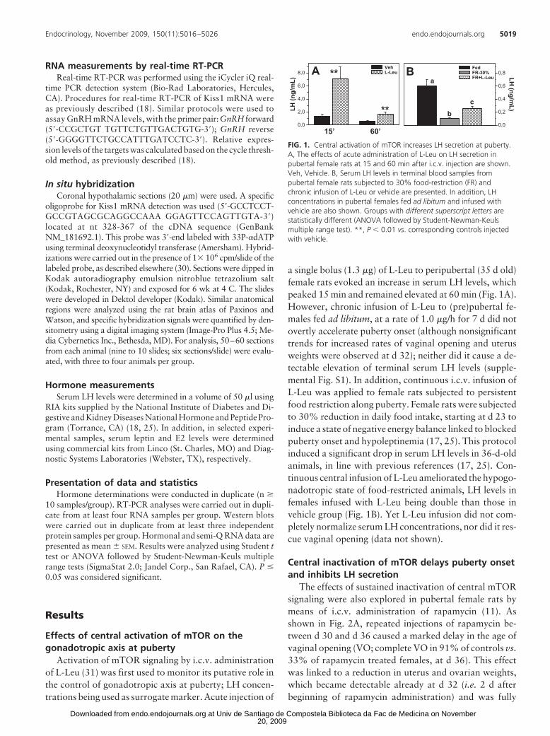
Activation of mTOR signaling by i.c.v. administrationof L-Leu (31) was first used to monitor its putative role inthe control of gonadotropic axis at puberty; LH concen-trations being used as surrogate marker. Acute injection of
a single bolus (1.3 �g) of L-Leu to peripubertal (35 d old)female rats evoked an increase in serum LH levels, whichpeaked 15 min and remained elevated at 60 min (Fig. 1A).However, chronic infusion of L-Leu to (pre)pubertal fe-males fed ad libitum, at a rate of 1.0 �g/h for 7 d did notovertly accelerate puberty onset (although nonsignificanttrends for increased rates of vaginal opening and uterusweights were observed at d 32); neither did it cause a de-tectable elevation of terminal serum LH levels (supple-mental Fig. S1). In addition, continuous i.c.v. infusion ofL-Leu was applied to female rats subjected to persistentfood restriction along puberty. Female rats were subjectedto 30% reduction in daily food intake, starting at d 23 toinduce a state of negative energy balance linked to blockedpuberty onset and hypoleptinemia (17, 25). This protocolinduced a significant drop in serum LH levels in 36-d-oldanimals, in line with previous references (17, 25). Con-tinuous central infusion of L-Leu ameliorated the hypogo-nadotropic state of food-restricted animals, LH levels infemales infused with L-Leu being double than those invehicle group (Fig. 1B). Yet L-Leu infusion did not com-pletely normalize serum LH concentrations, nor did it res-cue vaginal opening (data not shown).
Central inactivation of mTOR delays puberty onsetand inhibits LH secretion
The effects of sustained inactivation of central mTORsignaling were also explored in pubertal female rats bymeans of i.c.v. administration of rapamycin (11). Asshown in Fig. 2A, repeated injections of rapamycin be-tween d 30 and d 36 caused a marked delay in the age ofvaginal opening (VO; complete VO in 91% of controls vs.33% of rapamycin treated females, at d 36). This effectwas linked to a reduction in uterus and ovarian weights,which became detectable already at d 32 (i.e. 2 d afterbeginning of rapamycin administration) and was fully
LH (n
g/m
L)
0,0
2,0
4,0
6,0
8,0VehL-LeuA **
15’
**
60’
LH (ng/m
L)
0,0
0,2
0,4
0,6
0,8FedFR-30%FR+L-Leu
Ba
bc
FIG. 1. Central activation of mTOR increases LH secretion at puberty.A, The effects of acute administration of L-Leu on LH secretion inpubertal female rats at 15 and 60 min after i.c.v. injection are shown.Veh, Vehicle. B, Serum LH levels in terminal blood samples frompubertal female rats subjected to 30% food-restriction (FR) andchronic infusion of L-Leu or vehicle are presented. In addition, LHconcentrations in pubertal females fed ad libitum and infused withvehicle are also shown. Groups with different superscript letters arestatistically different (ANOVA followed by Student-Newman-Keulsmultiple range test). **, P � 0.01 vs. corresponding controls injectedwith vehicle.
Endocrinology, November 2009, 150(11):5016–5026 endo.endojournals.org 5019
20, 2009 at Univ de Santiago de Compostela Biblioteca da Fac de Medicina on Novemberendo.endojournals.orgDownloaded from
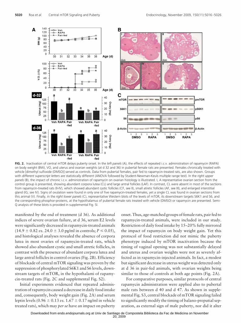
manifested by the end of treatment (d 36). As additionalindices of severe ovarian failure, at d 36, serum E2 levelswere significantly decreased in rapamycin-treated animals(14.9 � 0.82 vs. 24.0 � 3.0 pg/ml in controls; P � 0.05),and histological analyses revealed the absence of corporalutea in most ovaries of rapamycin-treated rats, whichshowed also abundant cystic and small atretic follicles, incontrast with the presence of abundant corpora lutea andlarge antral follicles in control ovaries (Fig. 2B). Efficiencyof blockade of central mTOR signaling was proven by thesuppression of phosphorylated S6K1 and S6 levels, down-stream targets of mTOR, in the hypothalami of rapamy-cin-treated rats (Fig. 2C and supplemental Fig. S2).
Initial experiments evidenced that repeated adminis-tration of rapamycin caused a decrease in daily food intakeand, consequently, body weight gain (Fig. 2A) and serumleptin levels (0.96 � 0.13 vs. 1.67 � 0.17 ng/ml in vehicletreated rats), which may per se have an impact on puberty
onset. Thus, age-matched groups of female rats, pair fed torapamycin-treated animals, were included in our study.Restriction of daily food intake by 15–20% fully mirroredthe impact of rapamycin on body weight gain. Yet thisprotocol of food restriction did not mimic the pubertyphenotype induced by mTOR inactivation because thetiming of vaginal opening was not substantially delayedand uterus and ovarian weights were not as severely af-fected as in rapamycin-injected animals. In fact, a modestbut significant decrease in uterus weight was detected onlyat d 36 in pair-fed animals, with ovarian weights beingsimilar to those of controls at both age points (Fig. 2A).
For comparative purposes, similar protocols of centralrapamycin administration were applied also to pubertalmale rats between d 40 and d 47. As shown in supple-mental Fig. S3, central blockade of mTOR signaling failedto significantly modify the timing of balano-preputial sep-aration, as external sign of male puberty, nor did it alter
FIG. 2. Inactivation of central mTOR delays puberty onset. In the left panels (A), the effects of repeated i.c.v. administration of rapamycin (RAPA)on body weight (BW), VO, and uterus and ovarian weights (at d 32 and 36) in pubertal female rats are presented. Females chronically treated withvehicle [dimethyl sulfoxide (DMSO)] served as controls. Data from pubertal females, pair fed to rapamycin-treated rats, are also shown. Groupswith different superscript letters are statistically different (ANOVA followed by Student-Newman-Keuls multiple range test). In the right upperpanels (B), the impact of chronic i.c.v. administration of rapamycin on ovarian histology is illustrated. I, A representative ovarian section from thecontrol group is presented, showing abundant corpora lutea (CL) and large antral follicles (LAF). In contrast, CL were absent in most of the sectionsfrom rapamycin-treated rats (II-IV), which showed abundant cystic follicles (CF, see II), small atretic follicles (AF, see III), and enlarged interstitialgland (IG; see IV). Signs of ovulation were found in only one of five rapamycin-treated females, yet a single CL was found in ovarian sections fromthis animal (V). Finally, in the right lower panels (C), representative Western blots of the levels of mTOR, its downstream targets S6K1 and S6, andthe corresponding phosphor-proteins, at the hypothalamus of pubertal female rats treated with vehicle (DMSO) or rapamycin are presented. Semi-Q analysis of these blots is provided in supplemental Fig. SI.
5020 Roa et al. Central mTOR Signaling and Puberty Endocrinology, November 2009, 150(11):5016–5026
20, 2009 at Univ de Santiago de Compostela Biblioteca da Fac de Medicina on Novemberendo.endojournals.orgDownloaded from
testicular weights at the end of treatment. However,chronic rapamycin administration evoked a detectabledrop in the weights of epididymis, seminal vesicles, andprostate, thus suggesting the suppression of the gonado-tropic axis also in the pubertal male.
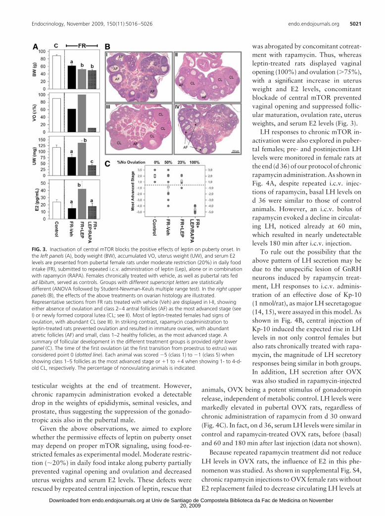
Given the above observations, we aimed to explorewhether the permissive effects of leptin on puberty onsetmay depend on proper mTOR signaling, using food-re-stricted females as experimental model. Moderate restric-tion (�20%) in daily food intake along puberty partiallyprevented vaginal opening and ovulation and decreaseduterus weights and serum E2 levels. These defects wererescued by repeated central injection of leptin, rescue that
was abrogated by concomitant cotreat-ment with rapamycin. Thus, whereasleptin-treated rats displayed vaginalopening (100%) and ovulation (75%),with a significant increase in uterusweight and E2 levels, concomitantblockade of central mTOR preventedvaginal opening and suppressed follic-ular maturation, ovulation rate, uterusweights, and serum E2 levels (Fig. 3).
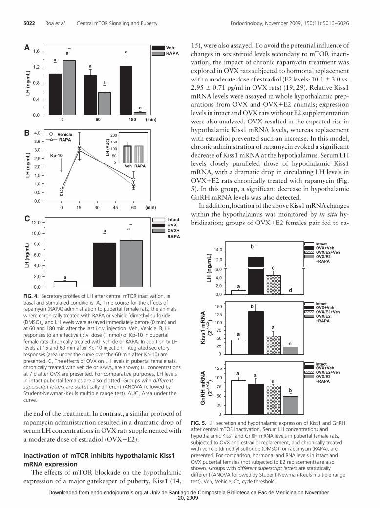
LH responses to chronic mTOR in-activation were also explored in puber-tal females; pre- and postinjection LHlevels were monitored in female rats atthe end (d 36) of our protocol of chronicrapamycin administration. As shown inFig. 4A, despite repeated i.c.v. injec-tions of rapamycin, basal LH levels ond 36 were similar to those of controlanimals. However, an i.c.v. bolus ofrapamycin evoked a decline in circulat-ing LH, noticed already at 60 min,which resulted in nearly undetectablelevels 180 min after i.c.v. injection.
To rule out the possibility that theabove pattern of LH secretion may bedue to the unspecific lesion of GnRHneurons induced by rapamycin treat-ment, LH responses to i.c.v. adminis-tration of an effective dose of Kp-10(1 nmol/rat), as major LH secretagogue(14, 15), were assayed in this model. Asshown in Fig. 4B, central injection ofKp-10 induced the expected rise in LHlevels in not only control females butalso rats chronically treated with rapa-mycin, the magnitude of LH secretoryresponses being similar in both groups.In addition, LH secretion after OVXwas also studied in rapamycin-injected
animals, OVX being a potent stimulus of gonadotropinrelease, independent of metabolic control. LH levels weremarkedly elevated in pubertal OVX rats, regardless ofchronic administration of rapamycin from d 30 onward(Fig. 4C). In fact, on d 36, serum LH levels were similar incontrol and rapamycin-treated OVX rats, before (basal)and 60 and 180 min after last injection (data not shown).
Because repeated rapamycin treatment did not reduceLH levels in OVX rats, the influence of E2 in this phe-nomenon was studied. As shown in supplemental Fig. S4,chronic rapamycin injections to OVX female rats withoutE2 replacement failed to decrease circulating LH levels at
FIG. 3. Inactivation of central mTOR blocks the positive effects of leptin on puberty onset. Inthe left panels (A), body weight (BW), accumulated VO, uterus weight (UW), and serum E2levels are presented from pubertal female rats under moderate restriction (20%) in daily foodintake (FR), submitted to repeated i.c.v. administration of leptin (Lep), alone or in combinationwith rapamycin (RAPA). Females chronically treated with vehicle, as well as pubertal rats fedad libitum, served as controls. Groups with different superscript letters are statisticallydifferent (ANOVA followed by Student-Newman-Keuls multiple range test). In the right upperpanels (B), the effects of the above treatments on ovarian histology are illustrated.Representative sections from FR rats treated with vehicle (Veh) are displayed in I-II, showingeither absence of ovulation and class 2–4 antral follicles (AF) as the most advanced stage (seeI) or newly formed corporal lutea (CL; see II). Most of leptin-treated females had signs ofovulation, with abundant CL (see III). In striking contrast, rapamycin coadministration toleptin-treated rats prevented ovulation and resulted in immature ovaries, with abundantatretic follicles (AF) and small, class 1–2 healthy follicles, as the most advanced stage. Asummary of follicular development in the different treatment groups is provided right lowerpanel (C). The time of the first ovulation (at the first transition from proestrus to estrus) wasconsidered point 0 (dotted line). Each animal was scored �5 (class 1) to �1 (class 5) whenshowing class 1–5 follicles as the most advanced stage or �1 to �4 when showing 1- to 4-d-old CL, respectively. The percentage of nonovulating animals is indicated.
Endocrinology, November 2009, 150(11):5016–5026 endo.endojournals.org 5021
20, 2009 at Univ de Santiago de Compostela Biblioteca da Fac de Medicina on Novemberendo.endojournals.orgDownloaded from
the end of the treatment. In contrast, a similar protocol ofrapamycin administration resulted in a dramatic drop ofserum LH concentrations in OVX rats supplemented witha moderate dose of estradiol (OVX�E2).
Inactivation of mTOR inhibits hypothalamic Kiss1mRNA expression
The effects of mTOR blockade on the hypothalamicexpression of a major gatekeeper of puberty, Kiss1 (14,
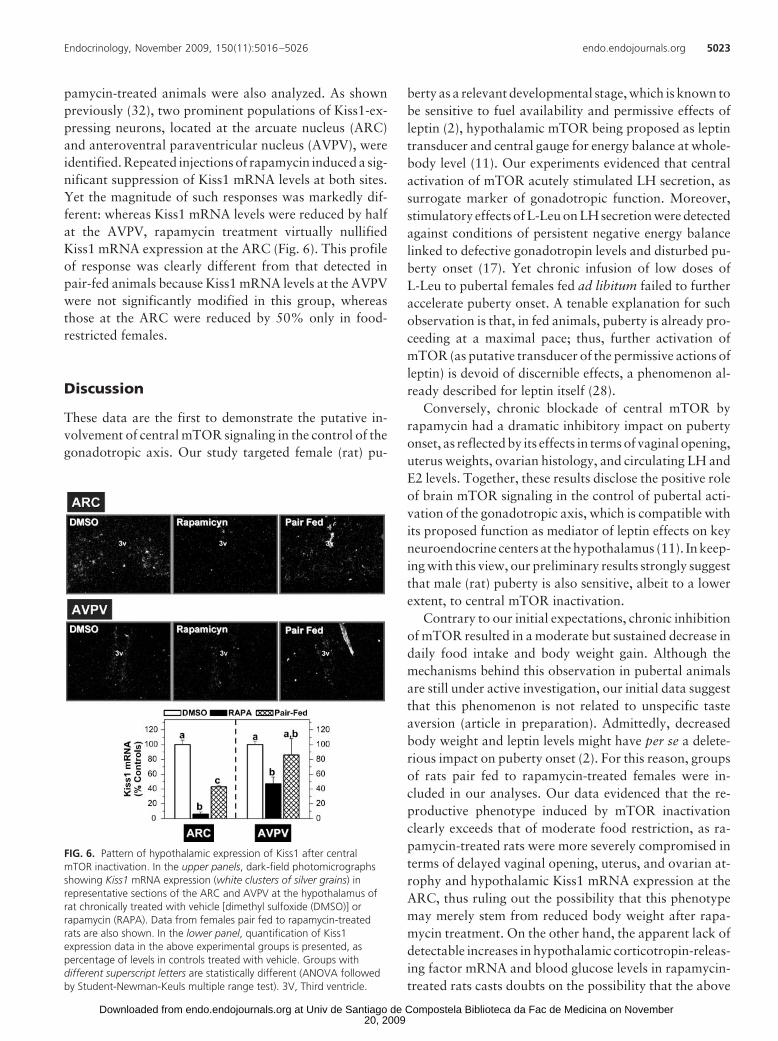
15), were also assayed. To avoid the potential influence ofchanges in sex steroid levels secondary to mTOR inacti-vation, the impact of chronic rapamycin treatment wasexplored in OVX rats subjected to hormonal replacementwith a moderate dose of estradiol (E2 levels: 10.1 � 3.0 vs.2.95 � 0.71 pg/ml in OVX rats) (19, 29). Relative Kiss1mRNA levels were assayed in whole hypothalamic prep-arations from OVX and OVX�E2 animals; expressionlevels in intact and OVX rats without E2 supplementationwere also analyzed. OVX resulted in the expected rise inhypothalamic Kiss1 mRNA levels, whereas replacementwith estradiol prevented such an increase. In this model,chronic administration of rapamycin evoked a significantdecrease of Kiss1 mRNA at the hypothalamus. Serum LHlevels closely paralleled those of hypothalamic Kiss1mRNA, with a dramatic drop in circulating LH levels inOVX�E2 rats chronically treated with rapamycin (Fig.5). In this group, a significant decrease in hypothalamicGnRH mRNA levels was also detected.
In addition, location of the above Kiss1 mRNA changeswithin the hypothalamus was monitored by in situ hy-bridization; groups of OVX�E2 females pair fed to ra-
LH (n
g/m
L)
0,0
0,4
0,8
1,2
1,6VehRAPA
0 60 180 (min)
aa
a
a
b
c
0 15 30 45 60
LH (n
g/m
L)
0,0
0,5
1,0
1,5
2,0
2,5
3,0
3,5
4,0 VehicleRAPA
LH (A
UC
)
0
50
100
150
200
Kp-10
Veh RAPA
LH (n
g/m
L)
0,0
2,0
4,0
6,0
8,0
10,0
12,0 IntactOVXOVX+RAPA
(min)
a
a a
A
B
C
FIG. 4. Secretory profiles of LH after central mTOR inactivation, inbasal and stimulated conditions. A, Time course for the effects ofrapamycin (RAPA) administration to pubertal female rats; the animalswhere chronically treated with RAPA or vehicle [dimethyl sulfoxide(DMSO)], and LH levels were assayed immediately before (0 min) andat 60 and 180 min after the last i.c.v. injection. Veh, Vehicle. B, LHresponses to an effective i.c.v. dose (1 nmol) of Kp-10 in pubertalfemale rats chronically treated with vehicle or RAPA. In addition to LHlevels at 15 and 60 min after Kp-10 injection, integrated secretoryresponses (area under the curve over the 60 min after Kp-10) arepresented. C, The effects of OVX on LH levels in pubertal female rats,chronically treated with vehicle or RAPA, are shown; LH concentrationsat 7 d after OVX are presented. For comparative purposes, LH levelsin intact pubertal females are also plotted. Groups with differentsuperscript letters are statistically different (ANOVA followed byStudent-Newman-Keuls multiple range test). AUC, Area under thecurve.
LH (n
g/m
L)
0,0
2,0
4,0
6,0
12,0
14,0IntactOVX+VehOVX/E2+VehOVX/E2+RAPA
a
b
d
c
Kis
s1 m
RN
A(2
- ∆∆
Ct )
0
25
50
75
100
125
150IntactOVX+VehOVX/E2+VehOVX/E2+RAPA
a
b
c
a
GnR
H m
RN
A(2
- ∆∆
Ct )
0
25
50
75
100
125IntactOVX+VehOVX/E2+VehOVX/E2+RAPA
a a
b
a
FIG. 5. LH secretion and hypothalamic expression of Kiss1 and GnRHafter central mTOR inactivation. Serum LH concentrations andhypothalamic Kiss1 and GnRH mRNA levels in pubertal female rats,subjected to OVX and estradiol replacement, and chronically treatedwith vehicle [dimethyl sulfoxide (DMSO)] or rapamycin (RAPA), arepresented. For comparison, hormonal and RNA levels in intact andOVX pubertal females (not subjected to E2 replacement) are alsoshown. Groups with different superscript letters are statisticallydifferent (ANOVA followed by Student-Newman-Keuls multiple rangetest). Veh, Vehicle; Ct, cycle threshold.
5022 Roa et al. Central mTOR Signaling and Puberty Endocrinology, November 2009, 150(11):5016–5026
20, 2009 at Univ de Santiago de Compostela Biblioteca da Fac de Medicina on Novemberendo.endojournals.orgDownloaded from
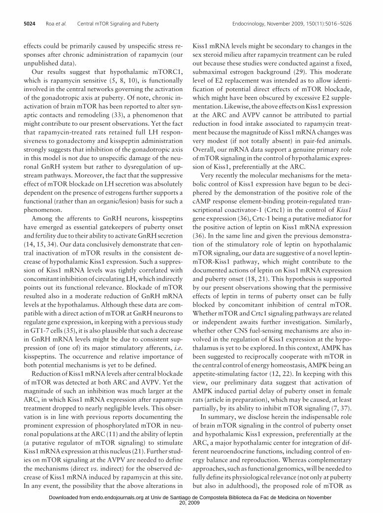
pamycin-treated animals were also analyzed. As shownpreviously (32), two prominent populations of Kiss1-ex-pressing neurons, located at the arcuate nucleus (ARC)and anteroventral paraventricular nucleus (AVPV), wereidentified. Repeated injections of rapamycin induced a sig-nificant suppression of Kiss1 mRNA levels at both sites.Yet the magnitude of such responses was markedly dif-ferent: whereas Kiss1 mRNA levels were reduced by halfat the AVPV, rapamycin treatment virtually nullifiedKiss1 mRNA expression at the ARC (Fig. 6). This profileof response was clearly different from that detected inpair-fed animals because Kiss1 mRNA levels at the AVPVwere not significantly modified in this group, whereasthose at the ARC were reduced by 50% only in food-restricted females.
Discussion
These data are the first to demonstrate the putative in-volvement of central mTOR signaling in the control of thegonadotropic axis. Our study targeted female (rat) pu-
berty as a relevant developmental stage, which is known tobe sensitive to fuel availability and permissive effects ofleptin (2), hypothalamic mTOR being proposed as leptintransducer and central gauge for energy balance at whole-body level (11). Our experiments evidenced that centralactivation of mTOR acutely stimulated LH secretion, assurrogate marker of gonadotropic function. Moreover,stimulatory effects of L-Leu on LH secretion were detectedagainst conditions of persistent negative energy balancelinked to defective gonadotropin levels and disturbed pu-berty onset (17). Yet chronic infusion of low doses ofL-Leu to pubertal females fed ad libitum failed to furtheraccelerate puberty onset. A tenable explanation for suchobservation is that, in fed animals, puberty is already pro-ceeding at a maximal pace; thus, further activation ofmTOR (as putative transducer of the permissive actions ofleptin) is devoid of discernible effects, a phenomenon al-ready described for leptin itself (28).
Conversely, chronic blockade of central mTOR byrapamycin had a dramatic inhibitory impact on pubertyonset, as reflected by its effects in terms of vaginal opening,uterus weights, ovarian histology, and circulating LH andE2 levels. Together, these results disclose the positive roleof brain mTOR signaling in the control of pubertal acti-vation of the gonadotropic axis, which is compatible withits proposed function as mediator of leptin effects on keyneuroendocrine centers at the hypothalamus (11). In keep-ing with this view, our preliminary results strongly suggestthat male (rat) puberty is also sensitive, albeit to a lowerextent, to central mTOR inactivation.
Contrary to our initial expectations, chronic inhibitionof mTOR resulted in a moderate but sustained decrease indaily food intake and body weight gain. Although themechanisms behind this observation in pubertal animalsare still under active investigation, our initial data suggestthat this phenomenon is not related to unspecific tasteaversion (article in preparation). Admittedly, decreasedbody weight and leptin levels might have per se a delete-rious impact on puberty onset (2). For this reason, groupsof rats pair fed to rapamycin-treated females were in-cluded in our analyses. Our data evidenced that the re-productive phenotype induced by mTOR inactivationclearly exceeds that of moderate food restriction, as ra-pamycin-treated rats were more severely compromised interms of delayed vaginal opening, uterus, and ovarian at-rophy and hypothalamic Kiss1 mRNA expression at theARC, thus ruling out the possibility that this phenotypemay merely stem from reduced body weight after rapa-mycin treatment. On the other hand, the apparent lack ofdetectable increases in hypothalamic corticotropin-releas-ing factor mRNA and blood glucose levels in rapamycin-treated rats casts doubts on the possibility that the above
FIG. 6. Pattern of hypothalamic expression of Kiss1 after centralmTOR inactivation. In the upper panels, dark-field photomicrographsshowing Kiss1 mRNA expression (white clusters of silver grains) inrepresentative sections of the ARC and AVPV at the hypothalamus ofrat chronically treated with vehicle [dimethyl sulfoxide (DMSO)] orrapamycin (RAPA). Data from females pair fed to rapamycin-treatedrats are also shown. In the lower panel, quantification of Kiss1expression data in the above experimental groups is presented, aspercentage of levels in controls treated with vehicle. Groups withdifferent superscript letters are statistically different (ANOVA followedby Student-Newman-Keuls multiple range test). 3V, Third ventricle.
Endocrinology, November 2009, 150(11):5016–5026 endo.endojournals.org 5023
20, 2009 at Univ de Santiago de Compostela Biblioteca da Fac de Medicina on Novemberendo.endojournals.orgDownloaded from
effects could be primarily caused by unspecific stress re-sponses after chronic administration of rapamycin (ourunpublished data).
Our results suggest that hypothalamic mTORC1,which is rapamycin sensitive (5, 8, 10), is functionallyinvolved in the central networks governing the activationof the gonadotropic axis at puberty. Of note, chronic in-activation of brain mTOR has been reported to alter syn-aptic contacts and remodeling (33), a phenomenon thatmight contribute to our present observations. Yet the factthat rapamycin-treated rats retained full LH respon-siveness to gonadectomy and kisspeptin administrationstrongly suggests that inhibition of the gonadotropic axisin this model is not due to unspecific damage of the neu-ronal GnRH system but rather to dysregulation of up-stream pathways. Moreover, the fact that the suppressiveeffect of mTOR blockade on LH secretion was absolutelydependent on the presence of estrogens further supports afunctional (rather than an organic/lesion) basis for such aphenomenon.
Among the afferents to GnRH neurons, kisspeptinshave emerged as essential gatekeepers of puberty onsetand fertility due to their ability to activate GnRH secretion(14, 15, 34). Our data conclusively demonstrate that cen-tral inactivation of mTOR results in the consistent de-crease of hypothalamic Kiss1 expression. Such a suppres-sion of Kiss1 mRNA levels was tightly correlated withconcomitant inhibition of circulating LH, which indirectlypoints out its functional relevance. Blockade of mTORresulted also in a moderate reduction of GnRH mRNAlevels at the hypothalamus. Although these data are com-patible with a direct action of mTOR at GnRH neurons toregulate gene expression, in keeping with a previous studyin GT1-7 cells (35), it is also plausible that such a decreasein GnRH mRNA levels might be due to consistent sup-pression of (one of) its major stimulatory afferents, i.e.kisspeptins. The occurrence and relative importance ofboth potential mechanisms is yet to be defined.
Reduction of Kiss1 mRNA levels after central blockadeof mTOR was detected at both ARC and AVPV. Yet themagnitude of such an inhibition was much larger at theARC, in which Kiss1 mRNA expression after rapamycintreatment dropped to nearly negligible levels. This obser-vation is in line with previous reports documenting theprominent expression of phosphorylated mTOR in neu-ronal populations at the ARC (11) and the ability of leptin(a putative regulator of mTOR signaling) to stimulateKiss1 mRNA expression at this nucleus (21). Further stud-ies on mTOR signaling at the AVPV are needed to definethe mechanisms (direct vs. indirect) for the observed de-crease of Kiss1 mRNA induced by rapamycin at this site.In any event, the possibility that the above alterations in
Kiss1 mRNA levels might be secondary to changes in thesex steroid milieu after rapamycin treatment can be ruledout because these studies were conducted against a fixed,submaximal estrogen background (29). This moderatelevel of E2 replacement was intended as to allow identi-fication of potential direct effects of mTOR blockade,which might have been obscured by excessive E2 supple-mentation.Likewise, theabove effectsonKiss1 expressionat the ARC and AVPV cannot be attributed to partialreduction in food intake associated to rapamycin treat-ment because the magnitude of Kiss1 mRNA changes wasvery modest (if not totally absent) in pair-fed animals.Overall, our mRNA data support a genuine primary roleof mTOR signaling in the control of hypothalamic expres-sion of Kiss1, preferentially at the ARC.
Very recently the molecular mechanisms for the meta-bolic control of Kiss1 expression have begun to be deci-phered by the demonstration of the positive role of thecAMP response element-binding protein-regulated tran-scriptional coactivator-1 (Crtc1) in the control of Kiss1gene expression (36), Crtc-1 being a putative mediator forthe positive action of leptin on Kiss1 mRNA expression(36). In the same line and given the previous demonstra-tion of the stimulatory role of leptin on hypothalamicmTOR signaling, our data are suggestive of a novel leptin-mTOR-Kiss1 pathway, which might contribute to thedocumented actions of leptin on Kiss1 mRNA expressionand puberty onset (18, 21). This hypothesis is supportedby our present observations showing that the permissiveeffects of leptin in terms of puberty onset can be fullyblocked by concomitant inhibition of central mTOR.Whether mTOR and Crtc1 signaling pathways are relatedor independent awaits further investigation. Similarly,whether other CNS fuel-sensing mechanisms are also in-volved in the regulation of Kiss1 expression at the hypo-thalamus is yet to be explored. In this context, AMPK hasbeen suggested to reciprocally cooperate with mTOR inthe central control of energy homeostasis, AMPK being anappetite-stimulating factor (12, 22). In keeping with thisview, our preliminary data suggest that activation ofAMPK induced partial delay of puberty onset in femalerats (article in preparation), which may be caused, at leastpartially, by its ability to inhibit mTOR signaling (7, 37).
In summary, we disclose herein the indispensable roleof brain mTOR signaling in the control of puberty onsetand hypothalamic Kiss1 expression, preferentially at theARC, a major hypothalamic center for integration of dif-ferent neuroendocrine functions, including control of en-ergy balance and reproduction. Whereas complementaryapproaches, such as functional genomics, will be needed tofully define its physiological relevance (not only at pubertybut also in adulthood), the proposed role of mTOR as
5024 Roa et al. Central mTOR Signaling and Puberty Endocrinology, November 2009, 150(11):5016–5026
20, 2009 at Univ de Santiago de Compostela Biblioteca da Fac de Medicina on Novemberendo.endojournals.orgDownloaded from
crossroad for the metabolic gating of cellular and systemicresponses (5, 6, 8, 9) and the putative function of kisspep-tin neurons as key elements for the metabolic control offertility (14, 16) make it tempting to hypothesize that thisnovel pathway may participate in the integrated regula-tion of energy homeostasis and reproductive function. Inaddition to their physiological implications, our presentfindings also may provide novel mechanistic insights forthe reported impact of mTOR antagonists, of clinicaluse in the management of some types of cancer andorgan transplantation (6 – 8) on reproductive functionin humans.
Acknowledgments
Address all correspondence and requests for reprints to: ManuelTena-Sempere, Physiology Section, Department of Cell Biology,Physiology, and Immunology, Faculty of Medicine, University ofCordoba, Avda. Menendez Pidal s/n, 14004 Cordoba, Spain.E-mail: [email protected].
This work was supported by Grants BFU 2005-07446 andBFU 2008-00984 from Ministerio de Ciencia e Innovacion,Spain; Project PI042082 (Ministerio de Sanidad, Spain); ProjectP08-CVI-03788 (Junta de Andalucía, Spain); and EuropeanUnion Research Contract DEER FP7-ENV-2007-1. Centro deInvestigacion Biomedica en Red Fisiopatología de la Obesidad yNutricion is an initiative of the Instituto de Salud Carlos III,Ministerio de Sanidad, Spain.
Disclosure Summary: The authors have nothing to disclose.
References
1. Cunningham MJ, Clifton DK, Steiner RA 1999 Leptin’s actions onthe reproductive axis: perspectives and mechanisms. Biol Reprod60:216–222
2. Fernandez-Fernandez R, Martini AC, Navarro VM, Castellano JM,Dieguez C, Aguilar E, Pinilla L, Tena-Sempere M 2006 Novel signalsfor the integration of energy balance and reproduction. Mol CellEndocrinol 254–255:127–132
3. Smith MS, Grove KL 2002 Integration of the regulation of repro-ductive function and energy balance: lactation as a model. FrontNeuroendocrinol 23:225–256
4. Tena-Sempere M 2008 Ghrelin as a pleotrophic modulator of go-nadal function and reproduction. Nat Clin Pract Endocrinol Metab4:666–674
5. Wullschleger S, Loewith R, Hall MN 2006 TOR signaling in growthand metabolism. Cell 124:471–484
6. Chiang GG, Abraham RT 2007 Targeting the mTOR signaling net-work in cancer. Trends Mol Med 13:433–442
7. Tsang CK, Qi H, Liu LF, Zheng XF 2007 Targeting mammaliantarget of rapamycin (mTOR) for health and diseases. Drug DiscovToday 12:112–124
8. Martin DE, Hall MN 2005 The expanding TOR signaling network.Curr Opin Cell Biol 17:158–166
9. Schmelzle T, Hall MN 2000 TOR, a central controller of cellgrowth. Cell 103:253–262
10. Loewith R, Jacinto E, Wullschleger S, Lorberg A, Crespo JL,Bonenfant D, Oppliger W, Jenoe P, Hall MN 2002 Two TORcomplexes, only one of which is rapamycin sensitive, have distinctroles in cell growth control. Mol Cell 10:457– 468
11. Cota D, Proulx K, Smith KA, Kozma SC, Thomas G, Woods SC,Seeley RJ 2006 Hypothalamic mTOR signaling regulates food in-take. Science 312:927–930
12. Kahn BB, Alquier T, Carling D, Hardie DG 2005 AMP-activatedprotein kinase: ancient energy gauge provides clues to modern un-derstanding of metabolism. Cell Metab 1:15–25
13. Hursting SD, Lashinger LM, Wheatley KW, Rogers CJ, Colbert LH,Nunez NP, Perkins SN 2008 Reducing the weight of cancer: mech-anistic targets for breaking the obesity-carcinogenesis link. BestPract Res Clin Endocrinol Metab 22:659–669
14. Roa J, Aguilar E, Dieguez C, Pinilla L, Tena-Sempere M 2008 Newfrontiers in kisspeptin/GPR54 physiology as fundamental gatekeep-ers of reproductive function. Front Neuroendocrinol 29:48–69
15. Popa SM, Clifton DK, Steiner RA 2008 The role of kisspeptins andGPR54 in the neuroendocrine regulation of reproduction. Annu RevPhysiol 70:213–238
16. Tena-SempereM2006KiSS-1andreproduction: focuson its role in themetabolic regulation of fertility. Neuroendocrinology 83:275–281
17. Castellano JM, Navarro VM, Fernandez-Fernandez R, NogueirasR, Tovar S, Roa J, Vazquez MJ, Vigo E, Casanueva FF, Aguilar E,Pinilla L, Dieguez C, Tena-Sempere M 2005 Changes in hypotha-lamic KiSS-1 system and restoration of pubertal activation of thereproductive axis by kisspeptin in undernutrition. Endocrinology146:3917–3925
18. Castellano JM, Navarro VM, Fernandez-Fernandez R, Roa J, VigoE, Pineda R, Dieguez C, Aguilar E, Pinilla L, Tena-Sempere M 2006Expression of hypothalamic KiSS-1 system and rescue of defectivegonadotropic responses by kisspeptin in streptozotocin-induced di-abetic male rats. Diabetes 55:2602–2610
19. Kalamatianos T, Grimshaw SE, Poorun R, Hahn JD, Coen CW2008 Fasting reduces KiSS-1 expression in the anteroventralperiventricular nucleus (AVPV): effects of fasting on the expressionof KiSS-1 and neuropeptide Y in the AVPV or arcuate nucleus offemale rats. J Neuroendocrinol 20:1089–1097
20. Luque RM, Kineman RD, Tena-Sempere M 2007 Regulation ofhypothalamic expression of KiSS-1 and GPR54 genes by metabolicfactors: analyses using mouse models and a cell line. Endocrinology148:4601–4611
21. Smith JT, Acohido BV, Clifton DK, Steiner RA 2006 KiSS-1 neu-rones are direct targets for leptin in the ob/ob mouse. J Neuroen-docrinol 18:298–303
22. Cota D, Proulx K, Seeley RJ 2007 The role of CNS fuel sensing inenergy and glucose regulation. Gastroenterology 132:2158–2168
23. Kaczmarek I, Groetzner J, Adamidis I, Landwehr P, Mueller M,Vogeser M, Gerstorfer M, Uberfuhr P, Meiser B, Reichart B 2004Sirolimus impairs gonadal function in heart transplant recipients.Am J Transplant 4:1084–1088
24. Navarro VM, Fernandez-Fernandez R, Castellano JM, Roa J,Mayen A, Barreiro ML, Gaytan F, Aguilar E, Pinilla L, Dieguez C,Tena-Sempere M 2004 Advanced vaginal opening and precociousactivation of the reproductive axis by KiSS-1 peptide, the endoge-nous ligand of GPR54. J Physiol 561:379–386
25. Roa J, Vigo E, García-Galiano D, Castellano JM, Navarro VM, PinedaR, Dieguez C, Aguilar E, Pinilla L, Tena-Sempere M 2008 Desensiti-zation of gonadotropin responses to kisspeptin in the female rat: anal-yses of LH and FSH secretion at different developmental and metabolicstates. Am J Physiol Endocrinol Metab 294:E1088–E1096
26. Pinilla L, Castellano JM, Romero M, Tena-Sempere M, Gaytan F,Aguilar E 2009 Delayed puberty in spontaneously hypertensive ratsinvolves a primary ovarian failure independent of the hypothalamicKiSS-1/GPR54/GnRH system. Endocrinology 150:2889–2897
27. Tena-Sempere M 2007 Roles of ghrelin and leptin in the control ofreproductive function. Neuroendocrinology 86:229–241
28. Cheung CC, Thornton JE, Kuijper JL, Weigle DS, Clifton DK,Steiner RA 1997 Leptin is a metabolic gate for the onset of pubertyin the female rat. Endocrinology 138:855–858
29. Yamada S, Uenoyama Y, Kinoshita M, Iwata K, Takase K, MatsuiH, Adachi S, Inoue K, Maeda KI, Tsukamura H 2007 Inhibition of
Endocrinology, November 2009, 150(11):5016–5026 endo.endojournals.org 5025
20, 2009 at Univ de Santiago de Compostela Biblioteca da Fac de Medicina on Novemberendo.endojournals.orgDownloaded from
metastin (kisspeptin-54)-GPR54 signaling in the arcuate nucleus-median eminence region during lactation in rats. Endocrinology148:2226–2232
30. Lopez M, Seoane LM, Tovar S, Nogueiras R, Dieguez C, Senarís R2004 Orexin-A regulates growth hormone-releasing hormonemRNA content in a nucleus-specific manner and somatostatinmRNA content in a growth hormone-dependent fashion in the rathypothalamus. Eur J Neurosci 19:2080–2088
31. Meijer AJ, Dubbelhuis PF 2004 Amino acid signalling and the integra-tion of metabolism. Biochem Biophys Res Commun 313:397–403
32. Smith JT, Cunningham MJ, Rissman EF, Clifton DK, Steiner RA2005 Regulation of Kiss1 gene expression in the brain of the femalemouse. Endocrinology 146:3686–3692
33. Swiech L, Perycz M, Malik A, Jaworski J 2008 Role of mTOR inphysiology and pathology of the nervous system. Biochim BiophysActa 1784:116–132
34. Tena-Sempere M 2006 GPR54 and kisspeptin in reproduction.Hum Reprod Update 12:631–639
35. Morrison CD, Xi X, White CL, Ye J, Martin RJ 2007 Amino acidsinhibit Agrp gene expression via an mTOR-dependent mechanism.Am J Physiol Endocrinol Metab 293:E165–E171
36. Altarejos JY, Goebel N, Conkright MD, Inoue H, Xie J, Arias CM,Sawchenko PE, Montminy M 2008 The Creb1 coactivator Crtc1 isrequired for energy balance and fertility. Nat Med 14:1112–1117
37. Inoki K, Zhu T, Guan KL 2003 TSC2 mediates cellular energy re-sponse to control cell growth and survival. Cell 115:577–590
5026 Roa et al. Central mTOR Signaling and Puberty Endocrinology, November 2009, 150(11):5016–5026
20, 2009 at Univ de Santiago de Compostela Biblioteca da Fac de Medicina on Novemberendo.endojournals.orgDownloaded from