Relaxin-3/RXFP3 Signaling and Neuroendocrine Function – A Perspective on Extrinsic Hypothalamic...
11
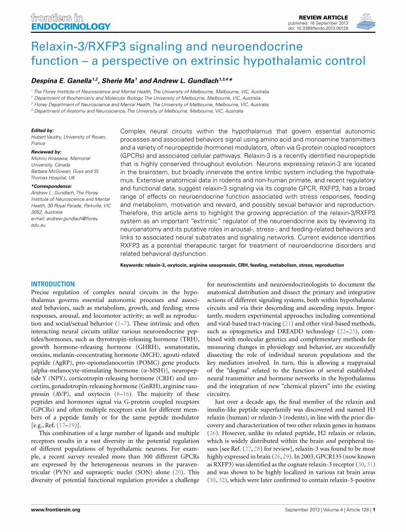
REVIEW ARTICLE published: 18 September 2013 doi: 10.3389/fendo.2013.00128 Relaxin-3/RXFP3 signaling and neuroendocrine function – a perspective on extrinsic hypothalamic control Despina E. Ganella 1,2 , Sherie Ma 1 and Andrew L. Gundlach 1,3,4 * 1 The Florey Institute of Neuroscience and Mental Health, The University of Melbourne, Melbourne, VIC, Australia 2 Department of Biochemistry and Molecular Biology,The University of Melbourne, Melbourne, VIC, Australia 3 Florey Department of Neuroscience and Mental Health, The University of Melbourne, Melbourne, VIC, Australia 4 Department of Anatomy and Neuroscience, The University of Melbourne, Melbourne, VIC, Australia Edited by: Hubert Vaudry, University of Rouen, France Reviewed by: Michiru Hirasawa, Memorial University, Canada Barbara McGowan, Guys and St. Thomas Hospital, UK *Correspondence: Andrew L. Gundlach, The Florey Institute of Neuroscience and Mental Health, 30 Royal Parade, Parkville, VIC 3052, Australia e-mail: andrew.gundlach@florey. edu.au Complex neural circuits within the hypothalamus that govern essential autonomic processes and associated behaviors signal using amino acid and monoamine transmitters and a variety of neuropeptide (hormone) modulators, often via G-protein coupled receptors (GPCRs) and associated cellular pathways. Relaxin-3 is a recently identified neuropeptide that is highly conserved throughout evolution. Neurons expressing relaxin-3 are located in the brainstem, but broadly innervate the entire limbic system including the hypothala- mus. Extensive anatomical data in rodents and non-human primate, and recent regulatory and functional data, suggest relaxin-3 signaling via its cognate GPCR, RXFP3, has a broad range of effects on neuroendocrine function associated with stress responses, feeding and metabolism, motivation and reward, and possibly sexual behavior and reproduction. Therefore, this article aims to highlight the growing appreciation of the relaxin-3/RXFP3 system as an important “extrinsic” regulator of the neuroendocrine axis by reviewing its neuroanatomy and its putative roles in arousal-, stress-, and feeding-related behaviors and links to associated neural substrates and signaling networks. Current evidence identifies RXFP3 as a potential therapeutic target for treatment of neuroendocrine disorders and related behavioral dysfunction. Keywords: relaxin-3, oxytocin, arginine vasopressin, CRH, feeding, metabolism, stress, reproduction INTRODUCTION Precise regulation of complex neural circuits in the hypo- thalamus governs essential autonomic processes and associ- ated behaviors, such as metabolism, growth, and feeding; stress responses, arousal, and locomotor activity; as well as reproduc- tion and social/sexual behavior (1–7). These intrinsic and often interacting neural circuits utilize various neuroendocrine pep- tides/hormones, such as thyrotropin-releasing hormone (TRH), growth hormone-releasing hormone (GHRH), somatostatin, orexins, melanin-concentrating hormone (MCH), agouti-related peptide (AgRP), pro-opiomelanocortin (POMC) gene products [alpha-melanocyte-stimulating hormone (α-MSH)], neuropep- tide Y (NPY), corticotropin-releasing hormone (CRH) and uro- cortins, gonadotropin-releasing hormone (GnRH), arginine vaso- pressin (AVP), and oxytocin (8–16). The majority of these peptides and hormones signal via G-protein coupled receptors (GPCRs) and often multiple receptors exist for different mem- bers of a peptide family or for the same peptide modulator [e.g., Ref. (17–19)]. This combination of a large number of ligands and multiple receptors results in a vast diversity in the potential regulation of different populations of hypothalamic neurons. For exam- ple, a recent survey revealed more than 300 different GPCRs are expressed by the heterogeneous neurons in the paraven- tricular (PVN) and supraoptic nuclei (SON) alone (20). This diversity of potential functional regulation provides a challenge for neuroscientists and neuroendocrinologists to document the anatomical distribution and dissect the primary and integrative actions of different signaling systems, both within hypothalamic circuits and via their descending and ascending inputs. Impor- tantly, modern experimental approaches including conventional and viral-based tract-tracing (21) and other viral-based methods, such as optogenetics and DREADD technology (22–25), com- bined with molecular genetics and complementary methods for measuring changes in physiology and behavior, are successfully dissecting the role of individual neuron populations and the key mediators involved. In turn, this is allowing a reappraisal of the “dogma” related to the function of several established neural transmitter and hormone networks in the hypothalamus and the integration of new “chemical players” into the existing circuitry. Just over a decade ago, the final member of the relaxin and insulin-like peptide superfamily was discovered and named H3 relaxin (human) or relaxin-3 (rodents), in line with the prior dis- covery and characterization of two other relaxin genes in humans (26). However, unlike its related peptide, H2 relaxin or relaxin, which is widely distributed within the brain and peripheral tis- sues [see Ref. (27, 28) for review], relaxin-3 was found to be most highly expressed in brain (26, 29). In 2003, GPCR135 (now known as RXFP3) was identified as the cognate relaxin-3 receptor (30, 31) and was shown to be highly localized in various rat brain areas (30, 32), which were later confirmed to contain relaxin-3-positive www.frontiersin.org September 2013 |Volume 4 | Article 128 | 1
-
Upload
independent -
Category
Documents
-
view
4 -
download
0
Transcript of Relaxin-3/RXFP3 Signaling and Neuroendocrine Function – A Perspective on Extrinsic Hypothalamic...
REVIEW ARTICLEpublished: 18 September 2013doi: 10.3389/fendo.2013.00128
Relaxin-3/RXFP3 signaling and neuroendocrinefunction – a perspective on extrinsic hypothalamic controlDespina E. Ganella1,2, Sherie Ma1 and Andrew L. Gundlach1,3,4*1 The Florey Institute of Neuroscience and Mental Health, The University of Melbourne, Melbourne, VIC, Australia2 Department of Biochemistry and Molecular Biology, The University of Melbourne, Melbourne, VIC, Australia3 Florey Department of Neuroscience and Mental Health, The University of Melbourne, Melbourne, VIC, Australia4 Department of Anatomy and Neuroscience, The University of Melbourne, Melbourne, VIC, Australia
Edited by:Hubert Vaudry, University of Rouen,France
Reviewed by:Michiru Hirasawa, MemorialUniversity, CanadaBarbara McGowan, Guys and St.Thomas Hospital, UK
*Correspondence:Andrew L. Gundlach, The FloreyInstitute of Neuroscience and MentalHealth, 30 Royal Parade, Parkville, VIC3052, Australiae-mail: [email protected]
Complex neural circuits within the hypothalamus that govern essential autonomicprocesses and associated behaviors signal using amino acid and monoamine transmittersand a variety of neuropeptide (hormone) modulators, often via G-protein coupled receptors(GPCRs) and associated cellular pathways. Relaxin-3 is a recently identified neuropeptidethat is highly conserved throughout evolution. Neurons expressing relaxin-3 are locatedin the brainstem, but broadly innervate the entire limbic system including the hypothala-mus. Extensive anatomical data in rodents and non-human primate, and recent regulatoryand functional data, suggest relaxin-3 signaling via its cognate GPCR, RXFP3, has a broadrange of effects on neuroendocrine function associated with stress responses, feedingand metabolism, motivation and reward, and possibly sexual behavior and reproduction.Therefore, this article aims to highlight the growing appreciation of the relaxin-3/RXFP3system as an important “extrinsic” regulator of the neuroendocrine axis by reviewing itsneuroanatomy and its putative roles in arousal-, stress-, and feeding-related behaviors andlinks to associated neural substrates and signaling networks. Current evidence identifiesRXFP3 as a potential therapeutic target for treatment of neuroendocrine disorders andrelated behavioral dysfunction.
Keywords: relaxin-3, oxytocin, arginine vasopressin, CRH, feeding, metabolism, stress, reproduction
INTRODUCTIONPrecise regulation of complex neural circuits in the hypo-thalamus governs essential autonomic processes and associ-ated behaviors, such as metabolism, growth, and feeding; stressresponses, arousal, and locomotor activity; as well as reproduc-tion and social/sexual behavior (1–7). These intrinsic and ofteninteracting neural circuits utilize various neuroendocrine pep-tides/hormones, such as thyrotropin-releasing hormone (TRH),growth hormone-releasing hormone (GHRH), somatostatin,orexins, melanin-concentrating hormone (MCH), agouti-relatedpeptide (AgRP), pro-opiomelanocortin (POMC) gene products[alpha-melanocyte-stimulating hormone (α-MSH)], neuropep-tide Y (NPY), corticotropin-releasing hormone (CRH) and uro-cortins, gonadotropin-releasing hormone (GnRH), arginine vaso-pressin (AVP), and oxytocin (8–16). The majority of thesepeptides and hormones signal via G-protein coupled receptors(GPCRs) and often multiple receptors exist for different mem-bers of a peptide family or for the same peptide modulator[e.g., Ref. (17–19)].
This combination of a large number of ligands and multiplereceptors results in a vast diversity in the potential regulationof different populations of hypothalamic neurons. For exam-ple, a recent survey revealed more than 300 different GPCRsare expressed by the heterogeneous neurons in the paraven-tricular (PVN) and supraoptic nuclei (SON) alone (20). Thisdiversity of potential functional regulation provides a challenge
for neuroscientists and neuroendocrinologists to document theanatomical distribution and dissect the primary and integrativeactions of different signaling systems, both within hypothalamiccircuits and via their descending and ascending inputs. Impor-tantly, modern experimental approaches including conventionaland viral-based tract-tracing (21) and other viral-based methods,such as optogenetics and DREADD technology (22–25), com-bined with molecular genetics and complementary methods formeasuring changes in physiology and behavior, are successfullydissecting the role of individual neuron populations and thekey mediators involved. In turn, this is allowing a reappraisalof the “dogma” related to the function of several establishedneural transmitter and hormone networks in the hypothalamusand the integration of new “chemical players” into the existingcircuitry.
Just over a decade ago, the final member of the relaxin andinsulin-like peptide superfamily was discovered and named H3relaxin (human) or relaxin-3 (rodents), in line with the prior dis-covery and characterization of two other relaxin genes in humans(26). However, unlike its related peptide, H2 relaxin or relaxin,which is widely distributed within the brain and peripheral tis-sues [see Ref. (27, 28) for review], relaxin-3 was found to be mosthighly expressed in brain (26, 29). In 2003, GPCR135 (now knownas RXFP3) was identified as the cognate relaxin-3 receptor (30, 31)and was shown to be highly localized in various rat brain areas(30, 32), which were later confirmed to contain relaxin-3-positive
www.frontiersin.org September 2013 | Volume 4 | Article 128 | 1
Ganella et al. Relaxin-3/RXFP3 signaling and neuroendocrine responses
axonal projections and terminations (33). A similar central distri-bution of relaxin-3 neurons and projections to that reported inthe rat was subsequently observed in the mouse [Ref. (34); AllenBrain Atlas1] and macaque brain (35, 36), suggesting that thisneuropeptide system has been highly conserved throughout evo-lution. Indeed, bioinformatic studies revealed that a relaxin-3-likeancestral peptide gave rise to the relaxin and insulin-like peptidesuperfamily and its sequence has been highly conserved by strongpurifying selection, consistent with a highly conserved function inthe central nervous system (37, 38).
After their discovery, characterization of the neuroanatomicaldistribution of relaxin-3- and RXFP3-expressing neurons pro-vided insights into putative functions of relaxin-3; and a growingnumber of experimental studies have subsequently confirmedroles for relaxin-3/RXFP3 signaling in arousal, feeding, stressresponses, and cognition [see Ref. (39) for review]. Several of theseactions of relaxin-3 likely involve effects on RXFP3-positive hypo-thalamic neuron populations. Therefore, in this article we willprovide a summary of the hypothalamic distribution of RXFP3
1www.brain-map.org
mRNA and protein, and relaxin-3 projections, a concise review ofexperimental data indicating that this neuropeptide/receptor sys-tem is a modulator of hypothalamic function, and a perspectiveon the future studies required to better understand this system andto exploit its therapeutic potential.
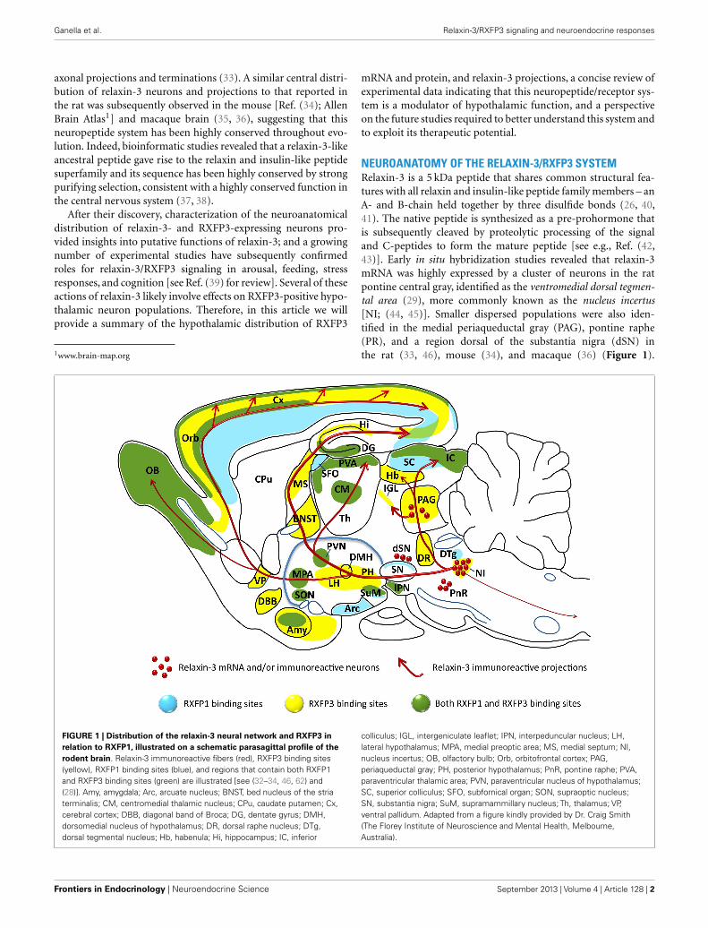
NEUROANATOMY OF THE RELAXIN-3/RXFP3 SYSTEMRelaxin-3 is a 5 kDa peptide that shares common structural fea-tures with all relaxin and insulin-like peptide family members – anA- and B-chain held together by three disulfide bonds (26, 40,41). The native peptide is synthesized as a pre-prohormone thatis subsequently cleaved by proteolytic processing of the signaland C-peptides to form the mature peptide [see e.g., Ref. (42,43)]. Early in situ hybridization studies revealed that relaxin-3mRNA was highly expressed by a cluster of neurons in the ratpontine central gray, identified as the ventromedial dorsal tegmen-tal area (29), more commonly known as the nucleus incertus[NI; (44, 45)]. Smaller dispersed populations were also iden-tified in the medial periaqueductal gray (PAG), pontine raphe(PR), and a region dorsal of the substantia nigra (dSN) inthe rat (33, 46), mouse (34), and macaque (36) (Figure 1).
FIGURE 1 | Distribution of the relaxin-3 neural network and RXFP3 inrelation to RXFP1, illustrated on a schematic parasagittal profile of therodent brain. Relaxin-3 immunoreactive fibers (red), RXFP3 binding sites(yellow), RXFP1 binding sites (blue), and regions that contain both RXFP1and RXFP3 binding sites (green) are illustrated [see (32–34, 46, 62) and(28)]. Amy, amygdala; Arc, arcuate nucleus; BNST, bed nucleus of the striaterminalis; CM, centromedial thalamic nucleus; CPu, caudate putamen; Cx,cerebral cortex; DBB, diagonal band of Broca; DG, dentate gyrus; DMH,dorsomedial nucleus of hypothalamus; DR, dorsal raphe nucleus; DTg,dorsal tegmental nucleus; Hb, habenula; Hi, hippocampus; IC, inferior
colliculus; IGL, intergeniculate leaflet; IPN, interpeduncular nucleus; LH,lateral hypothalamus; MPA, medial preoptic area; MS, medial septum; NI,nucleus incertus; OB, olfactory bulb; Orb, orbitofrontal cortex; PAG,periaqueductal gray; PH, posterior hypothalamus; PnR, pontine raphe; PVA,paraventricular thalamic area; PVN, paraventricular nucleus of hypothalamus;SC, superior colliculus; SFO, subfornical organ; SON, supraoptic nucleus;SN, substantia nigra; SuM, supramammillary nucleus; Th, thalamus; VP,ventral pallidum. Adapted from a figure kindly provided by Dr. Craig Smith(The Florey Institute of Neuroscience and Mental Health, Melbourne,Australia).
Frontiers in Endocrinology | Neuroendocrine Science September 2013 | Volume 4 | Article 128 | 2
Ganella et al. Relaxin-3/RXFP3 signaling and neuroendocrine responses
Ultrastructural analysis of relaxin-3 immunoreactivity in the ratNI identified the peptide in the rough endoplasmic reticulumand Golgi apparatus in the cell soma and within dense-core vesi-cles adjacent to synapses in nerve terminals of distant targetregions such as the lateral hypothalamus (46) and medial sep-tum (47), indicating that relaxin-3 is processed and released as atransmitter.
The efferent and afferent connections of the rat NI havebeen characterized (44, 45, 48–50) and many NI projectiontarget regions, including the hypothalamus, contain relaxin-3immunoreactive fibers and terminals, and neurons expressingRXFP3 (33, 50) (Figure 1; Table 1), suggesting many of theseareas are innervated by NI relaxin-3 neurons. Not surprisingly, NIrelaxin-3 neurons have been the focus of the majority of func-tional studies to date, which indicate they are highly responsive toneurogenic stressors and CRH (46, 51–53).
Less is known about the connections, regulation and func-tion of other relaxin-3 neuron populations, but a recent studydemonstrated that PAG relaxin-3 neurons strongly innervate andmodulate neuronal activity in the intergeniculate leaflet [IGL;(46, 54, 55)], a region that is known to contribute to the reg-ulation of arousal and circadian activity (56–58). In brain slicestudies, patch-clamp recordings of IGL neurons, revealed that acti-vation of RXFP3 by bath application of the agonist peptide, R3/I5,produced depolarization of identified NPY-containing neurons(55), which are known to project to the suprachiasmatic nucleusvia the geniculohypothalamic tract (56, 58). Neurons in the IGLalso project to a number of other hypothalamic areas, including
the anterior and lateral hypothalamic areas, and the dorsomedialnucleus (58).
There are, however, several brain regions like the afore-mentioned IGL and the amygdala that contain dense relaxin-3immunoreactivity and/or RXFP3, but sparse NI projections; sug-gesting they are also more strongly innervated by other relaxin-3populations. In fact, there is anatomical evidence, chiefly fromneural tract-tracing studies, to suggest the various RXFP3-positiveregions in the hypothalamus are also innervated by relaxin-3 neu-rons in the NI and other relaxin-3 groups. For example, a recentstudy of brainstem inputs to the PVN and surrounding area in therat (59) revealed projections from the medial PAG, PR, and “dorsalto substantia nigra” regions, which contain relaxin-3 neurons.
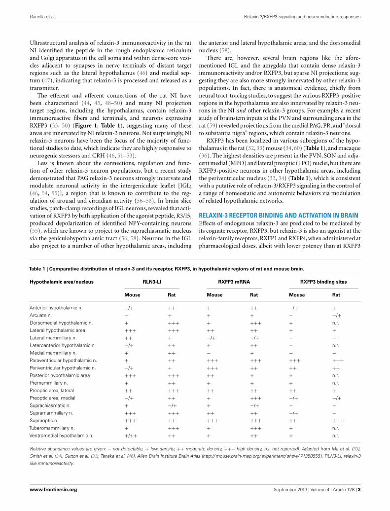
RXFP3 has been localized in various subregions of the hypo-thalamus in the rat (32, 33) mouse (34, 60) (Table 1), and macaque(36). The highest densities are present in the PVN, SON and adja-cent medial (MPO) and lateral preoptic (LPO) nuclei, but there areRXFP3-positive neurons in other hypothalamic areas, includingthe periventricular nucleus (33, 34) (Table 1), which is consistentwith a putative role of relaxin-3/RXFP3 signaling in the control ofa range of homeostatic and autonomic behaviors via modulationof related hypothalamic networks.
RELAXIN-3 RECEPTOR BINDING AND ACTIVATION IN BRAINEffects of endogenous relaxin-3 are predicted to be mediated byits cognate receptor, RXFP3, but relaxin-3 is also an agonist at therelaxin-family receptors, RXFP1 and RXFP4, when administered atpharmacological doses, albeit with lower potency than at RXFP3
Table 1 | Comparative distribution of relaxin-3 and its receptor, RXFP3, in hypothalamic regions of rat and mouse brain.
Hypothalamic area/nucleus RLN3-LI RXFP3 mRNA RXFP3 binding sites
Mouse Rat Mouse Rat Mouse Rat
Anterior hypothalamic n. −/+ ++ + ++ −/+ +
Arcuate n. − + + + − −/+
Dorsomedial hypothalamic n. + +++ + +++ + n.r.
Lateral hypothalamic area +++ +++ ++ ++ + +
Lateral mammillary n. ++ + −/+ −/+ − −
Lateroanterior hypothalamic n. −/+ ++ + ++ − n.r.
Medial mammillary n. + ++ − + − −
Paraventricular hypothalamic n. + ++ +++ +++ +++ +++
Periventricular hypothalamic n. −/+ + +++ ++ ++ ++
Posterior hypothalamic area +++ +++ ++ + + n.r.
Premammillary n. + ++ + + + n.r.
Preoptic area, lateral ++ +++ ++ ++ ++ +
Preoptic area, medial −/+ ++ + +++ −/+ −/+
Suprachiasmatic n. + −/+ + −/+ − −
Supramammillary n. +++ +++ ++ ++ −/+ −
Supraoptic n. +++ ++ +++ +++ ++ +++
Tuberomammillary n. + +++ + +++ + n.r.
Ventromedial hypothalamic n. +/++ ++ + ++ + n.r.
Relative abundance values are given: − not detectable, + low density, ++ moderate density, +++ high density, n.r. not reported). Adapted from Ma et al. (33),
Smith et al. (34), Sutton et al. (32), Tanaka et al. (46), Allen Brain Institute Brain Atlas (http:// mouse.brain-map.org/ experiment/ show/ 71358555). RLN3-LI, relaxin-3
like immunoreactivity.
www.frontiersin.org September 2013 | Volume 4 | Article 128 | 3
Ganella et al. Relaxin-3/RXFP3 signaling and neuroendocrine responses
(30, 61). RXFP1 and RXFP4 are likely expressed in the humanbrain, along with RXFP3, but based on animal studies these recep-tors and the peptides which bind and activate them (relaxin,insulin-like peptide 5, and relaxin-3, respectively) are expressedin quite distinct regions of the brain and at very different levels[e.g., Ref. (26, 29, 30, 33, 46, 62)], so a current working hypothesisthat can be tested is that these receptors mediate distinct func-tional effects in the brain, which modulate different homeostaticprocesses and behaviors.
Cell signaling events associated with the relaxin-family recep-tors have been studied in different cell lines transfected with thehuman receptors (28, 63) and activation of RXFP1 and RXFP3/4by H3 relaxin leads to different intracellular responses in vitro. InChinese hamster ovary (CHO) cells, RXFP3 and RXFP4 coupleto the inhibitory Gαi/Gαo-protein system and receptor activa-tion leads to sequestration of these G-proteins and inhibition ofadenylate cyclase (AC), and subsequent cAMP accumulation (30,64). The intracellular signaling pathway of the relaxin-3-RXFP3interaction has also been studied in the SN56 neuronal-like cellline, in which the Gαi/Gαo pathway was recruited, suggesting aninhibitory intracellular pathway may be activated in vivo whenrelaxin-3 binds to RXFP3-expressing neurons within the brain(63), an idea that can be tested in different functional networks [seeRef. (54, 55)]. In contrast, RXFP1 activation by either relaxin orrelaxin-3 in mammalian cell expression systems initiates a down-stream accumulation of cAMP, as it is predominantly coupled tothe stimulatory Gs-protein. These in vitro studies suggest acti-vation of different receptors may lead to different downstreameffects in neurons in vivo. Little is known, however, about thenative intracellular signaling of any of the relaxin family of recep-tors (RXFP1–RXFP4) in specific neuronal populations, apart froma recent in vitro study that revealed activation of RXFP3 by theagonist peptide, R3/I5 (65), produced depolarization of identi-fied NPY-containing neurons and hyperpolarization of adjacentnon-NPY neurons (55).
Although there is no definitive evidence that major biologicaleffects mediated by relaxin-3 are caused by activation of eitherRXFP1 and/or RXFP4, the ability of relaxin-3 to activate RXFP1and RXFP4 as well as RXFP3 must be considered as a confoundingfactor when using pharmacological doses of peptides in vivo, inattempts to study neuropeptide function in the rodent. From apractical viewpoint, the rat is suited to studies of the neurobiologyof relaxin-3/RXFP3 signaling, since RXFP4 is a pseudogene in thisspecies, and so not a “confound.” However, in situ hybridizationand radioligand binding site studies indicate RXFP1 is expressedin the rat brain in a number of regions positive for RXFP3, includ-ing the cerebral cortex, amygdala, thalamus, and hypothalamus(32, 33, 62). Consequently RXFP1 activity must be consideredin studies of exogenous relaxin-3 peptide administration [e.g.,Ref. (40, 66–70)]. The relative degree to which RXFP1 activationhas impacted outcomes in studies of relaxin-3 actions within thehypothalamus and other brain areas is hard to gage, although insome cases comparative effects of relaxin were reported [see Ref.(71) below]. Fortunately, the development more recently of ago-nist and antagonist peptides which selectively activate or inhibitRXFP3 [e.g., Ref. (65, 72–74)] has facilitated investigations of thebehavioral and physiological effects of specific relaxin-3/RXFP3
interactions. Indeed, several studies have used these peptides toassess relaxin-3/RXFP3 related functions associated with the hypo-thalamus, although their use is not as widespread as it mightbe [e.g., Ref. (69, 70)], particularly in light of the availabilityof a chemically less-complex, single-chain peptide antagonist forRXFP3 (73).
ACTIONS OF THE RELAXIN-3/RXFP3 SYSTEM – FOCUS ONHYPOTHALAMUSFEEDING AND ENERGY BALANCEThe PVN and arcuate nucleus (ARC) are two hypothalamic nucleiwhich tightly regulate food intake and energy homeostasis [e.g.,Ref. (3, 4, 12, 15, 75)]. Relaxin-3 immunoreactivity and RXFP3mRNA and binding sites have been identified in the PVN and ARC(30, 32, 33) (Table 1) and extensive research has demonstrated thatH3 relaxin can alter feeding and appetite in rats [see Ref. (76)].Central (icv) administration of H3 relaxin caused hyperphagia insatiated male Wistar rats in the first hour after treatment in boththe light and the dark phase, while equivalent doses of H2 relaxindid not produce a similar effect (66). The doses of H3 relaxinadministered icv (180 pmol) or directly into the PVN (18 pmol) atwhich a significant increase in feeding was observed (see below)were similar to doses of other “feeding” peptides which elicit anincreased feeding response upon similar administration [e.g., NPY∼80 pmol iPVN; (77)], consistent with a physiological role forrelaxin-3/RXFP3 signaling in modulating appetite and feedingbehavior. Importantly, icv administration of the selective agonist,R3/I5, produced an increase in first-hour food intake and thiseffect was inhibited by co-administration of the selective antago-nist, ∆R3/I5 (72), further suggesting RXFP3 involvement. In thesefeeding studies, the food intake was monitored for only 24 h post-injection, as it was predicted the injected peptide would be quiterapidly degraded by proteolysis. In a study of longer term effects ofrelaxin-3 on feeding and body weight, H3 relaxin (600 pmol/day)was infused icv into rats for 14 days via osmotic mini-pump, whichproduced a significant increase in food intake and body weightgain, and plasma leptin and insulin levels, compared to vehicle(78). This data indicated that the relaxin-3 induced increase infood intake could be sustained and lead to an increase in bodyweight and associated biochemical changes in the rat.
Given the primary role of the hypothalamus in energy balance,the effect on feeding of local hypothalamic injections of relaxin-3 was assessed. Acute H3 relaxin injection into the PVN (iPVN)increased food intake over the first hour (67). Sub-chronic iPVNH3 relaxin administration in ad libitum fed rats also produced anincrease in food intake and cumulative body weight gain c.f. vehi-cle (67). These authors also used “Fos-activation” mapping aftericv administration of H3 relaxin to identify activated hypothala-mic nuclei. In addition to the PVN, the SON, ARC, and anteriorpreoptic area displayed increased Fos staining (79). When injecteddirectly into these and other hypothalamic nuclei H3 relaxin stim-ulated a significant increase in food intake within the first hour,relative to control (79), suggesting multiple hypothalamic nucleimay mediate these potent orexigenic effects. Unfortunately thepotential involvement of RXFP1 activation in the observed effectscannot be reliably excluded, as RXFP3-selective peptides were notutilized. It is also possible the injected peptide is able to diffuse
Frontiers in Endocrinology | Neuroendocrine Science September 2013 | Volume 4 | Article 128 | 4
Ganella et al. Relaxin-3/RXFP3 signaling and neuroendocrine responses
from the injection site to adjacent areas that are primarily involvedin the activation of feeding, although some targeted sites were notassociated with feeding (79).
In order to circumvent issues associated with acute peptideadministration and cross-reactivity of H3 relaxin at RXFP1, weused a viral strategy to investigate the effect of chronic R3/I5mediated RXFP3 activation within the hypothalamic PVN (43).Using a recombinant adeno-associated virus (rAAV) engineeredto locally secrete bioactive R3/I5 (rAAV-R3/I5), we demonstratedan increase in food intake in the R3/I5 expressing rats (∼5.2 g/daymore than control) which was sustained for up to 2 months, lead-ing to an ∼23% increase in cumulative body weight gain (43). Inan attempt to identify targets of RXFP3 activation within the hypo-thalamus, levels of mRNA for a number of genes were also assessedin dissected hypothalamic tissue blocks using quantitative reversetranscription PCR. Notably, no major differences were identifiedin expression of some “major” feeding peptide genes (NPY, AgRP,POMC) between rAAV-R3/I5 and rAAV-control treated groups,whereas the levels of oxytocin and AVP mRNA were altered. This is,however, consistent with the strong expression of RXFP3 by neu-rons in the PVN and SON (30, 32, 33) (Table 1), which containoxytocin- and AVP-containing magnocellular neurons. Chronicviral-mediated expression of R3/I5 in the hypothalamus for up to∼14 weeks led to a marked reduction in oxytocin mRNA expres-sion in PVN (∼50%) and a smaller reduction in AVP (∼25%),compared to control expression (43). These data suggest the acuteorexigenic effect of RXFP3 activation may be mediated by changesin oxytocin release (and perhaps AVP) [see Ref. (80)], an idea thatcan be tested experimentally. Furthermore, there is a substantialliterature relating to oxytocin as a satiety factor that also supportsthis putative acute and chronic mechanism of action.
INTERACTION WITH OXYTOCIN AND ARGININE VASOPRESSINSYSTEMS IN THE PVNOxytocin is a peptide hormone highly expressed in magnocel-lular and some parvocellular neurons of the hypothalamic PVNand SON (81–83). Oxytocin is classically known as a reproductivehormone with roles in parturition, lactation, sexual behavior, andpair bonding and attachment [see Ref. (84, 85) for review]. How-ever, early studies revealed that central administration of oxytocindose-dependently reduced food consumption and time spent eat-ing and increased the latency to the first meal in pre-fasted rats;an effect prevented by co-administration of an oxytocin receptorantagonist (86). A number of studies have since confirmed cen-tral oxytocin administration inhibits food intake, strengtheningthe hypothesis that oxytocin serves a key role in appetite control(87–89). More recent data is consistent with this view, as oxy-tocin null mutation mice display enhanced intake of sweet andnon-sweet carbohydrate solutions (90, 91) and develop late-onsetobesity (92).
The magnitude of the increase in feeding and body weight gainobserved in our hypothalamic R3/I5 expression studies (43) wasmodest relative to those seen after similar viral-mediated NPYand AgRP over-expression, consistent with actions independent ofdirect effects on these neurons. The down-regulation of oxytocinand AVP mRNA expression observed (43), suggests the R3/I5 ago-nist peptide is activating RXFP3 on oxytocin- and AVP-containing
neurons, which results in downstream effects on the transcrip-tion of oxytocin and AVP mRNA in these neurons (e.g., Ref.(93, 94)] and subsequent effects on production and release ofthis anorexigenic hormone. If RXFP3 associated cell signaling inthese neurons is similar to effects reported in vitro, this could beassociated with reduction in cellular cAMP levels (via inhibitoryGi-protein coupling) and inhibition or hyperpolarization of targetneurons – a possibility that can be assessed experimentally usingin vitro or in vivo electrophysiological and biochemical methods[e.g., Ref. (55, 95)].
It is also necessary to establish whether it is the proposedreduction in oxytocin production and release which producesthe observed increase in food intake in AAV-R3/I5-treated rats,or whether the chronic increase in food intake caused by hypo-thalamic RXFP3 activation over time leads to down-regulation ofoxytocin mRNA via a distinct mechanism. This could be exploredby conducting acute peptide administration studies or shortertime course viral infusion studies, and assessing oxytocin mRNA,peptide and release levels, before any marked changes in bodyweight have occurred.
Indeed, a recent study examined the effect of icv H3 relaxin onanxiety-like behavior in rats and observed an anxiolytic effect inthe elevated plus maze test and the shock probe-burying test (70),consistent with our studies demonstrating that icv administrationof a selective RXFP3 agonist peptide reduced anxiety-like behaviorin the light-dark box and elevated plus maze (96). Notably, theseauthors used microarray and peptidomics approaches to iden-tify associated downstream signaling targets in the hypothalamusaltered by icv H3 relaxin and detected a relatively acute (6–24 h)and quite specific increase in the level of hypothalamic oxytocinmRNA and peptide levels (70). This data is, however, more consis-tent with the ability of H3 relaxin to activate neurons via RXFP1[see e.g., Ref. (40, 67, 68)] rather than via RXFP3, which based onpredicted signaling (28, 63) might be expected to decrease oxy-tocin neuron activity (and mRNA and peptide levels), as seen inour study (43). Therefore, further studies are required to identifyspecific effects of RXFP3 (and RXFP1) activation in hypothalamusand other areas in altered feeding and metabolism and anxiety-likebehavior (76, 96).
Further evidence for an association of a down-regulation/inhibition of PVN oxytocin activity with a hyperphagic phe-notype was reported recently. Optogenetic electrophysiologicalstudies revealed that GABA/AgRP neurons in the ARC projectto a population of oxytocin neurons in the PVN and stronglyinhibit their activity, and this suppression of oxytocin neurons byAgRP neuronal activation drives evoked feeding (23). This studyalso demonstrated that increased food seeking and consumptionin response to GABA/AgRP neuron activation is similarly inducedby suppressing the activity of the PVN neurons that (selectively)express the single-minded 1 (SIM1) transcription factor (23, 97).
Importantly from a clinical perspective, a number of dis-ease states in which hyperphagia is a symptom are associatedwith hypothalamic oxytocin dysregulation. A small populationof PVN oxytocin neurons is selectively lost in Prader–Willi syn-drome, which is a condition involving insatiable hunger (98); anddisruption of synaptic release of hypothalamic oxytocin results inovereating (99). Oxytocin deficits in SIM1 haploinsufficient mice
www.frontiersin.org September 2013 | Volume 4 | Article 128 | 5

Ganella et al. Relaxin-3/RXFP3 signaling and neuroendocrine responses
and mutations in the SIM1 gene in humans lead to hyperphagicobesity (97, 100, 101). In the mouse model, an ∼80% reduction inoxytocin and ∼30% reduction in AVP was observed (97). Thesestudies illustrate that modulation of relaxin-3 signaling and asso-ciated alterations in oxytocin neuron activity may be a fruitful areato explore for treating disease states associated with eating disor-ders. For example, a recent clinical cross-sectional study reportedthat female patients with anorexia nervosa, characterized by foodrestriction, low weight, and hypoleptinemia, had higher mean cir-culating oxytocin levels than healthy controls at all times assessed(102). The severity of disease psychopathology was also positivelyassociated with circulating oxytocin levels (102).
HYPOTHALAMIC-PITUITARY-ADRENAL AXIS AND STRESS RESPONSESIntegration of the stress response via the hypothalamic-pituitary-adrenal (HPA) axis occurs via interaction between brain areaswhich are sensitive to stress and neuroendocrine neurons of thehypothalamic PVN, particularly those in the parvocellular regionproducing CRH [see Ref. (1, 6, 103) for review]. CRH stimu-lates the secretion and synthesis of adrenocorticotropin hormone(ACTH) from the pituitary and is the main regulator of HPA axisactivity during stress.
Early regulatory studies revealed that neurons in the NI andspecifically relaxin-3 expressing neurons respond to stress andCRH (46, 104), and that relaxin-3-containing neurons in the NIexpress CRH type 1 receptors (CRH-R1) (46). Upon icv admin-istration of CRH, 65% of relaxin-3-positive neurons underwentactivation (detected using Fos-immunostaining 2 h post-infusion)(46). Electrophysiological characterization of NI neurons revealedthat a significant population increased firing following icv admin-istration of CRH, of which the majority were relaxin-3-positive,though an almost equal number of non-relaxin-3 neurons exhib-ited a decrease in firing in the anesthetized rat (53). These find-ings are consistent with semi-quantitative immunohistochemicalstudies of the NI revealing this heterogeneous neuron popula-tion consists of relaxin-3 neurons that all co-express CRH-R1,though not all CRH-R1 contain relaxin-3, in addition to a sig-nificant population that are CRH-R1 negative (53). Rats tested ina water immersion-restraint stress paradigm, displayed increasedFos-immunostaining and an up-regulation of relaxin-3 mRNA inNI neurons (46). Subsequently, the effect of a repeat forced swimon relaxin-3 expression in the NI was examined and led to a rapidincrease in relaxin-3 mRNA expression (51). This increase waslargely mediated by CRH activation of CRH-R1 located on NIneurons, as pre-treatment with the CRH antagonist, antalarmin,reduced the increase in relaxin-3 mRNA expression by 70–80%.Levels of relaxin-3 heteronuclear (hn) RNA were also increased inNI neurons after the repeat forced swim (51). Changes in hnRNAare a measure of transcriptional activity and are thought to reflectthe level of encoded peptide synthesis (105), suggesting in thiscase, an increase in relaxin-3 utilization.
Initial insights into the hypothalamic action of relaxin-3 inrelation to the stress response have been obtained by monitor-ing the effect of icv administration of relaxin-3 in male rats (69).Increased Fos staining in the PVN and SON was observed 1 hpost-administration and levels of c-fos and CRH mRNA in thePVN were also increased 2 h after H3 relaxin administration.
Central H3 relaxin administration also elicited an increase in circu-lating plasma ACTH (69). These data suggest a role for relaxin-3 inthe acute hypothalamus-pituitary CRH-ACTH system response,but these studies did not directly identify the presence of RXFP3 onCRH neurons or measure the direct acute excitatory or inhibitoryeffect of RXFP3 activation on these neurons [see Ref. (55)].
Notably, a recent report also suggests sex-specific regulationof CRH and relaxin-3 systems in response to combined stres-sors. Chronically stressed and repeatedly food-restricted femalerats consumed more standard chow during recovery and hadan increased bodyweight relative to controls, whereas male ratsexposed to this regime had a reduction in bodyweight (106).Stressed/food-restricted female rats had elevated plasma corticos-terone and low PVN CRH mRNA levels. CRH neurons in themedial preoptic area were identified as a source of increased CRHproduction/release during stress in female brain, i.e., CRH mRNAlevels in this area were – higher in female than male rats, increasedby chronic stress, and increased in female, not male, rats afterrepeated food restriction (106).
Further studies are now required to determine precisely how therobust, consistently observed stress and CRH-induced activationof NI and relaxin-3 neurons (46, 51, 53, 106) directly or indirectlyactivates (or possibly inhibits) the PVN and the main componentsof the HPA axis; and whether and how these processes are regulatedby steroid and other hormones under different conditions.
EFFECTS ON REPRODUCTIVE NEUROENDOCRINE SYSTEMSPreliminary studies have indicated a putative role for relaxin-3 in reproductive physiology. Following injection into the PVNand surrounds, H3 relaxin increased levels of marker hormonesof the hypothalamic-pituitary-gonadal (HPG) axis (71, 107);and anatomical studies have identified RXFP3 in many areas inthe hypothalamus relevant to reproductive neuroendocrinology,including the preoptic area and the PVN and SON (33). H3 relaxinadministered icv or iPVN significantly increased plasma luteiniz-ing hormone (LH) levels 30 min post-injection in male Wistar rats,an effect blocked by peripheral pre-treatment with a GnRH antag-onist, consistent with increased central GnRH release (71). In theseinitial studies, H2 relaxin administration via the same routes pro-duced a small,non-significant increase in LH,suggesting a strongerrelaxin-3/RXFP3-mediated effect (71). Activity of the endoge-nous peptide at RXFP1 cannot be completely excluded from aninvolvement in these effects, however, as RXFP1 is expressed in theanterior hypothalamus and PVN of the rat (62). If so, high levelsof endogenous relaxin-3 may act via RXFP1 to stimulate the HPGaxis, an idea that could be tested experimentally. It would also beof interest to observe the effects of icv or iPVN administration ofRXFP3-selective peptides (65, 72–74) on the HPG axis in femalerats to check for similar hormone changes to those observed inmale rats.
Notably, treatment of mouse hypothalamic neuron-like (GT1–7) cells or hypothalamic explants with synthetic H3 relaxin, pro-duced a dose-dependent increase in secretion of GnRH (71).However, it is again unclear if this is an RXFP3-mediated effect,as GT1–7 cells and hypothalamic explants also express RXFP1;and H3 relaxin binds and activates these receptors. As these cellsexpress both RXFP1 and RXFP3, the relaxin-3 mediated secretion
Frontiers in Endocrinology | Neuroendocrine Science September 2013 | Volume 4 | Article 128 | 6
Ganella et al. Relaxin-3/RXFP3 signaling and neuroendocrine responses
of GnRH should be assessed for sensitivity to blockade with anRXFP3 antagonist (71–73).
In our recent study, we assessed the effect of chronic(∼14 weeks) viral-mediated expression of R3/I5 in the hypo-thalamus on GnRH mRNA levels (43). Although the differencebetween groups was not statistically significant, there was a trendfor increased GnRH mRNA levels in AAV-R3/I5-treated com-pared to control rats, with considerable variability in individualvalues. Given the dispersed nature of GnRH expressing neuronsin the hypothalamus, the variability observed suggests that dif-ferent numbers (populations) of GnRH expressing neurons mayhave been dissected in these assays and/or there may be a genuineincrease in GnRH transcription as a result of the treatment. Thisimportant question needs to be further investigated using suitablequantitative methods, in conjunction with further assessments ofrelaxin3/RXFP3 indices relative to GnRH and other reproductivepeptides and receptors during different stages of pregnancy, birthand lactation.
OTHER HYPOTHALAMIC SITES OF RELAXIN-3/RXFP3 ACTIONThe relaxin-3/RXFP3 system may have actions in other hypo-thalamic nuclei, demonstrated by the presence of both relaxin-3immunoreactive fibers and RXFP3 mRNA/protein in the lateraland medial preoptic areas, anterior, posterior, dorsomedial, andventromedial regions, and the adjacent supramammillary nucleus(SuM) (33) (Table 1). The functions of these regions will be brieflyreviewed, in view of the postulated role of relaxin-3 in arousal,feeding, behavioral state, and cognition (39).
The SuM receives a moderate relaxin-3 innervation in the rat(33) and the mouse (34), and this nucleus represents a major tar-get of the peptide network in the macaque brain (35). In contrastto other “hypothalamic” regions, this nucleus is best characterizedas a key input to and modulator of the hippocampus and hip-pocampal theta rhythm (108, 109), a synchronous activity between4 and 12 Hz associated with active waking behavior and REMsleep (110), mnemonic processing (111, 112), spatial navigationand exploration (113), and sensorimotor integration (114, 115).There is growing evidence that the NI is a key player in mediat-ing brainstem-elicited theta rhythm (47, 53, 116). In addition toprojections to the SuM, there are strong relaxin-3-containing NIprojections to other regions subserving theta rhythm generation,such as medial septum (48–50), reticularis pontine oralis, and thehippocampus, as well as the median raphe, which is involved intheta desynchronization [see (52) for review]. In fact, relaxin-3projections to the medial septum have been shown to promotehippocampal theta rhythm and are necessary for normal spatialnavigation of rats in a spontaneous alternation task (47). Thus,the SuM is likely a further node at which this neuropeptide sys-tem acts to regulate hippocampal theta activity and associatedcognitive/autonomic processes.
The lateral and medial preoptic areas of hypothalamus are alsorich in relaxin-3 projections and RXFP3. The LPO area containspopulations of neurons that have identified roles in thermoreg-ulation (117, 118) and sleep-wakefulness (ventrolateral neurons)(119), so it will be of interest, to assess the precise topography ofRXFP3 in these areas and to assess the effects of RXFP3 activa-tion/inhibition in these regions on these physiological parameters,
particularly as our laboratory has anatomical and functional dataconsistent with effects of relaxin-3 on sleep-wake activity in mice(34, 120). Similarly, as discussed, the role of relaxin-3/RXFP3 sig-naling in the medial preoptic area may be important in stressresponses in female rats and the possible regulation of CRHneurons in the area (106).
RELAXIN-3/RXFP3 IN MOUSE HYPOTHALAMUS – SPECIESDIFFERENCESThe distribution of relaxin-3 neurons and projections, as well asthe distribution of RXFP3 mRNA and binding sites in the mousebrain, is regionally similar to that in the rat both generally through-out the forebrain and within the hypothalamus (33, 34). Further-more, the distribution of Rxfp3 mRNA in the C57BL6/J mousedetailed in the Allen Brain Institute Brain Atlas (see text footnote 1)using a digoxigenin-labeled RNA probe is similar to that observedin our studies using radioactively labeled oligonucleotides (34)(Table 1). However, a more detailed analysis is required to deter-mine whether identical populations of neurons are targeted withinkey hypothalamus areas, such as the arcuate, periventricular, PVN,and SON of rat and mouse to assess whether relaxin-3 signalingmight regulate similar neuroendocrine processes in these species.
We have conducted several studies to assess the possible role ofrelaxin-3/RXFP3 in food intake in mice (121). After the adminis-tration of similar or higher levels of relaxin-3 or various RXFP3-selective agonists (R3/I5, RXFP3-A2) used in studies on rats [see(76) for review], we did not observe an increase in food intake ofsatiated C57BL/6J mice. In more recent studies, we have observedthat the icv administration of the RXFP3 antagonist, R3(B1–22)R(73) produced a reduction in the robust feeding that occurred inmice offered access to regular chow after a 4 h period of isolatedhousing in a novel cage without bedding and in the absence of food(Smith and Gundlach, unpublished data). These particular con-ditions are presumed to induce a level of stress, hypothermia, andenergy deficiency in the mice, which motivates their feeding; andsuggests that relaxin-3/RXFP3 signaling in mice may not influencefeeding significantly under basal conditions, but may play a roleunder altered stress conditions. These possibilities are currentlybeing explored experimentally.
Consistent with these pharmacological studies, however, thereis no genotypic difference between relaxin-3 KO and wild typelittermates in bodyweight, total food consumption, or circadianrhythm of food consumption (120, 122). These findings are in con-trast to data obtained by Sutton et al. (123) in a separately derivedcolony of relaxin-3 KO mice on a mixed C57BL6/J/SV129 back-ground, which displayed a markedly reduced body weight relativeto wild type mice when both genotypes were fed a diet with a mod-erately elevated fat content. However, subsequent studies of a nullmutation Rxfp3 KO mouse strain revealed no body weight-relatedphenotype, but did reveal an identical circadian hypoactivity phe-notype to the relaxin-3 KO mouse (120,124,125). This suggests therelaxin-3 KO phenotype reported by Sutton et al. (123) was asso-ciated with genetic differences independent of relaxin-3/RXFP3[see (39)].
With regard to the specificity of pharmacological studies ofRXFP3 signaling in the mouse, while the presence of insulin-like peptide 5 (INSL5) and RXFP4 in the mouse brain has been
www.frontiersin.org September 2013 | Volume 4 | Article 128 | 7
Ganella et al. Relaxin-3/RXFP3 signaling and neuroendocrine responses
reported (126), these findings have not been independently val-idated; and in a separate study, the presence of INSL5-sensitivereceptor binding sites could not be identified (127). Furthermore,the ligand/receptor expression profile suggests the INSL5/RXFP4system acts primarily within the gastrointestinal tract and largeintestine (28, 128). While it is possible that peripheral INSL5 sig-naling may alter central (hypothalamic) function, at this stage nosuch data is available.
CONCLUSION AND PERSPECTIVESA decade of research has revealed the basic structural frame-work of central relaxin-3/RXFP3 networks and their likely func-tional importance in mammalian brain (27, 39, 76); and sev-eral studies have highlighted the interaction between hypothal-amic homeostatic systems and relaxin-3/RXFP3 signaling, pre-dominantly in pharmacological and regulatory studies in therat. Together, these studies point to a role for relaxin-3/RXFP3in regulating hypothalamic activity, with evidence suggestingit does so via interactions with oxytocin, AVP, and/or CRH,to modulate neuroendocrine function associated with stress,
feeding, and metabolism, and motivation and reward. Furtherstudies are now required to clarify the nature of these var-ious effects and how they are coordinated and regulated byhormonal and transmitter inputs onto relaxin-3 neurons andintegrated signaling at the level of hypothalamic loops (e.g.,HPA, HPG axes). Recent research strongly suggests the relaxin-3/RXFP3 system is an important regulator of the neuroendocrineaxis, with potential as a novel therapeutic target for the treat-ment of neuroendocrine disorders and associated behavioraldysfunction.
ACKNOWLEDGMENTSResearch in the authors’ laboratory is supported by grants fromthe National Health and Medical Research Council (NHMRC)of Australia (Andrew L. Gundlach, Sherie Ma), the Besen Familyand Pratt Foundations (Andrew L. Gundlach), and by the Vic-torian Government Operational Infrastructure Support Program.Despina E. Ganella was the recipient of a Commonwealth Aus-tralian Postgraduate Award. Andrew L. Gundlach is the recipientof an NHMRC (Australia) Research Fellowship.
REFERENCES1. Herman JP, Cullinan WE, Ziegler
DR, Tasker JG. Role of the par-aventricular nucleus microenvi-ronment in stress integration. EurJ Neurosci (2002) 16:381–5. doi:10.1046/j.1460-9568.2002.02133.x
2. Small CJ, Stanley SA, BloomSR. Appetite control and repro-duction: leptin and beyond.Semin Reprod Med (2002)20:389–98. doi:10.1055/s-2002-36712
3. Morton GJ, Cummings DE, BaskinDG, Barsh GS, Schwartz MW. Cen-tral nervous system control of foodintake and body weight. Nature(2006) 443:289–95. doi:10.1038/nature05026
4. Gao Q, Horvath TL. Neurobiol-ogy of feeding and energy expen-diture. Annu Rev Neurosci (2007)30:367–98. doi:10.1146/annurev.neuro.30.051606.094324
5. Adamantidis A, de Lecea L. Phys-iological arousal: a role for hypo-thalamic systems. Cell Mol Life Sci(2008) 65:1475–88. doi:10.1007/s00018-008-7521-8
6. Ulrich-Lai YM, Herman JP. Neuralregulation of endocrine and auto-nomic stress responses. Nat RevNeurosci (2009) 10:397–409. doi:10.1038/nrn2647
7. Zeltser LM, Seeley RJ, Tschöp MH.Synaptic plasticity in neuronal cir-cuits regulating energy balance.Nat Neurosci (2012) 15:1336–42.doi:10.1038/nn.3219
8. Lantos TA, Görcs TJ, PalkovitsM. Immunohistochemical map-ping of neuropeptides in the pre-mamillary region of the hypo-thalamus in rats. Brain Res Rev
(1995) 20:209–49. doi:10.1016/0165-0173(94)00013-F
9. Broberger C, De Lecea L, SutcliffeJG, Hokfelt T. Hypocretin/orexin-and melanin-concentratinghormone-expressing cells formdistinct populations in the rodentlateral hypothalamus: relation-ship to the neuropeptide Y andagouti gene-related protein sys-tems. J Comp Neurol (1998)402:460–74. doi:10.1002/(SICI)1096-9861(19981228)402:4<460::AID-CNE3>3.3.CO;2-J
10. Carlin KM, Vale WW, Bale TL.Vital functions of corticotropin-releasing factor (CRF) pathwaysin maintenance and regulationof energy homeostasis. ProcNatl Acad Sci U S A (2006)103:3462–7. doi:10.1073/pnas.0511320103
11. Lechan RM, Fekete C. The TRHneuron: a hypothalamic integra-tor of energy metabolism. ProgBrain Res (2006) 153:209–35. doi:10.1016/S0079-6123(06)53012-2
12. Meister B. Neurotransmitters inkey neurons of the hypothalamusthat regulate feeding behavior andbody weight. Physiol Behav (2007)92:263–71. doi:10.1016/j.physbeh.2007.05.021
13. Sakurai T. The neural circuit oforexin (hypocretin): maintainingsleep and wakefulness. Nat RevNeurosci (2007) 8:171–81. doi:10.1038/nrn2092
14. Saito Y, Nagasaki H. Themelanin-concentrating hor-mone system and its physio-logical functions. Results ProblCell Differ (2008) 46:159–79.doi:10.1007/400_2007_052
15. Williams KW, Elmquist JK. Light-ing up the hypothalamus: coor-dinated control of feeding behav-ior. Nat Neurosci (2011) 14:277–8.doi:10.1038/nn0311-277
16. Parker JA, Bloom SR. Hypothala-mic neuropeptides and the regula-tion of appetite. Neuropharmacol-ogy (2012) 63:18–30. doi:10.1016/j.neuropharm.2012.02.004
17. Michel MC, Beck-Sickinger A, CoxH, Doods HN, Herzog H, Larham-mar D, et al. XVI. InternationalUnion of Pharmacology recom-mendations for the nomenclatureof neuropeptide Y, peptide YY, andpancreatic polypeptide receptors.Pharmacol Rev (1998) 50:143–50.
18. Bale TL, Vale WW. CRF and CRFreceptors: role in stress respon-sivity and other behaviors. AnnuRev Pharmacol Toxicol (2004)44:525–57. doi:10.1146/annurev.pharmtox.44.101802.121410
19. Alexander SP, Mathie A, Peters JA.Guide to receptors and channels(GRAC), 5th edition. Br J Pharma-col (2011) 164(Suppl 1):S1–324.doi:10.1111/j.1476-5381.2011.01649_1.x
20. Hazell GG, Hindmarch CC,Pope GR, Roper JA, Light-man SL, Murphy D, et al. Gprotein-coupled receptors in thehypothalamic paraventricularand supraoptic nuclei – ser-pentine gateways to neuroen-docrine homeostasis. FrontNeuroendocrinol (2012) 33:45–66.doi:10.1016/j.yfrne.2011.07.002
21. Knobloch HS, Charlet A, Hoff-mann LC, Eliava M, Khrulev S,Cetin AH, et al. Evoked axonaloxytocin release in the central
amygdala attenuates fear response.Neuron (2012) 73:553–66. doi:10.1016/j.neuron.2011.11.030
22. Krashes MJ, Koda S, Ye CP, RoganSC, Adarns AC, Cusher DS, etal. Rapid, reversible activationof AgRP neurons drives feedingbehavior in mice. J Clin Invest(2011) 121:1424–8. doi:10.1172/JCI46229
23. Atasoy D, Betley JN, Su HH,Sternson SM. Deconstruction of aneural circuit for hunger. Nature(2012) 488:172–7. doi:10.1038/nature11270
24. de Lecea L, Carter ME, Adaman-tidis A. Shining light on wakeful-ness and arousal. Biol Psychiatry(2012) 71:1046–52. doi:10.1016/j.biopsych.2012.01.032
25. Zhan C, Zhou JF, Feng QR,Zhang JE, Lin SL, Bao JH, et al.Acute and long-term suppressionof feeding behavior by POMC neu-rons in the brainstem and hypo-thalamus, respectively. J Neurosci(2013) 33:3624–32. doi:10.1523/JNEUROSCI.2742-12.2013
26. Bathgate RAD, Samuel CS, BurazinTCD, Layfield S, Claasz AA, Rey-tomas IGT, et al. Human relaxingene 3 (H3) and the equiva-lent mouse relaxin (M3) gene:novel members of the relaxinpeptide family. J Biol Chem(2002) 277:1148–57. doi:10.1074/jbc.M107882200
27. Ma S, Gundlach AL. Relaxin-family peptide and receptorsystems in brain: insights fromrecent anatomical and functionalstudies. Adv Exp Med Biol (2007)612:119–37. doi:10.1007/978-0-387-74672-2_9
Frontiers in Endocrinology | Neuroendocrine Science September 2013 | Volume 4 | Article 128 | 8
Ganella et al. Relaxin-3/RXFP3 signaling and neuroendocrine responses
28. Bathgate RAD, Halls ML, vander Westhuizen ET, Callander GE,Kocan M, Summers RJ. Relaxinfamily peptides and their recep-tors. Physiol Rev (2013) 93:405–80.doi:10.1152/physrev.00001.2012
29. Burazin TCD, Bathgate RAD,Macris M, Layfield S, Gundlach AL,Tregear GW. Restricted, but abun-dant, expression of the novel ratgene-3 (R3) relaxin in the dorsaltegmental region of brain. J Neu-rochem (2002) 82:1553–7. doi:10.1046/j.1471-4159.2002.01114.x
30. Liu C, Eriste E, Sutton S, ChenJ, Roland B, Kuei C, et al. Iden-tification of relaxin-3/INSL7as an endogenous ligand forthe orphan G-protein cou-pled receptor GPCR135. J BiolChem (2003) 278:50754–64.doi:10.1074/jbc.M308995200
31. Bathgate RAD, Ivell R, SanbornBM, Sherwood OD, Summers RJ.International Union of Pharma-cology LVII: recommendations forthe nomenclature of receptors forrelaxin family peptides. PharmacolRev (2006) 58:7–31. doi:10.1124/pr.58.1.9
32. Sutton SW, Bonaventure P, Kuei C,Roland B, Chen J, Nepomuceno D,et al. Distribution of G-protein-coupled receptor (GPCR)135binding sites and receptor mRNAin the rat brain suggests a rolefor relaxin-3 in neuroendocrineand sensory processing. Neuroen-docrinology (2004) 80:298–307.doi:10.1159/000083656
33. Ma S, Bonaventure P, Ferraro T,Shen PJ, Burazin TCD, BathgateRAD, et al. Relaxin-3 in GABAprojection neurons of nucleusincertus suggests widespreadinfluence on forebrain circuitsvia G-protein-coupled receptor-135 in the rat. Neuroscience(2007) 144:165–90. doi:10.1016/j.neuroscience.2006.08.072
34. Smith CM, Shen PJ, Banerjee A,Bonaventure P, Ma S, BathgateRAD, et al. Distribution of relaxin-3 and RXFP3 within arousal, stress,affective, and cognitive circuitsof mouse brain. J Comp Neurol(2010) 518:4016–45. doi:10.1002/cne.22442
35. Ma S, Sang Q, Lanciego JL, Gund-lach AL. Localization of relaxin-3 in brain of Macaca fascicu-laris – identification of nucleusincertus in primate. J Comp Neurol(2009) 517:856–72. doi:10.1002/cne.22197
36. Ma S, Shen P-J, Sang Q, LanciegoJL, Gundlach AL. Distribu-tion of relaxin-3 mRNA and
immunoreactivity, and RXFP3binding sites in the brain of themacaque, Macaca fascicularis. AnnN Y Acad Sci (2009) 1160:256–8.doi:10.1111/j.1749-6632.2009.03954.x
37. Hsu SYT. New insights into theevolution of the relaxin-LGR sig-naling system. Trends EndocrinolMetab (2003) 14:303–9. doi:10.1016/S1043-2760(03)00106-1
38. Wilkinson TN, Speed TP, TregearGW, Bathgate RAD. Evolution ofthe relaxin-like peptide family.BMC Evol Biol (2005) 5:14. doi:10.1186/1471-2148-5-14
39. Smith CM, Ryan PJ, Hosken IT,Ma S, Gundlach AL. Relaxin-3 systems in the brain – thefirst 10 years. J Chem Neuroanat(2011) 42:262–75. doi:10.1016/j.jchemneu.2011.05.013
40. Bathgate RAD, Lin F, Hanson NF,Otvos LJ, Guidolin A, GiannakisC, et al. Relaxin-3: improved syn-thesis strategy and demonstrationof its high-affinity interaction withthe relaxin receptor LGR7 bothin vitro and in vivo. Biochemistry(2006) 45:1043–53. doi:10.1021/bi052233e
41. Hossain MA, Smith CM, Ryan PJ,Büchler E, Bathgate RAD, Gund-lach AL, et al. Chemical syn-thesis and orexigenic activity ofrat/mouse relaxin-3. Amino Acids(2013) 44:1529–36. doi:10.1007/s00726-013-1478-0
42. Sherwood OD. Relaxin’s phys-iological roles and otherdiverse actions. EndocrRev (2004) 25:205–34.doi:10.1210/er.2003-0013
43. Ganella DE, Callander GE, MaS, Bye CR, Gundlach AL, Bath-gate RAD. Modulation of feed-ing by chronic rAAV expressionof a relaxin-3 peptide agonistin rat hypothalamus. Gene Ther(2013) 20:703–16. doi:10.1038/gt.2012.83
44. Goto M, Swanson LW, CanterasNS. Connections of the nucleusincertus. J Comp Neurol (2001)438:86–122. doi:10.1002/cne.1303
45. Olucha-Bordonau FE, Teruel V,Barcia-Gonzalez J, Ruiz-Torner A,Valverde-Navarro AA, Martinez-Soriano F. Cytoarchitecture andefferent projections of the nucleusincertus of the rat. J Comp Neu-rol (2003) 464:62–97. doi:10.1002/cne.10774
46. Tanaka M, Iijima N, MiyamotoY, Fukusumi S, Itoh Y, Ozawa H,et al. Neurons expressing relaxin3/INSL7 in the nucleus incertusrespond to stress. Eur J Neurosci
(2005) 21:1659–70. doi:10.1111/j.1460-9568.2005.03980.x
47. Ma S, Olucha-Bordonau FE, Hos-sain MA, Lin F, Kuei C, Liu C,et al. Modulation of hippocam-pal theta oscillations and spatialmemory by relaxin-3 neurons ofthe nucleus incertus. Learn Mem(2009) 16:730–42. doi:10.1101/lm.1438109
48. Teruel-Marti V, Cervera-FerriA, Nunez A, Valverde-NavarroAA, Olucha-Bordonau FE, Ruiz-Torner A. Anatomical evidencefor a ponto-septal pathway via thenucleus incertus in the rat. BrainRes (2008) 1218:87–96. doi:10.1016/j.brainres.2008.04.022
49. Cervera-Ferri A, Rahmani Y,Martínez-Bellver S, Teruel-MartíV, Martínez-Ricós J. Gluta-matergic projection from thenucleus incertus to the sep-tohippocampal system. Neu-rosci Lett (2012) 517:71–6.doi:10.1016/j.neulet.2012.04.014
50. Olucha-Bordonau FE, Otero-García M, Sánchez-Pérez AM,Núñez A, Ma S, Gundlach AL.Distribution and targets of therelaxin-3 innervation of theseptal area in the rat. J CompNeurol (2012) 520:1903–39.doi:10.1002/cne.23018
51. Banerjee A, Shen PJ, Ma S,Bathgate RAD, Gundlach AL.Swim stress excitation of nucleusincertus and rapid induction ofrelaxin-3 expression via CRF1
activation. Neuropharmacology(2010) 58:145–55. doi:10.1016/j.neuropharm.2009.06.019
52. Ryan PJ, Ma S, Olucha-BordonauFE, Gundlach AL. Nucleus incer-tus – an emerging modula-tory role in arousal, stress andmemory. Neurosci Biobehav Rev(2011) 35:1326–41. doi:10.1016/j.neubiorev.2011.02.004
53. Ma S, Blasiak A, Olucha-BordonauFE, Verberne AJM, GundlachAL. Heterogeneous responsesof nucleus incertus neuronsto corticotropin-releasing fac-tor and coherent activity withhippocampal theta rhythm. JPhysiol (2013) 591:3981–4001.doi:10.1113/jphysiol.2013.254300
54. Blasiak A, Blasiak T, Hossain MA,Wade JD, Gundlach AL. Relaxin-3 innervation and modulation ofthe intergeniculate leaflet of the ratthalamus. Neuropeptides (2009)43:430.
55. Blasiak A, Blasiak T, LewandowskiMH, Hossain MA,Wade JD, Gund-lach AL. Relaxin-3 innervation ofthe intergeniculate leaflet of the
rat thalamus – neuronal tract-tracing and in vitro electrophys-iological studies. Eur J Neurosci(2013) 37:1284–94. doi:10.1111/ejn.12155
56. Morin LP, Allen CN. The circa-dian visual system, 2005. Brain ResRev (2006) 51:1–60. doi:10.1016/j.brainresrev.2005.08.003
57. Delogu A, Sellers K, ZagoraiouL, Bocianowska-Zbrog A, Man-dal S, Guimera J, et al. Sub-cortical visual shell nuclei tar-geted by ipRGCs develop froma Sox14+-GABAergic progenitorand require Sox14 to regulate dailyactivity rhythms. Neuron (2012)75:648–62. doi:10.1016/j.neuron.2012.06.013
58. Morin LP. Neuroanatomy of theextended circadian rhythm system.Exp Neurol (2013) 243:4–20. doi:10.1016/j.expneurol.2012.06.026
59. Ziegler DR, Edwards MR, Ulrich-Lai YM, Herman JP, Cullinan WE.Brainstem origins of glutamater-gic innervation of the rat hypo-thalamic paraventricular nucleus. JComp Neurol (2012) 520:2369–94.doi:10.1002/cne.23043
60. Boels K, Hermans-BorgmeyerI, Schaller HC. Identification ofa mouse orthologue of the G-protein-coupled receptor SALPRand its expression in adult mousebrain and during development.Dev Brain Res (2004) 152:265–8.doi:10.1016/j.devbrainres.2004.06.002
61. Sudo S, Kumagai J, Nishi S,Layfield S, Ferraro T, BathgateRA, et al. H3 Relaxin is a specificligand for LGR7 and activatesthe receptor by interacting withboth the ectodomain and theexoloop 2. J Biol Chem (2003)278:7855–62. doi:10.1074/jbc.M212457200
62. Ma S, Shen P-J, Burazin TCD,Tregear GW, Gundlach AL.Comparative localization ofleucine-rich repeat-containingG-protein-coupled receptor-7(RXFP1) mRNA and [33P]-relaxinbinding sites in rat brain: restrictedsomatic co-expression a clue torelaxin action? Neuroscience(2006) 141:329–44. doi:10.1016/j.neuroscience.2006.03.076
63. van der Westhuizen ET, WerryTD, Sexton PM, Summers RJ.The relaxin family peptide recep-tor 3 activates extracellular signal-regulated kinase 1/2 through a pro-tein kinase C-dependent mech-anism. Mol Pharmacol (2007)71:1618–29. doi:10.1124/mol.106.032763
www.frontiersin.org September 2013 | Volume 4 | Article 128 | 9
Ganella et al. Relaxin-3/RXFP3 signaling and neuroendocrine responses
64. Liu C, Chen J, Sutton S, RolandB, Kuei C, Farmer N, et al. Iden-tification of relaxin-3/INSL7 asa ligand for GPCR142. J BiolChem (2003) 278:50765–70. doi:10.1074/jbc.M308996200
65. Liu C, Chen J, Kuei C, Sutton S,Nepomuceno D, Bonaventure P, etal. Relaxin-3/Insulin-like peptide 5chimeric peptide, a selective lig-and for G protein-coupled recep-tor (GPCR)135 and GPCR142 overleucine-rich repeat-containing Gprotein-coupled receptor 7. MolPharmacol (2005) 67:231–40. doi:10.1124/mol.104.006700
66. McGowan BM, Stanley SA,Smith KL, White NE, Con-nolly MM, Thompson EL, etal. Central relaxin-3 adminis-tration causes hyperphagia inmale wistar rats. Endocrinol-ogy (2005) 146:3295–300.doi:10.1210/en.2004-1532
67. McGowan BM, Stanley SA, SmithKL, Minnion JS, Donovan J,Thompson EL, et al. Effectsof acute and chronic relaxin-3 on food intake and energyexpenditure in rats. Regul Pept(2006) 136:72–7. doi:10.1016/j.regpep.2006.04.009
68. Otsubo H, Onaka T, Suzuki H,Katoh A, Ohbuchi T, TodorokiM, et al. Centrally administeredrelaxin-3 induces Fos expressionin the osmosensitive areas in ratbrain and facilitates water intake.Peptides (2010) 31:1124–30. doi:10.1016/j.peptides.2010.02.020
69. Watanabe Y, Miyamoto Y, MatsudaT, Tanaka M. Relaxin-3/INSL7 reg-ulates the stress-response systemin the rat hypothalamus. J MolNeurosci (2011) 43:169–74. doi:10.1007/s12031-010-9468-0
70. Nakazawa C, Shikata K, Uesugi M,Katayama H, Aoshima K, TaharaK, et al. Prediction of relaxin-3-induced downstream pathwayresulting in anxiolytic-like behav-iors in rats based on a microar-ray and peptidome analysis. JRecept Signal Transduct Res (2013)33:224–33. doi:10.3109/10799893.2012.756895
71. McGowan BM, Stanley SA,Donovan J, Thompson EL,Patterson M, Semjonous NM,et al. Relaxin-3 stimulates thehypothalamic-pituitary-gonadalaxis. Am J Physiol EndocrinolMetab (2008) 295:E278–86.doi:10.1152/ajpendo.00028.2008
72. Kuei C, Sutton S, BonaventureP, Pudiak C, Shelton J, Zhu J,et al. R3(Bδ23-27)R/I5 chimericpeptide, a selective antagonist for
GPCR135 and GPCR142 overrelaxin receptor LGR7: in vitroand in vivo characterization. J BiolChem (2007) 282:25425–35. doi:10.1074/jbc.M701416200
73. Haugaard-Kedstrom LM, Shaban-poor F, Hossain MA, Clark RJ,Ryan PJ, Craik DJ, et al. Design,synthesis, and characterization ofa single-chain peptide antagonistfor the relaxin-3 receptor RXFP3. JAm Chem Soc (2011) 133:4965–74.doi:10.1021/ja110567j
74. Shabanpoor F, Hossain MA, RyanPJ, Belgi A, Layfield S, KocanM, et al. Minimization of humanrelaxin-3 leading to high-affinityanalogues with increased selectiv-ity for relaxin-family peptide 3receptor (RXFP3) over RXFP1. JMed Chem (2012) 55:1671–81. doi:10.1021/jm201505p
75. Dietrich MO, Horvath TL. Feedingsignals and brain circuitry. EurJ Neurosci (2009) 30:1688–96.doi:10.1111/j.1460-9568.2009.06963.x
76. Ganella DE, Ryan PJ, BathgateRAD, Gundlach AL. Increasedfeeding and body weight gainafter acute/chronic hypothalamicactivation of RXFP3 by relaxin-3 and receptor-selective syntheticand rAAV-driven agonist pep-tides: functional and therapeu-tic implications. Behav Pharma-col (2012) 23:516–25. doi:10.1097/FBP.0b013e3283576999
77. Stanley BG, Daniel DR, ChinAS, Leibowitz SF. Paraventricularnucleus injections of peptide-YYand neuropeptide-Y preferentiallyenhance carbohydrate ingestion.Peptides (1985) 6:1205–11. doi:10.1016/0196-9781(85)90452-8
78. Hida T, Takahashi E, Shikata K,Hirohashi T, Sawai T, Seiki T,et al. Chronic intracerebroven-tricular administration of relaxin-3 increases body weight in rats.J Recept Signal Transduct Res(2006) 26:147–58. doi:10.1080/10799890600623373
79. McGowan BM, Stanley SA, WhiteNE, Spangeus A, Patterson M,Thompson EL, et al. Hypo-thalamic mapping of orexigenicaction and Fos-like immunoreac-tivity following relaxin-3 admin-istration in male Wistar rats. AmJ Physiol Endocrinol Metab (2007)292:E913–9. doi:10.1152/ajpendo.00346.2006
80. Landgraf R, Neumann ID. Vaso-pressin and oxytocin release withinthe brain: a dynamic concept ofmultiple and variable modes ofneuropeptide communication.
Front Neuroendocrinol (2004)25:150–76. doi:10.1016/j.yfrne.2004.05.001
81. Swaab DF, Nijveldt F, Pool CW.Distribution of oxytocin and vaso-pressin in the rat supraopticand paraventricular nucleus. JEndocrinol (1975) 67:461–2. doi:10.1677/joe.0.0670461
82. Poulain DA, Wakerley JB. Electro-physiology of hypothalamic mag-nocellular neurons secreting oxy-tocin and vasopressin. Neuro-science (1982) 7:773–808. doi:10.1016/0306-4522(82)90044-6
83. Buijs RM, De Vries G, VanLeeuwen FW, Swaab DF. Vaso-pressin and oxytocin: distributionand putative functions in thebrain. Prog Brain Res (1983)60:115–22. doi:10.1016/S0079-6123(08)64379-4
84. Neumann ID. Brain oxytocin:a key regulator of emotionaland social behaviours in bothfemales and males. J Neuroen-docrinol (2008) 20:858–65. doi:10.1111/j.1365-2826.2008.01726.x
85. Onaka T, Takayanagi Y, YoshidaM. Roles of oxytocin neuronesin the control of stress, energymetabolism, and social behav-iour. J Neuroendocrinol (2012)24:587–98. doi:10.1111/j.1365-2826.2012.02300.x
86. Arletti R, Benelli A, BertoliniA. Influence of oxytocin onfeeding-behaviour in the rat. Pep-tides (1989) 10:89–93. doi:10.1016/0196-9781(89)90082-X
87. Olson BR, Drutarosky MD,Chow MS, Hruby VJ, StrickerEM, Verbalis JG. Oxytocin andan oxytocin agonist adminis-tered centrally decrease food-intake in rats. Peptides (1991)12:113–8. doi:10.1016/0196-9781(91)90176-P
88. Olson BR, Drutarosky MD,Stricker EM, Verbalis JG. Brainoxytocin receptor antagonismblunts the effects of anorexi-genic treatment in rats – evi-dence for central oxytocininhibition of food-intake.Endocrinology (1991) 129:785–91.doi:10.1210/endo-129-2-785
89. Olszewski PK, Klockars A, SchiöthHB, Levine AS. Oxytocin as feed-ing inhibitor: maintaining home-ostasis in consummatory behavior.Pharmacol Biochem Behav (2010)97:47–54. doi:10.1016/j.pbb.2010.05.026
90. Amico JA, Vollmer RR, Cai H-M,Miedlar JA, Rinaman L. Enhancedinitial and sustained intake ofsucrose solution in mice with
an oxytocin gene deletion. Am JPhysiol Regul Integr Comp Phys-iol (2005) 289:R1798–806. doi:10.1152/ajpregu.00558.2005
91. Sclafani A, Rinaman L, VollmerRR, Amico JA. Oxytocin knock-out mice demonstrate enhancedintake of sweet and nonsweet car-bohydrate solutions. Am J Phys-iol Regul Integr Comp Phys-iol (2007) 292:R1828–33. doi:10.1152/ajpregu.00826.2006
92. Camerino C. Low sympathetictone and obese phenotype inoxytocin-deficient mice. Obesity(2009) 17:980–4. doi:10.1038/oby.2009.12
93. Douglas AJ, Meeren HK, John-stone LE, Pfaff DW, Russell JA,Brooks PJ. Stimulation of expres-sion of the oxytocin gene in ratsupraoptic neurons at parturition.Brain Res (1998) 782:167–74. doi:10.1016/S0006-8993(97)01275-4
94. Gundlach AL, Burazin TCD, LarmJA. Distribution, regulation androle of hypothalamic galaninsystems: renewed interest in apleiotropic peptide family. ClinExp Pharmacol Physiol (2001)28:100–5. doi:10.1046/j.1440-1681.2001.03411.x
95. Ebner K, Bosch OJ, KramerSA, Singewald N, NeumannID. Release of oxytocin in therat central amygdala modu-lates stress-coping behaviorand the release of excitatoryamino acids. Neuropsychophar-macology (2004) 30:223–30.doi:10.1038/sj.npp.1300607
96. Ryan PJ, Büchler E, Shaban-poor F, Hossain MA, Wade JD,Lawrence AJ, et al. Central relaxin-3 receptor (RXFP3) activationdecreases anxiety- and depressive-like behaviours in the rat. BehavBrain Res (2013) 244:142–51. doi:10.1016/j.bbr.2013.01.034
97. Kublaoui BM, Gemelli T, Tol-son KP, Wang Y, Zinn AR. Oxy-tocin deficiency mediates hyper-phagic obesity of Sim1 haploin-sufficient mice. Mol Endocrinol(2008) 22:1723–34. doi:10.1210/me.2008-0067
98. Swaab DF, Purba JS, Hofman MA.Alterations in the hypothalamicparaventricular nucleus and itsoxytocin neurons (putative satietycells) in Prader-Willi-Syndrome –a study of 5 cases. J Clin EndocrinolMetab (1995) 80:573–9. doi:10.1210/jc.80.2.573
99. Zhang G, Bai H, Zhang H, Dean C,Wu Q, Li J, et al. Neuropeptide exo-cytosis involving synaptotagmin-4 and oxytocin in hypothalamic
Frontiers in Endocrinology | Neuroendocrine Science September 2013 | Volume 4 | Article 128 | 10
Ganella et al. Relaxin-3/RXFP3 signaling and neuroendocrine responses
programming of body weight andenergy balance. Neuron (2011)69:523–35. doi:10.1016/j.neuron.2010.12.036
100. Holder JL, Butte NF, Zinn AR.Profound obesity associated witha balanced translocation that dis-rupts the SIM1 gene. Hum MolGenet (2000) 9:101–8. doi:10.1093/hmg/9.1.101
101. Traurig M, Mack J, Hanson RL,Ghoussaini M, Meyre D, KnowlerWC, et al. Common variation inSIM1 is reproducibly associatedwith BMI in pima Indians. Dia-betes (2009) 58:1682–9. doi:10.2337/db09-0028
102. Lawson EA, Holsen LM, Santin M,Meenaghan E, Eddy KT, BeckerAE, et al. Oxytocin secretion isassociated with severity of disor-dered eating psychopathology andinsular cortex hypoactivation inanorexia nervosa. J Clin EndocrinolMetab (2012) 97:E1898–908. doi:10.1210/jc.2012-1702
103. Herman JP, Cullinan WE. Neu-rocircuitry of stress: central con-trol of the hypothalamo-pituitary-adrenocortical axis. Trends Neu-rosci (1997) 20:78–84. doi:10.1016/S0166-2236(96)10069-2
104. Bittencourt JC, Sawchenko PE.Do centrally administered neu-ropeptides access cognate recep-tors? An analysis in the centralcorticotropin-releasing factor sys-tem. J Neurosci (2000) 20:1142–56.
105. Torres G, Horowitz JM, LaflammeN, Rivest S. Fluoxetine inducesthe transcription of genes encod-ing c-fos, corticotropin-releasingfactor and its type 1 recep-tor in rat brain. Neuroscience(1998) 87:463–77. doi:10.1016/S0306-4522(98)00147-X
106. Lenglos C, Mitra A, GuèvremontG, Timofeeva E. Sex differences inthe effects of chronic stress andfood restriction on body weightgain and brain expression of CRFand relaxin-3 in rats. Genes BrainBehav (2013) 12:370–87. doi:10.1111/gbb.12028
107. McGowan BM, Stanley SA, GhateiMA, Bloom SR. Relaxin-3 andits role in neuroendocrine func-tion. Ann N Y Acad Sci (2009)
1160:250–5. doi:10.1111/j.1749-6632.2008.03796.x
108. Vertes RP, Kocsis B. Brainstem-diencephalo-septohippocampalsystems controlling the thetarhythm of the hippocampus.Neuroscience (1997) 81:893–926.
109. Pan WX, McNaughton N. Therole of the medial supramammil-lary nucleus in the control of hip-pocampal theta activity and behav-iour in rats. Eur J Neurosci (2002)16:1797–809. doi:10.1046/j.1460-9568.2002.02267.x
110. Buzsaki G. Theta oscillationsin the hippocampus. Neuron(2002) 33:325–40. doi:10.1016/S0896-6273(02)00586-X
111. Hasselmo ME. What is the func-tion of hippocampal theta rhythm?Linking behavioral data to pha-sic properties of field potentialand unit recording data. Hip-pocampus (2005) 15:936–49. doi:10.1002/hipo.20116
112. Vertes RP. Hippocampaltheta rhythm: a tag forshort-term memory. Hip-pocampus (2005) 15:923–35.doi:10.1002/hipo.20118
113. Kahana MJ, Sekuler R, CaplanJB, Kirschen M, Madsen JR.Human theta oscillations exhibittask dependence during virtualmaze navigation. Nature (1999)399:781–4. doi:10.1038/21645
114. Bland BH, Oddie SD. Thetaband oscillation and synchronyin the hippocampal formationand associated structures: thecase for its role in sensorimo-tor integration. Behav Brain Res(2001) 127:119–36. doi:10.1016/S0166-4328(01)00358-8
115. Cruikshank LC, Singhal A, Huep-pelsheuser M, Caplan JB. Thetaoscillations reflect a putativeneural mechanism for humansensorimotor integration. JNeurophysiol (2012) 107:65–77.doi:10.1152/jn.00893.2010
116. Nunez A, Cervera-Ferri A, Olucha-Bordonau FE, Ruiz-Torner A,Teruel V. Nucleus incertus con-tribution to hippocampal thetarhythm generation. Eur J Neurosci(2006) 23:2731–8. doi:10.1111/j.1460-9568.2006.04797.x
117. Nakamura K. Central circuitriesfor body temperature regula-tion and fever. Am J Phys-iol Regul Integr Comp Phys-iol (2011) 301:R1207–28. doi:10.1152/ajpregu.00109.2011
118. Mallick HN, Kumar VM. Basalforebrain thermoregulatory mech-anism modulates auto-regulatedsleep. Front Neurol (2012) 3:102.doi:10.3389/fneur.2012.00102
119. Saper CB, Scammell TE, Lu J.Hypothalamic regulation of sleepand circadian rhythms. Nature(2005) 437:1257–63. doi:10.1038/nature04284
120. Smith CM, Hosken IT, SuttonSW, Lawrence AJ, Gundlach AL.Relaxin-3 null mutation micedisplay a circadian hypoactivityphenotype. Genes Brain Behav(2012) 11:94–104. doi:10.1111/j.1601-183X.2011.00730.x
121. Smith CM, Hosken IT, DownerNL, Chua BE, Hossain MA, WadeJD, et al. Pharmacological acti-vation of RXFP3 is not orexi-genic in C57BL/6J mice. Proceed-ings of 6th International Conferenceon Relaxin and Related Peptides.Florence: Florence University Press(Forthcoming 2013).
122. Smith CM, Lawrence AJ, SuttonSW, Gundlach AL. Behavioral phe-notyping of mixed background(129S5:B6) relaxin-3 knockoutmice. Ann N Y Acad Sci (2009)1160:236–41. doi:10.1111/j.1749-6632.2009.03953.x
123. Sutton SW, Shelton J, SmithCM, Williams J, Yun S, Mot-ley T, et al. Metabolic and neu-roendocrine responses to RXFP3modulation in the central ner-vous system. Ann N Y Acad Sci(2009) 1160:242–9. doi:10.1111/j.1749-6632.2008.03812.x
124. Hosken IT, Smith CM, BlasiakA, Gundlach AL. Elimination ofRXFP3 signalling causes reducedcircadian voluntary wheel runningin mice: implications for relaxin-3networks in arousal and sleep con-trol. J Sleep Res (2012) 21(Suppl1):120.
125. Hosken IT, Smith CM, Chua BE,Gundlach AL. Consequences ofrelaxin-3 null mutation in mice on
food-entrainable arousal. Proceed-ings of 6th International Conferenceon Relaxin and Related Peptides.Florence: Florence University Press(Forthcoming 2013).
126. Dun SL, Brailoiu E, Wang Y,Brailoiu GC, Liu-Chen LY, Yang J,et al. Insulin-like peptide 5: expres-sion in the mouse brain and mobi-lization of calcium. Endocrinology(2006) 147:3243–8. doi:10.1210/en.2006-0237
127. Liu C, Kuei C, Sutton S, Chen J,Bonaventure P, Wu J, et al. INSL5is a high affinity specific agonist forGPCR142 (GPR100). J Biol Chem(2005) 280:292–300.
128. Mashima H, Ohno H, Yamada Y,Sakai T, Ohnishi H. INSL5 maybe a unique marker of colorec-tal endocrine cells and neuroen-docrine tumors. Biochem BiophysRes Commun (2013) 432:586–92.doi:10.1016/j.bbrc.2013.02.042
Conflict of Interest Statement: Theauthors declare that the research wasconducted in the absence of any com-mercial or financial relationships thatcould be construed as a potential con-flict of interest.
Received: 22 July 2013; paper pendingpublished: 06 August 2013; accepted: 02September 2013; published online: 18September 2013.Citation: Ganella DE, Ma S and Gund-lach AL (2013) Relaxin-3/RXFP3 sig-naling and neuroendocrine function –a perspective on extrinsic hypothalamiccontrol. Front. Endocrinol. 4:128. doi:10.3389/fendo.2013.00128This article was submitted to Neuroen-docrine Science, a section of the journalFrontiers in Endocrinology.Copyright © 2013 Ganella, Ma andGundlach. This is an open-access articledistributed under the terms of the Cre-ative Commons Attribution License (CCBY). The use, distribution or reproduc-tion in other forums is permitted, pro-vided the original author(s) or licensorare credited and that the original publica-tion in this journal is cited, in accordancewith accepted academic practice. No use,distribution or reproduction is permittedwhich does not comply with these terms.
www.frontiersin.org September 2013 | Volume 4 | Article 128 | 11