Human relaxin-2: historical perspectives and role in cancer biology
10
REVIEW ARTICLE Human relaxin-2: historical perspectives and role in cancer biology Vinojini B. Nair • Chrishan S. Samuel • Frances Separovic • Mohammed Akhter Hossain • John D. Wade Received: 19 July 2012 / Accepted: 20 July 2012 / Published online: 2 August 2012 Ó Springer-Verlag 2012 Abstract One of the most recognised and studied family of peptide hormones is the insulin superfamily. Within this family is the relaxin subfamily which comprises seven members: relaxin-1, -2 and -3 and insulin-like peptides 3, 4, 5 and 6. Besides exhibiting sequence similarities, each member exists as an active A–B heterodimer linked by three disulfide bonds. This mini-review is divided into three broad themes: an overview of all insulin superfamily members (including structural similarities); roles of each superfamily member and finally, a focus on the pleiotropic peptide hormone, human relaxin-2. In addition to promot- ing vasodilatory effects leading to evaluation in Phase III clinical trials for the treatment of acute heart failure, relaxin has recently been shown to be highly expressed by cancer cells, aiding in their proliferation, invasiveness and metastasis. These contrary effects of relaxin are discussed together with current efforts in the development of relaxin antagonists that may possess future therapeutic potential for the treatment of certain cancers. Keywords H2 relaxin Á Relaxin Á RXFP1 Á Cancer Á Tumour development Introduction Evolution: insulin and relaxin family of peptides In the early 1920s, Frederick Hisaw alongside his then PhD student, Alexander Albert and other co-workers observed pelvic ligament softening and broadening in pregnant female guinea pigs, which aided in offspring delivery (Hisaw 1926). Albert sectioned sow corpura lutea and extracted and partially purified the hormone involved in the guinea pigs’ interpubic ligament relaxation. Injection of this extracted hormone into virgin guinea pigs induced similar effects as seen in the pregnant guinea pigs. Thus, the hormone was termed ‘‘relaxin’’ (Hisaw 1926; Ziel 2000). In 1945, Robert Kroc, another of Hisaw’s former PhD students, took on the pioneering role of developing bioas- says to enable the structural study of relaxin. Due to obstacles associated with protein-isolating techniques and the difficulty of preparing pure relaxin, the study the physiology and chemistry of relaxin was a challenge (Friedman 2003). It was not until the mid-1970s that improved techniques to isolate and produce large quantities of purified relaxin enabled determination of the first relaxin primary structure (Bathgate et al. 2006b). Relaxin was shown to share remarkable structural conservation with insulin (approximately 25 % structural similarity) in that it consisted of two chains held together by three disulfide V. B. Nair Á C. S. Samuel Á M. A. Hossain (&) Á J. D. Wade (&) Florey Neuroscience Institutes, University of Melbourne, Melbourne, VIC 3010, Australia e-mail: akhter.hossain@florey.edu.au J. D. Wade e-mail: john.wade@florey.edu.au V. B. Nair Á F. Separovic Á M. A. Hossain Á J. D. Wade School of Chemistry, University of Melbourne, Melbourne, VIC 3010, Australia C. S. Samuel Department of Biochemistry and Molecular Biology, University of Melbourne, Melbourne, VIC 3010, Australia C. S. Samuel Department of Pharmacology, Monash University, Melbourne, VIC 3010, Australia 123 Amino Acids (2012) 43:1131–1140 DOI 10.1007/s00726-012-1375-y
-
Upload
independent -
Category
Documents
-
view
3 -
download
0
Transcript of Human relaxin-2: historical perspectives and role in cancer biology
REVIEW ARTICLE
Human relaxin-2: historical perspectives and rolein cancer biology
Vinojini B. Nair • Chrishan S. Samuel •
Frances Separovic • Mohammed Akhter Hossain •
John D. Wade
Received: 19 July 2012 / Accepted: 20 July 2012 / Published online: 2 August 2012
� Springer-Verlag 2012
Abstract One of the most recognised and studied family
of peptide hormones is the insulin superfamily. Within this
family is the relaxin subfamily which comprises seven
members: relaxin-1, -2 and -3 and insulin-like peptides 3,
4, 5 and 6. Besides exhibiting sequence similarities, each
member exists as an active A–B heterodimer linked by
three disulfide bonds. This mini-review is divided into
three broad themes: an overview of all insulin superfamily
members (including structural similarities); roles of each
superfamily member and finally, a focus on the pleiotropic
peptide hormone, human relaxin-2. In addition to promot-
ing vasodilatory effects leading to evaluation in Phase III
clinical trials for the treatment of acute heart failure,
relaxin has recently been shown to be highly expressed by
cancer cells, aiding in their proliferation, invasiveness and
metastasis. These contrary effects of relaxin are discussed
together with current efforts in the development of relaxin
antagonists that may possess future therapeutic potential
for the treatment of certain cancers.
Keywords H2 relaxin � Relaxin � RXFP1 � Cancer �Tumour development
Introduction
Evolution: insulin and relaxin family of peptides
In the early 1920s, Frederick Hisaw alongside his then PhD
student, Alexander Albert and other co-workers observed
pelvic ligament softening and broadening in pregnant
female guinea pigs, which aided in offspring delivery
(Hisaw 1926). Albert sectioned sow corpura lutea and
extracted and partially purified the hormone involved in the
guinea pigs’ interpubic ligament relaxation. Injection of
this extracted hormone into virgin guinea pigs induced
similar effects as seen in the pregnant guinea pigs. Thus,
the hormone was termed ‘‘relaxin’’ (Hisaw 1926; Ziel
2000).
In 1945, Robert Kroc, another of Hisaw’s former PhD
students, took on the pioneering role of developing bioas-
says to enable the structural study of relaxin. Due to
obstacles associated with protein-isolating techniques and
the difficulty of preparing pure relaxin, the study the
physiology and chemistry of relaxin was a challenge
(Friedman 2003). It was not until the mid-1970s that
improved techniques to isolate and produce large quantities
of purified relaxin enabled determination of the first relaxin
primary structure (Bathgate et al. 2006b). Relaxin was
shown to share remarkable structural conservation with
insulin (approximately 25 % structural similarity) in that it
consisted of two chains held together by three disulfide
V. B. Nair � C. S. Samuel � M. A. Hossain (&) �J. D. Wade (&)
Florey Neuroscience Institutes, University of Melbourne,
Melbourne, VIC 3010, Australia
e-mail: [email protected]
J. D. Wade
e-mail: [email protected]
V. B. Nair � F. Separovic � M. A. Hossain � J. D. Wade
School of Chemistry, University of Melbourne,
Melbourne, VIC 3010, Australia
C. S. Samuel
Department of Biochemistry and Molecular Biology,
University of Melbourne, Melbourne, VIC 3010, Australia
C. S. Samuel
Department of Pharmacology, Monash University,
Melbourne, VIC 3010, Australia
123
Amino Acids (2012) 43:1131–1140
DOI 10.1007/s00726-012-1375-y
bonds (Bathgate et al. 2006b; Friedman 2003). It was
subsequently shown that like insulin, relaxin was also
synthesised initially as a single-chain pre-prohormone
(Sherwood and O’Byrne 1974). Today, with the advance-
ment of DNA sequencing and genomic database searching
technologies, primary structures of relaxin from more than
20 different species have been determined (Bathgate et al.
2006a).
Further studies have shown that there are several other
insulin-like peptides in the genome. These make up what is
now known as the insulin superfamily. Within this family,
the relaxin subfamily consists of two main groups, the
insulin-like peptides (INSL3, 4, 5 and 6) and relaxins
(relaxin-1, -2 and -3) (Park et al. 2005) as shown in Fig. 1.
While the evolutionary pattern and partial sequence
homology between the relaxin-1 (RLN1) and -2 (RLN2)
genes suggest some similarities between them (Kong et al.
2010), subsequent studies have shown that the RLN1 gene
is in fact a pseudo-gene that does not get translated into a
functional protein (Bathgate et al. 2006b), whereas the
RLN2 gene encodes the major stored and circulating form
of relaxin in humans. Interestingly, lower primates and
rodents lack an orthologue of the RLN1 gene, but contain
the RLX and RLX3 genes, which encode for relaxin and
relaxin-3, the species-equivalent of human relaxin-2 and
relaxin-3 (Wilkinson 2005), respectively. Like human
relaxin-2, the equivalent relaxin peptide in other species
represents the major stored and detectable form. For the
purposes of this review, genes expressed in the human are
systematically named as RLN and those expressed in other
species termed RLX (Bathgate et al. 2006b; Wilkinson
2005).
Overview: relaxin family structures
The principal reason members of relaxin family were
classified within the insulin superfamily is because of the
structural similarities between the two (Fig. 2).
Furthermore, there is clear evidence towards the evolution
of relaxin from early vertebrates from an ancestral insulin
gene (Bathgate et al. 2006a).
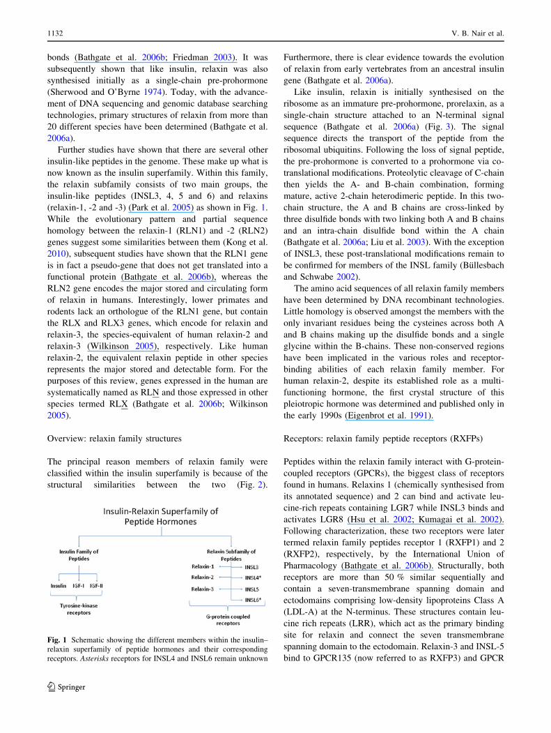
Like insulin, relaxin is initially synthesised on the
ribosome as an immature pre-prohormone, prorelaxin, as a
single-chain structure attached to an N-terminal signal
sequence (Bathgate et al. 2006a) (Fig. 3). The signal
sequence directs the transport of the peptide from the
ribosomal ubiquitins. Following the loss of signal peptide,
the pre-prohormone is converted to a prohormone via co-
translational modifications. Proteolytic cleavage of C-chain
then yields the A- and B-chain combination, forming
mature, active 2-chain heterodimeric peptide. In this two-
chain structure, the A and B chains are cross-linked by
three disulfide bonds with two linking both A and B chains
and an intra-chain disulfide bond within the A chain
(Bathgate et al. 2006a; Liu et al. 2003). With the exception
of INSL3, these post-translational modifications remain to
be confirmed for members of the INSL family (Bullesbach
and Schwabe 2002).
The amino acid sequences of all relaxin family members
have been determined by DNA recombinant technologies.
Little homology is observed amongst the members with the
only invariant residues being the cysteines across both A
and B chains making up the disulfide bonds and a single
glycine within the B-chains. These non-conserved regions
have been implicated in the various roles and receptor-
binding abilities of each relaxin family member. For
human relaxin-2, despite its established role as a multi-
functioning hormone, the first crystal structure of this
pleiotropic hormone was determined and published only in
the early 1990s (Eigenbrot et al. 1991).
Receptors: relaxin family peptide receptors (RXFPs)
Peptides within the relaxin family interact with G-protein-
coupled receptors (GPCRs), the biggest class of receptors
found in humans. Relaxins 1 (chemically synthesised from
its annotated sequence) and 2 can bind and activate leu-
cine-rich repeats containing LGR7 while INSL3 binds and
activates LGR8 (Hsu et al. 2002; Kumagai et al. 2002).
Following characterization, these two receptors were later
termed relaxin family peptides receptor 1 (RXFP1) and 2
(RXFP2), respectively, by the International Union of
Pharmacology (Bathgate et al. 2006b). Structurally, both
receptors are more than 50 % similar sequentially and
contain a seven-transmembrane spanning domain and
ectodomains comprising low-density lipoproteins Class A
(LDL-A) at the N-terminus. These structures contain leu-
cine rich repeats (LRR), which act as the primary binding
site for relaxin and connect the seven transmembrane
spanning domain to the ectodomain. Relaxin-3 and INSL-5
bind to GPCR135 (now referred to as RXFP3) and GPCR
Fig. 1 Schematic showing the different members within the insulin–
relaxin superfamily of peptide hormones and their corresponding
receptors. Asterisks receptors for INSL4 and INSL6 remain unknown
1132 V. B. Nair et al.
123
142 (RXFP4), respectively (Liu and Lovenberg 2008; Zhu
et al. 2008). These latter two receptors lack the ectodomain
region, LRR, and LDL-A modules. Activation of RXFP1
and RXFP2 results in intracellular cAMP production, via
receptor coupling to GaS and GaoS. In contrast, RXFP3 and
RXFP4 activation leads to inhibition of cAMP production
due to downward actions of inhibitory G proteins (Lu et al.
2006; Lin et al. 2004).
Due to the structural similarity of peptides within the
relaxin superfamily, cross activation of non-native recep-
tors does exist. For example, relaxin-3 has been shown to
bind and activate RXFP1 (Bathgate 2006; Hossain et al.
2011; Zhang et al. 2012) and RXFP4 as well as its native
receptor, RXFP3. Likewise, relaxin-2 binds to and acti-
vates RXFP2, the native INSL3 receptor (Sherwood 2004).
Relaxin peptide subfamily members: roles
and therapeutic applications
Insulin-like peptides (INSLs)
INSL3 has been implicated as having reproductive and
non-reproductive roles. In male rats, INSL3 has been
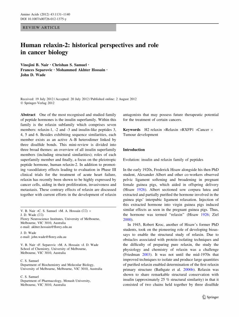
Fig. 2 Sequence homology of
members within the human
insulin–relaxin superfamily of
peptide hormones. Conserved
cysteines highlighted in bluedenote the intra-molecular
disulfide bond within the
A-chain; yellow and green inter-
disulfide between A- and
B-chains, respectively. Glycines
highlighted in mauve denote
conserved residues unique to the
B-chain (colour figure online)
Fig. 3 Representation of post-
translational modifications
undergone by single-chain pre-
prohormone to its active
heterodimeric stage. Signal
peptide in pre-prohormones
leads the transport of the peptide
from the ribosomal ubiquitins.
Following the loss of signal
peptide, the pre-prohormone is
converted to a prohormone via
co-translational modifications.
Proteolytic cleavage of C-chain
then yields the A- and B- chain
combination, forming mature,
active 2-chain heterodimeric
peptide
Human relaxin-2: historical perspectives and role in cancer biology 1133
123
shown to prevent male germ cells from entering the
apoptosis stage of the cell cycle (Kawamura et al. 2004).
Additionally, the previously termed Leydig cell insulin-like
peptides (Adham et al. 1993) are produced by foetal Ley-
dig cells and are involved in testicular descent via the
growth of gubernacular ligament (Ivell et al. 2005)
(Table 1). In females, INSL3 is also implicated in a similar
anti-apoptotic role, especially in ovarian follicles and the-
cal cells, during the follicle selection processes (Kawamura
et al. 2004; Ferlin et al. 2009). INSL3 antagonists are thus
expected to be potential male and female contraceptive
agents (Shabanpoor et al. 2010).
The foetal perichondrium and cytotrophoblasts have
been shown to express INSL4 and accounts for it also
being known as early placenta insulin-like peptide (Bellet
et al. 1997). Similar to the other members of this super-
family, INSL4 is expressed as a 15-kDa precursor, which
then undergoes cleavage to form its tertiary structure
(either two or three chain structures) (Bullesbach and
Schwabe 2001). INSL4 has been implicated in being
involved in trophoblastic development, including early cell
proliferation and development (Laurent et al. 1998).
Despite being exclusive to higher primates, the exact signal
transduction and physiological roles of INSL4 remain
unknown.
Examination of the expressed sequence tags databases
resulted in the discovery of a novel insulin-like sequence,
termed INSL5. The primary physiological function of
INSL5 remains unknown but recent studies imply it may
play a role in appetite control and gut motility (Belgi et al.
2011). The peptide has been produced by both chemical
synthesis and recombinant DNA expression (Hossain et al.
2008, Luo et al. 2010) which, in turn, is aiding the iden-
tification of its functional roles.
Another member of this ancient superfamily of func-
tionally diverse peptide hormones is INSL6. It is predom-
inantly expressed in the primary reproductive male organ,
the testis. However, published results do not concur
regarding the specific cell type expressing INSL6. For
example, studies by Hsu (1999) concluded INSL6 is
expressed in Leydig cells but studies by Lok et al. (2000)
established expression in spermatids but none in Leydig
cells. Despite this discordance, INSL6 has been shown to
be heavily involved in progression of spermatogenesis (Lu
et al. 2006). Further research is required to identify the
specific receptor for INSL6 and its exact physiological
chemistry.
Relaxin
Despite having two peptide-coding genes, relaxin gene 1
(RLN1) and RLN2, the major stored and circulatory form
of relaxin in humans is relaxin-2. Relaxin-2 is produced in
the prostate by males (Feng et al. 2007) and corpus lutea in
females (Shabanpoor et al. 2009). Since relaxin-1 is a
pseudogene, which does not translate into a functional
peptide in rodents, humans and other non-human species,
both relaxin-1 and -2 will be referred to as relaxin here
forth.
As mentioned previously, relaxin is the most compre-
hensively studied member of the relaxin subfamily.
Structure–activity studies of relaxin have revealed the
receptor ‘‘binding cassette’’ of this multi-functional peptide
hormone lies within its B-chain (Arg13-X-X-X-Arg17-X-X-
Ile20) and interacts with the binding pocket in native
receptor, RXFP1. Bullesbach et al. (2000) have shown the
importance of the binding cassette by replacing the argi-
nine at position 13 and 17 with citrulline, lysine and ala-
nine, rendering inactive the relaxin native receptor (no
interaction with RXFP1). Arg13 and Arg17 residues on the
B-chain of relaxin interact with a network of two aspartic
acid/glutamic acid pairs in the LRR within the ectodomain
of its native receptor, RXFP1. In addition, the Ile20 residue
on the relaxin B-chain interacts with tryptophan-iso-
leucine-leucine region of the LRR. Non-specific binding is
observed on deletion of any of the aforementioned three
residues on the ligand (Bullesbach and Schwabe 2000;
Hossain and Wade 2010).
The effect of relaxin, particularly during pregnancy, is
well established in rodents. Levels of relaxin change with
the different stages of pregnancy and these patterns are
dissimilar across species. In rodents, sows and dogs,
relaxin is untraceable early in the gestation but increases
Table 1 Summary of recognized functions of relaxin peptide sub-
family members, performing roles undertaken by specific peptide
hormones in humans unless otherwise specified
Peptide
Hormone
Functions
Relaxin
(relaxin-2)
Aids embryo implantation via uterine
vascularisation and differentiation of endometrial
cells sperm motility in male reproductive system
promotes collagen breakdown increased
vascularisation and renal functions in pregnant
females, lead to haemodynamic roles in acute
heart failure patients (Phase III clinical trials)
produced by cancer cells and acts on its receptor
(autocrine signalling) to promote cancer growth
and metastasis
Relaxin-3 Regulation of energy homeostasis and appetite
regulation
INSL3 Involved in testicular descent regulates germ cell
maturation
INSL4 Possibly involved in trophoblastic development
INSL5 Associated with feeding functions
INSL6 Involved in spermatogenesis progression
See text for specific references
1134 V. B. Nair et al.
123
and reaches a maximum before labour (Hwang et al. 1989;
Johnson et al. 1991). Conversely, maximum circulating
relaxin is observed in humans within the first trimester of
pregnancy. Plasma relaxin then reduces and somewhat
plateaus for the remaining period of pregnancy, almost in a
contrary pattern to rodents, sows and dogs (Burger and
Sherwood 1998; Eppel et al. 1999). As mentioned earlier,
the main source of circulating relaxin is from the corpus
luteum in females (Shabanpoor et al. 2009) and has been
shown to aid in embryo implantation via uterine vascu-
larisation and differentiation of endometrial cells (Eppel
et al.1999). Besides aiding in pelvic ligament and cervical
softening of birth canals, relaxin has been shown to be
involved in remodelling and development of mammary
glands and nipples of mice (O’Day et al. 1989).
Furthermore, relaxin has been shown to increase oocytes
fertility (Brener et al. 1984). Besides exerting its effect in
female reproduction, relaxin is also involved in maintain-
ing sperm motility in the male reproductive system (Weiss
1989). This conclusion was drawn when increased pene-
tration was observed in human cervical mucous for human
sperm incubated with porcine relaxin compared with buffer
mixture (Weiss 1989; Pupula et al. 1986). These exciting
findings point to the therapeutic potential of relaxin
towards assisting with infertility in humans.
Relaxin has also been shown to be involved in non-
reproductive functions. For example, relaxin plays a crucial
role in cardiovascular and renal systems (Conrad and
Novak 2004). Relaxin promotes collagen breakdown in
systemic tissues (Unemori and Amento 1990), rendering a
high possibility for an ability to reduce systemic fibrosis
(Unemori et al. 1996; Samuel 2005). Fibrosis occurs when
collagens, glycoproteins and other extracellular matrix
accumulate in organs. When relaxin is introduced to sys-
temic organs such as the heart and lungs, over-expression
of collagen has been reduced (Samuel et al. 2004). More-
over, relaxin has also been observed to increase vasodila-
tion and renal functions in pregnant females. This, along
with its cardioprotective actions (Dschietzig et al. 2006;
Samuel et al. 2006) has led to efforts to exploit its hae-
modynamic roles and consequently relaxin is currently in
Phase III clinical trials for the treatment of acute heart
failure. The clinical trials to date have demonstrated relaxin
provided relief of dyspnoea (symptom of breathlessness)
and reduction of heart failure symptoms (Teerlink et al.
2009).
Relaxin-3
Relaxin-3 acts through its native receptor, RXFP3, which is
found in the hypothalamic paraventricular nucleus of the
brain. The expression site of this most recently discovered
insulin superfamily neuropeptide was found to be in the
nucleus incertus within the hypothalamus. These regions of
the brain have been extensively associated with regulation
of energy homeostasis and appetite regulation (McGowan
et al. 2005). Additionally, the paraventricular nucleus plays
a reproductive role during reproduction by providing
feedback to the hypothalamic gonadotrophin-releasing
hormone neurons (Chan et al. 2011). Interestingly, relaxin-
3 knockout mice have shown altered sleep patterns during
normal active (night) periods with increased sleep episodes
compared with their wildtype counterparts (Ma and
Gundlach 2007). Although the exact physiological mech-
anisms of relaxin-3 in humans still remain vague, studies
from rodents have suggested that relaxin-3 may coordinate
sleep, hunger and food intake regulation, (Chan et al. 2011;
Ma and Gundlach 2007) which make the developments of
relaxin-3/RXFP3 agonists and antagonists desirable thera-
peutic candidates.
Relaxin and its potential role in cancer biology
While well known for its reproductive and antifibrotic
roles, most recently relaxin has been associated with cancer
biology. A number of putative roles, including the modu-
lation of tumor growth, neovascularization, metastasis and
oncogenic progression, have been correlated to relaxin
overexpression (Silvertown et al. 2003). The following
sections will focus on the effects of downstream intracel-
lular relaxin signalling and its physiological implications.
Intracellular pathways associated with cancer biology
known to be activated by relaxin
In addition to its vasodilatory effects, relaxin has been
shown to promote nitric oxide (NO) production in renal,
cardiac and hepatic systems. In MCF-7 breast cancer cell
lines, production of NO was reported to be increased via
heightened nitric oxide synthase (iNOS) production (Failli
et al. 2002; Bani et al. 1999b). Increased NO production is
implicated in oncogenic cell migration and growth (Bani
et al. 1995; Jadeski et al. 2003). As highlighted earlier,
relaxin stimulates increased vasodilation in a range of
systemic tissues, including the skeletal and cardiac mus-
cles. Relaxin-induced NO production could possibly
encourage blood flow and growth of new blood vessels
(angiogenesis) in the MCF-7 mammary cancer cell line.
NO encourages cell apoptosis by inhibiting DNA synthesis
and mitochondrial respiration, decreasing rate of cellular
growth and multiplication (Jadeski et al. 2000). However,
studies have shown increased expression of iNOS in an
adenocarcinomic breast cancer cell line, MCF-7, incubated
with porcine relaxin. These observations may correlate
with relaxin assisting cancer cells avoid apoptosis. This
Human relaxin-2: historical perspectives and role in cancer biology 1135
123
could lead to further invasiveness and metastasis potential,
particularly with malignant oncogenic cells (Bani et al.
1995; Jadeski et al. 2000). Further studies investigating the
relationship of increased NO production and its impact on
oncogenic acceleration may be useful in understanding the
role of relaxin in cancer.
The influence of relaxin in cell growth, invasion
and angiogenesis
Besides its inherent biochemical signalling pathway, other
physiological roles of relaxin may also encourage tumour
advancement, further aggravating the severity of cancer.
Cancer progression involves oncogenic cell replication,
development (tumerogenesis) and spread of the tumour
mass from one organ or tissue to another (metastasis). All
of these physiological actions involve angiogenesis and
tissue growth, remodelling and apoptosis (Failli et al.
2002). These oncogenic ‘‘hallmarks’’ are closely related to
one another despite being classified as three separate
physiological events—cell growth, cell invasion and
angiogenesis. These three events can be seen in two gen-
der-specific leaders in oncogenic mortality: prostate cancer
and breast cancer. The following sections address these
‘‘hallmarks’’ and their consequential effects.
Relaxin and prostate cancer
The unresponsive state of uncontrolled cell division and
differentiation is a characteristic feature of cancer cells. As
previously mentioned, further growth of tumours are aided
by vascularisation due to over-expression of NO. The
changes observed in matrix metalloproteases contribute to
changes in the underlying framework of connective tis-
sues—this increased angiogenesis and changes in connec-
tive tissue framework are classic giveaways of relaxin-
mediated effects (Hansell et al. 1991; Samuel et al. 2004;
Bathgate et al. 2006a). Early studies which focus on
causatives of neoplastic prostatic cells, particularly abnor-
mal cell increase, conclude that peptides may also be
involved in hyperplasia (Ivell et al. 1989; Sokol et al. 1989)
as well as steroid hormones (Montie and Pienta 1994).
The state of hyperplasia was indicated by angiogenesis
and remodelling of the connective tissue framework. Fur-
thermore, marked uncontrolled rate of cell differentiation is
another indication of the aforementioned neoplastic pros-
tatic cells observed in men and male dogs (Barrett-Connor
et al. 1990; Nomura et al. 1988). Both observations are
classic, trademark effects of relaxin (Bathgate et al. 2006a).
Furthermore, a study has shown a human prostate adeno-
carcinoma cell line, LNCaP, to express high levels of a
relaxin mRNA, FGC. These findings were observed via
reverse transcription PCR and Northern blot analysis. This
may be reflective in vivo as besides being a product of the
RLN2 gene, relaxin found in the seminal fluid is expressed
by the prostate gland (Gunnersen et al. 1995). The high
levels of mRNA transcripts suggest a link to prostate
cancer. Increased neoplastic prostate xenografts have been
shown to be due to lentiviral-mediated relaxin delivery into
PC-3 prostate cancer cell line (Silvertown et al. 2006), and
an R273H p53 mutation directly targets downstream
relaxin production in prostatic carcinoma cells (Vinall et al.
2006).
Relaxin and breast cancer
Similar to its effects on prostate cancer cell lines, relaxin is
associated with impairment in the development of mam-
mary cells, leading to neoplastic mammary tissues (Bani
and Bigazzi 1984; Binder et al. 2001). Elevated relaxin
transcripts in neoplastic mammary tissues compared with
non-neoplastic tissues were reported in 1994 (Sacchi et al.
1994). Increased circulating relaxin levels were also
observed in women diagnosed with breast cancer (Binder
et al. 2004). The canine relaxin precursor, prorelaxin 2, has
been shown to increase invasiveness of canine mammary
cells (Silvertown et al. 2003). In rodents, relaxin has been
shown to encourage replication and differentiation of
mammary cells, particularly glands responsible for milk
delivery post-gestation (Min and Sherwood 1996; Winn
et al. 1994). Similarly, relaxin in human mammary cells
carries out comparable roles alongside oestrogen and pro-
gesterone (Bani and Bigazzi 1984). The presence of relaxin
has been demonstrated in all malignant and benign mam-
mary cell samples in comparison with equivalent cells with
normal cell proliferation rate, correlating with elevated
circulating relaxin levels amongst metastatic breast cancer
patients (Binder et al. 2001). Studies which focus on in
vitro aetiology of mammary adenocarcinogenic mammary
cells show low amounts of relaxin encourage cell metas-
tasis over a short time (Sacchi et al. 1994; Bani et al.
1999a). An independent study determined higher relaxin
plasma concentration amongst metastatic breast cancer
patients, correlating with the previous observation (Binder
et al. 2004).
Despite prostate and breast cancers being the ‘‘focus’’ of
most studies, relaxin has been shown to be associated with
other types of cancers in vitro including gastrointestinal
tract, colorectum, thyroid and endometrial cancer cells
(Kamat et al. 2006; Hombach-Klonisch et al. 2006). Most
of these cancers have been shown to increase matrix
metalloprotease activity (Binder et al. 2002). This causes
downstream expression of vascular endothelial growth
factors which have been previously shown to increase
tumour vascularisation and angiogenesis (Liang et al.
2006).
1136 V. B. Nair et al.
123
Potential therapeutics towards relaxin-induced cancer
Relaxin expression interference in vitro successfully
reduced metastasis which led to increased prostate adeno-
carcinoma cells death (Feng et al. 2007). This particular
study was aimed at observing the hallmark characteristics
of prostate adenoma: invasion and metastasis rate of
prostate cancer cells. Suppression of relaxin levels or
reducing the autocrine/paracrine signalling to its native
receptor, RXFP1, via short interfering RNAs (siRNA)
significantly reduced prostate cancer cell growth and
metastasis. Moreover, prostate cancer cell apoptosis was
also increased with the suppression of relaxin. These
observations highlight the importance of relaxin signalling
and its role in cancer cells development (Feng et al. 2007).
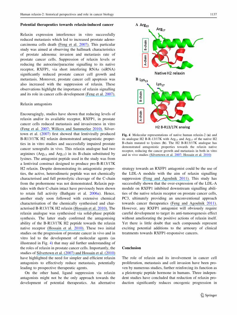
Relaxin antagonists
Encouragingly, studies have shown that reducing levels of
relaxin and/or its available receptor, RXFP1, in prostate
cancer cells reduced metastasis and invasiveness in vitro
(Feng et al. 2007; Willcox and Summerlee 2010). Silver-
town et al. (2007) first showed that lentivirally produced
B-R13/17K H2 relaxin demonstrated antagonistic proper-
ties in in vitro studies and successfully impaired prostate
cancer xenografts in vivo. This relaxin analogue had two
arginines (Arg13 and Arg17) in its B-chain substituted by
lysines. The antagonist peptide used in the study was from
a lentiviral construct designed to produce pro-B-R13/17K
H2 relaxin. Despite demonstrating its antagonistic proper-
ties, the active, heterodimeric peptide was not chemically
characterised and full proteolytic cleavage of the C-chain
from the prohormone was not demonstrated. Relaxin pep-
tides with their C-chain intact have previously been shown
to retain full activity (Bathgate et al. 2006a). Hence,
another study soon followed with extensive chemical
characterisation of the chemically synthesised and char-
acterised B-R13/17K H2 relaxin (Hossain et al. 2010). The
relaxin analogue was synthesised via solid-phase peptide
synthesis. The latter study confirmed the antagonising
ability of the B-R13/17K H2 peptide towards the relaxin
native receptor (Hossain et al. 2010). These two initial
studies on the progression of prostate cancer in vivo and in
vitro led to the development of molecular agents (as
illustrated in Fig. 4) that may aid further understanding of
the roles of relaxin in prostate cancer cells. Importantly, the
studies of Silvertown et al. (2007) and Hossain et al. (2010)
have highlighted the need for simpler and efficient relaxin
antagonists to effectively reduce metastasis, potentially
leading to prospective therapeutic agents.
On the other hand, ligand suppression via relaxin
antagonists might not be the only approach towards the
development of potential therapeutics. An alternative
strategy towards an RXFP1 antagonist could be the use of
the LDL-A module with the aim of relaxin signalling
suppression (Feng and Agoulnik 2011). This study has
successfully shown that the over-expression of the LDL-A
module on RXFP1 inhibited downstream signalling abili-
ties of the native relaxin receptor on prostate cancer cells,
PC3, ultimately providing an unconventional approach
towards cancer therapeutics (Feng and Agoulnik 2011).
However, any RXFP1 antagonist will obviously require
careful development to target its anti-tumorogenesis effect
without ameliorating the positive actions of relaxin itself.
Yet there is little doubt that such compounds represent
exciting potential additions to the armoury of clinical
treatments towards RXFP1-responsive cancers.
Conclusion
The role of relaxin and its involvement in cancer cell
proliferation, metastasis and cell invasion have been pro-
ven by numerous studies, further reinforcing its function as
a pleiotropic peptide hormone in humans. Three indepen-
dent studies have concluded that reduction of relaxin pro-
duction significantly reduces oncogenic progression in
Fig. 4 Molecular representations of native human relaxin-2 (a) and
its analogue H2 B-R-13/17K with Arg13 and Arg17 of the native H2
B-chain mutated to lysines (b). The H2 B-R13/17K analogue has
demonstrated antagonistic properties towards the relaxin native
receptor, reducing the cancer growth and metastasis in both in vitro
and in vivo studies (Silvertown et al. 2007; Hossain et al. 2010)
Human relaxin-2: historical perspectives and role in cancer biology 1137
123
vitro and in vivo (Feng et al. 2007), particularly with the
aid of a relaxin antagonist (Hossain et al. 2010; Silvertown
et al. 2007). These exciting findings illustrate the need for
further study of relaxin and its role in cancer biology. In
turn, this may improve the aforementioned anti-relaxin
agent and lead to greater investigation of tumour devel-
opment and cancers related to relaxin.
Acknowledgments V.B.N. is a recipient of a Melbourne Research
Scholarship by the University of Melbourne; C.S.S. is supported by a
National Heart Foundation of Australia and National Health and
Medical Research Council (NHMRC) of Australia RD Wright Fel-
lowship; M.A.H was the recipient of Reid Trust and Florey Foun-
dation Trust Fellowships and J.D.W is an NHMRC Principal
Research Fellow. Some of the authors’ research reported in this
review was supported by NHMRC Project Grants 508995 and
1023078. Research at the FNI was supported by the Victorian Gov-
ernment’s Operational Infrastructure Support Program.
Conflict of interest The authors declare that they have no conflict
of interest.
References
Adham IM, Burkhardt E, Benahmed M (1993) Cloning of a cDNA for
a novel insulin-like peptide of the testicular Leydig cells. J Biol
Chem 268:26668–26672
Bani G, Bigazzi M (1984) Morphological changes induced in mouse
mammary gland by porcine and human relaxin. Acta Anat
119:149–154
Bani D, Masini E, Bello MG, Bigazzi M, Sacchi TB (1995) Relaxin
activates the L-arginine-nitric oxide pathway in human breast
cancer cells. Cancer Res 55:5272–5275
Bani D, Flagiello D, Poupon MF, Nistri S, Poirson-Birhcat F, Bigazzi
M, Bani G, Sacchi TB (1999a) Relaxin promotes differentiation
of human breast cancer cells MCF-7 transplanted into nude mice.
Virchows Arch 435:509–519. doi:10.1007/s004280050435
Bani D, Baccari MC, Nistri S, Calamai F, Bigazzi M, Sacchi TB
(1999b) Relaxin up-regulates the nitric oxide biosynthetic
pathway in the mouse uterus: involvement in the inhibition of
myometrial contractility. Endocrinology 140:4434–4441. doi:
10.1210/en.140.10.4434
Barrett-Connor E, Garland C, McPhillips JB, Khaw KT, Wingard DL
(1990) A prospective, population-based study of androstenedi-
one, estrogens, and prostatic cancer. Cancer Res 50:169–173
Bathgate RAD (2006) Relaxin-3: improved synthesis strategy and
demonstration of its high-affinity interaction with the relaxin
receptor LGR7 both in vitro and in vivo. Biochemistry
45:1043–1053. doi:10.1021/bi052233e
Bathgate R, Hsueh A, Sherwood OD (2006a) Physiology and
molecular biology of the relaxin peptide family. In: Neill JD
(ed) Knobil and Neill’s physiology of reproduction, 3rd edn.
Academic Press, San Diego, pp 679–768
Bathgate RAD, Ivell R, Sanborn BM, Sherwood OD, Summers RJ
(2006b) International Union of Pharmacology LVII: recommen-
dations for the nomenclature of receptors for relaxin family
peptides. Pharmacol Rev 58:7–31. doi:10.1124/pr.58.1.9
Belgi A, Hossain MA, Shabanpoor F, Zhang S, Bathgate RAD,
Tregear GW, Wade JD (2011) Structure and function relation-
ship of murine insulin-like peptide 5 (INSL5): free C-terminus is
essential for RXFP4 receptor binding and activation. Biochem-
istry 50:8352–8361
Bellet D, Lavaissiere L, Mock P, Laurent A, Sabourin JC, Bedossa P,
Le Bouteiller P, Frydman R, Troalen F, Bidart J (1997)
Identification of pro-EPIL and EPIL peptides translated from
insulin-like 4 (INSL4) mRNA in human placenta. J Clin
Endocrinol Metab 82:3169–3172
Binder C, Binder L, Gurlit L, Einspanier A (2001) High serum
concentrations of relaxin correlate with dissemination of breast
cancer. In: Tregear GW, Ivell R, Bathgate RA, Wade JD (eds)
Relaxin 2000. Kluwer Academic Publishers, Netherlands,
pp 423–432
Binder C, Hagemann T, Husen B, Schulz M, Einspanier A (2002)
Relaxin enhances in vitro invasiveness of breast cancer cell lines
by up-regulation of matrix metalloproteases. MHR Basic Sci
Reprod Med 8:789–796. doi:10.1093/molehr/8.9.789
Binder C, Simon A, Binder L, Hagemann T, Schulz M, Emons G,
Trumper L, Einspanier A (2004) Elevated concentrations of
serum relaxin are associated with metastatic disease in breast
cancer patients. Breast Cancer Res Treat 87:157–166. doi:
10.1023/b:brea.0000041622.30169.16
Brener SHL, Schoenfeld C, Amelar RD, Dubin L, Weiss G (1984)
Stimulation of human sperm cervical mucus penetration in vitro
by relaxin. Fertil Steril 42:92–96
Bullesbach EE, Schwabe C (2000) The relaxin receptor-binding site
geometry suggests a novel gripping mode of interaction. J Biol
Chem 275:35276–35280. doi:10.1074/jbc.M005728200
Bullesbach EE, Schwabe C (2001) Synthesis and conformational
analysis of the insulin-like 4 gene product. J Pept Res 57:77–83
Bullesbach EE, Schwabe C (2002) The primary structure and the
disulfide links of the bovine relaxin-like factor (RLF). Biochem-
istry 41:274–281. doi:10.1021/bi0117302
Burger LL, Sherwood OD (1998) Relaxin increases the accumulation
of new epithelial and stromal cells in the rat cervix during the
second half of pregnancy. Endocrinology 139:3984–3995. doi:
10.1210/en.139.9.3984
Chan LJ, Hossain MA, Samuel CS, Separovic F, Wade JD (2011) The
relaxin peptide family—structure, function and clinical applica-
tions. Protein Pept Lett 18:220–229
Conrad KP, Novak J (2004) Emerging role of relaxin in renal and
cardiovascular function. Am J Physiol Regul Integr Comp
Physiol 287:250–261. doi:10.1152/ajpregu.00672.2003
Dschietzig T, Bartsch C, Baumann G, Stangl K (2006) Relaxin—a
pleiotropic hormone and its emerging role for experimental and
clinical therapeutics. Pharm Ther 112:38–56. doi:10.1016/j.
pharmthera.2006.03.004
Eigenbrot C, Randal M, Quan C, Burnier J, O’Connell L, Rinderkn-
echt E, Kossiakoff AA (1991) X-ray structure of human relaxin
at 1.5 A. Comparison to insulin and implications for receptor
binding determinants. J Mol Biol 221(1):15–21
Eppel W, Kucera E, Bielglmyer C (1999) Relationship of serum
levels of endogenous relaxin to cervical size in the second
trimester and to cervical ripening at term. Br J Obstet Gynaecol
106:917–923
Failli P, Nistri S, Quattrone S, Mazzetti L, Bigazzi M, Sacchi TB,
Bani D (2002) Relaxin up-regulates inducible nitric oxide
synthase expression and nitric oxide generation in rat coronary
endothelial cells. FASEB J 16:252–254
Feng S, Agoulnik A (2011) Expression of LDL-A module of relaxin
receptor in prostate cancer cells inhibits tumorigenesis. Int J
Oncol 39:1559–1565
Feng S, Agoulnik IU, Bogatcheva NV, Kamat AA, Kwabi-Addo B, Li
R, Ayala G, Ittmann MM, Agoulnik AI (2007) Relaxin promotes
prostate cancer progression. Clinical Cancer Res 13:1695–1702.
doi:10.1158/1078-0432.ccr-06-2492
1138 V. B. Nair et al.
123

Ferlin A, Pepe A, Gianesello L, Garolla A, Feng S, Facciolli A,
Morello R, Agoulnik AI, Foresta C (2009) New roles for INSL3
in adults. Ann N Y Acad Sci 1160:215–218. doi:10.1111/
j.1749-6632.2008.03787.x
Friedman A (2003) Remembrance: the contributions of Frederick
Hisaw. J Clin Endocrinol Metab 88:524–527. doi:
10.1210/jc.2002-021457
Gunnersen JM, Roche PJ, Tregear GW, Crawford RJ (1995)
Characterization of human relaxin gene regulation in the
relaxin-expressing human prostate adenocarcinoma cell line
LNCaP.FGC. J Mol Endocrinol 15:153–166. doi:10.1677/jme.
0.0150153
Hansell DJ, Bryant-Greenwood GD, Greenwood FC (1991) Expres-
sion of the human relaxin H1 gene in the decidua, trophoblast,
and prostate. J Clin Endocrinol Metab 72:899–904. doi:
10.1210/jcem-72-4-899
Hisaw F (1926) Experimental relaxation of the pubic ligament of the
guinea pig. Proc Soc Exper Biol Med 23:661–663
Hombach-Klonisch S, Bialek J, Trojanowicz B, Weber E, Holzhausen
HJ, Silvertown JD, Summerlee AJ, Dralle H, Hoang-Vu C,
Klonisch T (2006) Relaxin enhances the oncogenic potential of
human thyroid carcinoma cells. Am J Pathol 169:617–632. doi:
10.2353/ajpath.2006.050876
Hossain MA, Wade JD (2010) The roles of the A- and B-chains of
human relaxin-2 and -3 on their biological activity. Curr Protein
Pept Sci 11:719–724
Hossain MA, Bathgate RA, Kong C, Shabanpoor F, Zhang S,
Haugaard-Jonsson LM, Rosengren KJ, Tregear GW, Wade JD
(2008) Synthesis, conformation and receptor binding activity of
human insulin-like peptide 5 (INSL5). ChemBioChem
9:1816–1822
Hossain MA, Samuel CS, Binder C, Hewitson TD, Tregear GW,
Wade JD, Bathgate RAD (2010) The chemically synthesized
human relaxin-2 analog, B-R13/17K H2, is an RXFP1 antago-
nist. Amino Acids 39:409–416. doi:10.1007/s00726-009-0454-1
Hossain MA, Chow BSM, Zhao C, Xu Q, Du XJ, Wade JD, Samuel
CS (2011) H3 relaxin demonstrates antifibrotic properties via the
RXFP1 receptor. Biochemistry 50:1368–1375. doi:10.1021/
bi1013968
Hsu SY (1999) Cloning of two novel mammalian paralogs of relaxin/
insulin family proteins and their expression in testis and kidney.
Mol Endocrinol 13:2163–2167. doi:10.1210/me.13.12.2163
Hsu SY, Nakabayashi K, Nishi S, Kumagai J, Kudo M, Sherwood
OD, Hsueh AJW (2002) Activation of orphan receptors by the
hormone relaxin. Science 295:671–674
Hwang JJ, Shanks RD, Sherwood OD (1989) Monoclonal antibodies
specific for rat relaxin. IV. Passive immunization with mono-
clonal antibodies during the antepartum period reduces cervical
growth and extensibility, disrupts birth, and reduces pup survival
in intact rats. Endocrinology 125:260–266. doi:10.1210/endo-
125-1-260
Ivell R, Hunt N, Khan-Dawood F, Dawood MY (1989) Expression of
the human relaxin gene in the corpus luteum of the menstrual
cycle and in the prostate. Mol Cellular Endocrinol 66:251–255.
doi:10.1016/0303-7207(89)90037-3
Ivell R, Hartung S, Anand-Ivell R (2005) Insulin-like factor 3: where
are we now? Ann NY Acad Sci 1041:486–496. doi:10.1196/
annals.1282.073
Jadeski LC, Hum KO, Chakraborty C, Lala PK (2000) Nitric oxide
promotes murine mammary tumour growth and metastasis by
stimulating tumour cell migration, invasiveness and angiogen-
esis. Int J Cancer 86:30–39. doi:10.1002/(sici)1097-0215
(20000401)86:1\30::aid-ijc5[3.0.co;2-i
Jadeski LC, Chakraborty C, Lala PK (2003) Nitric oxide-mediated
promotion of mammary tumour cell migration requires sequen-
tial activation of nitric oxide synthase, guanylate cyclase and
mitogen-activated protein kinase. Int J Cancer 106:496. doi:
10.1002/ijc.11268
Johnson MR, Abdalla H, Allman AC, Wren ME, Kirkland A,
Lighman SL (1991) Relaxin levels in ovum donation pregnan-
cies. Fertil Steril 56:59–61
Kamat AA, Feng S, Agoulnik IU, Kheradmand F, Bogatcheva NV,
Coffey D, Sood AK, Agoulnik AI (2006) The role of relaxin in
endometrial cancer. Cancer Biol Therap 5:71–77. doi:
10.4161/cbt.5.1.2289
Kawamura K, Kumagai J, Sudo S, Chun SY, Pisarska M, Morita H,
Toppari J, Fu P, Wade JD, Bathgate RAD, Hsueh AJ (2004)
Paracrine regulation of mammalian oocyte maturation and male
germ cell survival. Proc Natl Acad Sci USA 101:7323–7328.
doi:10.1073/pnas.0307061101
Kong RCK, Shiling PJ, Lobb DK, Gooley PR, Bathgate RAD (2010)
Membrane receptors: structure and function of the relaxin family
peptide receptors. Mol Cell Endocrinol 320:1–15. doi:10.1016/
j.mce.2010.02.003
Kumagai J, Hsu SY, Matsumi H, Roh JS, Fu P, Wade JD, Bathgate
RAD, Hsueh AJ (2002) INSL3/Leydig insulin-like peptide
activates the LGR8 receptor important in testis descent. J Biol
Chem 277:31283–31286. doi:10.1074/jbc.C200398200
Laurent A, Rouillac C, Delezoide AL, Giovangrandi Y, Vekemans M,
Bellet D, Abitbol M, Vidaud M (1998) Insulin-like 4 (INSL4)
gene expression in human embryonic and trophoblastic tissues.
Mol Reprod Dev 51:123–129. doi:10.1002/(sici)1098-2795
(199810)51:2\123:aid-mrd1[3.0.co2-s
Liang WC, Wu X, Peale FV, Lee CV, Meng YG, Gutierrez J, Fu L,
Malik AK, Gerber HP, Ferrara N, Fuh G (2006) Cross-species
vascular endothelial growth factor (VEGF)-blocking antibodies
completely inhibit the growth of human tumor xenografts and
measure the contribution of stromal VEGF. J Biol Chem 281:
951–961. doi:10.1074/jbc.M508199200
Lin F, Otvos L Jr, Kumagai J, Tregear GW, Bathgate RA, Wade JD
(2004) Synthetic human insulin 4 does not activate the G-pro-
tein-coupled receptors LGR7 or LGR8. J Pept Sci 10:257–264.
doi:10.1002/psc.521
Liu C, Lovenberg T (2008) Relaxin-3, INSL5, and their receptors.
Orphan G protein-coupled receptors and novel neuropeptides.
Results Probl Cell Differ 46:213–237. doi:10.1007/400_2007_055
Liu C, Eriste E, Sutton S (2003) Identification of relaxin-3/INSL7 as
an endogenous ligand for the orphan G-protein-coupled receptor
GPCR135. J Biol Chem 278:50754–50764. doi:10.1074/jbc.
M308995200
Lok S, Johnston DS, Conklin D, Lofton-Day CE, Adams RL,
Jelmberg AC, Whitmore TE, Schrader S, Grisworld MD, Jaspers
SR (2000) Identification of INSL6, a new member of the insulin
family that is expressed in the testis of the human and rat. Biol
Reprod 62:1593–1599. doi:10.1095/biolreprod62.6.1593
Lu CX, Walker WH, Sun J, Weisz OA, Gibbs RB, Witchel SF,
Sperling MA, Menon RK (2006) Insulin-like peptide 6: charac-
terization of secretory status and posttranslational modifications.
Endocrinology 147:5611–5623. doi:10.1210/en.2006-0503
Luo X, Bathgate RAD, Zhang W-J, Liu Y-L, Shao X–X, Wade JD,
Guo Z-Y (2010) Design and recombinant expression of insulin-
like peptide 5 (INSL5) precursors and the preparation of mature
human INSL5. Amino Acids 39:1343–1352
Ma S, Gundlach AL (2007) Relaxin-family peptide and receptor
systems in brain: insights from recent anatomical and functional
studies. Adv Exp Med Biol 612:119–137. doi:10.1007/978-
0-387-74672-2_9
McGowan BM, Stanley SA, Smith KL, White NE, Connolly MM,
Thompson EL, Gardiner JV, Murphy KG, Ghatei MA, Bloom
SR (2005) Central relaxin-3 administration causes hyperphagia
in male Wistar rats. Endocrinology 146:3295–3300. doi:10.1210/
en.2004-1532
Human relaxin-2: historical perspectives and role in cancer biology 1139
123
Min G, Sherwood OD (1996) Identification of specific relaxin-binding
cells in the cervix, mammary glands, nipples, small intestine, and
skin of pregnant pigs. Biol Reprod 55:1243–1252. doi:
10.1095/biolreprod55.6.1243
Montie J, Pienta K (1994) Review of the role of androgenic hormones
in the epidemiology of benign prostatic hyperplasia and prostate
cancer. Urology 43:892–899
Nomura A, Heilbrun LK, Stemmermann GN, Judd HL (1988)
Prediagnostic serum hormones and the risk of prostate cancer.
Cancer Res 48:3515–3517
O’Day MB, Winn RJ, Easter RA, Dzuik PJ, Sherwood OD (1989)
Hormonal control of the cervix in pregnant gilts. II. Relaxin
promotes changes in the physical properties of the cervix in
ovariectomized hormone-treated pregnant gilts. Endocrinology
125:3004–3010. doi:10.1210/endo-125-6-3004
Park J, Chang C, Hsu S (2005) New Insights into biological roles of
relaxin and relaxin-related peptides. Rev Endocr Metab Disor-
ders 6:291–296. doi:10.1007/s11154-005-6187-x
Pupula M, Quinn P, Maclennan A (1986) The effect of porcine
relaxin on the fertilisation of mouse oocytes in vitro. Clin Reprod
Fertil 4:383–387
Sacchi TB, Bani D, Brandi ML, Falchetti A, Bigazzi M (1994)
Relaxin influences growth, differentiation and cell–cell adhesion
of human breast-cancer cells in culture. Int J Cancer 57:129–134.
doi:10.1002/ijc.2910570123
Samuel CS (2005) Relaxin: antifibrotic properties and effects in
models of disease. Clin Med Res 3:241–249
Samuel CS, Unemori EN, Mookerjee I, Bathgate RAD, Layfield SL,
Mak J, Tregear GW, Du X-J (2004) Relaxin modulates cardiac
fibroblast proliferation, differentiation, and collagen production
and reverses cardiac fibrosis in vivo. Endocrinology
145:4125–4133. doi:10.1210/en.2004-0209
Samuel CS, Du XJ, Bathgate RAD, Summers RJ (2006) ‘Relaxin’ the
stiffened heart and arteries: the therapeutic potential for relaxin
in the treatment of cardiovascular disease. Pharm Ther
112:529–552. doi:10.1016/j.pharmthera.2005.05.012
Shabanpoor F, Separovic F, Wade JD (2009) The human insulin
superfamily of polypeptide hormones. Vitam Horm 80:1–31.
doi:10.1016/s0083-6729(08)00601-8
Shabanpoor F, Hughes RA, Zhang S, Bathgate RAD, Layfield S,
Hossain MA, Tregear GW, Separovic F, Wade JD (2010) Effect of
helix-promoting strategies on the LGR8 receptor activity of novel
analogues of the B-chain of human insulin-like peptide 3, INSL3.
Amino Acids 38:121–131. doi:10.1007/s00726-008-0219-2
Sherwood OD (2004) Relaxin’s physiological roles and other diverse
actions. Endocr Rev 25:205–234. doi:10.1210/er.2003-0013
Sherwood CD, O’Byrne EM (1974) Purification and characterization
of porcine relaxin. Arch Biochem Biophys 160:185–196. doi:
10.1016/s0003-9861(74)80025-1
Silvertown JD, Geddes BJ, Summerlee AJ (2003) Adenovirus-
mediated expression of human prorelaxin promotes the invasive
potential of canine mammary cancer cells. Endocrinology
144:3683–3691. doi:10.1210/en.2003-0248
Silvertown JD, Ng J, Sato T, Summerlee AJ, Medin JA (2006) H2
relaxin overexpression increases in vivo prostate xenograft
tumor growth and angiogenesis. Int J Cancer 118:62–73. doi:
10.1002/ijc.21288
Silvertown JD, Symes JC, Neschadim A, Nonaka T, Kao JCH,
Summerlee AJ, Medin JA (2007) Analog of H2 relaxin exhibits
antagonistic properties and impairs prostate tumor growth.
FASEB J 21:754–765. doi:10.1096/fj.06-6847com
Sokol RZ, Wang XS, Lechago J, Johnston PD, Swerdloff RS
(1989) Immunohistochemical localization of relaxin in human
prostate. J Histochem Cytochem 37:1253–1255. doi:10.1177/
37.8.2666509
Teerlink JR, Metra M, Felker MG, Ponikowski P, Voors AA,
Weatherley BD, Marmor A, Katz A, Grzybowski J, Unemori E,
Teichman SL, Cotter G (2009) Relaxin for the treatment of
patients with acute heart failure (Pre-RELAX-AHF): a multi-
centre, randomised, placebo-controlled, parallel-group, dose-
finding phase IIb study. Lancet 373:1429–1439. doi:10.1016/
s0140-6736(09)60622-x
Unemori EN, Amento EP (1990) Relaxin modulates synthesis and
secretion of procollagenase and collagen by human dermal
fibroblasts. J Biol Chem 265:10681–10685
Unemori EN, Pickford LB, Salles AL, Piercy CE, Grove BH, Erikson
ME, Amento EP (1996) Relaxin induces an extracellular matrix-
degrading phenotype in human lung fibroblasts in vitro and
inhibits lung fibrosis in a murine model in vivo. J Clin Investig
98:2739–2745. doi:10.1172/jci119099
Vinall RL, Tepper CG, Shi XB, Xue LA, Gandour-Edwards R, de
Vere White RW (2006) The R273H p53 mutation can facilitate
the androgen-independent growth of LNCaP by a mechanism
that involves H2 relaxin and its cognate receptor LGR7.
Oncogene 25:2082–2093
Weiss G (1989) Relaxin in the male. Biol Reprod 40:197. doi:
10.1095/biolreprod40.2.197
Wilkinson T (2005) Coevolution of the relaxin-like peptides and their
receptors. Ann N Y Acad Sci 1041:534–539. doi:10.1196/annals.
1282.080
Willcox J, Summerlee A (2010) RLN2 and its role in cancer. In:
Huret J (ed) Atlas of genetics and cytogenetics in oncology and
haematology. INIST-CNRS, France, pp 1768–3262
Winn RJ, Baker MD, Merle CA, Sherwood OD (1994) Individual and
combined effects of relaxin, estrogen, and progesterone in
ovariectomized gilts. II. Effects on mammary development.
Endocrinology 135:1250–1255. doi:10.1210/en.135.3.1250
Zhang W-J, Luo X, Liu Y-L, Shao X-X, Wade JD, Bathgate RAD,
Guo Z-Y (2012) Site-specific DOTA/europium-labeling of the
recombinant human relaxin-3 for receptor-ligand interaction.
Amino Acids 43:983–992
Zhu J, Kuei C, Sutton S, Kamme F, Yu J, Bonaventure P, Atack J,
Lovenberg TW, Liu C (2008) Identification of the domains in
RXFP4 (GPCR142) responsible for the high affinity binding and
agonistic activity of INSL5 at RXFP4 compared to RXFP3
(GPCR135). Eur J Pharmacol 590:43–52. doi:10.1016/j.ejphar.
2008.05.025
Ziel HK (2000) Historical note Frederick L. Hisaw (1891–1972) and
the discovery of relaxin. Endocrinologist 10:215218. doi:
10.1097/00019616-200010040-00001
1140 V. B. Nair et al.
123