Senior Biology - cbbiology
230
Senior Biology Higher Level Leaving Certificate Second Edition 2012 Declan Cathcart [email protected]
-
Upload
khangminh22 -
Category
Documents
-
view
1 -
download
0
Transcript of Senior Biology - cbbiology
Senior Biology Higher Level Leaving Certificate Second Edition 2012
Declan Cathcart
2
Table of Contents
The Scientific Method ...................................................................................... 4
The Characteristics of Life ............................................................................... 6
The Molecules of Life ...................................................................................... 8
Ecology - General Principles ........................................................................... 12
Woodland Field Ecology ................................................................................. 21
Human Impact on the Environment ................................................................ 26
Cell Structure ................................................................................................ 29
Movement through Membranes ...................................................................... 34
Cell Continuity .............................................................................................. 36
Enzymes and Metabolism .............................................................................. 41
Photosynthesis .............................................................................................. 45
Respiration ................................................................................................... 51
DNA and RNA ............................................................................................... 58
Protein Synthesis .......................................................................................... 62
Genetic Engineering ...................................................................................... 66
DNA Profiling ................................................................................................ 70
Genetics ....................................................................................................... 72
Genetic Variation .......................................................................................... 87
Kingdom Protista........................................................................................... 90
Kingdom Fungi .............................................................................................. 91
Kingdom Monera ........................................................................................... 95
Blood ......................................................................................................... 101
The Heart and Circulation ............................................................................ 104
Lymphatic System ....................................................................................... 112
The Breathing System in the Human ............................................................ 114
The Digestive System .................................................................................. 117
The Liver .................................................................................................... 124
The Kidneys ................................................................................................ 125
The Skin ..................................................................................................... 129
3
The Nervous System ................................................................................... 131
The Senses ................................................................................................. 137
The Musculoskeletal System ........................................................................ 139
The Endocrine System ................................................................................. 146
Human Defence System .............................................................................. 150
Viruses ....................................................................................................... 155
Sexual Reproduction in Humans ................................................................... 157
Plant Anatomy ............................................................................................ 170
Plant Transport ........................................................................................... 175
Gas Exchange in Plants ............................................................................... 178
Responses in Flowering Plants ..................................................................... 180
Vegetative Propagation – Plant Asexual Reproduction ................................... 183
Sexual Reproduction in Flowering Plants ....................................................... 185
Seeds, Dispersal, Dormancy and Germination ............................................... 190
Mandatory Practicals (MPA’s) ....................................................................... 194
Essential Biology Definitions ........................................................................ 224
4
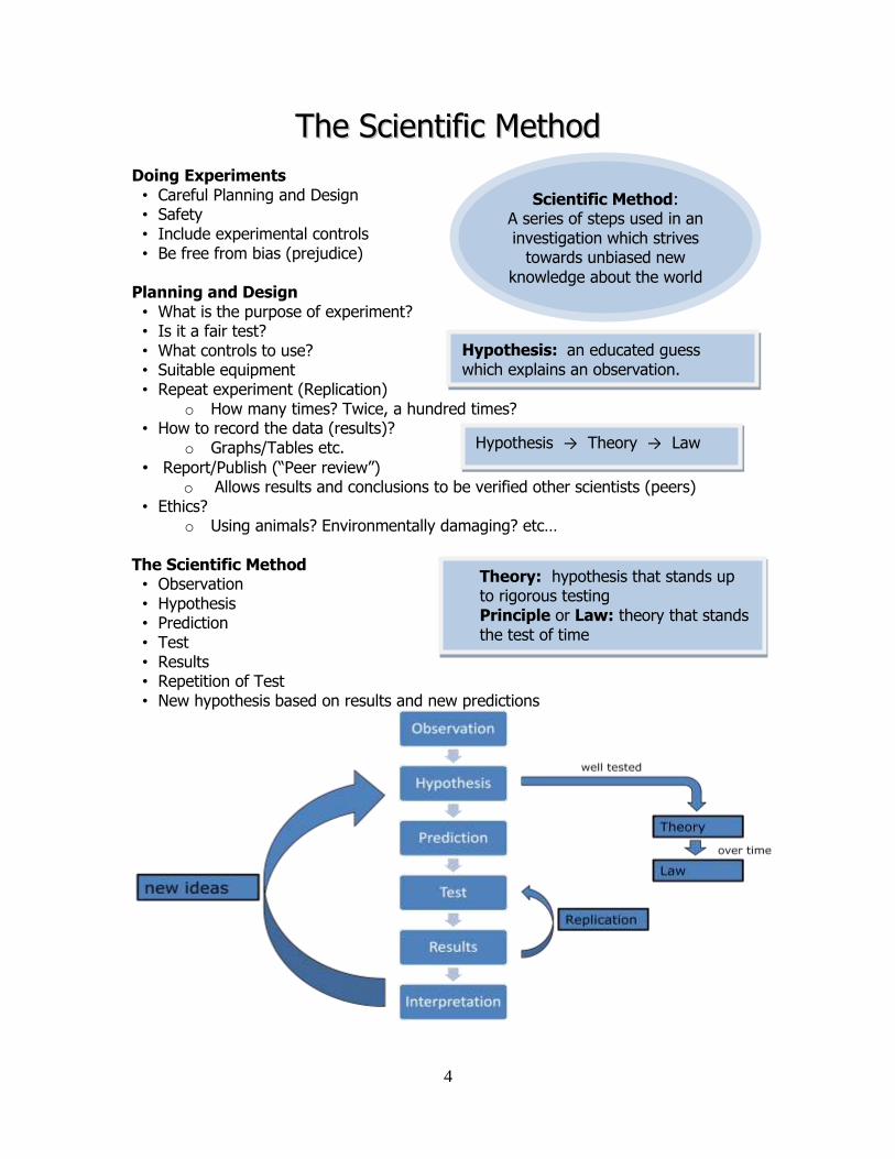
Scientific Method: A series of steps used in an investigation which strives
towards unbiased new
knowledge about the world
TThhee SScciieennttiiffiicc MMeetthhoodd Doing Experiments • Careful Planning and Design • Safety • Include experimental controls • Be free from bias (prejudice)
Planning and Design • What is the purpose of experiment? • Is it a fair test? • What controls to use? • Suitable equipment • Repeat experiment (Replication)
o How many times? Twice, a hundred times? • How to record the data (results)?
o Graphs/Tables etc.
• Report/Publish (“Peer review”) o Allows results and conclusions to be verified other scientists (peers)
• Ethics? o Using animals? Environmentally damaging? etc…
The Scientific Method • Observation • Hypothesis • Prediction • Test • Results • Repetition of Test • New hypothesis based on results and new predictions
Hypothesis → Theory → Law
Theory: hypothesis that stands up to rigorous testing Principle or Law: theory that stands the test of time
Hypothesis: an educated guess which explains an observation.
5
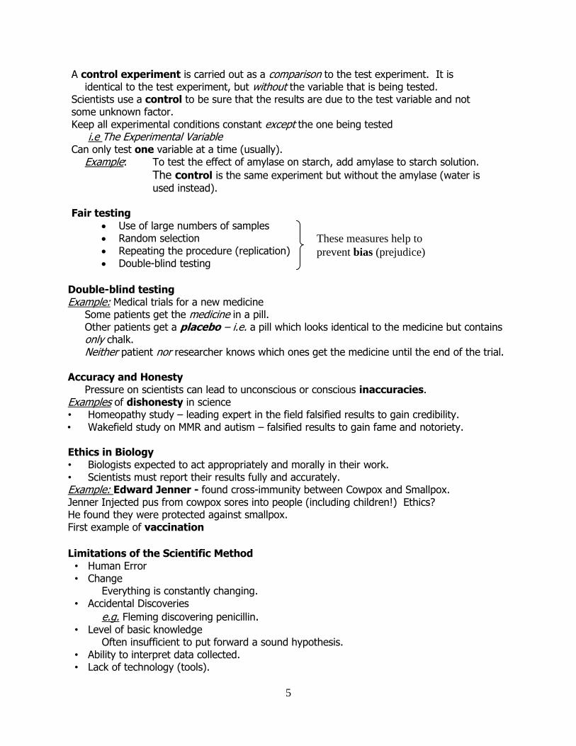
A control experiment is carried out as a comparison to the test experiment. It is identical to the test experiment, but without the variable that is being tested.
Scientists use a control to be sure that the results are due to the test variable and not some unknown factor. Keep all experimental conditions constant except the one being tested
i.e The Experimental Variable Can only test one variable at a time (usually).
Example: To test the effect of amylase on starch, add amylase to starch solution.
The control is the same experiment but without the amylase (water is
used instead). Fair testing
Use of large numbers of samples Random selection Repeating the procedure (replication) Double-blind testing
Double-blind testing Example: Medical trials for a new medicine
Some patients get the medicine in a pill. Other patients get a placebo – i.e. a pill which looks identical to the medicine but contains only chalk. Neither patient nor researcher knows which ones get the medicine until the end of the trial.
Accuracy and Honesty
Pressure on scientists can lead to unconscious or conscious inaccuracies. Examples of dishonesty in science • Homeopathy study – leading expert in the field falsified results to gain credibility. • Wakefield study on MMR and autism – falsified results to gain fame and notoriety. Ethics in Biology • Biologists expected to act appropriately and morally in their work. • Scientists must report their results fully and accurately. Example: Edward Jenner - found cross-immunity between Cowpox and Smallpox. Jenner Injected pus from cowpox sores into people (including children!) Ethics? He found they were protected against smallpox. First example of vaccination
Limitations of the Scientific Method • Human Error • Change
Everything is constantly changing. • Accidental Discoveries
e.g. Fleming discovering penicillin. • Level of basic knowledge
Often insufficient to put forward a sound hypothesis. • Ability to interpret data collected. • Lack of technology (tools).
These measures help to
prevent bias (prejudice)
6
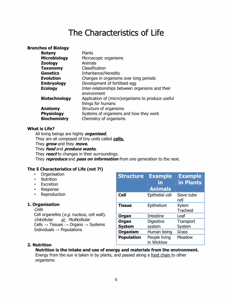
TThhee CChhaarraacctteerriissttiiccss ooff LLiiffee
Branches of Biology Botany Plants Microbiology Microscopic organisms Zoology Animals Taxonomy Classification Genetics Inheritance/Heredity Evolution Changes in organisms over long periods Embryology Development of fertilised egg Ecology Inter-relationships between organisms and their environment Biotechnology Application of (micro)organisms to produce useful things for humans Anatomy Structure of organisms Physiology Systems of organisms and how they work Biochemistry Chemistry of organisms What is Life?
All living beings are highly organised. They are all composed of tiny units called cells. They grow and they move. They feed and produce waste. They react to changes in their surroundings. They reproduce and pass on information from one generation to the next.
The 5 Characteristics of Life (not 7!)
• Organisation • Nutrition • Excretion • Response • Reproduction
1. Organisation
Cells Cell organelles (e.g. nucleus, cell wall). Unicellular or Multicellular Cells → Tissues → Organs → Systems
Individuals → Populations
2. Nutrition
Nutrition is the intake and use of energy and materials from the environment. Energy from the sun is taken in by plants, and passed along a food chain to other organisms.
Structure Example in
Animals
Example in Plants
Cell Epithelial cell Sieve tube cell
Tissue Epithelium Xylem Tracheid
Organ Intestine Leaf
Organ System
Digestive system
Transport System
Organism Human being Grass
Population People living in Wicklow
Meadow
7
Autotrophs make their own food from simple inorganic raw materials such as water and carbon dioxide.
Example: plants. Heterotrophs obtain their nutrients by either eating plants or other organisms Example: animals.
3. Excretion
Excretion is the removal of waste products of metabolism from the body of an organism. Metabolic reactions cause harmful toxic waste products to be produced in the cells. The kidneys excrete a mixture of water, salt and urea. Urea is made in the liver. Plants often move waste to the leaves and drop them in the autumn.
4. Response
Response is the ability of organisms to react to changes (stimuli) both inside and outside their bodies. The ability of an organism to react to changes in the environment ensures survival.
5. Reproduction
Reproduction is the ability of an organism to produce new individuals of its own kind. 2 Types:
Asexual – involves only one parent. Example: amoeba, bacteria, vegetative reproduction in plants.
Sexual – involves two parents producing gametes which fuse to form a zygote. Sexual reproduction produces individuals which carry genes from both parents.
Metabolism Metabolism refers to the sum of all the chemical reactions that occur in the cells of organisms. Chemical reactions needed for growth, repair, response and reproduction. Metabolism is controlled by the huge variety of enzymes found in cells.
Catabolic Reactions: the breakdown of molecules with the release of energy.
e.g. cellular respiration C6H12O6 + 6O2 → 6CO2 + 6H2O + energy
Anabolic Reactions: the building of larger molecules from smaller ones, using energy. e.g. photosynthesis 6CO2 + 6H2O + energy → C6H12O6 + 6O2
Continuity Continuity of life is the ability of organisms to exist from one generation to the next. i.e. living things arise from other living things of the same type. Living things don’t arise from non-living material.
Classification of Living things: All living things belong to one of 5 Kingdoms:
Plant, Animals, Fungi, Monera, and Protista
8
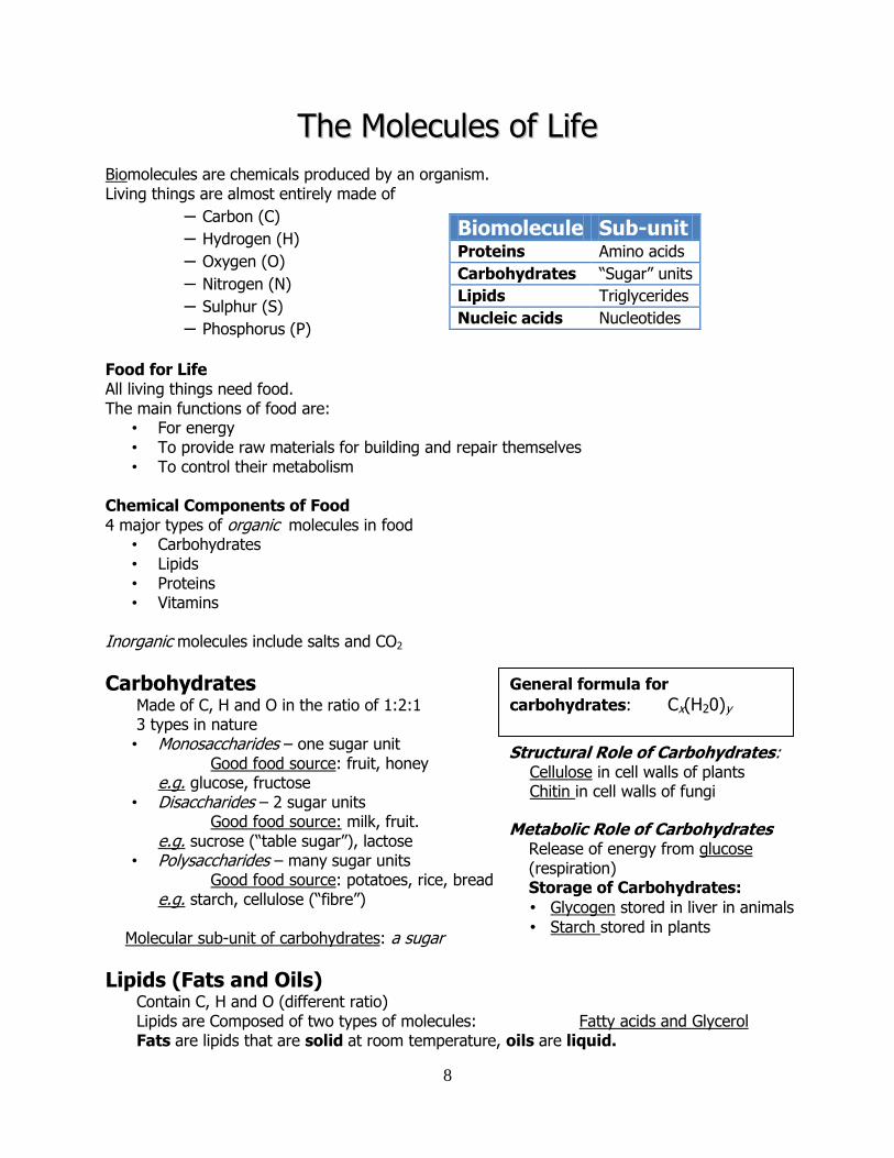
TThhee MMoolleeccuulleess ooff LLiiffee
Biomolecules are chemicals produced by an organism. Living things are almost entirely made of
– Carbon (C)
– Hydrogen (H)
– Oxygen (O)
– Nitrogen (N)
– Sulphur (S)
– Phosphorus (P)
Food for Life All living things need food. The main functions of food are:
• For energy • To provide raw materials for building and repair themselves • To control their metabolism
Chemical Components of Food 4 major types of organic molecules in food
• Carbohydrates • Lipids • Proteins • Vitamins
Inorganic molecules include salts and CO2
Carbohydrates Made of C, H and O in the ratio of 1:2:1 3 types in nature • Monosaccharides – one sugar unit
Good food source: fruit, honey e.g. glucose, fructose
• Disaccharides – 2 sugar units Good food source: milk, fruit. e.g. sucrose (“table sugar”), lactose
• Polysaccharides – many sugar units Good food source: potatoes, rice, bread e.g. starch, cellulose (“fibre”)
Molecular sub-unit of carbohydrates: a sugar
Lipids (Fats and Oils) Contain C, H and O (different ratio) Lipids are Composed of two types of molecules: Fatty acids and Glycerol Fats are lipids that are solid at room temperature, oils are liquid.
Biomolecule Sub-unit Proteins Amino acids
Carbohydrates “Sugar” units
Lipids Triglycerides
Nucleic acids Nucleotides
Structural Role of Carbohydrates: Cellulose in cell walls of plants Chitin in cell walls of fungi
Metabolic Role of Carbohydrates Release of energy from glucose
(respiration) Storage of Carbohydrates:
• Glycogen stored in liver in animals
• Starch stored in plants
General formula for
carbohydrates: Cx(H20)y
9
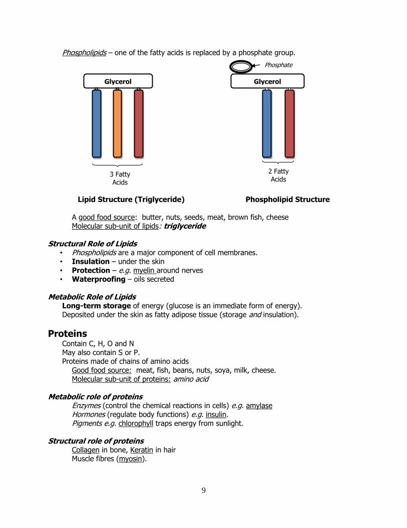
Phospholipids – one of the fatty acids is replaced by a phosphate group.
Lipid Structure (Triglyceride) Phospholipid Structure A good food source: butter, nuts, seeds, meat, brown fish, cheese Molecular sub-unit of lipids: triglyceride Structural Role of Lipids
• Phospholipids are a major component of cell membranes. • Insulation – under the skin • Protection – e.g. myelin around nerves • Waterproofing – oils secreted
Metabolic Role of Lipids
Long-term storage of energy (glucose is an immediate form of energy). Deposited under the skin as fatty adipose tissue (storage and insulation).
Proteins Contain C, H, O and N May also contain S or P. Proteins made of chains of amino acids
Good food source: meat, fish, beans, nuts, soya, milk, cheese. Molecular sub-unit of proteins: amino acid Metabolic role of proteins Enzymes (control the chemical reactions in cells) e.g. amylase Hormones (regulate body functions) e.g. insulin. Pigments e.g. chlorophyll traps energy from sunlight. Structural role of proteins Collagen in bone, Keratin in hair Muscle fibres (myosin).
2 Fatty Acids
Glycerol
Phosphate
Glycerol
3 Fatty
Acids
10
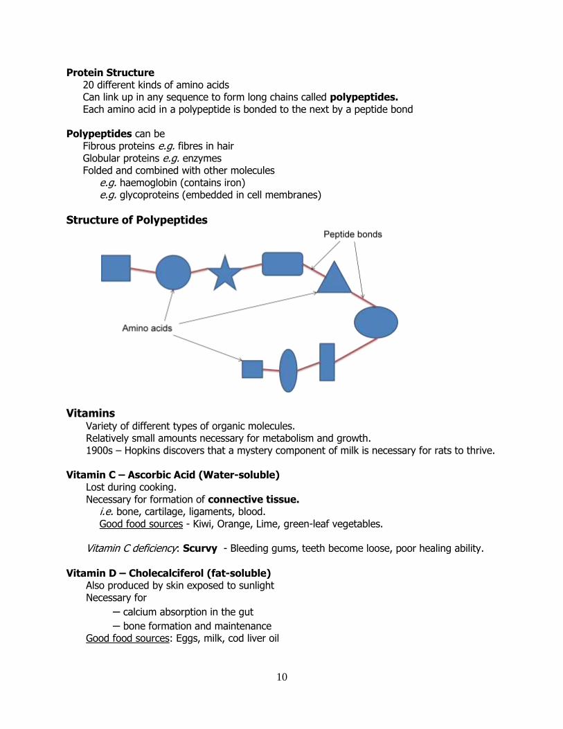
Protein Structure 20 different kinds of amino acids Can link up in any sequence to form long chains called polypeptides. Each amino acid in a polypeptide is bonded to the next by a peptide bond
Polypeptides can be Fibrous proteins e.g. fibres in hair Globular proteins e.g. enzymes Folded and combined with other molecules e.g. haemoglobin (contains iron) e.g. glycoproteins (embedded in cell membranes)
Structure of Polypeptides
Vitamins
Variety of different types of organic molecules. Relatively small amounts necessary for metabolism and growth. 1900s – Hopkins discovers that a mystery component of milk is necessary for rats to thrive.
Vitamin C – Ascorbic Acid (Water-soluble)
Lost during cooking. Necessary for formation of connective tissue. i.e. bone, cartilage, ligaments, blood. Good food sources - Kiwi, Orange, Lime, green-leaf vegetables. Vitamin C deficiency: Scurvy - Bleeding gums, teeth become loose, poor healing ability.
Vitamin D – Cholecalciferol (fat-soluble) Also produced by skin exposed to sunlight Necessary for
– calcium absorption in the gut
– bone formation and maintenance Good food sources: Eggs, milk, cod liver oil
11
Vitamin D deficiency Rickets in children Osteomalacia in adults - Deformed limbs, brittle bones
Minerals
• Inorganic substances needed in small amounts by organisms. • Plant absorb these from the soil • Animals obtain them in their diet
Role of Minerals Parts of rigid body structure
e.g. calcium for bones and teeth e.g. calcium in middle lamella (the “cement” in between plant cells)
Part of certain pigments e.g. iron in haemoglobin e.g. magnesium in chlorophyll
Water • Cytoplasm is 90% water, blood 92% water • Excellent solvent
o Necessary for cells’ metabolic activities • Carries materials in and out of cells
o Diffusion of dissolved substances • High specific heat capacity
o absorbs heat well without large increase in temp. • Gives shape to cells
o plant cells (turgor, stomata) o red blood cells
12
EEccoollooggyy -- GGeenneerraall PPrriinncciipplleess
Ecological Definitions
Ecology: the study of the how organisms interact with each other and with their environment.
Habitat: the place in which an organism lives.
Ecosystem: the interacting living and non-living components of a particular area.
Biosphere: the part of the Earth inhabited by living organisms.
Population: a group of organisms of the same species in the same habitat.
Community: a group of different populations living in the same habitat.
Carnivore: a flesh-eating animal, i.e. a meat-eater
Herbivore: an animal specially adapted to feed on plants.
Omnivore: an animal that eats both plants and meat.
Environmental Factors affecting living organisms Biotic Factors: the external influences on an organism by other living organisms. Abiotic Factors: the external influences on an organism by the non-living components of its environment.
Abiotic Factors Edaphic factors: soil features that influence the growth of plants or animals. e.g. pH; humus content; mineral composition; drainage. Climatic factors: the influences of prevailing weather conditions on living organisms in the ecosystem. e.g. wind direction, speed, light, temp, humidity, rainfall. Aquatic Factors: factors relating to aquatic habitats tides, waves, currents, temperature, salinity.
Biotic Factors Food organisms Parasites
Competition Predation
Pollinators Seed dispersal organisms
Producers and Consumers Producer: an organism that makes its own food from inorganic material, using energy from light (photosynthetic) or from chemical reactions (chemosynthetic). Producers are also called autotrophs. Consumer: organism that cannot make its own food, but instead must obtain it by eating. Consumers are also known as heterotrophs.
13
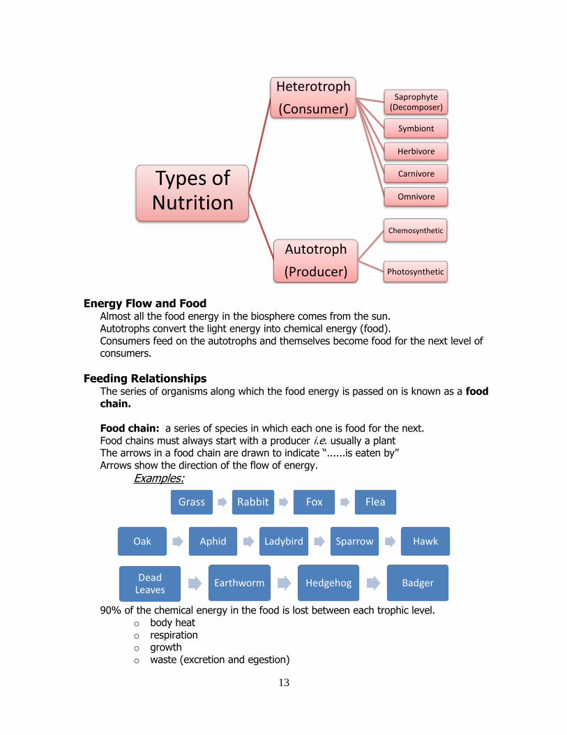
Energy Flow and Food Almost all the food energy in the biosphere comes from the sun. Autotrophs convert the light energy into chemical energy (food). Consumers feed on the autotrophs and themselves become food for the next level of consumers.
Feeding Relationships The series of organisms along which the food energy is passed on is known as a food chain. Food chain: a series of species in which each one is food for the next. Food chains must always start with a producer i.e. usually a plant The arrows in a food chain are drawn to indicate “......is eaten by” Arrows show the direction of the flow of energy.
Examples:
90% of the chemical energy in the food is lost between each trophic level.
o body heat o respiration o growth o waste (excretion and egestion)
Types of Nutrition
Heterotroph
(Consumer) Saprophyte
(Decomposer)
Symbiont
Herbivore
Carnivore
Omnivore
Autotroph
(Producer)
Chemosynthetic
Photosynthetic
Grass Rabbit Fox Flea
Oak Aphid Ladybird Sparrow Hawk
Dead Leaves
Earthworm Hedgehog Badger
14
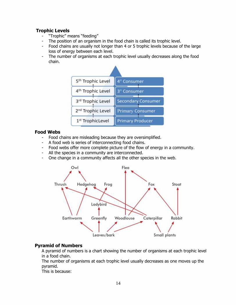
Trophic Levels - “Trophic” means “feeding” - The position of an organism in the food chain is called its trophic level. - Food chains are usually not longer than 4 or 5 trophic levels because of the large
loss of energy between each level. - The number of organisms at each trophic level usually decreases along the food
chain.
Food Webs
- Food chains are misleading because they are oversimplified. - A food web is series of interconnecting food chains. - Food webs offer more complete picture of the flow of energy in a community. - All the species in a community are interconnected. - One change in a community affects all the other species in the web.
Pyramid of Numbers A pyramid of numbers is a chart showing the number of organisms at each trophic level in a food chain. The number of organisms at each trophic level usually decreases as one moves up the pyramid. This is because:
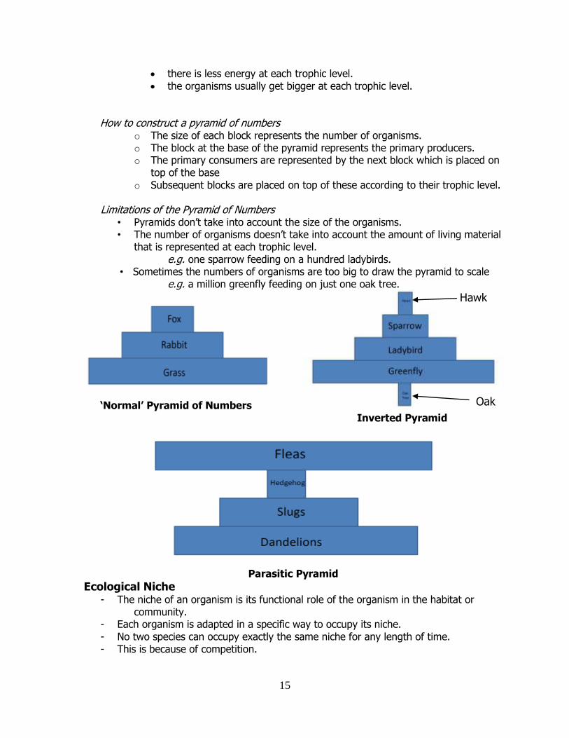
15
there is less energy at each trophic level. the organisms usually get bigger at each trophic level.
How to construct a pyramid of numbers
o The size of each block represents the number of organisms. o The block at the base of the pyramid represents the primary producers. o The primary consumers are represented by the next block which is placed on
top of the base o Subsequent blocks are placed on top of these according to their trophic level.
Limitations of the Pyramid of Numbers
• Pyramids don’t take into account the size of the organisms. • The number of organisms doesn’t take into account the amount of living material
that is represented at each trophic level. e.g. one sparrow feeding on a hundred ladybirds.
• Sometimes the numbers of organisms are too big to draw the pyramid to scale e.g. a million greenfly feeding on just one oak tree.
‘Normal’ Pyramid of Numbers
Inverted Pyramid
Parasitic Pyramid
Ecological Niche - The niche of an organism is its functional role of the organism in the habitat or
community. - Each organism is adapted in a specific way to occupy its niche. - No two species can occupy exactly the same niche for any length of time. - This is because of competition.
Hawk
Oak Tree
16
Ecological Relationships • Competition • Predation • Symbiosis
- Parasitism - Commensalism - Mutualism
These relationships are factors that control populations.
Competition Competition is the struggle between organisms for resources that are in limited supply. Competition can be between the same species or different species.
- Rivalry between organisms of the same species. Plants compete for light, water, space. Animals compete for food, mates. Scramble Competition “everybody loses”
- Each organism tries to get as much of the resource as possible while it is available. - There is no direct opposition. - Competition is not an issue if the resource is in plentiful supply. - If the resource is in short supply competition can lead to a severe drop in the
population. Example: rabbits in the Australian grasslands Contest Competition “somebody wins” Direct conflict between individuals where only one is successful in gaining the resource. The aim of the competition is to win the resource and deny it to others. Examples:
- territorial behaviour of blackbirds singing. - direct fighting between robins and sparrows for territory (nesting and food
resources in the area). - Stag deer rutting to compete for mating rights in the herd.
Role of Competition in the Ecosystem
- Controls the size of the population of competing individuals. - It’s the driving force behind adaptation and therefore evolution. - Only the ‘fittest’ survive. - Natural selection.
Adaptation Any change in the structure or behaviour of an organism that makes it better suited to its environment. Examples of adaptation to survive competition
Blackbirds song to discourage others. Grass produces huge numbers of pollen grains to increase chances of reproduction. Thistles produce extensive roots to increase chances of obtaining water and
minerals.
17
Predation - The catching and killing of an animal for food. - A predator is an animal that hunts and kills another animal (the prey) for food. - The prey is usually killed before feeding. - Normally the predator is bigger and less common than prey.
Predator Adaptations
- Eyesight, hearing, smell, dentition.
- Easy-to-catch prey. - Switching prey as numbers
change. - Living in packs, hunting together. - Migration to areas plentiful with
prey. - Camouflage.
Prey Adaptations - Thorns, spines, stings (nettles,
holly, etc) - Nasty taste (e.g. giant
hogweed). - Faster than predator at
swimming, running, flying. - Mimicking organisms undesirable
to predators. - Staying in large groups. - Warning colouration to look like
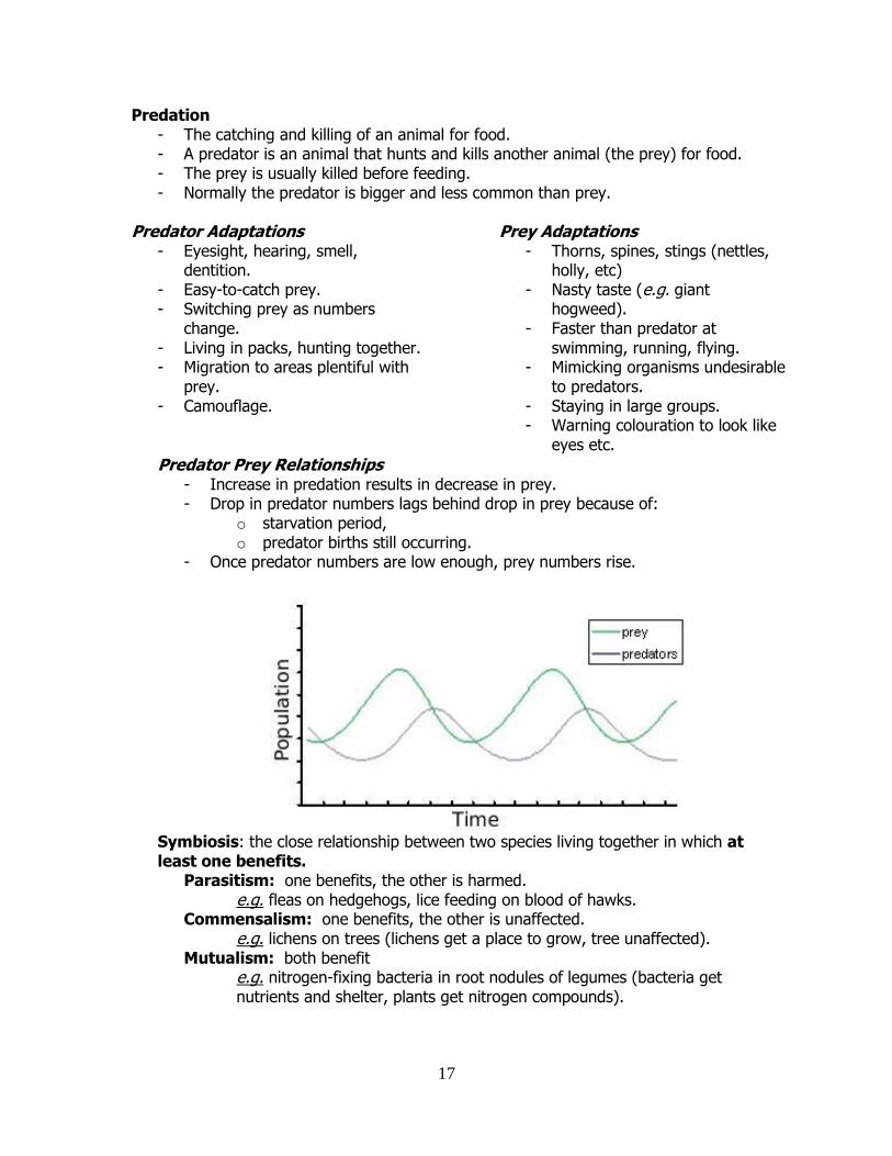
eyes etc. Predator Prey Relationships
- Increase in predation results in decrease in prey. - Drop in predator numbers lags behind drop in prey because of:
o starvation period, o predator births still occurring.
- Once predator numbers are low enough, prey numbers rise.
Symbiosis: the close relationship between two species living together in which at least one benefits. Parasitism: one benefits, the other is harmed. e.g. fleas on hedgehogs, lice feeding on blood of hawks. Commensalism: one benefits, the other is unaffected. e.g. lichens on trees (lichens get a place to grow, tree unaffected). Mutualism: both benefit e.g. nitrogen-fixing bacteria in root nodules of legumes (bacteria get nutrients and shelter, plants get nitrogen compounds).
18
Nutrient Recycling - This is the movement of essential elements from the abiotic environment into
living organisms and back again. - The return of elements ensures that they are always available to organisms. - Plants generally obtain their elements from salts in the soil. - Animals obtain elements from other organisms as food. - Decomposers generally return elements to the abiotic environment.
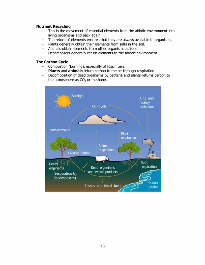
The Carbon Cycle
- Combustion (burning), especially of fossil fuels. - Plants and animals return carbon to the air through respiration. - Decomposition of dead organisms by bacteria and plants returns carbon to
the atmosphere as CO2 or methane.
(respiration by
decomposers)
19
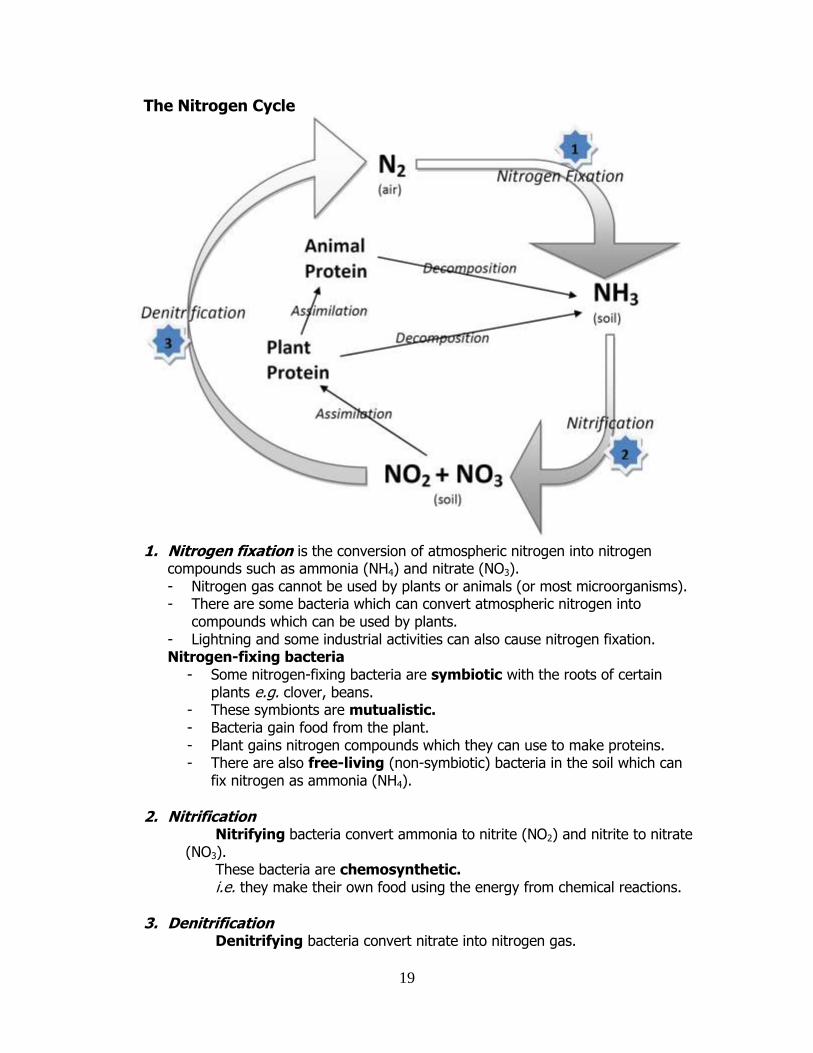
The Nitrogen Cycle
1. Nitrogen fixation is the conversion of atmospheric nitrogen into nitrogen
compounds such as ammonia (NH4) and nitrate (NO3). - Nitrogen gas cannot be used by plants or animals (or most microorganisms). - There are some bacteria which can convert atmospheric nitrogen into
compounds which can be used by plants. - Lightning and some industrial activities can also cause nitrogen fixation. Nitrogen-fixing bacteria
- Some nitrogen-fixing bacteria are symbiotic with the roots of certain plants e.g. clover, beans.
- These symbionts are mutualistic. - Bacteria gain food from the plant. - Plant gains nitrogen compounds which they can use to make proteins. - There are also free-living (non-symbiotic) bacteria in the soil which can
fix nitrogen as ammonia (NH4).
2. Nitrification Nitrifying bacteria convert ammonia to nitrite (NO2) and nitrite to nitrate
(NO3). These bacteria are chemosynthetic. i.e. they make their own food using the energy from chemical reactions.
3. Denitrification Denitrifying bacteria convert nitrate into nitrogen gas.
20
These bacteria are anaerobic and live in stagnant water-logged soil. Population Dynamics The study of changes in a population and the factors that cause these changes. Population stabilises eventually.
Factors affecting population growth
• Food • Space • Overcrowding • Waste • Disease • Easier prey
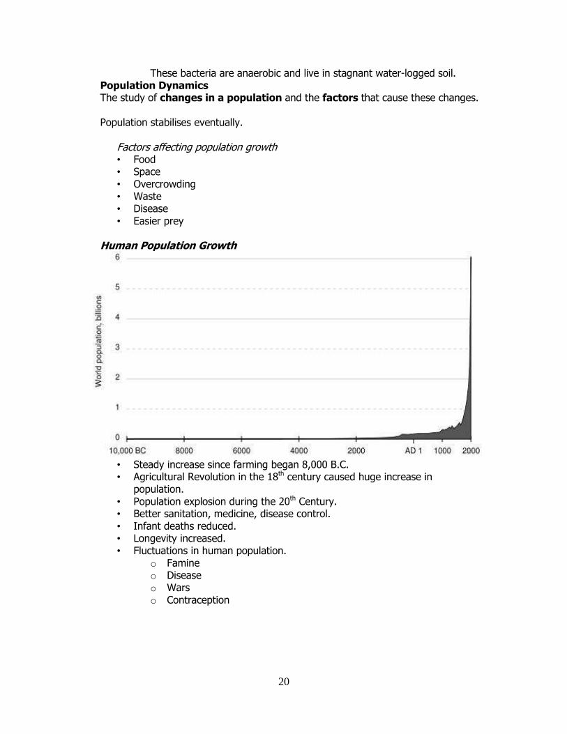
Human Population Growth
• Steady increase since farming began 8,000 B.C. • Agricultural Revolution in the 18th century caused huge increase in
population. • Population explosion during the 20th Century. • Better sanitation, medicine, disease control. • Infant deaths reduced. • Longevity increased. • Fluctuations in human population.
o Famine o Disease o Wars o Contraception
21
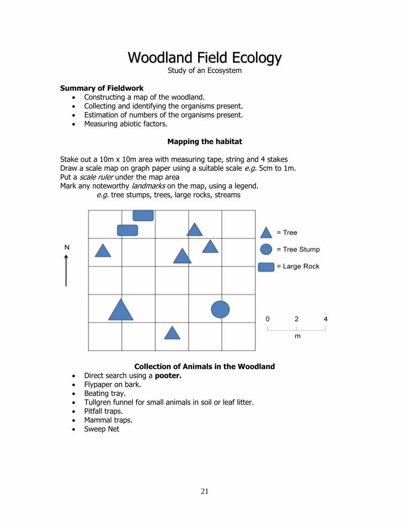
WWooooddllaanndd FFiieelldd EEccoollooggyy Study of an Ecosystem
Summary of Fieldwork
Constructing a map of the woodland. Collecting and identifying the organisms present. Estimation of numbers of the organisms present. Measuring abiotic factors.
Mapping the habitat
Stake out a 10m x 10m area with measuring tape, string and 4 stakes Draw a scale map on graph paper using a suitable scale e.g. 5cm to 1m. Put a scale ruler under the map area Mark any noteworthy landmarks on the map, using a legend.
e.g. tree stumps, trees, large rocks, streams
Collection of Animals in the Woodland
Direct search using a pooter. Flypaper on bark. Beating tray. Tullgren funnel for small animals in soil or leaf litter. Pitfall traps. Mammal traps. Sweep Net
22
Animal Identification Collecting insects is usually done with a pooter. Small mammals can be captured using a mammal trap. More common method involve non-disturbance
– birds identified at a distance with binoculars – owls identified by their regurgitated pellet of hair and feathers – droppings to identify badgers, wild goat etc. – mice vs. squirrels identified by comparing the holes on acorns.
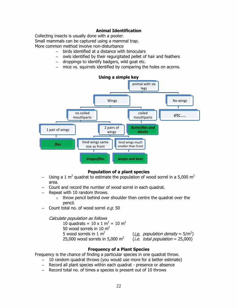
Using a simple key
Population of a plant species – Using a 1 m2 quadrat to estimate the population of wood sorrel in a 5,000 m2
area. – Count and record the number of wood sorrel in each quadrat. – Repeat with 10 random throws.
o throw pencil behind over shoulder then centre the quadrat over the pencil.
– Count total no. of wood sorrel e.g. 50 Calculate population as follows
10 quadrats = 10 x 1 m2 = 10 m2
50 wood sorrels in 10 m2
5 wood sorrels in 1 m2 (i.e. population density = 5/m2) 25,000 wood sorrels in 5,000 m2 (i.e. total population = 25,000)
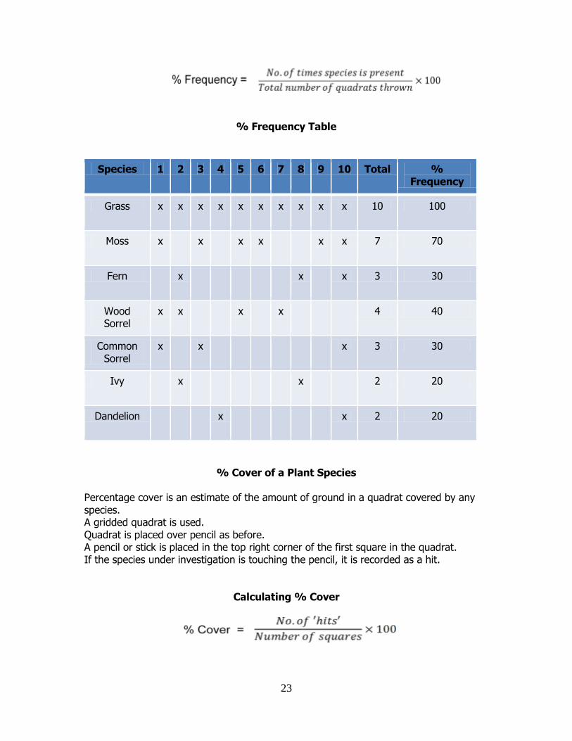
Frequency of a Plant Species
Frequency is the chance of finding a particular species in one quadrat throw. – 10 random quadrat throws (you would use more for a better estimate) – Record all plant species within each quadrat - presence or absence – Record total no. of times a species is present out of 10 throws
animal with six legs
Wings
no coiled mouthparts
1 pair of wings
flies
2 pairs of wings
hind wings same size as front
dragonflies
hind wings much smaller than front
wasps and bees
coiled mouthparts
Butterflies and Moths
No wings
etc....
23
% Frequency Table
Species 1 2 3 4 5 6 7 8 9 10 Total % Frequency
Grass x x x x x x x x x x 10 100
Moss x x x x x x 7 70
Fern x x x 3 30
Wood Sorrel
x x x x 4 40
Common Sorrel
x x x 3 30
Ivy x x 2 20
Dandelion x x 2 20
% Cover of a Plant Species Percentage cover is an estimate of the amount of ground in a quadrat covered by any species. A gridded quadrat is used. Quadrat is placed over pencil as before. A pencil or stick is placed in the top right corner of the first square in the quadrat. If the species under investigation is touching the pencil, it is recorded as a hit.
Calculating % Cover
24
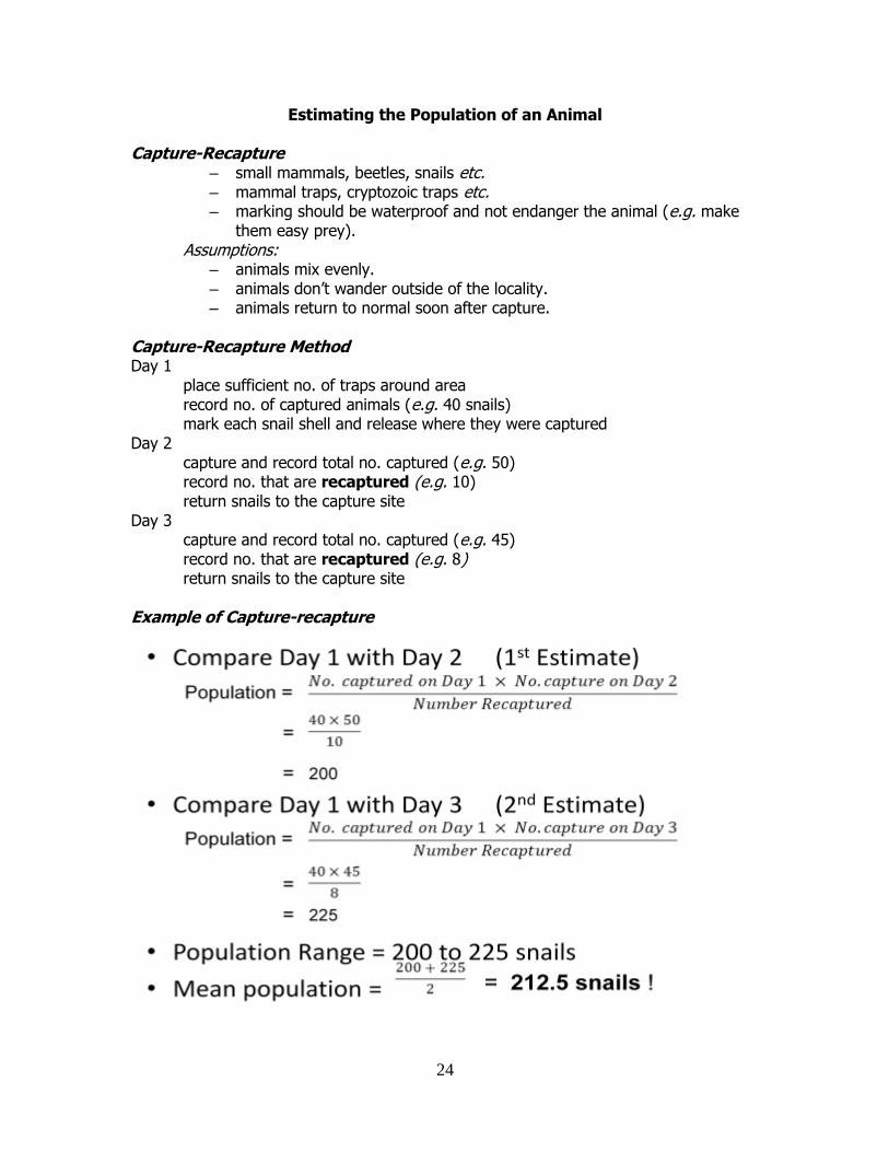
Estimating the Population of an Animal Capture-Recapture
– small mammals, beetles, snails etc. – mammal traps, cryptozoic traps etc. – marking should be waterproof and not endanger the animal (e.g. make
them easy prey). Assumptions:
– animals mix evenly. – animals don’t wander outside of the locality. – animals return to normal soon after capture.
Capture-Recapture Method Day 1
place sufficient no. of traps around area record no. of captured animals (e.g. 40 snails) mark each snail shell and release where they were captured
Day 2 capture and record total no. captured (e.g. 50) record no. that are recaptured (e.g. 10) return snails to the capture site
Day 3 capture and record total no. captured (e.g. 45) record no. that are recaptured (e.g. 8) return snails to the capture site
Example of Capture-recapture
25
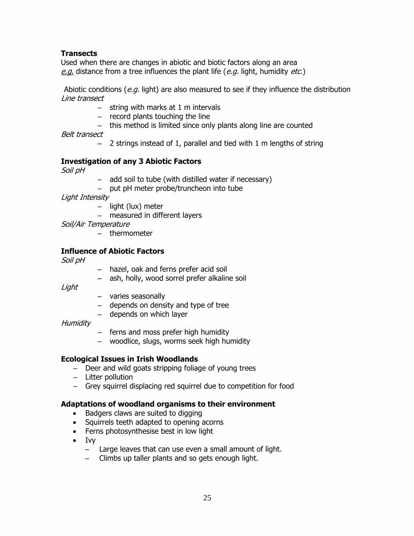
Transects Used when there are changes in abiotic and biotic factors along an area e.g. distance from a tree influences the plant life (e.g. light, humidity etc.)
Abiotic conditions (e.g. light) are also measured to see if they influence the distribution
Line transect – string with marks at 1 m intervals – record plants touching the line – this method is limited since only plants along line are counted
Belt transect – 2 strings instead of 1, parallel and tied with 1 m lengths of string
Investigation of any 3 Abiotic Factors Soil pH
– add soil to tube (with distilled water if necessary) – put pH meter probe/truncheon into tube
Light Intensity – light (lux) meter – measured in different layers
Soil/Air Temperature – thermometer
Influence of Abiotic Factors Soil pH
– hazel, oak and ferns prefer acid soil – ash, holly, wood sorrel prefer alkaline soil
Light – varies seasonally – depends on density and type of tree – depends on which layer
Humidity – ferns and moss prefer high humidity – woodlice, slugs, worms seek high humidity
Ecological Issues in Irish Woodlands
– Deer and wild goats stripping foliage of young trees – Litter pollution – Grey squirrel displacing red squirrel due to competition for food
Adaptations of woodland organisms to their environment
Badgers claws are suited to digging Squirrels teeth adapted to opening acorns Ferns photosynthesise best in low light Ivy
– Large leaves that can use even a small amount of light. – Climbs up taller plants and so gets enough light.
26
HHuummaann IImmppaacctt oonn tthhee EEnnvviirroonnmmeenntt
Some “Environmental” Issues • Human Population • Greenhouse emissions • Climate Change (Global Warming) • Biodiversity • Waste • Industrial Pollution • Power • Transport • Genetically Modified Organisms
Pollution
• Greenhouse gases • Acidifying gases • Water pollution
– fertilizers, pesticides, slurry, silage effluent, domestic and industrial waste • Land pollution
– pesticides, fertilizers, acid rain, industrial emissions, domestic and industrial waste
Greenhouse Gases
• CO2 – oil, coal, transport, deforestation. • CH4 (methane) – domestic waste, agriculture. • N2O (nitrous oxide) – agriculture.
Acidifying gases & Ozone precursors
• NH3 (ammonia) – agriculture, fertilizer use • SO2 (sulphur dioxide) – combustion, power stations • NOx (nitrogen oxides) – combustion, transport
Water Pollution
• Fertilizers • Pesticides • Slurry • Silage effluent • Domestic waste • Industrial waste
Pesticides and Fertilizers
• Insecticides – also affect beneficial insects – DDT causing thinning of the egg-shells of the peregrine falcon, sparrowhawk
• Herbicides - weeds develop resistance • Fungicides – can be toxic to humans and wildlife
27
Acid Rain • Sulphur dioxide • Burning of sulphur-containing fuels i.e. coal and oil. • Dissolves in rainwater to form sulphurous and sulphuric acid. • Soil pH lowered, nutrients leach away • SO2 prevents plants forming chlorophyll • Conifers especially affected
•• Causes problems for fish, bacteria, insects
Indicator Species
• These are organisms which are sensitive to the harmful effects of SO2 • Lichens are a symbiotic combination of a fungus and one of the green algae • Presence of lichens in an area, on tree-trunks and on walls indicates “clean” air. • Leaf yeasts are also an indicator of air quality .
Reducing SO2 emissions
• Alternative energy sources • Solar • Wind • Hydro
• Low-sulphur coal in power stations • Using Natural gas instead of other fossil fuels • Tall chimney stacks • Extract the SO2 in the stack before releasing smoke.
Conservation
• “The wise management of our environment to ensure the survival of organisms and their habitat”
•• Balancing our use of resources with the preservation of wildlife. • NNational Parks
e.g. Wicklow Mountains, Letterfrack, Killarney, The Burren, Glenveagh.
Example of a Conservation Activity
• Agriculture - “set-aside” • Farming reduces biodiversity and results in the reduction of native species • Part of the farm is given over or “set aside” to allow reintroduction of the natural
communities The Major Environmental Challenges
• Waste • Eutrophication of Inland Waters
– addition of nutrients to fresh water causing lack of oxygen • The Urban Environment and Transport • Climate Change and Greenhouse Gases • Biodiversity and Protection of Natural Resources
28
Waste Management • Reduce, reuse, recycle (3 R’s)
– minimisation is better than e.g. recycling. • Sawdust ‘waste’ from mills
– now compressed into blocks for fuel. • Whey (‘waste’ from dairy industry)
– now used to as food to grow microorganisms. • Domestic cooked food ‘waste’
– now collected in Wexford to make fuel • Glass, paper, metal cans, recycled back for use again.
Waste Disposal • Landfill
– groundwater pollution – landfill sites aarree nearly full – nobody wants a dump nearby – protests
• Incineration – doesn’t require much land use – may release toxic dioxins – some energy yield iiss possible
TThhee Role of Microorganisms Waste Treatment
– MMicrobes used to break down organic waste such as sewage, waste food, whey etc.
– Digesters can be open (aerobic) or closed (anaerobic) – Gas produced can be used as fuel – The mass of waste is vastly reduced and made safe.
Bioremediation
• Bioremediation is the use of microbes to remove pollution from an area and allow the area to return to its unpolluted state.
• Certain kinds of bacteria can be used to clean up contaminated sites • oil spillages • pesticide residues • chemical contamination.
29
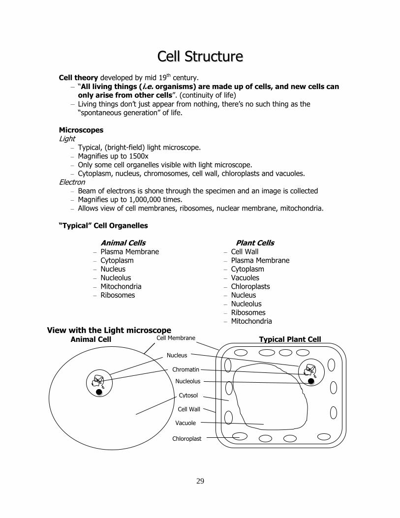
CCeellll SSttrruuccttuurree Cell theory developed by mid 19th century.
– “All living things (i.e. organisms) are made up of cells, and new cells can only arise from other cells”. (continuity of life)
– Living things don’t just appear from nothing, there’s no such thing as the “spontaneous generation” of life.
Microscopes Light
– Typical, (bright-field) light microscope. – Magnifies up to 1500x – Only some cell organelles visible with light microscope. – Cytoplasm, nucleus, chromosomes, cell wall, chloroplasts and vacuoles.
Electron – Beam of electrons is shone through the specimen and an image is collected – Magnifies up to 1,000,000 times. – Allows view of cell membranes, ribosomes, nuclear membrane, mitochondria.
“Typical” Cell Organelles
Animal Cells – Plasma Membrane – Cytoplasm – Nucleus – Nucleolus – Mitochondria – Ribosomes
Plant Cells – Cell Wall – Plasma Membrane – Cytoplasm – Vacuoles – Chloroplasts – Nucleus – Nucleolus – Ribosomes – Mitochondria
View with the Light microscope Animal Cell Typical Plant Cell
Cytosol
Cell Membrane
Cell Wall
Vacuole
Chloroplast
Nucleus
Nucleolus
Chromatin
30
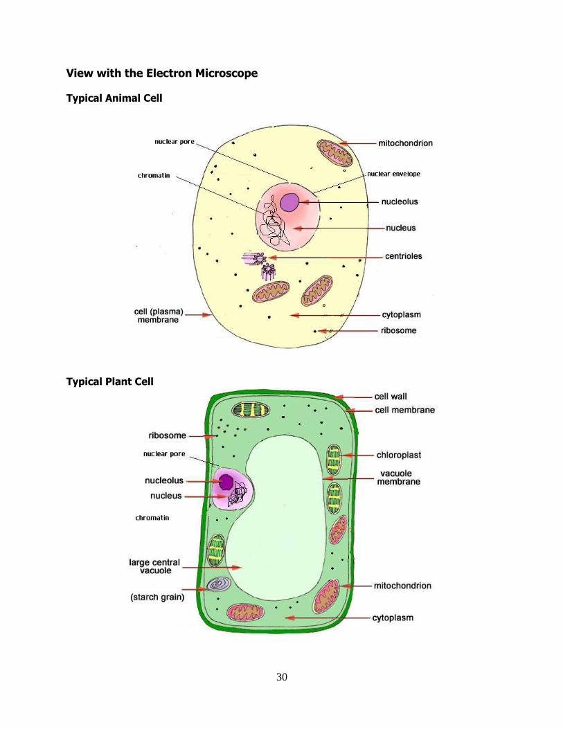
View with the Electron Microscope
Typical Animal Cell
Typical Plant Cell
31
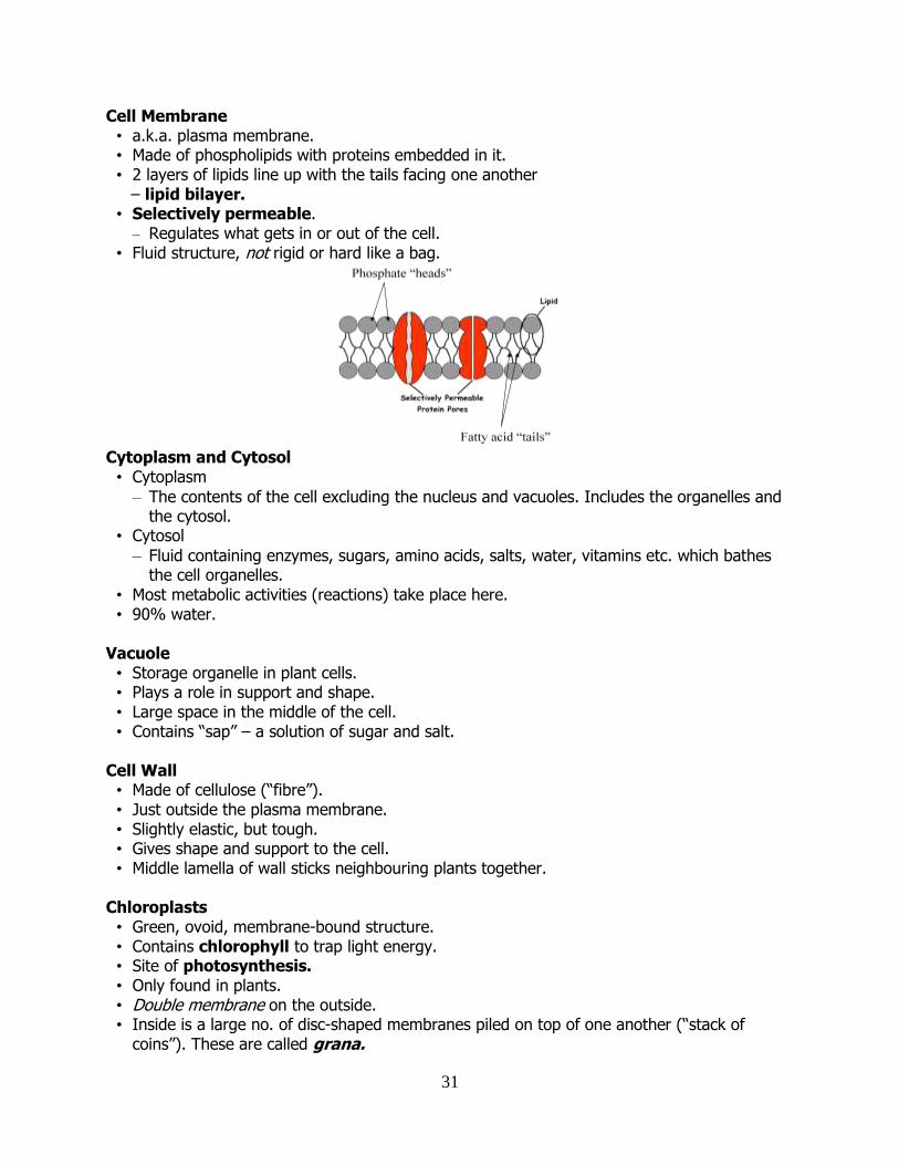
Cell Membrane • a.k.a. plasma membrane. • Made of phospholipids with proteins embedded in it. • 2 layers of lipids line up with the tails facing one another – lipid bilayer.
• Selectively permeable. – Regulates what gets in or out of the cell.
• Fluid structure, not rigid or hard like a bag.
Cytoplasm and Cytosol • Cytoplasm
– The contents of the cell excluding the nucleus and vacuoles. Includes the organelles and the cytosol.
• Cytosol
– Fluid containing enzymes, sugars, amino acids, salts, water, vitamins etc. which bathes the cell organelles.
• Most metabolic activities (reactions) take place here. • 90% water.
Vacuole • Storage organelle in plant cells. • Plays a role in support and shape. • Large space in the middle of the cell. • Contains “sap” – a solution of sugar and salt.
Cell Wall • Made of cellulose (“fibre”). • Just outside the plasma membrane. • Slightly elastic, but tough. • Gives shape and support to the cell. • Middle lamella of wall sticks neighbouring plants together.
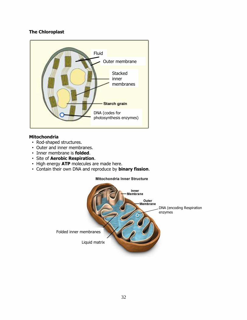
Chloroplasts • Green, ovoid, membrane-bound structure. • Contains chlorophyll to trap light energy. • Site of photosynthesis. • Only found in plants. • Double membrane on the outside. • Inside is a large no. of disc-shaped membranes piled on top of one another (“stack of
coins”). These are called grana.
32
The Chloroplast
Mitochondria • Rod-shaped structures. • Outer and inner membranes. • Inner membrane is folded. • Site of Aerobic Respiration. • High energy ATP molecules are made here. • Contain their own DNA and reproduce by binary fission.
Fluid
Stacked inner membranes
DNA (codes for photosynthesis enzymes)
Outer membrane
Folded inner membranes
Liquid matrix
DNA (encoding Respiration enzymes
33
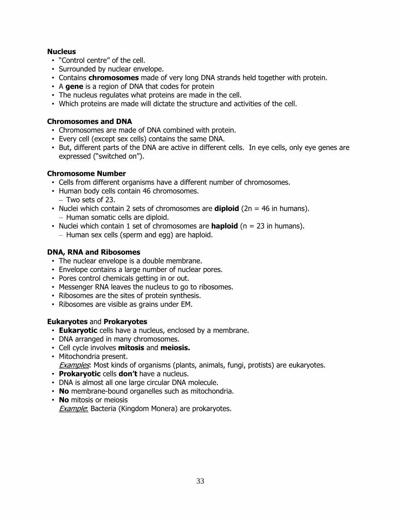
Nucleus • “Control centre” of the cell. • Surrounded by nuclear envelope. • Contains chromosomes made of very long DNA strands held together with protein. • A gene is a region of DNA that codes for protein • The nucleus regulates what proteins are made in the cell. • Which proteins are made will dictate the structure and activities of the cell.
Chromosomes and DNA • Chromosomes are made of DNA combined with protein. • Every cell (except sex cells) contains the same DNA. • But, different parts of the DNA are active in different cells. In eye cells, only eye genes are
expressed (“switched on”).
Chromosome Number • Cells from different organisms have a different number of chromosomes. • Human body cells contain 46 chromosomes.
– Two sets of 23. • Nuclei which contain 2 sets of chromosomes are diploid (2n = 46 in humans).
– Human somatic cells are diploid. • Nuclei which contain 1 set of chromosomes are haploid (n = 23 in humans).
– Human sex cells (sperm and egg) are haploid.
DNA, RNA and Ribosomes • The nuclear envelope is a double membrane. • Envelope contains a large number of nuclear pores. • Pores control chemicals getting in or out. • Messenger RNA leaves the nucleus to go to ribosomes. • Ribosomes are the sites of protein synthesis. • Ribosomes are visible as grains under EM.
Eukaryotes and Prokaryotes • Eukaryotic cells have a nucleus, enclosed by a membrane. • DNA arranged in many chromosomes. • Cell cycle involves mitosis and meiosis. • Mitochondria present.
Examples: Most kinds of organisms (plants, animals, fungi, protists) are eukaryotes. • Prokaryotic cells don’t have a nucleus. • DNA is almost all one large circular DNA molecule. • No membrane-bound organelles such as mitochondria. • No mitosis or meiosis
Example: Bacteria (Kingdom Monera) are prokaryotes.
34
MMoovveemmeenntt tthhrroouugghh MMeemmbbrraanneess Diffusion • Diffusion is the passive movement of molecules from areas of high concentration to area of
low concentration. • Gases (O2, CO2) diffuse from blood into and out of cells. • The difference in concentration between two areas is called a concentration gradient. • Diffusion is a passive process i.e. no energy input required. • Diffusion stops when there is no difference in concentration between the 2 areas.
Examples of Diffusion
- Alveoli - exchange of gases between capillaries and air. - Synapse - movement of neurotransmitter to the target cell. - Stomata and lenticels - gas exchange.
Osmosis • Osmosis is the movement of water from an area of high water concentration to an area of
low water concentration across a selectively permeable membrane. • In terms of solutes (dissolved substances) osmosis involves movement of water from a
dilute region to a concentrated region. • Osmosis is a special form of diffusion.
Selectively-permeable membranes • The cell membrane (plasma membrane) is a selectively permeable membrane. • Proteins in cell membranes select or regulate what kind of molecules go in and out of cells,
i.e. membrane proteins control permeability. • Some dissolved substances can move freely, others cannot.
Example of Osmosis • Amoeba - Contractile vacuole regulates water uptake. • Nephron - Water reabsorption. • Plant roots - Water enters root hairs by osmosis. • Stomata - Guard cells take in water, causing opening of stomata.
Multicellular organisms • Need to monitor the environment inside their bodies for water and salt. • Cells won’t work properly if the concentration of the blood is not right. • Control of water and salt concentration in the blood is an example of osmoregulation. • Osmoregulation is carried out by the kidneys in humans and many other animals • Osmoregulation refers to ways that organisms have of keeping the concentration of a
solution at an optimum. Amoeba • Only the cell membrane separates the cell contents from the environment. • Amoeba lives in fresh water, so the concentration of salt is much higher inside the cell than
outside. • Because of the concentration gradient, water flows into the cell. • Amoeba’s contractile vacuole gets rid of the water (requires energy) before the cell bursts.
35
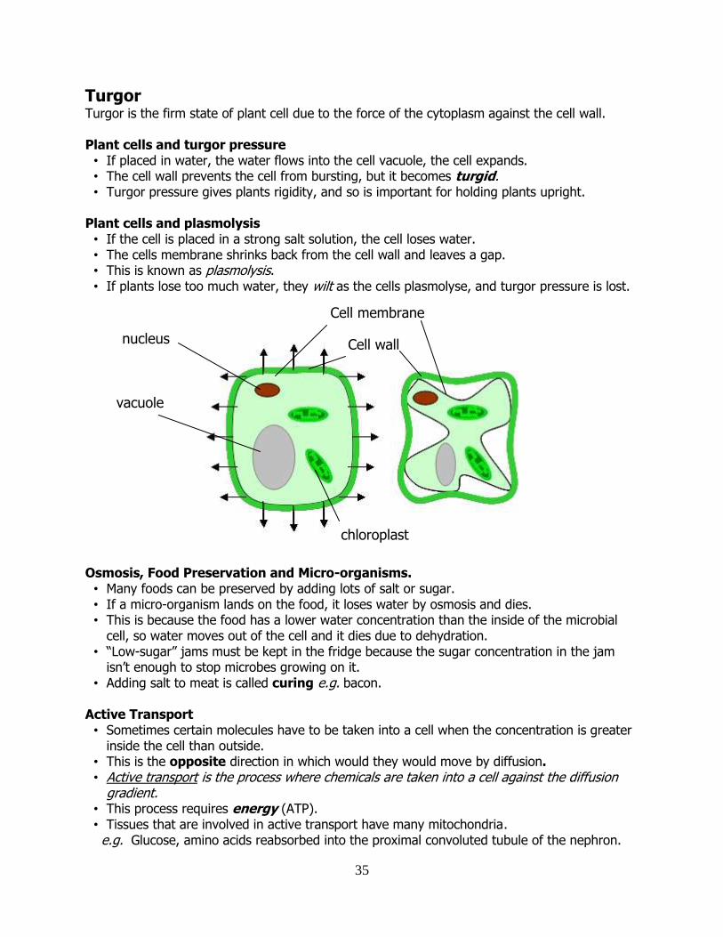
Turgor Turgor is the firm state of plant cell due to the force of the cytoplasm against the cell wall.
Plant cells and turgor pressure • If placed in water, the water flows into the cell vacuole, the cell expands. • The cell wall prevents the cell from bursting, but it becomes turgid. • Turgor pressure gives plants rigidity, and so is important for holding plants upright.
Plant cells and plasmolysis • If the cell is placed in a strong salt solution, the cell loses water. • The cells membrane shrinks back from the cell wall and leaves a gap. • This is known as plasmolysis. • If plants lose too much water, they wilt as the cells plasmolyse, and turgor pressure is lost.
Osmosis, Food Preservation and Micro-organisms. • Many foods can be preserved by adding lots of salt or sugar. • If a micro-organism lands on the food, it loses water by osmosis and dies. • This is because the food has a lower water concentration than the inside of the microbial
cell, so water moves out of the cell and it dies due to dehydration. • “Low-sugar” jams must be kept in the fridge because the sugar concentration in the jam
isn’t enough to stop microbes growing on it. • Adding salt to meat is called curing e.g. bacon.
Active Transport • Sometimes certain molecules have to be taken into a cell when the concentration is greater
inside the cell than outside. • This is the opposite direction in which would they would move by diffusion. • Active transport is the process where chemicals are taken into a cell against the diffusion
gradient. • This process requires energy (ATP). • Tissues that are involved in active transport have many mitochondria.
e.g. Glucose, amino acids reabsorbed into the proximal convoluted tubule of the nephron.
Cell wall
Cell membrane
nucleus
vacuole
chloroplast
36
CCeellll CCoonnttiinnuuiittyy
Definitions Chromosome – tightly packaged structures found in the nucleus composed of DNA and protein, and
consisting of a particular set of genes. Chromatin – the fibrous mixture of DNA and protein that chromosomes are made of,
chromosomes loosen to form strands of chromatin. Chromatid – one among the two identical copies of DNA making up a replicated chromosome,
which are joined at their centromeres Centromere – location on a chromosome where the sister chromatids are attached
Cell Continuity
• New living cells are produced by the division of existing living cells • All life on earth has resulted from the unbroken series of cell divisions going back nearly 4
billion years • Living cells can only be produced by cell division.
The Cell Cycle
• The sequence of events which result in the formation of new cells. • The cell cycle results in the production of new “daughter” cells from the original “mother”
cell. • There are 3 stages
– Interphase – Nuclear division – Cytokinesis
Interphase
• Cells are actively growing and doing their job • New organelles and enzymes are produced • Protein synthesis
– ribosomes are usually busy during early interphase • DNA Replication
– Before entering the next phase (nuclear division), the cell makes an identical copy of each of its chromosomes
Nuclear Division
• Mitosis – division of the nucleus to produce 2 genetically identical daughter cells with the
same number of chromosomes as the mother cell • Meiosis
– division of a diploid nucleus to produce 4 genetically different daughter cells which are haploid. Sometimes called reduction division
Note: meiosis can only occur with diploid cells
37
Cytokinesis • Once nuclear division (mitosis or meiosis) is complete, the cell divides to form new cells. • The cytoplasm is usually shared equally between the new daughter cells
– organelles – biomolecules
• Cytokinesis often begins to occur even before nuclear division i.e. the 2 stages can overlap
Mitosis Mitosis is nuclear division which produces genetically identical daughter nuclei
Haploid and Diploid Haploid (n): one set of chromosomes present in the nucleus of a cell i.e. one of each type of chromosome is present in the nucleus Diploid (2n): two sets of chromosomes present in the nucleus of a cell i.e. two of each type of chromosome is present in the nucleus
• Mitosis results in the formation of identical daughter cells • These mother and daughters have
– the same number of chromosomes – the same number of genes – the same type of genes as one another
• During mitosis – haploid cells produce haploid daughter cells – diploid cells produce diploid daughter cells
The Role of Mitosis
• Growth and repair of multicellular organisms • Reproduction of single-celled eukaryotes
– prokaryotes (bacteria) do not have a nucleus, therefore do not undergo mitosis or meiosis
• Formation of gametes in plant sexual reproduction • Passing on of identical genetic information from one generation of cells to the next.
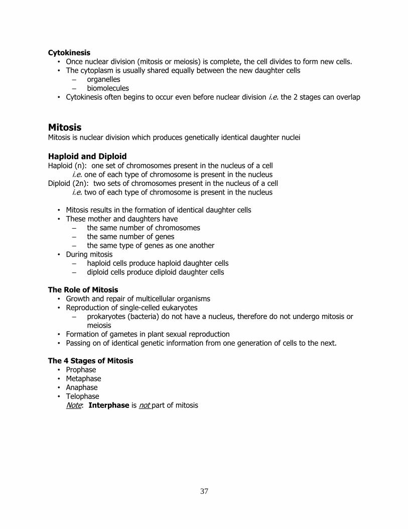
The 4 Stages of Mitosis
• Prophase • Metaphase • Anaphase • Telophase
Note: Interphase is not part of mitosis
38
Nuclear
membrane
Chromosome
Centromere
Sister Chromatids
Sister
Chromatids
Nuclear Membrane
reforming
Spindle fibres
39
Prophase • Chromatin strands start to condense. • Individual chromosomes start to become visible. • The nucleolus disappears. • The nuclear membrane begins to break down. • Spindle fibres begin to form.
Metaphase
• The nuclear membrane fully breaks down. • The chromosomes line up at the equator of the cell. • Spindle fibres attach to the centromeres of the chromosomes.
Anaphase
• The spindle fibres start to contract (shorten). • The sister chromosomes are pulled apart to opposite poles of the cell.
– Note: now that they have been separated, the sister chromatids are called chromosomes.
• Each end of the cell now contains a complete set of chromosomes. Telophase
• Chromosomes start to loosen to become chromatin. • Individual chromosomes become difficult to distinguish. • A nuclear membrane begins to form around each new set of chromosomes at the poles of
the cell. • Daughter nuclei are genetically identical
– Note: cytokinesis (cell division) is not part of telophase, but the cell often divides before telophase is finished.
Tumours
• Mitosis and cell division is normally under tight control by a number of important genes. • Sometimes these genes may undergo mutations and loses control of cell division. • An individual cell may then grow and divide to form a mass of identical cells called a
tumour. • Benign tumour
– Cells soon stop dividing and do not invade tissues. • Malignant tumour
– Uncontrolled division of cells which can then invade surrounding tissue.
Cancer • A malignant tumour is also known as a “cancer”. • A cancer is a mass of cells which have lost the ability to control the rate and frequency of
mitosis, and which invade and disrupt surrounding tissue. • Cancerous cells can break off the break off from the tumour and travel in the circulatory
system to new sites to form new tumours. • This is known as metastasis.
40
Causes of Cancer • Certain genes are crucial to maintain normal cell behaviour. • If these genes are mutated, cancers can result. • These genes are known as oncogenes. • Carcinogens are cancer-causing agents which cause mutations in oncogenes. • Examples:
– cigarette smoke – UV radiation – certain viruses
Meiosis
Meiosis is the division of a diploid nucleus resulting in 4 genetically different daughter nuclei. Role of Meiosis
• formation of gametes in animals. • formation of female megaspores and male microspores in plants. • prevents doubling of chromosome number during sexual reproduction.
Site of Meiosis Animals
• sperm formed in the testis • ova formed in ovary
Plants
• male microspores formed in the anther of the stamen • female megaspores formed in the ovule of the ovary
The Products of Meiosis and Mitosis
Mitosis Meiosis
Two daughter cells produced Four daughter cells produced
Chromosome number stays the same
Chromosome number is halved
Daughters genetically identical to parents
Daughters genetically different to parents
41
EEnnzzyymmeess aanndd MMeettaabboolliissmm Metabolism is the term used to describe chemical reactions in cells
Metabolism = the sum of all cellular chemical reactions in a cell or organism
Enzymes • Enzymes are biological catalysts
– catalysts are chemicals that speed up chemical reactions without being changed permanently by them
• Enzymes are normally proteins • They are made of one or more chains of amino acids folded into a particular 3D shape • The correct folded shape of an active enzyme is called its native shape
General Properties of Enzymes • Specific catalysts made of protein
– one enzyme for one reaction • Reversible
– can catalyse the reaction in both directions • Rate of action affected by temperature and pH • Denatured by high temperature and extremes of pH • Inhibitors are chemicals which denature enzymes
– They are often enzyme-specific, e.g. some antibiotics affect a bacterial enzyme but don’t affect human enzymes, so that it is safe for us to take them to kill infections.
Role of enzymes
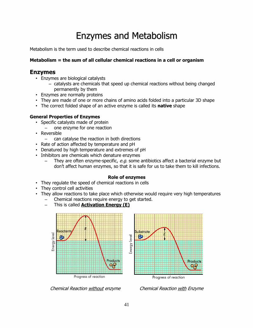
• They regulate the speed of chemical reactions in cells • They control cell activities • They allow reactions to take place which otherwise would require very high temperatures
– Chemical reactions require energy to get started. – This is called Activation Energy (E)
Chemical Reaction without enzyme Chemical Reaction with Enzyme
42
Substrate and Product • The chemical(s) that an enzyme reacts with is called the substrate. • The chemical(s) produced at the end of the reaction is called the product.
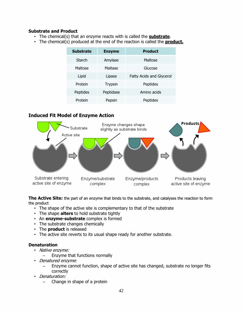
Induced Fit Model of Enzyme Action
The Active Site: tthhee ppaarrtt ooff aann eennzzyymmee tthhaatt bbiinnddss ttoo tthhee ssuubbssttrraattee,, aanndd ccaattaallyysseess tthhee rreeaaccttiioonn ttoo ffoorrmm
tthhee pprroodduucctt • The shape of the active site is complementary to that of the substrate • The shape alters to hold substrate tightly • An enzyme-substrate complex is formed • The substrate changes chemically • The product is released • The active site reverts to its usual shape ready for another substrate.
Denaturation
• Native enzyme: – Enzyme that functions normally
• Denatured enzyme: – Enzyme cannot function, shape of active site has changed, substrate no longer fits
correctly • Denaturation:
– Change in shape of a protein
Substrate Enzyme Product
Starch Amylase Maltose
Maltose Maltase Glucose
Lipid Lipase Fatty Acids and Glycerol
Protein Trypsin Peptides
Peptides Peptidase Amino acids
Protein Pepsin Peptides
43
– No longer able to function – Usually irreversible
• Renatured enzyme: – Enzyme may recover its shape if temperature and/or pH return to normal.
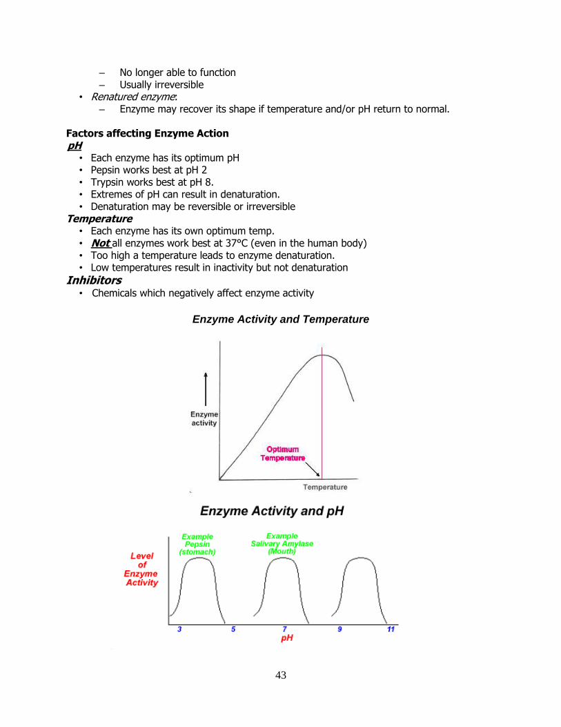
Factors affecting Enzyme Action pH
• Each enzyme has its optimum pH • Pepsin works best at pH 2 • Trypsin works best at pH 8. • Extremes of pH can result in denaturation. • Denaturation may be reversible or irreversible
Temperature • Each enzyme has its own optimum temp. • Not all enzymes work best at 37°C (even in the human body) • Too high a temperature leads to enzyme denaturation. • Low temperatures result in inactivity but not denaturation
Inhibitors • Chemicals which negatively affect enzyme activity
Enzyme Activity and Temperature
44
Immobilised Enzymes • Immobilised Enzymes are enzymes which are not free in solution; they are trapped in a
soft permeable gel or attached to the internal surface of a porous solid. • Many cellular reactions involve enzymes which are immobilised
e.g. enzymes on membranes in the Electron Transport Chain of Respiration. • Immobilised enzymes are often used in industry in a bioreactor. This process is known as
bioprocessing.
Advantages of Immobilised Enzymes • More economical – can be reused. • Easier separation from product. • Continuous production of product. • Immobilised enzymes are more stable, so they last longer. • Large scale manufacture is easier.
Bioprocessing with immobilised enzymes
• Bioprocessing is the use of living cells, their components or enzymes to make products of commercial or scientific value or destroy harmful wastes.
• Examples of Bioprocessing – Production of glucose syrup from sucrose using invertase (sucrase). – Production of glucose from cellulose using cellulases.
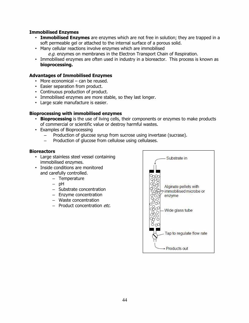
Bioreactors
• Large stainless steel vessel containing immobilised enzymes.
• Inside conditions are monitored and carefully controlled.
– Temperature – pH – Substrate concentration – Enzyme concentration – Waste concentration – Product concentration etc.
45
PPhhoottoossyynntthheessiiss The manufacture of carbohydrate by living organisms from inorganic molecules using the energy from light. Photosynthetic organisms use a range of molecules known as collectively as chlorophyll to trap light. The light energy is converted to chemical energy. Note that photosynthetic organisms do not make carbohydrate from light. They make carbohydrate from inorganic molecules such as carbon dioxide and water, and use the energy from light to carry out the reactions required to do this.
chlorophyll
6CO2 + 6H2O + light C6H12O6 + 6O2
Photosynthesis • Plants require chlorophyll for photosynthesis • Chlorophyll is only found in chloroplasts • There is a high concentration of chloroplasts in leaves. • More chloroplasts in the upper layer of the leaf (palisade mesophyll). • Plants from low light habitats have high concentration of chloroplasts
Chlorophyll
• Chlorophyll molecules are pigments found in photosynthetic organisms • These molecules trap or absorb light energy • When a light energy is absorbed it is transferred to electrons • These electrons become energised or ‘excited’ • The excited electrons often give off their energy as heat. • Sometimes, the electrons energy can be used to generate ATP
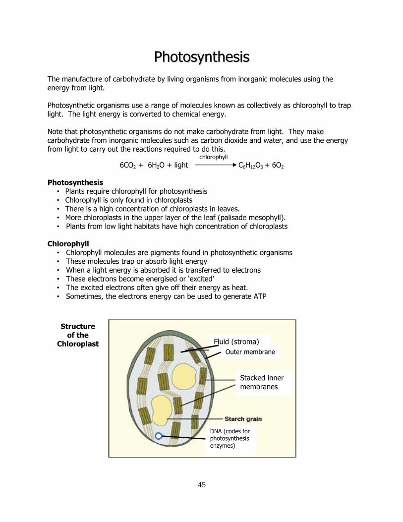
Structure of the
Chloroplast Fluid (stroma)
Stacked inner
membranes
DNA (codes for photosynthesis enzymes)
Outer membrane
46
Inside the chloroplast • Double outer membrane • Chlorophyll-rich inner membranes
Light-dependent stage occurs here Folded structure increases surface area for increased light absorption
• Liquid phase interior Light-independent stage occurs here
Light Stage
• ATP is generated using the energy from electrons • Water is split into
H+ ions electrons (e-) O2 (mostly waste released through stomata)
Dark Stage
• ATP energy is used to make glucose by combining H+ ions electrons CO2 (mostly from the air, some from mitochondria)
The Biochemistry of Photosynthesis Light-dependent stage (“Light stage”)
Only happens in the presence of light NADPH and ATP are produced Water is split, O2 is released Chlorophyll is used
Light-independent stage (“Dark stage”)
Light is not required NADPH and ATP produced in the 1st stage is used CO2 reduced to sugar by the H+ ions and electrons NADPH provides H+ and electrons for the production of glucose
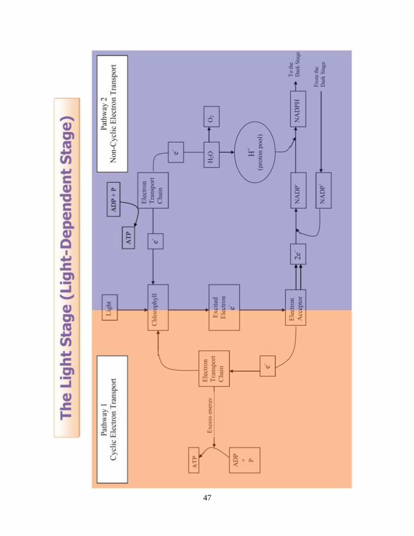
The Light Stage (Light-Dependent Stage)
• Only takes place in the presence of light. • 2 pathways • Occurs on internal membranes of chloroplast • Pathway 1 is cyclic
also known as cyclic photophosphorylation excited electrons leave chlorophyll and then return after they have released their
energy • Pathway 2 is non-cyclic
also known as non-cyclic photophosphorylation excited electrons leave chlorophyll, then is carried by NADP- to the Dark stage electron doesn’t return to chlorophyll
Photophosphorylation is the phosphorylation of ADP to form ATP using the energy of sunlight
47
48
Pathway 1 - Cyclic • Chlorophyll is hit by a photon of light. • Electron becomes “excited” or energised. • Energised electron leaves chlorophyll. • An electron acceptor molecule takes this electron and passes to the electron transport
chain (E.T.C.). • As the electron moves along chain of transport molecules it gives up its energy. • This energy is used to make ATP using ADP and phosphates. • Electron returns to chlorophyll.
Pathway 2 – Non-cyclic
• Chlorophyll is hit by a photon of light. • Electron becomes “excited” (high energy). • Energised electron leaves chlorophyll. • An electron acceptor molecule takes this electron and passes it to NADP+. • NADP+ becomes NADP. • NADP then takes another electron to become NADP-. • NADP- attracts a H+ ion (proton) from the pool of protons and becomes NADPH. • NADPH now carries the 2 electrons and the proton into the Dark Stage. • Meanwhile, chlorophyll is still lacking electrons (which have been taken by NADPH). • The chlorophyll “pulls” electrons from nearby water molecules. • This causes the splitting of water into oxygen, protons (H+) and electrons.
the splitting of water using light is called photolysis • The electrons replace those lost by chlorophyll. • The oxygen diffuses out of the chloroplast. • The protons form part of the pool of protons available in the chloroplast.
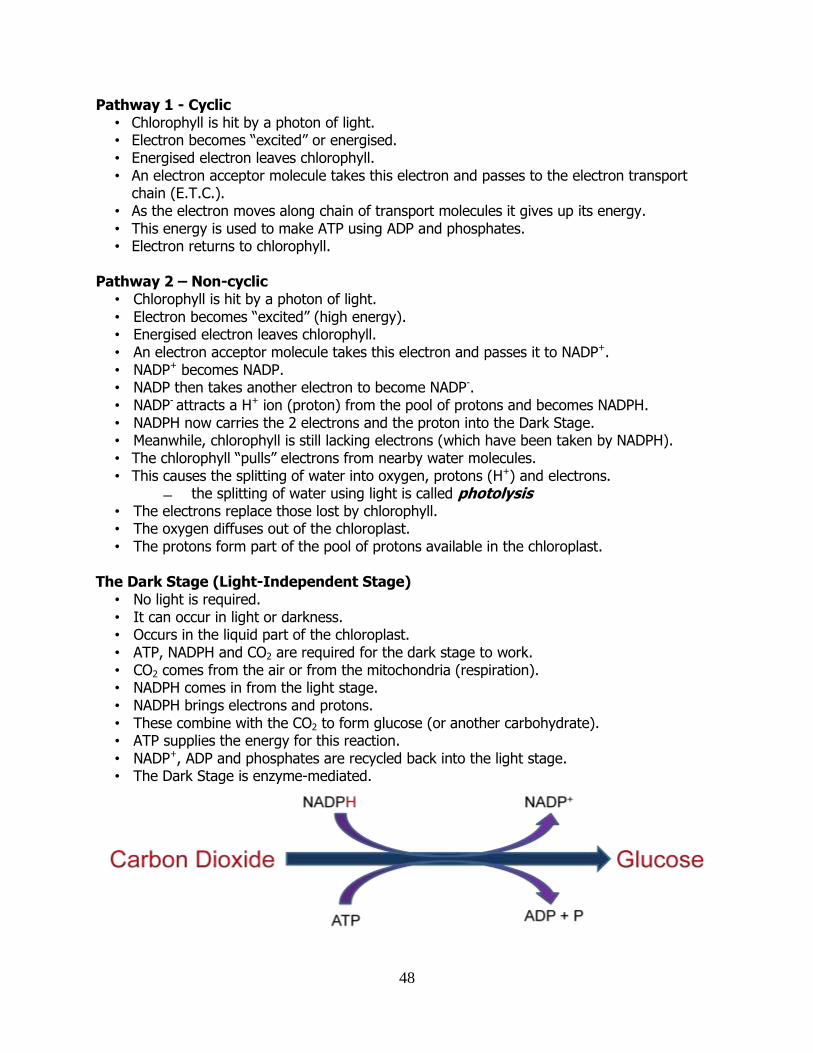
The Dark Stage (Light-Independent Stage)
• No light is required. • It can occur in light or darkness. • Occurs in the liquid part of the chloroplast. • ATP, NADPH and CO2 are required for the dark stage to work. • CO2 comes from the air or from the mitochondria (respiration). • NADPH comes in from the light stage. • NADPH brings electrons and protons. • These combine with the CO2 to form glucose (or another carbohydrate). • ATP supplies the energy for this reaction. • NADP+, ADP and phosphates are recycled back into the light stage. • The Dark Stage is enzyme-mediated.
49
Products of the Light and Dark Stages Light Stage
ATP NADPH Oxygen
Dark Stage Glucose NADP+
ADP Phosphates
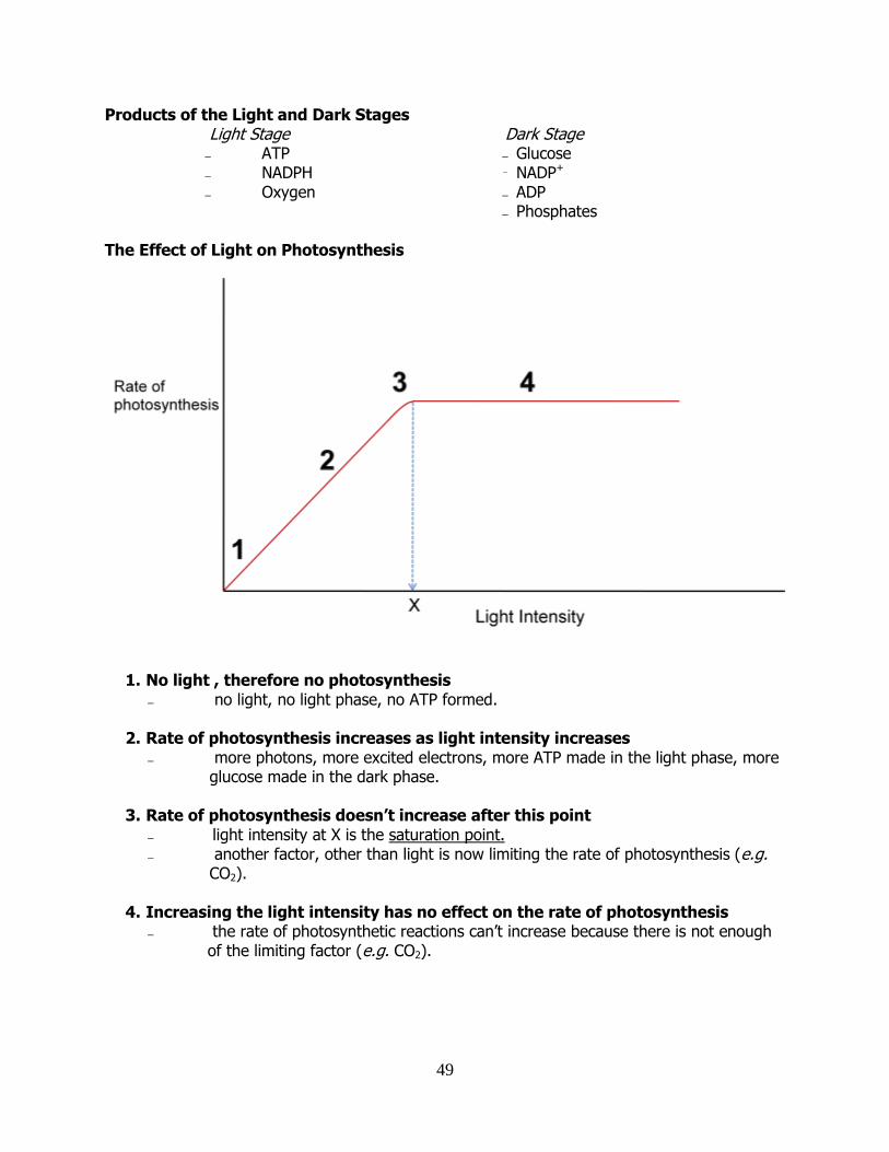
The Effect of Light on Photosynthesis
1. No light , therefore no photosynthesis no light, no light phase, no ATP formed.
2. Rate of photosynthesis increases as light intensity increases
more photons, more excited electrons, more ATP made in the light phase, more glucose made in the dark phase.
3. Rate of photosynthesis doesn’t increase after this point
light intensity at X is the saturation point. another factor, other than light is now limiting the rate of photosynthesis (e.g.
CO2).
4. Increasing the light intensity has no effect on the rate of photosynthesis the rate of photosynthetic reactions can’t increase because there is not enough
of the limiting factor (e.g. CO2).
50
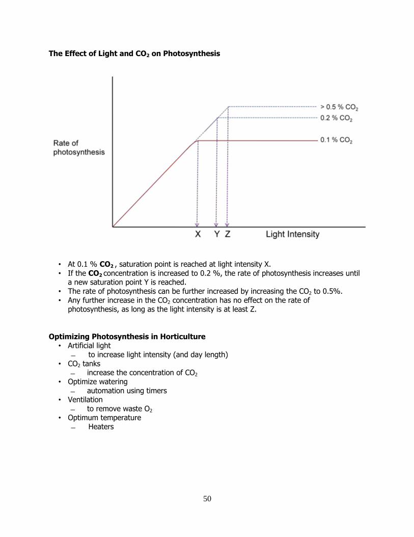
The Effect of Light and CO2 on Photosynthesis
• At 0.1 % CO2 , saturation point is reached at light intensity X. • If the CO2 concentration is increased to 0.2 %, the rate of photosynthesis increases until a new saturation point Y is reached. • The rate of photosynthesis can be further increased by increasing the CO2 to 0.5%. • Any further increase in the CO2 concentration has no effect on the rate of photosynthesis, as long as the light intensity is at least Z.
Optimizing Photosynthesis in Horticulture
• Artificial light to increase light intensity (and day length)
• CO2 tanks increase the concentration of CO2
• Optimize watering automation using timers
• Ventilation to remove waste O2
• Optimum temperature Heaters
51
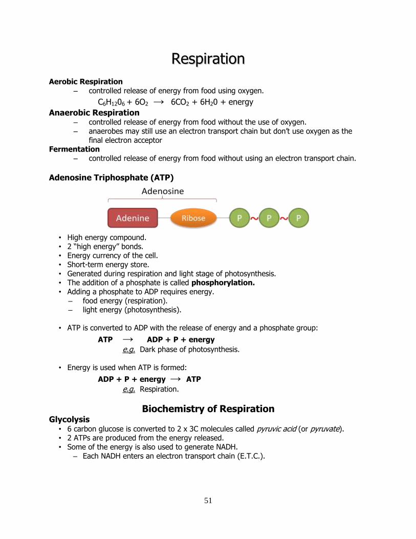
RReessppiirraattiioonn
Aerobic Respiration – controlled release of energy from food using oxygen.
C6H1206 + 6O2 → 6CO2 + 6H20 + energy
Anaerobic Respiration
– controlled release of energy from food without the use of oxygen. – anaerobes may still use an electron transport chain but don’t use oxygen as the
final electron acceptor Fermentation
– controlled release of energy from food without using an electron transport chain.
Adenosine Triphosphate (ATP)
• High energy compound. • 2 “high energy” bonds. • Energy currency of the cell. • Short-term energy store. • Generated during respiration and light stage of photosynthesis. • The addition of a phosphate is called phosphorylation. • Adding a phosphate to ADP requires energy.
– food energy (respiration). – light energy (photosynthesis).
• ATP is converted to ADP with the release of energy and a phosphate group:
ATP → ADP + P + energy
e.g. Dark phase of photosynthesis.
• Energy is used when ATP is formed:
ADP + P + energy → ATP
e.g. Respiration.
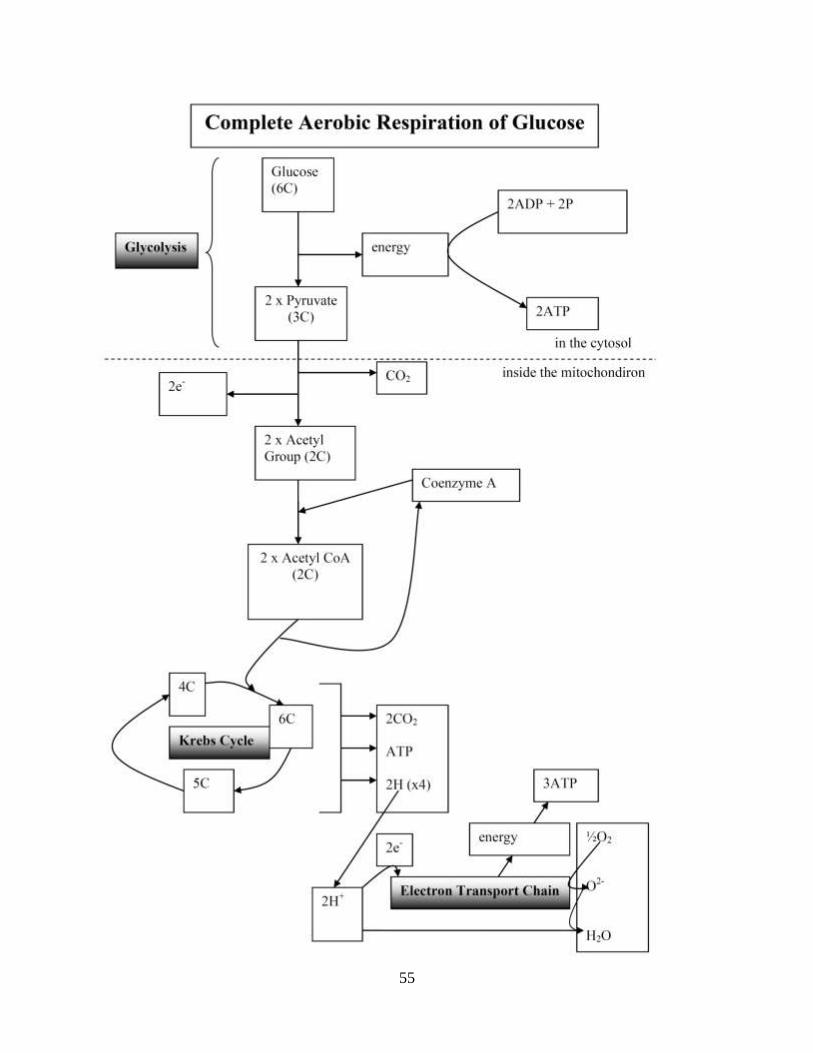
Biochemistry of Respiration Glycolysis
• 6 carbon glucose is converted to 2 x 3C molecules called pyruvic acid (or pyruvate). • 2 ATPs are produced from the energy released. • Some of the energy is also used to generate NADH.
– Each NADH enters an electron transport chain (E.T.C.).
52
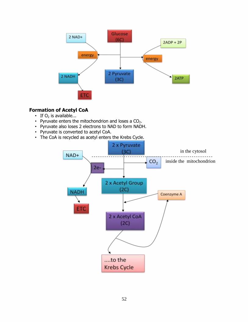
Formation of Acetyl CoA • If O2 is available... • Pyruvate enters the mitochondrion and loses a CO2. • Pyruvate also loses 2 electrons to NAD to form NADH. • Pyruvate is converted to acetyl CoA. • The CoA is recycled as acetyl enters the Krebs Cycle.
in the cytosol
inside the mitochondrion
53
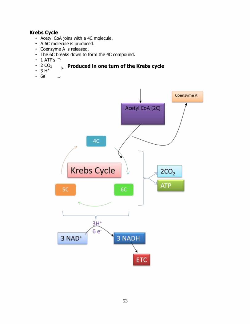
Krebs Cycle • Acetyl CoA joins with a 4C molecule. • A 6C molecule is produced. • Coenzyme A is released. • The 6C breaks down to form the 4C compound. • 1 ATP’s • 2 CO2 • 3 H+ • 6e-
Produced in one turn of the Krebs cycle
54
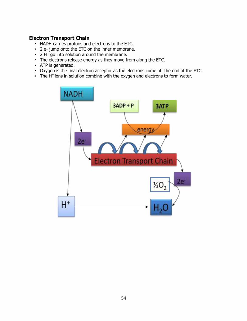
Electron Transport Chain
• NADH carries protons and electrons to the ETC. • 2 e- jump onto the ETC on the inner membrane. • 2 H+ go into solution around the membrane. • The electrons release energy as they move from along the ETC. • ATP is generated. • Oxygen is the final electron acceptor as the electrons come off the end of the ETC. • The H+ ions in solution combine with the oxygen and electrons to form water.
55
56
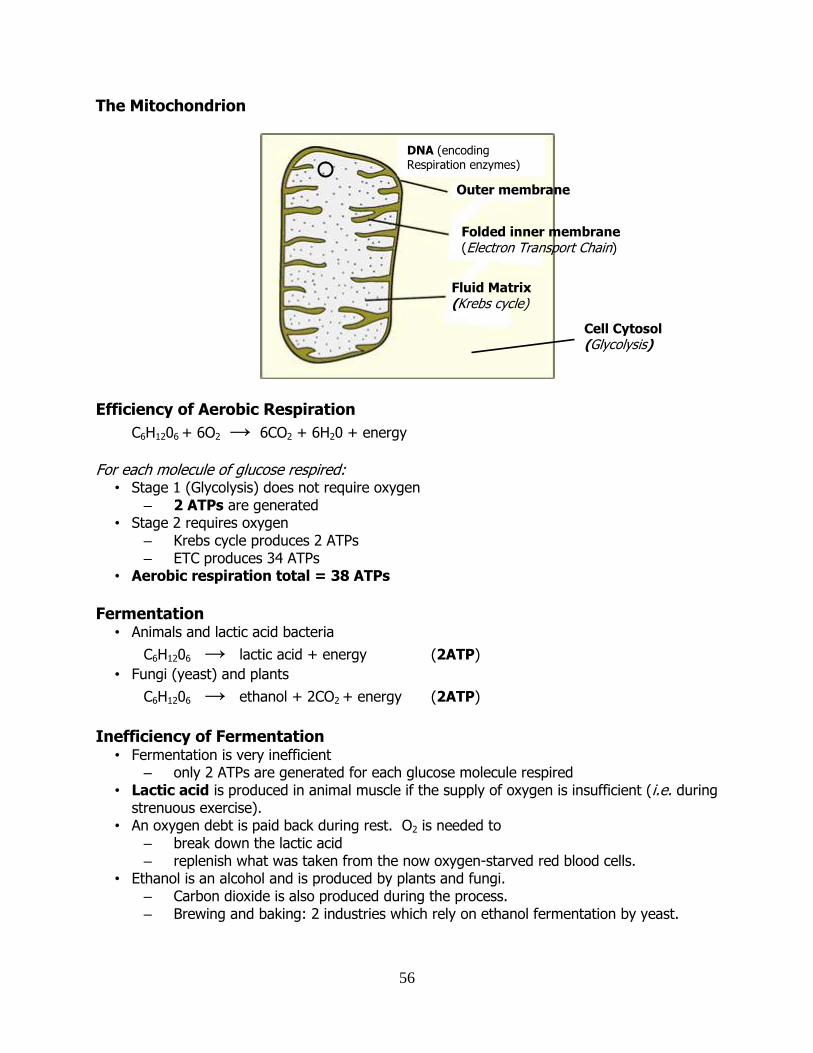
The Mitochondrion
Efficiency of Aerobic Respiration
C6H1206 + 6O2 → 6CO2 + 6H20 + energy
For each molecule of glucose respired:
• Stage 1 (Glycolysis) does not require oxygen – 2 ATPs are generated
• Stage 2 requires oxygen – Krebs cycle produces 2 ATPs – ETC produces 34 ATPs
• Aerobic respiration total = 38 ATPs
Fermentation
• Animals and lactic acid bacteria
C6H1206 → lactic acid + energy (2ATP) • Fungi (yeast) and plants
C6H1206 → ethanol + 2CO2 + energy (2ATP)
Inefficiency of Fermentation
• Fermentation is very inefficient – only 2 ATPs are generated for each glucose molecule respired
• Lactic acid is produced in animal muscle if the supply of oxygen is insufficient (i.e. during strenuous exercise).
• An oxygen debt is paid back during rest. O2 is needed to – break down the lactic acid – replenish what was taken from the now oxygen-starved red blood cells.
• Ethanol is an alcohol and is produced by plants and fungi. – Carbon dioxide is also produced during the process. – Brewing and baking: 2 industries which rely on ethanol fermentation by yeast.
Outer membrane
Folded inner membrane (Electron Transport Chain)
Fluid Matrix (Krebs cycle)
Cell Cytosol (Glycolysis)
DNA (encoding Respiration enzymes)
57
Industrial Fermentations • A huge variety of different products can be produced by growing microorganisms in
culture. • A bioreactor or fermentation vessel can be used for this purpose. • A bioreactor is a container in which a living thing is used in the production of something
useful. It can be sterilised, temperature controlled, stirrers, aerators etc. • Immobilised cells can be used in a bioreactor to carry our conversions.
• Cells can be immobilised in the same way as enzymes are immobilised (see Enzymes).
• Example: Immobilised yeast cells are used in the industrial fermentation of alcohol.
Advantages of using immobilized cells for fermentations
• Set-up is more suitable to continuous flow process which is more efficient than batch
fermentation.
• Product is easier to purify.
• Cells are more easily recovered and reused.
• Cells are in better condition during process since they are not physically stirred.
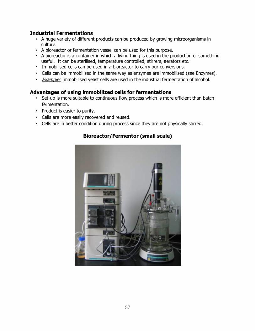
Bioreactor/Fermentor (small scale)
58
DDNNAA aanndd RRNNAA The Structure and Function of Nucleic Acids
Deoxyribonucleic Acid (DNA) • Double helix structure discovered 1953 by Watson and Crick. • DNA is the genetic material. • Found in the nucleus of eukaryotes. • Also found in mitochondria and chloroplasts. • Made up 4 different types of units called nucleotides joined together in long strands. • The sequence of the nucleotides is the “genetic code”.
Nucleotides • Units of DNA made up of 3 parts
– A sugar – A phosphate – A base
• There are 4 different bases – Adenine (A) – Guanine (G) – Cytosine (C) – Thymine (T)
Double-stranded DNA • DNA is generally found as long double-stranded molecules (“strands”). • Two strands are held together by hydrogen bonds. • Hydrogen bonds are between the bases on opposite strands. • Two complementary strands are in opposite directions.
Nucleotide Bases • Adenine and Guanine are called purines. • Cytosine and Thymine are called pyrimidines. • A is complementary with T. • G is complementary with C. • A binds to T by a double hydrogen bond. • G binds to C by a triple hydrogen bond. • 2 complementary strands form a double-stranded helical structure – the double helix.
59
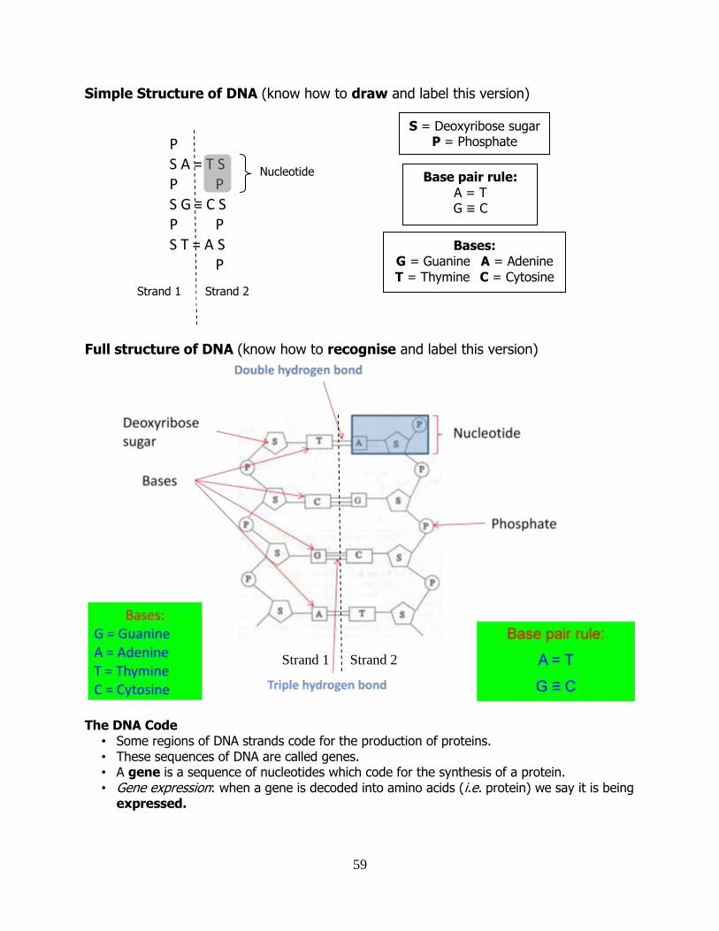
Simple Structure of DNA (know how to draw and label this version)
P S A = T S P P S G ≡ C S P P S T = A S P
Full structure of DNA (know how to recognise and label this version)
The DNA Code
• Some regions of DNA strands code for the production of proteins. • These sequences of DNA are called genes. • A gene is a sequence of nucleotides which code for the synthesis of a protein. • Gene expression: when a gene is decoded into amino acids (i.e. protein) we say it is being
expressed.
Nucleotide
S = Deoxyribose sugar P = Phosphate
Bases: G = Guanine A = Adenine
T = Thymine C = Cytosine
Base pair rule: A = T G ≡ C
Strand 1 Strand 2
Strand 1 Strand 2
60
Non-coding DNA • Between genes (and sometimes within genes) there are regions of DNA that do not code
for the production of any protein. • Non-coding DNA is sometimes known (inaccurately) as “junk DNA”. • Non-coding DNA has various functions e.g DNA folding. • > 90 % of human DNA is made up of non-coding regions.
The Language of DNA • The sequence of bases that make up a gene can be “read” like words. • The 4 bases are the letters. • Each triplet of bases codes for one amino acid. • These triplets are known as codons, and are the words of the language. • A sequence of codons (words), makes up a gene (like a recipe). • The sum of all the genes in an organisms DNA is called its genome (like a “cook-book” of
DNA recipes).
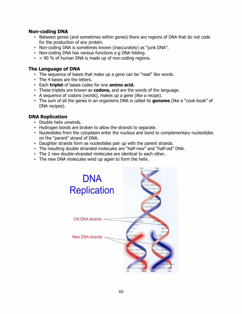
DNA Replication • Double helix unwinds. • Hydrogen bonds are broken to allow the strands to separate. • Nucleotides from the cytoplasm enter the nucleus and bond to complementary nucleotides
on the “parent” strand of DNA. • Daughter strands form as nucleotides pair up with the parent strands. • The resulting double stranded molecules are “half-new” and “half-old” DNA. • The 2 new double-stranded molecules are identical to each other. • The new DNA molecules wind up again to form the helix.
61
Ribonucleic Acid (RNA) There are 3 different types of RNA molecules
Messenger RNA (mRNA)
– a copy (transcript) of a coding region DNA
Ribosomal RNA (rRNA)
– ribosomes are made of rRNA and protein
Transfer RNA (tRNA)
– carries amino acids to the ribosomes during translation
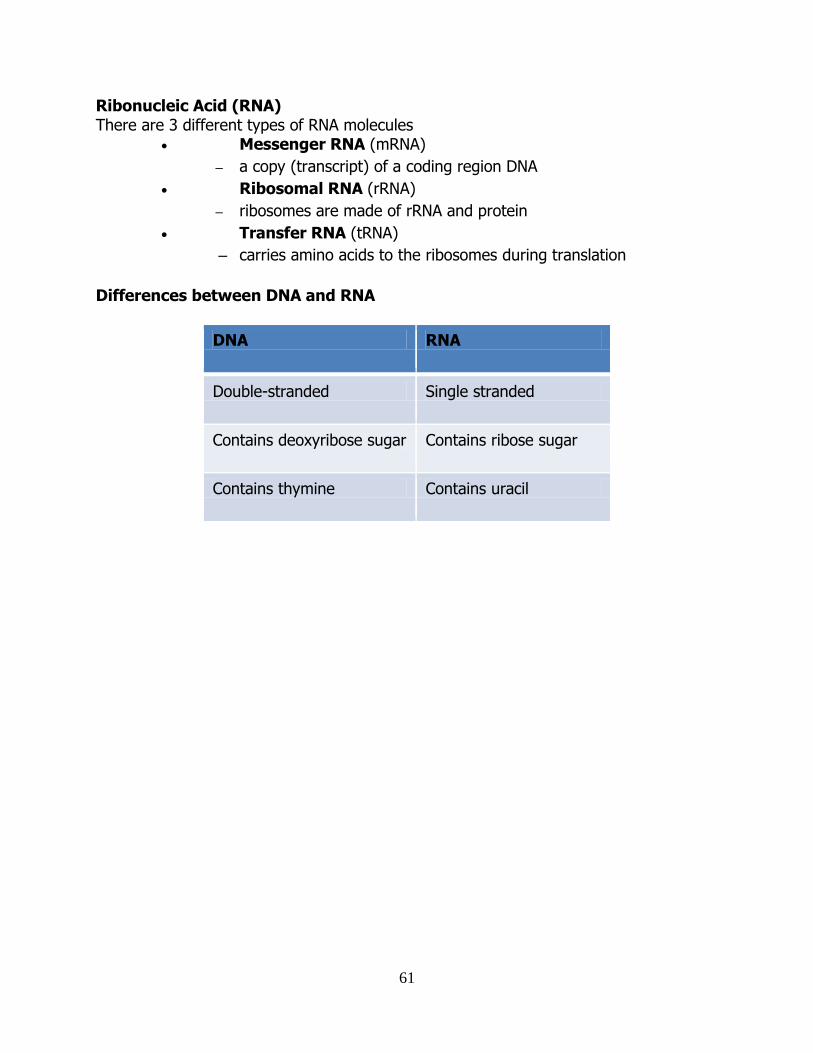
Differences between DNA and RNA
DNA RNA
Double-stranded Single stranded
Contains deoxyribose sugar Contains ribose sugar
Contains thymine Contains uracil
62
PPrrootteeiinn SSyynntthheessiiss
Protein Synthesis is the combination of the transcription of a region of DNA followed by translation into protein For Protein Synthesis, cells need:
• A supply of amino acids – the cytoplasm
• Instructions on what amino acids to join together – the genetic code
• An assembly line – the ribosomes
• A messenger to carry the information from DNA to ribosomes – mRNA
The Language of DNA – the Genetic Code
• The sequence of bases that make up a gene can be “read” like words. • The 4 bases are the letters. • Each triplet of bases codes for one amino acid. • These triplets are known as codons, and are the words of the language. • A sequence of codons (words), makes up a gene (like a recipe). • The sum of all the genes in an organisms DNA is called its genome (like a “cook-book” of
DNA recipes).
2 stages of Protein Synthesis: 1. Transcription – the copying of the DNA sequence into an mRNA sequence. 2. Translation – the production of a protein according to the mRNA sequence.
Transcription
• The transfer of information in the nucleus from a DNA molecule to an RNA molecule. • The DNA is unwound. • Only 1 DNA strand serves as the template. • Complementary RNA bases bond with the template strand. • The enzyme RNA polymerase joins the RNA bases together. • A new strand of mRNA is formed according to the base pair rule. • Uracil (rather than Thymine) is complementary to Adenine during mRNA synthesis. • Transcription
– starts at promoter DNA (AUG) – ends at terminator DNA (stop)
• mRNA is processed by removing non-coding regions. • mRNA molecule is released from the nucleus into the cytoplasm
63
Translation Translation involves all of the following components:
• mRNA (codons) • tRNA (anticodons) • rRNA (ribosomes) • amino acids
3 Types of RNA
• messenger RNA (mRNA) • transfer RNA (tRNA) • ribosomal RNA (rRNA)
Messenger RNA (mRNA) • Carries the information for a specific protein. • Made up of 500 to 1000 nucleotides long. • Made up of codons (sequence of three bases) • Each codon is specific for one amino acid.
Transfer RNA (tRNA)
• Made up of 75 to 80 nucleotides long. • Picks up the appropriate amino acid floating in the cytoplasm. • Transports amino acids to the mRNA. • Has anticodons that are complementary to mRNA codons. • Recognizes the appropriate codons on the mRNA and bonds to them with H-bonds.
Ribosomal RNA (rRNA)
• Ribosomes are made of RNA and protein. • Ribosomes consist of a large and a small subunit which “sandwich” the mRNA transcripts
during protein synthesis.
Ribosomes
– They are composed of rRNA (40%) and proteins (60%). – Each ribosome has a large and a small subunit. – Both units come together and help bind the mRNA and tRNA.
Summary of Translation There are 3 stages in translation:
1. Initiation: the mRNA binds to a ribosome. 2. Elongation: the strand of mRNA is pulled through the ribosome three bases at a
time, in triplets. – each of these triplets on the mRNA strand is called a codon. – tRNA molecules add amino acids to a growing peptide chain.
3. Termination: the ribosome comes to a stop codon and the peptide chain and the
mRNA detach from the ribosome.
all are produced in the nucleus

64
Initiation: • A strand of mRNA moves out of the nucleus to the cytoplasm.
• In the cytoplasm, the mRNA binds to rRNA in a ribosome.
• The mRNA binds to a small ribosomal sub-unit.
• A transfer RNA (tRNA) with a complementary anticodon binds to the “start” codon of the
mRNA.
- There is a supply of tRNA’s in the cytosol. Each one carries a particular amino acid
that corresponds to its anticodon.
• The large ribosomal subunit binds so that the mRNA is sandwiched between the 2
ribosomal sub-units.
- Each ribosome has space for 2 tRNA’s at a time. Each anticodon on a tRNA is
complimentary to a codon on the mRNA.
• This first tRNA carries the first amino acid of the protein.
Elongation:
• A second tRNA arrives, bringing with it a second amino acid.
• This second tRNA has an anticodon matching the second codon on the mRNA
• It lands right next to the first tRNA.
• The adjacent amino acids join together with a peptide bond.
• The first tRNA leaves without its amino acid.
• As it leaves, it pulls the mRNA strand through the ribosome.
• Another tRNA lands bringing with it the third amino acid.
• As each codon is read, the next tRNA brings in a new amino acid and the polypeptide
(protein) chain grows.
• The process requires ATP and enzymes to work.
Termination:
• tRNA’s continue to bind until a stop codon is reached.
• The polypeptide chain is complete, and it falls away from the ribosome, as does the
mRNA.
Functional Protein: the newly formed polypeptide chain now has to undergo folding and
processing to become the fully functional protein.
DNA Coding Strand A T G G G C T C C
Template Strand T A C C C G A G G
mRNA Codons A U G G G C U C C
tRNA Anticodons U A C C C G A G G
Protein Amino acids Methionine – Glycine – Serine

65
Transcription of DNA to mRNA Translation of mRNA into protein
(in the nucleus) (at the ribosomes)
tRNA
Ribosome
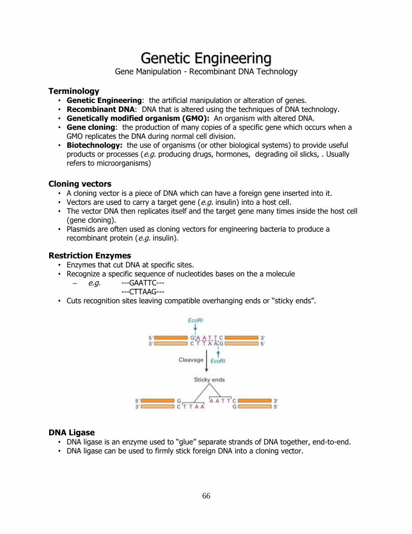
66
GGeenneettiicc EEnnggiinneeeerriinngg Gene Manipulation - Recombinant DNA Technology
Terminology
• Genetic Engineering: the artificial manipulation or alteration of genes. • Recombinant DNA: DNA that is altered using the techniques of DNA technology. • Genetically modified organism (GMO): An organism with altered DNA. • Gene cloning: the production of many copies of a specific gene which occurs when a
GMO replicates the DNA during normal cell division. • Biotechnology: the use of organisms (or other biological systems) to provide useful
products or processes (e.g. producing drugs, hormones, degrading oil slicks, . Usually refers to microorganisms)
Cloning vectors
• A cloning vector is a piece of DNA which can have a foreign gene inserted into it. • Vectors are used to carry a target gene (e.g. insulin) into a host cell. • The vector DNA then replicates itself and the target gene many times inside the host cell
(gene cloning). • Plasmids are often used as cloning vectors for engineering bacteria to produce a
recombinant protein (e.g. insulin).
Restriction Enzymes • Enzymes that cut DNA at specific sites. • Recognize a specific sequence of nucleotides bases on the a molecule
– e.g. ---GAATTC--- ---CTTAAG---
• Cuts recognition sites leaving compatible overhanging ends or “sticky ends”.
DNA Ligase
• DNA ligase is an enzyme used to “glue” separate strands of DNA together, end-to-end. • DNA ligase can be used to firmly stick foreign DNA into a cloning vector.
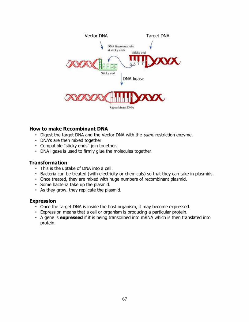
67
How to make Recombinant DNA • Digest the target DNA and the Vector DNA with the same restriction enzyme. • DNA’s are then mixed together. • Compatible “sticky ends” join together. • DNA ligase is used to firmly glue the molecules together.
Transformation
• This is the uptake of DNA into a cell. • Bacteria can be treated (with electricity or chemicals) so that they can take in plasmids. • Once treated, they are mixed with huge numbers of recombinant plasmid. • Some bacteria take up the plasmid. • As they grow, they replicate the plasmid.
Expression • Once the target DNA is inside the host organism, it may become expressed. • Expression means that a cell or organism is producing a particular protein. • A gene is expressed if it is being transcribed into mRNA which is then translated into
protein.
DNA ligase
Vector DNA Target DNA
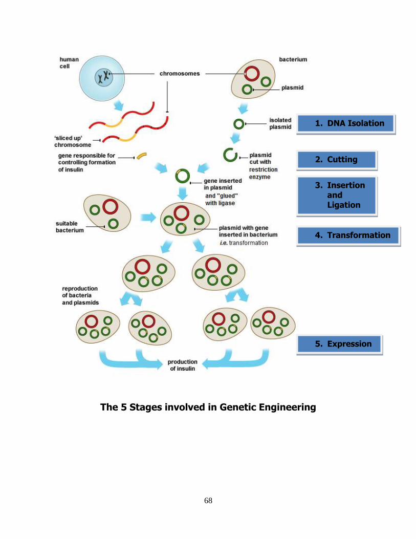
68
The 5 Stages involved in Genetic Engineering
2. Cutting
1. DNA Isolation
3. Insertion and Ligation
4. Transformation
5. Expression
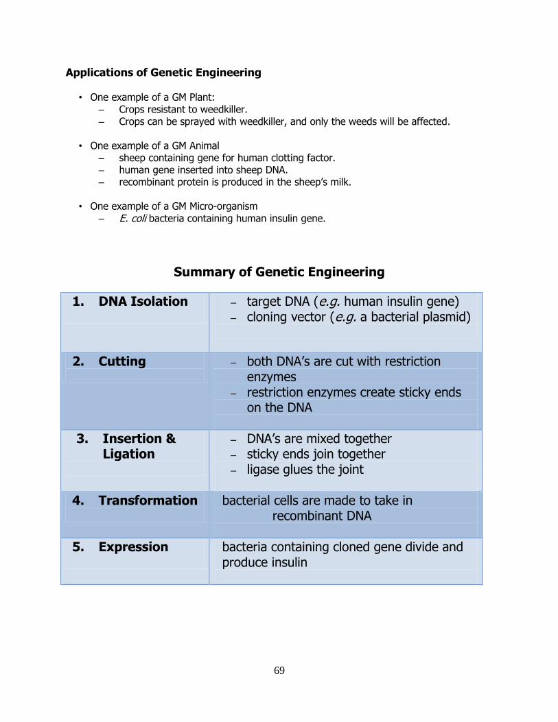
69
Applications of Genetic Engineering
• One example of a GM Plant: – Crops resistant to weedkiller. – Crops can be sprayed with weedkiller, and only the weeds will be affected.
• One example of a GM Animal
– sheep containing gene for human clotting factor. – human gene inserted into sheep DNA. – recombinant protein is produced in the sheep’s milk.
• One example of a GM Micro-organism
– E. coli bacteria containing human insulin gene.
Summary of Genetic Engineering
1. DNA Isolation
– target DNA (e.g. human insulin gene) – cloning vector (e.g. a bacterial plasmid)
2. Cutting
– both DNA’s are cut with restriction enzymes
– restriction enzymes create sticky ends on the DNA
3. Insertion & Ligation
– DNA’s are mixed together – sticky ends join together – ligase glues the joint
4. Transformation
bacterial cells are made to take in recombinant DNA
5. Expression
bacteria containing cloned gene divide and produce insulin
70
DDNNAA PPrrooffiilliinngg “Genetic Fingerprinting”
What is DNA Profiling? • The process of making a unique pattern of bands (like a barcode) from the DNA of a
person. which can be distinguished from the pattern of another person’s DNA. • If two people are genetically different, then they will have different DNA profiles. • Some areas of our (non-coding) DNA are more variable than others. • These hyper-variable regions are analysed in DNA profiling.
The 4 Steps of DNA Profiling
1. DNA Extraction – DNA is isolated from tissue.
2. Cutting of DNA – restriction enzymes are used to digest the DNA.
3. Separation of DNA Fragments – restriction fragments are separated using gels.
4. Use of DNA Probe – some of the fragments are highlighted to produce the barcode pattern.
DNA Extraction
• Suitable tissue samples are processed – blood, skin, hair, semen etc.
• Cells are disrupted so that DNA is released. • DNA is separated from the cell debris. • If the amount of DNA is very small, it can be easily copied in the laboratory to produce
sufficient quantities.
Cutting of the DNA • A restriction enzyme is used to cut the DNA into a collection of different sized fragments. • Restriction enzymes recognise and cut at specific short sequences in the DNA. • Where these sequences occur varies from person to person (especially in the hyper-
variable regions of the non-coding DNA). • This digestion of DNA results in a very large collection of different sized fragments of DNA.
Separation of DNA Fragments • The collection of DNA fragments is placed into one end of a slab of gel. • An electric current is applied across the gel. • The DNA is at the negative terminal. • Since DNA is negatively charged it moves towards the positive terminal. • Smaller fragments will move more quickly through the gel, larger ones move slowly.
DNA Probe
• Specially designed, short pieces of radioactive or fluorescent DNA. • These will bind only to some of the separated fragments in the gel. • The fragments that the probe binds to become highlighted. • The barcode-like pattern of highlighted fragments is the “DNA fingerprint”.
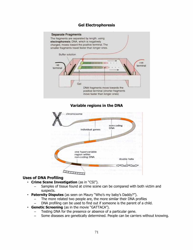
71
Gel Electrophoresis
Variable regions in the DNA
Uses of DNA Profiling
• Crime Scene Investigation (as in “CSI”). – Samples of tissue found at crime scene can be compared with both victim and
suspects. • Paternity Disputes (as seen on Maury “Who’s my baby’s Daddy?”).
– The more related two people are, the more similar their DNA profiles – DNA profiling can be used to find out if someone is the parent of a child.
• Genetic Screening (as in the movie “GATTACA”). – Testing DNA for the presence or absence of a particular gene. – Some diseases are genetically determined. People can be carriers without knowing.
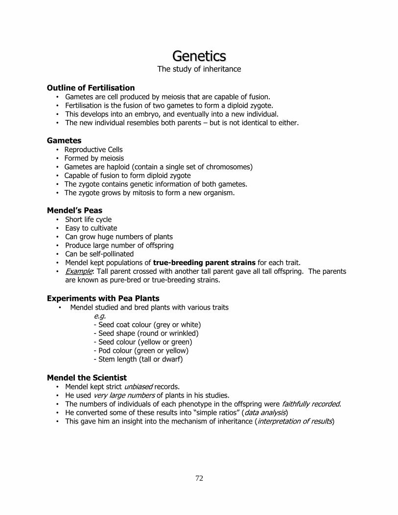
72
GGeenneettiiccss The study of inheritance
Outline of Fertilisation
• Gametes are cell produced by meiosis that are capable of fusion. • Fertilisation is the fusion of two gametes to form a diploid zygote. • This develops into an embryo, and eventually into a new individual. • The new individual resembles both parents – but is not identical to either.
Gametes • Reproductive Cells • Formed by meiosis • Gametes are haploid (contain a single set of chromosomes) • Capable of fusion to form diploid zygote • The zygote contains genetic information of both gametes. • The zygote grows by mitosis to form a new organism.
Mendel’s Peas • Short life cycle • Easy to cultivate • Can grow huge numbers of plants • Produce large number of offspring • Can be self-pollinated • Mendel kept populations of true-breeding parent strains for each trait. • Example: Tall parent crossed with another tall parent gave all tall offspring. The parents
are known as pure-bred or true-breeding strains.
Experiments with Pea Plants
• Mendel studied and bred plants with various traits e.g.
- Seed coat colour (grey or white) - Seed shape (round or wrinkled) - Seed colour (yellow or green) - Pod colour (green or yellow) - Stem length (tall or dwarf)
Mendel the Scientist
• Mendel kept strict unbiased records. • He used very large numbers of plants in his studies. • The numbers of individuals of each phenotype in the offspring were faithfully recorded. • He converted some of these results into “simple ratios” (data analysis) • This gave him an insight into the mechanism of inheritance (interpretation of results)
73
Mendel’s Experiments Cross 1
• Mendel crossed pea plants for his studies, looking at many traits. Example: flower colour: • He crossed true-breeding purple plants with true-breeding white-flowered plants. • He found that all the progeny (the “F1 generation”) had purple flowers.
Cross 2:
• He crossed these F1 purple plants with themselves (a “self-cross”). • This time he found that the colour of flowers of the progeny (F2) were in the ratio 3
purple: 1 white. • Mendel arrived at this ratio after repeating the crosses many times. • He also used other traits such as tall/small and smooth/wrinkled to replicate his findings.
Results from Mendel's Experiments
Parental Cross
F1 Phenotype
F2 Phenotypic
Ratio
F2 Ratio
Round x Wrinkled
Seed
Round 5474 Round :
1850 Wrinkled
2.96:1
Yellow x Green
Seeds
Yellow 6022 Yellow :
2001 Green
3.01:1
Axial x Terminal
Flower Position
Axial
705 Axial :
224 Terminal
3.15:1
Tall x Dwarf Plants
Tall 787 Tall :
227 Dwarf
2.84:1
Mendel’s Puzzle Mendel was puzzled by the following facts:
1. When the original pea plants were crossed purple with white, all the progeny were purple
2. When the first generation purple offspring were self-crossed, the white colour was seen in the progeny (the 2nd generation) but only in 25% of them.
Mendel’s Explanation - The Law of Segregation (Mendel’s 1st Law) Mendel explained these results with the following deductions (Mendel’s 1st Law):
• Every characteristic is governed by a pair of factors (genes). • The factors separate during gamete formation. • Each gamete only receives one of each pair of factors. • At fertilisation a pair of factors is re-established for each characteristic.
Mendel’s First Law (put another way….) • Alleles of a gene come in pairs which separate at gamete formation, each gamete
receiving only one of each pair. • Pairs of genes are made again when the gametes fuse at fertilisation.
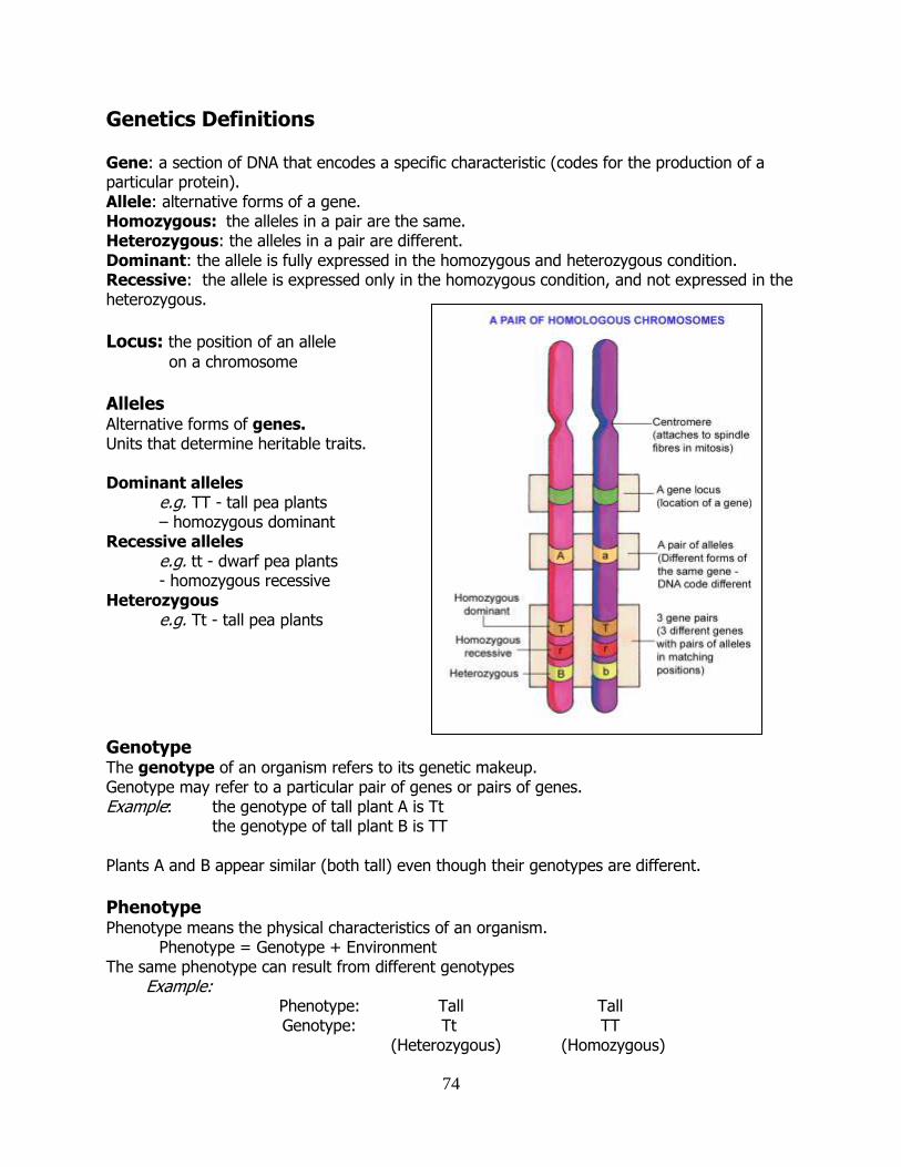
74
Genetics Definitions Gene: a section of DNA that encodes a specific characteristic (codes for the production of a particular protein). Allele: alternative forms of a gene. Homozygous: the alleles in a pair are the same. Heterozygous: the alleles in a pair are different. Dominant: the allele is fully expressed in the homozygous and heterozygous condition. Recessive: the allele is expressed only in the homozygous condition, and not expressed in the heterozygous. Locus: the position of an allele
on a chromosome Alleles Alternative forms of genes. Units that determine heritable traits. Dominant alleles e.g. TT - tall pea plants – homozygous dominant Recessive alleles e.g. tt - dwarf pea plants - homozygous recessive Heterozygous e.g. Tt - tall pea plants
Genotype The genotype of an organism refers to its genetic makeup. Genotype may refer to a particular pair of genes or pairs of genes. Example: the genotype of tall plant A is Tt the genotype of tall plant B is TT Plants A and B appear similar (both tall) even though their genotypes are different.
Phenotype Phenotype means the physical characteristics of an organism.
Phenotype = Genotype + Environment The same phenotype can result from different genotypes Example:
Phenotype: Tall Tall Genotype: Tt TT
(Heterozygous) (Homozygous)
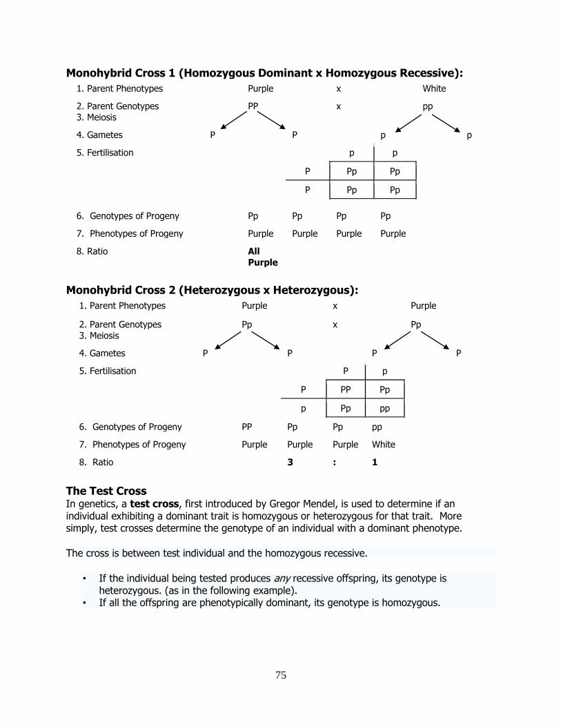
75
Monohybrid Cross 1 (Homozygous Dominant x Homozygous Recessive):
1. Parent Phenotypes Purple x White
2. Parent Genotypes
3. Meiosis
PP x pp
4. Gametes P P p p
5. Fertilisation p p
P Pp Pp
P Pp Pp
6. Genotypes of Progeny Pp Pp Pp Pp
7. Phenotypes of Progeny Purple Purple Purple Purple
8. Ratio All
Purple
Monohybrid Cross 2 (Heterozygous x Heterozygous):
1. Parent Phenotypes Purple x Purple
2. Parent Genotypes
3. Meiosis
Pp x Pp
4. Gametes P P P P
5. Fertilisation P p
P PP Pp
p Pp pp
6. Genotypes of Progeny PP Pp Pp pp
7. Phenotypes of Progeny Purple Purple Purple White
8. Ratio 3 : 1
The Test Cross In genetics, a test cross, first introduced by Gregor Mendel, is used to determine if an individual exhibiting a dominant trait is homozygous or heterozygous for that trait. More simply, test crosses determine the genotype of an individual with a dominant phenotype.
The cross is between test individual and the homozygous recessive.
• If the individual being tested produces any recessive offspring, its genotype is heterozygous. (as in the following example).
• If all the offspring are phenotypically dominant, its genotype is homozygous.
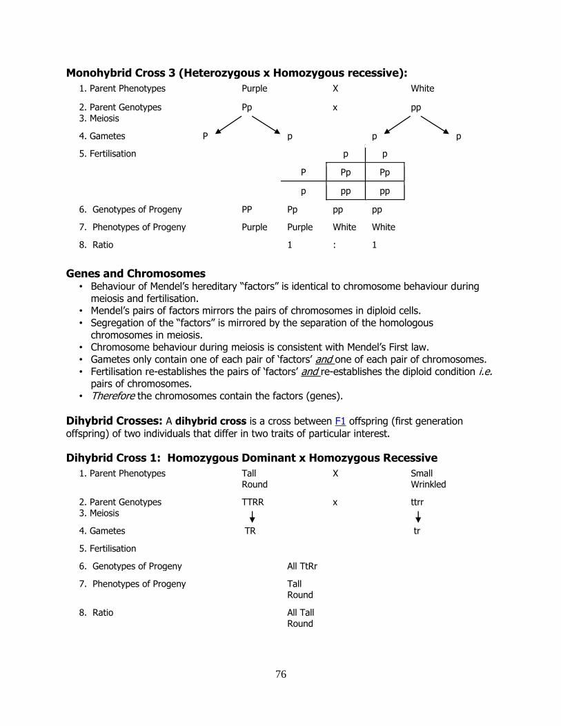
76
Monohybrid Cross 3 (Heterozygous x Homozygous recessive):
1. Parent Phenotypes Purple X White
2. Parent Genotypes
3. Meiosis
Pp x pp
4. Gametes P p p p
5. Fertilisation p p
P Pp Pp
p pp pp
6. Genotypes of Progeny PP Pp pp pp
7. Phenotypes of Progeny Purple Purple White White
8. Ratio 1 : 1
Genes and Chromosomes
• Behaviour of Mendel’s hereditary “factors” is identical to chromosome behaviour during meiosis and fertilisation.
• Mendel’s pairs of factors mirrors the pairs of chromosomes in diploid cells. • Segregation of the “factors” is mirrored by the separation of the homologous
chromosomes in meiosis. • Chromosome behaviour during meiosis is consistent with Mendel’s First law. • Gametes only contain one of each pair of ‘factors’ and one of each pair of chromosomes. • Fertilisation re-establishes the pairs of ‘factors’ and re-establishes the diploid condition i.e.
pairs of chromosomes. • Therefore the chromosomes contain the factors (genes).
Dihybrid Crosses: A dihybrid cross is a cross between F1 offspring (first generation
offspring) of two individuals that differ in two traits of particular interest.
Dihybrid Cross 1: Homozygous Dominant x Homozygous Recessive
1. Parent Phenotypes Tall Round
X Small Wrinkled
2. Parent Genotypes 3. Meiosis
TTRR x ttrr
4. Gametes TR tr
5. Fertilisation
6. Genotypes of Progeny All TtRr
7. Phenotypes of Progeny Tall Round
8. Ratio All Tall Round
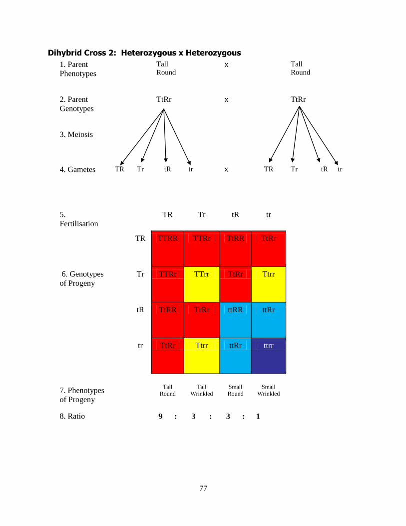
77
Dihybrid Cross 2: Heterozygous x Heterozygous
1. Parent
Phenotypes
Tall
Round x Tall
Round
2. Parent
Genotypes
TtRr x
TtRr
3. Meiosis
4. Gametes TR Tr tR tr x TR Tr tR tr
5.
Fertilisation
TR Tr tR tr
TR TTRR TTRr TtRR TtRr
6. Genotypes
of Progeny
Tr TTRr TTrr TtRr Ttrr
tR TtRR TrRr ttRR ttRr
tr TtRr Ttrr ttRr ttrr
7. Phenotypes
of Progeny
Tall
Round
Tall
Wrinkled
Small
Round
Small
Wrinkled
8. Ratio 9 : 3 : 3 : 1
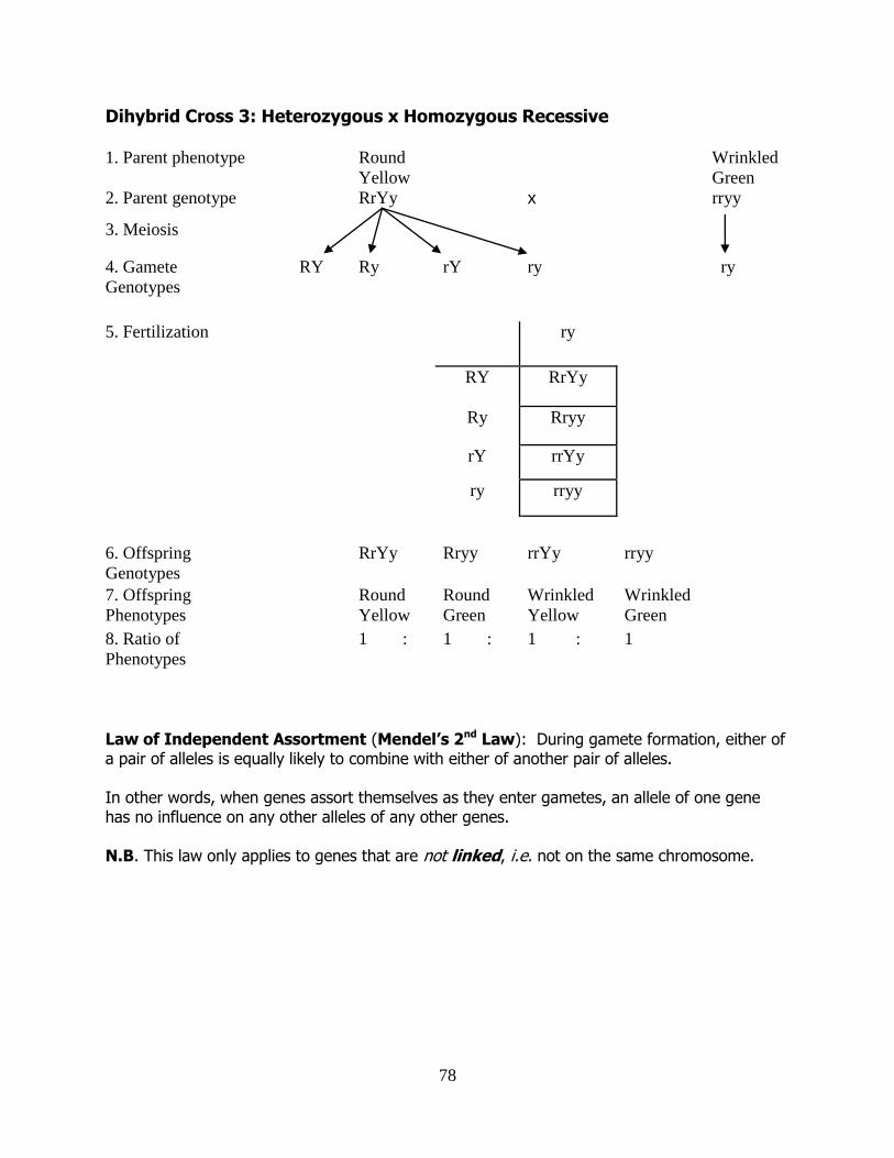
78
Dihybrid Cross 3: Heterozygous x Homozygous Recessive 1. Parent phenotype Round
Yellow
Wrinkled
Green
2. Parent genotype
RrYy x rryy
3. Meiosis
4. Gamete
Genotypes
RY Ry rY ry ry
5. Fertilization ry
RY RrYy
Ry Rryy
rY rrYy
ry rryy
6. Offspring
Genotypes
RrYy Rryy rrYy rryy
7. Offspring
Phenotypes
Round
Yellow
Round
Green
Wrinkled
Yellow
Wrinkled
Green
8. Ratio of
Phenotypes
1 : 1 : 1 : 1
Law of Independent Assortment (Mendel’s 2nd Law): During gamete formation, either of a pair of alleles is equally likely to combine with either of another pair of alleles. In other words, when genes assort themselves as they enter gametes, an allele of one gene has no influence on any other alleles of any other genes. N.B. This law only applies to genes that are not linked, i.e. not on the same chromosome.
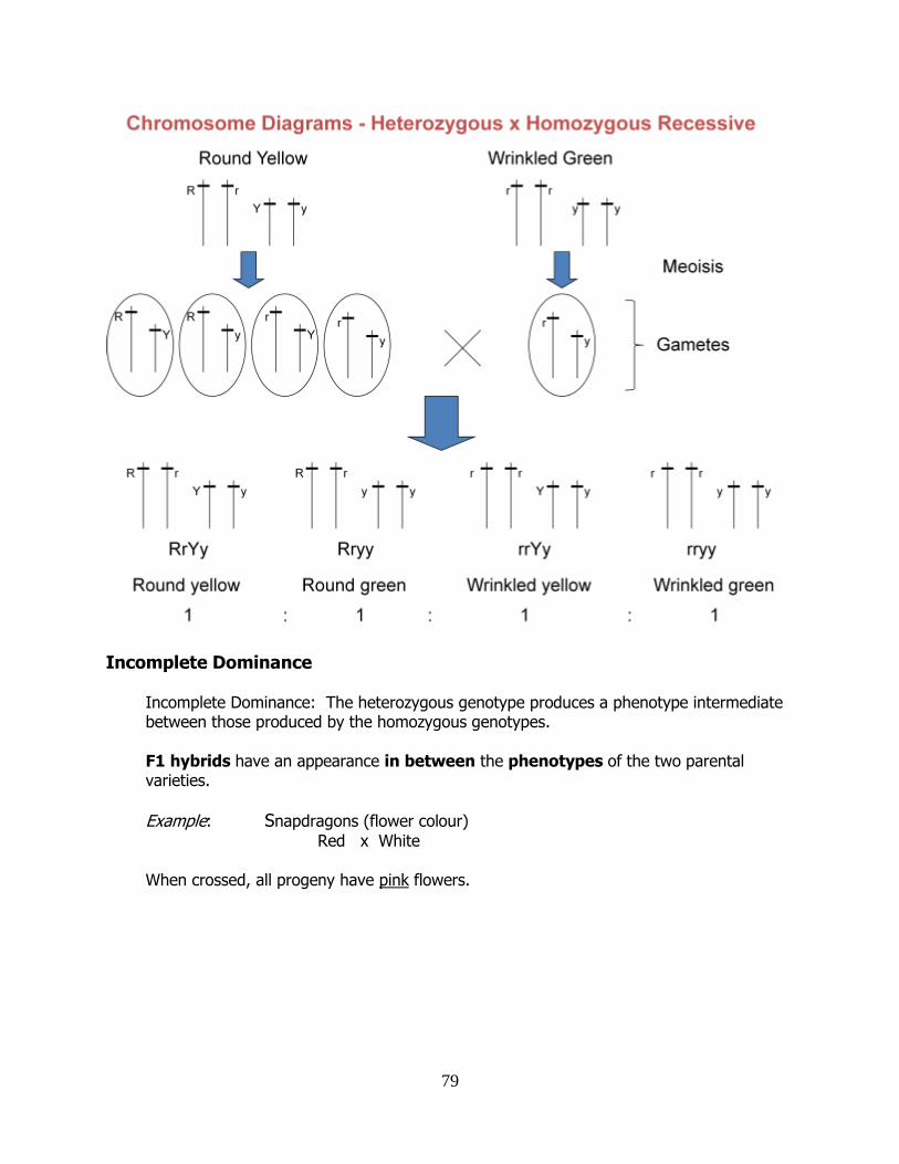
79
Incomplete Dominance Incomplete Dominance: The heterozygous genotype produces a phenotype intermediate between those produced by the homozygous genotypes. F1 hybrids have an appearance in between the phenotypes of the two parental varieties.
Example: Snapdragons (flower colour)
Red x White When crossed, all progeny have pink flowers.
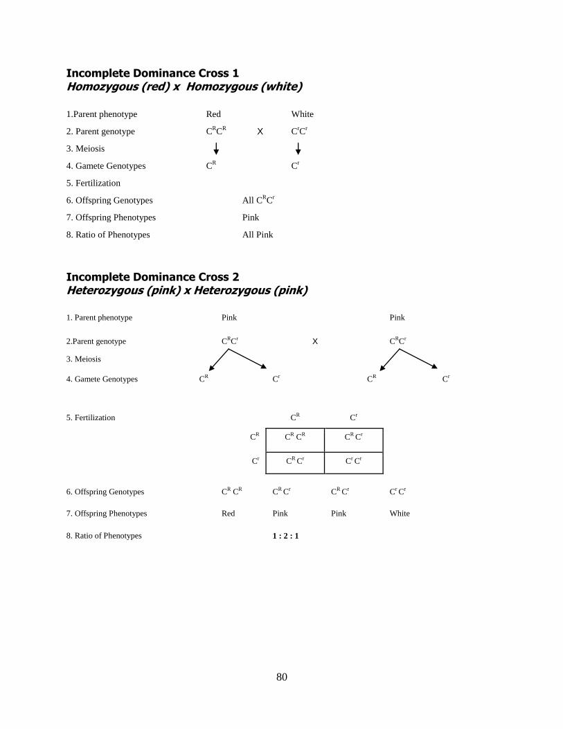
80
Incomplete Dominance Cross 1 Homozygous (red) x Homozygous (white)
1.Parent phenotype Red White
2. Parent genotype CRC
R X C
rC
r
3. Meiosis
4. Gamete Genotypes CR
Cr
5. Fertilization
6. Offspring Genotypes All CRC
r
7. Offspring Phenotypes Pink
8. Ratio of Phenotypes All Pink
Incomplete Dominance Cross 2 Heterozygous (pink) x Heterozygous (pink)
1. Parent phenotype Pink Pink
2.Parent genotype
CRCr X CRCr
3. Meiosis
4. Gamete Genotypes CR Cr CR Cr
5. Fertilization CR Cr
CR CR CR CR Cr
Cr CR Cr Cr Cr
6. Offspring Genotypes CR CR CR Cr CR Cr Cr Cr
7. Offspring Phenotypes Red Pink Pink White
8. Ratio of Phenotypes 1 : 2 : 1
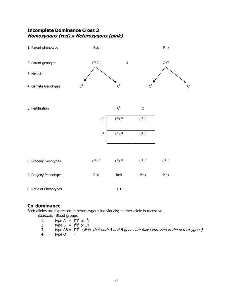
81
Incomplete Dominance Cross 3 Homozygous (red) x Heterozygous (pink)
1. Parent phenotype Red Pink
2. Parent genotype
CR CR X
CRCr
3. Meiosis
4. Gamete Genotypes CR CR CR Cr
5. Fertilization CR Cr
CR CR CR CR Cr
CR CR CR CR Cr
6. Progeny Genotypes CR CR CR CR CR Cr CR Cr
7. Progeny Phenotypes Red Red Pink Pink
8. Ratio of Phenotypes 1:1
Co-dominance Both alleles are expressed in heterozygous individuals; neither allele is recessive.
Example: Blood groups
1. type A = IAIA or IAi 2. type B = IBIB or IBi
3. type AB = IAIB (Note that both A and B genes are fully expressed in the heterozygous) 4. type O = ii
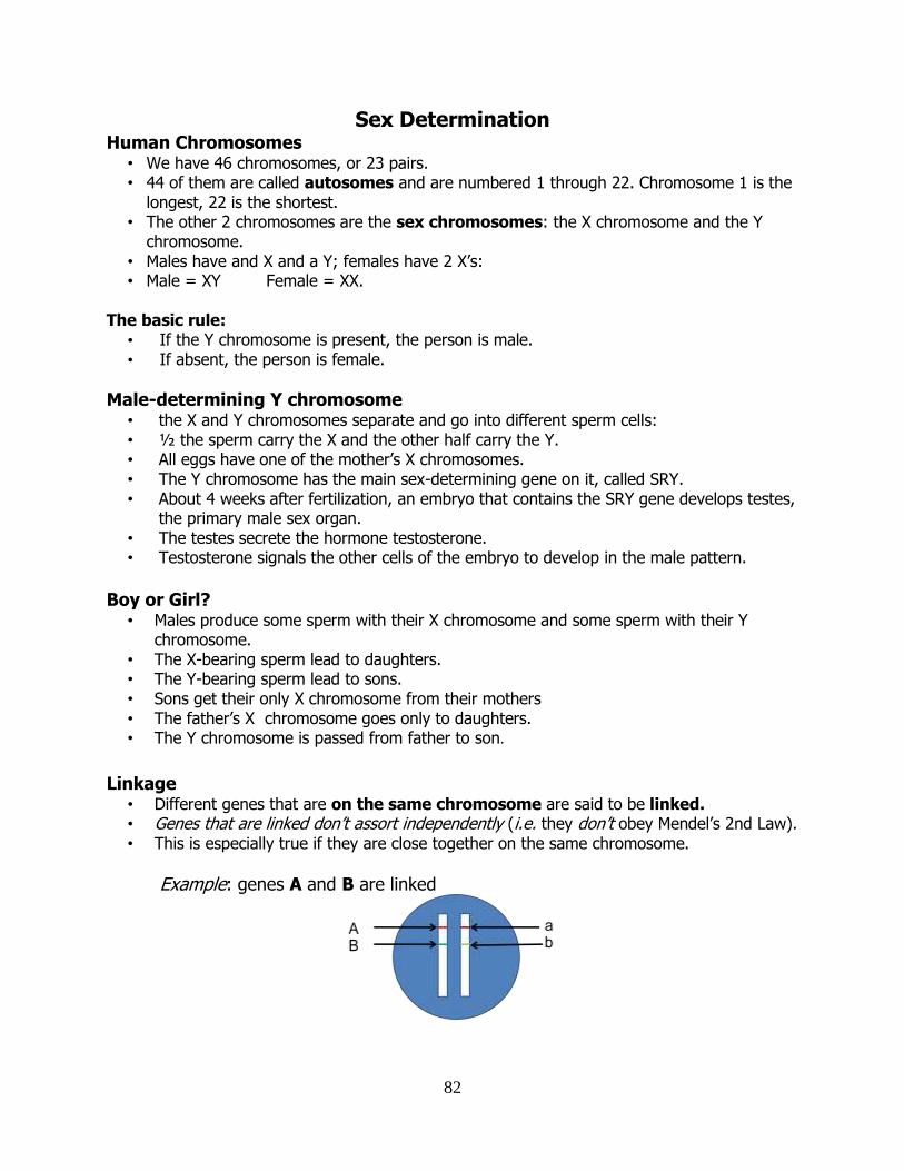
82
Sex Determination Human Chromosomes
• We have 46 chromosomes, or 23 pairs. • 44 of them are called autosomes and are numbered 1 through 22. Chromosome 1 is the
longest, 22 is the shortest. • The other 2 chromosomes are the sex chromosomes: the X chromosome and the Y
chromosome. • Males have and X and a Y; females have 2 X’s: • Male = XY Female = XX.
The basic rule:
• If the Y chromosome is present, the person is male. • If absent, the person is female.
Male-determining Y chromosome • the X and Y chromosomes separate and go into different sperm cells: • ½ the sperm carry the X and the other half carry the Y. • All eggs have one of the mother’s X chromosomes. • The Y chromosome has the main sex-determining gene on it, called SRY. • About 4 weeks after fertilization, an embryo that contains the SRY gene develops testes,
the primary male sex organ. • The testes secrete the hormone testosterone. • Testosterone signals the other cells of the embryo to develop in the male pattern.
Boy or Girl?
• Males produce some sperm with their X chromosome and some sperm with their Y chromosome.
• The X-bearing sperm lead to daughters. • The Y-bearing sperm lead to sons. • Sons get their only X chromosome from their mothers • The father’s X chromosome goes only to daughters. • The Y chromosome is passed from father to son.
Linkage
• Different genes that are on the same chromosome are said to be linked. • Genes that are linked don’t assort independently (i.e. they don’t obey Mendel’s 2nd Law). • This is especially true if they are close together on the same chromosome.
Example: genes A and B are linked
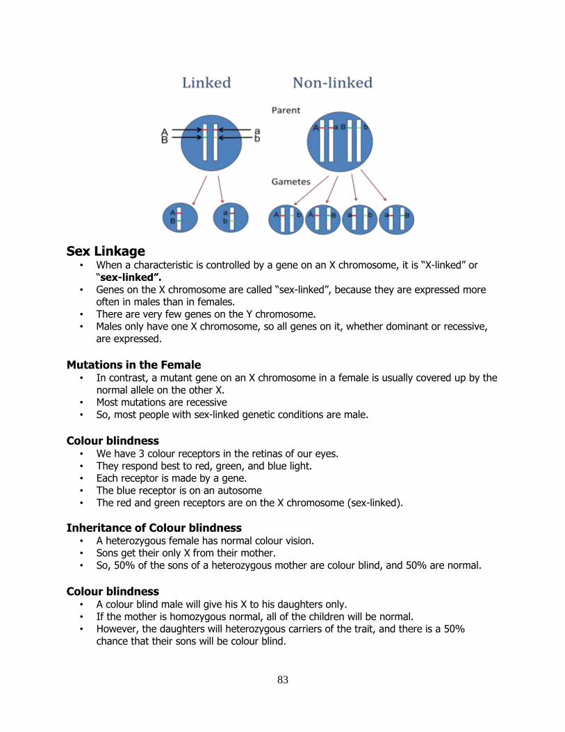
83
Sex Linkage • When a characteristic is controlled by a gene on an X chromosome, it is “X-linked” or
“sex-linked”. • Genes on the X chromosome are called “sex-linked”, because they are expressed more
often in males than in females. • There are very few genes on the Y chromosome. • Males only have one X chromosome, so all genes on it, whether dominant or recessive,
are expressed.
Mutations in the Female
• In contrast, a mutant gene on an X chromosome in a female is usually covered up by the normal allele on the other X.
• Most mutations are recessive • So, most people with sex-linked genetic conditions are male.
Colour blindness
• We have 3 colour receptors in the retinas of our eyes. • They respond best to red, green, and blue light. • Each receptor is made by a gene. • The blue receptor is on an autosome • The red and green receptors are on the X chromosome (sex-linked).
Inheritance of Colour blindness • A heterozygous female has normal colour vision. • Sons get their only X from their mother. • So, 50% of the sons of a heterozygous mother are colour blind, and 50% are normal.
Colour blindness
• A colour blind male will give his X to his daughters only. • If the mother is homozygous normal, all of the children will be normal. • However, the daughters will heterozygous carriers of the trait, and there is a 50%
chance that their sons will be colour blind.
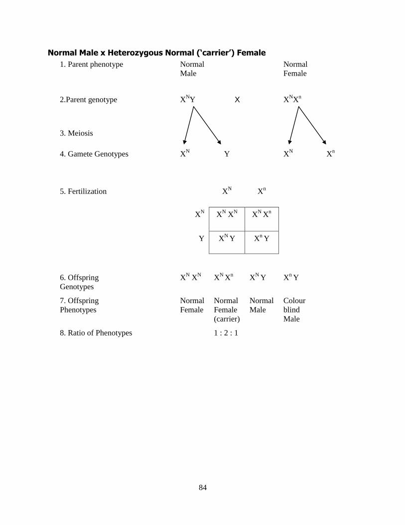
84
Normal Male x Heterozygous Normal (‘carrier’) Female
1. Parent phenotype Normal
Male
Normal
Female
2.Parent genotype XNY X X
NX
n
3. Meiosis
4. Gamete Genotypes XN
Y XN
Xn
5. Fertilization XN X
n
XN
XN X
N X
N X
n
Y X
N Y X
n Y
6. Offspring
Genotypes
XN X
N X
N X
n X
N Y X
n Y
7. Offspring
Phenotypes
Normal
Female
Normal
Female
(carrier)
Normal
Male
Colour
blind
Male
8. Ratio of Phenotypes 1 : 2 : 1
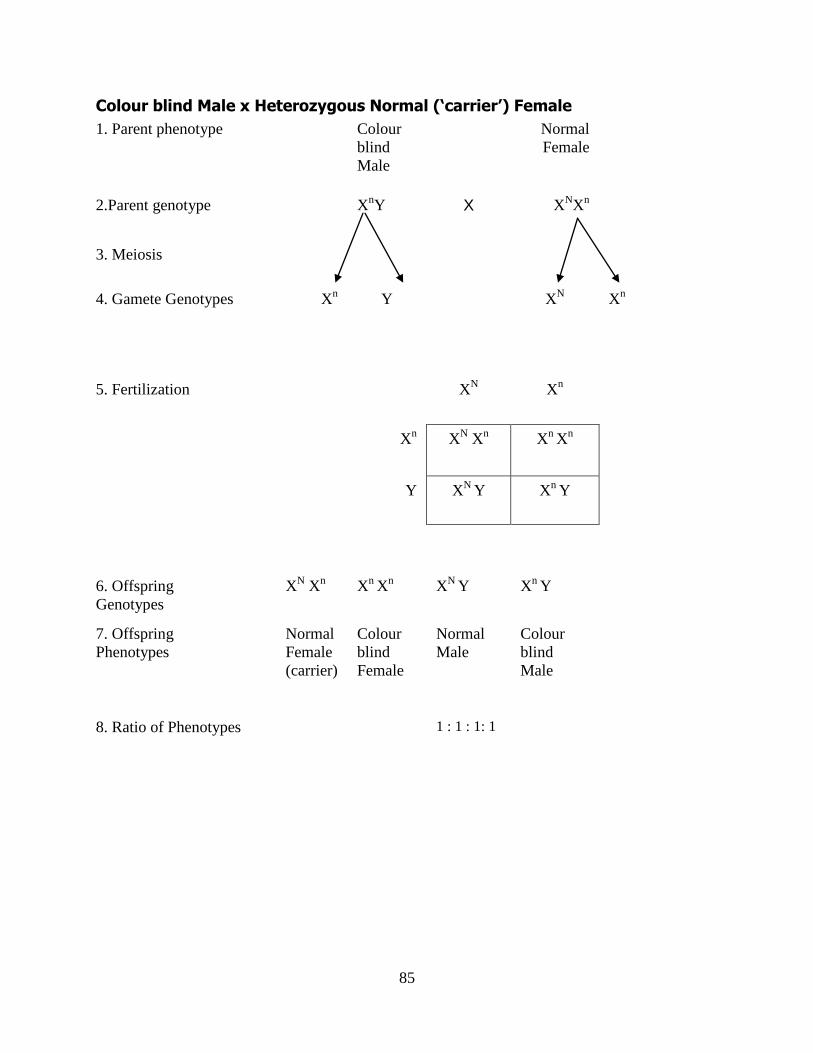
85
Colour blind Male x Heterozygous Normal (‘carrier’) Female
1. Parent phenotype Colour
blind
Male
Normal
Female
2.Parent genotype
XnY X X
NX
n
3. Meiosis
4. Gamete Genotypes Xn Y X
N X
n
5. Fertilization XN X
n
Xn X
N X
n X
n X
n
Y XN
Y Xn
Y
6. Offspring
Genotypes
XN X
n X
n X
n X
N Y X
n Y
7. Offspring
Phenotypes
Normal
Female
(carrier)
Colour
blind
Female
Normal
Male
Colour
blind
Male
8. Ratio of Phenotypes 1 : 1 : 1: 1
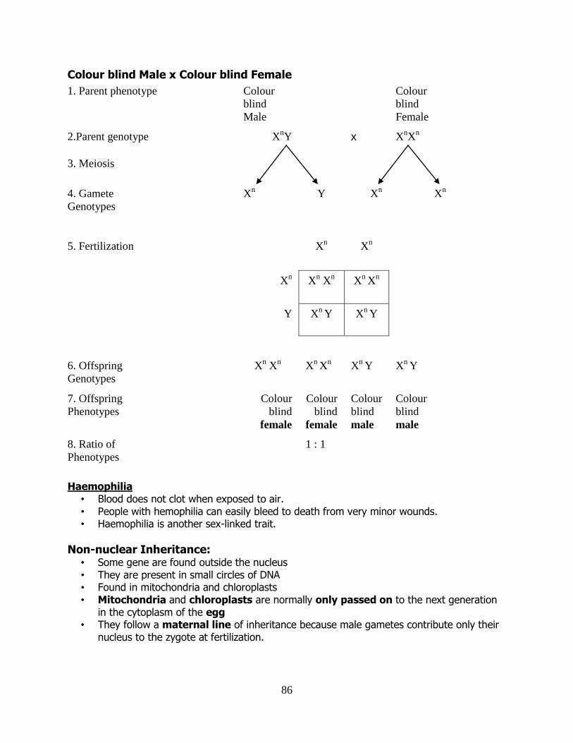
86
Colour blind Male x Colour blind Female
1. Parent phenotype Colour
blind
Male
Colour
blind
Female
2.Parent genotype XnY x X
nX
n
3. Meiosis
4. Gamete
Genotypes
Xn Y X
n X
n
5. Fertilization Xn X
n
Xn X
n X
n X
n X
n
Y X
n Y X
n Y
6. Offspring
Genotypes
Xn X
n X
n X
n X
n Y X
n Y
7. Offspring
Phenotypes
Colour
blind
female
Colour
blind
female
Colour
blind
male
Colour
blind
male
8. Ratio of
Phenotypes
1 : 1
Haemophilia • Blood does not clot when exposed to air. • People with hemophilia can easily bleed to death from very minor wounds. • Haemophilia is another sex-linked trait.
Non-nuclear Inheritance: • Some gene are found outside the nucleus • They are present in small circles of DNA • Found in mitochondria and chloroplasts • Mitochondria and chloroplasts are normally only passed on to the next generation
in the cytoplasm of the egg • They follow a maternal line of inheritance because male gametes contribute only their
nucleus to the zygote at fertilization.
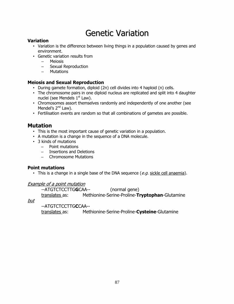
87
GGeenneettiicc VVaarriiaattiioonn Variation
• Variation is the difference between living things in a population caused by genes and environment.
• Genetic variation results from – Meiosis – Sexual Reproduction – Mutations
Meiosis and Sexual Reproduction
• During gamete formation, diploid (2n) cell divides into 4 haploid (n) cells. • The chromosome pairs in one diploid nucleus are replicated and split into 4 daughter
nuclei (see Mendels 1st Law). • Chromosomes assort themselves randomly and independently of one another (see
Mendel’s 2nd Law). • Fertilisation events are random so that all combinations of gametes are possible.
Mutation
• This is the most important cause of genetic variation in a population. • A mutation is a change in the sequence of a DNA molecule. • 3 kinds of mutations
– Point mutations – Insertions and Deletions – Chromosome Mutations
Point mutations
• This is a change in a single base of the DNA sequence (e.g. sickle cell anaemia).
Example of a point mutation --ATGTCTCCTTGGCAA-- (normal gene) translates as: Methionine-Serine-Proline-Tryptophan-Glutamine but --ATGTCTCCTTGCCAA-- translates as: Methionine-Serine-Proline-Cysteine-Glutamine
88
Insertions and Deletions • Insertions add one or more extra nucleotides into the DNA. • Deletions remove one or more nucleotides from the DNA. • Both types of mutations cause changes in the way the genes are transcribed and
translated.
Example of Insertion Mutation
--ATGTCTCCTTGGCAA-- (normal gene) translates as: Methionine-Serine-Proline-Tryptophan-Glutamine (amino acid sequence) but --ATGTCTCCTTTGGCAA-- (mutant gene) translates as: Methionine-Serine-Proline-Leucine-Alanine- (now different amino acids)
Chromosome mutations
• Chromosome mutations occur when there is a change in the structure or number of one or more chromosomes.
• e.g. Down syndrome caused by possessing 3 copies of chromosome 21 (i.e. one extra copy).
Mutagens • Agents that are responsible for mutations are called mutagens. • Examples:
– Nuclear radiation – Tobacco smoke – UV light – X-rays
Cancer
• Cancer is a genetic disease. • It is caused by a change or a mutation in the DNA of a cell. • This mutation affects the genes that control cell division. • These genes are called oncogenes and if mutations in arise in these genes, a cancerous
(malignant) tumour can result.
Carcinogens
• A carcinogen is any factor that results in a DNA mutation that causes cancer. • 3 main factors environmental factors that cause cancer.
– Chemicals – Radiation – Diet
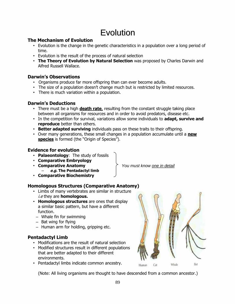
89
EEvvoolluuttiioonn The Mechanism of Evolution
• Evolution is the change in the genetic characteristics in a population over a long period of time.
• Evolution is the result of the process of natural selection • The Theory of Evolution by Natural Selection was proposed by Charles Darwin and
Alfred Russell Wallace.
Darwin’s Observations
• Organisms produce far more offspring than can ever become adults. • The size of a population doesn’t change much but is restricted by limited resources. • There is much variation within a population.
Darwin’s Deductions
• There must be a high death rate, resulting from the constant struggle taking place between all organisms for resources and in order to avoid predators, disease etc.
• In the competition for survival, variations allow some individuals to adapt, survive and reproduce better than others.
• Better adapted surviving individuals pass on these traits to their offspring. • Over many generations, these small changes in a population accumulate until a new
species is formed (the “Origin of Species”).
Evidence for evolution
• Palaeontology: The study of fossils • Comparative Embryology • Comparative Anatomy
– e.g. The Pentadactyl limb
• Comparative Biochemistry
Homologous Structures (Comparative Anatomy) • Limbs of many vertebrates are similar in structure
i.e they are homologous. • Homologous structures are ones that display
a similar basic pattern, but have a different function.
– Whale fin for swimming – Bat wing for flying – Human arm for holding, gripping etc.
Pentadactyl Limb • Modifications are the result of natural selection • Modified structures result in different populations
that are better adapted to their different environments.
• Pentadactyl limbs indicate common ancestry. (Note: All living organisms are thought to have descended from a common ancestor.)
You must know one in detail
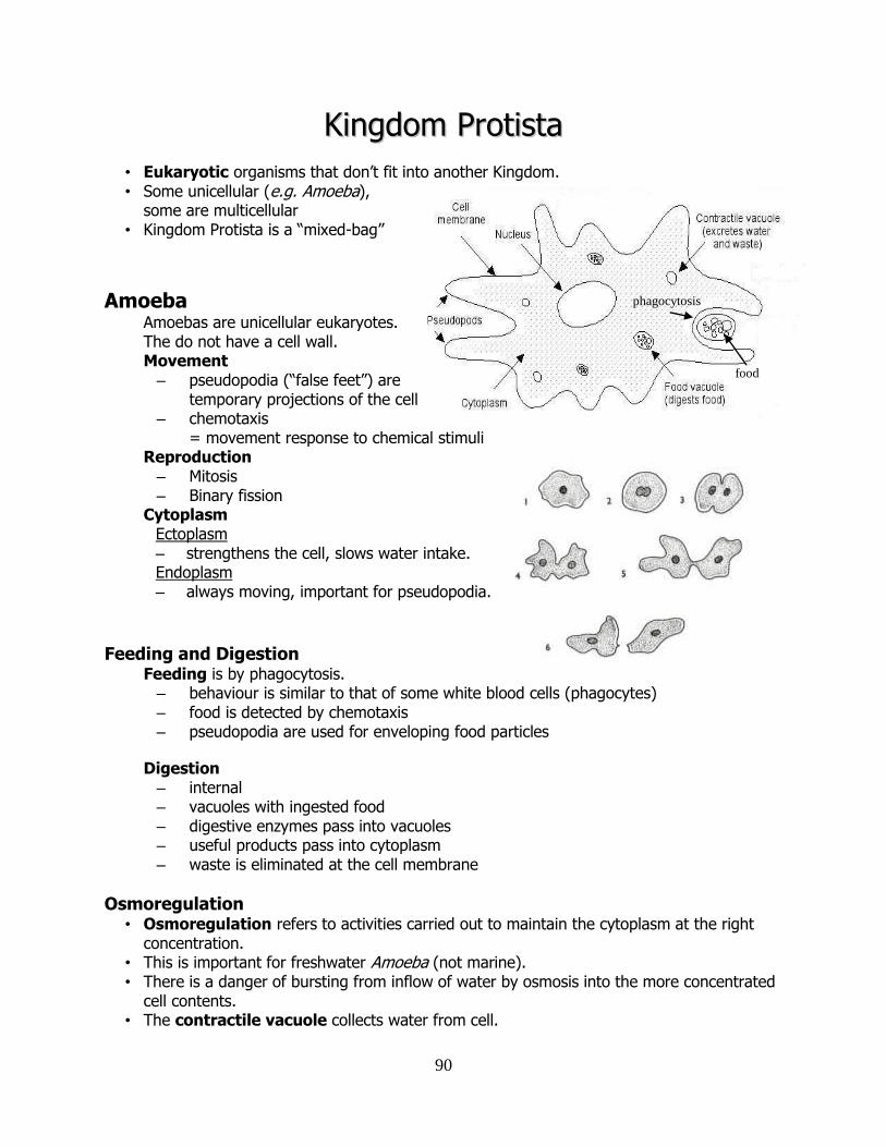
90
KKiinnggddoomm PPrroottiissttaa
• Eukaryotic organisms that don’t fit into another Kingdom. • Some unicellular (e.g. Amoeba),
some are multicellular • Kingdom Protista is a “mixed-bag”
Amoeba Amoebas are unicellular eukaryotes. The do not have a cell wall. Movement – pseudopodia (“false feet”) are
temporary projections of the cell – chemotaxis
= movement response to chemical stimuli Reproduction – Mitosis – Binary fission
Cytoplasm Ectoplasm – strengthens the cell, slows water intake. Endoplasm – always moving, important for pseudopodia.
Feeding and Digestion Feeding is by phagocytosis. – behaviour is similar to that of some white blood cells (phagocytes) – food is detected by chemotaxis – pseudopodia are used for enveloping food particles
Digestion – internal – vacuoles with ingested food – digestive enzymes pass into vacuoles – useful products pass into cytoplasm – waste is eliminated at the cell membrane
Osmoregulation • Osmoregulation refers to activities carried out to maintain the cytoplasm at the right
concentration. • This is important for freshwater Amoeba (not marine). • There is a danger of bursting from inflow of water by osmosis into the more concentrated
cell contents. • The contractile vacuole collects water from cell.
phagocytosis
food
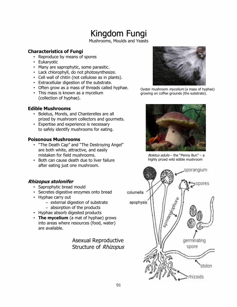
91
KKiinnggddoomm FFuunnggii Mushrooms, Moulds and Yeasts
Characteristics of Fungi • Reproduce by means of spores • Eukaryotic • Many are saprophytic, some parasitic. • Lack chlorophyll, do not photosynthesize. • Cell wall of chitin (not cellulose as in plants). • Extracellular digestion of the substrate. • Often grow as a mass of threads called hyphae. • This mass is known as a mycelium
(collection of hyphae).
Edible Mushrooms • Boletus, Morels, and Chanterelles are all
prized by mushroom collectors and gourmets. • Expertise and experience is necessary
to safely identify mushrooms for eating.
Poisonous Mushrooms • “The Death Cap” and “The Destroying Angel”
are both white, attractive, and easily mistaken for field mushrooms.
• Both can cause death due to liver failure after eating just one mushroom.
Rhizopus stolonifer
• Saprophytic bread mould • Secretes digestive enzymes onto bread • Hyphae carry out
– external digestion of substrate – absorption of the products
• Hyphae absorb digested products • The mycelium (a mat of hyphae) grows
into areas where resources (food, water) are available.
Asexual Reproductive Structure of Rhizopus
Oyster mushroom mycelium (a mass of hyphae) growing on coffee grounds (the substrate).
Boletus edulis – the “Penny Bun” – a highly prized wild edible mushroom
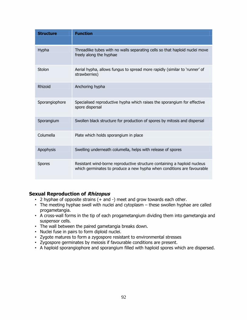
92
Structure Function
Hypha Threadlike tubes with no walls separating cells so that haploid nuclei move freely along the hyphae
Stolon Aerial hypha, allows fungus to spread more rapidly (similar to ‘runner’ of strawberries)
Rhizoid Anchoring hypha
Sporangiophore Specialised reproductive hypha which raises the sporangium for effective
spore dispersal
Sporangium Swollen black structure for production of spores by mitosis and dispersal
Columella Plate which holds sporangium in place
Apophysis Swelling underneath columella, helps with release of spores
Spores Resistant wind-borne reproductive structure containing a haploid nucleus
which germinates to produce a new hypha when conditions are favourable
Sexual Reproduction of Rhizopus • 2 hyphae of opposite strains (+ and -) meet and grow towards each other. • The meeting hyphae swell with nuclei and cytoplasm – these swollen hyphae are called
progametangia. • A cross-wall forms in the tip of each progametangium dividing them into gametangia and
suspensor cells. • The wall between the paired gametangia breaks down. • Nuclei fuse in pairs to form diploid nuclei. • Zygote matures to form a zygospore resistant to environmental stresses • Zygospore germinates by meiosis if favourable conditions are present. • A haploid sporangiophore and sporangium filled with haploid spores which are dispersed.
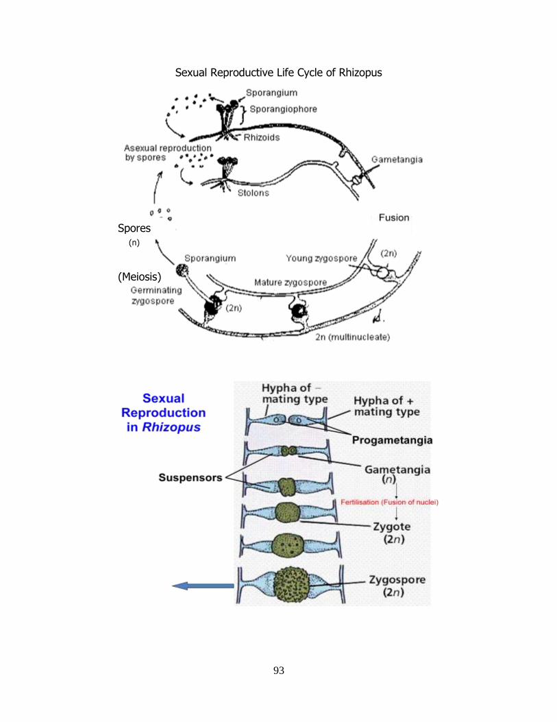
93
Sexual Reproductive Life Cycle of Rhizopus
Spores
(Meiosis)
(n)
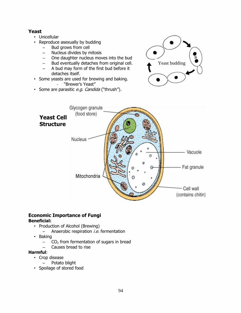
94
Yeast • Unicellular • Reproduce asexually by budding
– Bud grows from cell – Nucleus divides by mitosis – One daughter nucleus moves into the bud – Bud eventually detaches from original cell. – A bud may form of the first bud before it
detaches itself. • Some yeasts are used for brewing and baking.
- “Brewer’s Yeast” • Some are parasitic e.g. Candida (“thrush”).
Economic Importance of Fungi Beneficial:
• Production of Alcohol (Brewing) – Anaerobic respiration i.e. fermentation
• Baking – CO2 from fermentation of sugars in bread – Causes bread to rise
Harmful: • Crop disease
– Potato blight • Spoilage of stored food
Yeast budding
Yeast Cell Structure
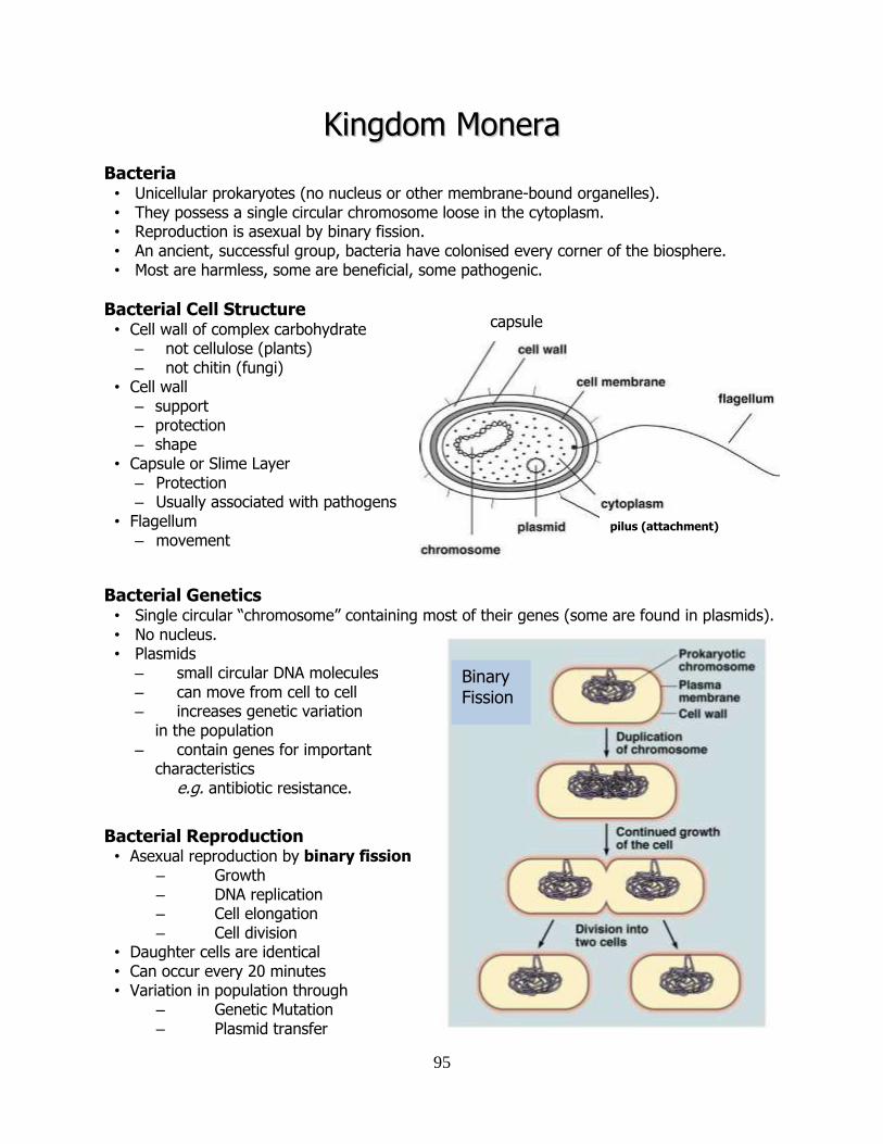
95
KKiinnggddoomm MMoonneerraa
Bacteria • Unicellular prokaryotes (no nucleus or other membrane-bound organelles). • They possess a single circular chromosome loose in the cytoplasm. • Reproduction is asexual by binary fission. • An ancient, successful group, bacteria have colonised every corner of the biosphere. • Most are harmless, some are beneficial, some pathogenic.
Bacterial Cell Structure • Cell wall of complex carbohydrate
– not cellulose (plants) – not chitin (fungi)
• Cell wall – support – protection – shape
• Capsule or Slime Layer – Protection – Usually associated with pathogens
• Flagellum – movement
Bacterial Genetics • Single circular “chromosome” containing most of their genes (some are found in plasmids). • No nucleus. • Plasmids
– small circular DNA molecules – can move from cell to cell – increases genetic variation in the population – contain genes for important
characteristics e.g. antibiotic resistance.
Bacterial Reproduction • Asexual reproduction by binary fission
– Growth – DNA replication – Cell elongation – Cell division
• Daughter cells are identical • Can occur every 20 minutes • Variation in population through
– Genetic Mutation – Plasmid transfer
capsule
pilus (attachment)
Binary Fission
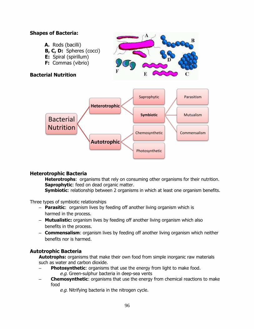
96
Shapes of Bacteria:
A. Rods (bacilli) B, C, D: Spheres (cocci) E: Spiral (spirillum) F: Commas (vibrio) Bacterial Nutrition
Heterotrophic Bacteria Heterotrophs: organisms that rely on consuming other organisms for their nutrition.
Saprophytic: feed on dead organic matter. Symbiotic: relationship between 2 organisms in which at least one organism benefits.
Three types of symbiotic relationships
– Parasitic: organism lives by feeding off another living organism which is
harmed in the process.
– Mutualistic: organism lives by feeding off another living organism which also
benefits in the process.
– Commensalism: organism lives by feeding off another living organism which neither
benefits nor is harmed.
Autotrophic Bacteria Autotrophs: organisms that make their own food from simple inorganic raw materials such as water and carbon dioxide. – Photosynthetic: organisms that use the energy from light to make food.
e.g. Green-sulphur bacteria in deep-sea vents – Chemosynthetic: organisms that use the energy from chemical reactions to make
food e.g. Nitrifying bacteria in the nitrogen cycle.
Bacterial Nutrition
Heterotrophic
Saprophytic
Symbiotic
Parasitism
Mutualism
Commensalism
Autotrophic
Chemosynthetic
Photosynthetic
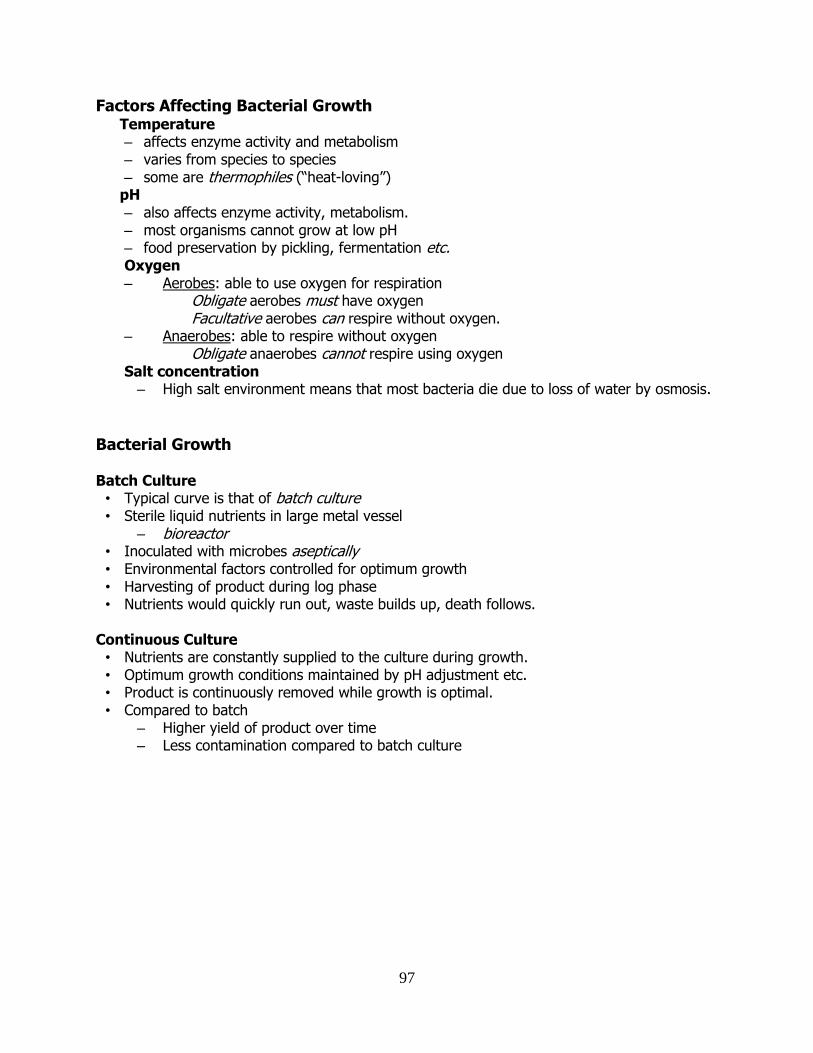
97
Factors Affecting Bacterial Growth Temperature – affects enzyme activity and metabolism – varies from species to species – some are thermophiles (“heat-loving”)
pH – also affects enzyme activity, metabolism. – most organisms cannot grow at low pH – food preservation by pickling, fermentation etc. Oxygen – Aerobes: able to use oxygen for respiration
Obligate aerobes must have oxygen Facultative aerobes can respire without oxygen.
– Anaerobes: able to respire without oxygen Obligate anaerobes cannot respire using oxygen
Salt concentration – High salt environment means that most bacteria die due to loss of water by osmosis.
Bacterial Growth
Batch Culture • Typical curve is that of batch culture • Sterile liquid nutrients in large metal vessel
– bioreactor • Inoculated with microbes aseptically • Environmental factors controlled for optimum growth • Harvesting of product during log phase • Nutrients would quickly run out, waste builds up, death follows.
Continuous Culture • Nutrients are constantly supplied to the culture during growth. • Optimum growth conditions maintained by pH adjustment etc. • Product is continuously removed while growth is optimal. • Compared to batch
– Higher yield of product over time – Less contamination compared to batch culture
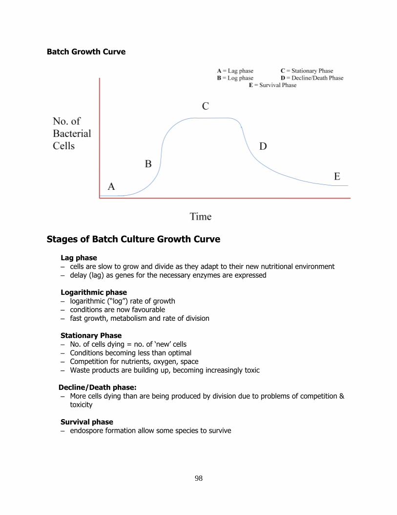
98
Batch Growth Curve
Stages of Batch Culture Growth Curve
Lag phase – cells are slow to grow and divide as they adapt to their new nutritional environment – delay (lag) as genes for the necessary enzymes are expressed Logarithmic phase – logarithmic (“log”) rate of growth – conditions are now favourable – fast growth, metabolism and rate of division Stationary Phase – No. of cells dying = no. of ‘new’ cells – Conditions becoming less than optimal – Competition for nutrients, oxygen, space – Waste products are building up, becoming increasingly toxic Decline/Death phase: – More cells dying than are being produced by division due to problems of competition & toxicity Survival phase – endospore formation allow some species to survive
99
Endospores • Formed within (hence endo-) cells of some bacteria when conditions unfavourable
e.g. heat, freezing, drying, radiation • Highly resistant • Can survive indefinitely • When conditions return to normal, endospore germinates
Endospore Formation • The cell’s DNA is replicated • The cytoplasm (containing a chromosome) is pulled to one end of the cell. • A membrane forms around the cytoplasm. • Thick protective wall forms and surrounds the membrane. • The original bacterial cell breaks down. • The endospore is released.
Endospore Germination • Germination only occurs when conditions are favourable. • Water is absorbed and the endospore wall softens. • A new cell emerges.
Endospores and Disease • Some pathogens can make endospores.
e.g. Clostridium botulinum which causes botulism • Endospores are not killed at 100°C. • Autoclaving (121°C for 20 minutes) is necessary to destroy them. • Sterilisation of glassware, etc in laboratory or hospital is often carried out in autoclaves.
Antibiotics • Antibiotics are chemicals produced by bacteria and fungi which kill or inhibit the growth of
other bacteria. • Produced by microbes in their habitat to kill off competitors • Antibiotics are
– ineffective against viruses. – non-toxic to humans and other animals.
Antibiotic Resistance • Genes confer resistance, resistance is acquired by
– mutation – gene transfer between bacteria
• Resistance is an advantage (competitive edge) in environments where antibiotics are present.
• A population of resistant bacteria builds up. • If antibiotics are commonplace, so will resistance be. • In the past, antibiotics were
– overprescribed
– used in agriculture in animal feed to boost weight of livestock
• Resistance to multiple antibiotics now occurs e.g. MRSA (hospital superbug).
100
Economically Beneficial Bacteria
Lactobacillus spp. – for fermenting milk (yoghurt), olives, grass (silage) etc. – some are probiotic (“health-promoting”)
e.g. L. casei immunitas (“Actimel”) E. coli
– used as cellular “tool” for doing groundwork in molecular biology research. – used in biotechnology to produce e.g. insulin.
Economically Harmful Bacteria
Mycobacterium tuberculosis – Tuberculosis (T.B.)
M. bovis – bovine T.B
Streptococcus pyogenes – Causes “streptococcal throat” (severe sore throat, white spots visible in throat) – Many work and school days lost due to this.
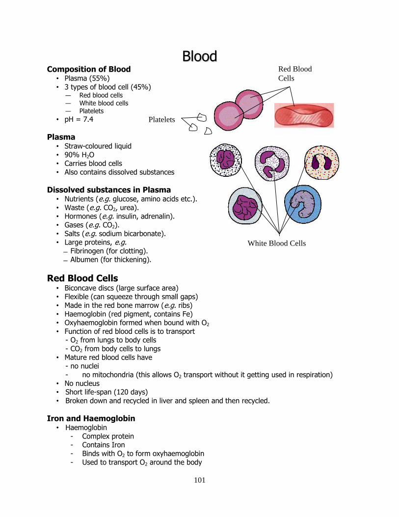
101
BBlloooodd Composition of Blood
• Plasma (55%) • 3 types of blood cell (45%)
— Red blood cells — White blood cells — Platelets
• pH = 7.4
Plasma • Straw-coloured liquid • 90% H2O • Carries blood cells • Also contains dissolved substances
Dissolved substances in Plasma • Nutrients (e.g. glucose, amino acids etc.). • Waste (e.g. CO2, urea). • Hormones (e.g. insulin, adrenalin). • Gases (e.g. CO2). • Salts (e.g. sodium bicarbonate). • Large proteins, e.g.
Fibrinogen (for clotting). Albumen (for thickening).
Red Blood Cells • Biconcave discs (large surface area) • Flexible (can squeeze through small gaps) • Made in the red bone marrow (e.g. ribs) • Haemoglobin (red pigment, contains Fe) • Oxyhaemoglobin formed when bound with O2 • Function of red blood cells is to transport
- O2 from lungs to body cells - CO2 from body cells to lungs
• Mature red blood cells have - no nuclei - no mitochondria (this allows O2 transport without it getting used in respiration)
• No nucleus • Short life-span (120 days) • Broken down and recycled in liver and spleen and then recycled.
Iron and Haemoglobin • Haemoglobin
- Complex protein - Contains Iron - Binds with O2 to form oxyhaemoglobin - Used to transport O2 around the body
Platelets
White Blood Cells
Red Blood
Cells
102
• Lack of iron in diet causes anaemia - Iron rich foods - i.e. red meat, eggs, nuts, bananas, green veg
White Blood Cells • Larger than red blood cells • Do not contain haemoglobin • Do have a nucleus • Formed in the red bone marrow • Protect against disease • Recognise and destroy foreign agents • Red : White = 700 : 1
Monocytes (75%) • Largest of the white blood cells
• Become aggressive macrophages when they leave the blood
• Also known as phagocytes - engulf foreign cells by phagocytosis
• “Clean up” - cause “pus” at the site of infection.
Lymphocytes (25%) : 2 types
1. B-lymphocytes mature in lymphoid tissue. • They produce antibodies which bind to foreign antigens. • Some B-lymphocytes remain after infection as memory cells.
2. T-lymphocytes mature in the thymus.
4 types of T-Lymphcytes (T-cells): Memory T Cells Like memory B cells, memory T cells remain in the blood after the infection has gone in order to better launch a response in the event of another attack from the same antigen. Helper T Cells Stimulate other T and B cells to proliferate
Killer T Cells: produce perforin which produces holes in the cell membrane of the target.
Suppressor T Cells: dampens down the immune response after the antigen/pathogen has been dealt with.
Platelets • Small fragments of larger cells. • Made in the bone marrow. • No nucleus. • Function in blood clotting – seals ruptured blood vessels. • “Packages” of enzymes – released when blood vessels become damaged.
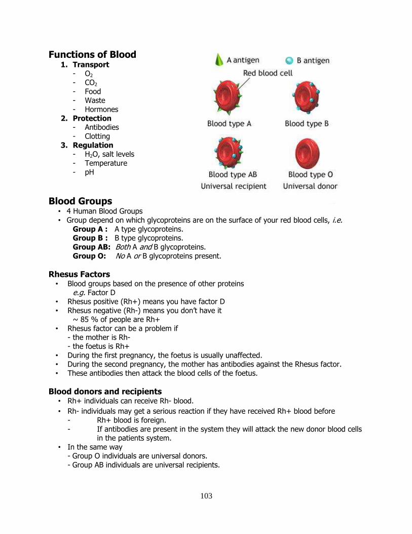
103
Functions of Blood 1. Transport
- O2 - CO2 - Food - Waste - Hormones
2. Protection - Antibodies - Clotting
3. Regulation - H2O, salt levels - Temperature - pH
Blood Groups • 4 Human Blood Groups • Group depend on which glycoproteins are on the surface of your red blood cells, i.e.
Group A : A type glycoproteins. Group B : B type glycoproteins. Group AB: Both A and B glycoproteins. Group O: No A or B glycoproteins present.
Rhesus Factors • Blood groups based on the presence of other proteins
e.g. Factor D • Rhesus positive (Rh+) means you have factor D • Rhesus negative (Rh-) means you don’t have it
~ 85 % of people are Rh+ • Rhesus factor can be a problem if
- the mother is Rh- - the foetus is Rh+
• During the first pregnancy, the foetus is usually unaffected. • During the second pregnancy, the mother has antibodies against the Rhesus factor. • These antibodies then attack the blood cells of the foetus.
Blood donors and recipients • Rh+ individuals can receive Rh- blood.
• Rh- individuals may get a serious reaction if they have received Rh+ blood before - Rh+ blood is foreign. - If antibodies are present in the system they will attack the new donor blood cells
in the patients system. • In the same way
- Group O individuals are universal donors. - Group AB individuals are universal recipients.
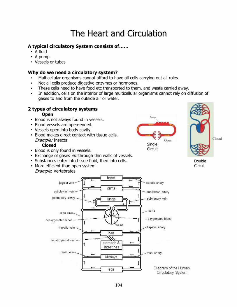
104
TThhee HHeeaarrtt aanndd CCiirrccuullaattiioonn
A typical circulatory System consists of…… • A fluid • A pump • Vessels or tubes
Why do we need a circulatory system? • Multicellular organisms cannot afford to have all cells carrying out all roles. • Not all cells produce digestive enzymes or hormones. • These cells need to have food etc transported to them, and waste carried away. • In addition, cells on the interior of large multicellular organisms cannot rely on diffusion of
gases to and from the outside air or water.
2 types of circulatory systems Open • Blood is not always found in vessels. • Blood vessels are open-ended. • Vessels open into body cavity. • Blood makes direct contact with tissue cells.
Example: Insects Closed • Blood is only found in vessels. • Exchange of gases etc through thin walls of vessels. • Substances enter into tissue fluid, then into cells. • More efficient than open system.
Example: Vertebrates
Double
Circuit
Single Circuit
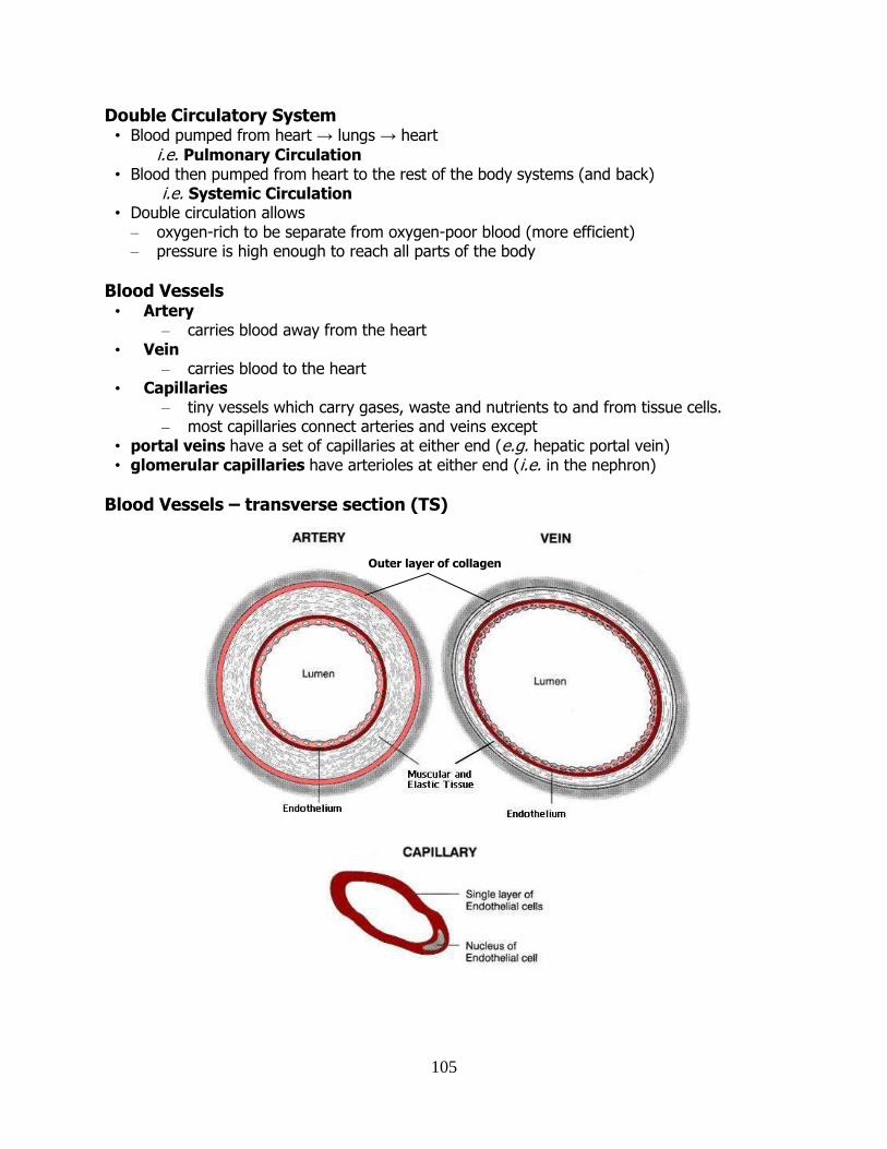
105
Double Circulatory System • Blood pumped from heart → lungs → heart
i.e. Pulmonary Circulation • Blood then pumped from heart to the rest of the body systems (and back)
i.e. Systemic Circulation • Double circulation allows
– oxygen-rich to be separate from oxygen-poor blood (more efficient) – pressure is high enough to reach all parts of the body
Blood Vessels • Artery
– carries blood away from the heart • Vein
– carries blood to the heart • Capillaries
– tiny vessels which carry gases, waste and nutrients to and from tissue cells. – most capillaries connect arteries and veins except
• portal veins have a set of capillaries at either end (e.g. hepatic portal vein) • glomerular capillaries have arterioles at either end (i.e. in the nephron)
Blood Vessels – transverse section (TS)
Outer layer of collagen
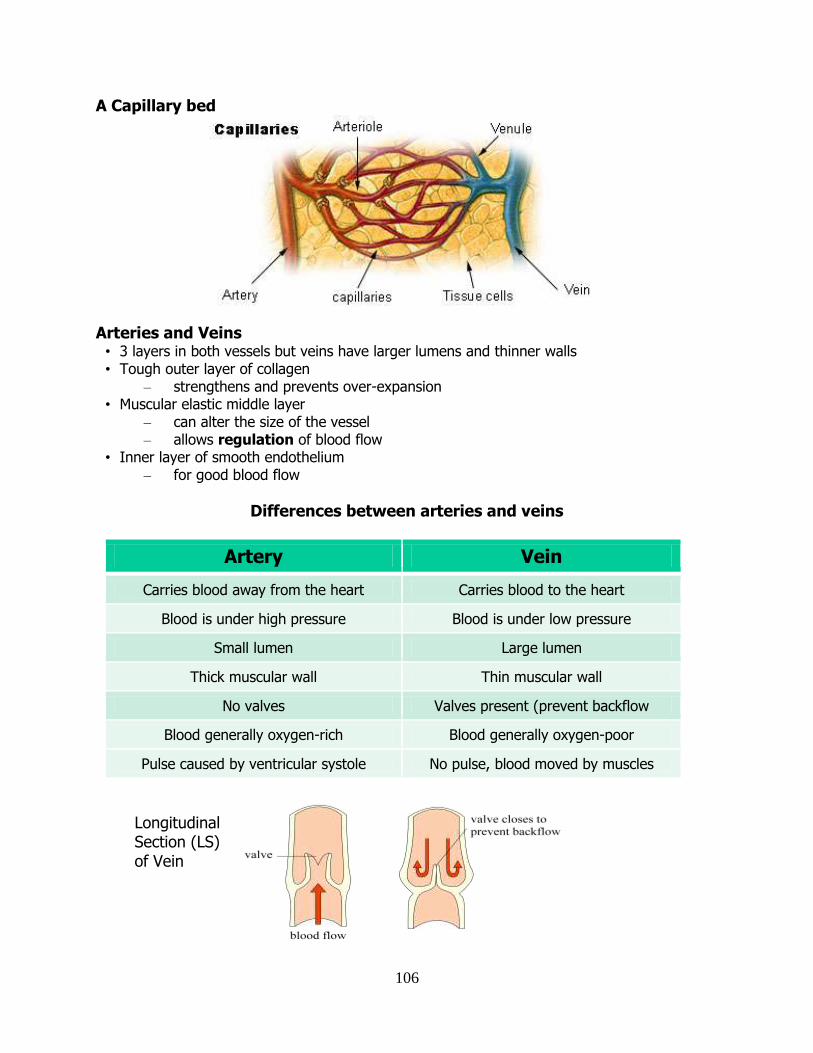
106
A Capillary bed
Arteries and Veins • 3 layers in both vessels but veins have larger lumens and thinner walls • Tough outer layer of collagen
– strengthens and prevents over-expansion • Muscular elastic middle layer
– can alter the size of the vessel – allows regulation of blood flow
• Inner layer of smooth endothelium – for good blood flow
Differences between arteries and veins
Artery Vein
Carries blood away from the heart Carries blood to the heart
Blood is under high pressure Blood is under low pressure
Small lumen Large lumen
Thick muscular wall Thin muscular wall
No valves Valves present (prevent backflow
Blood generally oxygen-rich Blood generally oxygen-poor
Pulse caused by ventricular systole No pulse, blood moved by muscles
Longitudinal Section (LS) of Vein
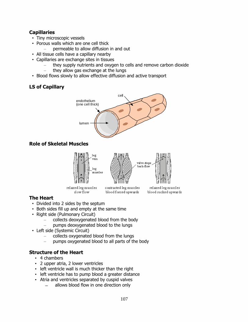
107
Capillaries • Tiny microscopic vessels • Porous walls which are one cell thick
– permeable to allow diffusion in and out • All tissue cells have a capillary nearby • Capillaries are exchange sites in tissues
– they supply nutrients and oxygen to cells and remove carbon dioxide – they allow gas exchange at the lungs
• Blood flows slowly to allow effective diffusion and active transport
LS of Capillary
Role of Skeletal Muscles
The Heart • Divided into 2 sides by the septum • Both sides fill up and empty at the same time • Right side (Pulmonary Circuit)
– collects deoxygenated blood from the body – pumps deoxygenated blood to the lungs
• Left side (Systemic Circuit) – collects oxygenated blood from the lungs – pumps oxygenated blood to all parts of the body
Structure of the Heart • 4 chambers • 2 upper atria, 2 lower ventricles • left ventricle wall is much thicker than the right • left ventricle has to pump blood a greater distance • Atria and ventricles separated by cuspid valves
allows blood flow in one direction only
lumen
endothelium(one cell thick)
cell
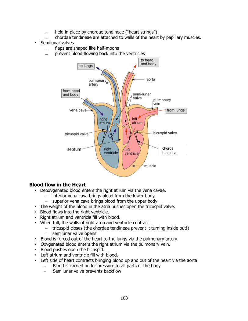
108
held in place by chordae tendineae (“heart strings”) chordae tendineae are attached to walls of the heart by papillary muscles.
• Semilunar valves flaps are shaped like half-moons prevent blood flowing back into the ventricles
Blood flow in the Heart
• Deoxygenated blood enters the right atrium via the vena cavae. – inferior vena cava brings blood from the lower body – superior vena cava brings blood from the upper body
• The weight of the blood in the atria pushes open the tricuspid valve. • Blood flows into the right ventricle. • Right atrium and ventricle fill with blood. • When full, the walls of right atria and ventricle contract
– tricuspid closes (the chordae tendineae prevent it turning inside out!) – semilunar valve opens
• Blood is forced out of the heart to the lungs via the pulmonary artery. • Oxygenated blood enters the right atrium via the pulmonary vein. • Blood pushes open the bicuspid. • Left atrium and ventricle fill with blood. • Left side of heart contracts bringing blood up and out of the heart via the aorta
– Blood is carried under pressure to all parts of the body – Semilunar valve prevents backflow
chorda tendinea
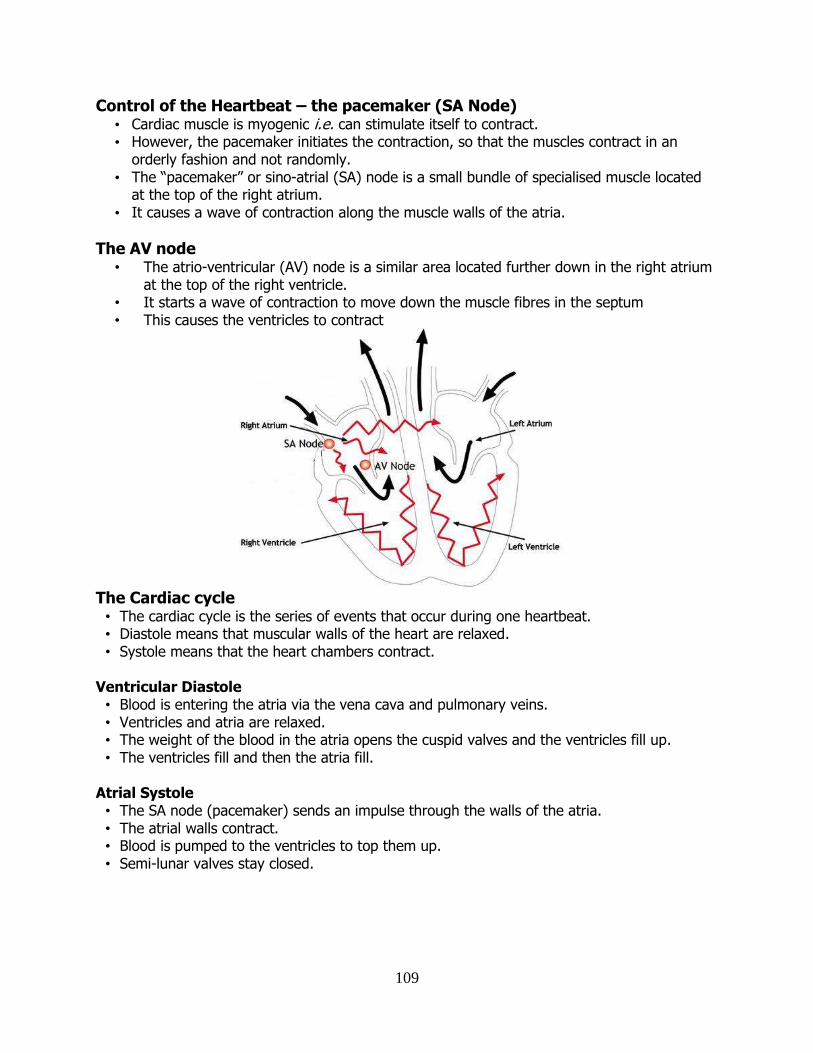
109
Control of the Heartbeat – the pacemaker (SA Node) • Cardiac muscle is myogenic i.e. can stimulate itself to contract. • However, the pacemaker initiates the contraction, so that the muscles contract in an
orderly fashion and not randomly. • The “pacemaker” or sino-atrial (SA) node is a small bundle of specialised muscle located
at the top of the right atrium. • It causes a wave of contraction along the muscle walls of the atria.
The AV node • The atrio-ventricular (AV) node is a similar area located further down in the right atrium at the top of the right ventricle. • It starts a wave of contraction to move down the muscle fibres in the septum • This causes the ventricles to contract
The Cardiac cycle • The cardiac cycle is the series of events that occur during one heartbeat. • Diastole means that muscular walls of the heart are relaxed. • Systole means that the heart chambers contract.
Ventricular Diastole • Blood is entering the atria via the vena cava and pulmonary veins. • Ventricles and atria are relaxed. • The weight of the blood in the atria opens the cuspid valves and the ventricles fill up. • The ventricles fill and then the atria fill.
Atrial Systole • The SA node (pacemaker) sends an impulse through the walls of the atria. • The atrial walls contract. • Blood is pumped to the ventricles to top them up. • Semi-lunar valves stay closed.
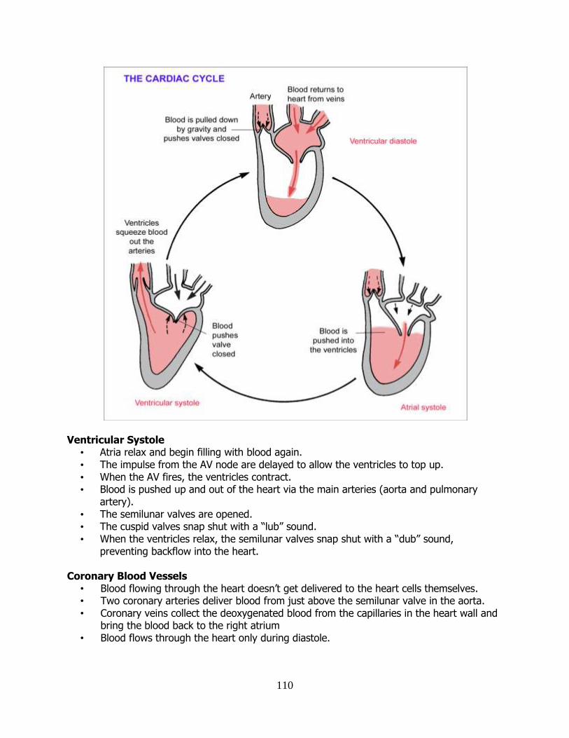
110
Ventricular Systole • Atria relax and begin filling with blood again. • The impulse from the AV node are delayed to allow the ventricles to top up. • When the AV fires, the ventricles contract. • Blood is pushed up and out of the heart via the main arteries (aorta and pulmonary
artery). • The semilunar valves are opened. • The cuspid valves snap shut with a “lub” sound. • When the ventricles relax, the semilunar valves snap shut with a “dub” sound,
preventing backflow into the heart.
Coronary Blood Vessels • Blood flowing through the heart doesn’t get delivered to the heart cells themselves. • Two coronary arteries deliver blood from just above the semilunar valve in the aorta. • Coronary veins collect the deoxygenated blood from the capillaries in the heart wall and bring the blood back to the right atrium • Blood flows through the heart only during diastole.
111
The Pulse • A pulse is caused by the expansion and contraction of an artery as blood is forced through it
under pressure. • This pressure wave is caused by the force of ventricular systole. • The pulse rate is the same as the heart rate. • However, the pulse travels faster than the blood does
i.e. the pulse is not a surge of blood along the artery. Blood Pressure • Blood pressure is the force of blood against the walls of the arteries • Generally, pressure is highest at the beginning of an artery, and lowest in a vein. • Blood pressure is at its highest in the opening of the aorta and decreases as the circulating
blood moves away from the heart, through the capillaries and back to the heart through veins.
In order of decreasing blood pressure,
Aorta > arteries > arterioles > capillaries > venules > veins > vena cava
• Two values are measured when reading blood pressure: – the higher value is measured during systole – the lower value is measured during diastole
Effect of Smoking • Smoking damages the heart and blood vessels
– hardens the arteries – increases blood pressure – increases the chance of blood clots – increases the chance of stroke – reduces the ability of the blood to carry oxygen
Effect of Diet • Saturated fats causes build up of cholesterol in the arteries. • Too much salt causes raised blood pressure • Too little protein causes low blood pressure. • Obesity causes raised blood pressure and increased risk of heart attack.
Effect of Exercise • Strengthens the heart • Improves blood circulation generally • Keeps weight down • Lowers the resting heart rate
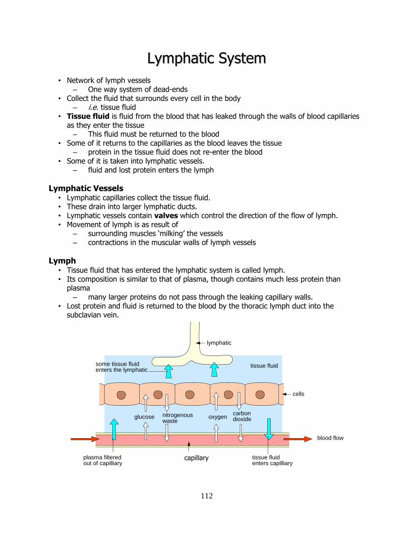
112
LLyymmpphhaattiicc SSyysstteemm
• Network of lymph vessels – One way system of dead-ends
• Collect the fluid that surrounds every cell in the body – i.e. tissue fluid
• Tissue fluid is fluid from the blood that has leaked through the walls of blood capillaries as they enter the tissue – This fluid must be returned to the blood
• Some of it returns to the capillaries as the blood leaves the tissue – protein in the tissue fluid does not re-enter the blood
• Some of it is taken into lymphatic vessels. – fluid and lost protein enters the lymph
Lymphatic Vessels • Lymphatic capillaries collect the tissue fluid. • These drain into larger lymphatic ducts. • Lymphatic vessels contain valves which control the direction of the flow of lymph. • Movement of lymph is as result of
– surrounding muscles ‘milking’ the vessels – contractions in the muscular walls of lymph vessels
Lymph • Tissue fluid that has entered the lymphatic system is called lymph. • Its composition is similar to that of plasma, though contains much less protein than
plasma – many larger proteins do not pass through the leaking capillary walls.
• Lost protein and fluid is returned to the blood by the thoracic lymph duct into the subclavian vein.
glucose nitrogenouswaste
oxygencarbondioxide
tissue fluid
capilliary
blood flow
plasma filteredout of capilliary
tissue fluidenters capilliary
lymphatic
cells
some tissue fluidenters the lymphatic
capillary
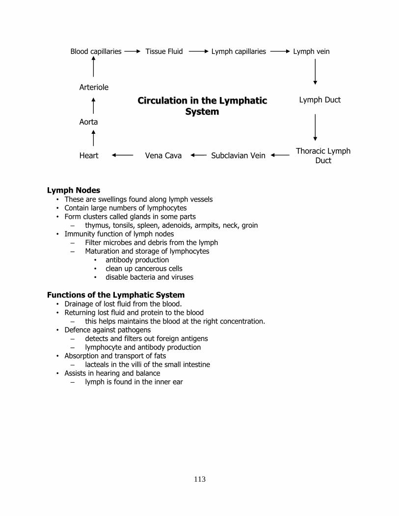
113
Blood capillaries Tissue Fluid Lymph capillaries Lymph vein
Lymph Nodes
• These are swellings found along lymph vessels • Contain large numbers of lymphocytes • Form clusters called glands in some parts
– thymus, tonsils, spleen, adenoids, armpits, neck, groin • Immunity function of lymph nodes
– Filter microbes and debris from the lymph – Maturation and storage of lymphocytes
• antibody production • clean up cancerous cells • disable bacteria and viruses
Functions of the Lymphatic System • Drainage of lost fluid from the blood. • Returning lost fluid and protein to the blood
– this helps maintains the blood at the right concentration. • Defence against pathogens
– detects and filters out foreign antigens – lymphocyte and antibody production
• Absorption and transport of fats – lacteals in the villi of the small intestine
• Assists in hearing and balance – lymph is found in the inner ear
Lymph Duct
Thoracic Lymph Duct
Vena Cava Subclavian Vein Heart
Aorta
Arteriole
CCiirrccuullaattiioonn iinn tthhee LLyymmpphhaattiicc
SSyysstteemm
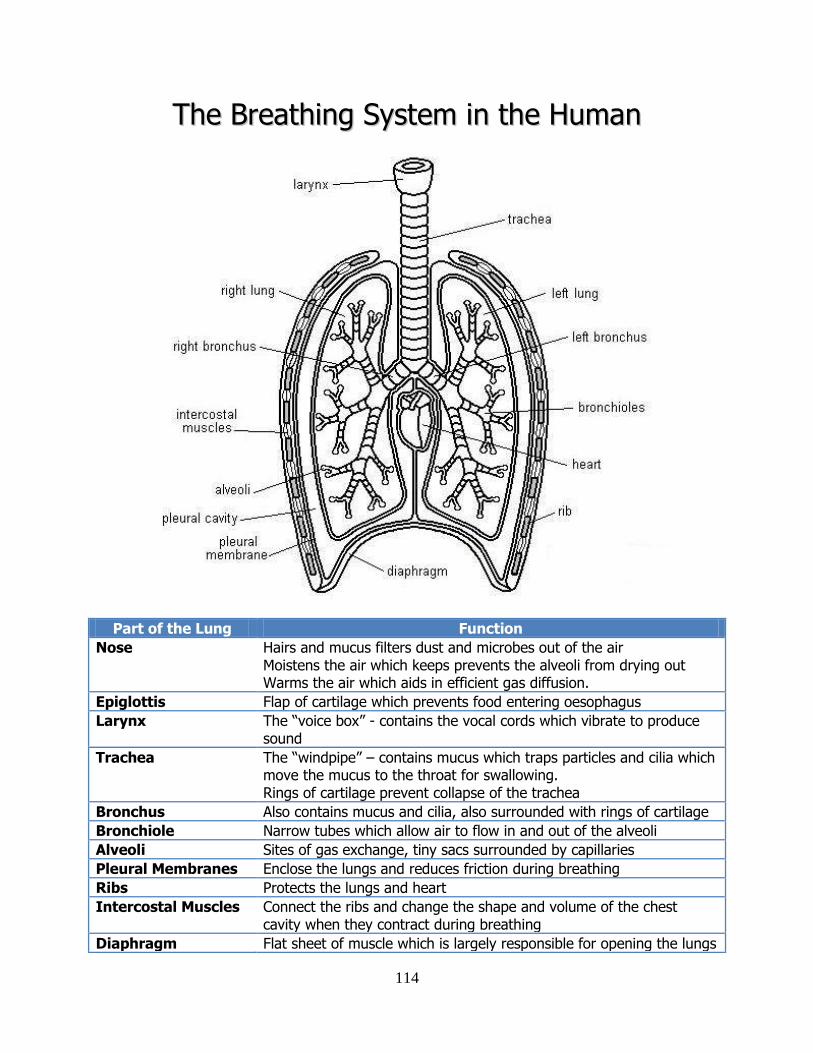
114
TThhee BBrreeaatthhiinngg SSyysstteemm iinn tthhee HHuummaann
Part of the Lung Function
Nose Hairs and mucus filters dust and microbes out of the air Moistens the air which keeps prevents the alveoli from drying out Warms the air which aids in efficient gas diffusion.
Epiglottis Flap of cartilage which prevents food entering oesophagus
Larynx The “voice box” - contains the vocal cords which vibrate to produce sound
Trachea The “windpipe” – contains mucus which traps particles and cilia which move the mucus to the throat for swallowing. Rings of cartilage prevent collapse of the trachea
Bronchus Also contains mucus and cilia, also surrounded with rings of cartilage
Bronchiole Narrow tubes which allow air to flow in and out of the alveoli
Alveoli Sites of gas exchange, tiny sacs surrounded by capillaries
Pleural Membranes Enclose the lungs and reduces friction during breathing
Ribs Protects the lungs and heart
Intercostal Muscles Connect the ribs and change the shape and volume of the chest cavity when they contract during breathing
Diaphragm Flat sheet of muscle which is largely responsible for opening the lungs
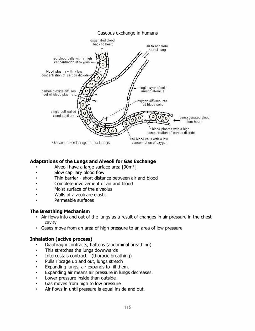
115
Gaseous exchange in humans
Adaptations of the Lungs and Alveoli for Gas Exchange • Alveoli have a large surface area [90m²] • Slow capillary blood flow • Thin barrier - short distance between air and blood • Complete involvement of air and blood • Moist surface of the alveolus • Walls of alveoli are elastic • Permeable surfaces
The Breathing Mechanism
• Air flows into and out of the lungs as a result of changes in air pressure in the chest cavity
• Gases move from an area of high pressure to an area of low pressure
Inhalation (active process) • Diaphragm contracts, flattens (abdominal breathing) • This stretches the lungs downwards • Intercostals contract (thoracic breathing) • Pulls ribcage up and out, lungs stretch • Expanding lungs, air expands to fill them. • Expanding air means air pressure in lungs decreases. • Lower pressure inside than outside • Gas moves from high to low pressure • Air flows in until pressure is equal inside and out.
116
Exhalation (passive process) • Diaphragm and intercostals relax • Force on the lungs has been removed • Lungs shrink to their original size • Volume of air in the lungs decreases • This causes an increase in air pressure inside the lungs • Air inside is at a higher pressure than outside • Gases move from higher to lower pressure • Air flows out of the lungs until pressure is equalised
Transport of Gases in the blood
• Red blood cells have haemoglobin in them. • Red blood cells carry 97% of the oxygen. • The other 3% is carried in the plasma. • Most CO2 is carried in the Plasma as
bicarbonate ions or as dissolved carbon dioxide. • A small % of CO2 is carried by the red blood cells .
The Role of CO2 in breathing control
• CO2 levels influence rate and depth of breathing • Rise in blood CO2 stimulates medulla • Medulla sends nerve impulses to breathing muscles • Diaphragm and intercostals contract, causing inhalation • Nervous message sent from expanded alveoli back to medulla switches off stimulation. • Muscles relax, exhalation.
Role of the Brain in Breathing
• Mostly breathing rhythm is unconsciously set by the brain • We can voluntarily change the rate at which we breath • Holding your breath leads eventually leads to unconsciousness, normal breathing then
resumes. • Death from holding your breath is impossible.
Breathing Disorders
• Asthma – inflammation & constriction of bronchi. • Bronchitis • Emphysema – destruction of alveoli • TB – elasticity reduced (bacteria) • Pneumonia – fills with fluid
Asthma Cause: largely unknown; allergens. Symptoms
• Coughing • Wheezing • Breathlessness • Chest tightness
Treatment • Use specific drug treatment • Inhalers
Bronchodilators (“reliever”) Steroids (“preventer”)
117
TThhee DDiiggeessttiivvee SSyysstteemm
Nutrition: The way an organism obtains and uses its food Autotrophic: The organism make its own food e.g. a plant Heterotrophic: The organism cannot make its own food so it must rely on eating outside sources e.g. an animal Herbivores: Animals that feed only on plants e.g. rabbit Carnivores: Animals that feed only on animals e.g. fox Omnivores: Animals that feed on plants and animals e.g. Badger Digestion: The physical and chemical breakdown of food into smaller soluble particles that can be absorbed by the body. The need for digestion: To breakdown large food particles until they are small enough to pass into body cells.
The need for a digestive system
• Individual cells don’t all have to contain a full range of digestive enzymes. • It’s more efficient if food digestion is carried out by a system of specialised cells tissues
and organs.
A Balanced Diet
• A balanced diet is a healthy eating habit which includes all of the essential nutrients in the correct proportions.
• This means eating a variety of foods each day from the various food groups – Protein, carbohydrates (including fibre), fats, vitamins, minerals, and water
• Factors affecting a balanced diet – Level of physical activity – Gender – Age
Stages in Human Nutrition Ingestion: Food is taken into the alimentary canal. Digestion: Food is broken down into smaller soluble pieces. Absorption: The movement of digested from the alimentary canal into the blood system. Egestion: Removal of undigested and unabsorbed material as faeces.
Fat in the Diet • Too much or too little fat in the diet can cause various problems. • Obesity refers to an unhealthy high % of body fat
– Heart disease – Diabetes – High blood pressure
• Anorexia refers to an unhealthy low % of body fat – Low heart rate
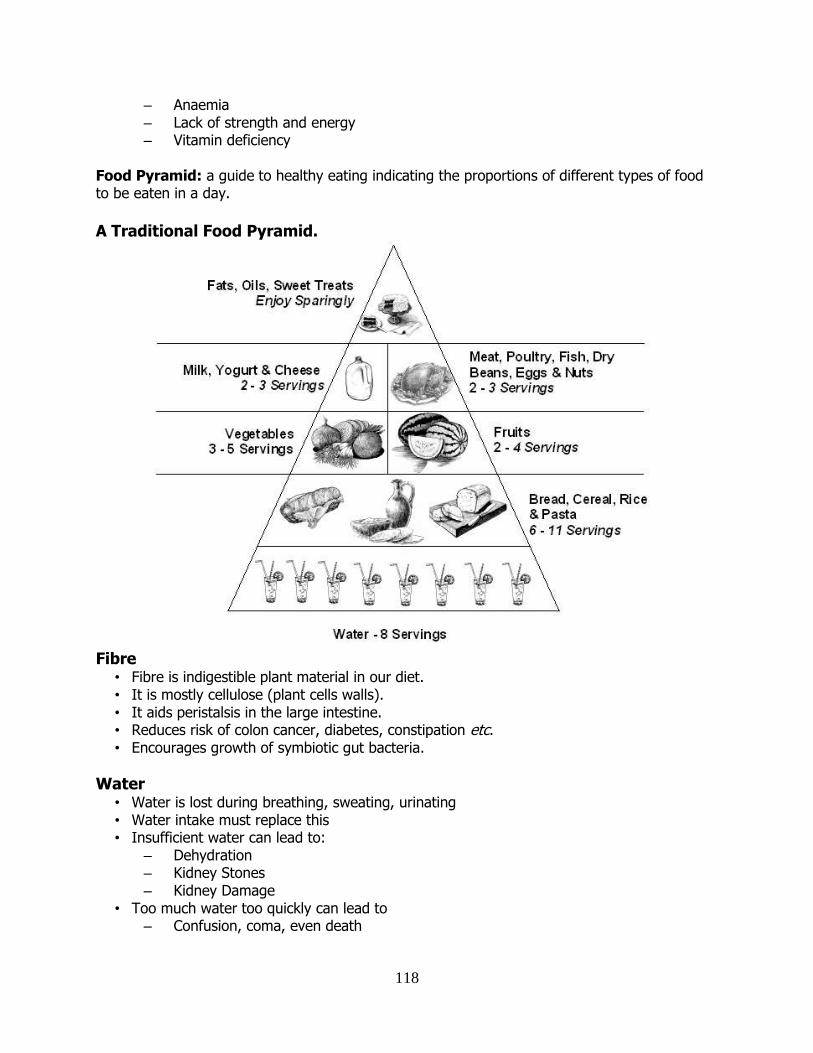
118
– Anaemia – Lack of strength and energy – Vitamin deficiency
Food Pyramid: a guide to healthy eating indicating the proportions of different types of food to be eaten in a day.
A Traditional Food Pyramid.
Fibre
• Fibre is indigestible plant material in our diet. • It is mostly cellulose (plant cells walls). • It aids peristalsis in the large intestine. • Reduces risk of colon cancer, diabetes, constipation etc. • Encourages growth of symbiotic gut bacteria.
Water • Water is lost during breathing, sweating, urinating • Water intake must replace this • Insufficient water can lead to:
– Dehydration – Kidney Stones – Kidney Damage
• Too much water too quickly can lead to – Confusion, coma, even death
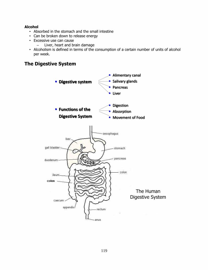
119
Alcohol • Absorbed in the stomach and the small intestine • Can be broken down to release energy • Excessive use can cause
– Liver, heart and brain damage • Alcoholism is defined in terms of the consumption of a certain number of units of alcohol
per week.
The Digestive System
The Human Digestive System
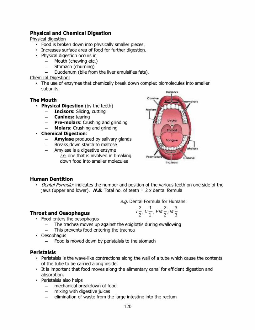
120
Physical and Chemical Digestion Physical digestion
• Food is broken down into physically smaller pieces. • Increases surface area of food for further digestion. • Physical digestion occurs in
– Mouth (chewing etc.) – Stomach (churning) – Duodenum (bile from the liver emulsifies fats).
Chemical Digestion: • The use of enzymes that chemically break down complex biomolecules into smaller
subunits.
The Mouth • Physical Digestion (by the teeth)
– Incisors: Slicing, cutting – Canines: tearing – Pre-molars: Crushing and grinding – Molars: Crushing and grinding
• Chemical Digestion: – Amylase produced by salivary glands – Breaks down starch to maltose – Amylase is a digestive enzyme i.e. one that is involved in breaking down food into smaller molecules
Human Dentition
• Dental Formula: indicates the number and position of the various teeth on one side of the jaws (upper and lower). N.B. Total no. of teeth = 2 x dental formula e.g. Dental Formula for Humans:
Throat and Oesophagus
• Food enters the oesophagus – The trachea moves up against the epiglottis during swallowing – This prevents food entering the trachea
• Oesophagus – Food is moved down by peristalsis to the stomach
Peristalsis • Peristalsis is the wave-like contractions along the wall of a tube which cause the contents
of the tube to be carried along inside. • It is important that food moves along the alimentary canal for efficient digestion and
absorption. • Peristalsis also helps
– mechanical breakdown of food – mixing with digestive juices – elimination of waste from the large intestine into the rectum
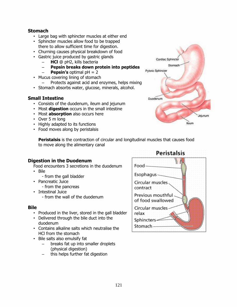
121
Stomach • Large bag with sphincter muscles at either end • Sphincter muscles allow food to be trapped
there to allow sufficient time for digestion. • Churning causes physical breakdown of food • Gastric juice produced by gastric glands
– HCl @ pH2, kills bacteria – Pepsin breaks down protein into peptides – Pepsin’s optimal pH = 2
• Mucus covering lining of stomach – Protects against acid and enzymes, helps mixing
• Stomach absorbs water, glucose, minerals, alcohol.
Small Intestine • Consists of the duodenum, ileum and jejunum • Most digestion occurs in the small intestine • Most absorption also occurs here • Over 5 m long • Highly adapted to its functions • Food moves along by peristalsis
Peristalsis is the contraction of circular and longitudinal muscles that causes food to move along the alimentary canal
Digestion in the Duodenum Food encounters 3 secretions in the duodenum • Bile
- from the gall bladder • Pancreatic Juice
- from the pancreas • Intestinal Juice
- from the wall of the duodenum
Bile
• Produced in the liver, stored in the gall bladder • Delivered through the bile duct into the
duodenum • Contains alkaline salts which neutralise the
HCl from the stomach • Bile salts also emulsify fat
– breaks fat up into smaller droplets (physical digestion)
– this helps further fat digestion
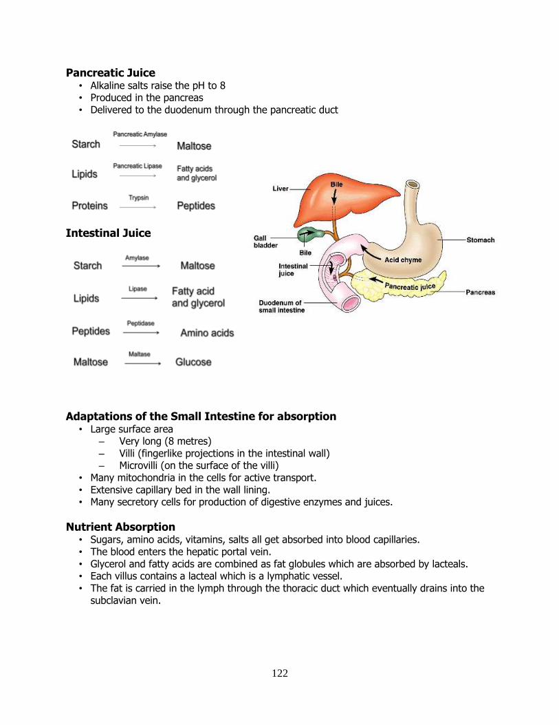
122
Pancreatic Juice • Alkaline salts raise the pH to 8 • Produced in the pancreas • Delivered to the duodenum through the pancreatic duct
Intestinal Juice
Adaptations of the Small Intestine for absorption
• Large surface area – Very long (8 metres) – Villi (fingerlike projections in the intestinal wall) – Microvilli (on the surface of the villi)
• Many mitochondria in the cells for active transport. • Extensive capillary bed in the wall lining. • Many secretory cells for production of digestive enzymes and juices.
Nutrient Absorption • Sugars, amino acids, vitamins, salts all get absorbed into blood capillaries. • The blood enters the hepatic portal vein. • Glycerol and fatty acids are combined as fat globules which are absorbed by lacteals. • Each villus contains a lacteal which is a lymphatic vessel. • The fat is carried in the lymph through the thoracic duct which eventually drains into the
subclavian vein.
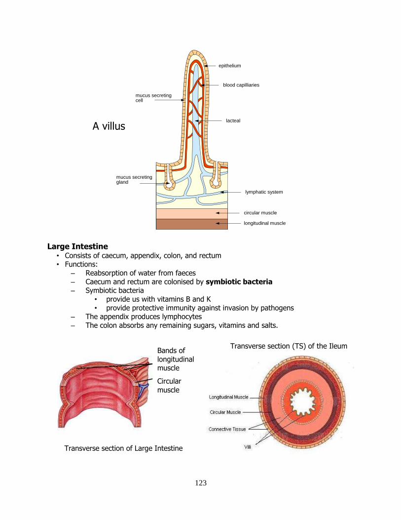
123
Large Intestine
• Consists of caecum, appendix, colon, and rectum • Functions:
– Reabsorption of water from faeces – Caecum and rectum are colonised by symbiotic bacteria – Symbiotic bacteria
• provide us with vitamins B and K • provide protective immunity against invasion by pathogens
– The appendix produces lymphocytes – The colon absorbs any remaining sugars, vitamins and salts.
epithelium
blood capilliaries
lacteal
lymphatic system
mucus secretingcell
mucus secretinggland
circular muscle
longitudinal muscle
Transverse section (TS) of the Ileum
Transverse section of Large Intestine
Bands of longitudinal muscle
Circular
muscle
A villus
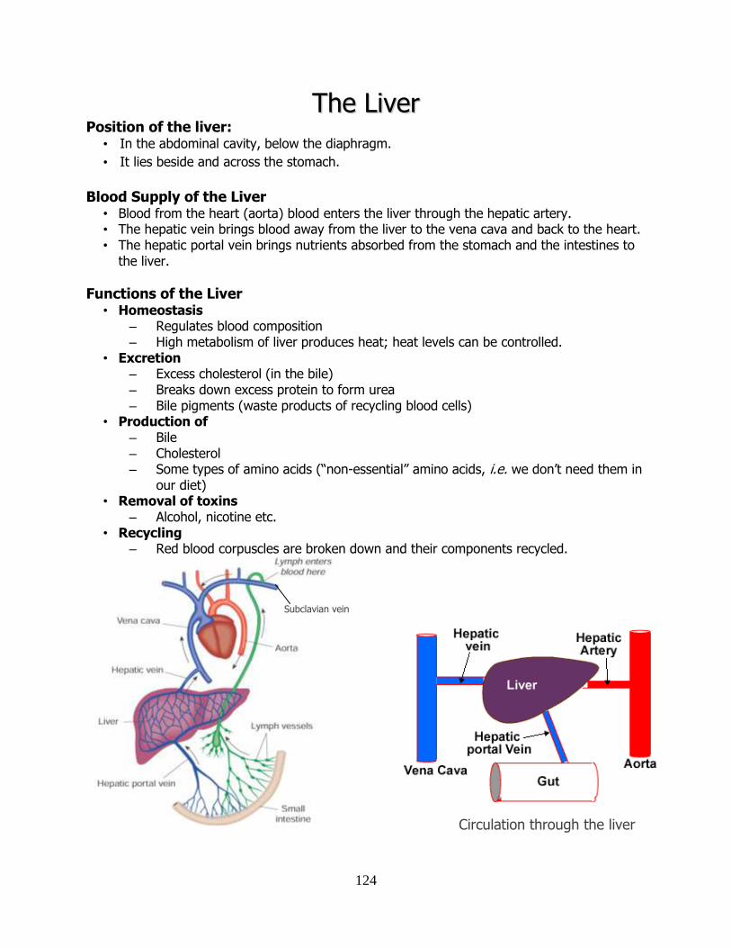
124
TThhee LLiivveerr Position of the liver:
• In the abdominal cavity, below the diaphragm.
• It lies beside and across the stomach.
Blood Supply of the Liver • Blood from the heart (aorta) blood enters the liver through the hepatic artery. • The hepatic vein brings blood away from the liver to the vena cava and back to the heart. • The hepatic portal vein brings nutrients absorbed from the stomach and the intestines to
the liver.
Functions of the Liver • Homeostasis
– Regulates blood composition – High metabolism of liver produces heat; heat levels can be controlled.
• Excretion – Excess cholesterol (in the bile) – Breaks down excess protein to form urea – Bile pigments (waste products of recycling blood cells)
• Production of – Bile – Cholesterol – Some types of amino acids (“non-essential” amino acids, i.e. we don’t need them in
our diet) • Removal of toxins
– Alcohol, nicotine etc. • Recycling
– Red blood corpuscles are broken down and their components recycled.
Subclavian vein
Circulation through the liver
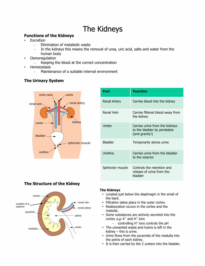
125
TThhee KKiiddnneeyyss Functions of the Kidneys • Excretion
– Elimination of metabolic waste – In the kidneys this means the removal of urea, uric acid, salts and water from the
human body • Osmoregulation
– Keeping the blood at the correct concentration • Homeostasis
– Maintenance of a suitable internal environment
The Urinary System
Part Function
Renal Artery Carries blood into the kidney
Renal Vein Carries filtered blood away from the kidney
Ureter Carries urine from the kidneys to the bladder by peristalsis
(and gravity!)
Bladder Temporarily stores urine
Urethra Carries urine from the bladder to the exterior
Sphincter muscle Controls the retention and
release of urine from the bladder
The Structure of the Kidney
cortex
renal artery
renal vein
ureter
pelvis
medulla
pyramid
vena cava aorta
renal arteryrenal vein
kidney
bladder
sphincter muscle
urethra
ureter
Location of a nephron
The Kidneys
• Located just below the diaphragm in the small of the back.
• Filtration takes place in the outer cortex.
• Reabsorption occurs in the cortex and the
medulla.
• Some substances are actively secreted into the
cortex e.g. K+ and H+ ions
– controlling H+ ions controls the pH
• The unwanted waste and toxins is left in the
kidney – this is urine.
• Urine flows from the pyramids of the medulla into the pelvis of each kidney.
• It is then carried by the 2 ureters into the bladder.
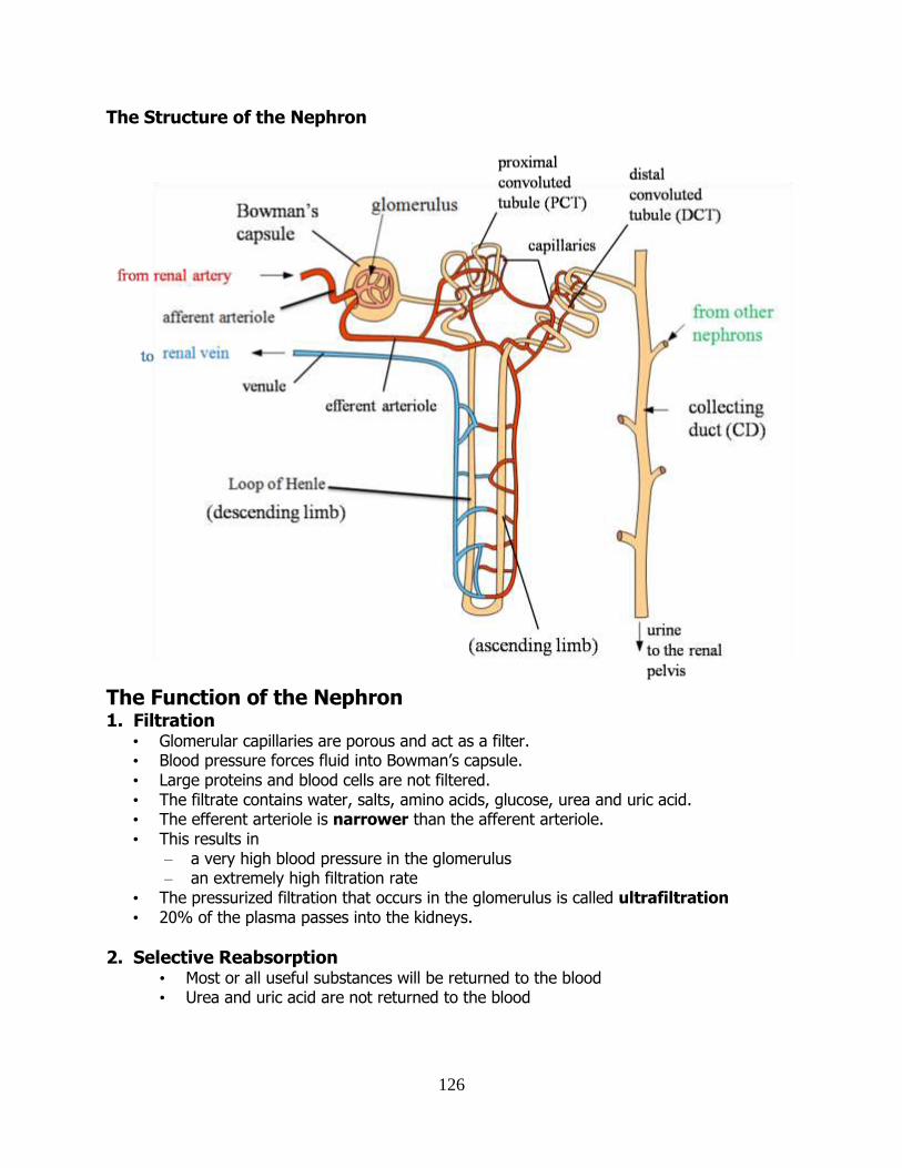
126
The Structure of the Nephron
The Function of the Nephron 1. Filtration
• Glomerular capillaries are porous and act as a filter. • Blood pressure forces fluid into Bowman’s capsule. • Large proteins and blood cells are not filtered. • The filtrate contains water, salts, amino acids, glucose, urea and uric acid. • The efferent arteriole is narrower than the afferent arteriole. • This results in
– a very high blood pressure in the glomerulus – an extremely high filtration rate
• The pressurized filtration that occurs in the glomerulus is called ultrafiltration • 20% of the plasma passes into the kidneys.
2. Selective Reabsorption • Most or all useful substances will be returned to the blood • Urea and uric acid are not returned to the blood
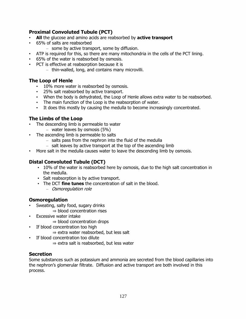
127
Proximal Convoluted Tubule (PCT) • All the glucose and amino acids are reabsorbed by active transport • 65% of salts are reabsorbed
– some by active transport, some by diffusion. • ATP is required for this, so there are many mitochondria in the cells of the PCT lining. • 65% of the water is reabsorbed by osmosis. • PCT is effective at reabsorption because it is
– thin-walled, long, and contains many microvilli.
The Loop of Henle • 10% more water is reabsorbed by osmosis. • 25% salt reabsorbed by active transport. • When the body is dehydrated, the Loop of Henle allows extra water to be reabsorbed. • The main function of the Loop is the reabsorption of water. • It does this mostly by causing the medulla to become increasingly concentrated.
The Limbs of the Loop • The descending limb is permeable to water
– water leaves by osmosis (5%) • The ascending limb is permeable to salts
– salts pass from the nephron into the fluid of the medulla – salt leaves by active transport at the top of the ascending limb
• More salt in the medulla causes water to leave the descending limb by osmosis.
Distal Convoluted Tubule (DCT) • 10% of the water is reabsorbed here by osmosis, due to the high salt concentration in
the medulla. • Salt reabsorption is by active transport. • The DCT fine tunes the concentration of salt in the blood.
– Osmoregulation role
Osmoregulation • Sweating, salty food, sugary drinks
⇒ blood concentration rises • Excessive water intake
⇒ blood concentration drops • If blood concentration too high
⇒ extra water reabsorbed, but less salt • If blood concentration too dilute
⇒ extra salt is reabsorbed, but less water
Secretion Some substances such as potassium and ammonia are secreted from the blood capillaries into the nephron’s glomerular filtrate. Diffusion and active transport are both involved in this process.
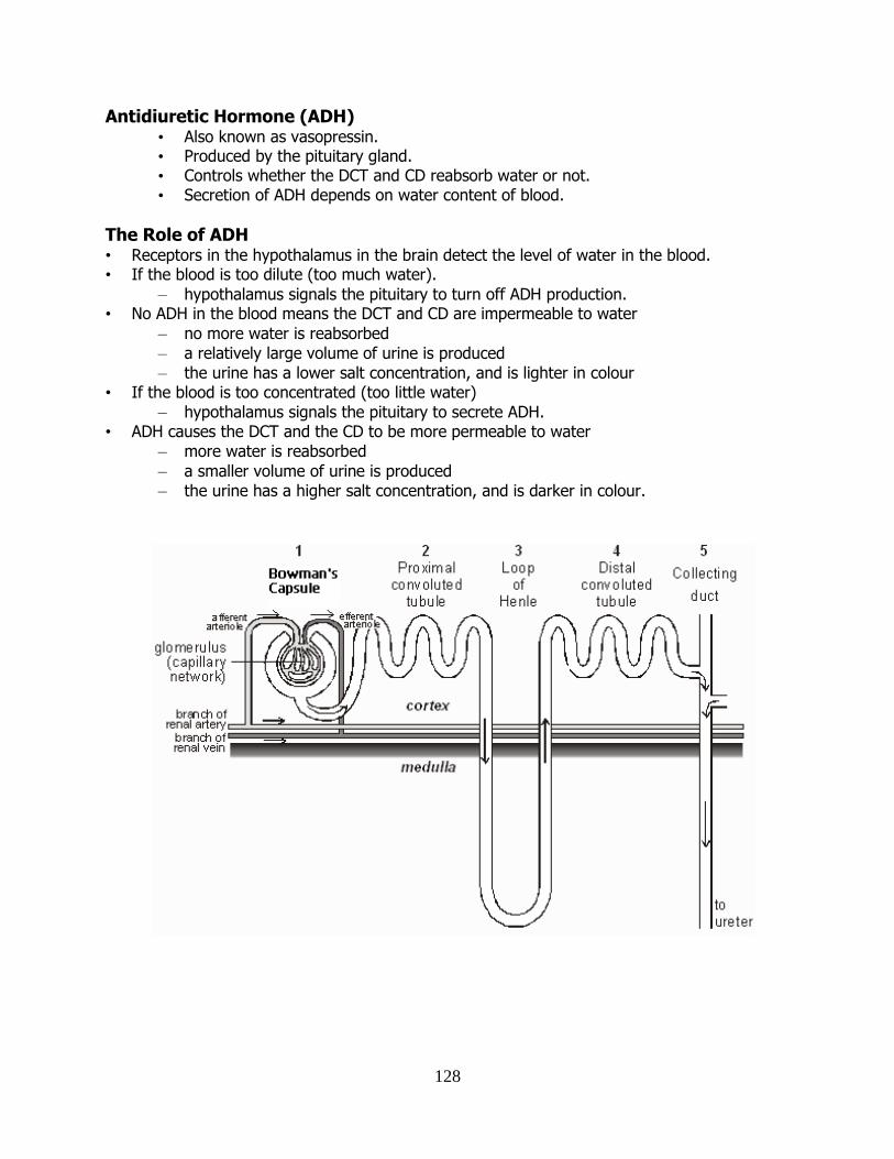
128
Antidiuretic Hormone (ADH) • Also known as vasopressin. • Produced by the pituitary gland. • Controls whether the DCT and CD reabsorb water or not. • Secretion of ADH depends on water content of blood.
The Role of ADH • Receptors in the hypothalamus in the brain detect the level of water in the blood. • If the blood is too dilute (too much water).
– hypothalamus signals the pituitary to turn off ADH production. • No ADH in the blood means the DCT and CD are impermeable to water
– no more water is reabsorbed
– a relatively large volume of urine is produced
– the urine has a lower salt concentration, and is lighter in colour • If the blood is too concentrated (too little water)
– hypothalamus signals the pituitary to secrete ADH. • ADH causes the DCT and the CD to be more permeable to water
– more water is reabsorbed
– a smaller volume of urine is produced
– the urine has a higher salt concentration, and is darker in colour.
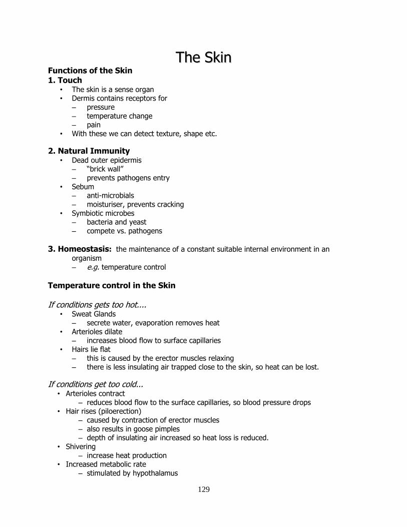
129
TThhee SSkkiinn Functions of the Skin 1. Touch
• The skin is a sense organ • Dermis contains receptors for
– pressure – temperature change – pain
• With these we can detect texture, shape etc.
2. Natural Immunity • Dead outer epidermis
– “brick wall” – prevents pathogens entry
• Sebum – anti-microbials – moisturiser, prevents cracking
• Symbiotic microbes – bacteria and yeast – compete vs. pathogens
3. Homeostasis: the maintenance of a constant suitable internal environment in an
organism – e.g. temperature control
Temperature control in the Skin
If conditions gets too hot.... • Sweat Glands
– secrete water, evaporation removes heat • Arterioles dilate
– increases blood flow to surface capillaries • Hairs lie flat
– this is caused by the erector muscles relaxing – there is less insulating air trapped close to the skin, so heat can be lost.
If conditions get too cold... • Arterioles contract
– reduces blood flow to the surface capillaries, so blood pressure drops • Hair rises (piloerection)
– caused by contraction of erector muscles – also results in goose pimples – depth of insulating air increased so heat loss is reduced.
• Shivering – increase heat production
• Increased metabolic rate – stimulated by hypothalamus
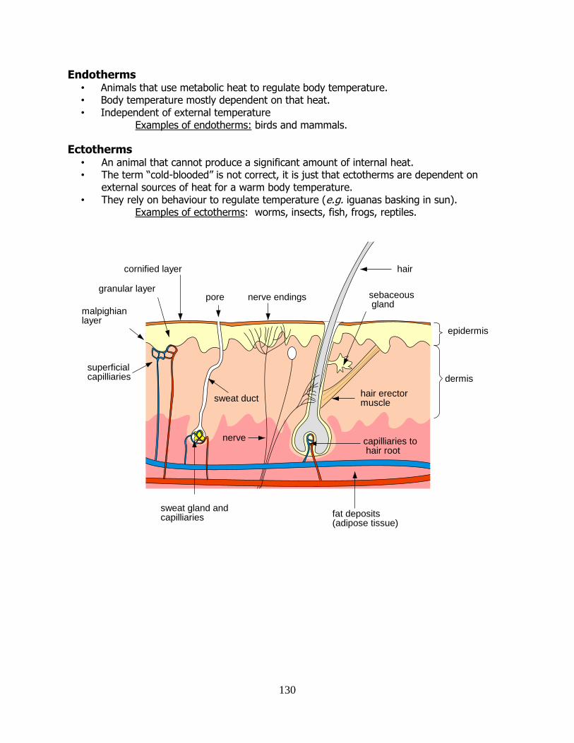
130
hair
pore nerve endings
hair erector muscle
sebaceous gland
superficialcapilliaries
sweat duct
nerve
fat deposits(adipose tissue)
sweat gland andcapilliaries
capilliaries to hair root
dermis
epidermis
cornified layer
granular layer
malpighianlayer
Endotherms • Animals that use metabolic heat to regulate body temperature. • Body temperature mostly dependent on that heat. • Independent of external temperature
Examples of endotherms: birds and mammals.
Ectotherms • An animal that cannot produce a significant amount of internal heat. • The term “cold-blooded” is not correct, it is just that ectotherms are dependent on
external sources of heat for a warm body temperature. • They rely on behaviour to regulate temperature (e.g. iguanas basking in sun).
Examples of ectotherms: worms, insects, fish, frogs, reptiles.
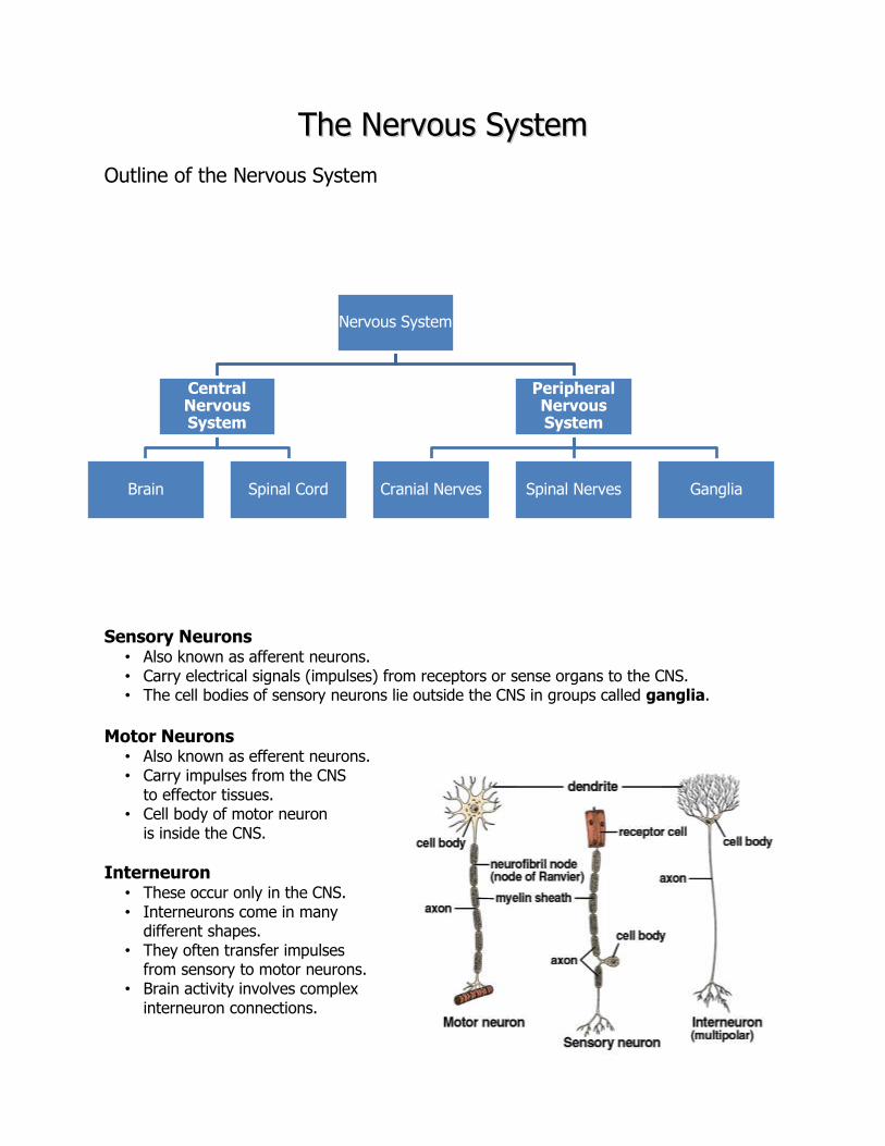
131
TThhee NNeerrvvoouuss SSyysstteemm
Outline of the Nervous System
Sensory Neurons • Also known as afferent neurons. • Carry electrical signals (impulses) from receptors or sense organs to the CNS. • The cell bodies of sensory neurons lie outside the CNS in groups called ganglia.
Motor Neurons
• Also known as efferent neurons. • Carry impulses from the CNS to effector tissues. • Cell body of motor neuron is inside the CNS.
Interneuron • These occur only in the CNS. • Interneurons come in many different shapes. • They often transfer impulses from sensory to motor neurons. • Brain activity involves complex interneuron connections.
Nervous System
Central Nervous System
Brain Spinal Cord
Peripheral Nervous System
Cranial Nerves Spinal Nerves Ganglia
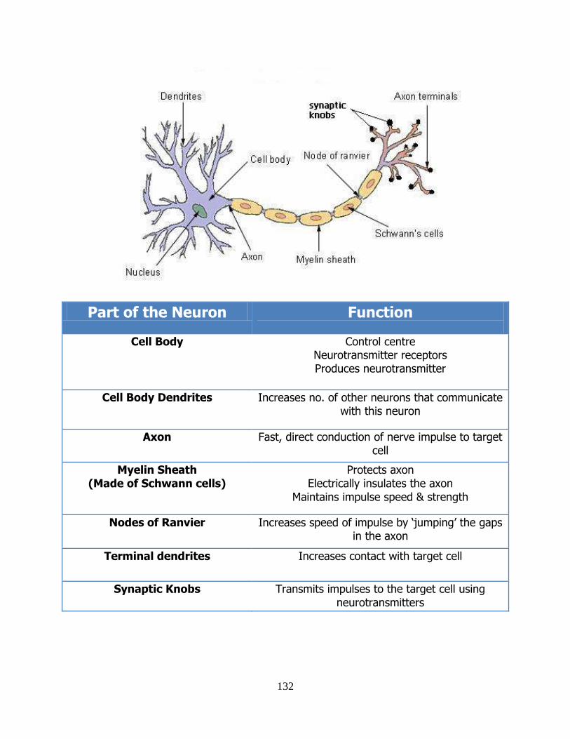
132
Part of the Neuron Function
Cell Body Control centre Neurotransmitter receptors Produces neurotransmitter
Cell Body Dendrites Increases no. of other neurons that communicate with this neuron
Axon Fast, direct conduction of nerve impulse to target cell
Myelin Sheath (Made of Schwann cells)
Protects axon Electrically insulates the axon
Maintains impulse speed & strength
Nodes of Ranvier Increases speed of impulse by ‘jumping’ the gaps in the axon
Terminal dendrites Increases contact with target cell
Synaptic Knobs Transmits impulses to the target cell using neurotransmitters
133
The Nerve Impulse • A nerve impulse is a self-propagating wave of electrical charge along a neuron. • The nerve impulse is exactly the same in all neurons. • The impulse is a wave of “depolarisation” that occurs due to influx on ions along the axon. • This requires ATP.
Nerve Impulse Terminology
• Stimulus • A change in the neurons environment that, if strong enough, will generate an
impulse. • Threshold
• The minimum intensity of stimulus needed to generate an impulse. • All-or-nothing
• an impulse is only generated if the stimulus is at or above threshold. • Refractory Period
• This is the slight delay that occurs between any two impulses
The Synapse
• Specialised junctions between a neuron and the target cell. • Target cell could be
– a muscle cell – a gland cell – another neuron
The Nerve Impulse
• The impulse arrives at the synapse. • Its arrival stimulates release of neurotransmitter from synaptic vesicles into the synaptic
cleft (gap). • Neurotransmitter molecules diffuse across to the target cell. • They bind to specific receptors in the target cell membrane (the “post synaptic
membrane”). • This neurotransmitter-receptor complex generates an impulse in the target cell. • An enzyme is released into the area and digests the neurotransmitter. • This switches off the response. • The breakdown products are reabsorbed through the pre-synaptic membrane. • Neurotransmitter is remade from the recycled breakdown products. • N.B. Example
• Acetylcholine (neurotransmitter) • Cholinesterase (enzyme)
• Drugs can be used to change nerve impulses by
• Binding to the receptor on the post-synaptic membrane.
• Blocking the production or release of neurotransmitter.
• Preventing release of enzyme into the synapse.
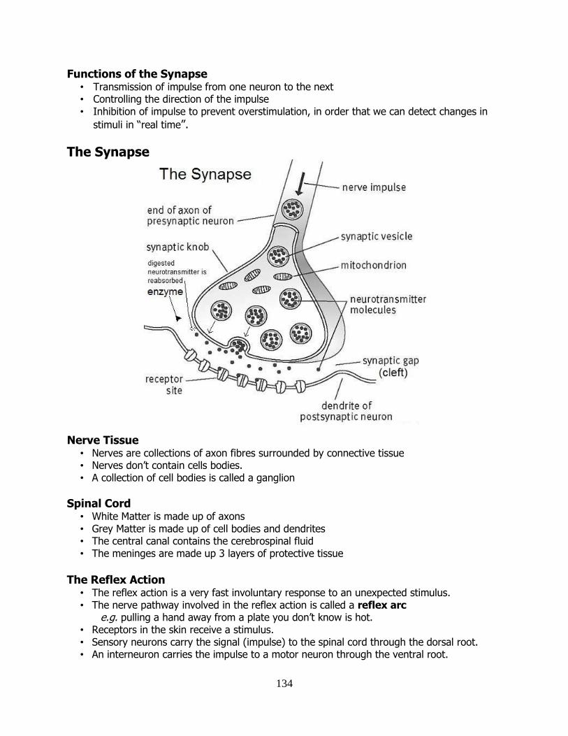
134
Functions of the Synapse • Transmission of impulse from one neuron to the next • Controlling the direction of the impulse • Inhibition of impulse to prevent overstimulation, in order that we can detect changes in
stimuli in “real time”.
The Synapse
Nerve Tissue
• Nerves are collections of axon fibres surrounded by connective tissue • Nerves don’t contain cells bodies. • A collection of cell bodies is called a ganglion
Spinal Cord • White Matter is made up of axons • Grey Matter is made up of cell bodies and dendrites • The central canal contains the cerebrospinal fluid • The meninges are made up 3 layers of protective tissue
The Reflex Action
• The reflex action is a very fast involuntary response to an unexpected stimulus. • The nerve pathway involved in the reflex action is called a reflex arc e.g. pulling a hand away from a plate you don’t know is hot. • Receptors in the skin receive a stimulus. • Sensory neurons carry the signal (impulse) to the spinal cord through the dorsal root. • An interneuron carries the impulse to a motor neuron through the ventral root.
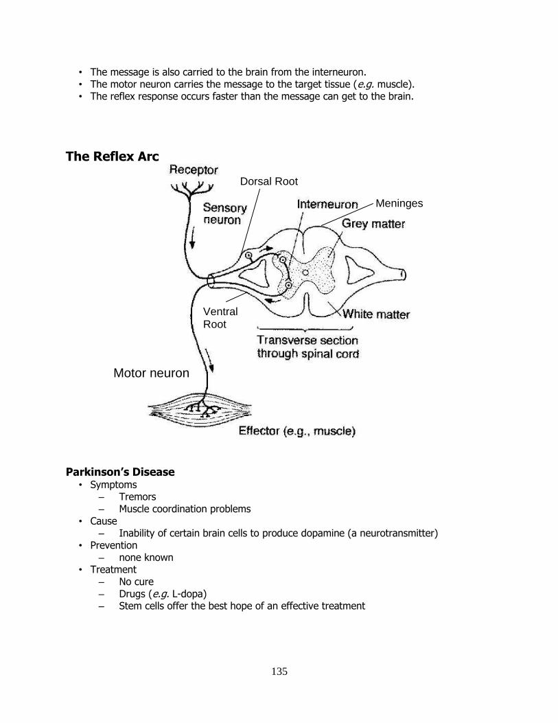
135
• The message is also carried to the brain from the interneuron. • The motor neuron carries the message to the target tissue (e.g. muscle). • The reflex response occurs faster than the message can get to the brain.
The Reflex Arc
Parkinson’s Disease • Symptoms
– Tremors – Muscle coordination problems
• Cause – Inability of certain brain cells to produce dopamine (a neurotransmitter)
• Prevention – none known
• Treatment – No cure – Drugs (e.g. L-dopa) – Stem cells offer the best hope of an effective treatment
Motor neuron
Dorsal Root
Ventral Root
Meninges
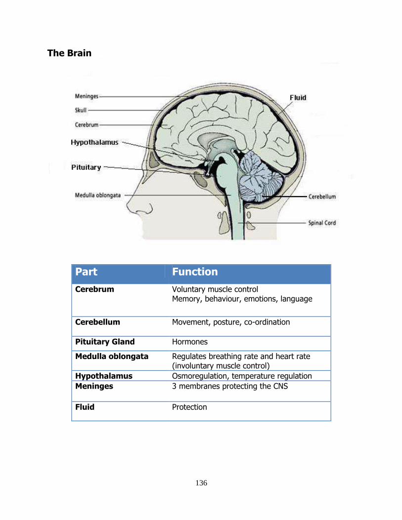
136
The Brain
Part Function
Cerebrum Voluntary muscle control Memory, behaviour, emotions, language
Cerebellum Movement, posture, co-ordination
Pituitary Gland Hormones
Medulla oblongata Regulates breathing rate and heart rate (involuntary muscle control)
Hypothalamus Osmoregulation, temperature regulation
Meninges
3 membranes protecting the CNS
Fluid Protection
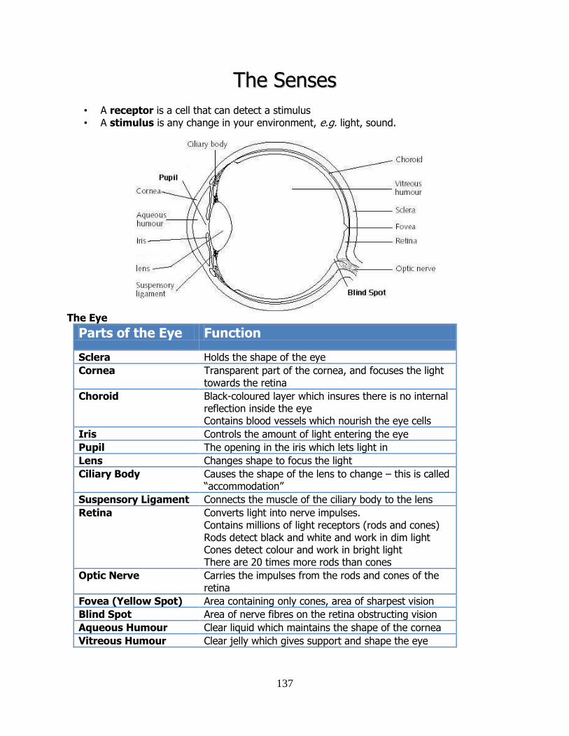
137
TThhee SSeennsseess
• A receptor is a cell that can detect a stimulus • A stimulus is any change in your environment, e.g. light, sound.
The Eye
Parts of the Eye Function
Sclera Holds the shape of the eye
Cornea Transparent part of the cornea, and focuses the light towards the retina
Choroid Black-coloured layer which insures there is no internal reflection inside the eye Contains blood vessels which nourish the eye cells
Iris Controls the amount of light entering the eye
Pupil The opening in the iris which lets light in
Lens Changes shape to focus the light
Ciliary Body Causes the shape of the lens to change – this is called “accommodation”
Suspensory Ligament Connects the muscle of the ciliary body to the lens
Retina Converts light into nerve impulses. Contains millions of light receptors (rods and cones) Rods detect black and white and work in dim light Cones detect colour and work in bright light There are 20 times more rods than cones
Optic Nerve Carries the impulses from the rods and cones of the retina
Fovea (Yellow Spot) Area containing only cones, area of sharpest vision
Blind Spot Area of nerve fibres on the retina obstructing vision
Aqueous Humour Clear liquid which maintains the shape of the cornea
Vitreous Humour Clear jelly which gives support and shape the eye
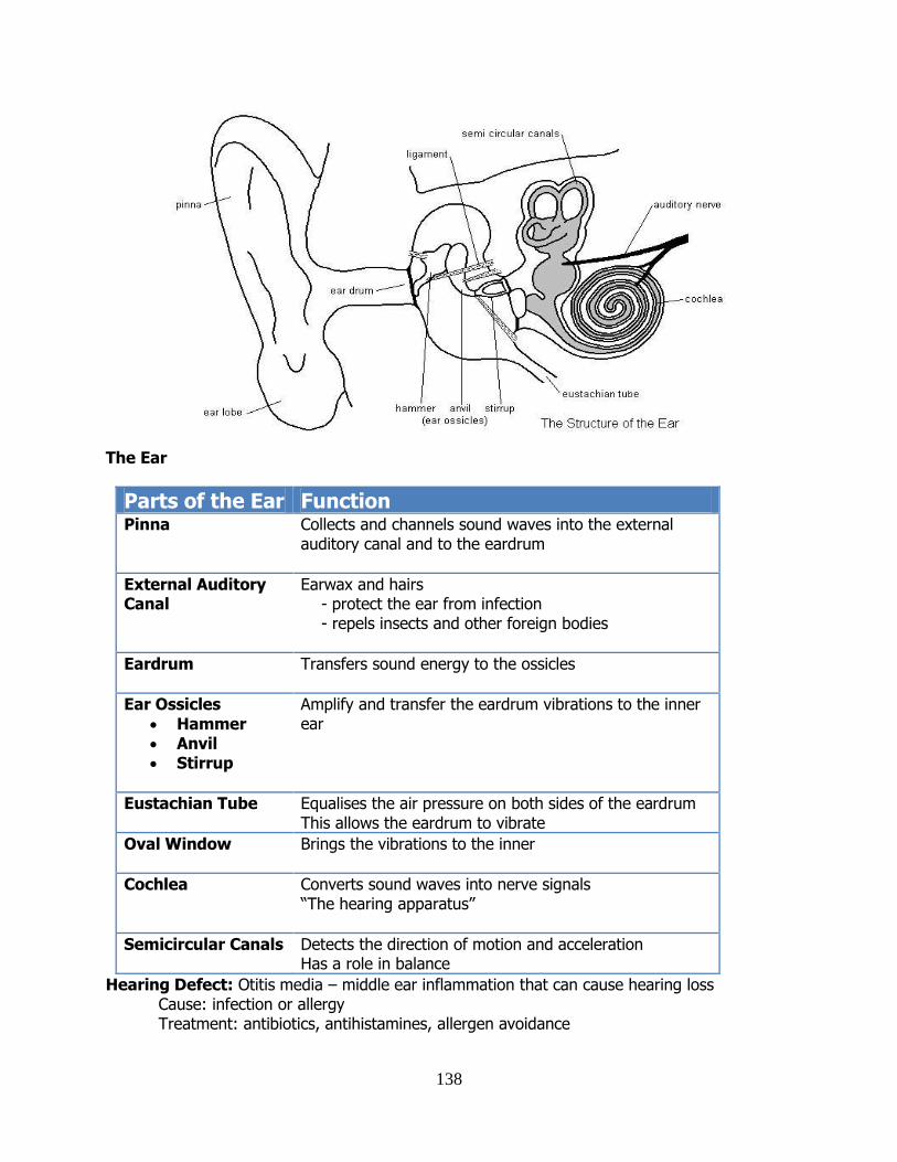
138
The Ear
Parts of the Ear Function Pinna Collects and channels sound waves into the external
auditory canal and to the eardrum
External Auditory Canal
Earwax and hairs - protect the ear from infection - repels insects and other foreign bodies
Eardrum
Transfers sound energy to the ossicles
Ear Ossicles
Hammer Anvil Stirrup
Amplify and transfer the eardrum vibrations to the inner ear
Eustachian Tube
Equalises the air pressure on both sides of the eardrum This allows the eardrum to vibrate
Oval Window
Brings the vibrations to the inner
Cochlea
Converts sound waves into nerve signals “The hearing apparatus”
Semicircular Canals Detects the direction of motion and acceleration Has a role in balance
Hearing Defect: Otitis media – middle ear inflammation that can cause hearing loss Cause: infection or allergy Treatment: antibiotics, antihistamines, allergen avoidance
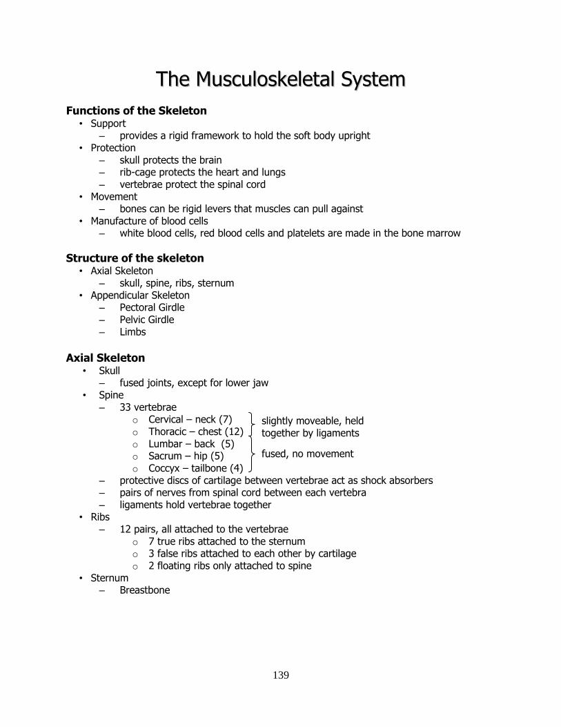
139
TThhee MMuussccuulloosskkeelleettaall SSyysstteemm Functions of the Skeleton
• Support – provides a rigid framework to hold the soft body upright
• Protection – skull protects the brain – rib-cage protects the heart and lungs – vertebrae protect the spinal cord
• Movement – bones can be rigid levers that muscles can pull against
• Manufacture of blood cells – white blood cells, red blood cells and platelets are made in the bone marrow
Structure of the skeleton • Axial Skeleton
– skull, spine, ribs, sternum • Appendicular Skeleton
– Pectoral Girdle – Pelvic Girdle – Limbs
Axial Skeleton
• Skull – fused joints, except for lower jaw
• Spine – 33 vertebrae
o Cervical – neck (7) o Thoracic – chest (12) o Lumbar – back (5) o Sacrum – hip (5) o Coccyx – tailbone (4)
– protective discs of cartilage between vertebrae act as shock absorbers – pairs of nerves from spinal cord between each vertebra – ligaments hold vertebrae together
• Ribs – 12 pairs, all attached to the vertebrae
o 7 true ribs attached to the sternum o 3 false ribs attached to each other by cartilage o 2 floating ribs only attached to spine
• Sternum – Breastbone
slightly moveable, held together by ligaments fused, no movement
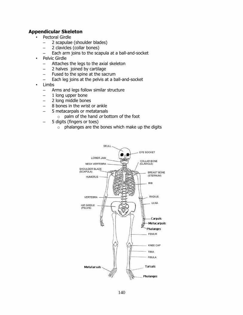
140
Appendicular Skeleton • Pectoral Girdle
– 2 scapulae (shoulder blades) – 2 clavicles (collar bones) – Each arm joins to the scapula at a ball-and-socket
• Pelvic Girdle – Attaches the legs to the axial skeleton – 2 halves joined by cartilage – Fused to the spine at the sacrum – Each leg joins at the pelvis at a ball-and-socket
• Limbs – Arms and legs follow similar structure – 1 long upper bone – 2 long middle bones – 8 bones in the wrist or ankle – 5 metacarpals or metatarsals
o palm of the hand or bottom of the foot – 5 digits (fingers or toes)
o phalanges are the bones which make up the digits

141
Structure of the long bone • The epiphysis is the wide cap at the end of the long bone.
– Both epiphyses are covered with protective cartilage • The diaphysis is the shaft of the bone • The periosteum is a membrane that lines the outer surface of all bones, except at the
ends of long bones. It provides an attachment for muscles and tendons.
• The medullary cavity is a hollow area at the centre of the diaphysis.
Compact bone
• Consists of living cells (osteoblasts) embedded in a matrix of calcium salts and protein (called collagen). – Inorganic calcium salts give strength – Organic collagen protein gives flexibility
• Very dense. • Found mainly at the outer edge of the diaphysis and as a layer around the epiphyses. • Blood vessels and nerves run through the bone.
Spongy bone • Gives moderate strength and rigidity to bones. • Consists of a network of thin, bony columns and plates. • These are separated by spaces • Spaces are filled with yellow or red marrow. • Makes bones lighter.
Bone Marrow • Marrow is contained within the medullary cavity • Red marrow makes blood cells (red, white and platelets). • Yellow marrow is inactive. It serves as a fat-rich, energy store. • The medullar cavity reduces the weight of a bone without reducing its strength
Bone growth
• Cartilage is replaced with bone during embryonic development. • Osteoblasts are bone-secreting cells. • Bones lengthen due to the activity of osteoblasts at the growth plates. (ceases after adolescence) • A growth plate is a band of cartilage between the epiphysis and the diaphysis. • New cartilage is formed here and turned to bone (ossified) by osteoblasts.
Epiphysis
Epiphysis
Diaphysis
Spongy Bone
Compact bone
Yellow marrow
Medullary cavity
Articular cartilage
Periosteum
Red marrow
Blood vessel
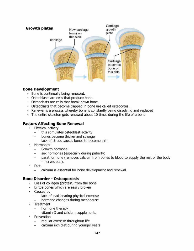
142
Bone Development
• Bone is continually being renewed. • Osteoblasts are cells that produce bone. • Osteoclasts are cells that break down bone. • Osteoblasts that become trapped in bone are called osteocytes.. • Renewal is a process whereby bone is constantly being dissolving and replaced • The entire skeleton gets renewed about 10 times during the life of a bone.
Factors Affecting Bone Renewal
• Physical activity – this stimulates osteoblast activity – bones become thicker and stronger – lack of stress causes bones to become thin.
• Hormones – Growth hormone – sex hormones (especially during puberty) – parathormone (removes calcium from bones to blood to supply the rest of the body
– nerves etc.). • Diet
– calcium is essential for bone development and renewal.
Bone Disorder - Osteoporosis • Loss of collagen (protein) from the bone • Brittle bones which are easily broken • Caused by
– lack of load-bearing physical exercise – hormone changes during menopause
• Treatment – hormone therapy – vitamin D and calcium supplements
• Prevention – regular exercise throughout life – calcium rich diet during younger years
Growth plates
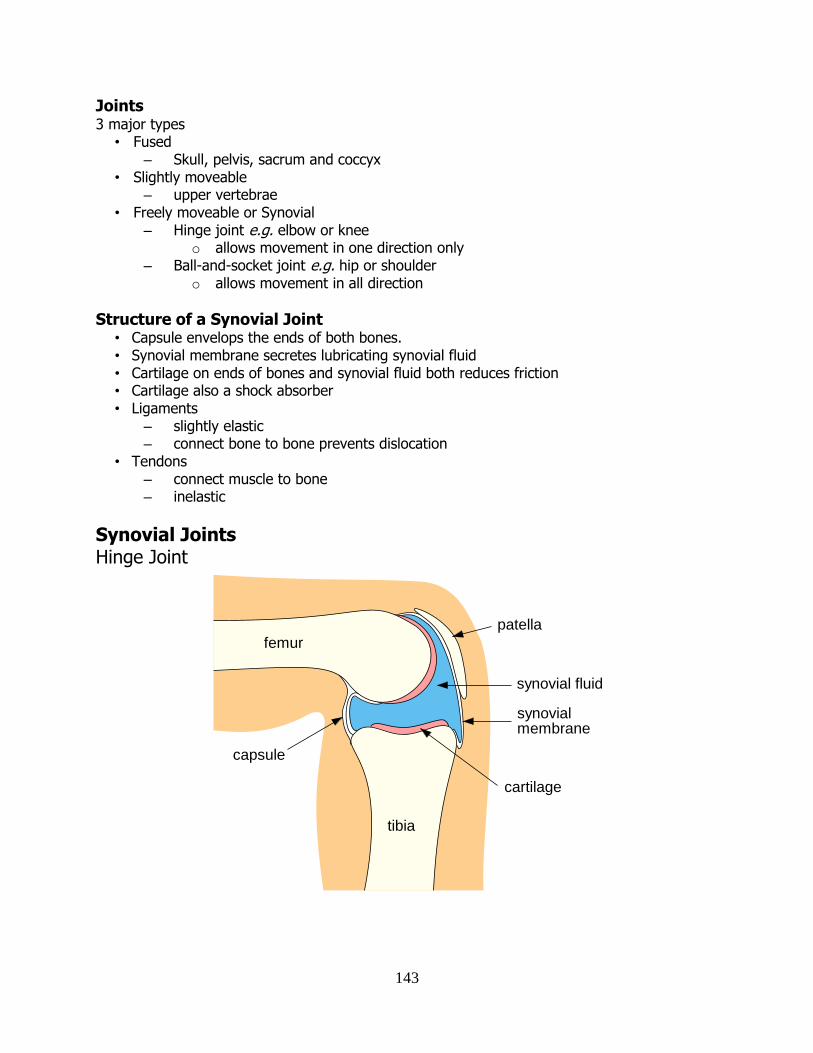
143
Joints 3 major types
• Fused – Skull, pelvis, sacrum and coccyx
• Slightly moveable – upper vertebrae
• Freely moveable or Synovial – Hinge joint e.g. elbow or knee
o allows movement in one direction only – Ball-and-socket joint e.g. hip or shoulder
o allows movement in all direction
Structure of a Synovial Joint • Capsule envelops the ends of both bones. • Synovial membrane secretes lubricating synovial fluid • Cartilage on ends of bones and synovial fluid both reduces friction • Cartilage also a shock absorber • Ligaments
– slightly elastic – connect bone to bone prevents dislocation
• Tendons – connect muscle to bone – inelastic
Synovial Joints Hinge Joint
femur
tibia
patella
synovial fluid
capsule
synovial membrane
cartilage
144
Ball-and-socket Joint
Muscles
• Skeletal Muscle – striped muscle for body movement and support – can contract very quickly but tires easily – under voluntary control
• Smooth muscle – contracts slowly, but doesn’t tire easily – under involuntary, unconscious control – Bladder, uterus, digestive tract
• Cardiac muscle – involuntary control
Antagonistic Pairs • Pairs of muscles that have opposite effects on each other • Muscles can only pull, they can’t push
– they can shorten (contract) but not lengthen • Raising the forearm
– Biceps muscle contract to pull 2 tendons connected to shoulder – Triceps relaxed
• Lowering the forearm – Triceps contract to pull 3 tendons connected to shoulder – Biceps relax
pelvis
femur
synovial fluid
cartilage
capsule
ligament
synovial membrane
ligament
145
An Antagonistic Pair of Muscles
Ligaments
• Ligaments are strong, slightly elastic fibres.
• They connect bone to bone.
• They are more flexible when warm (hence “warming up” before exercise to prevent
damage).
Tendons
• Tendons are strong inelastic fibres.
• They connect muscle to bone.
• They are composed of collagen.
ulna
radius
humerus
biceps triceps
146
TThhee EEnnddooccrriinnee SSyysstteemm Endocrine and Exocrine Glands • Exocrine glands release substances into ducts.
e.g. salivary glands, sweat glands. • Endocrine glands are ductless, and secrete hormones into tissue fluid from which they pass
into the blood. • A hormone is a chemical messenger produced by an endocrine gland and carried by the
bloodstream to another part of the body where it has a specific effect. • Most hormones are proteins, some are steroids (lipid-like).
Major Endocrine Glands
Gland Hormone Located in Regulating Function
Hypothalamus Growth Hormone Releasing Factor
Base of the brain
Controls the pituitary
Links endocrine and nervous
systems
Pituitary Growth Hormone (GH), FSH, LH
Under the brain Bone growth
Thyroid Thyroxin Neck (trachea) Metabolism
Parathyroid Parathormone Within the thyroid
Calcium levels in the blood
Thymus Thymosin Upper chest Maturation and activation of lymphocytes
Pancreas Insulin Below the stomach
Stimulates cells to absorb glucose
from the blood.
Adrenals Adrenaline At the top of the kidneys
Increases heart beat, opens
bronchioles, increases strength, mental alertness
Ovary Oestrogen Progesterone
Lower abdomen Development and maintenance of
the endometrium
Testis Testosterone Groin Primary and secondary sexual
characteristics
Pineal Melatonin Base of brain Not known.
147
Differences between the Nervous System and Endocrine System
Endocrine System Nervous System
Cells involved Gland Sense receptor
Message Chemical (Hormone) Electrical (Impulse)
Carried by Blood Nerve cell
Message sent to Cells throughout the body A specific cell or tissue
Received by Target organ Effector (muscle or gland)
Speed of transmission
Usually slow Rapid
Effects Can be widespread Localised usually
Duration Long-lasting (hours) Usually brief (seconds)
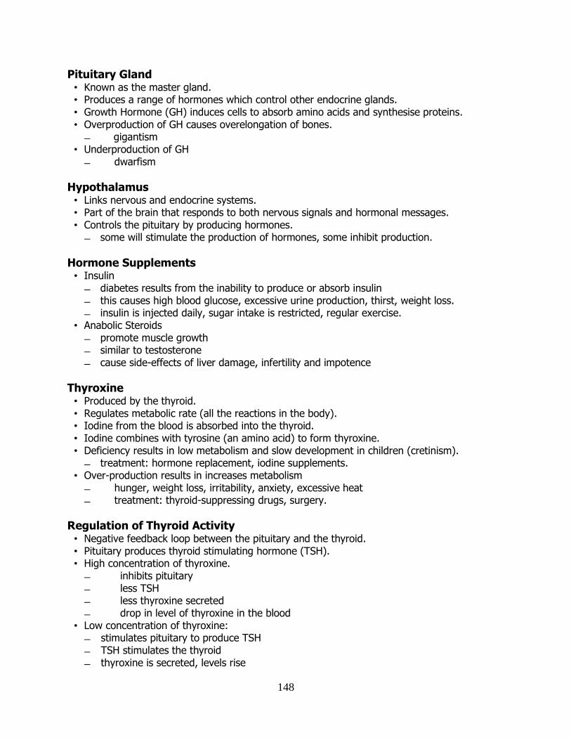
148
Pituitary Gland • Known as the master gland. • Produces a range of hormones which control other endocrine glands. • Growth Hormone (GH) induces cells to absorb amino acids and synthesise proteins. • Overproduction of GH causes overelongation of bones.
gigantism • Underproduction of GH
dwarfism
Hypothalamus • Links nervous and endocrine systems. • Part of the brain that responds to both nervous signals and hormonal messages. • Controls the pituitary by producing hormones.
some will stimulate the production of hormones, some inhibit production.
Hormone Supplements • Insulin
diabetes results from the inability to produce or absorb insulin this causes high blood glucose, excessive urine production, thirst, weight loss. insulin is injected daily, sugar intake is restricted, regular exercise.
• Anabolic Steroids promote muscle growth similar to testosterone cause side-effects of liver damage, infertility and impotence
Thyroxine • Produced by the thyroid. • Regulates metabolic rate (all the reactions in the body). • Iodine from the blood is absorbed into the thyroid. • Iodine combines with tyrosine (an amino acid) to form thyroxine. • Deficiency results in low metabolism and slow development in children (cretinism).
treatment: hormone replacement, iodine supplements. • Over-production results in increases metabolism
hunger, weight loss, irritability, anxiety, excessive heat treatment: thyroid-suppressing drugs, surgery.
Regulation of Thyroid Activity • Negative feedback loop between the pituitary and the thyroid. • Pituitary produces thyroid stimulating hormone (TSH). • High concentration of thyroxine.
inhibits pituitary less TSH less thyroxine secreted drop in level of thyroxine in the blood
• Low concentration of thyroxine: stimulates pituitary to produce TSH TSH stimulates the thyroid thyroxine is secreted, levels rise
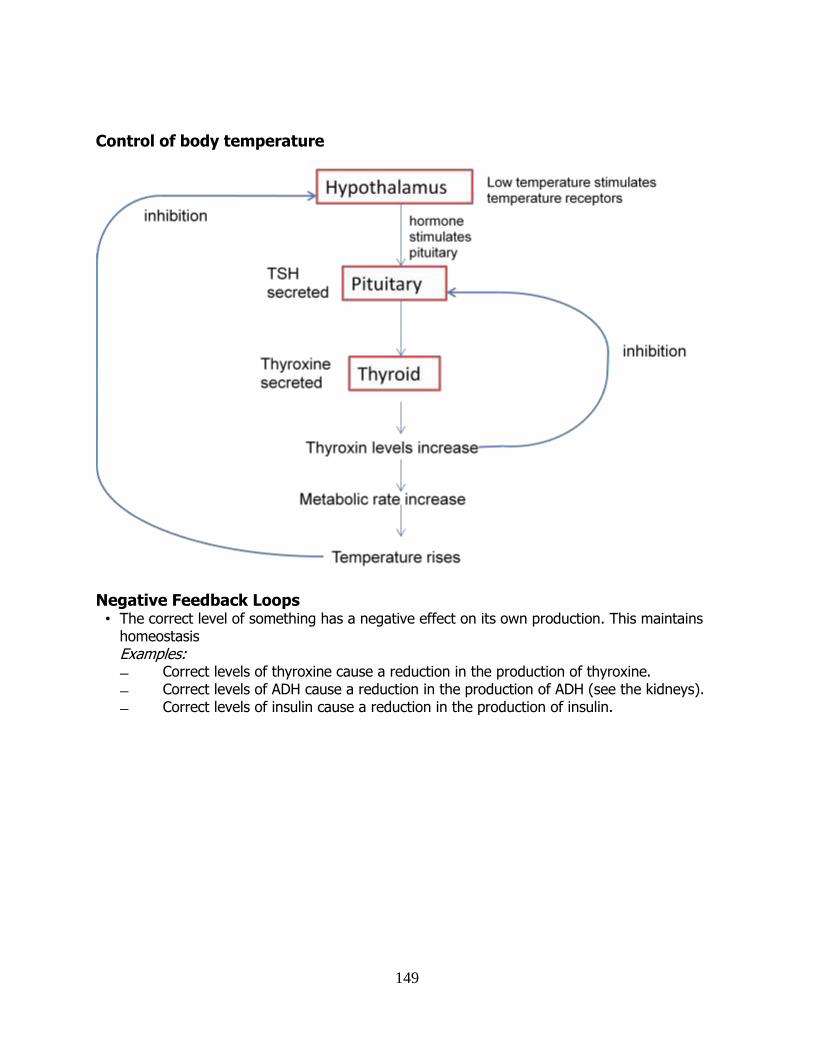
149
Control of body temperature
Negative Feedback Loops • The correct level of something has a negative effect on its own production. This maintains
homeostasis Examples: Correct levels of thyroxine cause a reduction in the production of thyroxine. Correct levels of ADH cause a reduction in the production of ADH (see the kidneys). Correct levels of insulin cause a reduction in the production of insulin.
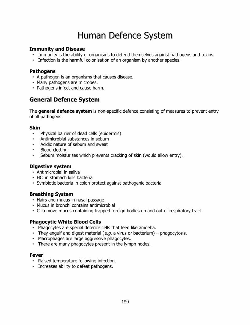
150
HHuummaann DDeeffeennccee SSyysstteemm
Immunity and Disease • Immunity is the ability of organisms to defend themselves against pathogens and toxins. • Infection is the harmful colonisation of an organism by another species.
Pathogens • A pathogen is an organisms that causes disease. • Many pathogens are microbes. • Pathogens infect and cause harm.
General Defence System
The general defence system is non-specific defence consisting of measures to prevent entry of all pathogens.
Skin • Physical barrier of dead cells (epidermis) • Antimicrobial substances in sebum • Acidic nature of sebum and sweat • Blood clotting • Sebum moisturises which prevents cracking of skin (would allow entry).
Digestive system • Antimicrobial in saliva • HCl in stomach kills bacteria • Symbiotic bacteria in colon protect against pathogenic bacteria
Breathing System • Hairs and mucus in nasal passage • Mucus in bronchi contains antimicrobial • Cilia move mucus containing trapped foreign bodies up and out of respiratory tract.
Phagocytic White Blood Cells • Phagocytes are special defence cells that feed like amoeba. • They engulf and digest material (e.g. a virus or bacterium) – phagocytosis. • Macrophages are large aggressive phagocytes. • There are many phagocytes present in the lymph nodes.
Fever • Raised temperature following infection. • Increases ability to defeat pathogens.
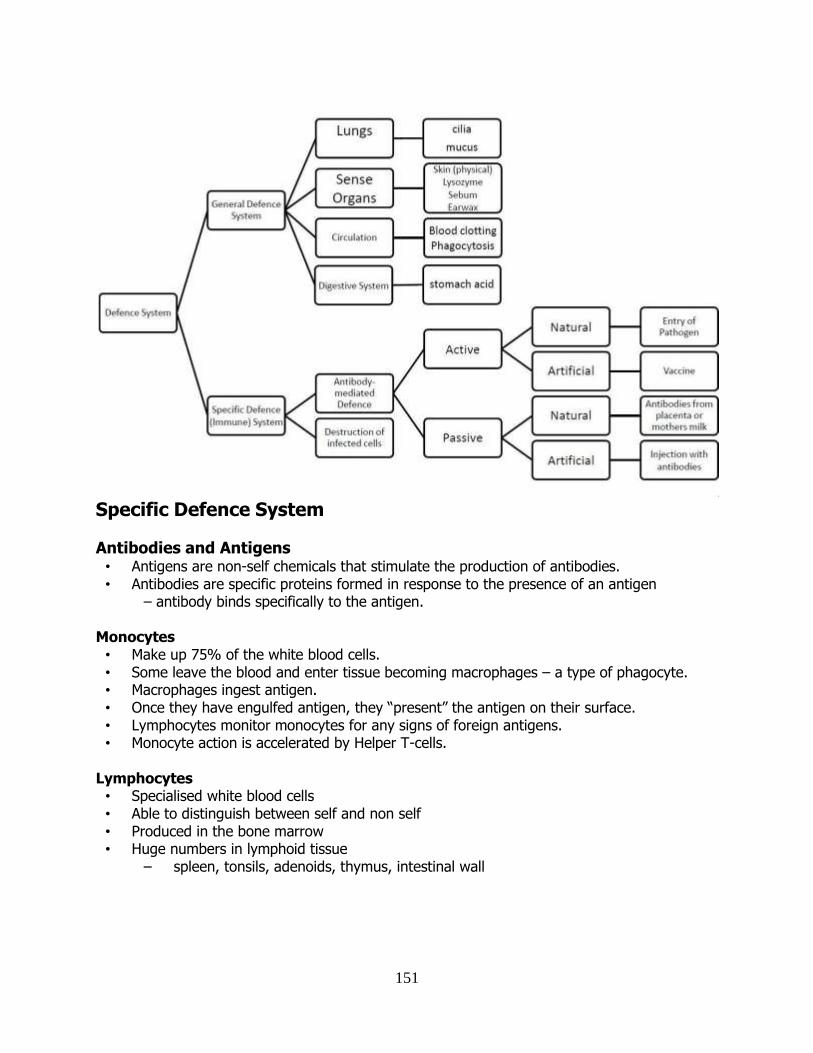
151
Specific Defence System
Antibodies and Antigens • Antigens are non-self chemicals that stimulate the production of antibodies. • Antibodies are specific proteins formed in response to the presence of an antigen
– antibody binds specifically to the antigen.
Monocytes • Make up 75% of the white blood cells. • Some leave the blood and enter tissue becoming macrophages – a type of phagocyte. • Macrophages ingest antigen. • Once they have engulfed antigen, they “present” the antigen on their surface. • Lymphocytes monitor monocytes for any signs of foreign antigens. • Monocyte action is accelerated by Helper T-cells.
Lymphocytes • Specialised white blood cells • Able to distinguish between self and non self • Produced in the bone marrow • Huge numbers in lymphoid tissue
– spleen, tonsils, adenoids, thymus, intestinal wall
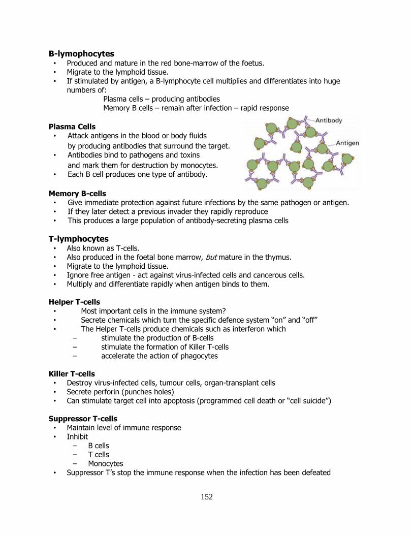
152
B-lymophocytes • Produced and mature in the red bone-marrow of the foetus. • Migrate to the lymphoid tissue. • If stimulated by antigen, a B-lymphocyte cell multiplies and differentiates into huge
numbers of: Plasma cells – producing antibodies Memory B cells – remain after infection – rapid response
Plasma Cells • Attack antigens in the blood or body fluids
by producing antibodies that surround the target. • Antibodies bind to pathogens and toxins
and mark them for destruction by monocytes. • Each B cell produces one type of antibody.
Memory B-cells • Give immediate protection against future infections by the same pathogen or antigen. • If they later detect a previous invader they rapidly reproduce • This produces a large population of antibody-secreting plasma cells
T-lymphocytes • Also known as T-cells. • Also produced in the foetal bone marrow, but mature in the thymus. • Migrate to the lymphoid tissue. • Ignore free antigen - act against virus-infected cells and cancerous cells. • Multiply and differentiate rapidly when antigen binds to them.
Helper T-cells • Most important cells in the immune system? • Secrete chemicals which turn the specific defence system “on” and “off” • The Helper T-cells produce chemicals such as interferon which
– stimulate the production of B-cells – stimulate the formation of Killer T-cells – accelerate the action of phagocytes
Killer T-cells • Destroy virus-infected cells, tumour cells, organ-transplant cells • Secrete perforin (punches holes) • Can stimulate target cell into apoptosis (programmed cell death or “cell suicide”)
Suppressor T-cells • Maintain level of immune response • Inhibit
– B cells – T cells – Monocytes
• Suppressor T’s stop the immune response when the infection has been defeated
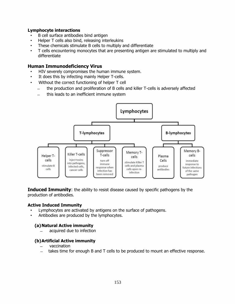
153
Lymphocyte interactions • B cell surface antibodies bind antigen • Helper T cells also bind, releasing interleukins • These chemicals stimulate B cells to multiply and differentiate • T cells encountering monocytes that are presenting antigen are stimulated to multiply and
differentiate
Human Immunodeficiency Virus • HIV severely compromises the human immune system. • It does this by infecting mainly Helper T-cells.
• Without the correct functioning of helper T cell
the production and proliferation of B cells and killer T-cells is adversely affected
this leads to an inefficient immune system
Induced Immunity: the ability to resist disease caused by specific pathogens by the
production of antibodies. Active Induced Immunity • Lymphocytes are activated by antigens on the surface of pathogens. • Antibodies are produced by the lymphocytes.
(a) Natural Active immunity
acquired due to infection
(b) Artificial Active immunity vaccination takes time for enough B and T cells to be produced to mount an effective response.
154
Passive Induced Immunity • B and T cells are not activated and plasma cells have not produced antibodies. • The antigen doesn’t have to be encountered for the body to make the antibodies. • Antibodies appear immediately in blood but protection is only temporary.
(a) Artificial Passive immunity
Used when a very rapid immune response is needed e.g. after infection with tetanus.
Human antibodies are injected. In the case of tetanus these are antitoxin antibodies.
Antibodies come from blood donors who have recently had the tetanus vaccination. Only provides short term protection as abs destroyed by phagocytes in spleen and
liver.
(b) Natural Passive immunity A mother’s antibodies pass across the placenta to the foetus and remain for several
months. Colostrum (the first breast milk) contains lots of IgA which remain on surface of the
baby’s gut wall and pass into blood. Vaccination • A vaccine is a preparation which gives artificial active immunity to a pathogen. • May contain live or dead inactivated pathogen, or part of the pathogen. • This provides a harmless first encounter with the antigens of the potential pathogen.
Benefits of Vaccination • Provides immunity without suffering the symptoms of the disease • Gives long-term protection • No long-term drug-taking required. • Examples of vaccines:
– MMR (Measles, mumps and rubella) – 3 in 1 (whooping cough, diptheria and tetanus – BCG (tuberculosis)
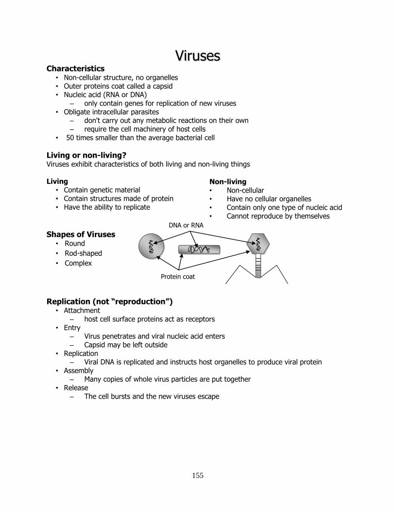
155
VViirruusseess Characteristics
• Non-cellular structure, no organelles • Outer proteins coat called a capsid • Nucleic acid (RNA or DNA)
– only contain genes for replication of new viruses • Obligate intracellular parasites
– don't carry out any metabolic reactions on their own – require the cell machinery of host cells
• 50 times smaller than the average bacterial cell
Living or non-living? Viruses exhibit characteristics of both living and non-living things Living
• Contain genetic material • Contain structures made of protein • Have the ability to replicate
Non-living
• Non-cellular • Have no cellular organelles • Contain only one type of nucleic acid • Cannot reproduce by themselves
Shapes of Viruses
• Round
• Rod-shaped
• Complex
Replication (not “reproduction”) • Attachment
– host cell surface proteins act as receptors • Entry
– Virus penetrates and viral nucleic acid enters – Capsid may be left outside
• Replication – Viral DNA is replicated and instructs host organelles to produce viral protein
• Assembly – Many copies of whole virus particles are put together
• Release – The cell bursts and the new viruses escape
DNA or RNA
Protein coat
156
Economic Importance Advantages
– Used to transfer genes from one organism to another in genetic engineering
– Used to control pests
e.g. insect virus against European pine sawfly
Disadvantages
– Crop diseases
e.g. tomato mosaic virus
– animal livestock diseases
e.g. foot and mouth disease
Medical Importance
Advantages
– vaccine manufacture (e.g. Measles, mumps and rubella - MMR)
– gene therapy (using viruses to replace abnormal gene with normal one in diseased
tissues)
Disadvantages
– cause many infectious diseases
e.g. Measles, mumps, rubella, influenza, common cold, rabies
– some cancers are caused by viral infections
Human Diseases caused by viruses
• AIDS
• Cold-sores
• Measles, mumps and rubella (“MMR”)
• Influenza and the common cold
• Chicken pox and shingles
• Viral meningitis
• SARS
• Haemorrhagic fever and Ebola
• Genital warts and genital herpes
Treatment of viral disease
• Antibiotics do not work against viruses
– they are only effective against bacteria
• Antibodies
– injections of antibodies are often used as vaccine but also as a treatment
• There has only been limited success with anti-viral drugs
– Acyclovir (“Zovirax”) against Herpes simplex
i.e. treatment of coldsores.
– AZT against HIV in treatment of HIV/AIDS.
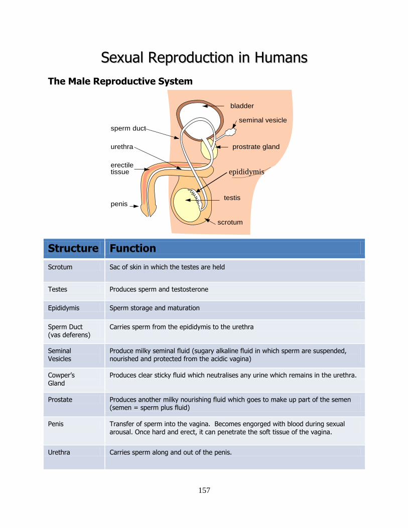
157
SSeexxuuaall RReepprroodduuccttiioonn iinn HHuummaannss
The Male Reproductive System
Structure Function
Scrotum Sac of skin in which the testes are held
Testes Produces sperm and testosterone
Epididymis Sperm storage and maturation
Sperm Duct (vas deferens)
Carries sperm from the epididymis to the urethra
Seminal Vesicles
Produce milky seminal fluid (sugary alkaline fluid in which sperm are suspended, nourished and protected from the acidic vagina)
Cowper’s
Gland
Produces clear sticky fluid which neutralises any urine which remains in the urethra.
Prostate Produces another milky nourishing fluid which goes to make up part of the semen (semen = sperm plus fluid)
Penis Transfer of sperm into the vagina. Becomes engorged with blood during sexual
arousal. Once hard and erect, it can penetrate the soft tissue of the vagina.
Urethra Carries sperm along and out of the penis.
bladder
seminal vesicle
prostrate gland
testis
scrotum
penis
erectile tissue
sperm duct
urethra
epididymis
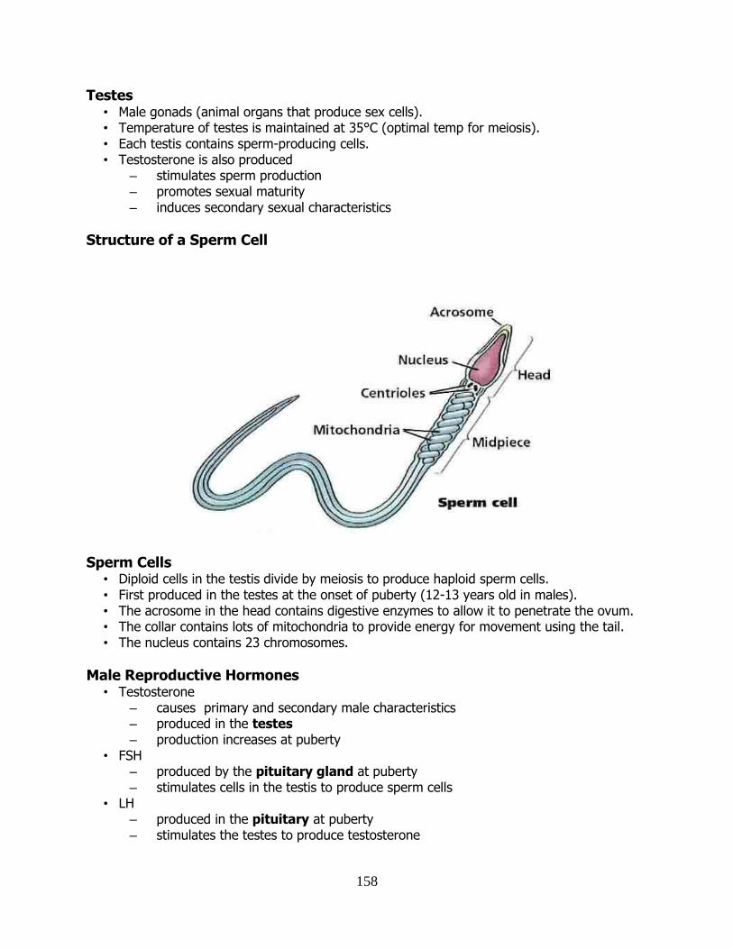
158
Testes • Male gonads (animal organs that produce sex cells). • Temperature of testes is maintained at 35°C (optimal temp for meiosis). • Each testis contains sperm-producing cells. • Testosterone is also produced
– stimulates sperm production – promotes sexual maturity – induces secondary sexual characteristics
Structure of a Sperm Cell
Sperm Cells
• Diploid cells in the testis divide by meiosis to produce haploid sperm cells. • First produced in the testes at the onset of puberty (12-13 years old in males). • The acrosome in the head contains digestive enzymes to allow it to penetrate the ovum. • The collar contains lots of mitochondria to provide energy for movement using the tail. • The nucleus contains 23 chromosomes.
Male Reproductive Hormones • Testosterone
– causes primary and secondary male characteristics – produced in the testes – production increases at puberty
• FSH – produced by the pituitary gland at puberty – stimulates cells in the testis to produce sperm cells
• LH – produced in the pituitary at puberty – stimulates the testes to produce testosterone
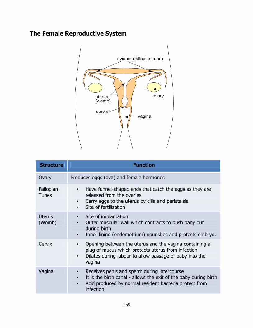
159
The Female Reproductive System
Structure Function
Ovary Produces eggs (ova) and female hormones
Fallopian Tubes
• Have funnel-shaped ends that catch the eggs as they are released from the ovaries
• Carry eggs to the uterus by cilia and peristalsis • Site of fertilisation
Uterus (Womb)
• Site of implantation • Outer muscular wall which contracts to push baby out
during birth • Inner lining (endometrium) nourishes and protects embryo.
Cervix • Opening between the uterus and the vagina containing a plug of mucus which protects uterus from infection
• Dilates during labour to allow passage of baby into the vagina
Vagina • Receives penis and sperm during intercourse • It is the birth canal - allows the exit of the baby during birth • Acid produced by normal resident bacteria protect from
infection
ovary
oviduct (fallopian tube)
uterus(womb)
vaginacervix
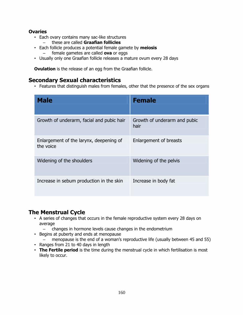
160
Ovaries • Each ovary contains many sac-like structures
– these are called Graafian follicles • Each follicle produces a potential female gamete by meiosis
– female gametes are called ova or eggs • Usually only one Graafian follicle releases a mature ovum every 28 days Ovulation is the release of an egg from the Graafian follicle.
Secondary Sexual characteristics • Features that distinguish males from females, other that the presence of the sex organs
Male Female
Growth of underarm, facial and pubic hair Growth of underarm and pubic hair
Enlargement of the larynx, deepening of the voice
Enlargement of breasts
Widening of the shoulders Widening of the pelvis
Increase in sebum production in the skin Increase in body fat
The Menstrual Cycle
• A series of changes that occurs in the female reproductive system every 28 days on average – changes in hormone levels cause changes in the endometrium
• Begins at puberty and ends at menopause – menopause is the end of a woman’s reproductive life (usually between 45 and 55)
• Ranges from 21 to 40 days in length • The Fertile period is the time during the menstrual cycle in which fertilisation is most
likely to occur.
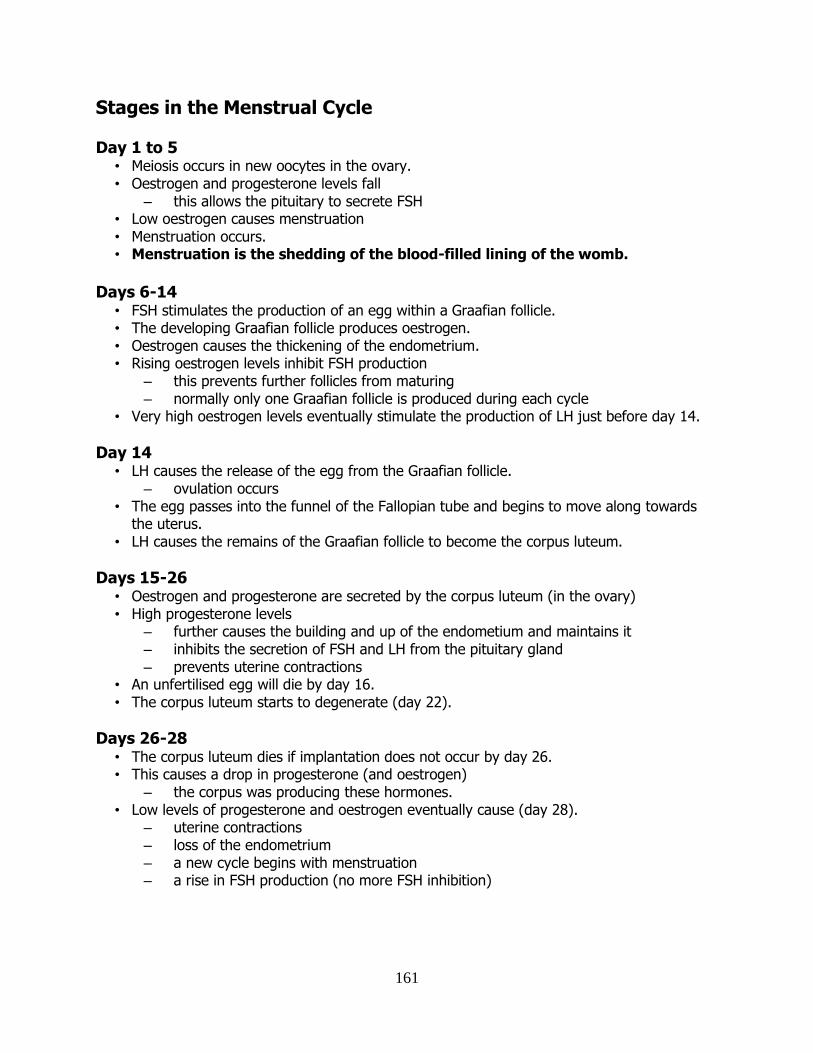
161
Stages in the Menstrual Cycle
Day 1 to 5 • Meiosis occurs in new oocytes in the ovary. • Oestrogen and progesterone levels fall
– this allows the pituitary to secrete FSH • Low oestrogen causes menstruation • Menstruation occurs. • Menstruation is the shedding of the blood-filled lining of the womb.
Days 6-14
• FSH stimulates the production of an egg within a Graafian follicle. • The developing Graafian follicle produces oestrogen. • Oestrogen causes the thickening of the endometrium. • Rising oestrogen levels inhibit FSH production
– this prevents further follicles from maturing – normally only one Graafian follicle is produced during each cycle
• Very high oestrogen levels eventually stimulate the production of LH just before day 14.
Day 14 • LH causes the release of the egg from the Graafian follicle.
– ovulation occurs • The egg passes into the funnel of the Fallopian tube and begins to move along towards
the uterus. • LH causes the remains of the Graafian follicle to become the corpus luteum.
Days 15-26 • Oestrogen and progesterone are secreted by the corpus luteum (in the ovary) • High progesterone levels
– further causes the building and up of the endometium and maintains it – inhibits the secretion of FSH and LH from the pituitary gland – prevents uterine contractions
• An unfertilised egg will die by day 16. • The corpus luteum starts to degenerate (day 22).
Days 26-28 • The corpus luteum dies if implantation does not occur by day 26. • This causes a drop in progesterone (and oestrogen)
– the corpus was producing these hormones. • Low levels of progesterone and oestrogen eventually cause (day 28).
– uterine contractions – loss of the endometrium – a new cycle begins with menstruation – a rise in FSH production (no more FSH inhibition)
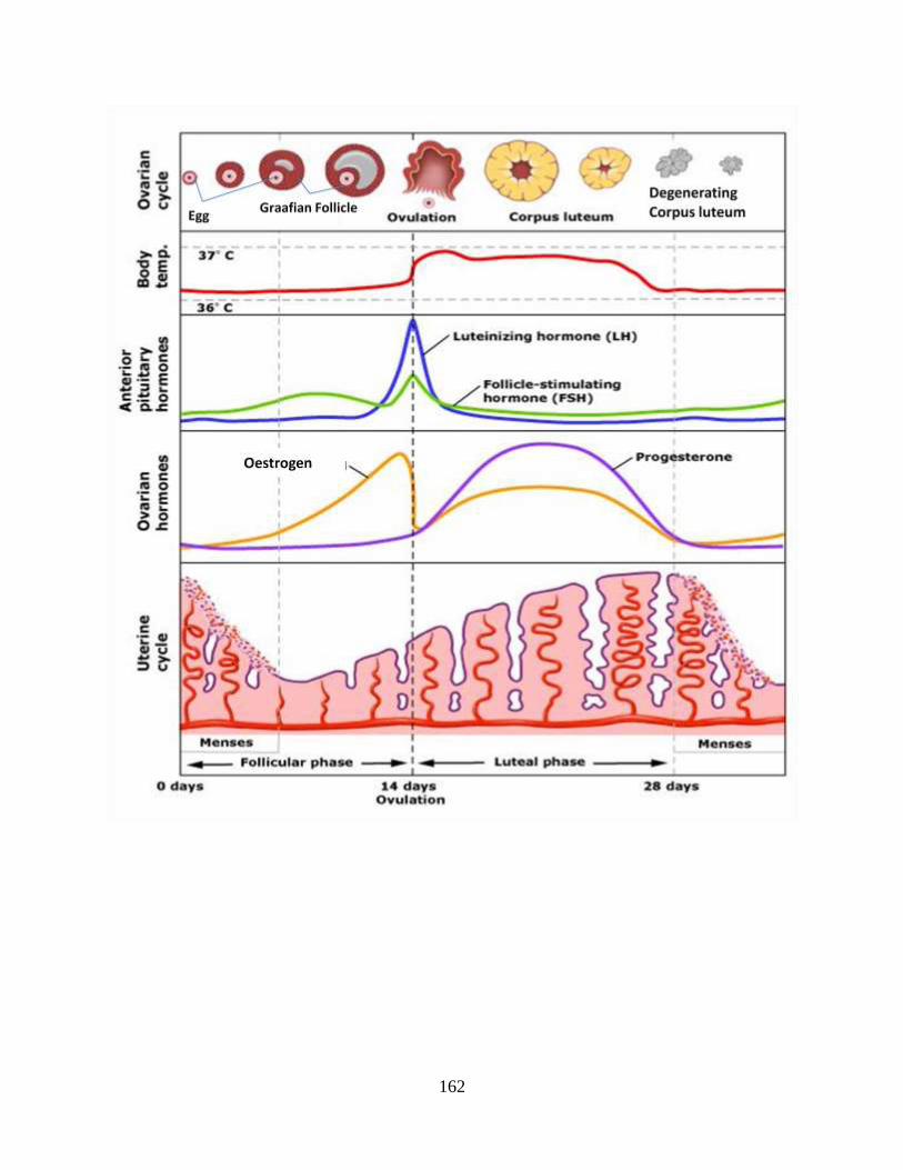
162
163
The Role of Hormones in the Menstrual Cycle Role of Oestrogen
• Produced in the ovary (by the Graafian follicle). • Causes the endometrium to thicken for first 14 days. • Inhibits FSH production (negative feedback)
– prevents further Graafian follicles from maturing – this ensures only one egg produced per cycle
• High levels stimulate the pituitary to produce LH.
Role of Progesterone • Produced in the ovary (by the corpus luteum). • Causes the endometrium to thicken for last 14 days and maintains its structure. • Inhibits FSH production (negative feedback)
– stops the development of more Graafian follicles. • Inhibits LH production
– prevents ovulation. • Prevents muscular contractions of the uterus.
Role of Follicle Stimulating Hormone
• Produced in the pituitary. • Causes Graafian follicles (containing eggs) to develop and mature. • Indirectly causes the production of oestrogen (since the follicle produces oestrogen).
Role of Luteinising Hormone • Produced in the pituitary gland. • Causes ovulation. • Causes the remains of the Graafian follicle to become the corpus luteum. • Indirectly results in the production of progesterone (since the corpus luteum produces
progesterone).
Infertility
• Infertility (male or female) is the inability to produce offspring • Affects 1 in 6 couples, but 50% are successfully treated
Infertility Cause Treatment
Male Low sperm count due to smoking, alcohol/drug abuse, stress.
Stop or reduce smoking, drugs, alcohol, stress.
Female Failure to ovulate (due to hormone disorder).
Hormone treatment (e.g. FSH stimulates ovulation)
164
Male Infertility • Male infertility is the inability to produce sperm capable of fertilizing an egg cell. • Cause: low sperm count
due to excessive alcohol or drug use; stress • Treatment: avoidance of possible reasons for low sperm count
alcohol and drug use stress
Female Infertility
• Female infertility is the inability to conceive. • Cause: failure to ovulate
due to hormone deficiency • Treatment: hormone therapy
In-vitro fertilisation (IVF)
• Method of treating infertility. • Eggs are removed from the ovary. • Fertility drugs are given to encourage egg development. • Eggs are surgically removed on day 14. • Sperm and eggs are incubated together in vitro (in a glass dish). • If fertilisation occurs, a number of embryos are placed in the uterus to implant.
Menstrual Disorder - Fibroids
• Benign tumours of the uterus. • Most common in 35-45 year olds. • If they grow too large they can cause pain, heavy bleeding, miscarriage, infertility • Cause is thought to be abnormal response to oestrogen. • Treatment
– if small, no treatment – if large, surgery to remove them – hysterectomy in severe cases
Stages in Sexual Intercourse
• Sexual arousal – penis becomes erect – vagina becomes lubricated
• Copulation – coitus or sexual intercourse
• Orgasm - involuntary muscle spasms cause – ejaculation in males – uterine and vaginal contractions in female
• Insemination – Release of semen into the vagina
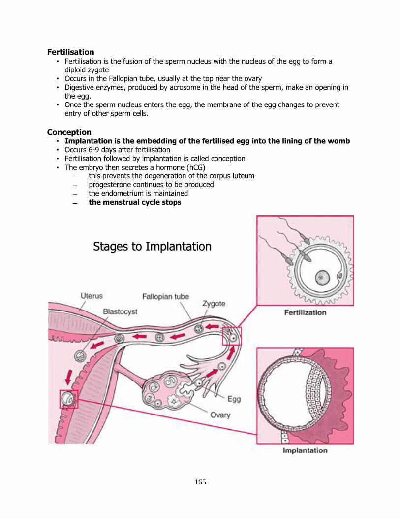
165
Fertilisation • Fertilisation is the fusion of the sperm nucleus with the nucleus of the egg to form a
diploid zygote • Occurs in the Fallopian tube, usually at the top near the ovary • Digestive enzymes, produced by acrosome in the head of the sperm, make an opening in
the egg. • Once the sperm nucleus enters the egg, the membrane of the egg changes to prevent
entry of other sperm cells.
Conception
• Implantation is the embedding of the fertilised egg into the lining of the womb • Occurs 6-9 days after fertilisation • Fertilisation followed by implantation is called conception • The embryo then secretes a hormone (hCG)
this prevents the degeneration of the corpus luteum progesterone continues to be produced the endometrium is maintained the menstrual cycle stops
SSttaaggeess ttoo IImmppllaannttaattiioonn
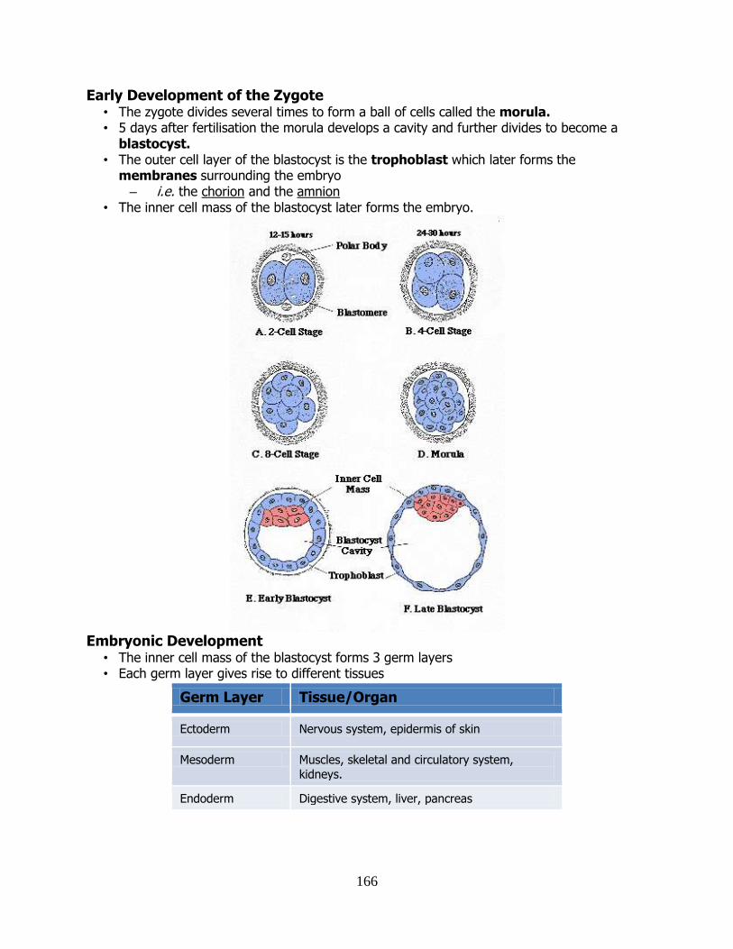
166
Early Development of the Zygote • The zygote divides several times to form a ball of cells called the morula. • 5 days after fertilisation the morula develops a cavity and further divides to become a
blastocyst. • The outer cell layer of the blastocyst is the trophoblast which later forms the
membranes surrounding the embryo – i.e. the chorion and the amnion
• The inner cell mass of the blastocyst later forms the embryo.
Embryonic Development
• The inner cell mass of the blastocyst forms 3 germ layers • Each germ layer gives rise to different tissues
Germ Layer Tissue/Organ
Ectoderm Nervous system, epidermis of skin
Mesoderm Muscles, skeletal and circulatory system,
kidneys.
Endoderm Digestive system, liver, pancreas
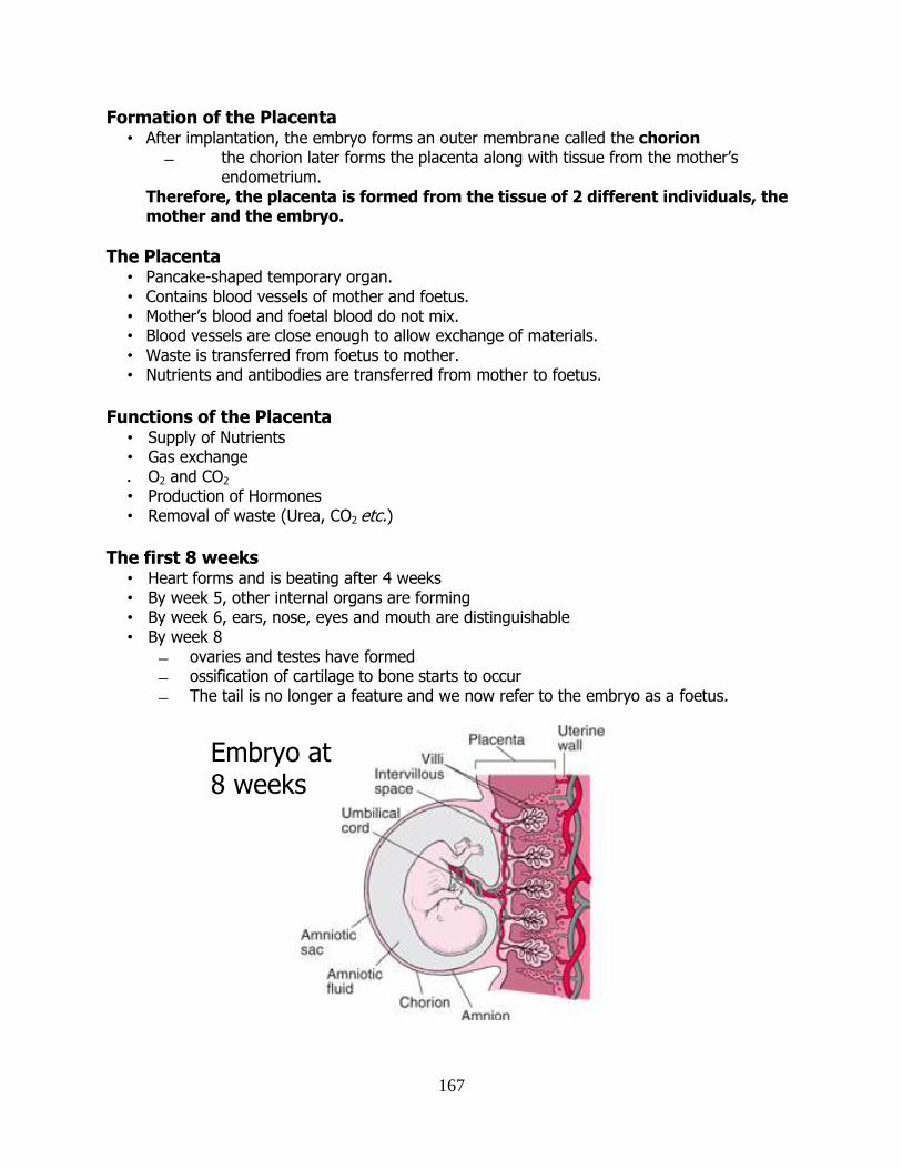
167
Formation of the Placenta • After implantation, the embryo forms an outer membrane called the chorion
the chorion later forms the placenta along with tissue from the mother’s endometrium.
Therefore, the placenta is formed from the tissue of 2 different individuals, the mother and the embryo.
The Placenta • Pancake-shaped temporary organ. • Contains blood vessels of mother and foetus. • Mother’s blood and foetal blood do not mix. • Blood vessels are close enough to allow exchange of materials. • Waste is transferred from foetus to mother. • Nutrients and antibodies are transferred from mother to foetus.
Functions of the Placenta
• Supply of Nutrients • Gas exchange
• O2 and CO2
• Production of Hormones • Removal of waste (Urea, CO2 etc.)
The first 8 weeks
• Heart forms and is beating after 4 weeks • By week 5, other internal organs are forming • By week 6, ears, nose, eyes and mouth are distinguishable • By week 8
ovaries and testes have formed ossification of cartilage to bone starts to occur The tail is no longer a feature and we now refer to the embryo as a foetus.
Embryo at 8 weeks

168
By 12 weeks • All major organs are formed. • Nerves and muscles become co-ordinated. • Limbs begin to move, the foetus can kick. • Teeth grow, breathing starts. • Urinary system begins to work.
Week 12 to birth
• No new organs are formed. • Growth and development is rapid. • Gestation is the length of time spend in the uterus from fertilisation to birth. • Gestation time is 38 weeks from the day of fertilisation.
Hormones during Pregnancy
• The corpus luteum produces progesterone and oestrogen during the first 10 weeks. • The placenta takes over this role after this. • Just before birth
Progesterone production stops Oestrogen production increases.
Birth Stage 1 - Dilation
• Cervix dilates to allow passage of the baby. • Amniotic fluid is released from the amniotic sac. • Muscular contractions begin. • The mucus plug in the cervix is lost.
Stage 2 - Delivery
• Contractions increase as oxytocin levels increase. • Baby passes through the birth canal head first.
Stage 3 - Placental
• Release of the placenta and foetal membrane.
Lactation
• Milk Production by the mammary glands. • Colostrum is the first milk produced in the days just after the birth
rich in protein and antibodies (passive natural immunity) • Prolactin is produced
this hormone promotes milk production. progesterone inhibits prolactin secretion during pregnancy.
Benefits of Breast-feeding • Mother milk is the perfectly balanced source of nutrients for the baby. • Contains antibodies which provide natural passive immunity to the baby. • Helps the mother recover after the birth by helping the uterus to tighten again. • Helps the mother lose fat put on during pregnancy.
169
Birth Control or Family Planning Birth control involves taking steps to reduce the number of children born. Contraception: methods used to prevent fertilisation or implantation.
Natural • Abstaining from sexual intercourse completely (0% failure rate!). • Abstaining from intercourse during the fertile period (~25% failure rate).
Mechanical • Condom – latex sheath covering the penis (10% failure rate). • Diaphragm – dome-shaped rubber cap which fits over the cervix (10% failure rate). • Intrauterine Device – loop or coil which prevents implantation (5% failure rate).
Chemical • Oral Contraceptive Pill – hormones such as progesterone and oestrogen which prevent
ovulation (6% failure rate). • Injected Contraceptive – progesterone (0.4% failure rate). • Spermicides – chemicals which kill sperm (20% failure rate) but usually used in
conjunction with barrier methods such as the diaphragm or condom. • Morning after pill: high concentration of oestrogen and progesterone taken within 72
hours of intercourse (3% failure rate).
Combination
• Intrauterine System (IUS – “Mirena Coil”) – an IUD which releases a hormone (0.2% failure rate).
Surgical • Vasectomy – cutting the sperm ducts (0.4% failure rate). • Tubal ligation – cutting or tying the Fallopian tubes (0.4% failure rate).
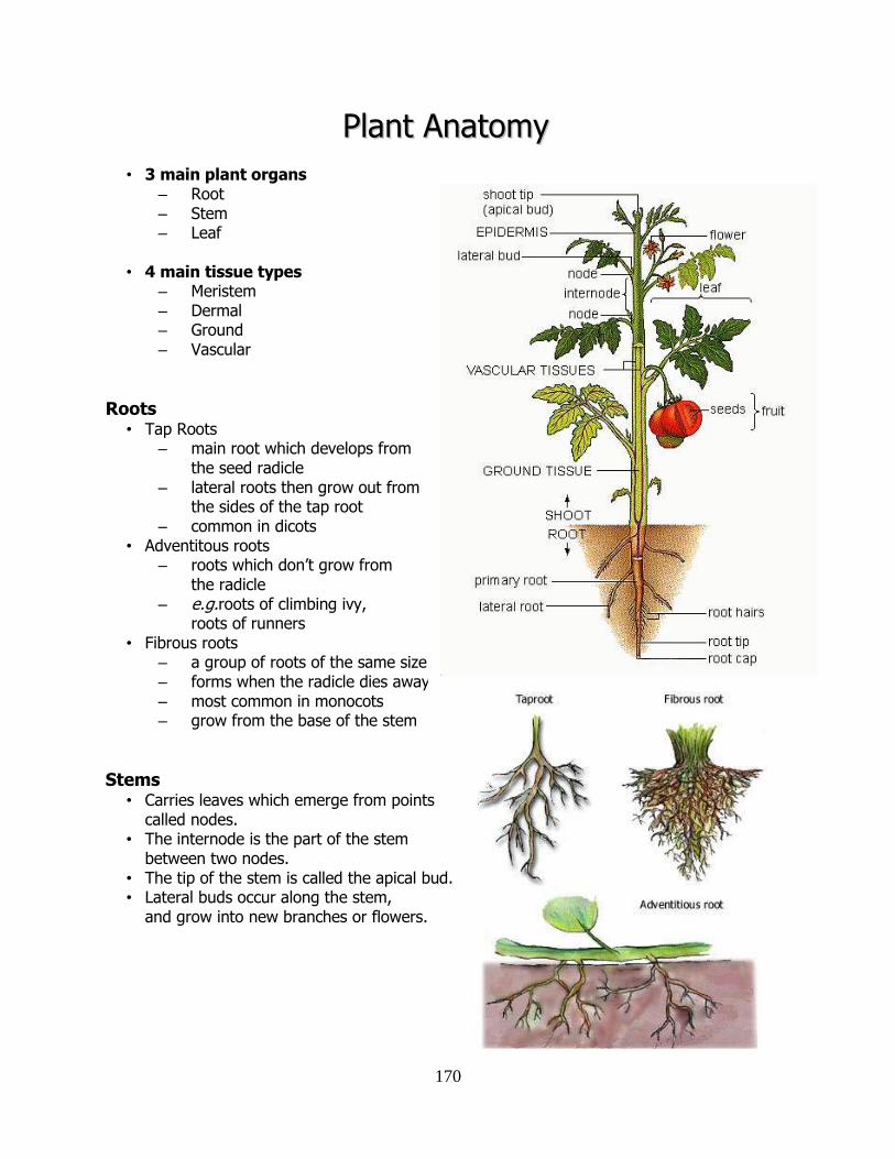
170
PPllaanntt AAnnaattoommyy
• 3 main plant organs – Root – Stem – Leaf
• 4 main tissue types – Meristem – Dermal – Ground – Vascular
Roots • Tap Roots
– main root which develops from the seed radicle
– lateral roots then grow out from the sides of the tap root
– common in dicots • Adventitous roots
– roots which don’t grow from the radicle
– e.g.roots of climbing ivy, roots of runners
• Fibrous roots – a group of roots of the same size – forms when the radicle dies away – most common in monocots – grow from the base of the stem
Stems • Carries leaves which emerge from points
called nodes. • The internode is the part of the stem
between two nodes. • The tip of the stem is called the apical bud. • Lateral buds occur along the stem,
and grow into new branches or flowers.
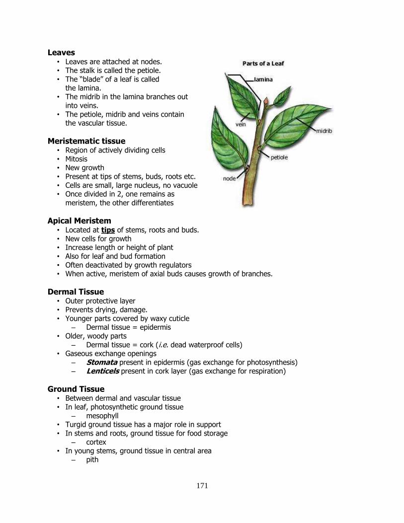
171
Leaves • Leaves are attached at nodes. • The stalk is called the petiole. • The “blade” of a leaf is called
the lamina. • The midrib in the lamina branches out
into veins. • The petiole, midrib and veins contain
the vascular tissue.
Meristematic tissue • Region of actively dividing cells • Mitosis • New growth • Present at tips of stems, buds, roots etc. • Cells are small, large nucleus, no vacuole • Once divided in 2, one remains as
meristem, the other differentiates
Apical Meristem • Located at tips of stems, roots and buds. • New cells for growth • Increase length or height of plant • Also for leaf and bud formation • Often deactivated by growth regulators • When active, meristem of axial buds causes growth of branches.
Dermal Tissue • Outer protective layer • Prevents drying, damage. • Younger parts covered by waxy cuticle
– Dermal tissue = epidermis • Older, woody parts
– Dermal tissue = cork (i.e. dead waterproof cells) • Gaseous exchange openings
– Stomata present in epidermis (gas exchange for photosynthesis) – Lenticels present in cork layer (gas exchange for respiration)
Ground Tissue • Between dermal and vascular tissue • In leaf, photosynthetic ground tissue
– mesophyll • Turgid ground tissue has a major role in support • In stems and roots, ground tissue for food storage
– cortex • In young stems, ground tissue in central area
– pith
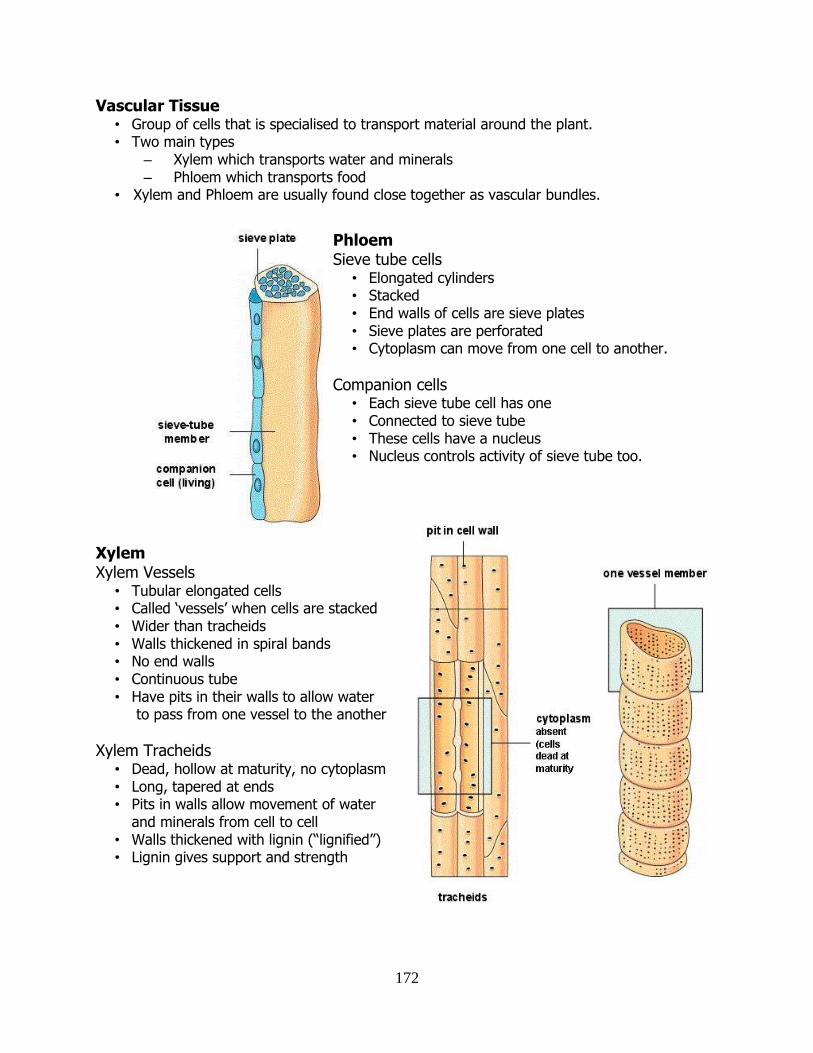
172
Vascular Tissue • Group of cells that is specialised to transport material around the plant. • Two main types
– Xylem which transports water and minerals – Phloem which transports food
• Xylem and Phloem are usually found close together as vascular bundles.
Xylem Xylem Vessels
• Tubular elongated cells • Called ‘vessels’ when cells are stacked • Wider than tracheids • Walls thickened in spiral bands • No end walls • Continuous tube • Have pits in their walls to allow water
to pass from one vessel to the another
Xylem Tracheids • Dead, hollow at maturity, no cytoplasm • Long, tapered at ends • Pits in walls allow movement of water
and minerals from cell to cell • Walls thickened with lignin (“lignified”) • Lignin gives support and strength
Phloem Sieve tube cells
• Elongated cylinders • Stacked • End walls of cells are sieve plates • Sieve plates are perforated • Cytoplasm can move from one cell to another.
Companion cells • Each sieve tube cell has one • Connected to sieve tube • These cells have a nucleus • Nucleus controls activity of sieve tube too.
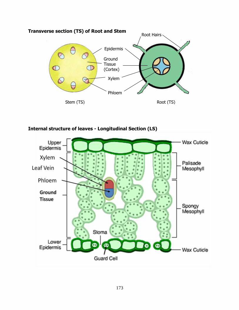
173
Transverse section (TS) of Root and Stem
Internal structure of leaves - Longitudinal Section (LS)
Ground Tissue (Cortex)
Xylem
Epidermis
Root Hairs
Phloem
Stem (TS) Root (TS)
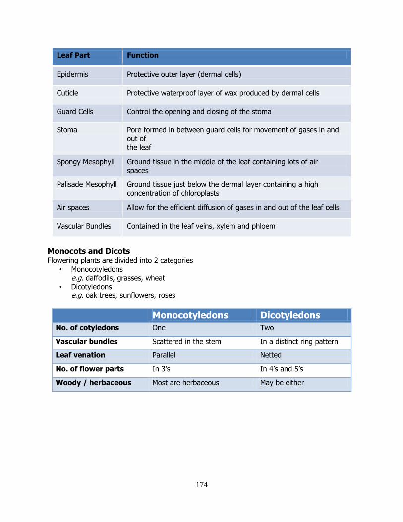
174
Leaf Part Function
Epidermis Protective outer layer (dermal cells)
Cuticle Protective waterproof layer of wax produced by dermal cells
Guard Cells Control the opening and closing of the stoma
Stoma Pore formed in between guard cells for movement of gases in and out of the leaf
Spongy Mesophyll Ground tissue in the middle of the leaf containing lots of air spaces
Palisade Mesophyll Ground tissue just below the dermal layer containing a high concentration of chloroplasts
Air spaces Allow for the efficient diffusion of gases in and out of the leaf cells
Vascular Bundles Contained in the leaf veins, xylem and phloem
Monocots and Dicots Flowering plants are divided into 2 categories
• Monocotyledons e.g. daffodils, grasses, wheat
• Dicotyledons
e.g. oak trees, sunflowers, roses
Monocotyledons Dicotyledons No. of cotyledons One Two
Vascular bundles Scattered in the stem In a distinct ring pattern
Leaf venation Parallel Netted
No. of flower parts In 3’s In 4’s and 5’s
Woody / herbaceous Most are herbaceous May be either
175
PPllaanntt TTrraannssppoorrtt
• Plants (autotrophs) need to transport water for photosynthesis (making food). • They also have to transport food to where it is needed. • Minerals, gases and growth regulators are also transported. • Oxygen is needed for photosynthesis. • Carbon is dioxide needed for respiration.
Water Transport
• Water is absorbed through root hairs by osmosis (hairs give large surface area). • There is no cuticle on root hairs (allows absorption). • Water diffuses across cortex into xylem. • It then travels up the xylem into the leaves. • Only 1% of the absorbed water is used by the plant. • What about the other 99%?
Absorption of water into root hairs
Role of Transpiration
• Transpiration is the loss of water vapour from the surface of the plant. • 99% of water absorbed by the plant exits through the stomata in the leaf. • Leaf cells therefore need replacement water. • Water enters them by osmosis from xylem vessels. • This causes more water to be “pulled up” into the roots from the ground.
Role of Root Pressure • Water entering the roots causes root pressure. • Helps “push” the water up the xylem. • Root pressure is the force which can push water up a stem from the root. • So, water rises up the plant through a combination of ‘push’ (root pressure) and ‘pull’
(transpiration).
nucleus
root hair
water soil
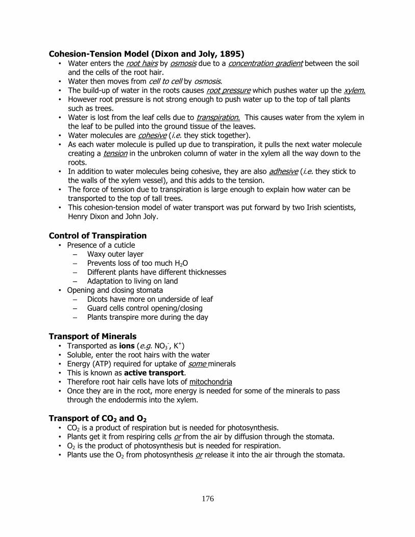
176
Cohesion-Tension Model (Dixon and Joly, 1895) • Water enters the root hairs by osmosis due to a concentration gradient between the soil
and the cells of the root hair. • Water then moves from cell to cell by osmosis. • The build-up of water in the roots causes root pressure which pushes water up the xylem. • However root pressure is not strong enough to push water up to the top of tall plants
such as trees. • Water is lost from the leaf cells due to transpiration. This causes water from the xylem in
the leaf to be pulled into the ground tissue of the leaves. • Water molecules are cohesive (i.e. they stick together). • As each water molecule is pulled up due to transpiration, it pulls the next water molecule
creating a tension in the unbroken column of water in the xylem all the way down to the roots.
• In addition to water molecules being cohesive, they are also adhesive (i.e. they stick to the walls of the xylem vessel), and this adds to the tension.
• The force of tension due to transpiration is large enough to explain how water can be transported to the top of tall trees.
• This cohesion-tension model of water transport was put forward by two Irish scientists, Henry Dixon and John Joly.
Control of Transpiration
• Presence of a cuticle – Waxy outer layer – Prevents loss of too much H2O – Different plants have different thicknesses – Adaptation to living on land
• Opening and closing stomata – Dicots have more on underside of leaf – Guard cells control opening/closing – Plants transpire more during the day
Transport of Minerals
• Transported as ions (e.g. NO3-, K+)
• Soluble, enter the root hairs with the water • Energy (ATP) required for uptake of some minerals • This is known as active transport. • Therefore root hair cells have lots of mitochondria • Once they are in the root, more energy is needed for some of the minerals to pass
through the endodermis into the xylem.
Transport of CO2 and O2
• CO2 is a product of respiration but is needed for photosynthesis. • Plants get it from respiring cells or from the air by diffusion through the stomata. • O2 is the product of photosynthesis but is needed for respiration. • Plants use the O2 from photosynthesis or release it into the air through the stomata.
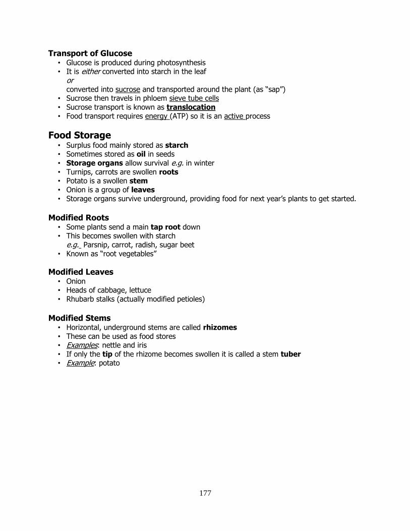
177
Transport of Glucose • Glucose is produced during photosynthesis • It is either converted into starch in the leaf
or converted into sucrose and transported around the plant (as “sap”)
• Sucrose then travels in phloem sieve tube cells • Sucrose transport is known as translocation • Food transport requires energy (ATP) so it is an active process
Food Storage • Surplus food mainly stored as starch • Sometimes stored as oil in seeds • Storage organs allow survival e.g. in winter • Turnips, carrots are swollen roots • Potato is a swollen stem • Onion is a group of leaves • Storage organs survive underground, providing food for next year’s plants to get started.
Modified Roots
• Some plants send a main tap root down • This becomes swollen with starch e.g. Parsnip, carrot, radish, sugar beet • Known as “root vegetables”
Modified Leaves • Onion • Heads of cabbage, lettuce • Rhubarb stalks (actually modified petioles)
Modified Stems
• Horizontal, underground stems are called rhizomes • These can be used as food stores • Examples: nettle and iris • If only the tip of the rhizome becomes swollen it is called a stem tuber • Example: potato
178
GGaass EExxcchhaannggee iinn PPllaannttss Control of Transpiration in leaves
• Loss of water vapour must be controlled by leaves or they will wilt and die • Waxy cuticles
– prevent water passing through • Stomata only on underside of leaf
– more evaporation on the upper side • Guard cells
– control the opening and closing of the stoma
Stomata Generally
• Open by day – allows gas exchange during photosynthesis
• Closed by night – gas exchange is not necessary – reduces transpiration at night
However • Stomata will close during the day
– if the temperature is too high – if the plant is losing too much water
Adaptations of the leaf for Gas Exchange
• Stomata – Allow gases to taken in and out of the leaf quickly
• Thin lamina (leaf blade) – short distance for CO2 to diffuse
• Large surface area • Air spaces
– diffusion of CO2 and O2 is faster than in liquid • Moist tissue
– necessary for transfer of gases in and out of cells
Leaf Adaptations for Photosynthesis
• Higher density of chloroplasts in the palisade layer – cells nearest the light
• Chloroplasts are at the edges of the cells • Flat shape • Large surface area • Leaf petiole positions the leaf in order to maximise light absorption
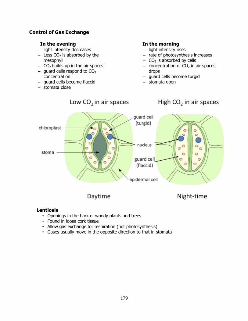
179
Control of Gas Exchange In the evening
– light intensity decreases – Less CO2 is absorbed by the
mesophyll – CO2 builds up in the air spaces – guard cells respond to CO2
concentration – guard cells become flaccid – stomata close
In the morning – light intensity rises – rate of photosynthesis increases – CO2 is absorbed by cells – concentration of CO2 in air spaces
drops – guard cells become turgid – stomata open
Lenticels • Openings in the bark of woody plants and trees • Found in loose cork tissue • Allow gas exchange for respiration (not photosynthesis) • Gases usually move in the opposite direction to that in stomata
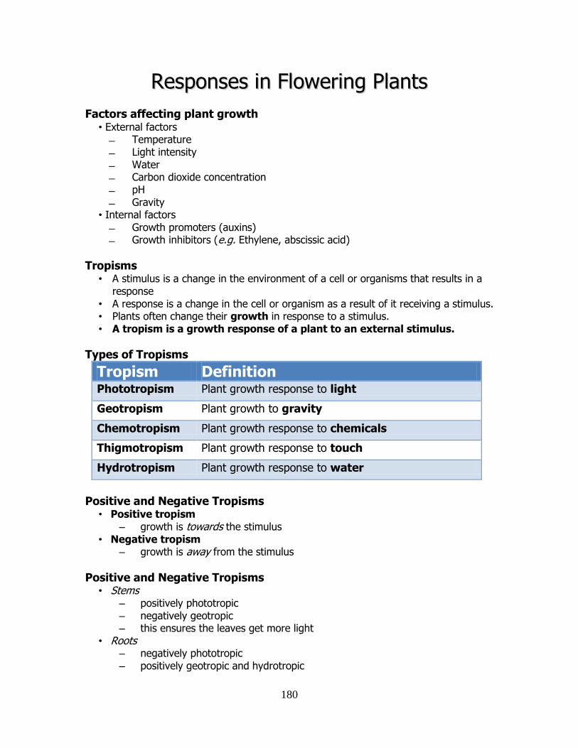
180
RReessppoonnsseess iinn FFlloowweerriinngg PPllaannttss Factors affecting plant growth
• External factors Temperature Light intensity Water Carbon dioxide concentration pH Gravity
• Internal factors Growth promoters (auxins) Growth inhibitors (e.g. Ethylene, abscissic acid)
Tropisms • A stimulus is a change in the environment of a cell or organisms that results in a
response • A response is a change in the cell or organism as a result of it receiving a stimulus. • Plants often change their growth in response to a stimulus. • A tropism is a growth response of a plant to an external stimulus.
Types of Tropisms
Tropism Definition Phototropism Plant growth response to light
Geotropism Plant growth to gravity
Chemotropism Plant growth response to chemicals
Thigmotropism Plant growth response to touch
Hydrotropism Plant growth response to water
Positive and Negative Tropisms
• Positive tropism – growth is towards the stimulus
• Negative tropism – growth is away from the stimulus
Positive and Negative Tropisms • Stems
– positively phototropic – negatively geotropic – this ensures the leaves get more light
• Roots – negatively phototropic – positively geotropic and hydrotropic
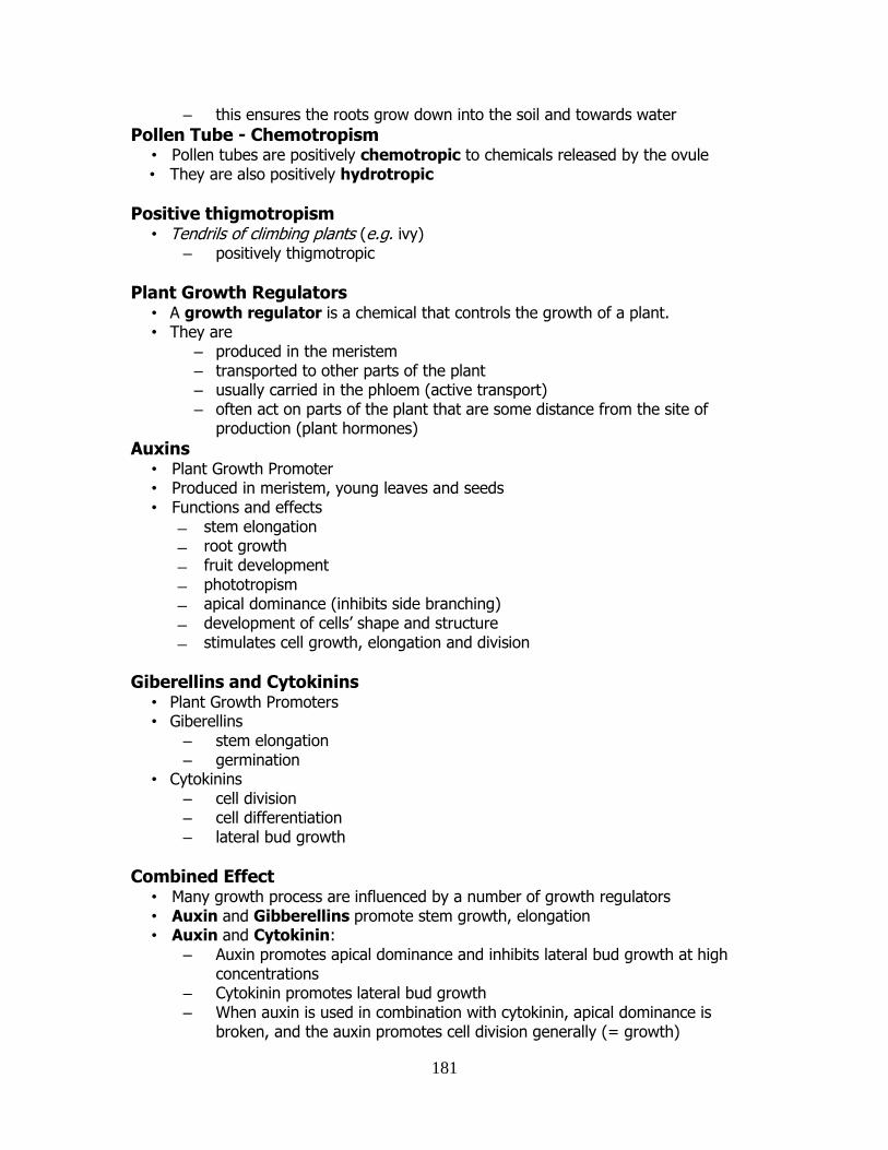
181
– this ensures the roots grow down into the soil and towards water
Pollen Tube - Chemotropism • Pollen tubes are positively chemotropic to chemicals released by the ovule • They are also positively hydrotropic
Positive thigmotropism • Tendrils of climbing plants (e.g. ivy)
– positively thigmotropic
Plant Growth Regulators • A growth regulator is a chemical that controls the growth of a plant. • They are
– produced in the meristem – transported to other parts of the plant – usually carried in the phloem (active transport) – often act on parts of the plant that are some distance from the site of
production (plant hormones)
Auxins • Plant Growth Promoter • Produced in meristem, young leaves and seeds • Functions and effects
stem elongation root growth fruit development phototropism apical dominance (inhibits side branching) development of cells’ shape and structure stimulates cell growth, elongation and division
Giberellins and Cytokinins • Plant Growth Promoters • Giberellins
– stem elongation – germination
• Cytokinins – cell division – cell differentiation – lateral bud growth
Combined Effect • Many growth process are influenced by a number of growth regulators • Auxin and Gibberellins promote stem growth, elongation • Auxin and Cytokinin:
– Auxin promotes apical dominance and inhibits lateral bud growth at high concentrations
– Cytokinin promotes lateral bud growth – When auxin is used in combination with cytokinin, apical dominance is
broken, and the auxin promotes cell division generally (= growth)
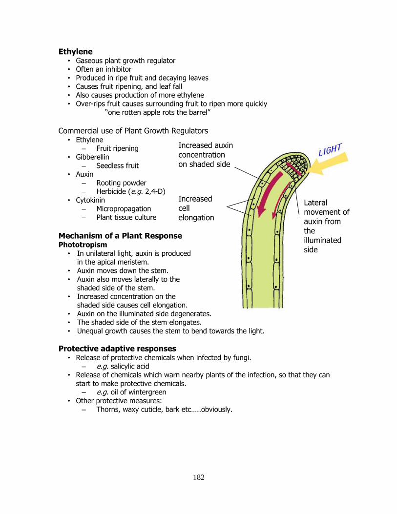
182
Ethylene • Gaseous plant growth regulator • Often an inhibitor • Produced in ripe fruit and decaying leaves • Causes fruit ripening, and leaf fall • Also causes production of more ethylene • Over-rips fruit causes surrounding fruit to ripen more quickly
“one rotten apple rots the barrel”
Commercial use of Plant Growth Regulators
• Ethylene – Fruit ripening
• Gibberellin – Seedless fruit
• Auxin – Rooting powder – Herbicide (e.g. 2,4-D)
• Cytokinin – Micropropagation – Plant tissue culture
Mechanism of a Plant Response Phototropism
• In unilateral light, auxin is produced in the apical meristem.
• Auxin moves down the stem. • Auxin also moves laterally to the
shaded side of the stem. • Increased concentration on the
shaded side causes cell elongation. • Auxin on the illuminated side degenerates. • The shaded side of the stem elongates. • Unequal growth causes the stem to bend towards the light.
Protective adaptive responses • Release of protective chemicals when infected by fungi.
– e.g. salicylic acid • Release of chemicals which warn nearby plants of the infection, so that they can
start to make protective chemicals. – e.g. oil of wintergreen
• Other protective measures: – Thorns, waxy cuticle, bark etc…..obviously.
Increased auxin concentration on shaded side
Increased cell elongation on shaded side
Lateral movement of auxin from the illuminated side
183
VVeeggeettaattiivvee PPrrooppaaggaattiioonn –– PPllaanntt AAsseexxuuaall
RReepprroodduuccttiioonn
• The production of new plants without the fusion of gametes. • All the offspring are identical to the parent. • Genes are identical - offspring are known as clones.
Runners • Branch of the main stem. • “Runs” or grows along the surface of the ground. • Runners develop from axillary bud at the base of the stem. • Roots and shoots grow from terminal bud • Connecting bit eventually dies off • New plants formed e.g. strawberries
Stem tubers • Underground stem swollen with stored food • Capable of vegetative reproduction • Eyes on potato are lateral buds • Produce new shoots and roots using stored food • Old tuber eventually dies off • All commercial potatoes grown from tubers i.e. seed potatoes
Root Tubers • Modified root, swollen with stored food • Axillary bud at the top of each tuber • Parent dies off in autumn • Tubers with their buds stay behind • In spring/summer, new plants form • Can split tubers by hand, each one becoming a new plant
Modified Leaves • Leaves produce plantlets • When these “mini plants” reach a certain size, they fall off • Once on the ground they root and grown into new plants • Example: Bryophyllum
Modified Buds • A bulb is a modified bud • V small stem with a terminal flower bud • One of more lateral buds • Surrounded by thick fleshy leaves • Leaves swollen with food • Terminal bud produces new leaves with the flower • After flowering, leaves still make food for storage in the bulb. • New bulbs can develop from the lateral buds
184
Artificial Propagation of Plants Grafting
• Attaching cut surfaces of similar plants • Meristematic layers must be side by side • Plant to be propagated = scion • Plant on which the scion is grafted = stock • Grafting faster than cutting or seeds • E.g. Apple trees, roses, grape vines.
Layering • Strong, healthy stem cut across at the internode. • Cut stem is pegged down into the soil. • Roots stimulated to form at cut. • A new shoot eventually forms. • Example: blackberry
Micropropagation • Plant Tissue Culture plants whereby whole plants are grown from small pieces
of tissue. • Clones formed on a sterile environment in artificial media in petri dishes or flasks. • Each one identical to parent. • Example: oil palm trees. • Has revolutionised plant breeding, genetically modified crops.
Tissue Culture • Small piece of tissue taken from plant tip. • Apical meristem is removed. • Tissue is placed in sterile agar containing minerals, sugars, vitamins, hormones. • Plantlets placed under artificial light in incubator room.
Advantages of Tissue Culture • Huge numbers of plants can be produced. • Only a small amount of tissue is required. • Can be successful where other methods fail. • Improves chances of propagating rare plants. • Useful with genetic engineering which usually only yields small amounts of cells.
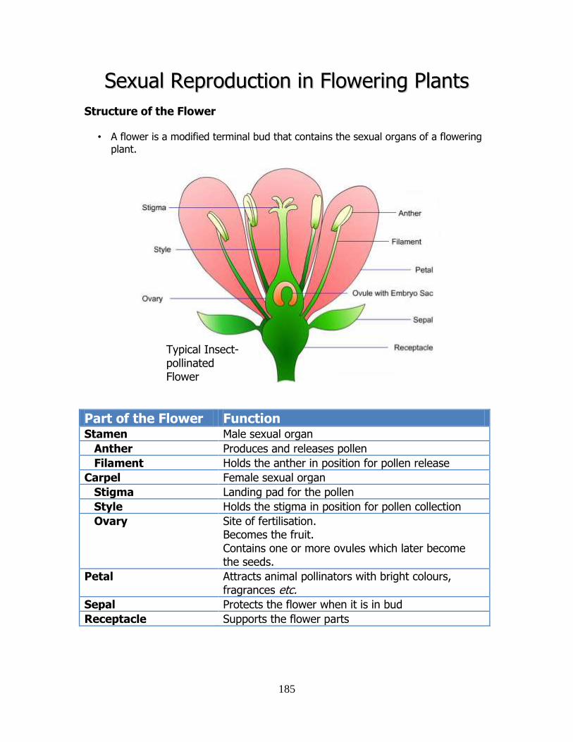
185
SSeexxuuaall RReepprroodduuccttiioonn iinn FFlloowweerriinngg PPllaannttss
Structure of the Flower
• A flower is a modified terminal bud that contains the sexual organs of a flowering plant.
Part of the Flower Function Stamen Male sexual organ
Anther Produces and releases pollen
Filament Holds the anther in position for pollen release
Carpel Female sexual organ
Stigma Landing pad for the pollen
Style Holds the stigma in position for pollen collection
Ovary Site of fertilisation. Becomes the fruit. Contains one or more ovules which later become the seeds.
Petal Attracts animal pollinators with bright colours, fragrances etc.
Sepal Protects the flower when it is in bud
Receptacle Supports the flower parts
Typical Insect- pollinated Flower
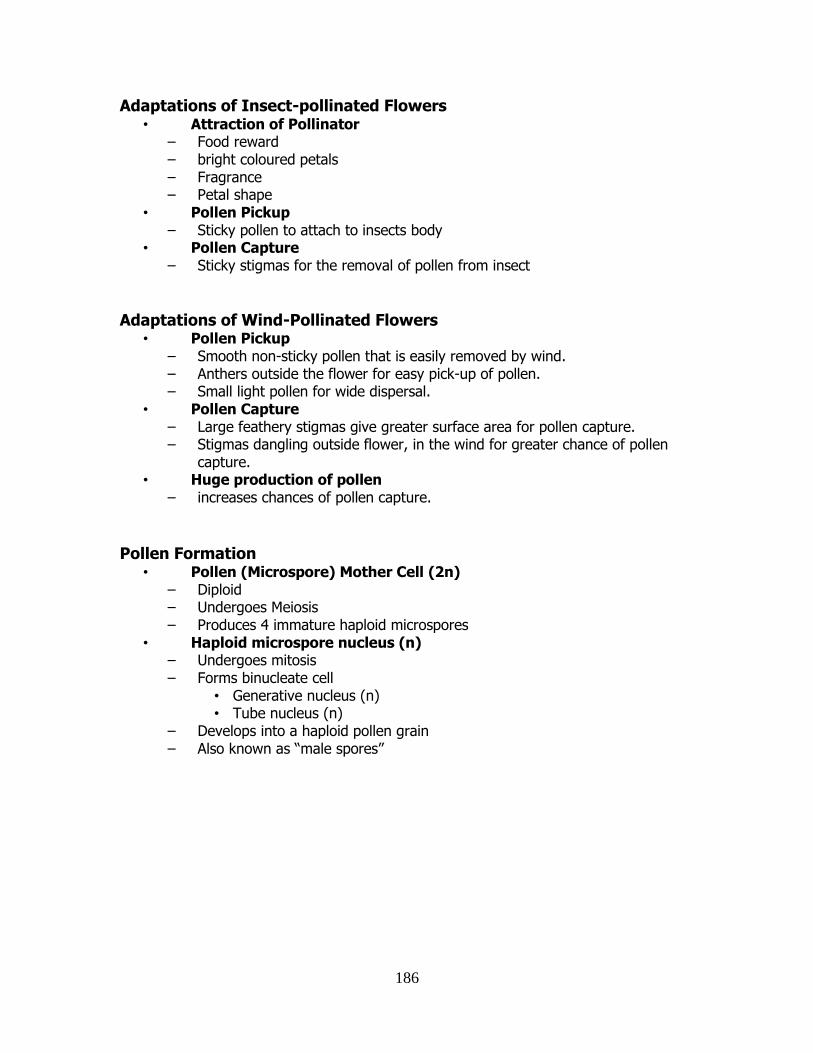
186
Adaptations of Insect-pollinated Flowers • Attraction of Pollinator
– Food reward – bright coloured petals – Fragrance – Petal shape
• Pollen Pickup – Sticky pollen to attach to insects body
• Pollen Capture – Sticky stigmas for the removal of pollen from insect
Adaptations of Wind-Pollinated Flowers
• Pollen Pickup – Smooth non-sticky pollen that is easily removed by wind. – Anthers outside the flower for easy pick-up of pollen. – Small light pollen for wide dispersal.
• Pollen Capture – Large feathery stigmas give greater surface area for pollen capture. – Stigmas dangling outside flower, in the wind for greater chance of pollen
capture. • Huge production of pollen
– increases chances of pollen capture.
Pollen Formation • Pollen (Microspore) Mother Cell (2n)
– Diploid – Undergoes Meiosis – Produces 4 immature haploid microspores
• Haploid microspore nucleus (n) – Undergoes mitosis – Forms binucleate cell
• Generative nucleus (n) • Tube nucleus (n)
– Develops into a haploid pollen grain – Also known as “male spores”
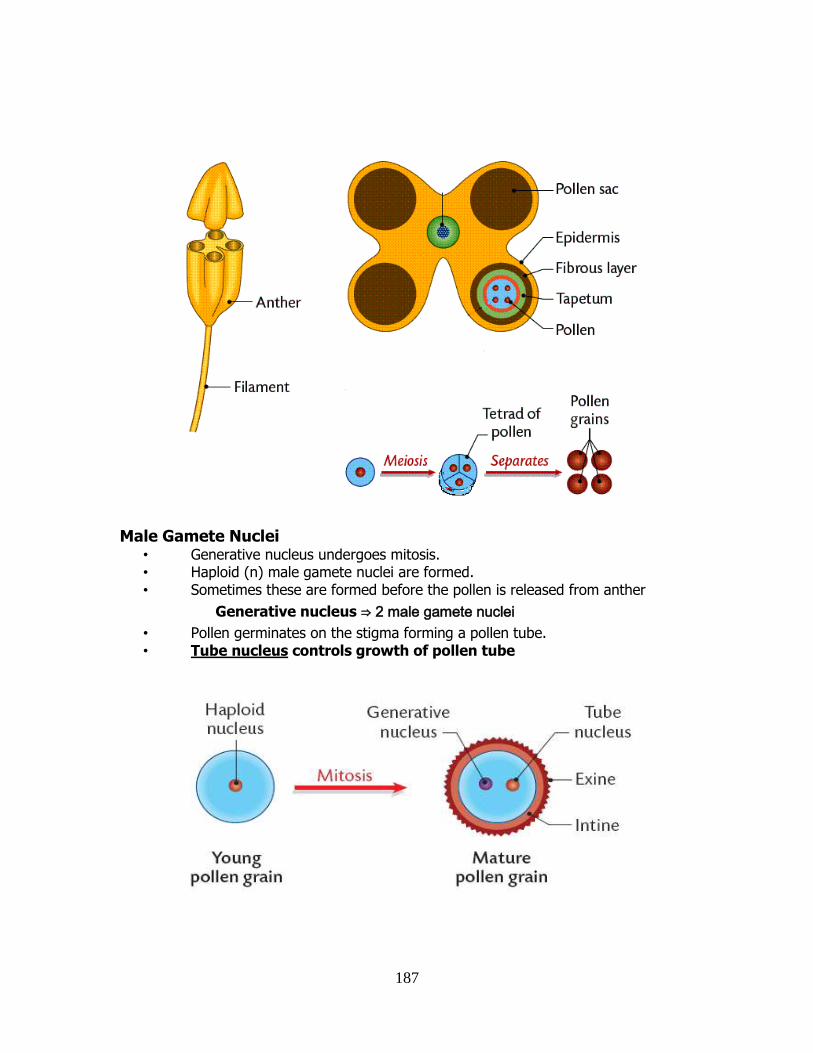
187
Male Gamete Nuclei • Generative nucleus undergoes mitosis. • Haploid (n) male gamete nuclei are formed. • Sometimes these are formed before the pollen is released from anther
Generative nucleus ⇒ 2 male gamete nuclei
• Pollen germinates on the stigma forming a pollen tube. • Tube nucleus controls growth of pollen tube
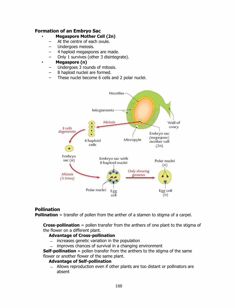
188
Formation of an Embryo Sac • Megaspore Mother Cell (2n)
– At the centre of each ovule. – Undergoes meiosis. – 4 haploid megaspores are made. – Only 1 survives (other 3 disintegrate).
• Megaspore (n) – Undergoes 3 rounds of mitosis. – 8 haploid nucleii are formed. – These nuclei become 6 cells and 2 polar nuclei.
Pollination Pollination = transfer of pollen from the anther of a stamen to stigma of a carpel.
Cross-pollination = pollen transfer from the anthers of one plant to the stigma of the flower on a different plant.
Advantage of Cross-pollination increases genetic variation in the population improves chances of survival in a changing environment
Self-pollination = pollen transfer from the anthers to the stigma of the same flower or another flower of the same plant.
Advantage of Self-pollination Allows reproduction even if other plants are too distant or pollinators are
absent
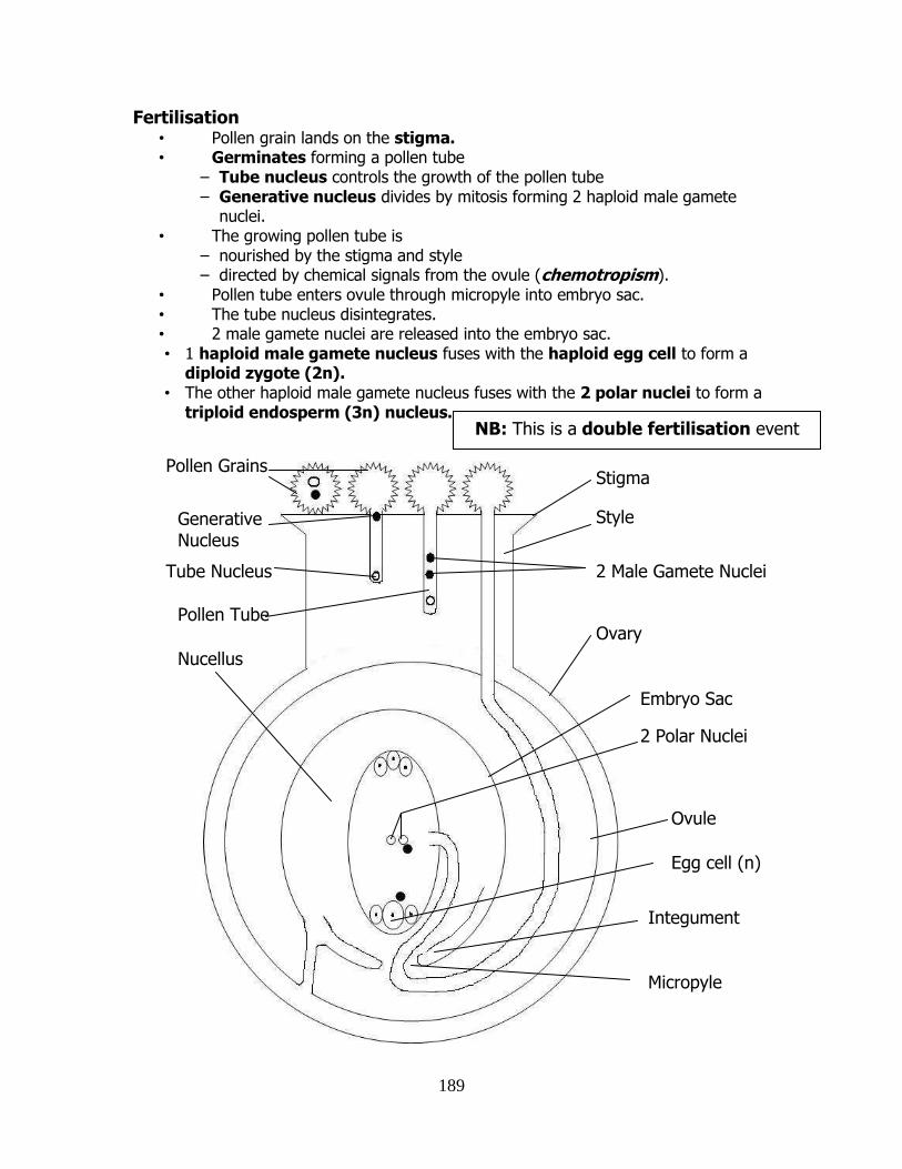
189
Fertilisation • Pollen grain lands on the stigma. • Germinates forming a pollen tube
– Tube nucleus controls the growth of the pollen tube – Generative nucleus divides by mitosis forming 2 haploid male gamete
nuclei. • The growing pollen tube is
– nourished by the stigma and style – directed by chemical signals from the ovule (chemotropism).
• Pollen tube enters ovule through micropyle into embryo sac. • The tube nucleus disintegrates. • 2 male gamete nuclei are released into the embryo sac. • 1 haploid male gamete nucleus fuses with the haploid egg cell to form a
diploid zygote (2n). • The other haploid male gamete nucleus fuses with the 2 polar nuclei to form a
triploid endosperm (3n) nucleus.
Pollen Tube
Tube Nucleus
Generative Nucleus
Micropyle
Ovule
Integument
Embryo Sac
Ovary
2 Male Gamete Nuclei
Style
Stigma Pollen Grains
Nucellus
NB: This is a double fertilisation event
2 Polar Nuclei
Egg cell (n)
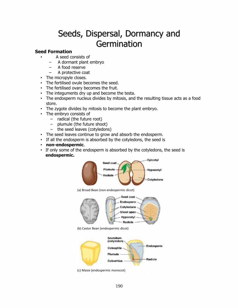
190
SSeeeeddss,, DDiissppeerrssaall,, DDoorrmmaannccyy aanndd
GGeerrmmiinnaattiioonn Seed Formation
• A seed consists of – A dormant plant embryo – A food reserve – A protective coat
• The micropyle closes. • The fertilised ovule becomes the seed. • The fertilised ovary becomes the fruit. • The integuments dry up and become the testa. • The endosperm nucleus divides by mitosis, and the resulting tissue acts as a food
store. • The zygote divides by mitosis to become the plant embryo. • The embryo consists of
– radical (the future root) – plumule (the future shoot) – the seed leaves (cotyledons)
• The seed leaves continue to grow and absorb the endosperm. • If all the endosperm is absorbed by the cotyledons, the seed is • non-endospermic. • If only some of the endosperm is absorbed by the cotyledons, the seed is
endospermic.
(c) Maize (endospermic monocot)
(a) Broad Bean (non-endospermic dicot)
(b) Castor Bean (endospermic dicot)
191
Seed Structure • A seeds consists of a plant embryo and a food reserve contained within a
protective coat (the testa). • The food reserve may be:
In the endosperm (as in endospermous seeds) e.g. Maize, Castor Beans.
In the cotyledon (as in non-endospermous seeds) e.g. Broad Beans.
Fruit formation • A fruit is a fertilised, ripened, ovary of a flower which contains the seeds. • The fruit protects the seed and helps in dispersal. • A tomato is a true fruit, so is a grape. • The inner area of an apple or a pear is a fruit, which is contained within the
outer sweet flesh of the pear. • This outer area is a “false fruit” or swollen receptacle.
Seedless Fruit • Some are seedless varieties, and this is due to genetics
– oranges: pollination occurs but fertilisation doesn’t occur – bananas: fertilisation occurs but seeds don’t develop
• Spraying with plant growth regulators – Auxin can stimulate fruit formation without fertilisation.
Seed Dispersal • Dispersal is the transfer of a seed away from the parent plant. • Reasons for dispersal
– Avoids competition with each other and parents – Finding new areas to colonise – Increases chances of survival of larger numbers of plants
Wind Dispersal • Tiny light seeds
– e.g. Orchids • `Parachutes’ for increased air travel time
– e.g. Dandelion, Thistle • Fruit with wings
– e.g. Sycamore
Water dispersal • Air-filled fruits which can float • Large distances can be covered • Seeds can travel by river and streams
– e.g. Water lilies • Seeds can even travel on the open sea to other land masses
– e.g. sea-beans, coconuts
192
Animal Dispersal • Animals can travel long distances. • Animals often live in places where seeds can germinate (e.g. underground). • 2 types of Fruits
Hooked or barbed fruits e.g. Burdock, goose grass Edible fruits e.g. Tomato, Blackberry, Acorns
Self Dispersal
• Explosive structures, catapults etc. • Examples:
– Peas – Gorse – Poppies
Dormancy • Dormancy is a resting period when seeds undergo no growth and have
reduced cell activity or metabolism, even though the environmental conditions are suitable for growth.
Causes of Dormancy • Growth inhibitors in the outer parts of the seed. • Testa is too thick to allow water or oxygen in. • Growing embryo can’t get out during germination because the testa is too
tough. • A lack of growth regulators promoters (auxins).
Breaking Dormancy • Sometimes dormancy is broken by soaking or scraping.
– softens or breaks the testa • Often a cold period is needed to break dormancy
– breaks down the growth inhibitors causing the dormancy – stimulates the production of growth promoters – light or warmth (e.g. in spring) may stimulate production of growth
promoters.
Advantages of Dormancy • Allows the embryo time to develop. • Allows time for seed dispersal. • Helps plant avoid winter conditions. • The plant grows during the spring and summer months
– optimum conditions for growth. • Duration of dormancy varies
– Staggered germinations allows seed banks to develop in the soil.
Dormancy in Agriculture • Some seeds may need to be treated in a certain way to break dormancy. • Delayed and staggered germination is a disadvantage in horticulture.
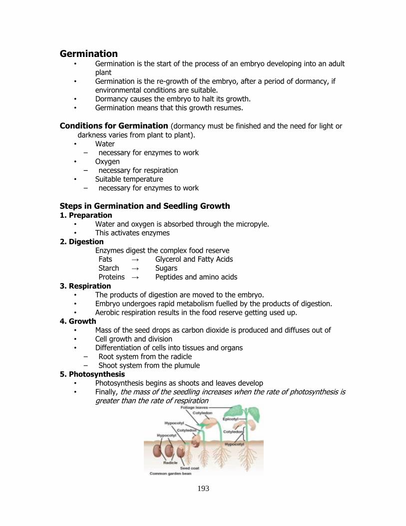
193
Germination • Germination is the start of the process of an embryo developing into an adult
plant • Germination is the re-growth of the embryo, after a period of dormancy, if
environmental conditions are suitable. • Dormancy causes the embryo to halt its growth. • Germination means that this growth resumes.
Conditions for Germination (dormancy must be finished and the need for light or
darkness varies from plant to plant). • Water
– necessary for enzymes to work • Oxygen
– necessary for respiration • Suitable temperature
– necessary for enzymes to work
Steps in Germination and Seedling Growth 1. Preparation
• Water and oxygen is absorbed through the micropyle. • This activates enzymes
2. Digestion Enzymes digest the complex food reserve
Fats → Glycerol and Fatty Acids
Starch → Sugars Proteins → Peptides and amino acids
3. Respiration • The products of digestion are moved to the embryo. • Embryo undergoes rapid metabolism fuelled by the products of digestion. • Aerobic respiration results in the food reserve getting used up.
4. Growth • Mass of the seed drops as carbon dioxide is produced and diffuses out of • Cell growth and division • Differentiation of cells into tissues and organs
– Root system from the radicle – Shoot system from the plumule
5. Photosynthesis • Photosynthesis begins as shoots and leaves develop • Finally, the mass of the seedling increases when the rate of photosynthesis is
greater than the rate of respiration
194
MMaannddaattoorryy PPrraaccttiiccaallss ((MMPPAA’’ss))
1. Conduct a qualitative test for: starch, fat, a reducing sugar, a protein.
2. *Identify any five fauna and any five flora using simple keys. Identify a variety of
habitats within the selected ecosystem.
3. *Identify and use various apparatus required for collection methods in an ecological
study.
4. *Conduct a quantitative study of plants and animals of a sample area of the selected
ecosystem. Transfer results to tables, diagrams, graphs, histograms or any relevent mode.
5. *Investigate any three abiotic factors present in the selected ecosystem. Relate results to
choice of habitat selected by each organism identified in this study.
6. Be familiar with and use the light microscope.
7. Prepare and examine one animal cell and one plant cell – unstained and stained – using
the light microscope (×100, ×400).
8. Investigate the effect of pH on the rate of one of the following: amylase, pepsin or
catalase activity.
9. Investigate the effect of temperature on the rate of one of the following: amylase, pepsin
or catalase activity.
10. Prepare one enzyme immobilisation and examine its application.
11. Investigate the influence of light intensity or carbon dioxide on the rate of photosynthesis.
12. Prepare and show the production of alcohol by yeast.
13. Conduct any activity to demonstrate osmosis.
14. Investigate the effect of heat denaturation on the activity of one enzyme.
15. Isolate DNA from a plant tissue.
16. Investigate the growth of leaf yeast using agar plates and controls.
17. Prepare and examine microscopically the transverse section of a dicotyledonous stem
(×100, ×400).
18. Dissect, display and identify an ox’s or a sheep’s heart.
19. Investigate the effect of exercise on the breathing rate or pulse rate of a human.
20. Investigate the effect of IAA growth regulator on plant tissue.
21. Investigate the effect of water, oxygen and temperature on germination.
22. Use starch agar or skimmed milk plates to show digestive activity during germination.
* see Woodland Field Ecology chapter for these practicals.
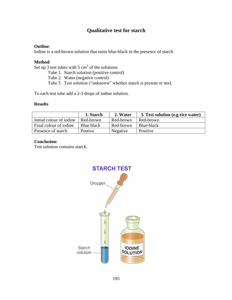
195
Qualitative test for starch
Outline:
Iodine is a red-brown solution that turns blue-black in the presence of starch
Method:
Set up 3 test tubes with 5 cm3 of the solutions
Tube 1. Starch solution (positive control)
Tube 2. Water (negative control)
Tube 3. Test solution (“unknown” whether starch is present or not).
To each test tube add a 2-3 drops of iodine solution.
Results
1. Starch 2. Water 3. Test solution (e.g rice water)
Initial colour of iodine Red-brown Red-brown Red-brown
Final colour of iodine Blue black Red-brown Blue-black
Presence of starch Postive Negative Positive
Conclusion:
Test solution contains starch.
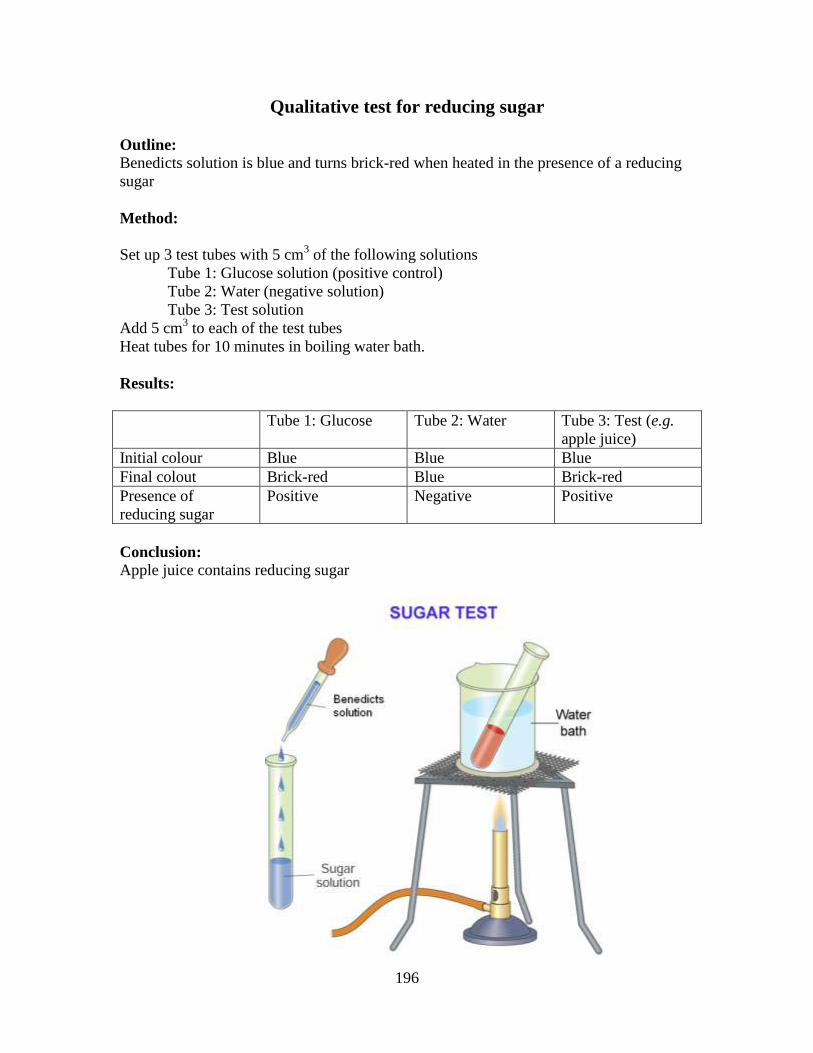
196
Qualitative test for reducing sugar
Outline:
Benedicts solution is blue and turns brick-red when heated in the presence of a reducing
sugar
Method:
Set up 3 test tubes with 5 cm3 of the following solutions
Tube 1: Glucose solution (positive control)
Tube 2: Water (negative solution)
Tube 3: Test solution
Add 5 cm3 to each of the test tubes
Heat tubes for 10 minutes in boiling water bath.
Results:
Tube 1: Glucose Tube 2: Water Tube 3: Test (e.g.
apple juice)
Initial colour Blue Blue Blue
Final colout Brick-red Blue Brick-red
Presence of
reducing sugar
Positive Negative Positive
Conclusion:
Apple juice contains reducing sugar
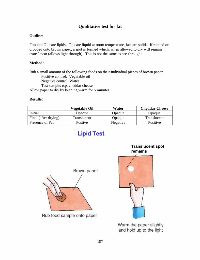
197
Qualitative test for fat
Outline:
Fats and Oils are lipids. Oils are liquid at room temperature, fats are solid. If rubbed or
dropped onto brown paper, a spot is formed which, when allowed to dry will remain
translucent (allows light through). This is not the same as see-through!
Method:
Rub a small amount of the following foods on their individual pieces of brown paper.
Positive control: Vegetable oil
Negative control: Water
Test sample: e.g. cheddar cheese
Allow paper to dry by keeping warm for 5 minutes
Results:
Vegetable Oil Water Cheddar Cheese
Initial Opaque Opaque Opaque
Final (after drying) Translucent Opaque Translucent
Presence of Fat Postive Negative Positive
Lipid Test
Translucent spot
remains
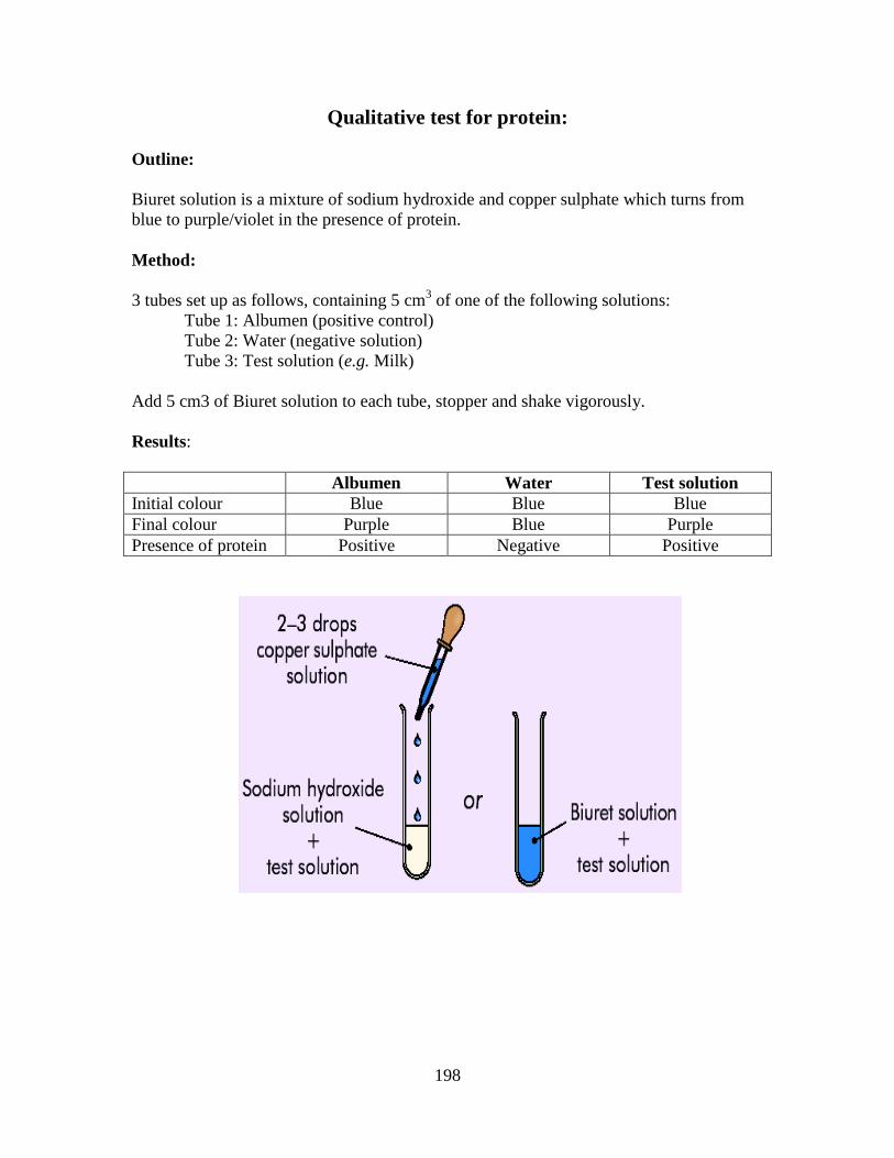
198
Qualitative test for protein:
Outline:
Biuret solution is a mixture of sodium hydroxide and copper sulphate which turns from
blue to purple/violet in the presence of protein.
Method:
3 tubes set up as follows, containing 5 cm3 of one of the following solutions:
Tube 1: Albumen (positive control)
Tube 2: Water (negative solution)
Tube 3: Test solution (e.g. Milk)
Add 5 cm3 of Biuret solution to each tube, stopper and shake vigorously.
Results:
Albumen Water Test solution
Initial colour Blue Blue Blue
Final colour Purple Blue Purple
Presence of protein Positive Negative Positive
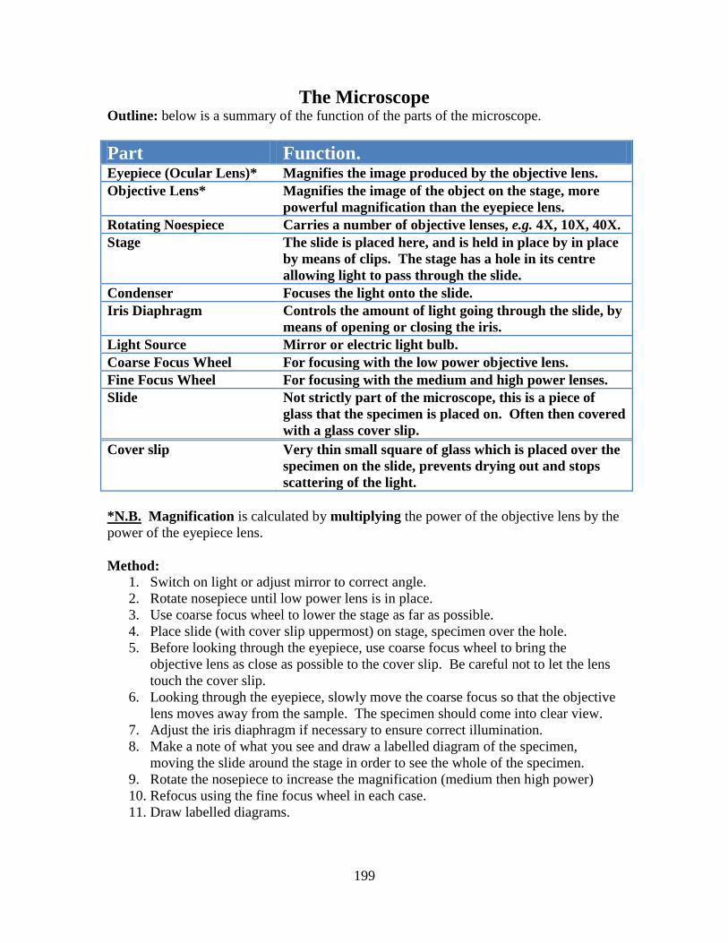
199
The Microscope Outline: below is a summary of the function of the parts of the microscope.
Part Function. Eyepiece (Ocular Lens)* Magnifies the image produced by the objective lens.
Objective Lens* Magnifies the image of the object on the stage, more
powerful magnification than the eyepiece lens.
Rotating Noespiece Carries a number of objective lenses, e.g. 4X, 10X, 40X.
Stage The slide is placed here, and is held in place by in place
by means of clips. The stage has a hole in its centre
allowing light to pass through the slide.
Condenser Focuses the light onto the slide.
Iris Diaphragm Controls the amount of light going through the slide, by
means of opening or closing the iris.
Light Source Mirror or electric light bulb.
Coarse Focus Wheel For focusing with the low power objective lens.
Fine Focus Wheel For focusing with the medium and high power lenses.
Slide Not strictly part of the microscope, this is a piece of
glass that the specimen is placed on. Often then covered
with a glass cover slip.
Cover slip Very thin small square of glass which is placed over the
specimen on the slide, prevents drying out and stops
scattering of the light.
*N.B. Magnification is calculated by multiplying the power of the objective lens by the
power of the eyepiece lens.
Method:
1. Switch on light or adjust mirror to correct angle.
2. Rotate nosepiece until low power lens is in place.
3. Use coarse focus wheel to lower the stage as far as possible.
4. Place slide (with cover slip uppermost) on stage, specimen over the hole.
5. Before looking through the eyepiece, use coarse focus wheel to bring the
objective lens as close as possible to the cover slip. Be careful not to let the lens
touch the cover slip.
6. Looking through the eyepiece, slowly move the coarse focus so that the objective
lens moves away from the sample. The specimen should come into clear view.
7. Adjust the iris diaphragm if necessary to ensure correct illumination.
8. Make a note of what you see and draw a labelled diagram of the specimen,
moving the slide around the stage in order to see the whole of the specimen.
9. Rotate the nosepiece to increase the magnification (medium then high power)
10. Refocus using the fine focus wheel in each case.
11. Draw labelled diagrams.
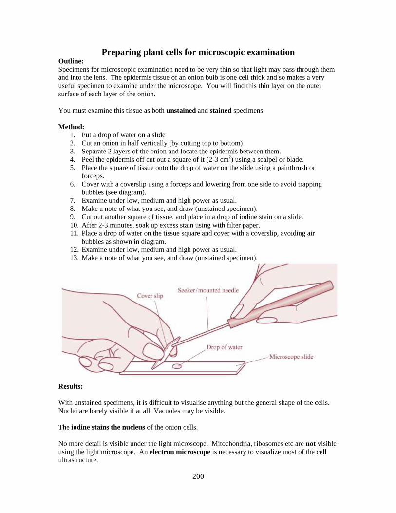
200
Preparing plant cells for microscopic examination Outline:
Specimens for microscopic examination need to be very thin so that light may pass through them
and into the lens. The epidermis tissue of an onion bulb is one cell thick and so makes a very
useful specimen to examine under the microscope. You will find this thin layer on the outer
surface of each layer of the onion.
You must examine this tissue as both unstained and stained specimens.
Method:
1. Put a drop of water on a slide
2. Cut an onion in half vertically (by cutting top to bottom)
3. Separate 2 layers of the onion and locate the epidermis between them.
4. Peel the epidermis off cut out a square of it (2-3 cm2) using a scalpel or blade.
5. Place the square of tissue onto the drop of water on the slide using a paintbrush or
forceps.
6. Cover with a coverslip using a forceps and lowering from one side to avoid trapping
bubbles (see diagram).
7. Examine under low, medium and high power as usual.
8. Make a note of what you see, and draw (unstained specimen).
9. Cut out another square of tissue, and place in a drop of iodine stain on a slide.
10. After 2-3 minutes, soak up excess stain using with filter paper.
11. Place a drop of water on the tissue square and cover with a coverslip, avoiding air
bubbles as shown in diagram.
12. Examine under low, medium and high power as usual.
13. Make a note of what you see, and draw (unstained specimen).
Results:
With unstained specimens, it is difficult to visualise anything but the general shape of the cells.
Nuclei are barely visible if at all. Vacuoles may be visible.
The iodine stains the nucleus of the onion cells.
No more detail is visible under the light microscope. Mitochondria, ribosomes etc are not visible
using the light microscope. An electron microscope is necessary to visualize most of the cell
ultrastructure.
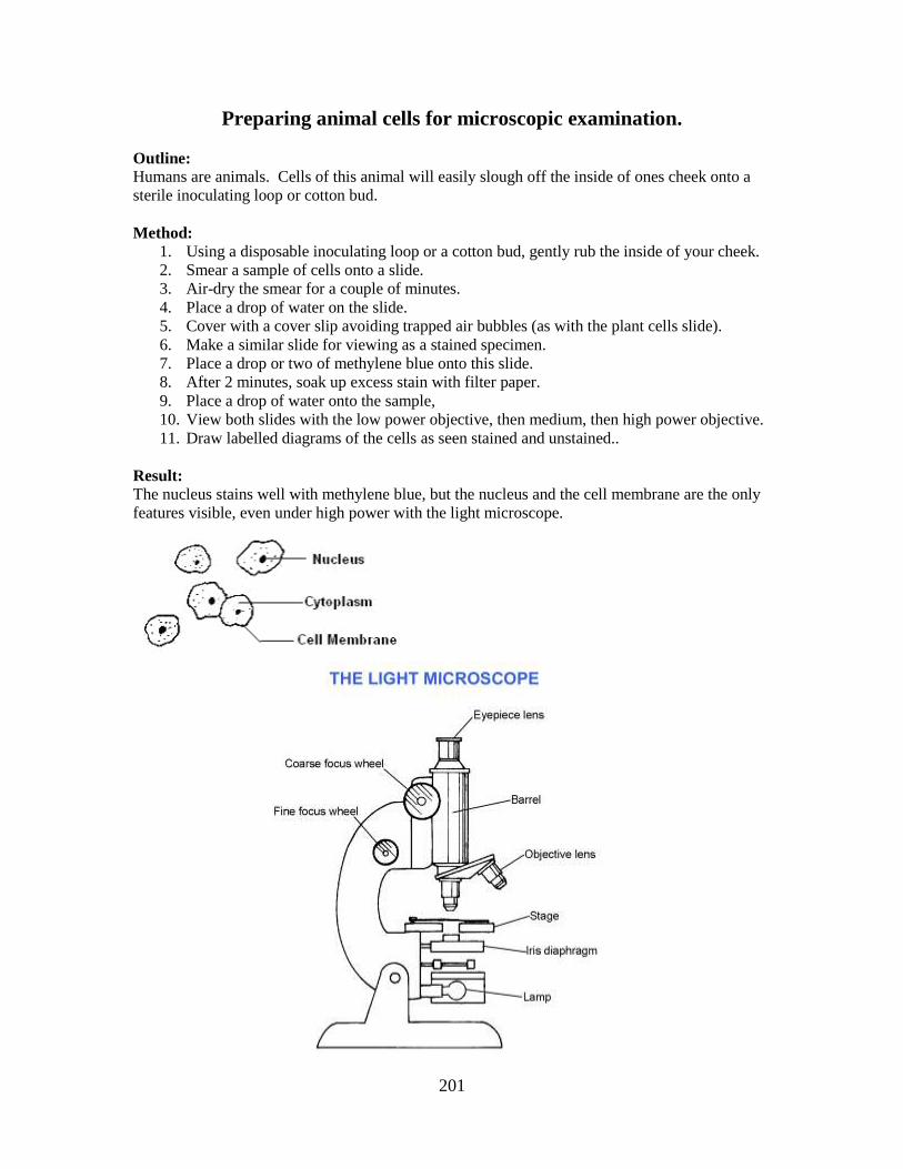
201
Preparing animal cells for microscopic examination.
Outline:
Humans are animals. Cells of this animal will easily slough off the inside of ones cheek onto a
sterile inoculating loop or cotton bud.
Method:
1. Using a disposable inoculating loop or a cotton bud, gently rub the inside of your cheek.
2. Smear a sample of cells onto a slide.
3. Air-dry the smear for a couple of minutes.
4. Place a drop of water on the slide.
5. Cover with a cover slip avoiding trapped air bubbles (as with the plant cells slide).
6. Make a similar slide for viewing as a stained specimen.
7. Place a drop or two of methylene blue onto this slide.
8. After 2 minutes, soak up excess stain with filter paper.
9. Place a drop of water onto the sample,
10. View both slides with the low power objective, then medium, then high power objective.
11. Draw labelled diagrams of the cells as seen stained and unstained..
Result:
The nucleus stains well with methylene blue, but the nucleus and the cell membrane are the only
features visible, even under high power with the light microscope.
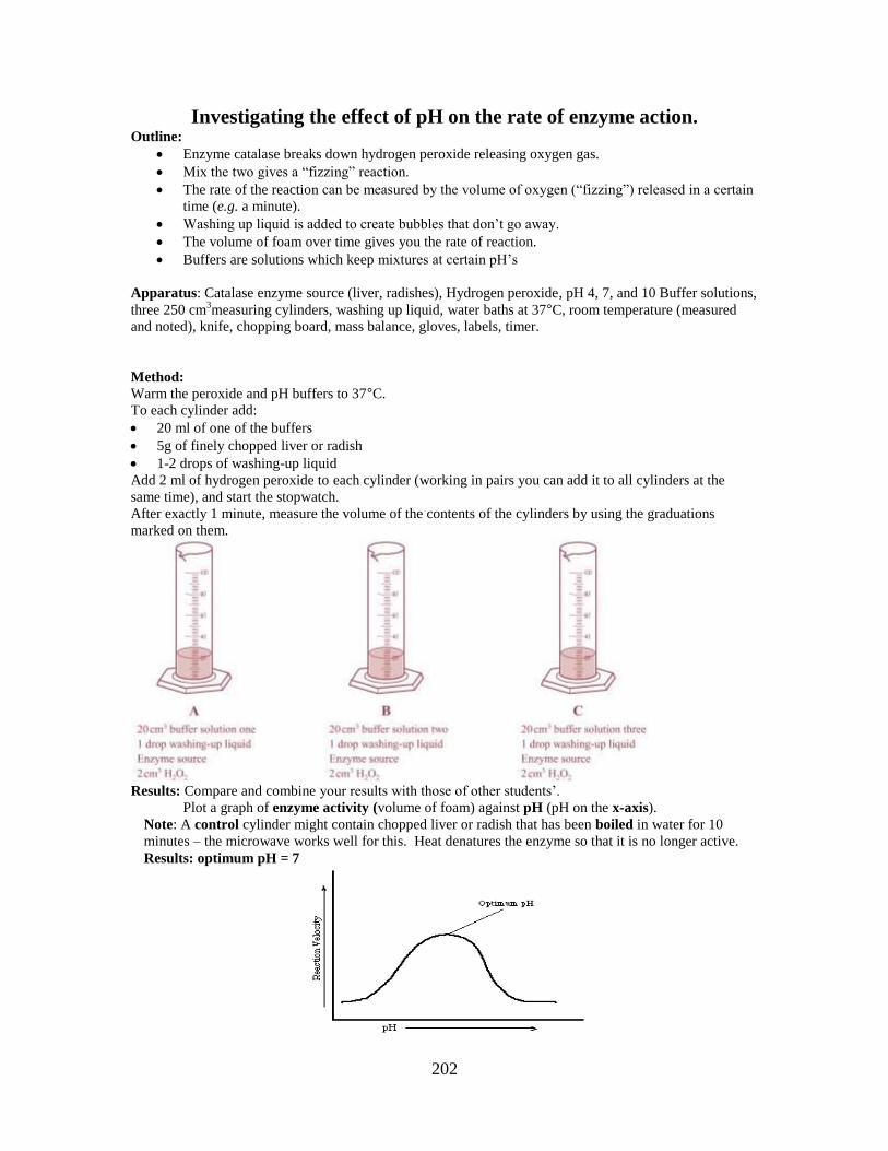
202
Investigating the effect of pH on the rate of enzyme action. Outline:
Enzyme catalase breaks down hydrogen peroxide releasing oxygen gas.
Mix the two gives a “fizzing” reaction.
The rate of the reaction can be measured by the volume of oxygen (“fizzing”) released in a certain
time (e.g. a minute).
Washing up liquid is added to create bubbles that don’t go away.
The volume of foam over time gives you the rate of reaction.
Buffers are solutions which keep mixtures at certain pH’s
Apparatus: Catalase enzyme source (liver, radishes), Hydrogen peroxide, pH 4, 7, and 10 Buffer solutions,
three 250 cm3measuring cylinders, washing up liquid, water baths at 37°C, room temperature (measured
and noted), knife, chopping board, mass balance, gloves, labels, timer.
Method:
Warm the peroxide and pH buffers to 37°C.
To each cylinder add:
20 ml of one of the buffers
5g of finely chopped liver or radish
1-2 drops of washing-up liquid
Add 2 ml of hydrogen peroxide to each cylinder (working in pairs you can add it to all cylinders at the
same time), and start the stopwatch.
After exactly 1 minute, measure the volume of the contents of the cylinders by using the graduations
marked on them.
Results: Compare and combine your results with those of other students’.
Plot a graph of enzyme activity (volume of foam) against pH (pH on the x-axis).
Note: A control cylinder might contain chopped liver or radish that has been boiled in water for 10
minutes – the microwave works well for this. Heat denatures the enzyme so that it is no longer active.
Results: optimum pH = 7
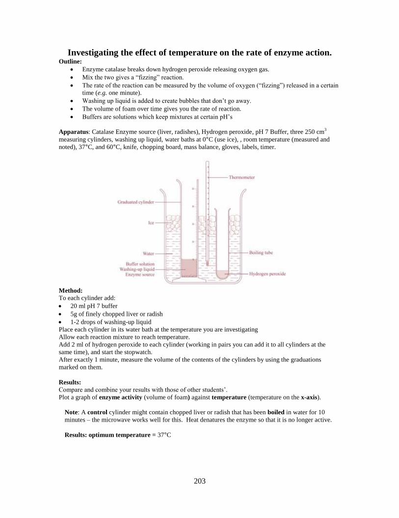
203
Investigating the effect of temperature on the rate of enzyme action. Outline:
Enzyme catalase breaks down hydrogen peroxide releasing oxygen gas.
Mix the two gives a “fizzing” reaction.
The rate of the reaction can be measured by the volume of oxygen (“fizzing”) released in a certain
time (e.g. one minute).
Washing up liquid is added to create bubbles that don’t go away.
The volume of foam over time gives you the rate of reaction.
Buffers are solutions which keep mixtures at certain pH’s
Apparatus: Catalase Enzyme source (liver, radishes), Hydrogen peroxide, pH 7 Buffer, three 250 cm3
measuring cylinders, washing up liquid, water baths at 0°C (use ice), , room temperature (measured and
noted), 37°C, and 60°C, knife, chopping board, mass balance, gloves, labels, timer.
Method:
To each cylinder add:
20 ml pH 7 buffer
5g of finely chopped liver or radish
1-2 drops of washing-up liquid
Place each cylinder in its water bath at the temperature you are investigating
Allow each reaction mixture to reach temperature.
Add 2 ml of hydrogen peroxide to each cylinder (working in pairs you can add it to all cylinders at the
same time), and start the stopwatch.
After exactly 1 minute, measure the volume of the contents of the cylinders by using the graduations
marked on them.
Results:
Compare and combine your results with those of other students’.
Plot a graph of enzyme activity (volume of foam) against temperature (temperature on the x-axis).
Note: A control cylinder might contain chopped liver or radish that has been boiled in water for 10
minutes – the microwave works well for this. Heat denatures the enzyme so that it is no longer active.
Results: optimum temperature = 37°C
204
The effect of heat denaturation on enzyme activity
Outline:
Many enzymes lose activity when they are exposed to high temperatures (or pH for that matter).
Heat causes a change in shape of the active site of the enzyme molecule, affecting its ability to
bind to the substrate effectively. Much of the time this denaturation is irreversible i.e. the activity
of the enzyme does not return (it does not revert to its original shape).
Method: A sample of amylase is heated in a boiling hot water bath (100°C) for 10 minutes, then cooled to
37°C before use. Starch solution is also warmed to 37°C.
Tube 1 contains starch and amylase (positive control)
Tube 2 contains starch and boiled amylase (experiment)
Tube 3 contains starch and water (negative control)
Tubes are then incubated at 37°C for 10 minutes.
Each solution is then tested with Benedict’s solution for the presence of reducing sugar (maltose
in this case).
Results:
Starch and
Amylase
Starch and
Boiled
Amylase
Starch and Water
Initial colour Blue Blue Blue
Final Colour Brick-red Blue Blue
Presence of Reducing sugar Positive Negative Negative
Conclusion:
The boiling of the amylase resulted in the loss of its enzymatic activity, despite being returned to
its optimum temperature. It was therefore irreversibly denatured.
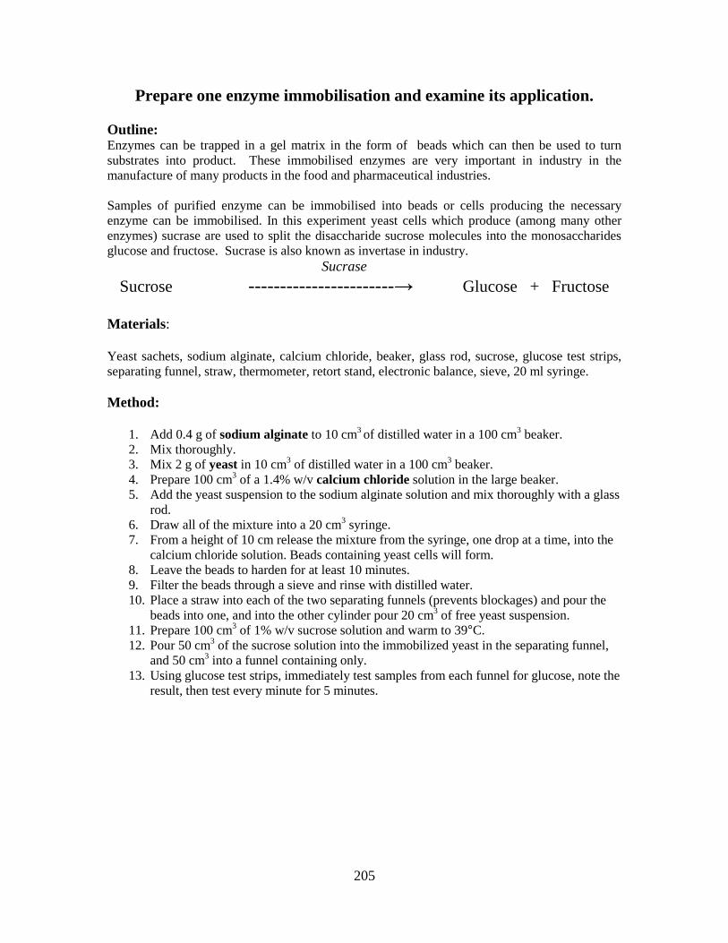
205
Prepare one enzyme immobilisation and examine its application.
Outline: Enzymes can be trapped in a gel matrix in the form of beads which can then be used to turn
substrates into product. These immobilised enzymes are very important in industry in the
manufacture of many products in the food and pharmaceutical industries.
Samples of purified enzyme can be immobilised into beads or cells producing the necessary
enzyme can be immobilised. In this experiment yeast cells which produce (among many other
enzymes) sucrase are used to split the disaccharide sucrose molecules into the monosaccharides
glucose and fructose. Sucrase is also known as invertase in industry.
Sucrase
Sucrose -----------------------→ Glucose + Fructose
Materials:
Yeast sachets, sodium alginate, calcium chloride, beaker, glass rod, sucrose, glucose test strips,
separating funnel, straw, thermometer, retort stand, electronic balance, sieve, 20 ml syringe.
Method:
1. Add 0.4 g of sodium alginate to 10 cm
3 of distilled water in a 100 cm
3 beaker.
2. Mix thoroughly.
3. Mix 2 g of yeast in 10 cm3 of distilled water in a 100 cm
3 beaker.
4. Prepare 100 cm3 of a 1.4% w/v calcium chloride solution in the large beaker.
5. Add the yeast suspension to the sodium alginate solution and mix thoroughly with a glass
rod.
6. Draw all of the mixture into a 20 cm3 syringe.
7. From a height of 10 cm release the mixture from the syringe, one drop at a time, into the
calcium chloride solution. Beads containing yeast cells will form.
8. Leave the beads to harden for at least 10 minutes.
9. Filter the beads through a sieve and rinse with distilled water.
10. Place a straw into each of the two separating funnels (prevents blockages) and pour the
beads into one, and into the other cylinder pour 20 cm3 of free yeast suspension.
11. Prepare 100 cm3 of 1% w/v sucrose solution and warm to 39°C.
12. Pour 50 cm3 of the sucrose solution into the immobilized yeast in the separating funnel,
and 50 cm3 into a funnel containing only.
13. Using glucose test strips, immediately test samples from each funnel for glucose, note the
result, then test every minute for 5 minutes.
206
Results and Conclusion:
1. Immobilised enzyme converts at a faster rate than the free enzyme.
2. The product is contaminant-free.
3. Enzyme beads can be re-used.
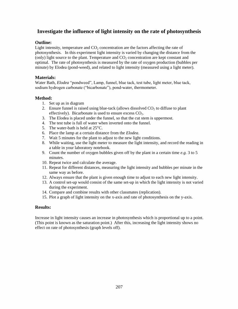
207
Investigate the influence of light intensity on the rate of photosynthesis
Outline: Light intensity, temperature and CO2 concentration are the factors affecting the rate of
photosynthesis. In this experiment light intensity is varied by changing the distance from the
(only) light source to the plant. Temperature and CO2 concentration are kept constant and
optimal. The rate of photosynthesis is measured by the rate of oxygen production (bubbles per
minute) by Elodea (pond-weed), and related to light intensity (measured using a light meter).
Materials: Water Bath, Elodea “pondweed”, Lamp, funnel, blue tack, test tube, light meter, blue tack,
sodium hydrogen carbonate (“bicarbonate”), pond-water, thermometer.
Method: 1. Set up as in diagram
2. Ensure funnel is raised using blue-tack (allows dissolved CO2 to diffuse to plant
effectively). Bicarbonate is used to ensure excess CO2.
3. The Elodea is placed under the funnel, so that the cut stem is uppermost.
4. The test tube is full of water when inverted onto the funnel.
5. The water-bath is held at 25°C.
6. Place the lamp at a certain distance from the Elodea.
7. Wait 5 minutes for the plant to adjust to the new light conditions.
8. While waiting, use the light meter to measure the light intensity, and record the reading in
a table in your laboratory notebook.
9. Count the number of oxygen bubbles given off by the plant in a certain time e.g. 3 to 5
minutes.
10. Repeat twice and calculate the average.
11. Repeat for different distances, measuring the light intensity and bubbles per minute in the
same way as before.
12. Always ensure that the plant is given enough time to adjust to each new light intensity.
13. A control set-up would consist of the same set-up in which the light intensity is not varied
during the experiment.
14. Compare and combine results with other classmates (replication).
15. Plot a graph of light intensity on the x-axis and rate of photosynthesis on the y-axis.
Results:
Increase in light intensity causes an increase in photosynthesis which is proportional up to a point.
(This point is known as the saturation point.) After this, increasing the light intensity shows no
effect on rate of photosynthesis (graph levels off).
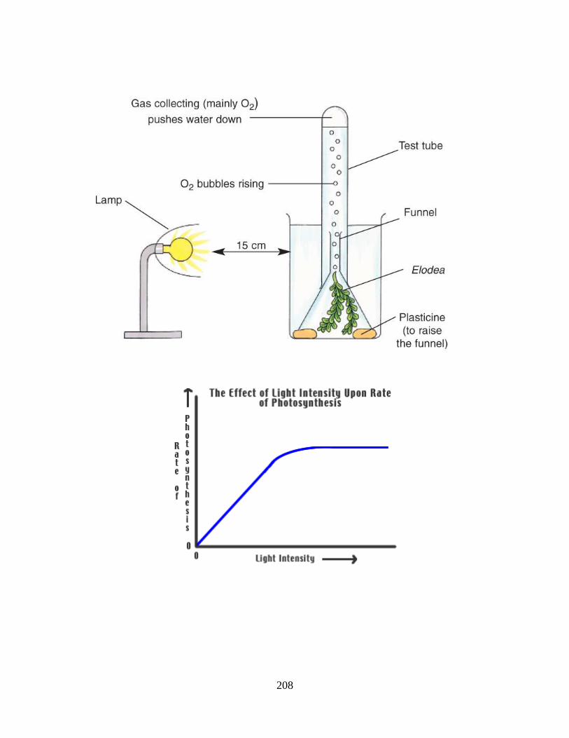
208
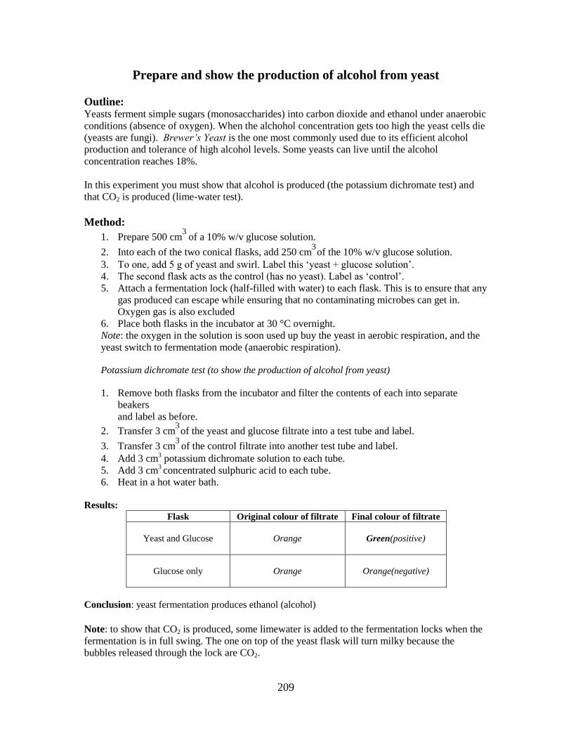
209
Prepare and show the production of alcohol from yeast
Outline: Yeasts ferment simple sugars (monosaccharides) into carbon dioxide and ethanol under anaerobic
conditions (absence of oxygen). When the alchohol concentration gets too high the yeast cells die
(yeasts are fungi). Brewer’s Yeast is the one most commonly used due to its efficient alcohol
production and tolerance of high alcohol levels. Some yeasts can live until the alcohol
concentration reaches 18%.
In this experiment you must show that alcohol is produced (the potassium dichromate test) and
that CO2 is produced (lime-water test).
Method:
1. Prepare 500 cm3
of a 10% w/v glucose solution.
2. Into each of the two conical flasks, add 250 cm3
of the 10% w/v glucose solution.
3. To one, add 5 g of yeast and swirl. Label this ‘yeast + glucose solution’.
4. The second flask acts as the control (has no yeast). Label as ‘control’.
5. Attach a fermentation lock (half-filled with water) to each flask. This is to ensure that any
gas produced can escape while ensuring that no contaminating microbes can get in.
Oxygen gas is also excluded
6. Place both flasks in the incubator at 30 °C overnight.
Note: the oxygen in the solution is soon used up buy the yeast in aerobic respiration, and the
yeast switch to fermentation mode (anaerobic respiration).
Potassium dichromate test (to show the production of alcohol from yeast)
1. Remove both flasks from the incubator and filter the contents of each into separate
beakers
and label as before.
2. Transfer 3 cm3
of the yeast and glucose filtrate into a test tube and label.
3. Transfer 3 cm3
of the control filtrate into another test tube and label.
4. Add 3 cm3 potassium dichromate solution to each tube.
5. Add 3 cm3 concentrated sulphuric acid to each tube.
6. Heat in a hot water bath.
Results:
Flask Original colour of filtrate Final colour of filtrate
Yeast and Glucose
Orange
Green(positive)
Glucose only
Orange
Orange(negative)
Conclusion: yeast fermentation produces ethanol (alcohol)
Note: to show that CO2 is produced, some limewater is added to the fermentation locks when the
fermentation is in full swing. The one on top of the yeast flask will turn milky because the
bubbles released through the lock are CO2.
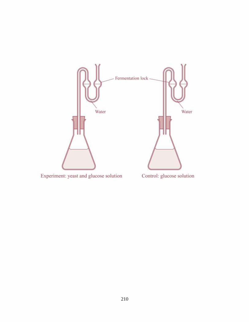
210
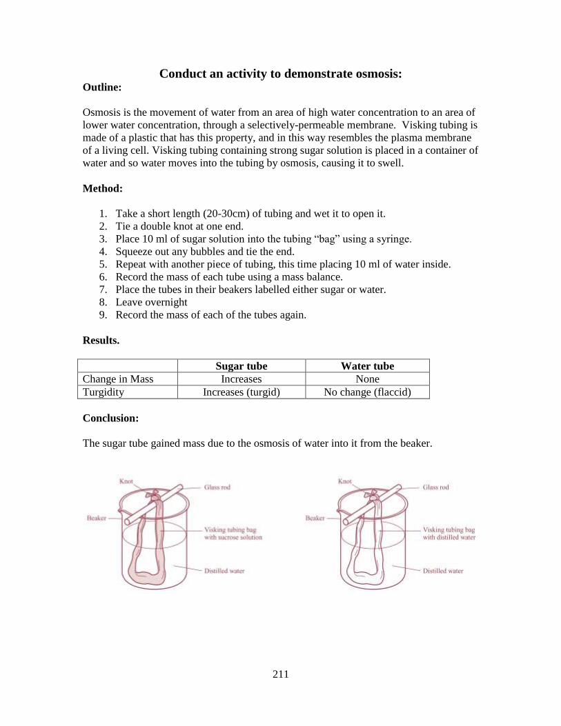
211
Conduct an activity to demonstrate osmosis: Outline:
Osmosis is the movement of water from an area of high water concentration to an area of
lower water concentration, through a selectively-permeable membrane. Visking tubing is
made of a plastic that has this property, and in this way resembles the plasma membrane
of a living cell. Visking tubing containing strong sugar solution is placed in a container of
water and so water moves into the tubing by osmosis, causing it to swell.
Method:
1. Take a short length (20-30cm) of tubing and wet it to open it.
2. Tie a double knot at one end.
3. Place 10 ml of sugar solution into the tubing “bag” using a syringe.
4. Squeeze out any bubbles and tie the end.
5. Repeat with another piece of tubing, this time placing 10 ml of water inside.
6. Record the mass of each tube using a mass balance.
7. Place the tubes in their beakers labelled either sugar or water.
8. Leave overnight
9. Record the mass of each of the tubes again.
Results.
Sugar tube Water tube
Change in Mass Increases None
Turgidity Increases (turgid) No change (flaccid)
Conclusion:
The sugar tube gained mass due to the osmosis of water into it from the beaker.
212
To isolate DNA from plant tissue
Outline:
Separating DNA from cells involves a number of steps because of the different types of
materials that the cell is made of. The main problem with the successful extraction is the
danger posed by nucleases. These are DNA-chopping enzymes produced by the cells
themselves when they are under attack. The method involves physical, chemical and
enzymatic steps which lead to the separation of the DNA from the cell debris.
Method:
1. Add 3 g of table salt to 10 cm3 of washing-up liquid in the beaker and make up to
100cm3 with distilled water.
2. Chop the onion into small pieces.
3. Add the chopped onion to the beaker with the salty washing-up liquid solution
and stir.
4. Put the beaker in the water bath at 60°C for exactly 15 minutes.
5. Cool the mixture by standing the beaker in the ice-water bath for 5 minutes,
occasionally stirring gently.
6. Pour the mixture into the blender and blend it for no more than 3 seconds.
7. Carefully filter the mixture into the second beaker using coffee filter paper and
not laboratory filter paper.
8. Transfer about 10 cm3 of this filtrate into the boiling tube.
9. Add 2 – 3 drops of protease to the filtrate and mix gently.
10. Trickle about 10 cm3
of the ice-cold ethanol, straight from the freezer, down the
side of the boiling tube, to form a layer on top of the filtrate. Leave the tube (on
ice if you have any) if necessary.
11. Observe any changes that take place at the interface of the alcohol and the filtrate.
12. Using a scratched glass rod, wooden splint, or wire loop, gently draw the DNA
out from the alcohol.
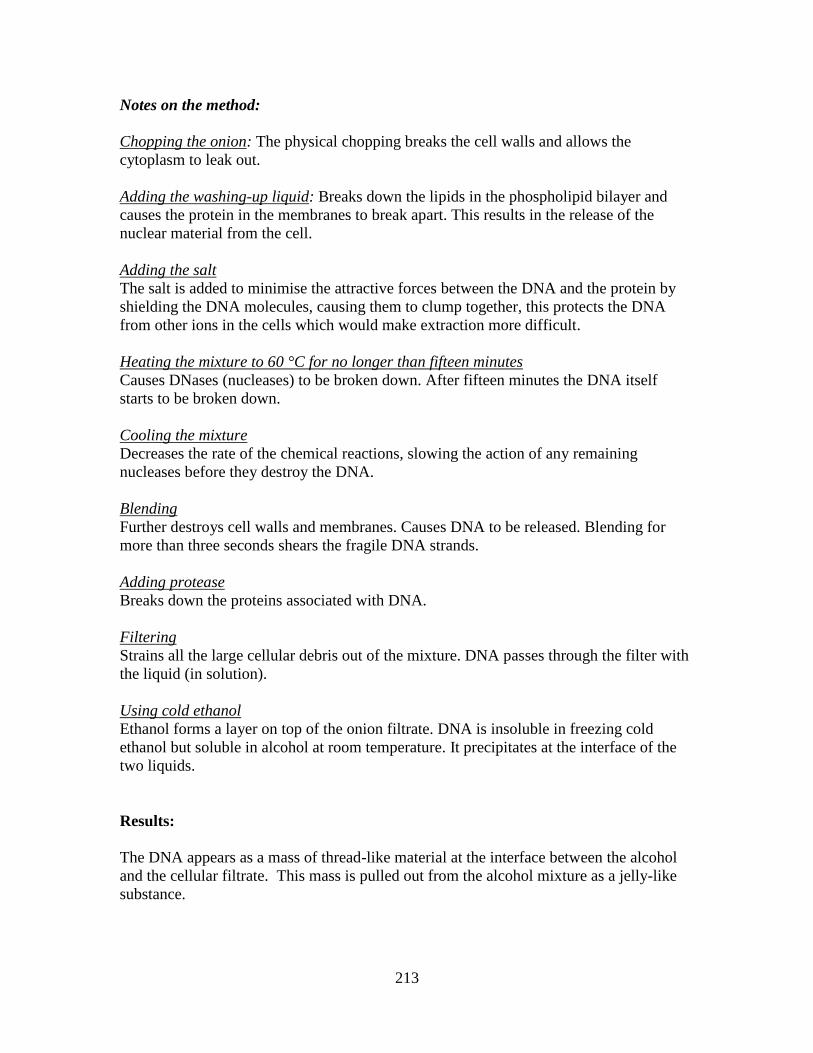
213
Notes on the method:
Chopping the onion: The physical chopping breaks the cell walls and allows the
cytoplasm to leak out.
Adding the washing-up liquid: Breaks down the lipids in the phospholipid bilayer and
causes the protein in the membranes to break apart. This results in the release of the
nuclear material from the cell.
Adding the salt
The salt is added to minimise the attractive forces between the DNA and the protein by
shielding the DNA molecules, causing them to clump together, this protects the DNA
from other ions in the cells which would make extraction more difficult.
Heating the mixture to 60 °C for no longer than fifteen minutes
Causes DNases (nucleases) to be broken down. After fifteen minutes the DNA itself
starts to be broken down.
Cooling the mixture
Decreases the rate of the chemical reactions, slowing the action of any remaining
nucleases before they destroy the DNA.
Blending
Further destroys cell walls and membranes. Causes DNA to be released. Blending for
more than three seconds shears the fragile DNA strands.
Adding protease
Breaks down the proteins associated with DNA.
Filtering
Strains all the large cellular debris out of the mixture. DNA passes through the filter with
the liquid (in solution).
Using cold ethanol
Ethanol forms a layer on top of the onion filtrate. DNA is insoluble in freezing cold
ethanol but soluble in alcohol at room temperature. It precipitates at the interface of the
two liquids.
Results:
The DNA appears as a mass of thread-like material at the interface between the alcohol
and the cellular filtrate. This mass is pulled out from the alcohol mixture as a jelly-like
substance.
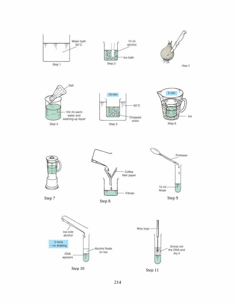
214
Step 7
Step 9
Step 10 Step 11
Step 8
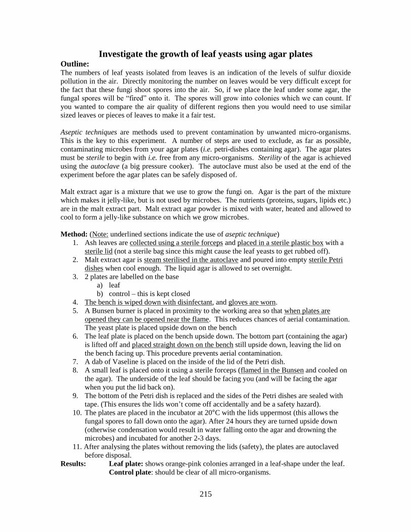
215
Investigate the growth of leaf yeasts using agar plates Outline: The numbers of leaf yeasts isolated from leaves is an indication of the levels of sulfur dioxide
pollution in the air. Directly monitoring the number on leaves would be very difficult except for
the fact that these fungi shoot spores into the air. So, if we place the leaf under some agar, the
fungal spores will be “fired” onto it. The spores will grow into colonies which we can count. If
you wanted to compare the air quality of different regions then you would need to use similar
sized leaves or pieces of leaves to make it a fair test.
Aseptic techniques are methods used to prevent contamination by unwanted micro-organisms.
This is the key to this experiment. A number of steps are used to exclude, as far as possible,
contaminating microbes from your agar plates (i.e. petri-dishes containing agar). The agar plates
must be sterile to begin with i.e. free from any micro-organisms. Sterility of the agar is achieved
using the autoclave (a big pressure cooker). The autoclave must also be used at the end of the
experiment before the agar plates can be safely disposed of.
Malt extract agar is a mixture that we use to grow the fungi on. Agar is the part of the mixture
which makes it jelly-like, but is not used by microbes. The nutrients (proteins, sugars, lipids etc.)
are in the malt extract part. Malt extract agar powder is mixed with water, heated and allowed to
cool to form a jelly-like substance on which we grow microbes.
Method: (Note: underlined sections indicate the use of aseptic technique)
1. Ash leaves are collected using a sterile forceps and placed in a sterile plastic box with a
sterile lid (not a sterile bag since this might cause the leaf yeasts to get rubbed off).
2. Malt extract agar is steam sterilised in the autoclave and poured into empty sterile Petri
dishes when cool enough. The liquid agar is allowed to set overnight.
3. 2 plates are labelled on the base
a) leaf
b) control – this is kept closed
4. The bench is wiped down with disinfectant, and gloves are worn.
5. A Bunsen burner is placed in proximity to the working area so that when plates are
opened they can be opened near the flame. This reduces chances of aerial contamination.
The yeast plate is placed upside down on the bench
6. The leaf plate is placed on the bench upside down. The bottom part (containing the agar)
is lifted off and placed straight down on the bench still upside down, leaving the lid on
the bench facing up. This procedure prevents aerial contamination.
7. A dab of Vaseline is placed on the inside of the lid of the Petri dish.
8. A small leaf is placed onto it using a sterile forceps (flamed in the Bunsen and cooled on
the agar). The underside of the leaf should be facing you (and will be facing the agar
when you put the lid back on).
9. The bottom of the Petri dish is replaced and the sides of the Petri dishes are sealed with
tape. (This ensures the lids won’t come off accidentally and be a safety hazard).
10. The plates are placed in the incubator at 20°C with the lids uppermost (this allows the
fungal spores to fall down onto the agar). After 24 hours they are turned upside down
(otherwise condensation would result in water falling onto the agar and drowning the
microbes) and incubated for another 2-3 days.
11. After analysing the plates without removing the lids (safety), the plates are autoclaved
before disposal.
Results: Leaf plate: shows orange-pink colonies arranged in a leaf-shape under the leaf.
Control plate: should be clear of all micro-organisms.
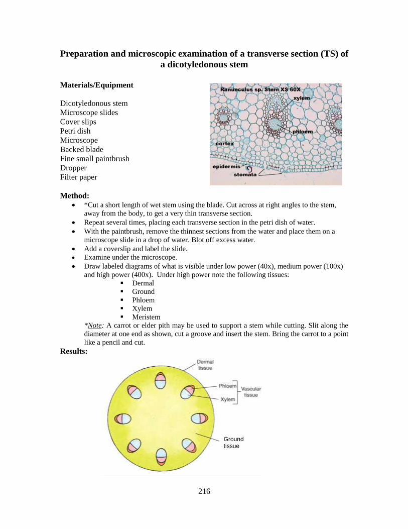
216
Preparation and microscopic examination of a transverse section (TS) of
a dicotyledonous stem
Materials/Equipment
Dicotyledonous stem
Microscope slides
Cover slips
Petri dish
Microscope
Backed blade
Fine small paintbrush
Dropper
Filter paper
Method:
*Cut a short length of wet stem using the blade. Cut across at right angles to the stem,
away from the body, to get a very thin transverse section.
Repeat several times, placing each transverse section in the petri dish of water.
With the paintbrush, remove the thinnest sections from the water and place them on a
microscope slide in a drop of water. Blot off excess water.
Add a coverslip and label the slide.
Examine under the microscope.
Draw labeled diagrams of what is visible under low power (40x), medium power (100x)
and high power (400x). Under high power note the following tissues:
Dermal
Ground
Phloem
Xylem
Meristem
*Note: A carrot or elder pith may be used to support a stem while cutting. Slit along the
diameter at one end as shown, cut a groove and insert the stem. Bring the carrot to a point
like a pencil and cut.
Results:
Ground tissue
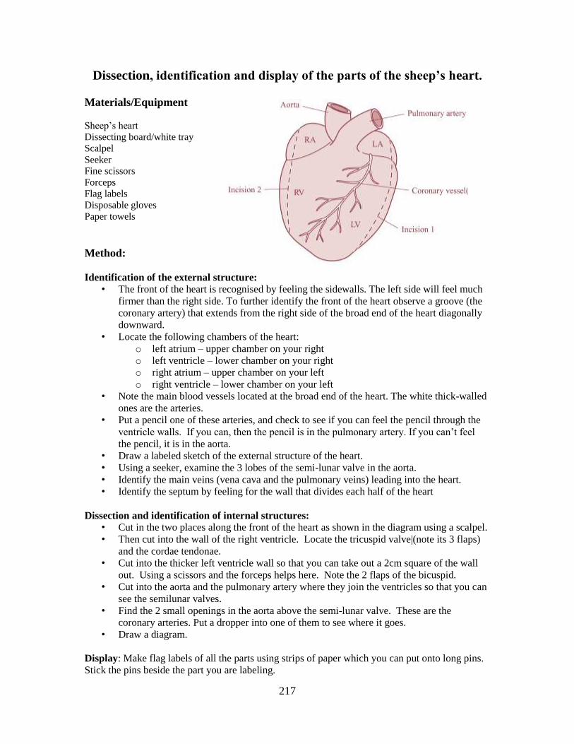
217
Dissection, identification and display of the parts of the sheep’s heart.
Materials/Equipment
Sheep’s heart
Dissecting board/white tray
Scalpel
Seeker
Fine scissors
Forceps
Flag labels
Disposable gloves
Paper towels
Method:
Identification of the external structure:
• The front of the heart is recognised by feeling the sidewalls. The left side will feel much
firmer than the right side. To further identify the front of the heart observe a groove (the
coronary artery) that extends from the right side of the broad end of the heart diagonally
downward.
• Locate the following chambers of the heart:
o left atrium – upper chamber on your right
o left ventricle – lower chamber on your right
o right atrium – upper chamber on your left
o right ventricle – lower chamber on your left
• Note the main blood vessels located at the broad end of the heart. The white thick-walled
ones are the arteries.
• Put a pencil one of these arteries, and check to see if you can feel the pencil through the
ventricle walls. If you can, then the pencil is in the pulmonary artery. If you can’t feel
the pencil, it is in the aorta.
• Draw a labeled sketch of the external structure of the heart.
• Using a seeker, examine the 3 lobes of the semi-lunar valve in the aorta.
• Identify the main veins (vena cava and the pulmonary veins) leading into the heart.
• Identify the septum by feeling for the wall that divides each half of the heart
Dissection and identification of internal structures:
• Cut in the two places along the front of the heart as shown in the diagram using a scalpel.
• Then cut into the wall of the right ventricle. Locate the tricuspid valve|(note its 3 flaps)
and the cordae tendonae.
• Cut into the thicker left ventricle wall so that you can take out a 2cm square of the wall
out. Using a scissors and the forceps helps here. Note the 2 flaps of the bicuspid.
• Cut into the aorta and the pulmonary artery where they join the ventricles so that you can
see the semilunar valves.
• Find the 2 small openings in the aorta above the semi-lunar valve. These are the
coronary arteries. Put a dropper into one of them to see where it goes.
• Draw a diagram.
Display: Make flag labels of all the parts using strips of paper which you can put onto long pins.
Stick the pins beside the part you are labeling.
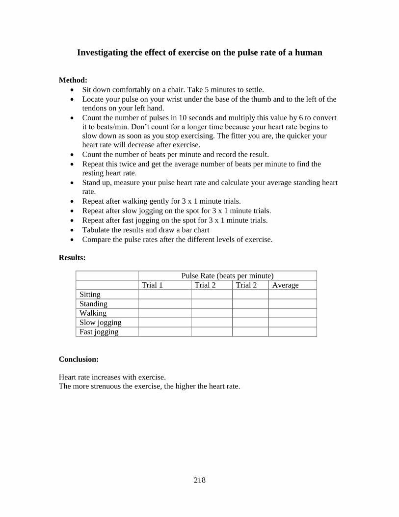
218
Investigating the effect of exercise on the pulse rate of a human
Method:
Sit down comfortably on a chair. Take 5 minutes to settle.
Locate your pulse on your wrist under the base of the thumb and to the left of the
tendons on your left hand.
Count the number of pulses in 10 seconds and multiply this value by 6 to convert
it to beats/min. Don’t count for a longer time because your heart rate begins to
slow down as soon as you stop exercising. The fitter you are, the quicker your
heart rate will decrease after exercise.
Count the number of beats per minute and record the result.
Repeat this twice and get the average number of beats per minute to find the
resting heart rate.
Stand up, measure your pulse heart rate and calculate your average standing heart
rate.
Repeat after walking gently for 3 x 1 minute trials.
Repeat after slow jogging on the spot for 3 x 1 minute trials.
Repeat after fast jogging on the spot for 3 x 1 minute trials.
Tabulate the results and draw a bar chart
Compare the pulse rates after the different levels of exercise.
Results:
Pulse Rate (beats per minute)
Trial 1 Trial 2 Trial 2 Average
Sitting
Standing
Walking
Slow jogging
Fast jogging
Conclusion:
Heart rate increases with exercise.
The more strenuous the exercise, the higher the heart rate.
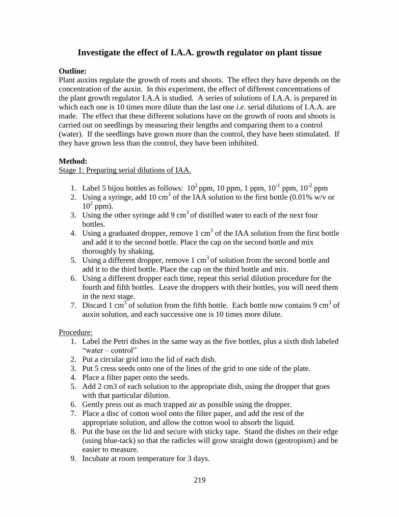
219
Investigate the effect of I.A.A. growth regulator on plant tissue
Outline:
Plant auxins regulate the growth of roots and shoots. The effect they have depends on the
concentration of the auxin. In this experiment, the effect of different concentrations of
the plant growth regulator I.A.A is studied. A series of solutions of I.A.A. is prepared in
which each one is 10 times more dilute than the last one i.e. serial dilutions of I.A.A. are
made. The effect that these different solutions have on the growth of roots and shoots is
carried out on seedlings by measuring their lengths and comparing them to a control
(water). If the seedlings have grown more than the control, they have been stimulated. If
they have grown less than the control, they have been inhibited.
Method:
Stage 1: Preparing serial dilutions of IAA.
1. Label 5 bijou bottles as follows: 102
ppm, 10 ppm, 1 ppm, 10-1
ppm, 10-2
ppm
2. Using a syringe, add 10 cm3 of the IAA solution to the first bottle (0.01% w/v or
102 ppm).
3. Using the other syringe add 9 cm3
of distilled water to each of the next four
bottles.
4. Using a graduated dropper, remove 1 cm3 of the IAA solution from the first bottle
and add it to the second bottle. Place the cap on the second bottle and mix
thoroughly by shaking.
5. Using a different dropper, remove 1 cm3
of solution from the second bottle and
add it to the third bottle. Place the cap on the third bottle and mix.
6. Using a different dropper each time, repeat this serial dilution procedure for the
fourth and fifth bottles. Leave the droppers with their bottles, you will need them
in the next stage.
7. Discard 1 cm3 of solution from the fifth bottle. Each bottle now contains 9 cm
3 of
auxin solution, and each successive one is 10 times more dilute.
Procedure:
1. Label the Petri dishes in the same way as the five bottles, plus a sixth dish labeled
“water – control”
2. Put a circular grid into the lid of each dish.
3. Put 5 cress seeds onto one of the lines of the grid to one side of the plate.
4. Place a filter paper onto the seeds.
5. Add 2 cm3 of each solution to the appropriate dish, using the dropper that goes
with that particular dilution.
6. Gently press out as much trapped air as possible using the dropper.
7. Place a disc of cotton wool onto the filter paper, and add the rest of the
appropriate solution, and allow the cotton wool to absorb the liquid.
8. Put the base on the lid and secure with sticky tape. Stand the dishes on their edge
(using blue-tack) so that the radicles will grow straight down (geotropism) and be
easier to measure.
9. Incubate at room temperature for 3 days.

220
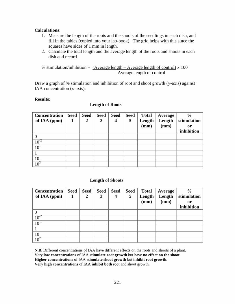
221
Calculations:
1. Measure the length of the roots and the shoots of the seedlings in each dish, and
fill in the tables (copied into your lab-book). The grid helps with this since the
squares have sides of 1 mm in length.
2. Calculate the total length and the average length of the roots and shoots in each
dish and record.
% stimulation/inhibition = (Average length – Average length of control) x 100
Average length of control
Draw a graph of % stimulation and inhibition of root and shoot growth (y-axis) against
IAA concentration (x-axis).
Results:
Length of Roots
Concentration
of IAA (ppm)
Seed
1
Seed
2
Seed
3
Seed
4
Seed
5
Total
Length
(mm)
Average
Length
(mm)
%
stimulation
or
inhibition
0
10-2
10-1
1
10
102
Length of Shoots
Concentration
of IAA (ppm)
Seed
1
Seed
2
Seed
3
Seed
4
Seed
5
Total
Length
(mm)
Average
Length
(mm)
%
stimulation
or
inhibition
0
10-2
10-1
1
10
102
N.B. Different concentrations of IAA have different effects on the roots and shoots of a plant.
Very low concentrations of IAA stimulate root growth but have no effect on the shoot.
Higher concentrations of IAA stimulate shoot growth but inhibit root growth.
Very high concentrations of IAA inhibit both root and shoot growth.
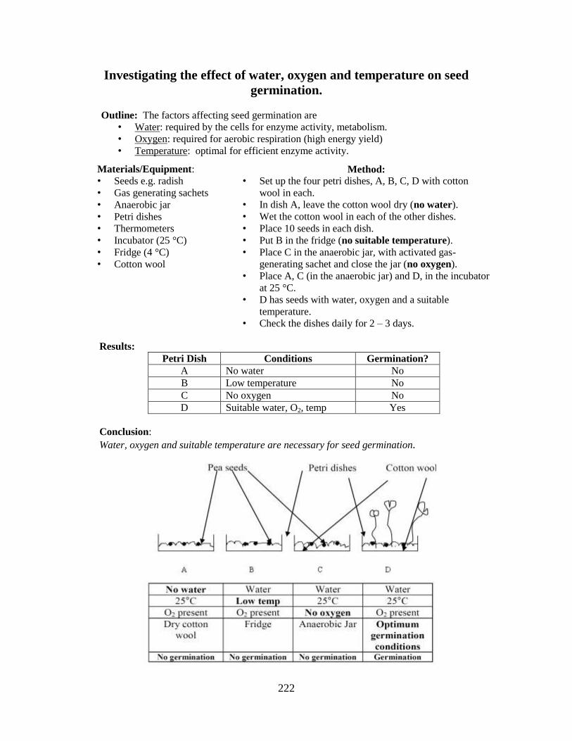
222
Investigating the effect of water, oxygen and temperature on seed
germination.
Outline: The factors affecting seed germination are
• Water: required by the cells for enzyme activity, metabolism.
• Oxygen: required for aerobic respiration (high energy yield)
• Temperature: optimal for efficient enzyme activity.
Method:
• Set up the four petri dishes, A, B, C, D with cotton
wool in each.
• In dish A, leave the cotton wool dry (no water).
• Wet the cotton wool in each of the other dishes.
• Place 10 seeds in each dish.
• Put B in the fridge (no suitable temperature).
• Place C in the anaerobic jar, with activated gas-
generating sachet and close the jar (no oxygen).
• Place A, C (in the anaerobic jar) and D, in the incubator
at 25 °C.
• D has seeds with water, oxygen and a suitable
temperature.
• Check the dishes daily for 2 – 3 days.
Materials/Equipment:
• Seeds e.g. radish
• Gas generating sachets
• Anaerobic jar
• Petri dishes
• Thermometers
• Incubator (25 °C)
• Fridge (4 °C)
• Cotton wool
Results:
Petri Dish Conditions Germination?
A No water No
B Low temperature No
C No oxygen No
D Suitable water, O2, temp Yes
Conclusion:
Water, oxygen and suitable temperature are necessary for seed germination.
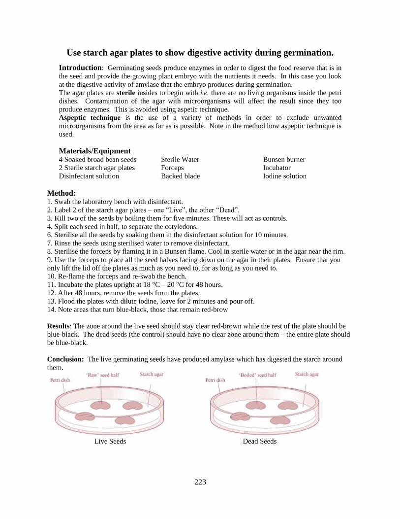
223
Use starch agar plates to show digestive activity during germination.
Introduction: Germinating seeds produce enzymes in order to digest the food reserve that is in
the seed and provide the growing plant embryo with the nutrients it needs. In this case you look
at the digestive activity of amylase that the embryo produces during germination.
The agar plates are sterile insides to begin with i.e. there are no living organisms inside the petri
dishes. Contamination of the agar with microorganisms will affect the result since they too
produce enzymes. This is avoided using aspetic technique.
Aspeptic technique is the use of a variety of methods in order to exclude unwanted
microorganisms from the area as far as is possible. Note in the method how aspeptic technique is
used.
Materials/Equipment 4 Soaked broad bean seeds
2 Sterile starch agar plates
Disinfectant solution
Sterile Water
Forceps
Backed blade
Bunsen burner
Incubator
Iodine solution
Method: 1. Swab the laboratory bench with disinfectant.
2. Label 2 of the starch agar plates – one “Live”, the other “Dead”.
3. Kill two of the seeds by boiling them for five minutes. These will act as controls.
4. Split each seed in half, to separate the cotyledons.
6. Sterilise all the seeds by soaking them in the disinfectant solution for 10 minutes.
7. Rinse the seeds using sterilised water to remove disinfectant.
8. Sterilise the forceps by flaming it in a Bunsen flame. Cool in sterile water or in the agar near the rim.
9. Use the forceps to place all the seed halves facing down on the agar in their plates. Ensure that you
only lift the lid off the plates as much as you need to, for as long as you need to.
10. Re-flame the forceps and re-swab the bench.
11. Incubate the plates upright at 18 °C – 20 °C for 48 hours.
12. After 48 hours, remove the seeds from the plates.
13. Flood the plates with dilute iodine, leave for 2 minutes and pour off.
14. Note areas that turn blue-black, those that remain red-brow
Results: The zone around the live seed should stay clear red-brown while the rest of the plate should be
blue-black. The dead seeds (the control) should have no clear zone around them – the entire plate should
be blue-black.
Conclusion: The live germinating seeds have produced amylase which has digested the starch around
them.
Live Seeds Dead Seeds
224
EEsssseennttiiaall BBiioollooggyy DDeeffiinniittiioonnss Abiotic Factors: the external influences on an organism by the non-living components of its
environment.
Absorption: taking nutrients, water and ions from the lumen of the alimentary canal into the blood or lymph.
Active immunity: the production of antibodies in response to the presence of antigens in the body.
Active site: the part of an enzyme that binds to the substrate, and catalyses the reaction to form the product.
Aerobic Respiration: the controlled release of energy from food by living cells using free oxygen.
Adaptation: any change in structure or behaviour that increases an organisms chances of survival and reproduction
Allele: different forms of the same gene
Anabolism: the biochemical reactions that produce larger, more complex biomolecules from smaller, simpler ones using energy.
Anaerobic respiration: the biochemical reactions requiring energy which are involved in building of
larger, more complex molecules from smaller, simpler ones.
Antibiotic: a chemical produced by bacteria or fungi that inhibits or kills other bacteria and/or fungi.
Artificial active immunity: the production of antibodies in response to antigens that have been
deliberately introduced to the body i.e. vaccination, either oral or injected.
Artificial passive immunity: the immunity gained when an individual is injected with antibodies made
by another organism. e.g. a rabbit.
Aseptic technique: measures that are taken to exclude, microorganisms from the environment.
Autotroph: an organism that can make its own food from inorganic substances.
Balanced diet: good eating habits that supply all the necessary nutrients in the correct quantities for
energy, health, growth and the maintenance of a suitable level of energy reserves.
Biosphere: the part of the earth inhabited by living organisms.
Biotic Factors: the external influences on an organism by other living organisms
Carnivore: a flesh-eating animal, i.e. meat-eater
Catabolism: the biochemical reactions that produce simpler substances by the breakdown of larger,
more complex molecules. Involves energy release.
Cell continuity: the uninterrupted production of cells since the dawn of life on Earth.
225
Characteristics of Life: these are the set of features that must be present for matter to be classified
as a living organism – organisation, nutrition, excretion, response, reproduction.
Chemotropism: a directional growth response of a plant turning towards or away from a specific chemical.
Chromosome: a coloured thread-like structure of condensed chromatin that carries the genetic information in its DNA
Chromatin: Nuclear genetic material composed of DNA and proteins that condenses to
form chromosomes during eukaryotic cell division.
Climatic factors: the influences of prevailing weather conditions on living organisms in the ecosystem.
Competition: the struggle between organisms for the same resource in a community.
Conservation: the protection and wise management of natural resources, the biodiversity of organisms
and their habitats.
Continuity of life: living things only arise from other living things of the same type.
Control: a comparison experiment in which only one variable is different from the real experiment.
Denaturation: a process in which proteins lose their shape as a result of stresses such as extremes of
pH or temperature.
Diffusion: passive movement of molecules of a substance from a region of its higher concentration to
regions of its lower concentration.
Digestion: breakdown of complex biomolecules into simple soluble, absorbable ones.
Dihybrid cross: the study of inheritance of two different genes or characteristics in an organism during
breeding.
Diploid: two sets of chromosomes are present in the nucleus r cell: two of each type of chromosomes
are present.
Dominance: an allele that is fully expressed in the heterozygous genotype.
Dormancy: the programmed failure of viable seeds to germinate despite favourable external growing conditions.
DNA Profiling (aka DNA Fingerprinting): techniques used to analyse the DNA of an individual so that the results can be used for identification purposes.
Ecology: study of the inter-relationships between living organisms, and between organisms and their
environment.
Ecosystem: the interacting living and non-living components of a particular area. (Note: includes the
habitat and the organisms that live there)
Edaphic factors: a soil feature that influences the growth of plants or animals.
226
Egestion: expulsion of undigested and unabsorbed material from the gut at the anus.
Endocrine gland: a ductless gland that secretes hormones directly into the blood or lymph.
Enzyme: a biological catalyst made of protein (some RNA’s also operate as catalysts)
Eukaryotic: cells or organisms that have membrane-bound nuclei.
Evolution: change in the characteristic of a population over time by natural selection.
Exocrine gland: an organ that secretes its products into a duct.
Excretion: the expulsion of metabolic waste from the living organism.
Fermentation: (i) anaerobic respiration by certain bacteria and yeasts, (ii) industrial growth of micro-
organisms, or (iii) anaerobic breakdown of organic materials by micro-organisms.
Fertilisation: the fusion of two haploid gametes forming a diploid zygote in sexual reproduction.
Frequency: this is the percentage chance of a species being within a randomly chosen quadrat.
Gamete: a haploid sex cell that fuses with another forming a diploid zygote.
Gene: a unit of genetic information: a section of DNA that contains the information for a particular
characteristic or the formation of a specific protein.
Gene expression: is the process by which information from a gene is used in the synthesis of a
functional gene product (i.e. a protein).
Genetic engineering (aka recombinant DNA technology): the direct manipulation of the genes of
an organism.
Genetic screening: the testing of DNA for the presence or absence of a gene or genes.
Genotype: the genetic make-up of the organism or the pair(s) or genes governing the characteristics
under study.
Geotropism: a directional growth response of a plant turning towards or away from gravity.
Germination: return to growth of a seed, spore, bud, or pollen grain. Note: Seed germination is the renewal of growth of a plant embryo when condition are favourable, after a period of dormancy.
Growth regulator: a chemical that is produced at a specific site in a plant and influences the growth at its production site or at a distant site.
Habitat: the place in which an organism lives.
Haploid: one set of chromosomes is present in the nucleus or cell; one of each type of chromosome is present.
Herbivore: an animal specially adapted to feed on plants.
Heredity: the passing on of genetically determined characteristic from one generation to another.
227
Heterotroph: an organism that cannot make its own food from inorganic materials, but must rely on an outside source of organic material (i.e. food)
Heterozygous: the pair of alleles controlling the characteristic is different, e.g. Tt
Homeostasis: maintaining a constant suitable internal environment within the organism for efficient metabolism.
Homozygous: the pair of alleles controlling the characteristic is the same e.g. TT, tt.
Hormone: chemical messenger secreted by an endocrine gland into the blood and transported
throughout the body, stimulating the target tissue to change its activity.
Hydrotropism: a directional growth response of a plant turning towards or away from water.
Hypothesis: an educated guess to explain an observation.
Implantation: the embedding of the fertilised egg into the lining of the womb.
Incomplete dominance: the heterozygous genotype produces a phenotype intermediate between those produced by the two homozygous genotypes; alleles where neither is dominant over or recessive to
the other.
Induced immunity: a defence response brought about by exposure to an antigen, resulting in the
production of specific antibodies and memory B-cells. There are two types of induced immunity - active and passive.
Infertility: the inability to produce offspring
Ingestion: taking food into the alimentary canal through the mouth opening.
In vitro fertilisation (IVF): the technique of fertilising eggs outside the body (“in vitro” literally means “in glass”, e.g. in a petri dish or similar).
Law or Principle: A theory that has stood the test of time and has been tested in every way possible.
Law of Segregation (Mendel’s 1st Law): Each characteristic is controlled by a pair of factors (genes) which separate at gamete formation such that a gamete receives only one of each pair. At fertilisation, a
pair of factors is re-established for each characteristic.
Law of Independent Assortment (Mendel’s 2nd Law): During gamete formation, either of a pair of
alleles is equally likely to combine with either of another pair of alleles.
Lenticels: openings in the stems of plants that allow gaseous exchange.
Linkage: genes that are present on the same chromosome: if genes are tightly linked (close to each
other) they tend to be inherited together and do not follow Mendel’s Second Law.
Locus: the position of a gene on a chromosome (plural loci)
Meiosis: the reduction division of a diploid nucleus producing four haploid daughter nuclei genetically different to each other and the original nucleus.
228
Menstrual cycle: a series of events that occurs every 28 days in the female if fertilisation has not
occurred.
Meristem: a distinct region of a plant where cells are actively dividing by mitosis, producing new cells for growth.
Metabolism: the sum total of all the chemical activity occurring in a living organism.
Mutation: any change in the amount or structure of DNA.
Mitosis: division of the nucleus into two nuclei genetically identical to each other and to the original.
Natural active immunity: the production of antibodies in response to the entry of a pathogen to the
body.
Natural passive immunity: the immunity gained by a child when it receives antibodies from its mother, either via the placenta during pregnancy, or from the mother’s milk.
Niche: the functional role of an organism or species in the community.
Nutrient recycling: the movement of essential elements from the abiotic environment into living organisms, along food chains and back again to the abiotic environment by decomposition.
Nutrition: the process by which an organism obtains the energy and materials it needs from its
environment in order to live, grow and reproduce.
Omnivore: an animal that eats both plants and meat.
Optimum activity (of an enzyme): the rate of enzyme activity is at its maximum because the pH is the
most suitable value, whereby all the enzymes are in their native condition, i.e. there are no denatured
enzymes.
Organ: a distinct structure composed of two or more tissues working together in co-operation to carry out specific functions for the organisms.
Organ system: a set of organs in co-operation to carry out specific functions for the organism
Organisation: one of the characteristics of living organisms; a living organisms has a complex orderly structure that fives rise to a highly co-ordinated dynamic growing reproduction system.
Osmosis: the diffusion of water through a semi-permeable membrane from a region of higher water
concentration to regions of lower water concentration, i.e. from a dilute to a more concentrated solution.
Ovulation: the release of an egg from the ovary.
Parasitism: an intimate relationship between two organisms where one, the parasite, obtains
nourishment by feeding on the living tissue of the other, its host.
Passive immunity: occurs when individuals receives antibodies from an outside source, i.e. another
organism.
Pathogen: a disease-causing organism, especially micro-organism.
229
Percentage Cover: this is the proportion of ground, given as a percentage, directly overhung by the
aerial parts of a species of plant or a sedentary animal species.
Phenotype: the physical appearance of the trait of an organism.
Photosynthesis: a biological process that transforms light energy into chemical energy into chemical
energy as carbohydrate.
Phototropism: a directional growth response of a plant turning towards or away from light.
Pollination: transfer of pollen from the anther of the stamen to the stigma of the carpel
Pollution: the harmful contamination of the environment by the waste materials of human activity.
Predation: the catching and killing of one organism (the prey) by another organism (the predator) for
food.
Prokaryotic cell: the cell never has a membrane-bound nucleus at any stage of its cell cycle.
Qualitative study: the recording of the presence or absence of an organism.
Quantitative study: the recording of the numbers of organisms present.
Recessive: the form of the gene not expressed in the heterozygous genotype; the recessive allele is
only expressed in the homozygous genotype.
Reflex action: the automatic, involuntary response to a stimulus
Reproduction: formation of new individuals of the same species.
Respiration: controlled release of energy from organic substances by living organisms.
Response: advantageous change in the activity of an organism upon detection of a stimulus.
Root Pressure: the build-up of water in the roots’ xylem tissue due to the uptake of water by osmosis,
causing water to be pushed up the stem of the plant.
Scientific Method: a process of investigation which consists of the collection of data through observation and experimentation, and the formulation and testing of hypotheses.
Secondary sexual characteristics: the features not directly involved in reproduction that develop at
puberty under the influence of testosterone in males and oestrogen in females, e.g. pattern of
musculature, bone growth, hair and fat distribution, breast development and larynx growth.
Species: a group of individuals that broadly have very similar characteristics and are able to breed among themselves, producing fertile offspring.
Sex linkage: the phenotypic expression of an allele that is located on a sex chromosome.
Symbiosis: an intimate relationship between two organisms of different species living together to the benefit of only one.
Theory: a hypothesis that is backed up by rigorous testing.
230
Thigmotropism: a directional growth response of a plant turning towards or away from physical contact.
Tissue: group of co-operating cells performing a particular task for the organism.
Transcription: the process of creating an equivalent RNA copy of a sequence of DNA
Translation: the process of decoding of a sequence messenger RNA (produced in transcription) into a chain of amino acids. Turgor: the outward pressure of liquid against the cell wall caused by the cell sap being more
concentrated than the external solution; the cell is in a slightly expanded, stiff condition due to the
tendency of water to enter the cell.