Musculoskeletal side effects of aromatase inhibitors treatment ...

Review
The effect of relaxin on the musculoskeletal system
F. Dehghan1, B. S. Haerian2, S. Muniandy3, A. Yusof4, J. L. Dragoo5, N. Salleh1
1Department of Physiology, Faculty of Medicine, University of Malaya, Kuala Lumpur, Malaysia, 2Department of Pharmacology,Faculty of Medicine, University of Malaya, Kuala Lumpur, Malaysia, 3Department of Molecular Medicine, Faculty of Medicine,University of Malaya, Kuala Lumpur, Malaysia, 4Department of Physiology, Sports Center, University of Malaya, Kuala Lumpur,Malaysia, 5Department of Orthopaedic Surgery, Stanford University, Redwood City, California, USACorresponding authors: Jason L. Dragoo, Department of Orthopaedic Surgery, Stanford University, Redwood City, CA 94063-6342,USA. Tel: +1650 723 5643, Fax: +1650 721 3422, E-mail: [email protected]; Naguib Salleh, Department of Physiology, Facultyof Medicine, University of Malaya, Kuala Lumpur 50603, Malaysia. Tel: +60 3 7967 7532, Fax: +60 3 7967 4775, E-mail:[email protected]
Accepted for publication 2 October 2013
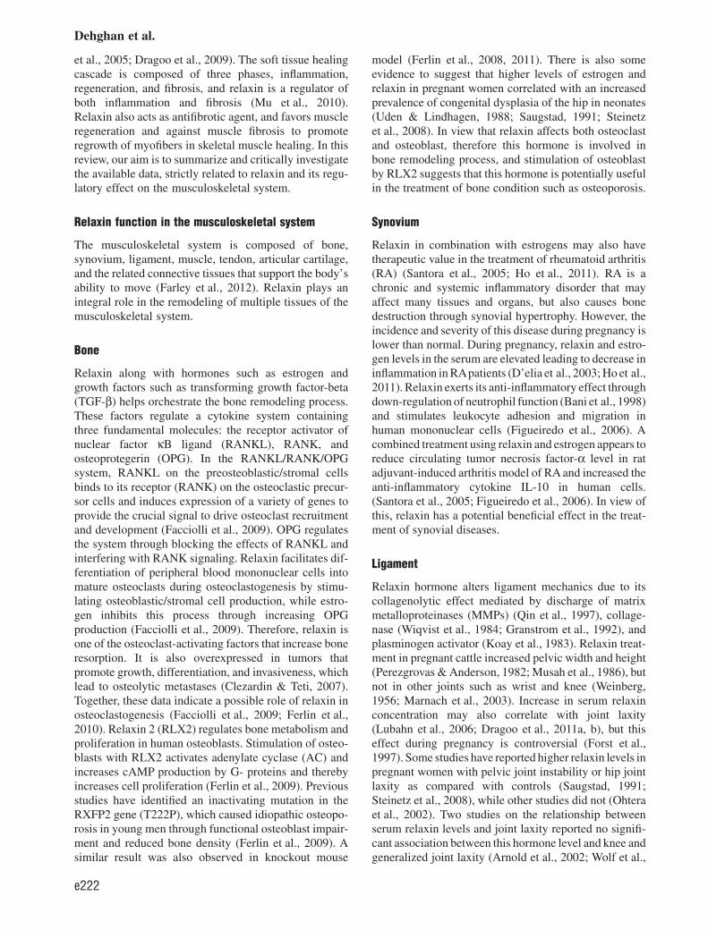
Relaxin is a hormone structurally related to insulin andinsulin-like growth factor, which exerts its regulatoryeffect on the musculoskeletal and other systems throughbinding to its receptor in various tissues, mediated bydifferent signaling pathways. Relaxin alters the proper-ties of cartilage and tendon by activating collagenase.
This hormone is also involved in bone remodeling andhealing of injured ligaments and skeletal muscle. In thisreview, we have summarized the literature on the effect ofrelaxin in musculoskeletal system to provide a broad per-spective for future studies in this field.
Relaxin, the mammalian 6-kDa heterodimeric polypep-tide hormone, is a member of the insulin-like superfam-ily (Hisaw, 1926) and consists of seven peptides of highstructural but low sequence similarity. Relaxin plays anessential role in the biological processes such as metabo-lism, growth, pregnancy, and parturition in differentspecies including humans and rodents. Relaxin circu-lates in these species during pregnancy emanating fromthe corpus luteum (Conrad & Baker, 2013) and placenta(Goh et al., 2013); however, temporal pattern of changeand serum concentrations of this hormone are different.In rodents, circulating relaxin peak concentrations at theend of pregnancy reach 100 ng/mL, two times greaterthan in human (Sherwood, 1994). While relaxin playsimportant role in collagen catabolism of the pubic sym-physis during gestation in lower mammals such as miceand rats (Samuel et al., 1998), the role of this hormoneon pubic symphysis of human is however unknown(Hashem et al., 2006; Wang et al., 2009). Several studieshave highlighted the therapeutic potential of relaxin forectopic pregnancy, male infertility, and heart failure, car-diovascular and musculoskeletal diseases. Currently,
there are seven known relaxin family peptides (RXFP)that are structurally related to insulin which includerelaxin (RLN)1, RLN2, RLN3, and insulin-like peptide(INSL)3, INSL4, INSL5, and INSL6 (Bathgateet al., 2013). RLN1 and RLN2 are strong regulators ofcollagen expression and metabolism in fibroblasts, andare differentially expressed in the corpus luteum,decidua, and endometrium, as well as prostate tissue,while RLN3 is specific to the brain (Sherwood, 2005).Relaxin1 and 2 reconcile the hemodynamic changesoccurring during pregnancy such as cardiac output, renalblood flow, and arterial compliance (Conrad, 2011), aswell as weakening the pelvic ligaments for parturition inspecies such as guinea pigs and mice (Sherwood et al.,1993). RLN3 is a highly conserved neuropeptide invertebrates, and is involved in a wide range ofneuroactivities such as response to stress and cognition,as well as in neurological disease (Smith et al., 2011).
Relaxin binds to RXFP receptors and exerts its actionthrough a ligand-receptor system in multiple pathways.The relaxin receptor is involved in signal transductionbetween extracellular/intracellular domains. Relaxin1–4hormones are ligands for the RXFP1, RXFP2, RXFP3,and RXFP4, respectively (Fig. 1). This family peptidesact on four G-protein-coupled receptors (GPCRs; for-merly LGR7, LGR8, GPCR135, and GPCR142) (Konget al., 2010). RXFP1 and RXFP2 are composed of largeextracellular domains which encompass of leucine-richrepeats. On the other hand, RXFP3 and RXFP4 proteinsare more similar to small peptide ligands (Summers et al.,
Jason L Dragoo and Naguib Salleh have equal contribution in prepara-tion of this manuscript.
This is an open access article under the terms of the Creative CommonsAttribution-NonCommercial-NoDerivs License, which permits use anddistribution in any medium, provided the original work is properly cited,the use is non-commercial and no modifications or adaptations are made.
The copyright line for this article was changed on 10 July 2014 afteroriginal online publication.
Scand J Med Sci Sports 2014: 24: e220–e229doi: 10.1111/sms.12149
© 2013 The Authors. Scandinavian Journal of Medicine & Science
in Sports published by John Wiley & Sons Ltd.
e220

2009). Recently, it has been shown that there is a differ-ence in the ligand binding mode between RXFP1 andRXFP2 (Scott et al., 2012). RXFP1 and RXFP2 exist inuterus, cervix, vagina, brain, and heart of a number ofanimal species. However, production of these proteinsdiffers among tissues of various species. For example,RXFP1 is expressed in rats and mice myometrium(Vodstrcil et al., 2010), whereas in human, this receptor ismainly localized to the endometrium (Campitiello et al.,2011). Moreover, RXFP1 is expressed in the rats and miceheart localized to the atria where it mediates positiveinotropic and chronotropic responses (Piedras-Renteriaet al., 1997), while there is currently no report of thisreceptor binding or function in the human heart.
Evidence also suggests that the functional domains ofRXFP1, the cell type in which it is expressed, and theligand used to activate the receptor all have importantroles in the musculoskeletal system (Fig. 2). Relaxinalters cartilage and tendon stiffness by activating colla-genase (Hashem et al., 2006; Pearson et al., 2011).Relaxin is also involved in bone remodeling process andin healing of injured ligaments and skeletal muscles (Li
RLN2
RLN1
RLN3
RXFP3
RXFP1
RXFP2
INSL3
RFL
Fig. 1. Interaction of RLN1, RLN2, and RLN3 proteins withtheir receptors RXFP1, RXFP2, and RXFP3, respectively, aswell as with insulin-like growth factor (INSL3) and rearrangedL-myc fusion (RFL) in the network (http://www.genecards.org/).
Fig. 2. A summary of relaxin role in the locomotor system.
Musculoskeletal system
e221
et al., 2005; Dragoo et al., 2009). The soft tissue healingcascade is composed of three phases, inflammation,regeneration, and fibrosis, and relaxin is a regulator ofboth inflammation and fibrosis (Mu et al., 2010).Relaxin also acts as antifibrotic agent, and favors muscleregeneration and against muscle fibrosis to promoteregrowth of myofibers in skeletal muscle healing. In thisreview, our aim is to summarize and critically investigatethe available data, strictly related to relaxin and its regu-latory effect on the musculoskeletal system.
Relaxin function in the musculoskeletal system
The musculoskeletal system is composed of bone,synovium, ligament, muscle, tendon, articular cartilage,and the related connective tissues that support the body’sability to move (Farley et al., 2012). Relaxin plays anintegral role in the remodeling of multiple tissues of themusculoskeletal system.
Bone
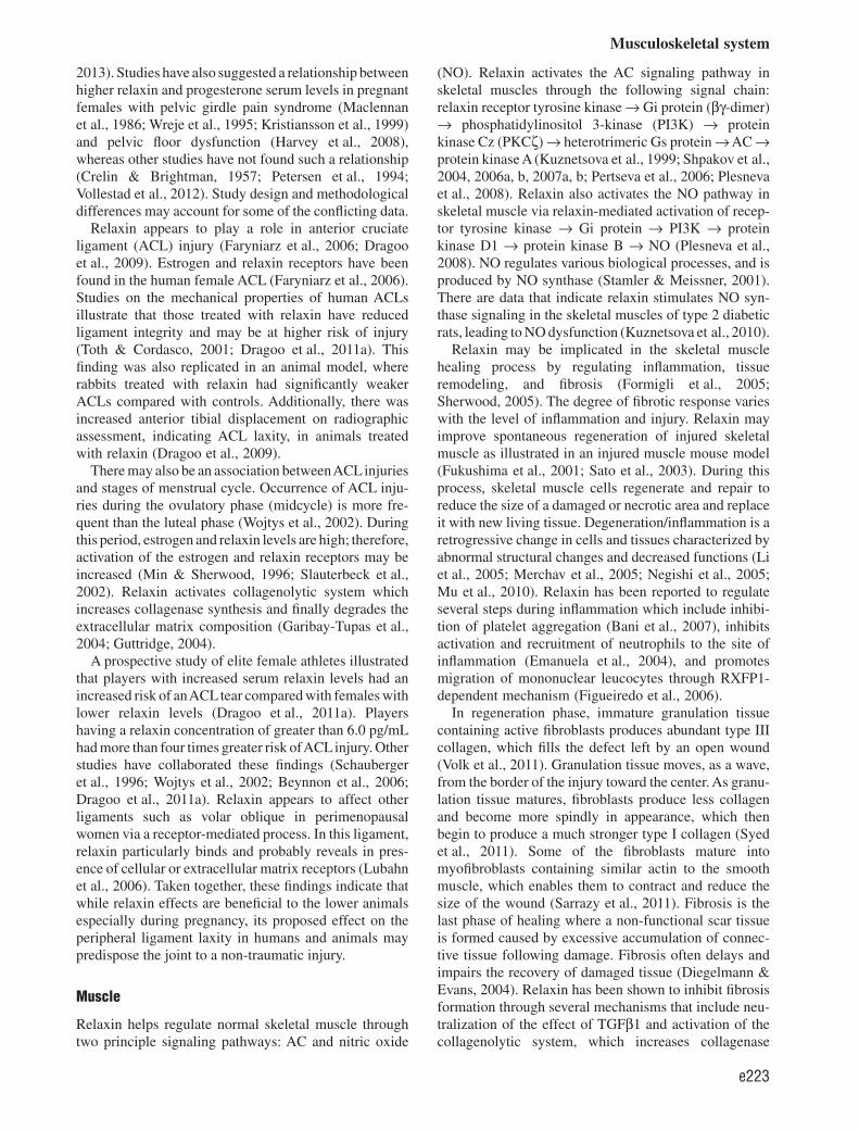
Relaxin along with hormones such as estrogen andgrowth factors such as transforming growth factor-beta(TGF-β) helps orchestrate the bone remodeling process.These factors regulate a cytokine system containingthree fundamental molecules: the receptor activator ofnuclear factor κB ligand (RANKL), RANK, andosteoprotegerin (OPG). In the RANKL/RANK/OPGsystem, RANKL on the preosteoblastic/stromal cellsbinds to its receptor (RANK) on the osteoclastic precur-sor cells and induces expression of a variety of genes toprovide the crucial signal to drive osteoclast recruitmentand development (Facciolli et al., 2009). OPG regulatesthe system through blocking the effects of RANKL andinterfering with RANK signaling. Relaxin facilitates dif-ferentiation of peripheral blood mononuclear cells intomature osteoclasts during osteoclastogenesis by stimu-lating osteoblastic/stromal cell production, while estro-gen inhibits this process through increasing OPGproduction (Facciolli et al., 2009). Therefore, relaxin isone of the osteoclast-activating factors that increase boneresorption. It is also overexpressed in tumors thatpromote growth, differentiation, and invasiveness, whichlead to osteolytic metastases (Clezardin & Teti, 2007).Together, these data indicate a possible role of relaxin inosteoclastogenesis (Facciolli et al., 2009; Ferlin et al.,2010). Relaxin 2 (RLX2) regulates bone metabolism andproliferation in human osteoblasts. Stimulation of osteo-blasts with RLX2 activates adenylate cyclase (AC) andincreases cAMP production by G- proteins and therebyincreases cell proliferation (Ferlin et al., 2009). Previousstudies have identified an inactivating mutation in theRXFP2 gene (T222P), which caused idiopathic osteopo-rosis in young men through functional osteoblast impair-ment and reduced bone density (Ferlin et al., 2009). Asimilar result was also observed in knockout mouse
model (Ferlin et al., 2008, 2011). There is also someevidence to suggest that higher levels of estrogen andrelaxin in pregnant women correlated with an increasedprevalence of congenital dysplasia of the hip in neonates(Uden & Lindhagen, 1988; Saugstad, 1991; Steinetzet al., 2008). In view that relaxin affects both osteoclastand osteoblast, therefore this hormone is involved inbone remodeling process, and stimulation of osteoblastby RLX2 suggests that this hormone is potentially usefulin the treatment of bone condition such as osteoporosis.
Synovium
Relaxin in combination with estrogens may also havetherapeutic value in the treatment of rheumatoid arthritis(RA) (Santora et al., 2005; Ho et al., 2011). RA is achronic and systemic inflammatory disorder that mayaffect many tissues and organs, but also causes bonedestruction through synovial hypertrophy. However, theincidence and severity of this disease during pregnancy islower than normal. During pregnancy, relaxin and estro-gen levels in the serum are elevated leading to decrease ininflammation in RApatients (D’elia et al., 2003; Ho et al.,2011). Relaxin exerts its anti-inflammatory effect throughdown-regulation of neutrophil function (Bani et al., 1998)and stimulates leukocyte adhesion and migration inhuman mononuclear cells (Figueiredo et al., 2006). Acombined treatment using relaxin and estrogen appears toreduce circulating tumor necrosis factor-α level in ratadjuvant-induced arthritis model of RA and increased theanti-inflammatory cytokine IL-10 in human cells.(Santora et al., 2005; Figueiredo et al., 2006). In view ofthis, relaxin has a potential beneficial effect in the treat-ment of synovial diseases.
Ligament
Relaxin hormone alters ligament mechanics due to itscollagenolytic effect mediated by discharge of matrixmetalloproteinases (MMPs) (Qin et al., 1997), collage-nase (Wiqvist et al., 1984; Granstrom et al., 1992), andplasminogen activator (Koay et al., 1983). Relaxin treat-ment in pregnant cattle increased pelvic width and height(Perezgrovas & Anderson, 1982; Musah et al., 1986), butnot in other joints such as wrist and knee (Weinberg,1956; Marnach et al., 2003). Increase in serum relaxinconcentration may also correlate with joint laxity(Lubahn et al., 2006; Dragoo et al., 2011a, b), but thiseffect during pregnancy is controversial (Forst et al.,1997). Some studies have reported higher relaxin levels inpregnant women with pelvic joint instability or hip jointlaxity as compared with controls (Saugstad, 1991;Steinetz et al., 2008), while other studies did not (Ohteraet al., 2002). Two studies on the relationship betweenserum relaxin levels and joint laxity reported no signifi-cant association between this hormone level and knee andgeneralized joint laxity (Arnold et al., 2002; Wolf et al.,
Dehghan et al.
e222
2013). Studies have also suggested a relationship betweenhigher relaxin and progesterone serum levels in pregnantfemales with pelvic girdle pain syndrome (Maclennanet al., 1986; Wreje et al., 1995; Kristiansson et al., 1999)and pelvic floor dysfunction (Harvey et al., 2008),whereas other studies have not found such a relationship(Crelin & Brightman, 1957; Petersen et al., 1994;Vollestad et al., 2012). Study design and methodologicaldifferences may account for some of the conflicting data.
Relaxin appears to play a role in anterior cruciateligament (ACL) injury (Faryniarz et al., 2006; Dragooet al., 2009). Estrogen and relaxin receptors have beenfound in the human female ACL (Faryniarz et al., 2006).Studies on the mechanical properties of human ACLsillustrate that those treated with relaxin have reducedligament integrity and may be at higher risk of injury(Toth & Cordasco, 2001; Dragoo et al., 2011a). Thisfinding was also replicated in an animal model, whererabbits treated with relaxin had significantly weakerACLs compared with controls. Additionally, there wasincreased anterior tibial displacement on radiographicassessment, indicating ACL laxity, in animals treatedwith relaxin (Dragoo et al., 2009).
There may also be an association betweenACLinjuriesand stages of menstrual cycle. Occurrence of ACL inju-ries during the ovulatory phase (midcycle) is more fre-quent than the luteal phase (Wojtys et al., 2002). Duringthis period, estrogen and relaxin levels are high; therefore,activation of the estrogen and relaxin receptors may beincreased (Min & Sherwood, 1996; Slauterbeck et al.,2002). Relaxin activates collagenolytic system whichincreases collagenase synthesis and finally degrades theextracellular matrix composition (Garibay-Tupas et al.,2004; Guttridge, 2004).
A prospective study of elite female athletes illustratedthat players with increased serum relaxin levels had anincreased risk of anACLtear compared with females withlower relaxin levels (Dragoo et al., 2011a). Playershaving a relaxin concentration of greater than 6.0 pg/mLhad more than four times greater risk ofACLinjury. Otherstudies have collaborated these findings (Schaubergeret al., 1996; Wojtys et al., 2002; Beynnon et al., 2006;Dragoo et al., 2011a). Relaxin appears to affect otherligaments such as volar oblique in perimenopausalwomen via a receptor-mediated process. In this ligament,relaxin particularly binds and probably reveals in pres-ence of cellular or extracellular matrix receptors (Lubahnet al., 2006). Taken together, these findings indicate thatwhile relaxin effects are beneficial to the lower animalsespecially during pregnancy, its proposed effect on theperipheral ligament laxity in humans and animals maypredispose the joint to a non-traumatic injury.
Muscle
Relaxin helps regulate normal skeletal muscle throughtwo principle signaling pathways: AC and nitric oxide
(NO). Relaxin activates the AC signaling pathway inskeletal muscles through the following signal chain:relaxin receptor tyrosine kinase → Gi protein (βγ-dimer)→ phosphatidylinositol 3-kinase (PI3K) → proteinkinase Cz (PKCζ) → heterotrimeric Gs protein → AC →protein kinase A (Kuznetsova et al., 1999; Shpakov et al.,2004, 2006a, b, 2007a, b; Pertseva et al., 2006; Plesnevaet al., 2008). Relaxin also activates the NO pathway inskeletal muscle via relaxin-mediated activation of recep-tor tyrosine kinase → Gi protein → PI3K → proteinkinase D1 → protein kinase B → NO (Plesneva et al.,2008). NO regulates various biological processes, and isproduced by NO synthase (Stamler & Meissner, 2001).There are data that indicate relaxin stimulates NO syn-thase signaling in the skeletal muscles of type 2 diabeticrats, leading to NO dysfunction (Kuznetsova et al., 2010).
Relaxin may be implicated in the skeletal musclehealing process by regulating inflammation, tissueremodeling, and fibrosis (Formigli et al., 2005;Sherwood, 2005). The degree of fibrotic response varieswith the level of inflammation and injury. Relaxin mayimprove spontaneous regeneration of injured skeletalmuscle as illustrated in an injured muscle mouse model(Fukushima et al., 2001; Sato et al., 2003). During thisprocess, skeletal muscle cells regenerate and repair toreduce the size of a damaged or necrotic area and replaceit with new living tissue. Degeneration/inflammation is aretrogressive change in cells and tissues characterized byabnormal structural changes and decreased functions (Liet al., 2005; Merchav et al., 2005; Negishi et al., 2005;Mu et al., 2010). Relaxin has been reported to regulateseveral steps during inflammation which include inhibi-tion of platelet aggregation (Bani et al., 2007), inhibitsactivation and recruitment of neutrophils to the site ofinflammation (Emanuela et al., 2004), and promotesmigration of mononuclear leucocytes through RXFP1-dependent mechanism (Figueiredo et al., 2006).
In regeneration phase, immature granulation tissuecontaining active fibroblasts produces abundant type IIIcollagen, which fills the defect left by an open wound(Volk et al., 2011). Granulation tissue moves, as a wave,from the border of the injury toward the center. As granu-lation tissue matures, fibroblasts produce less collagenand become more spindly in appearance, which thenbegin to produce a much stronger type I collagen (Syedet al., 2011). Some of the fibroblasts mature intomyofibroblasts containing similar actin to the smoothmuscle, which enables them to contract and reduce thesize of the wound (Sarrazy et al., 2011). Fibrosis is thelast phase of healing where a non-functional scar tissueis formed caused by excessive accumulation of connec-tive tissue following damage. Fibrosis often delays andimpairs the recovery of damaged tissue (Diegelmann &Evans, 2004). Relaxin has been shown to inhibit fibrosisformation through several mechanisms that include neu-tralization of the effect of TGFβ1 and activation of thecollagenolytic system, which increases collagenase
Musculoskeletal system
e223

synthesis (Garibay-Tupas et al., 2004; Guttridge, 2004;Mendias et al., 2004, 2012; Mu et al., 2010; Vinall et al.,2011). Through these mechanisms, relaxin reduces theformation of fibrous scar tissue (Fig. 3). Relaxin admin-istration to the injured skeletal muscle promotes activa-tion of satellite cells, induces angiogenesis andrevascularization, as well as represses the extendedinflammatory reaction (Mu et al., 2010). Recently,relaxin administration to diabetic wound in mice hasbeen shown to up-regulate the mRNA expression of vas-cular endothelial growth factor, epithelial NO, andstromal-cell-derived factor 1-α, stimulates angiogenesisand vasculogenesis, enhances MMP-11 expression, andincreases wound-breaking strength (Bitto et al., 2013).In view that relaxin plays important role in the healingprocess, it can potentially be used as a therapeutic agentto treat damaged skeletal muscle (Negishi et al., 2005).
Tendon
Relaxin has been reported to effect tendon metabolismby controlling the length of tendon growth (Maclennanet al., 1986; Wood et al., 2003) and reduce tendon stiff-ness by increasing tendon laxity through activation ofcollagenase (Pearson et al., 2011). An in vivo studyinvestigating the growth of rat tails and human patellartendons showed that relaxin levels correlate with tendonlength (Wood et al., 2003; Pearson et al., 2011). Rat tailtendons treated with relaxin exhibited alterations in col-lagen through interfering with fibril association and col-lagen sliding (Wood et al., 2003). Another study inwomen with normal menstrual cycle, who did not takeany contraception pills, demonstrated a significant linkbetween serum relaxin levels and patellar tendon stiff-ness (Pearson et al., 2011). Besides the reported effectsof relaxin on the tendon, potential benefits of relaxin ontendon repair and remodeling are largely unknown.
Cartilage
Relaxin appears to decrease knee articular cartilage stiff-ness (Bonaventure et al., 1988; Hellio Le Graverand
et al., 1998) through induction of collagenase-1,MMP-1, and MMP-3, which reduces collagen contentand expression in fibrocartilaginous cells. Modulation ofMMPs to loss of collagen by hormones may contributeselectively to degeneration of specific joints fibrocarti-laginous explants facilitated by proteinases (Naqvi et al.,2005; Hashem et al., 2006). The degradation of extracel-lular matrix in fibrocartilage is synergized byβ-estradiol. Relaxin exerts its effect through binding toRXFP1 and RXFP2 receptors (Hellio Le Graverandet al., 1998; Wang et al., 2009). The ratio of RXFP2 inknee meniscus of pregnant rabbits was shown to be morethan RXFP1, which may indicate differential role ofthese receptors in the remodeling of fibrocartilage(Hellio Le Graverand et al., 1998; Wang et al., 2009).Comparison of collagen content in articular cartilage ofnonpregnant and pregnant rabbits showed that the totalRNA levels and chondrocyte metabolism decreasedduring pregnancy. Depending on the level of skeletalmaturity, pregnancy can exert both general and specificeffects on the RNA levels in articular cartilage of therabbit knee (Hellio Le Graverand et al., 1998). Thus,relaxin may play a role in women’s susceptibility tomusculoskeletal disease (Naqvi et al., 2005). Takentogether, these findings suggested that in female,increased relaxin level may result in undesirable effectson the articular cartilage.
Perspective
Relaxin plays a vital role in biological processes includ-ing metabolism, growth, and reproduction. Among thefour relaxin types, RLN1 and RLN2 regulate the mus-culoskeletal system via multiple pathways through aligand-receptor system, depending on cell type andligand (Table 1). Our investigation of relaxin’s role inthe musculoskeletal system showed some limitations inthe literature. For example, most of the reports did notdelineate the relaxin isoform or its specific receptor.Additionally, despite relaxin’s accepted role in theregulation of AC and NO pathways, few studies havefocused on the regulatory effect of these pathways in the
Degeneration
RegenerationFibrosis Healing
TGF-β1
Relaxin
Relaxin
TGF-β1
Tissue injury
Fig. 3. A competition exists between fibrosis and regeneration during healing of damage tissue. Relaxin and transforming growthfactor-beta1 (TGF-β1) make imbalance between regeneration and fibrosis process. Increased relaxin and decreased TGF- β1 result inregeneration and consequently healing, while decreased relaxin and increased TGF- β1 lead to fibrosis.
Dehghan et al.
e224
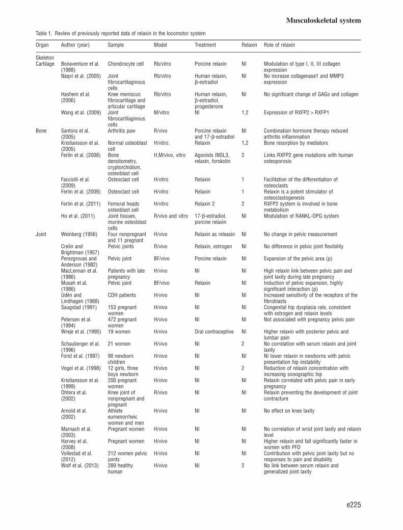
Table 1. Review of previously reported data of relaxin in the locomotor system
Organ Author (year) Sample Model Treatment Relaxin Role of relaxin
SkeletonCartilage Bonaventure et al.
(1988)Chondrocyte cell Rb/vitro Porcine relaxin NI Modulation of type I, II, III collagen
expressionNaqvi et al. (2005) Joint
fibrocartilaginouscells
Rb/vitro Human relaxin,β-estradiol
NI No increase collagenase1 and MMP3expression
Hashem et al.(2006)
Knee meniscusfibrocartilage andarticular cartilage
Rb/vitro Human relaxin,β-estradiol,progesterone
NI No significant change of GAGs and collagen
Wang et al. (2009) Jointfibrocartilaginouscells
M/vitro NI 1,2 Expression of RXFP2 > RXFP1
Bone Santora et al.(2005)
Arthritis paw R/vivo Porcine relaxinand 17-β-estradiol
NI Combination hormone therapy reducedarthritis inflammation
Kristiansson et al.(2005)
Normal osteoblastcell
H/vitro. Relaxin 1,2 Bone resorption by mediators
Ferlin et al. (2008) Bonedensitometry,cryptorchidism,osteoblast cell
H,M/vivo, vitro Agonists INSL3,relaxin, forskolin
2 Links RXFP2 gene mutations with humanosteoporosis
Facciolli et al.(2009)
Osteoclast cell H/vitro Relaxin 1 Facilitation of the differentiation ofosteoclasts
Ferlin et al. (2009) Osteoclast cell H/vitro Relaxin 1 Relaxin is a potent stimulator ofosteoclastogenesis
Ferlin et al. (2011) Femoral headsosteoblast cell
H/vitro Relaxin 2 2 RXFP2 system is involved in bonemetabolism
Ho et al. (2011) Joint tissues,murine osteoblastcells
R/vivo and vitro 17-β-estradiol,porcine relaxin
NI Modulation of RANKL-OPG system
Joint Weinberg (1956) Four nonpregnantand 11 pregnant
H/vivo Relaxin as releasin NI No change in pelvic measurement
Crelin andBrightman (1957)
Pelvic joints R/vivo Relaxin, estrogen NI No difference in pelvic joint flexibility
Perezgrovas andAnderson (1982)
Pelvic joint BF/vivo Porcine relaxin NI Expansion of the pelvic area (p)
MacLennan et al.(1986)
Patients with latepregnancy
H/vivo NI NI High relaxin link between pelvic pain andjoint laxity during late pregnancy
Musah et al.(1986)
Pelvic joint BF/vivo Relaxin NI Induction of pelvic expansion, highlysignificant interaction (p)
Udén andLindhagen (1988)
CDH patients H/vivo NI NI Increased sensitivity of the receptors of thefibroblasts
Saugstad (1991) 153 pregnantwomen
H/vivo NI NI Congenital hip dysplasia rate, consistentwith estrogen and relaxin levels
Petersen et al.(1994)
472 pregnantwomen
H/vivo NI NI Not associated with pregnancy pelvic pain
Wreje et al. (1995) 19 women H/vivo Oral contraceptive NI Higher relaxin with posterior pelvic andlumbar pain
Schauberger et al.(1996)
21 women H/vivo NI 2 No correlation with serum relaxin and jointlaxity
Forst et al. (1997) 90 newbornchildren
H/vivo NI NI NI lower relaxin in newborns with pelvicpresentation hip instability
Vogel et al. (1998) 12 girls, threeboys newborn
H/vivo NI 2 Reduction of relaxin concentration withincreasing sonographic hip
Kristiansson et al.(1999)
200 pregnantwomen
H/vivo NI NI Relaxin correlated with pelvic pain in earlypregnancy
Ohtera et al.(2002)
Knee joint ofnonpregnant andpregnant
R/vivo NI NI Relaxin preventing the development of jointcontracture
Arnold et al.(2002)
Athleteeumenorrheicwomen and men
H/vivo NI NI No effect on knee laxity
Marnach et al.(2003)
Pregnant women H/vivo NI NI No correlation of wrist joint laxity and relaxinlevel
Harvey et al.(2008)
Pregnant women H/vivo NI NI Higher relaxin and fall significantly faster inwomen with PFD
Vollestad et al.(2012)
212 women pelvicjoints
H/vivo NI NI Contribution with pelvic joint laxity but noresponses to pain and disability
Wolf et al. (2013) 289 healthyhuman
H/vivo NI 2 No link between serum relaxin andgeneralized joint laxity
Musculoskeletal system
e225
musculoskeletal organs. Although relaxin may affectmany ligaments and tendons of the musculoskeletalsystem, previous studies have mostly concentrated onthe anterior cruciate and wrist ligaments. Future studiesare warranted to gain a better understanding of relaxin’srole in the musculoskeletal system.
Key words: relaxin, motor organs, skeletal muscle,tendon, ligament.
Acknowledgements
This work is supported by PPP grant (PV104/2012A), Universityof Malaya, Kuala Lumpur, Malaysia.
References
Albert H, Godskesen M, Westergaard JG,Chard T, Gunn L. Circulating levels ofrelaxin are normal in pregnant womenwith pelvic pain. Eur J Obstet Gyn R B1997: 74 (1): 19–22.
Arnold C, Van Bell C, Rogers V, CooneyT. The relationship between serum
relaxin and knee joint laxity in femaleathletes. Orthopedics 2002: 25 (6):669–673.
Bani D, Masini E, Bello MG, Bigazzi M,Sacchi TB. Relaxin protects againstmyocardial injury caused byischemia and reperfusion in rat heart
[Research Support, Non-U.S. Gov’t].Am J Pathol 1998: 152 (5):1367–1376.
Bani D, Nistri S, Cinci L, Giannini L,Princivalle M, Elliott L, Bigazzi M,Masini E. A novel, simple bioactivityassay for relaxin based on inhibition of
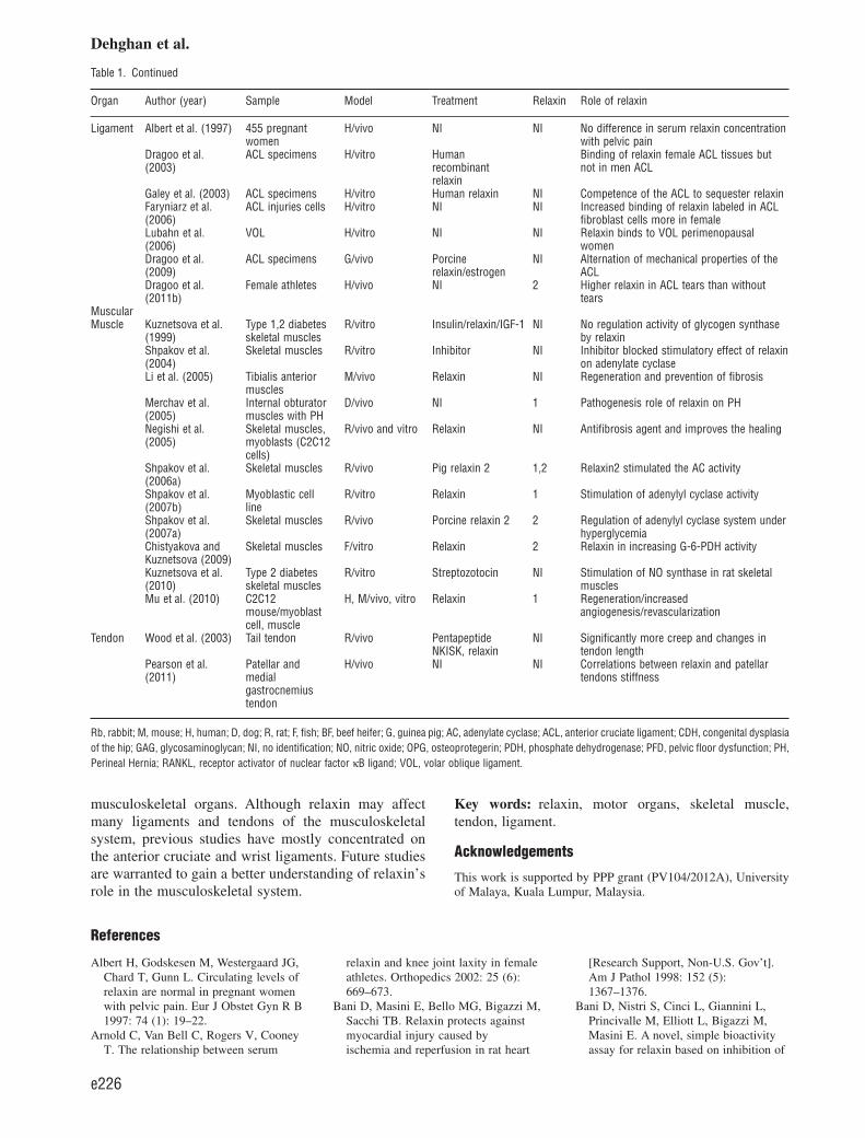
Table 1. Continued
Organ Author (year) Sample Model Treatment Relaxin Role of relaxin
Ligament Albert et al. (1997) 455 pregnantwomen
H/vivo NI NI No difference in serum relaxin concentrationwith pelvic pain
Dragoo et al.(2003)
ACL specimens H/vitro Humanrecombinantrelaxin
Binding of relaxin female ACL tissues butnot in men ACL
Galey et al. (2003) ACL specimens H/vitro Human relaxin NI Competence of the ACL to sequester relaxinFaryniarz et al.(2006)
ACL injuries cells H/vitro NI NI Increased binding of relaxin labeled in ACLfibroblast cells more in female
Lubahn et al.(2006)
VOL H/vitro NI NI Relaxin binds to VOL perimenopausalwomen
Dragoo et al.(2009)
ACL specimens G/vivo Porcinerelaxin/estrogen
NI Alternation of mechanical properties of theACL
Dragoo et al.(2011b)
Female athletes H/vivo NI 2 Higher relaxin in ACL tears than withouttears
MuscularMuscle Kuznetsova et al.
(1999)Type 1,2 diabetesskeletal muscles
R/vitro Insulin/relaxin/IGF-1 NI No regulation activity of glycogen synthaseby relaxin
Shpakov et al.(2004)
Skeletal muscles R/vitro Inhibitor NI Inhibitor blocked stimulatory effect of relaxinon adenylate cyclase
Li et al. (2005) Tibialis anteriormuscles
M/vivo Relaxin NI Regeneration and prevention of fibrosis
Merchav et al.(2005)
Internal obturatormuscles with PH
D/vivo NI 1 Pathogenesis role of relaxin on PH
Negishi et al.(2005)
Skeletal muscles,myoblasts (C2C12cells)
R/vivo and vitro Relaxin NI Antifibrosis agent and improves the healing
Shpakov et al.(2006a)
Skeletal muscles R/vivo Pig relaxin 2 1,2 Relaxin2 stimulated the AC activity
Shpakov et al.(2007b)
Myoblastic cellline
R/vitro Relaxin 1 Stimulation of adenylyl cyclase activity
Shpakov et al.(2007a)
Skeletal muscles R/vivo Porcine relaxin 2 2 Regulation of adenylyl cyclase system underhyperglycemia
Chistyakova andKuznetsova (2009)
Skeletal muscles F/vitro Relaxin 2 Relaxin in increasing G-6-PDH activity
Kuznetsova et al.(2010)
Type 2 diabetesskeletal muscles
R/vitro Streptozotocin NI Stimulation of NO synthase in rat skeletalmuscles
Mu et al. (2010) C2C12mouse/myoblastcell, muscle
H, M/vivo, vitro Relaxin 1 Regeneration/increasedangiogenesis/revascularization
Tendon Wood et al. (2003) Tail tendon R/vivo PentapeptideNKISK, relaxin
NI Significantly more creep and changes intendon length
Pearson et al.(2011)
Patellar andmedialgastrocnemiustendon
H/vivo NI NI Correlations between relaxin and patellartendons stiffness
Rb, rabbit; M, mouse; H, human; D, dog; R, rat; F, fish; BF, beef heifer; G, guinea pig; AC, adenylate cyclase; ACL, anterior cruciate ligament; CDH, congenital dysplasiaof the hip; GAG, glycosaminoglycan; NI, no identification; NO, nitric oxide; OPG, osteoprotegerin; PDH, phosphate dehydrogenase; PFD, pelvic floor dysfunction; PH,Perineal Hernia; RANKL, receptor activator of nuclear factor κB ligand; VOL, volar oblique ligament.
Dehghan et al.
e226

platelet aggregation. Regul Pept 2007:144 (1–3): 10–16.
Bathgate RAD, Halls ML,Van Der Westhuizen ET, Callander GE,Kocan M, Summers RJ. Relaxin familypeptides and their receptors. PhysiolRev 2013: 93 (1): 405–480.
Beynnon BD, Johnson RJ, Braun S,Sargent M, Bernstein IM, Skelly JM.The relationship between menstrualcycle phase and anterior cruciateligament injury. Am J Sports Med2006: 34 (5): 757–764.
Bitto A, Irrera N, Minutoli L, Calò M,Lo Cascio P, Caccia P, Pizzino G,Pallio G, Micali A, Vaccaro M, SaittaA, Squadrito F, Altavilla D. Relaxinimproves multiple markers of woundhealing and ameliorates the disturbedhealing pattern of genetically diabeticmice. Clin Sci (Lond) 2013: 125 (12):575–585.
Bonaventure J, Delatour B, Tsagris L,Eddie LW, Tregear G, Corvol MT.Effect of relaxin on the phenotype ofcollagens synthesized by cultured rabbitchondrocytes. Biochim Biophys Acta1988: 972 (2): 209–220.
Campitiello MR, De Franciscis P, MeleD, Izzo G, Sinisi A, Delrio G,Colacurci N. Endometrial LGR7expression during menstrual cycle.Fertil Steril 2011: 95 (8):2511–2514.
Chistyakova OV, Kuznetsova LA. Effectof peptides of the insulin superfamilyon glucose-6-phosphate dehydrogenaseactivity in skeletal muscles of riverlamprey (Lampetra fluviatilis) duringprespawning starvation. B Exp BiolMed+ 2009: 148 (1): 29–32.
Clezardin P, Teti A. Bone metastasis:pathogenesis and therapeuticimplications [Research Support,Non-U.S. Gov’t Review]. ClinExp Metastasis 2007: 24 (8):599–608.
Conrad KP. Maternal vasodilation inpregnancy: the emerging role ofrelaxin. Am J Physiol Regul IntegrComp Physiol 2011: 301 (2):R267–R275.
Conrad KP, Baker VL. Corpus lutealcontribution to maternal pregnancyphysiology and outcomes in assistedreproductive technologies. Am JPhysiol Regul Integr Comp Physiol2013: 304 (2): R69–R72.
Crelin ES, Brightman MW. The pelvis ofthe rat: its response to estrogen andrelaxin. Anat Rec 1957: 128 (3):467–483.
D’elia HF, Mattsson LA, Ohlsson C,Nordborg E, Carlsten H. Hormonereplacement therapy in rheumatoidarthritis is associated with lower serumlevels of soluble IL-6 receptor andhigher insulin-like growth factor 1.
Arthritis Res Ther 2003: 5 (4):R202–R209.
Diegelmann RF, Evans MC. Woundhealing: an overview of acute, fibroticand delayed healing. Front Biosci 2004:1 (9): 283–289.
Dragoo JL, Castillo TN, Braun HJ, RidleyBA, Kennedy AC, Golish SR.Prospective correlation between serumrelaxin concentration and anteriorcruciate ligament tears among elitecollegiate female athletes. Am J SportsMed 2011a: 39 (10): 2175–2180.
Dragoo JL, Castillo TN, Korotkova TA,Kennedy AC, Kim HJ, Stewart DR.Trends in serum relaxin concentrationamong elite collegiate female athletes.Int J Womens Health 2011b: 3:19–24.
Dragoo JL, Lee RS, Benhaim P, FinermanGA, Hame SL. Relaxin receptors in thehuman female anterior cruciateligament. Am J Sport Med 2003: 31(4): 577–584.
Dragoo JL, Padrez K, Workman R,Lindsey DP. The effect of relaxin onthe female anterior cruciate ligament:analysis of mechanical properties in ananimal model. Knee 2009: 16 (1):69–72.
Emanuela M, Nistri S, Vannacci A,Sacchi TB, Novelli A, Bani D. Relaxininhibits the activation of humanneutrophils: involvement of the nitricoxide pathway. Endocrinology 2004:145 (3): 1106–1112.
Facciolli A, Ferlin A, Gianesello L, PepeA, Foresta C. Role of relaxin in humanosteoclastogenesis. Ann N Y Acad Sci2009: 1160: 221–225.
Farley A, McLafferty E, Hendry C. Theanatomy and physiology of thelocomotor system. Nurs Stand 2012: 27(7): 35–43.
Faryniarz DA, Bhargava M, Lajam C,Attia ET, Hannafin JA. Quantitation ofestrogen receptors and relaxin bindingin human anterior cruciate ligamentfibroblasts [Comparative StudyResearch Support, Non-U.S. Gov’t]. InVitro Cell Dev Biol Anim 2006: 42 (7):176–181.
Ferlin A, Pepe A, Facciolli A, GianeselloL, Foresta C. Relaxin stimulatesosteoclast differentiation and activation.Bone 2010: 46 (2): 504–513.
Ferlin A, Pepe A, Gianesello L, GarollaA, Feng S, Facciolli A, Morello R,Agoulnik AI, Foresta C. New roles forINSL3 in adults [In Vitro]. Ann N YAcad Sci 2009: 1160: 215–218.
Ferlin A, Pepe A, Gianesello L, GarollaA, Feng S, Giannini S, Zaccolo M,Facciolli A, Morello R, Agoulnik AI,Foresta C. Mutations in the insulin-likefactor 3 receptor are associated withosteoporosis [Research Support, N.I.H.,Extramural Research Support,
Non-U.S. Gov’t]. J Bone Miner Res2008: 23 (5): 683–693.
Ferlin A, Perilli L, Gianesello L,Taglialavoro G, Foresta C. Profilinginsulin like factor 3 (INSL3) signalingin human osteoblasts. PLoS ONE 2011:6 (12): e29733.
Figueiredo KA, Mui AL, Nelson CC, CoxME. Relaxin stimulates leukocyteadhesion and migration through arelaxin receptor LGR7-dependentmechanism. J Biol Chem 2006: 281(6): 3030–3039.
Formigli L, Francini F, Chiappini L,Zecchi-Orlandini S, Bani D. Relaxinfavors the morphofunctional integrationbetween skeletal myoblasts and adultcardiomyocytes in coculture. Ann N YAcad Sci 2005: 1041: 444–445.
Forst J, Forst C, Forst R, Heller KD.Pathogenetic relevance of thepregnancy hormone relaxin to inbornhip instability. [Clinical Trial]. ArchOrthop Trauma Surg 1997: 116 (4):209–212.
Fukushima K, Badlani N, Usas A, RianoF, Fu FH, Huard J. The use of anantifibrosis agent to improve musclerecovery after laceration. Am J SportsMed 2001: 29 (4): 394–402.
Galey S, Konieczko EM, Arnold CA,Cooney TE. Immunohistologicaldetection of relaxin binding to anteriorcruciate ligaments. Orthopedics 2003:26 (12): 1201–1204.
Garibay-Tupas JL, Okazaki KJ, TashimaLS, Yamamoto S, Bryant-GreenwoodGD. Regulation of the human relaxingenes H1 and H2 by steroid hormones.Mol Cell Endocrinol 2004: 219 (1–2):115–125.
Goh W, Yamamoto SY, Thompson KS,Bryant-Greenwood GD. Relaxin, itsreceptor (RXFP1), and insulin-likepeptide 4 expression through gestationand in placenta accreta. Reprod Sci2013: 20 (8): 968–980.
Granstrom LM, Ekman GE, MalmstromA, Ulmsten U, Woessner JF Jr. Serumcollagenase levels in relation to thestate of the human cervix duringpregnancy and labor [Research Support,Non-U.S. Gov’t Research Support, U.S.Gov’t, P.H.S.]. Am J Obstet Gynecol1992: 167 (5): 1284–1288.
Guttridge DC. Signaling pathways weighin on decisions to make or breakskeletal muscle [Review]. Curr OpinClin Nutr Metab Care 2004: 7 (4):443–450.
Harvey MA, Johnston SL, Davies GA.Mid-trimester serum relaxinconcentrations and post-partum pelvicfloor dysfunction [Research Support,Non-U.S. Gov’t]. Acta Obstet GynecolScand 2008: 87 (12): 1315–1321.
Hashem G, Zhang Q, Hayami T, Chen J,Wang W, Kapila S. Relaxin and
Musculoskeletal system
e227

beta-estradiol modulate targeted matrixdegradation in specific synovial jointfibrocartilages: progesterone preventsmatrix loss [Research Support, N.I.H.,Extramural Research Support,Non-U.S. Gov’t]. Arthritis Res Ther2006: 8 (4): R98. doi: 10.1186/ar1978.
Hellio Le Graverand MP, Reno C, HartDA. Influence of pregnancy on geneexpression in rabbit articular cartilage[Research Support, Non-U.S. Gov’t].Osteoarthritis Cartilage 1998: 6 (5):341–350.
Hisaw FL. Experimental relaxation of thepubic ligament of guinea pig. Proc SocExp Biol Med 1926: 23: 661–663.
Ho TY, Santora K, Chen JC, FrankshunAL, Bagnell CA. Effects of relaxin andestrogens on bone remodeling markers,receptor activator of NF-kB ligand(RANKL) and osteoprotegerin (OPG),in rat adjuvant-induced arthritis[Research Support, Non-U.S. Gov’t].Bone 2011: 48 (6): 1346–1353.
Koay ES, Too CK, Greenwood FC,Bryant-Greenwood GD. Relaxinstimulates collagenase and plasminogenactivator secretion by dispersed humanamnion and chorion cells in vitro[Comparative Study]. JCEM 1983: 56(6): 1332–1334.
Kong RCK, Shilling PJ, Lobb DK,Gooley PR, Bathgate RAD. Membranereceptors: structure and function of therelaxin family peptide receptors. MolCell Endocrinol 2010: 320 (1–2):1–15.
Kristiansson P, Holding C, Hughes S,Haynes D. Does human relaxin-2 affectperipheral blood mononuclear cells toincrease inflammatory mediators inpathologic bone loss? Ann Ny Acad Sci2005: 1041 (1): 317–319.
Kristiansson P, Svardsudd K,Von Schoultz B. Reproductivehormones and aminoterminalpropeptide of type III procollagen inserum as early markers of pelvic painduring late pregnancy. Am J ObstetGynecol 1999: 180 (1 Pt 1):128–134.
Kuznetsova L, Plesneva S, Derjabina N,Omeljaniuk E, Pertseva M. On themechanism of relaxin action: theinvolvement of adenylyl cyclasesignalling system. Regul Pept 1999: 80(1-2): 33–39.
Kuznetsova LA, Chistyakova OV,Bondareva VM, Sharova TS, PertsevaMN. Disturbance of regulation of NOsynthase activity by peptides of insulinfamily in rat skeletal muscles instreptozotocin model of neonatal type 2diabetes mellitus. Dokl BiochemBiophys 2010: 432 (1): 123–125.
Li Y, Negishi S, Sakamoto M, Usas A,Huard J. The use of relaxin improveshealing in injured muscle [Research
Support, N.I.H., Extramural ResearchSupport, Non-U.S. Gov’t ResearchSupport, U.S. Gov’t, P.H.S.]. Ann N YAcad Sci 2005: 1041: 395–397.
Lubahn J, Ivance D, Konieczko E,Cooney T. Immunohistochemicaldetection of relaxin binding to the volaroblique ligament [Research Support,Non-U.S. Gov’t]. J Hand Surg [Am]2006: 31 (1): 80–84.
Maclennan AH, Nicolson R, Green RC,Bath M. Serum relaxin and pelvic painof pregnancy [Research Support,Non-U.S. Gov’t]. Lancet 1986: 2(8501): 243–245.
Marnach ML, Ramin KD, Ramsey PS,Song SW, Stensland JJ, An KN.Characterization of the relationshipbetween joint laxity and maternalhormones in pregnancy. ObstetGynecol 2003: 101 (2): 331–335.
Mendias CL, Gumucio JP, Davis ME,Bromley CW, Davis CS, Brooks SV.Transforming growth factor-betainduces skeletal muscle atrophy andfibrosis through the induction ofatrogin-1 and scleraxis. Muscle Nerve2012: 45 (1): 55–59.
Mendias CL, Tatsumi R, Allen RE. Roleof cyclooxygenase-1 and -2 in satellitecell proliferation, differentiation, andfusion. Muscle Nerve 2004: 30 (4):497–500.
Merchav R, Feuermann Y, Shamay A,Ranen E, Stein U, Johnston DE, ShaharR. Expression of relaxin receptorLRG7, canine relaxin, and relaxin-likefactor in the pelvic diaphragmmusculature of dogs with and withoutperineal hernia. Vet Surg 2005: 34 (5):476–481.
Min G, Sherwood OD. Identification ofspecific relaxin-binding cells in thecervix, mammary glands, nipples, smallintestine, and skin of pregnant pigs[Research Support, Non-U.S. Gov’t].Biol Reprod 1996: 55 (6):1243–1252.
Mu X, Urso ML, Murray K, Fu F, Li Y.Relaxin regulates MMP expression andpromotes satellite cell mobilizationduring muscle healing in both youngand aged mice. Am J Pathol 2010: 177(5): 2399–2410.
Musah AI, Schwabe C, Willham RL,Anderson LL. Pelvic development asaffected by relaxin in three geneticallyselected frame sizes of beef heifers.Biol Reprod 1986: 34: 363–369.
Naqvi T, Duong TT, Hashem G, Shiga M,Zhang Q, Kapila S. Relaxin’s inductionof metalloproteinases is associated withthe loss of collagen andglycosaminoglycans in synovial jointfibrocartilaginous explants. ArthritisRes Ther 2005: 7 (1): 1–11.
Negishi S, Li Y, Usas A, Fu FH, Huard J.The effect of relaxin treatment on
skeletal muscle injuries. Am J SportsMed 2005: 33 (12): 1816–1824.
Ohtera K, Zobitz ME, Luo ZP, MorreyBF, O’driscoll SW, Ramin KD, AnKN. Effect of pregnancy on jointcontracture in the rat knee [ResearchSupport, Non-U.S. Gov’t]. J ApplPhysiol 2002: 92 (4): 1494–1498.
Pearson SJ, Burgess KE, Onamb’El’EGL. Serum relaxin levels affect the invivo properties of some but not alltendons in normally menstruatingyoung women. Exp Physiol 2011: 96(7): 681–688.
Perezgrovas R, Anderson LL. Effect ofporcine relaxin on cervical dilatation,pelvic area and parturition in beefheifers. Biol Reprod 1982: 26 (4):765–776.
Pertseva M, Shpakov A, Kuznetsova L,Plesneva S, Omeljaniuk E. Adenylylcyclase signaling mechanisms ofrelaxin and insulin action: similaritiesand differences. Cell Biol Int 2006: 30(6): 533–540.
Petersen LK, Hvidman L, Uldbjerg N.Normal serum relaxin in women withdisabling pelvic pain during pregnancy.Gynecol Obstet Invest 1994: 38 (1):21–23.
Piedras-Renteria ES, Sherwood OD, BestPM. Effects of relaxin on rat atrialmyocytes. I. Inhibition of I(to) viaPKA-dependent phosphorylation. Am JPhysiol Heart Circ Physiol 1997: 272(4): H1791–H1797.
Plesneva SA, Kuznetsova LA, ShpakovAO, Sharova TS, Pertseva MN.Structural and functional organizationof the adenylyl cyclase signalingmechanism of insulin-like growthfactor 1 revealed in the muscle tissue invertebrates and invertebrates. Zh EvolBiokhim Fiziol 2008: 44 (5): 459–466.
Qin X, Garibay-Tupas J, Chua PK,Cachola L, Bryant-Greenwood GD. Anautocrine/paracrine role of humandecidual relaxin. I. Interstitialcollagenase (matrixmetalloproteinase-1) and tissueplasminogen activator [ResearchSupport, Non-U.S. Gov’t ResearchSupport, U.S. Gov’t, P.H.S.]. BiolReprod 1997: 56 (4): 800–811.
Samuel CS, Coghlan JP, Bateman JF.Effects of relaxin, pregnancy andparturition on collagen metabolism inthe rat pubic symphysis. J Endocrinol1998: 159 (1): 117–125.
Santora K, Rasa C, Visco D, Steinetz B,Bagnell C. Effects of relaxin in amodel of rat adjuvant-induced arthritis[Research Support, Non-U.S. Gov’t].Ann N Y Acad Sci 2005: 1041:481–485.
Sarrazy V, Billet F, Micallef L, CoulombB, Desmoulière A. Mechanisms ofpathological scarring: role of
Dehghan et al.
e228
myofibroblasts and currentdevelopments. Wound Repair Regen2011: 19: s10–s15.
Sato K, Li Y, Foster W, Fukushima K,Badlani N, Adachi N, Usas A, Fu FH,Huard J. Improvement of musclehealing through enhancement of muscleregeneration and prevention of fibrosis.Muscle Nerve 2003: 28 (3): 365–372.
Saugstad LF. Persistent pelvic pain andpelvic joint instability. Eur J ObstetGynecol Reprod Biol 1991: 41 (3):197–201.
Schauberger CW, Rooney BL, GoldsmithL, Shenton D, Silva PD, Schaper A.Peripheral joint laxity increases inpregnancy but does not correlate withserum relaxin levels. Am J ObstetGynecol 1996: 174 (2): 667–671.
Scott DJ, Rosengren KJ, Bathgate RAD.The different ligand-binding modes ofrelaxin family peptide receptors RXFP1and RXFP2. Mol Endocrinol 2012: 26(11): 1896–1906.
Sherwood OD. Relaxin. In: Knobil E,Neill J, eds. The physiology ofreproduction. New York: Raven, 1994:2: 861–1009.
Sherwood OD, ed. Relaxin and relatedpeptides. New York: Springer Science,2005: 1041.
Sherwood OD, Downing SJ, Guico-LammML, Hwang JJ, O’day-Bowman MB,Fields PA. The physiological effects ofrelaxin during pregnancy: studies inrats and pigs. Oxf Rev Reprod Biol1993: 15: 143–189.
Shpakov AO, Gur’ianov IA, KuznetsovaLA, Plesneva SA, Shpakova EA,Vlasov GP, Pertsev MN. Study ofmolecular mechanisms of the relaxinaction on adenylyl cyclase signalingsystem using synthetic peptides derivedfrom the relaxin receptor LGR7.Neurosci Behav Physiol 2006a: 92 (5):521–535.
Shpakov AO, Gur’yanov IA, KuznetsovaLA, Plesneva SA, Shpakova EA,Vlasov GP, Pertseva MN. Peptidederivatives of the LGR7 relaxinreceptor control the functional activityof relaxin-sensitive adenylate cyclase[Research Support, Non-U.S. Gov’t].Dokl Biochem Biophys 2006b: 407:109–112.
Shpakov AO, Kuznetsova LA, PlesnevaSA, Bondareva VM, Pertseva MN.Functional coupling of hormonereceptors with G proteins in theadenylate cyclase system of the ratmuscle tissues and brain underconditions of short-term hyperglycemia[In Vitro Research Support, Non-U.S.Gov’t]. Bull Exp Biol Med 2007a: 144(5): 684–688.
Shpakov AO, Kuznetsova LA, PlesnevaSA, Pertseva MN. Role of
phosphatidylinositol-3-kinase andprotein kinase Cζ in the adenylatecyclase signal mechanism of action ofrelaxin in muscle tissues of rats andmollusks. Bull Exp Biol Med 2004:138 (4): 372–375.
Shpakov AO, Kuznetsova LA, PlesnevaSA, Pertseva MN. Reactivity of theadenylyl cyclase system in rat tissues tobiogenic amines and peptide hormonesunder starvation condition. Ross FiziolZh Im I M Sechenova 2007b: 93 (4):345–356.
Slauterbeck JR, Fuzie SF, Smith MP,Clark RJ, Xu K, Starch DW, HardyDM. The menstrual cycle, sexhormones, and anterior cruciateligament injury. J Athl Train 2002: 37(3): 275–278.
Smith CM, Ryan PJ, Hosken IT, Ma S,Gundlach AL. Relaxin-3 systems in thebrain – the first 10 years [ResearchSupport, Non-U.S. Gov’t Review]. JChem Neuroanat 2011: 42 (4):262–275.
Stamler JS, Meissner G. Physiology ofnitric oxide in skeletal muscle[Research Support, U.S. Gov’t, P.H.S.Review]. Physiol Rev 2001: 81 (1):209–237.
Steinetz BG, Williams AJ, Lust G,Schwabe C, Bullesbach EE, GoldsmithLT. Transmission of relaxin andestrogens to suckling canine pups viamilk and possible association with hipjoint laxity. Am J Vet Res 2008: 69 (1):59–67.
Summers RJ, Bathgate RA, Wade JD,Van Der Westhuizen ET, Halls ML.Roles of the receptor, the ligand, andthe cell in the signal transductionpathways utilized by the relaxin familypeptide receptors 1-3 [ResearchSupport, Non-U.S. Gov’t]. Ann N YAcad Sci 2009: 1160: 99–104.
Syed F, Ahmadi E, Iqbal SA, Singh S,McGrouther DA, Bayat A. Fibroblastsfrom the growing margin of keloidscars produce higher levels of collagenI and III compared with intralesionaland extralesional sites: clinicalimplications for lesional site-directedtherapy. Br J Dermatol 2011: 164 (1):83–96.
Toth AP, Cordasco FA. Anterior cruciateligament injuries in the female athlete[Review]. J Gend Specif Med 2001: 4(4): 25–34.
Uden A, Lindhagen T. Inguinal hernia inpatients with congenital dislocation ofthe hip. A sign of general connectivetissue disorder [Research Support,Non-U.S. Gov’t]. Acta Orthop Scand1988: 59 (6): 667–668.
Vinall LR, Mahaffey MC, Davis RR, LuoZP, Regina GE, Ghosh MP, TepperGC, Vere White W, De R. Dual
blockade of PKA and NF–κB inhibitsH2 relaxin-mediated castrate-resistantgrowth of prostate cancer sublines andinduces apoptosis. Horm Cancer 2011:2 (4): 224–238.
Vodstrcil LA, Shynlova O, Verlander JW,Wlodek ME, Parry LJ. Decreasedexpression of the rat myometrialrelaxin receptor (RXFP1) in latepregnancy is partially mediated by thepresence of the conceptus. Biol Reprod2010: 83 (5): 818–824.
Vogel I, Andersson JE, Uldbjerg N.Serum relaxin in the newborn is not amarker of neonatal hip instability. JPediatr Orthoped 1998: 18 (4):535–537.
Volk SW, Wang Y, Mauldin EA, LiechtyKW, Adams SL. Diminished type IIIcollagen promotes myofibroblastdifferentiation and increases scardeposition in cutaneous wound healing.Cells Tissues Organs 2011: 194 (1):25–37.
Vollestad NK, Torjesen PA, Robinson HS.Association between the serum levelsof relaxin and responses to the activestraight leg raise test in pregnancy.Man Ther 2012: 17 (3): 225–230.
Wang W, Hayami T, Kapila S. Femalehormone receptors are differentiallyexpressed in mouse fibrocartilages[Research Support, N.I.H., Extramural].Osteoarthritis Cartilage 2009: 17 (5):646–654.
Weinberg A. An x-ray pelvimetric studyof relaxin extract in pelvic expansion.Surg Gynecol Obstet 1956: 103 (3):303–306.
Wiqvist I, Norstrom A, O’byrne E,Wiqvist N. Regulatory influence ofrelaxin on human cervical and uterineconnective tissue. Acta Endocrinol(Copenh) 1984: 106 (1): 127–132.
Wojtys EM, Huston LJ, Boynton MD,Spindler KP, Lindenfeld TN. The effectof the menstrual cycle on anteriorcruciate ligament injuries in women asdetermined by hormone levels. Am JSports Med 2002: 30 (2): 182–188.
Wolf JM, Williams AE, Delaronde S,Leger R, Clifton KB, King KB.Relationship of serum relaxin togeneralized and trapezial-metacarpaljoint laxity. J Hand Surg [Am] 2013:38 (4): 721–728.
Wood ML, Luthin WN, Lester GE,Dahners LE. Tendon creep ispotentiated by NKISK and relaxinwhich produce collagen fiber sliding.Iowa Orthop J 2003: 23: 75–79.
Wreje U, Kristiansson P, Aberg H,Bystrom B, Von Schoultz B. Serumlevels of relaxin during the menstrualcycle and oral contraceptive use.Gynecol Obstet Invest 1995: 39 (3):197–200.
Musculoskeletal system
e229
Copyright © 2022 FDOKUMEN