pathology of Musculoskeletal System and Skin
58
Department of pathology Faculty of Veterinary Medicine Cairo University Dr Sahar Samir Mahmoud Special Veterinary Pathology Course (401)
Transcript of pathology of Musculoskeletal System and Skin
Department of pathology
Faculty of Veterinary Medicine
Cairo University
Dr Sahar Samir Mahmoud
Special Veterinary Pathology
Course (401)
Musculoskeletal
System
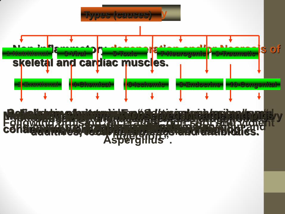
Myopathy
Non inflammatory degeneration and/or Necrosis of
skeletal and cardiac muscles. 1-Nutritional
2-Exertional
3-Viral 7-Neurogenic
6-Ischemic
5-Toxic
4-Chemical
Types (causes)
Deficiency of vitamin E and / or selenium in young
calves and foals. Muscular inactivity during periods of rest after heavy
continuous work “usually a Monday morning”.
In new born calves, sheep and goats “Akabane
and Foot & mouth disease virus infection”. Caused by chemical agents as drugs,food
additives, local anesthetics and antibiotics.
Caused by plant toxines “Solanum malacoxylon “
and mycotoxines “Tremorgenes in Penicillin and
Aspergillus”.
In cattle following recumbency for long period.
following traumatic injury and severance of the
nerve supplying the muscles.
8-Endocrine
9-Traumatic
10-Congenital
Following hyper adrenocortisism in animals and
hypothyroidism in dogs. Following bruising, laceration, gun shot and violent
injection.
Observed in cattle and pigs
Grossly
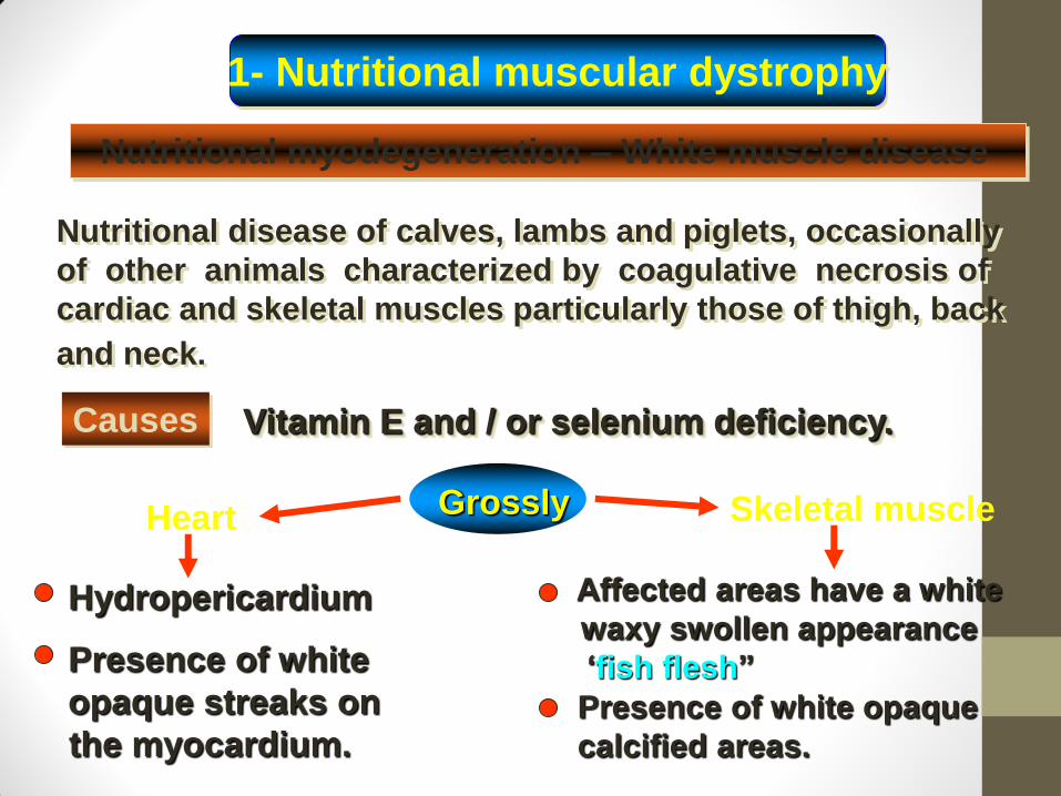
1- Nutritional muscular dystrophy
Nutritional myodegeneration – White muscle disease
Nutritional disease of calves, lambs and piglets, occasionally
of other animals characterized by coagulative necrosis of
cardiac and skeletal muscles particularly those of thigh, back
and neck.
Causes Vitamin E and / or selenium deficiency.
Heart Skeletal muscle
Hydropericardium
Presence of white
opaque streaks on
the myocardium.
Affected areas have a white
waxy swollen appearance
‘fish flesh”
Presence of white opaque
calcified areas.
Grossly
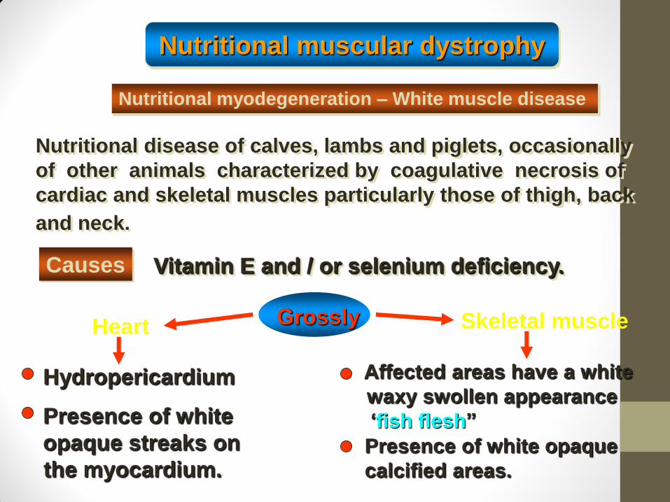
Nutritional muscular dystrophy
Nutritional myodegeneration – White muscle disease
Nutritional disease of calves, lambs and piglets, occasionally
of other animals characterized by coagulative necrosis of
cardiac and skeletal muscles particularly those of thigh, back
and neck.
Causes Vitamin E and / or selenium deficiency.
Heart Skeletal muscle
Hydropericardium
Presence of white
opaque streaks on
the myocardium.
Affected areas have a white
waxy swollen appearance
‘fish flesh”
Presence of white opaque
calcified areas.
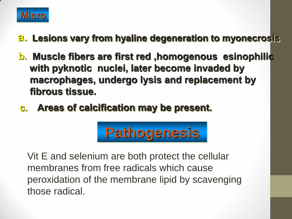
Lesions vary from hyaline degeneration to myonecrosis
c.
Pathogenesis
Areas of calcification may be present.
Micro
a.
Muscle fibers are first red ,homogenous esinophilic
with pyknotic nuclei, later become invaded by
macrophages, undergo lysis and replacement by
fibrous tissue.
b.
Vit E and selenium are both protect the cellular
membranes from free radicals which cause
peroxidation of the membrane lipid by scavenging
those radical.
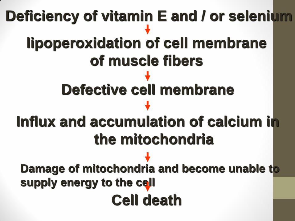
lipoperoxidation of cell membrane
of muscle fibers
Defective cell membrane
Influx and accumulation of calcium in
the mitochondria
Damage of mitochondria and become unable to
supply energy to the cell
Cell death
Deficiency of vitamin E and / or selenium

A disease of horses characterized by coagulative
necrosis of heavy muscles of shoulder and thighs.
Occurs in horses as equine paralytic myoglubinurea“azoturea “
and in sheep chased by dogs and in cattle after running wildly.
2- Exertional or post- exercise myopathy
Equine rhabdomyolysis
Azoturea, Monday morning disease , tying up syndrome
It is common in days following periods of rest after
heavy continuous work “usually a Monday morning”.
It appears suddenly after few steps, the horse becomes
unable to move, stands still, trembling, sweating in
severe pain.
Affected muscles are swollen and as hard as wood.
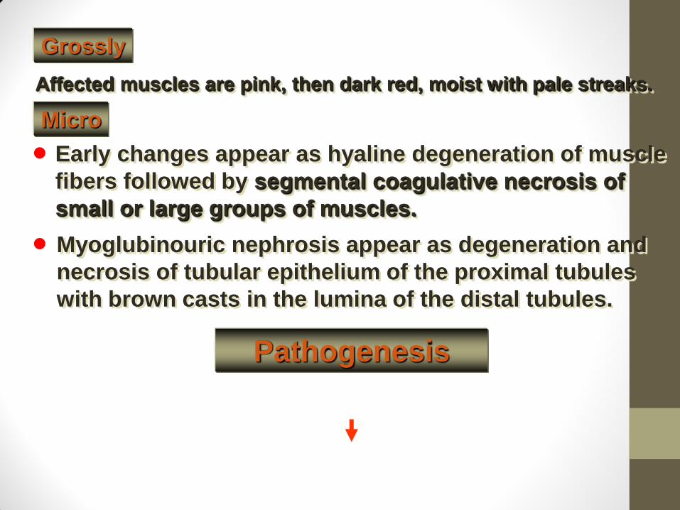
Grossly
Affected muscles are pink, then dark red, moist with pale streaks.
Early changes appear as hyaline degeneration of muscle
fibers followed by segmental coagulative necrosis of
small or large groups of muscles.
Myoglubinouric nephrosis appear as degeneration and
necrosis of tubular epithelium of the proximal tubules
with brown casts in the lumina of the distal tubules.
Micro
Pathogenesis
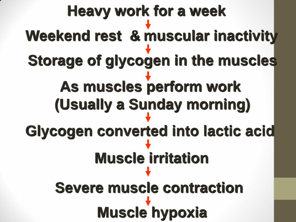
Weekend rest & muscular inactivity
Storage of glycogen in the muscles
As muscles perform work
(Usually a Sunday morning)
Glycogen converted into lactic acid
Muscle irritation
Severe muscle contraction
Muscle hypoxia
Heavy work for a week
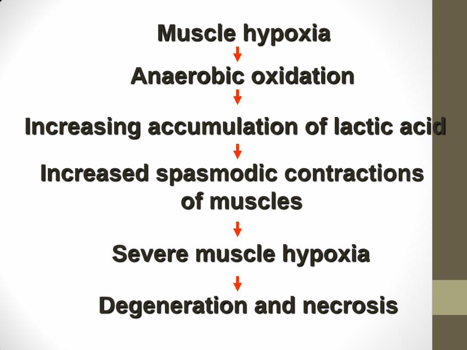
Muscle hypoxia
Anaerobic oxidation
Increasing accumulation of lactic acid
Increased spasmodic contractions
of muscles
Severe muscle hypoxia
Degeneration and necrosis
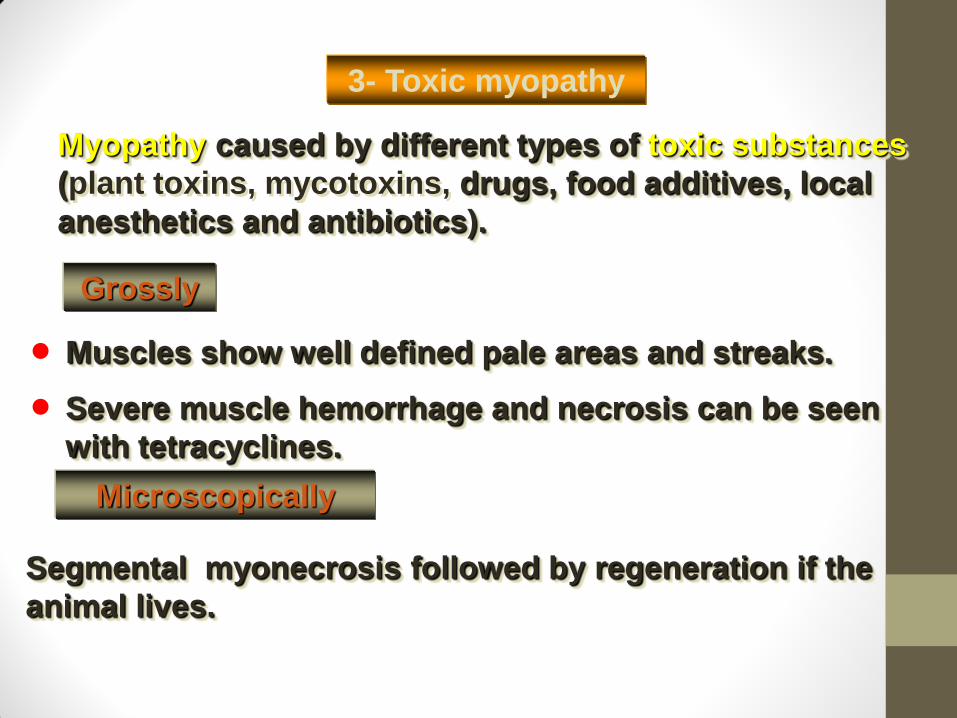
3- Toxic myopathy
Muscles show well defined pale areas and streaks.
Myopathy caused by different types of toxic substances
(plant toxins, mycotoxins, drugs, food additives, local
anesthetics and antibiotics).
Severe muscle hemorrhage and necrosis can be seen
with tetracyclines.
Grossly
Microscopically
Segmental myonecrosis followed by regeneration if the
animal lives.
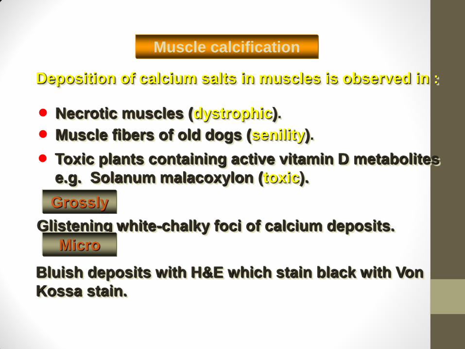
Deposition of calcium salts in muscles is observed in :
Necrotic muscles (dystrophic).
Muscle calcification
Muscle fibers of old dogs (senility).
Toxic plants containing active vitamin D metabolites
e.g. Solanum malacoxylon (toxic).
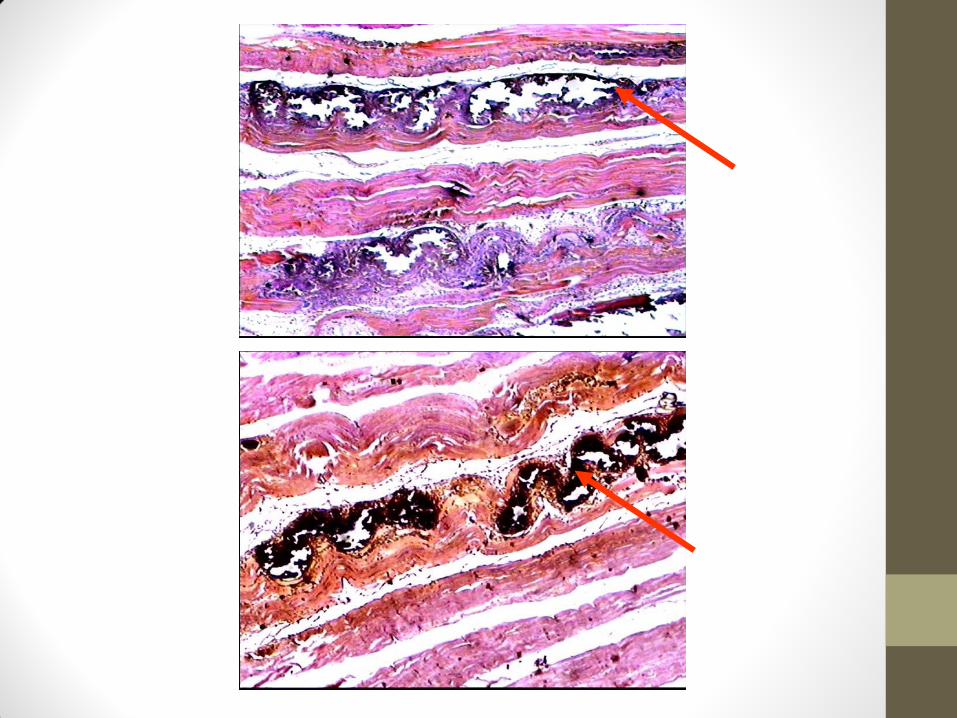
Grossly
Glistening white-chalky foci of calcium deposits.
Micro
Bluish deposits with H&E which stain black with Von
Kossa stain.

N.B:
Calcification is often confused with ossification. Calcification is synonymous with the formation of calcium-based salts and crystals within cells and tissue. It is a process that occurs during ossification, but not vice versa.
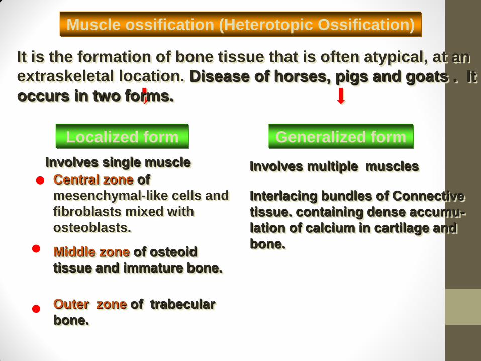
Interlacing bundles of Connective
tissue. containing dense accumu-
lation of calcium in cartilage and
bone.
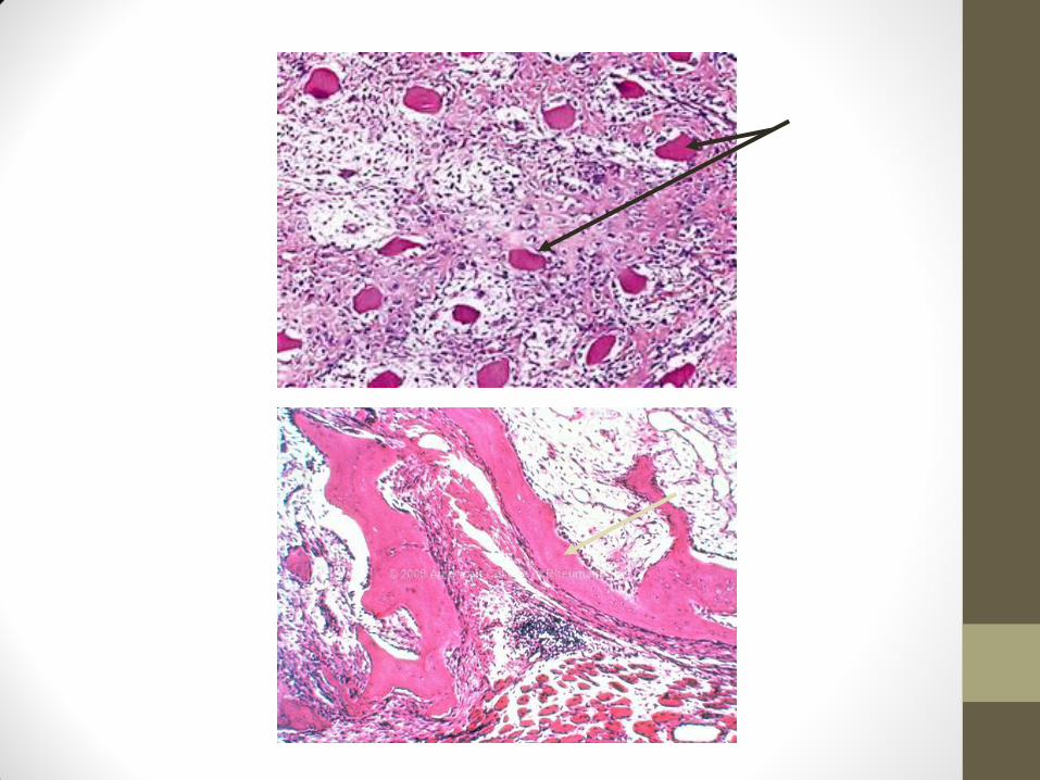
Central zone of
mesenchymal-like cells and
fibroblasts mixed with
osteoblasts.
It is the formation of bone tissue that is often atypical, at an
extraskeletal location. Disease of horses, pigs and goats . It
occurs in two forms.
Muscle ossification (Heterotopic Ossification)
Localized form Generalized form
Involves single muscle Involves multiple muscles
Middle zone of osteoid
tissue and immature bone.
Outer zone of trabecular
bone.
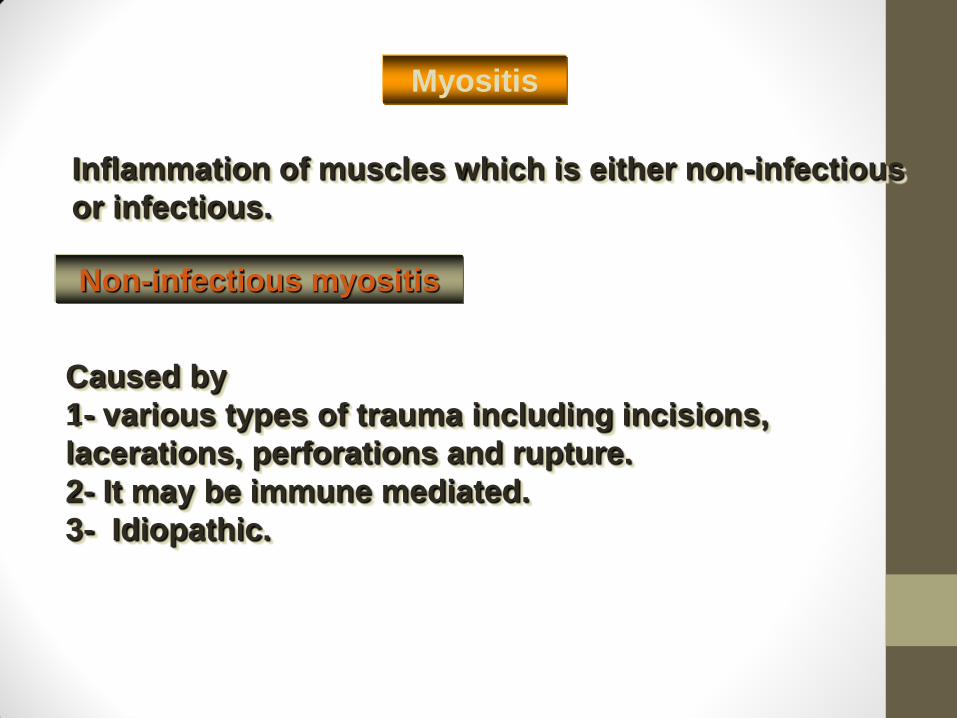
Inflammation of muscles which is either non-infectious
or infectious.
Caused by
1- various types of trauma including incisions,
lacerations, perforations and rupture.
2- It may be immune mediated.
3- Idiopathic.
Myositis
Non-infectious myositis
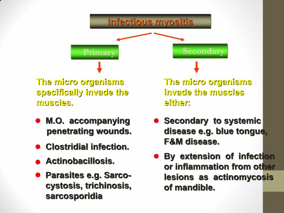
Primary Secondary
Infectious myositis
The micro organisms
specifically invade the
muscles.
The micro organisms
invade the muscles
either:
M.O. accompanying
penetrating wounds.
Clostridial infection.
Actinobacillosis.
Parasites e.g. Sarco-
cystosis, trichinosis,
sarcosporidia
Secondary to systemic
disease e.g. blue tongue,
F&M disease.
By extension of infection
or inflammation from other
lesions as actinomycosis
of mandible.
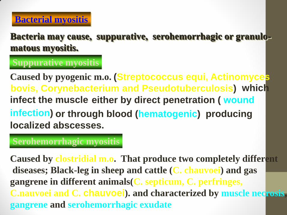
Bacterial myositis
Bacteria may cause, suppurative, serohemorrhagic or granulo-
matous myositis.
Suppurative myositis
Caused by pyogenic m.o.
Caused by clostridial m.o. That produce two completely different
diseases; Black-leg in sheep and cattle (C. chauvoei) and gas
gangrene in different animals(C. septicum, C. perfringes,
C.nauvoei and C. chauvoei). and characterized by muscle necrosis,
gangrene and serohemorrhagic exudate
Serohemorrhagic myositis
(Streptococcus equi, Actinomyces
bovis, Corynebacterium and Pseudotuberculosis) which
infect the muscle either by direct penetration ( wound
infection) or through blood (hematogenic) producing
localized abscesses.
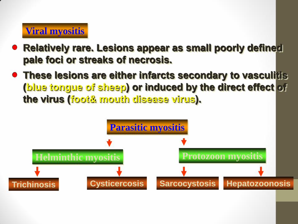
Parasitic myositis
Helminthic myositis Protozoon myositis
Trichinosis Cysticercosis Sarcocystosis Hepatozoonosis
Relatively rare. Lesions appear as small poorly defined
pale foci or streaks of necrosis.
These lesions are either infarcts secondary to vasculitis
(blue tongue of sheep) or induced by the direct effect of
the virus (foot& mouth disease virus).
Viral myositis
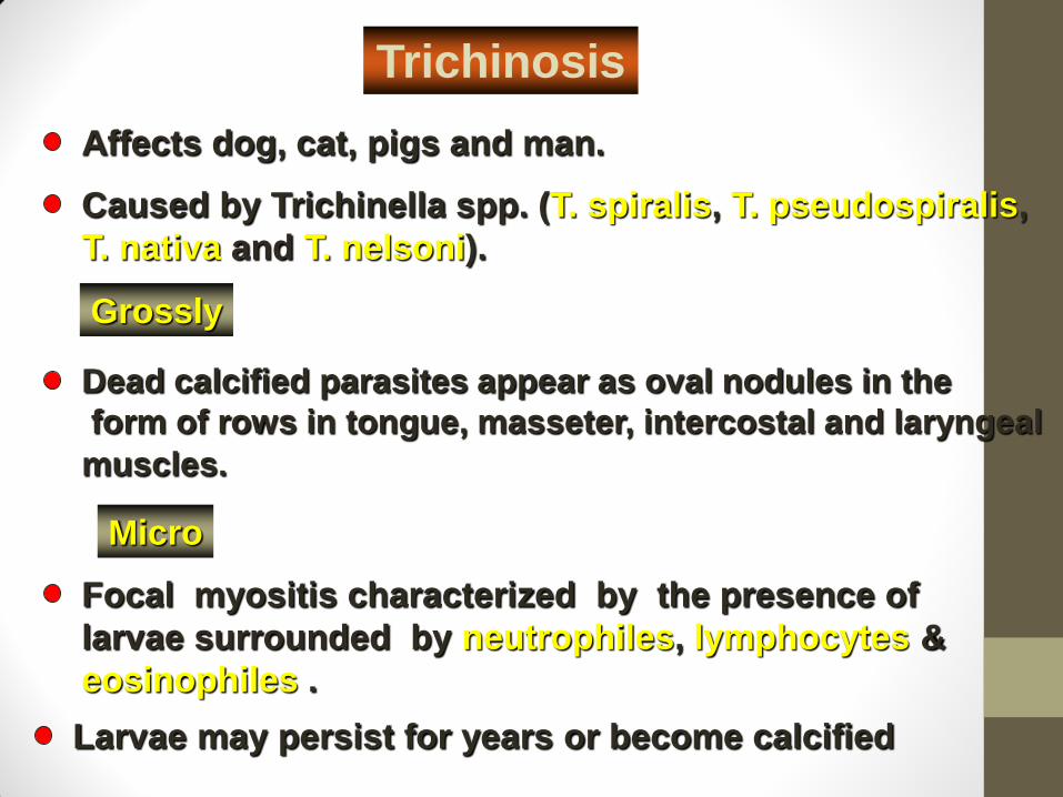
Affects dog, cat, pigs and man.
Caused by Trichinella spp. (T. spiralis, T. pseudospiralis,
T. nativa and T. nelsoni).
Dead calcified parasites appear as oval nodules in the
form of rows in tongue, masseter, intercostal and laryngeal
muscles.
Grossly
Micro
Focal myositis characterized by the presence of
larvae surrounded by neutrophiles, lymphocytes &
eosinophiles .
Larvae may persist for years or become calcified
Trichinosis
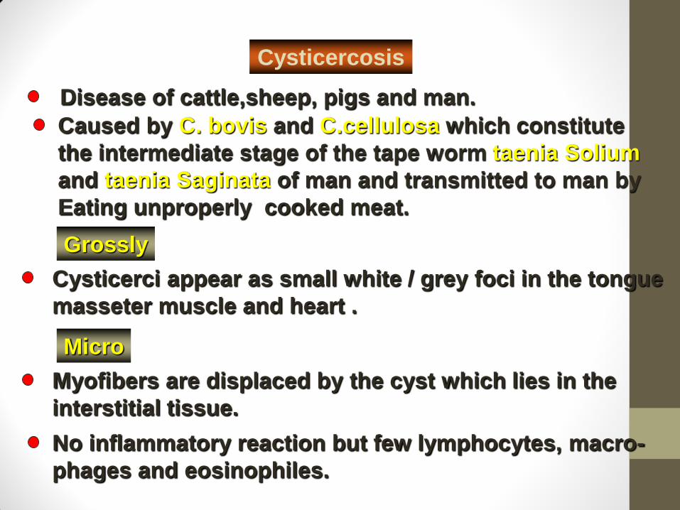
Disease of cattle,sheep, pigs and man.
Caused by C. bovis and C.cellulosa which constitute
the intermediate stage of the tape worm taenia Solium
and taenia Saginata of man and transmitted to man by
Eating unproperly cooked meat.
Cysticerci appear as small white / grey foci in the tongue
masseter muscle and heart .
Grossly
Micro
Myofibers are displaced by the cyst which lies in the
interstitial tissue.
No inflammatory reaction but few lymphocytes, macro-
phages and eosinophiles.
Cysticercosis
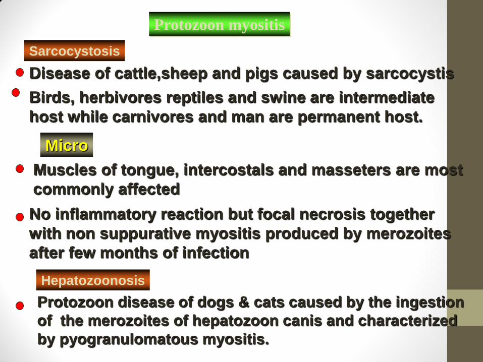
Disease of cattle,sheep and pigs caused by sarcocystis
Birds, herbivores reptiles and swine are intermediate
host while carnivores and man are permanent host.
Micro
Muscles of tongue, intercostals and masseters are most
commonly affected
No inflammatory reaction but focal necrosis together
with non suppurative myositis produced by merozoites
after few months of infection
Protozoon disease of dogs & cats caused by the ingestion
of the merozoites of hepatozoon canis and characterized
by pyogranulomatous myositis.
Hepatozoonosis
Sarcocystosis
Protozoon myositis
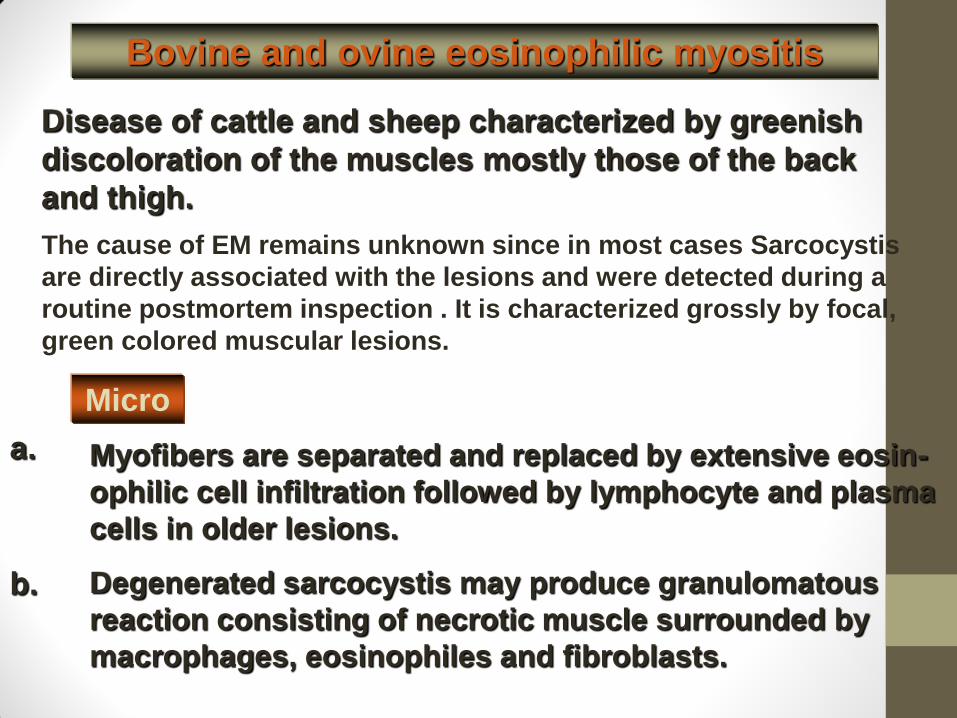
Bovine and ovine eosinophilic myositis
Disease of cattle and sheep characterized by greenish
discoloration of the muscles mostly those of the back
and thigh.
Micro
a. Myofibers are separated and replaced by extensive eosin-
ophilic cell infiltration followed by lymphocyte and plasma
cells in older lesions.
Degenerated sarcocystis may produce granulomatous
reaction consisting of necrotic muscle surrounded by
macrophages, eosinophiles and fibroblasts.
b.
The cause of EM remains unknown since in most cases Sarcocystis
are directly associated with the lesions and were detected during a
routine postmortem inspection . It is characterized grossly by focal,
green colored muscular lesions.

Skeletal system
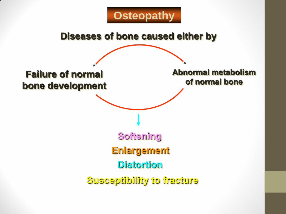
Diseases of bone caused either by
Osteopathy
Failure of normal
bone development
Abnormal metabolism
of normal bone
Distortion
Softening
Enlargement
Susceptibility to fracture
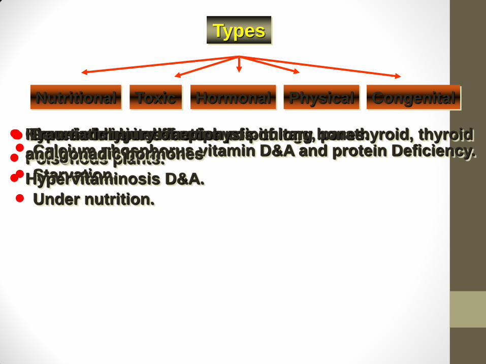
Types
Nutritional Toxic Congenital Physical Hormonal
Calcium,phosphorus,vitamin D&A and protein Deficiency.
Starvation.
Under nutrition.
Chronic lead and fluorine poisoning.
Poisonous plants.
Hypervitaminosis D&A.
g
Hypo and hyper secretion of pituitary, parathyroid, thyroid
and gonadic hormones
Genetic inherited factors Traumatic injury of epiphysis of long bones.
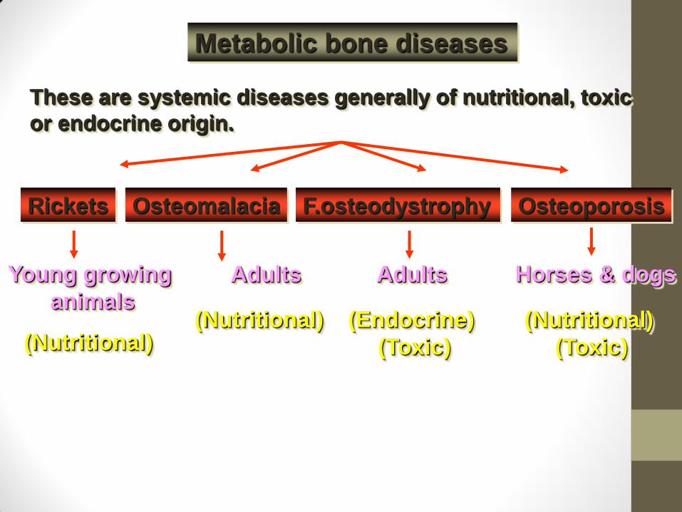
Metabolic bone diseases
These are systemic diseases generally of nutritional, toxic
or endocrine origin.
Rickets Osteomalacia Osteoporosis F.osteodystrophy
Young growing
animals
Adults Horses & dogs Adults
(Nutritional) (Nutritional) (Endocrine)
(Toxic)
(Nutritional)
(Toxic)
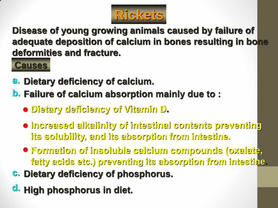
Rickets Disease of young growing animals caused by failure of
adequate deposition of calcium in bones resulting in bone
deformities and fracture.
Dietary deficiency of calcium.
Causes
Failure of calcium absorption mainly due to :
a.
b.
Formation of insoluble calcium compounds (oxalate,
fatty acids etc.) preventing its absorption from intestine.
Dietary deficiency of Vitamin D.
Increased alkalinity of intestinal contents preventing
its solubility, and its absorption from intestine.
c. Dietary deficiency of phosphorus.
d. High phosphorus in diet.
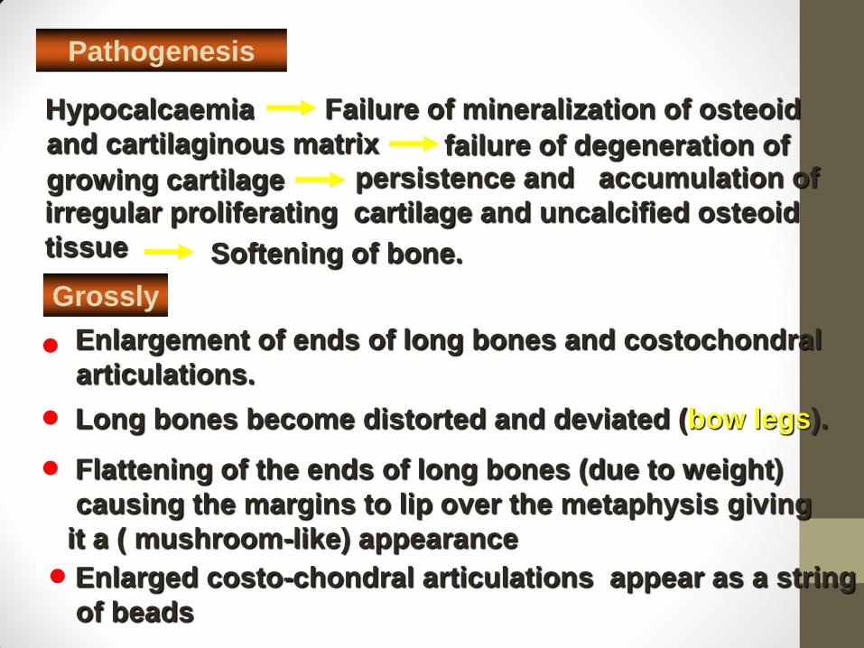
Pathogenesis
Hypocalcaemia
Grossly
Failure of mineralization of osteoid
and cartilaginous matrix failure of degeneration of
growing cartilage persistence and accumulation of
irregular proliferating cartilage and uncalcified osteoid
tissue Softening of bone.
Enlargement of ends of long bones and costochondral
articulations.
Long bones become distorted and deviated (bow legs).
Flattening of the ends of long bones (due to weight)
causing the margins to lip over the metaphysis giving
it a ( mushroom-like) appearance
Enlarged costo-chondral articulations appear as a string
of beads
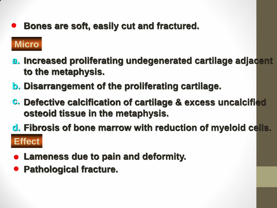
Micro
a.
b.
c.
d.
Increased proliferating undegenerated cartilage adjacent
to the metaphysis.
Disarrangement of the proliferating cartilage.
Defective calcification of cartilage & excess uncalcified
osteoid tissue in the metaphysis.
Fibrosis of bone marrow with reduction of myeloid cells.
Bones are soft, easily cut and fractured.
Effect
Lameness due to pain and deformity.
Pathological fracture.
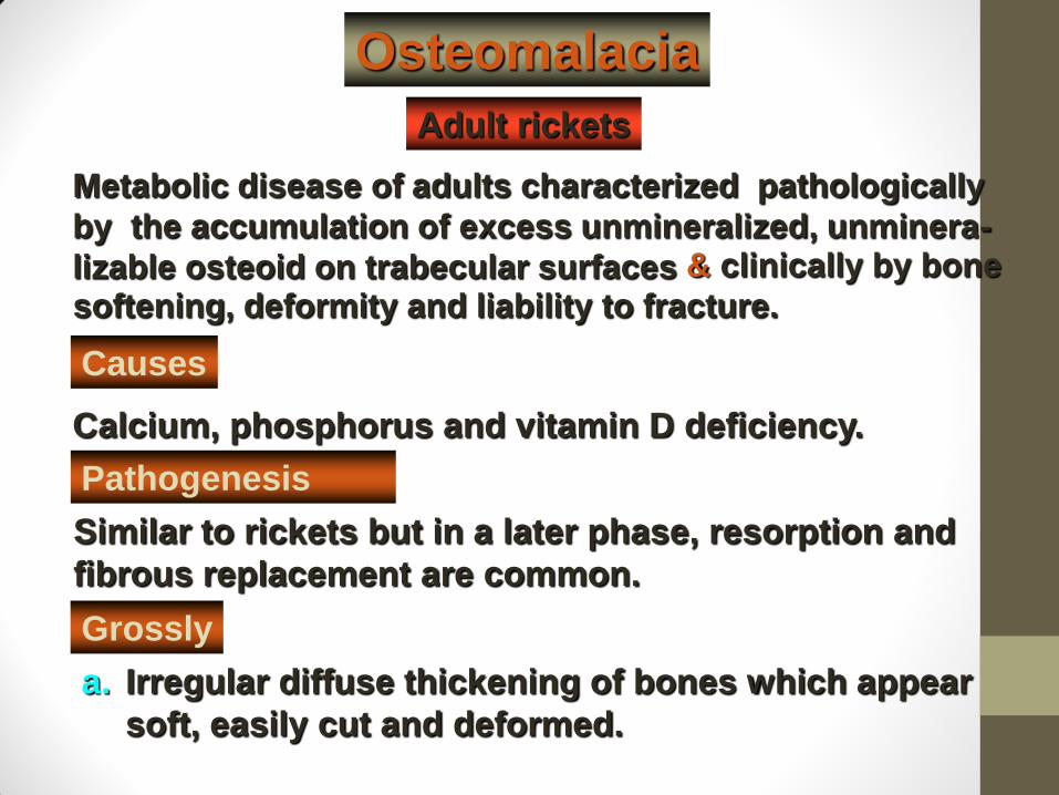
Osteomalacia
Adult rickets
Metabolic disease of adults characterized pathologically
by the accumulation of excess unmineralized, unminera-
lizable osteoid on trabecular surfaces
Causes
Calcium, phosphorus and vitamin D deficiency.
Irregular diffuse thickening of bones which appear
soft, easily cut and deformed.
& clinically by bone
softening, deformity and liability to fracture.
Pathogenesis
Similar to rickets but in a later phase, resorption and
fibrous replacement are common.
Grossly
a.
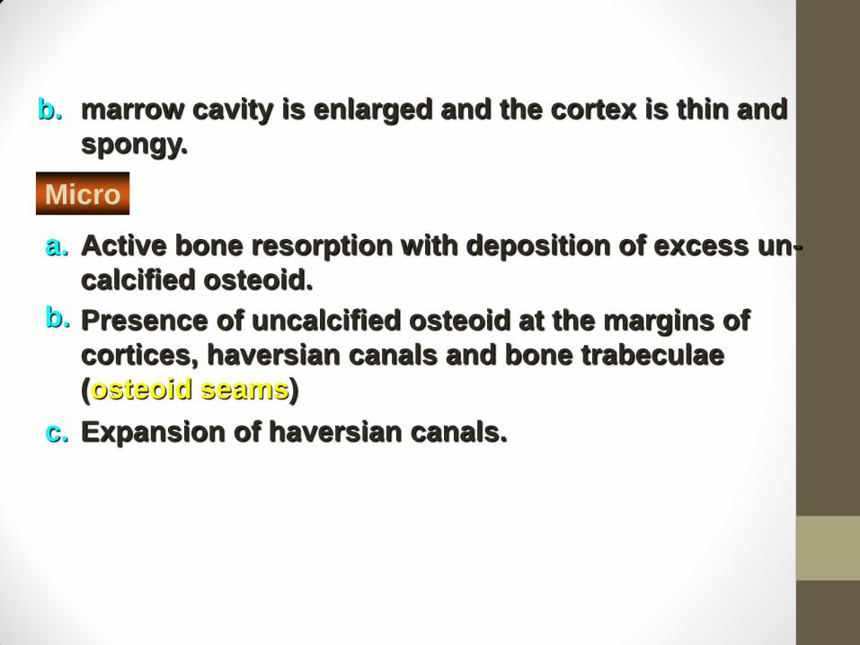
marrow cavity is enlarged and the cortex is thin and
spongy.
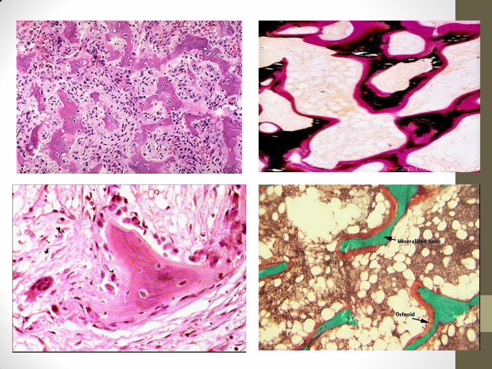
Presence of uncalcified osteoid at the margins of
cortices, haversian canals and bone trabeculae
(osteoid seams)
Active bone resorption with deposition of excess un-
calcified osteoid.
Expansion of haversian canals.
b.
Micro
a.
b.
c.
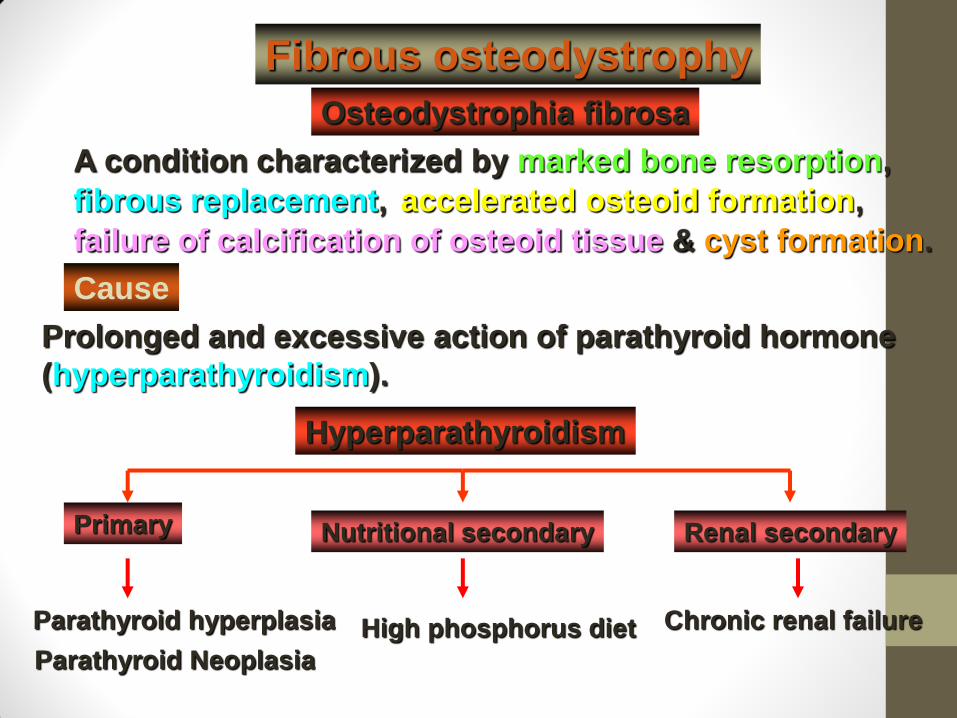
Hyperparathyroidism
Parathyroid hyperplasia Chronic renal failure High phosphorus diet
A condition characterized by marked bone resorption,
Fibrous osteodystrophy
fibrous replacement, accelerated osteoid formation,
failure of calcification of osteoid tissue & cyst formation.
Cause
Prolonged and excessive action of parathyroid hormone
(hyperparathyroidism).
Parathyroid Neoplasia
Osteodystrophia fibrosa
Primary Nutritional secondary Renal secondary
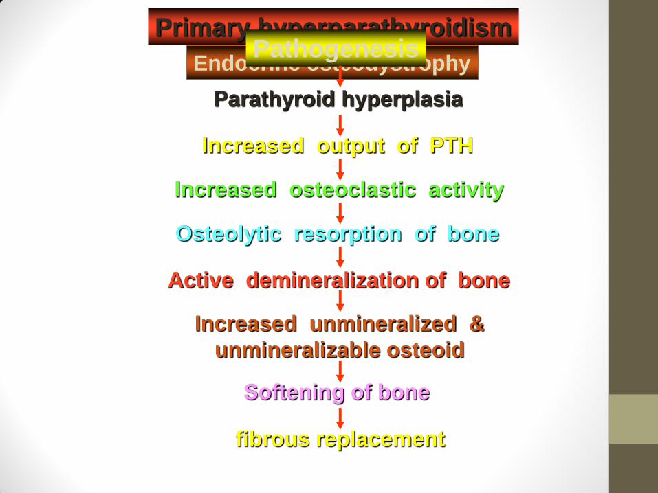
Primary hyperparathyroidism
Endocrine osteodystrophy Pathogenesis
Parathyroid hyperplasia
Increased output of PTH
Increased osteoclastic activity
Active demineralization of bone
Osteolytic resorption of bone
Increased unmineralized &
unmineralizable osteoid
Softening of bone
fibrous replacement
Grossly
Bones gradually soften, become flexible, deformed and
easily fractured
Multiple areas of rarification and cyst formation.
a.
b.
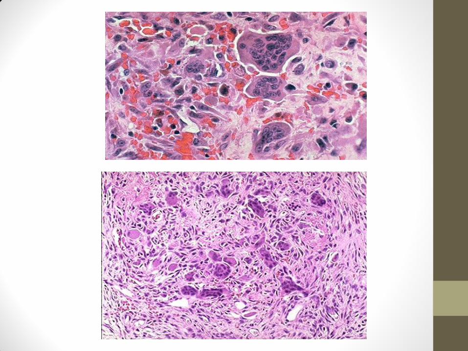
Micro
a. Marked bone resorption and increased osteoclastic
activity
Fibrous replacement of resorbed bone and bone marrow. b.
c. Thinning of trabecular bone and haversian canals.
Nutritional secondary hyperparathyroidism
Most commonly observed in young growing animals fed on
low calcium, high phosphorus diet (bran fed to horses.).
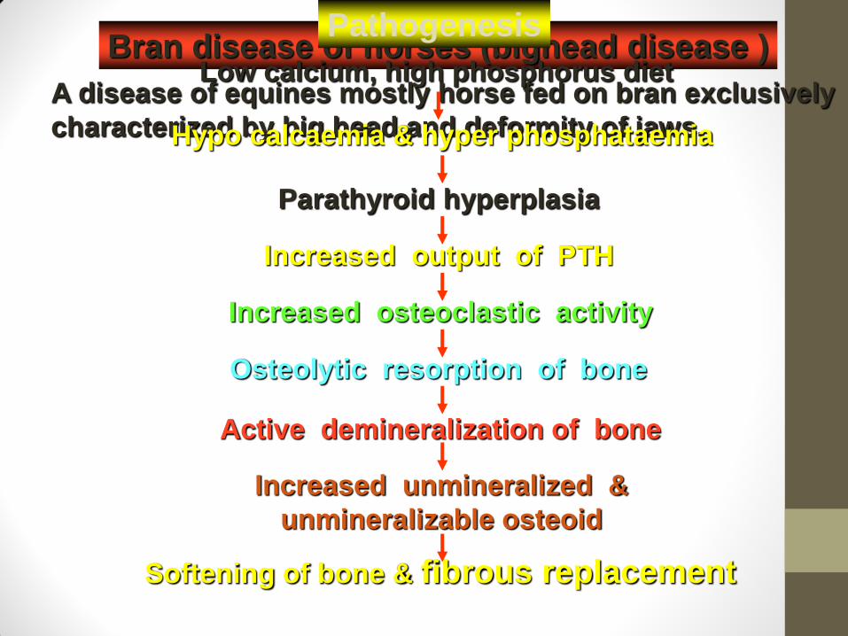
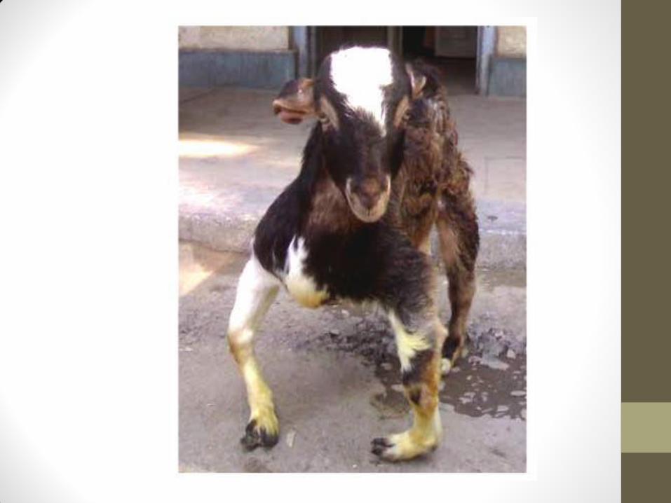
Bran disease of horses (bighead disease )
A disease of equines mostly horse fed on bran exclusively
characterized by big head and deformity of jaws.
Pathogenesis
Parathyroid hyperplasia
Increased output of PTH
Increased osteoclastic activity
Active demineralization of bone
Osteolytic resorption of bone
Increased unmineralized &
unmineralizable osteoid
Softening of bone & fibrous replacement
Low calcium, high phosphorus diet
Hypo calcaemia & hyper phosphataemia
Grossly
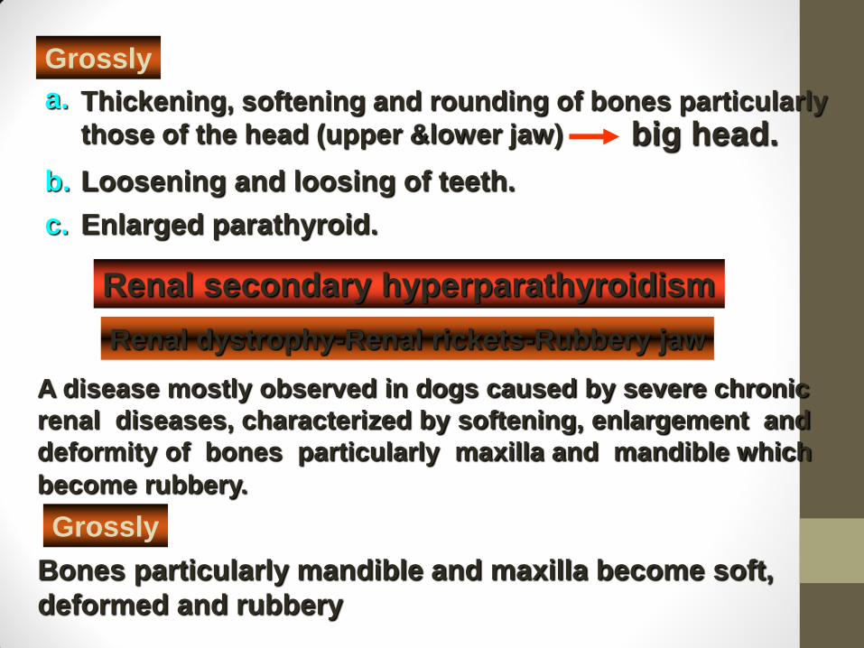
Thickening, softening and rounding of bones particularly
those of the head (upper &lower jaw)
Loosening and loosing of teeth.
a.
b.
big head.
c. Enlarged parathyroid.
Renal secondary hyperparathyroidism
A disease mostly observed in dogs caused by severe chronic
renal diseases, characterized by softening, enlargement and
deformity of bones particularly maxilla and mandible which
become rubbery.
Renal dystrophy-Renal rickets-Rubbery jaw
Grossly
Bones particularly mandible and maxilla become soft,
deformed and rubbery

Pathogenesis
Parathyroid hyperplasia
Increased out put of PTH
Increased osteoclastic activity
Osteolytic resorption and demineralization of bone
Increased unmineralized &
unmineralizable osteoid
Softening of bone & fibrous replacement
Renal insufficiency
Reduced glomerular filtration
Retention of phosphorus
Hyperphosphataemia
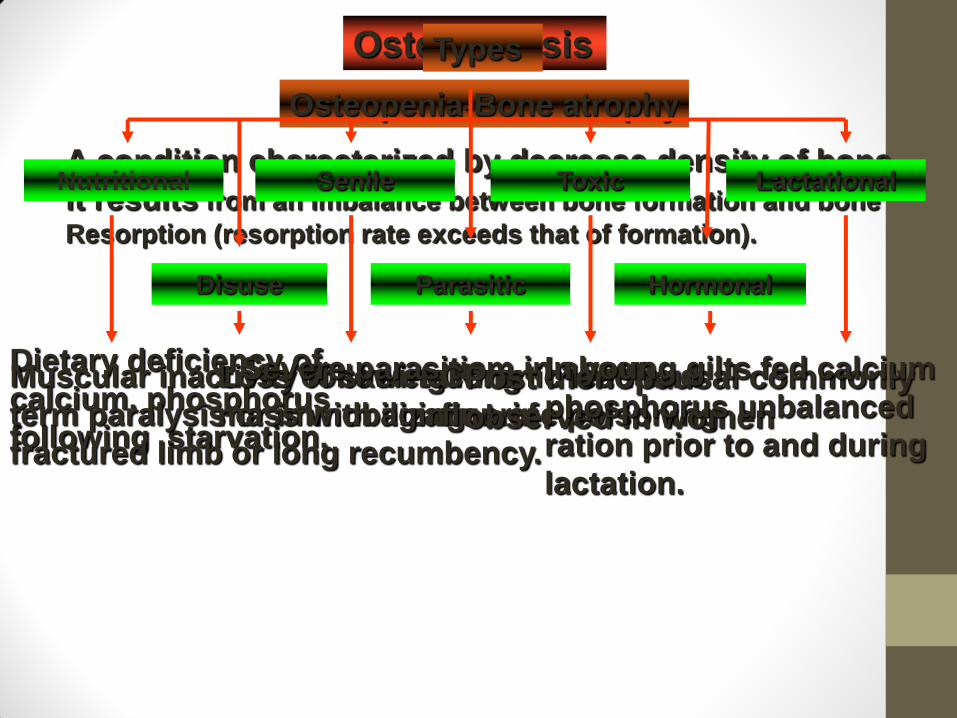
Osteoporosis
Osteopenia-Bone atrophy
A condition characterized by decrease density of bone,
it results from an imbalance between bone formation and bone
Resorption (resorption rate exceeds that of formation).
Nutritional
Disuse
Senile Lactational
Hormonal
Toxic
Parasitic
Types
Dietary deficiency of
calcium, phosphorus
following starvation.
Muscular inactivity following long
term paralysis or immobilization of
fractured limb or long recumbency.
Loss of skeletal
mass with aging.
Severe parasitism in sheep Chronic lead and
fluorine poisoning Post menopausal commonly
observed in women
In young gilts fed calcium
phosphorus unbalanced
ration prior to and during
lactation.
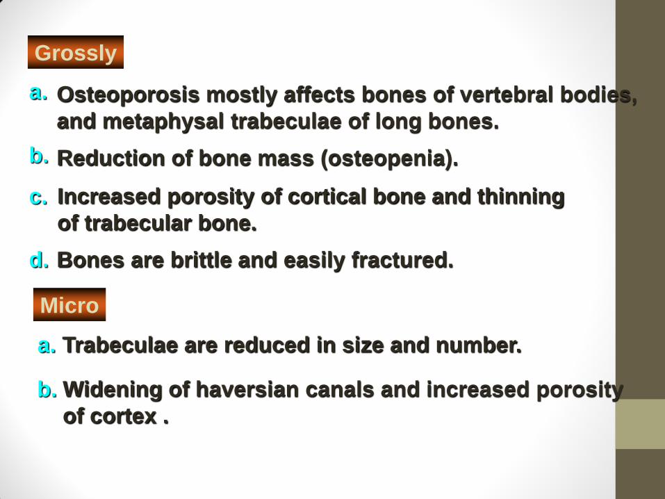
Grossly
Osteoporosis mostly affects bones of vertebral bodies,
and metaphysal trabeculae of long bones.
Reduction of bone mass (osteopenia).
a.
b.
c.
a.
d.
Increased porosity of cortical bone and thinning
of trabecular bone.
Bones are brittle and easily fractured.
Trabeculae are reduced in size and number.
Micro
b. Widening of haversian canals and increased porosity
of cortex .
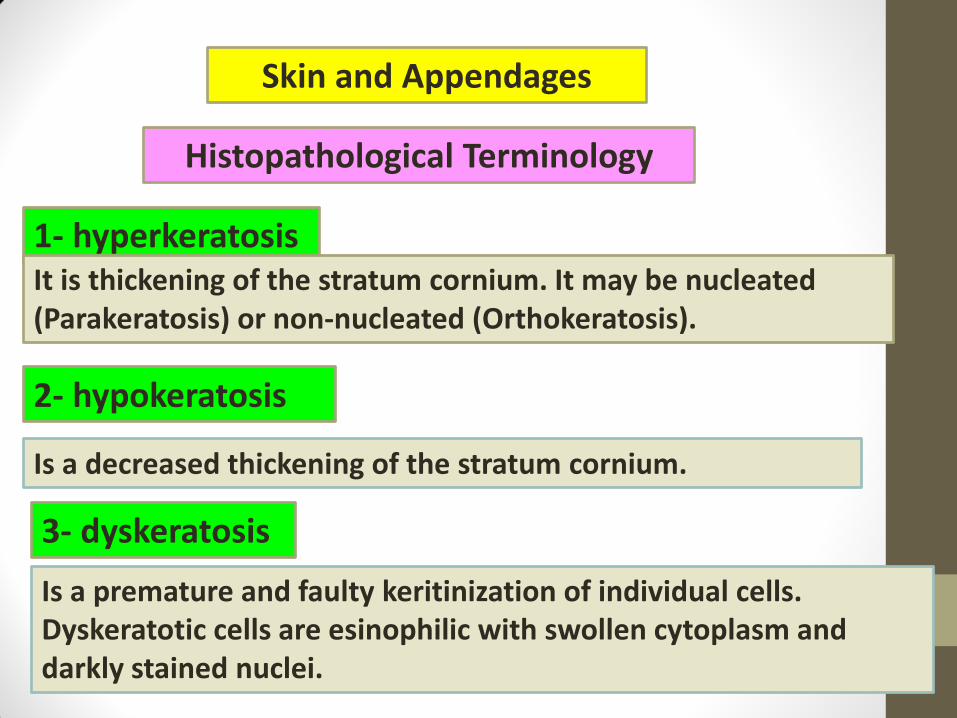
Skin and Appendages
Histopathological Terminology
1- hyperkeratosis It is thickening of the stratum cornium. It may be nucleated (Parakeratosis) or non-nucleated (Orthokeratosis).
2- hypokeratosis
Is a decreased thickening of the stratum cornium.
3- dyskeratosis
Is a premature and faulty keritinization of individual cells. Dyskeratotic cells are esinophilic with swollen cytoplasm and darkly stained nuclei.
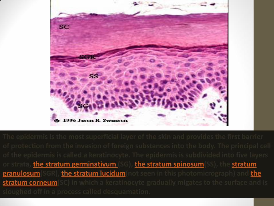
The epidermis is the most superficial layer of the skin and provides the first barrier of protection from the invasion of foreign substances into the body. The principal cell of the epidermis is called a keratinocyte. The epidermis is subdivided into five layers or strata, the stratum germinativum (SG), the stratum spinosum(SS), the stratum granulosum(SGR), the stratum lucidum(not seen in this photomicrograph) and the stratum corneum(SC) in which a keratinocyte gradually migates to the surface and is sloughed off in a process called desquamation.

4- Hypergranulosis and hypogranulosis
Indicates an increase or decrease thickening of the stratum granulosum.
5- Acanthosis
Is an increase thickeness f the stratum spinosum. It may be due to hyperplasia (true acanthosis) or due to hypertrophy (peudo acanthosis) of epidermal cells.
6- dysplasia
Is abnormal development of epidermal cells (disorganized formation). It may be precarcinogenic.
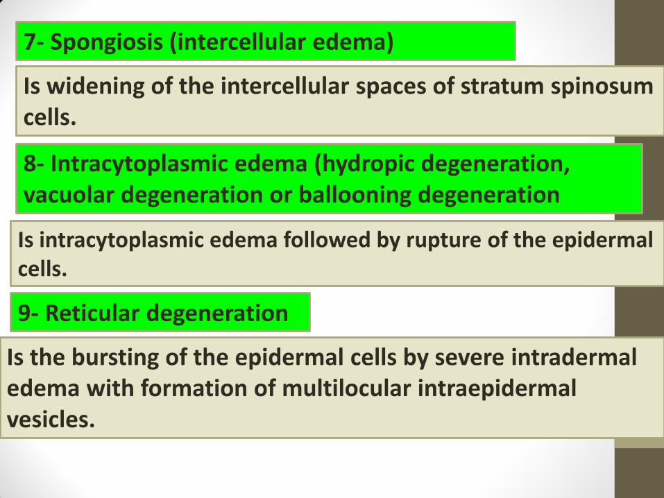
7- Spongiosis (intercellular edema)
Is widening of the intercellular spaces of stratum spinosum cells.
8- Intracytoplasmic edema (hydropic degeneration, vacuolar degeneration or ballooning degeneration
Is intracytoplasmic edema followed by rupture of the epidermal cells.
9- Reticular degeneration
Is the bursting of the epidermal cells by severe intradermal edema with formation of multilocular intraepidermal vesicles.
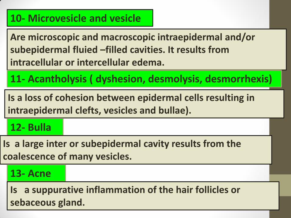
10- Microvesicle and vesicle
Are microscopic and macroscopic intraepidermal and/or subepidermal fluied –filled cavities. It results from intracellular or intercellular edema.
11- Acantholysis ( dyshesion, desmolysis, desmorrhexis)
Is a loss of cohesion between epidermal cells resulting in intraepidermal clefts, vesicles and bullae).
12- Bulla
Is a large inter or subepidermal cavity results from the coalescence of many vesicles.
13- Acne
Is a suppurative inflammation of the hair follicles or sebaceous gland.
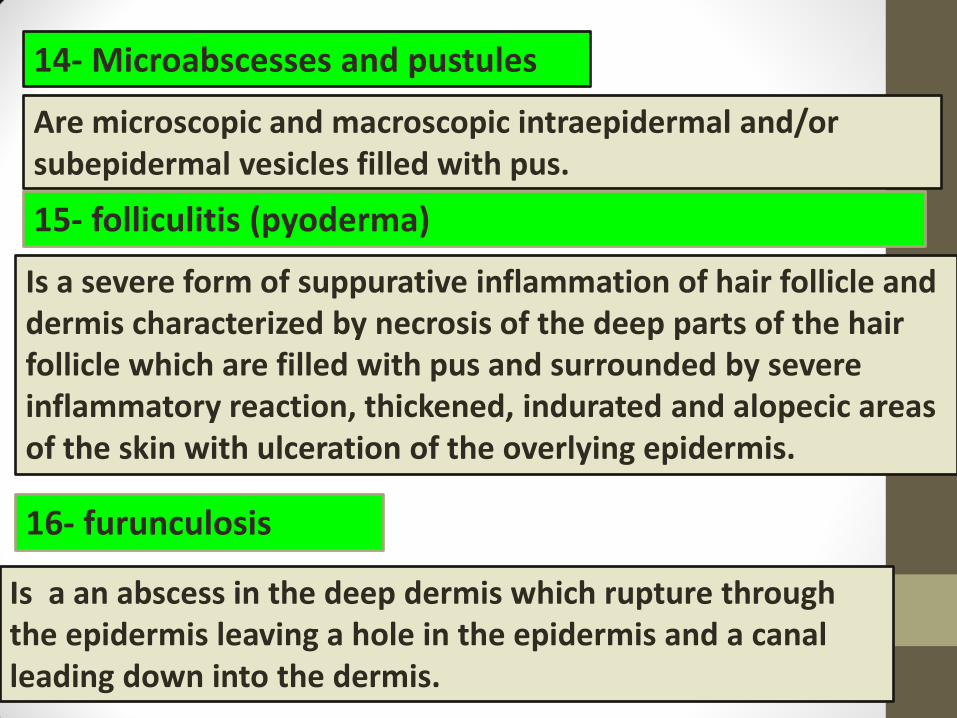
14- Microabscesses and pustules
Are microscopic and macroscopic intraepidermal and/or subepidermal vesicles filled with pus.
15- folliculitis (pyoderma)
Is a severe form of suppurative inflammation of hair follicle and dermis characterized by necrosis of the deep parts of the hair follicle which are filled with pus and surrounded by severe inflammatory reaction, thickened, indurated and alopecic areas of the skin with ulceration of the overlying epidermis.
16- furunculosis
Is a an abscess in the deep dermis which rupture through the epidermis leaving a hole in the epidermis and a canal leading down into the dermis.

17- Carbuncle
Is a deep dermal abscess which opens through many sinus tracts.
18- Exocytosis
Is the margenation of the inflammatory cells and/or erythrocytes through the intercellular spaces of the epidermis..
19- Pachyderma
Is a focal thickening of the skin involving all the skin layers.
20- Erosion (Excoriation)
Is a superficial loss of epidermal or epithelial cells
Is the loss of the epidermis with exposure of the underlying dermis..
21- ulcer

22- Fissure
Is a linear defect in the epidermis..
23- Erythema
Is a circumscribed area of edema and congestion of the papillary dermis.
24- Albinism
Is a generalized congenital lack of melanin pigment throughout the entire body due to lack of tyrosinase enzyme necessary for melanin synthesis.
25- Vitiligo
Is an aquired loss of melanin pigmnet in a focal areas of skin (burns or scars).
26- Acanthosis nigricans
Is a condition commonly observed in man and dogs characterized by patches of heavily pigmented rough thick skin ( acnthosis and hyperpigmentation with melanin).
27- Keloid
Is a hypertrophic scar characterized by heavy bundles of esinophilic collagen in the dermis, elevated epidermis which may become ulcerated
28- Crust Is a consolidated, desiccated surface mass composed of varying
combinations of keratin, serum, cellular debris and few microorganisms.
29- Xanthomatosis
Is a nodular accumulation of large epitheloid cells (laden with lipid substance) and multinucleated giant cells in the dermis and subcutis (in association with disturbance of lipid metabolism).
30- Congenital icthyosis
Is a condition of newborn calves characterized by thick, scaly, horny hairless skin which resembles skin of fish.

Dermatitis
Inflammation of the skin which may be
* acute
*subacute
*chronic
* focal
*diffuse
*Serus *Serohemorrhagic *Suppurative *granulomatous

*spongiosis
*Intercellular edema
*Leukocytic
infiltaration
*Vascular dilatation
*Perivascular cell
infiltartion
*similar to acute form
+
The reaction is
proliferative and
Infiltrative
+
Fibroplasia
Mononuclear cell
infiltration
*similar to acute from
+
•Epidermal hyperplasia.
•Ortho or parakeratotic
hyperkeratosis.
Acute form
characterized by
Chronic form
characterized by
Subacute form
characterized by
Generally the microscopic picture varies with the type of exudates and the nature of infection and causative agent.