Musculoskeletal Loads during Stationary Cycling and the ...
120
University of Tennessee, Knoxville University of Tennessee, Knoxville TRACE: Tennessee Research and Creative TRACE: Tennessee Research and Creative Exchange Exchange Doctoral Dissertations Graduate School 5-2017 Musculoskeletal Loads during Stationary Cycling and the Effects Musculoskeletal Loads during Stationary Cycling and the Effects of Pedal Modifications for Knee Osteoarthritis of Pedal Modifications for Knee Osteoarthritis Rachel Lynn Thompson University of Tennessee, Knoxville, [email protected] Follow this and additional works at: https://trace.tennessee.edu/utk_graddiss Part of the Biomechanics and Biotransport Commons Recommended Citation Recommended Citation Thompson, Rachel Lynn, "Musculoskeletal Loads during Stationary Cycling and the Effects of Pedal Modifications for Knee Osteoarthritis. " PhD diss., University of Tennessee, 2017. https://trace.tennessee.edu/utk_graddiss/4503 This Dissertation is brought to you for free and open access by the Graduate School at TRACE: Tennessee Research and Creative Exchange. It has been accepted for inclusion in Doctoral Dissertations by an authorized administrator of TRACE: Tennessee Research and Creative Exchange. For more information, please contact [email protected].
-
Upload
khangminh22 -
Category
Documents
-
view
1 -
download
0
Transcript of Musculoskeletal Loads during Stationary Cycling and the ...

University of Tennessee, Knoxville University of Tennessee, Knoxville
TRACE: Tennessee Research and Creative TRACE: Tennessee Research and Creative
Exchange Exchange
Doctoral Dissertations Graduate School
5-2017
Musculoskeletal Loads during Stationary Cycling and the Effects Musculoskeletal Loads during Stationary Cycling and the Effects
of Pedal Modifications for Knee Osteoarthritis of Pedal Modifications for Knee Osteoarthritis
Rachel Lynn Thompson University of Tennessee, Knoxville, [email protected]
Follow this and additional works at: https://trace.tennessee.edu/utk_graddiss
Part of the Biomechanics and Biotransport Commons
Recommended Citation Recommended Citation Thompson, Rachel Lynn, "Musculoskeletal Loads during Stationary Cycling and the Effects of Pedal Modifications for Knee Osteoarthritis. " PhD diss., University of Tennessee, 2017. https://trace.tennessee.edu/utk_graddiss/4503
This Dissertation is brought to you for free and open access by the Graduate School at TRACE: Tennessee Research and Creative Exchange. It has been accepted for inclusion in Doctoral Dissertations by an authorized administrator of TRACE: Tennessee Research and Creative Exchange. For more information, please contact [email protected].

To the Graduate Council:
I am submitting herewith a dissertation written by Rachel Lynn Thompson entitled
"Musculoskeletal Loads during Stationary Cycling and the Effects of Pedal Modifications for
Knee Osteoarthritis." I have examined the final electronic copy of this dissertation for form and
content and recommend that it be accepted in partial fulfillment of the requirements for the
degree of Doctor of Philosophy, with a major in Biomedical Engineering.
Jeffrey Reinbolt, Major Professor
We have read this dissertation and recommend its acceptance:
Songning Zhang, JAM Boulet, Eric Wade
Accepted for the Council:
Dixie L. Thompson
Vice Provost and Dean of the Graduate School
(Original signatures are on file with official student records.)

Musculoskeletal Loads during Stationary Cycling and the Effects of Pedal
Modifications for Knee Osteoarthritis
A Dissertation Presented for the
Doctor of Philosophy
Degree
The University of Tennessee, Knoxville
Rachel Lynn Thompson
May 2017

ii
Copyright © 2017 by Rachel L. Thompson
All rights reserved.

iii
DEDICATION
This dissertation is dedicated to my parents Ann and Brian Thompson, my siblings Nicole Ray
and Mathew Thompson. Without your unconditional love and unwavering support, this would
not have been possible.

iv
ACKNOWLEDGEMENTS
First, I would like to thank my advisor, Dr. Jeffrey A. Reinbolt, for giving me the
opportunity to purse my PhD in his laboratory. This dissertation would not have been possible if
it was not for Dr. Reinbolt with his never-ending support, supervision and guidance along the
way. Not only was he my advisor, he helped me develop as a person, engineer and an
independent researcher.
I would also like to thank Dr. Songning Zhang and Dr. Jacob Gardner who not only
collected the data but also allowed me to work with the data. This collaboration taught me a lot
over the past few years.
Finally, I would like to thank my committee members, Dr. JAM Boulet and Dr. Eric
Wade for your irreplaceable guidance and feedback. They each have added to my education and
research. They have made me become a better scientist and engineer without their help I would
not be the engineer I am today.

v
ABSTRACT
Knee OA is a deteriorating joint disease affecting 27 million people in the US and current
exercise prescriptions may be improved with new knowledge of their effects on muscle forces
and joint contact loads. Cycling rather than other exercise modalities is generally considered an
alternative for people with knee OA. If these research objectives were achieved, clinicians would
have additional tools related to joint contact loads for treating people with OA with an cycling
exercise while controlling progression of OA. The long-term goal of this research is to provide a
scientific basis for planning, evaluation and improvement of subject-specific rehabilitation for
subjects with knee osteoarthritis (OA).
The principles governing relationships between muscle forces, joint contact loads and
movements in people with knee OA, have not been discovered. Determining how to adjust
movements to optimize joint contact loads is difficult because experiments do not account for
these loads. In combination with experimental approaches, muscle-actuated inverse dynamic
simulations provide a scientific framework to estimate important variables and identify cause-
and-effect relationships. These activities challenge existing paradigms for exercise prescriptions
by including movements specifically designed for decreasing knee joint contact loads.
The research objective is to investigate muscle forces and joint contact loads that are
experienced by the knee during cycling. The overall hypothesis is a combination of
biomechanical cycling modifications that contribute to altered muscle forces and a reduction in
knee joint contact loads in subjects with and without knee OA during cycling; this may be
mitigated with a novel pedal design. The overall purpose of this research was to discover
relationships between muscle forces, joint contact loads, cycling and OA-friendly cycling
modifications for improving exercise prescriptions. The following objectives were addressed: 1)

vi
determine the effects of lateral pedal wedges and toe-in on joint biomechanics during cycling and
2) examine the potential of optimization to design subject-specific cycling modifications for
decreasing knee joint contact loads.

vii
PREFACE
This dissertation presents three studies conducted using inverse dynamic simulations of
muscle forces, joint contact loads and optimization analyses to modify and develop effective
cycling prescriptions for knee osteoarthritis. Each chapter is written as a separate technical paper
and an overview of the goals and methods employed in each study has been provided.
Additionally, each chapter provides an in-depth discussion of the study’s findings as well, as
how these findings can be used to answer the scientific questions posed. Chapter 5 provides a
summary of the results of the three studies in this dissertation and outlines on how they can be
applied to develop better treatment options for individuals at risk for osteoarthritis progression.
viii
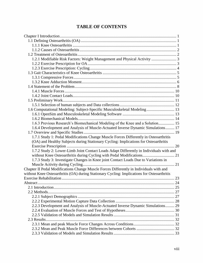
TABLE OF CONTENTS
Chapter I Introduction ..................................................................................................................... 1
1.1 Defining Osteoarthritis (OA) ................................................................................................ 1
1.1.1 Knee Osteoarthritis ........................................................................................................ 1
1.1.2 Causes of Osteoarthritis ................................................................................................. 2
1.2 Treatment of Osteoarthritis ................................................................................................... 2
1.2.1 Modifiable Risk Factors: Weight Management and Physical Activity ......................... 3
1.2.2 Exercise Prescription for OA ......................................................................................... 3
1.2.3 Exercise Prescription: Cycling ....................................................................................... 4
1.3 Gait Characteristics of Knee Osteoarthritis .......................................................................... 5
1.3.1 Compressive Forces ....................................................................................................... 5
1.3.2 Knee Adduction Moment ............................................................................................... 6
1.4 Statement of the Problem ...................................................................................................... 8
1.4.1 Muscle Forces .............................................................................................................. 10
1.4.2 Joint Contact Loads...................................................................................................... 10
1.5 Preliminary Work................................................................................................................ 11
1.5.1 Selection of human subjects and Data collections ....................................................... 12
1.6 Computational Modeling: Subject-Specific Musculoskeletal Modeling ............................ 13
1.6.1 OpenSim and Musculoskeletal Modeling Software .................................................... 13
1.6.2 Biomechanical Models................................................................................................. 14
1.6.3 Previous Research’s Biomechanical Modeling of the Knee and a Solution ................ 15
1.6.4 Development and Analysis of Muscle-Actuated Inverse Dynamic Simulations ......... 17
1.7 Overview and Specific Studies ........................................................................................... 19
1.7.1 Study 1: Pedal Modifications Change Muscle Forces Differently in Osteoarthritis
(OA) and Healthy Subjects during Stationary Cycling: Implications for Osteoarthritis
Exercise Prescription ............................................................................................................ 20
1.7.2 Study 2: Lower-Limb Joint Contact Loads Adapt Differently in Individuals with and
without Knee Osteoarthritis during Cycling with Pedal Modifications................................ 21
1.7.3 Study 3: Investigate Changes in Knee joint Contact Loads Due to Variations in
Muscle Activity during Cycling............................................................................................ 21
Chapter II Pedal Modifications Change Muscle Forces Differently in Individuals with and
without Knee Osteoarthritis (OA) during Stationary Cycling: Implications for Osteoarthritis
Exercise Rehabilitation ................................................................................................................. 23
Abstract ......................................................................................................................................... 24
2.1 Introduction ......................................................................................................................... 25
2.2 Methods............................................................................................................................... 27
2.2.1 Subject Demographics ................................................................................................. 27
2.2.2 Experimental Motion Capture Data Collection ........................................................... 28
2.2.3 Development and Analysis of Muscle-Actuated Inverse Dynamic Simulations ......... 29
2.2.4 Evaluation of Muscle Forces and Test of Hypotheses ................................................. 30
2.2.5 Validation of Models and Simulation Results ............................................................. 31
2.3 Results ................................................................................................................................. 32
2.3.1 Mean and peak Muscle Force Changes Across Conditions ......................................... 32
2.3.2 Mean and Peak Muscle Force Differences between Cohorts ...................................... 32
2.3.3 Validation of Models and Simulation Results ............................................................. 33
ix
2.4 Discussion ........................................................................................................................... 34
Chapter III Lower-Limb Joint Contact Loads Adapt Differently in Individuals with and without
Knee Osteoarthritis during Cycling with Pedal Modifications ..................................................... 38
Abstract ......................................................................................................................................... 39
3.1 Introduction ......................................................................................................................... 40
3.2 Methods............................................................................................................................... 41
3.2.1 Subject Demographics ................................................................................................. 41
3.2.2 Experimental Motion Capture Data Collection ........................................................... 42
3.2.3 Development and Analysis of Muscle-Actuated Inverse Dynamic Simulations ......... 42
3.2.4 Estimation of Joint Contact Loads ............................................................................... 44
3.2.5 Tests of the Hypothesis ................................................................................................ 44
3.2.6 Validation of the Model and Simulation Results ......................................................... 45
3.3 Results ................................................................................................................................. 46
3.3.1 Lateral Wedge Peak Joint Contact Load Differences across Conditions .................... 46
3.3.2 Toe-in Peak Joint Contact Load Differences across Conditions ................................. 47
3.3.3 Peak Joint Contact Loads Differences between Cohorts ............................................. 48
3.4 Discussion ........................................................................................................................... 49
Chapter IV Investigating Changes in tibiofemoral joint contact loads due to variations in muscle
activity during stationary cycling .................................................................................................. 54
Abstract ......................................................................................................................................... 55
4.1 Introduction ......................................................................................................................... 56
4.2 Methods............................................................................................................................... 58
4.2.1 Subject Demographics ................................................................................................. 58
4.2.2 Development and Analysis of Muscle-Actuated Inverse Dynamic Simulations ......... 58
4.2.3 Static Optimization and Joint Reaction Analysis......................................................... 59
4.2.4 Creating the Objective Function .................................................................................. 61
4.2.5 Exploring the Muscle Activation and their Effects on the Joint Contact Loads .......... 62
4.2.6 A Muscle Coordination Pattern Minimizing Compressive Tibiofemoral Force ......... 64
4.2.5 Change in Tibiofemoral Forces Due to Maximized Muscle Activations .................... 64
4.3 Results ................................................................................................................................. 65
4.3.1Minimize Muscle Activations ....................................................................................... 65
4.3.2 Minimize Joint Contact Forces .................................................................................... 67
4.3.3 Maximizing the muscle force to minimize the joint loads ........................................... 67
4.4 Discussion ........................................................................................................................... 68
Chapter V Conclusion ................................................................................................................... 72
5.1 Significance of Research..................................................................................................... 72
5.2 Research Innovation............................................................................................................ 73
5.3 Fundamental Contributions ................................................................................................. 74
5.4 Future Work ........................................................................................................................ 75
List of References ......................................................................................................................... 76
Appendix ....................................................................................................................................... 84
Vita .............................................................................................................................................. 101
x
LIST OF TABLES
Table 1: A muscle force change identity grid showing the significant differences within each
cohort in all lower extremity muscles compared to neutral pedal condition, for the mean
muscle forces. Red indicates an increase, while blue indicates a decrease compared to
neutral. The darker the color the greater the increase or decrease. ....................................... 90
Table 2: A muscle force change identity grid showing the significant differences within each
cohort in all muscles compared to neutral, for the peak muscle forces. Red indicates an
increase, while blue indicates a decrease compared to neutral; while, the darker the color the
greater the increase or decrease. ........................................................................................... 92
Table 3: Mean and peak muscle force significant differences between OA (+) and Healthy (-) in
all conditions. Red indicates knee OA cohort was greater within that conditions, while blue
indicates healthy cohort was higher within that conditions; the darker the color the greater
the difference. ....................................................................................................................... 93
Table 4: The joint contact loads change identity grid showing the significant differences within
each cohort in all contact loads compared to neutral, for the peak contact loads. Red
indicates an increase compared to neutral, while blue indicates a decrease compared to
neutral; while, the darker the color the greater the increase or decrease. ............................. 95
Table 5: The peak joint contact loads significant differences between OA (+) and Healthy (-)
across all conditions. Red indicates knee OA cohort was greater within the corresponding
condition, while blue indicates healthy cohort was higher within that condition. The darker
the color the greater the difference. ...................................................................................... 96
Table 6: A change identity grid showing the differences in the joint contact loads for the specific
muscles that changed from the normal joint reaction analysis. Results are shown for each
cohort for step 1’s muscle variation, for the subjects with and without knee OA in all
muscles used stands for a muscle weighting on 1, while don’t stands for. Red indicates an
increase compared to joint reaction analysis, while blue indicates a decrease compared to
joint reaction analysis. The darker the color the greater the increase or decrease. ............... 97

xi
LIST OF FIGURES
Figure 1: Illustrations showing the direction of the net external knee adduction moment (KAM)
represented by the blue curved arrows toward the midline or medial aspect of the body or
corresponding knee joint this is a result from a traditional inverse dynamics analysis
determining the net forces responsible for the movement. The important fact in the
difference between the adduction-abduction contact moments for this research is the net
KAM is determined without taking into account for the internal muscle forces. ................. 85
Figure 2: Showing what the a) lower limb looks like when there is a change in alignments, b)
shows a closer look as to what happens at the knee joint when the joint changes from
normal alignment to a more varus or adducted alignment. ................................................... 86
Figure 3: Many factors contribute to knee joint contact loads associated with OA disease
progression and disability during movements. The transformations between experimental
EMG patterns and coordinated multi-joint movement (shaded region) are complicated.
Furthermore, to make exercise prescription decisions, clinicians must predict joint load
changes after equipment adjustments. Exercise prescription alters musculoskeletal geometry
and multi-joint dynamics and these changes are not easily measured. The long-term goal of
our work is to provide a scientific basis for exercise prescription that reduces harmful knee
joint contact loads. ................................................................................................................ 87
Figure 4: Four-step procedure to generate a subject-specific inverse dynamic simulation that
reproduces experimental data. .............................................................................................. 88
Figure 5: Example subject during a) data collection and example b) subject-specific
musculoskeletal model for muscle force estimation with the added patella. ........................ 89
Figure 6: Some of the muscles that showed the most change across the conditions (at least 3
conditions compared to neutral) with a threshold set at greater than 10%BW, observed for
a) mean and b) peak muscle forces normalized by %BW. ................................................... 91
Figure 7: Comparison of muscle activations during cycling with the neutral pedal condition (at
80W and 60 RPM) and EMG from Neptune, et al., 1997 from elite male cyclists (at 127W
and 60 RPM). ........................................................................................................................ 94
Figure 8: An example of a few muscle activations for a) subjects with OA and b) Subjects
without OA. This tested the sensitivity of the joint contact loads by altering the muscle
activations weightings from normal static optimization (weighting of 1) shown in blue. The
red line is a weighting of 0 indicating being able to use the muscle as much as it wants to
carry out the cycling movement. The green line is with a weighting of 100 penalizing the
muscle with a and not allowing it to be used. Lastly, the black line is for maximizing the
muscle activations. ................................................................................................................ 98
Figure 9: Subjects with (left) and without (right) knee OA’s optimized adduction-abduction
moment (top) and the compressive load(bottom) results shown, from the minimized knee
joint contact loads and the maximized muscle forces in order to minimize joint contact
loads. Minimum knee contact loads shown in red and maximized muscle forces shown in
green were plotted against previously calculated joint contact loads from joint reaction
analysis shown in blue. ....................................................................................................... 100

xii
DEFINITION OF TERMS
Abduction Movement of a limb away from the midline (or center) of the body.
Adduction Movement of the limb toward the midline (or center) of the body.
Adduction-Abduction Motion of the limb being analyzed within the frontal plane as seen
by an observer positioned along the anterior-posterior axis of the
body.
Adductor Brevis This muscle acts to adducts and flex the thigh, as well as helping to
laterally rotate the thigh.
Adductor Longus Aids in the adduction and flexion of the thigh, while helping to
laterally rotate the hip joint.
Adductor Magnus Is a thigh adductor; which aids in the flexion of the thigh
Aerobic Capacity Is the highest amount of oxygen consumed during maximal exercise
in activities that use the large muscle groups in the legs or arms and
legs combined.
Anterior Refers to the front facing aspect of the body.
Anterior-Posterior
Axis
Also known as the sagittal axis, which passes horizontally through
the body from the posterior to the anterior.
Articular Cartilage Is the connective tissue within the joint, which is to provide a
smooth, lubricated surface for articulation and to facilitate the
transmission of the joint loads.
Biceps Femoris Long
Head
One of the lateral hamstring muscles, which functions to flex the
knee and extend the hip.
Biceps Femoris Short
Head
One of the lateral hamstring muscles, which functions to flex the
knee and extend the hip.
Bottom Dead Center When the bicycle pedal is in the lowest position of the crank cycle
possible.
Compartment Separate section of a specific joint structure.
Condyle The round prominence or projection on a bone.
Crank Cycle One revolution of the bicycle crank arm beginning and ending at
top dead center one cycle is 360°.

xiii
Distal The object or body that is further away or seemingly more distant
of two objects with respect to the point of reference also known as
inferior.
Dorsiflexion The motion that occurs when the toes move up toward the tibia.
Extension Movement that moves two limbs farther apart, increasing the angle
between them, which occurs in the sagittal plane.
Extensor Digitorum A muscle which extend the metatarsals (or toes) 2 - 5 and aids in
dorsiflexing the ankle.
Extensor Hallicus Is the extensor muscle of the big toe while aiding in the dorsiflexion
of the ankle.
External Knee
Adduction Moment
Is often defined as the negative of the internal three-dimensional
knee moment projected along the anterior axis (X-axis) of the
shank.
External Rotation Motion that rotates away from the midline of the body.
Flexion Movement that moves two limbs closer together, reducing the angle
between them, which occurs in the sagittal plane.
Flexion-Extension
Moment
Motion of the long axis of the body within the sagittal plane as seen
by an observer positioned along the medial-lateral axis of the thigh.
Flexor Digitorum Flexes toes 2 - 5; also helps in plantar flexion of ankle.
Flexor Hallicus Flexes the big toe, in order to aid in supinating and plantar flexor of
the ankle.
Force An action or effect applied to the body that tends to produce
acceleration.
Force Plate A transducer that is set in the floor (or pedal) to measure about
some specified point, the force and torque applied by the foot to the
ground (or pedal). This device provides a measurement of the three
components of the resultant ground (or pedal) reaction force vector,
three components of the resulting torque vector and the coordinates
of the center of pressure.
Forward Dynamics Utilizes know known forces and torques to calculate motion.
Frontal Plane This is one of three planes used to divide and describe the body.
xiv
This plane separates the anterior and posterior sections of the body.
Gemellus Rotates the thigh laterally; also helps abduct the flexed thigh.
Generalized
Coordinates
A set of coordinates (or parameters) that uniquely describes the
geometric position and orientation of a body or system of bodies.
Any set of coordinates that are used to describe the motion of a
physical system.
Genu A part of certain structures resembling a knee, in particular a bend
in the knee.
Gluteus Maximus Extension of hip; lateral rotation and abduction of thigh.
Gluteus Medius Medial Rotation & abduction of thigh.
Gluteus Minimus Abducts and medially rotates the hip joint.
Gracilis Flexes the knee, adducts the thigh as well as aids in medially
rotating the tibia.
Iliacus Acts as a flexor of the torso and thigh with respect one another.
In vivo Taking place inside an organism.
Inferior Refers to a segment of the body that is closer of the two objects to
the floor according to the point of reference, also known as distal.
Injury Describes damage to the tissue caused by physical trauma.
Internal Rotation Motion that rotates toward the midline of the body.
Internal-External
Rotation Moment
Motion of the medial-lateral (or adduction-abduction) movement of
the body with respect to the vertical axis of the body within the
transverse plane as viewed by an observer positioned along the
longitudinal axis of the shank.
Inverse Kinematics A process that derives joint angles from experimental marker data.
Joint Contact Loads Is the resulting forces and moments that result from the motion,
pedal reaction forces and muscle forces from static optimization.
Joint Space Narrowing Cartilage separates the bones in joints such as the knee; the area
between the bones is the joint space. In conditions like
osteoarthritis, the joint space gets narrow because the cartilage
wears away and the loading begins to shift.
Kinematics Describes movement without regard to the forces involved.
xv
Kinetics Is the branch of mechanics that deals with the actions of forces in
producing or changing the motion of masses. (For example moment
arms)
Knee Adduction Angle Frontal plane peak angular deviation of the tibia towards the
midline of the body relative to the femur around 90° of the power
phase of the crank cycle.
Knee External
Rotation Angle
Transverse plane peak angular deviation of the tibia away from the
midline of the body with respect to the femur.
Knee Flexion Angle Sagittal plane peak angular deviation of the tibia with respect to the
femur.
Knee Internal
Rotation Moment
Transverse plane moment produced by the knee internal rotator
muscles and ligaments around 90° of the power phase of the crank
cycle.
Lateral Located away from the midline or center of the body.
Lateral Gastrocnemius One of the muscles that makes up the calf muscle complex. It lies
on the lateral side of the posterior portion of the tibia. It functions to
plantar flex the foot and flex the knee.
Lateral wedge It medially plantar flexes the foot from a neutral or resting position
Medial Refers to the midline or center of the body.
Medial Gastrocnemius One of the muscles that makes up the calf muscle complex. It lies
on the medial side of the posterior portion of the tibia. It functions
to plantar flex the foot and flex the knee.
Moment The effect of a force that tends to rotate or bend a body or segment.
Pectineus Adducts the thigh and flexes the hip joint.
Pedal Cadence Is the number of revolutions or complete cycle in one minute.
Pedal Reaction Force Forces collected during data collection from an instrumented pedal
with a force plate .
Peroneus Brevis Everts foot and plantar flexes ankle.
Peroneus Longus Plantar flexes the ankle and everts the foot.
Peroneus Tertius Works in conjunction with the extensor digitorum longus to
dorsiflex the foot as well as evert and abduct.
xvi
Piriformis Aids in lateral rotation of the hip joint; and helps abducting the hip
when in a flexed position.
Posterior Refers to the back of the sagittal plane body.
Proximal The closer of two or more objects with respect to the origin or point
of reference.
Psoas Major Flex the torso and thigh with respect to each other.
Quadratus Femoris Rotates the hip laterally; also helps adduct the hip.
Rectus Femoris It is one of the quadriceps muscles that reside in the middle of the
thigh functions to extend the knee.
Sagittal Plane One of three planes used to divide and describe the body. This
plane divides the right and left halves of the body. Knee flexion-
extension occurs in this plane.
Sartorius Flexes and laterally rotates the hip joint and flexes the knee.
Semimembranosus A medial muscle located in the hamstring. It is further medial than
the semitendinosus muscle. It functions to flex the knee and
extending the hip.
Semitendinosus A medial muscle located next to the semimembranosus in the
hamstring. It functions to flex the knee and extend the hip.
Soleus This muscle functions as a plantar flexor the foot.
Superior Refers to the upper portion of the body of a structure or body
(towards the head).
Tensor Fascia Lata Extends knee, aids in abduction & medial rotation of thigh.
Tibia One of two bones located between the knee and ankle joint.
Tibialis Anterior This muscle acts to invert the foot and dorsiflexes the ankle.
Tibialis Posterior It functions to invert the foot and plantar flexes the foot about the
ankle.
Toe-In Turning the foot in to a certain degree toward the midline of the
body.
Top Dead Center When the bicycle pedal is in the highest position possible and the
attached leg is at a 90° from the torso.
Transverse Plane One of three planes used to divide and describe the body. This

xvii
plane divides the body into superior and inferior (or proximal and
distal) halves of the body. Knee internal-external rotation occurring
in this plane.
Valgus It is an abnormal deformity in which the anatomical body part is
turned outward away from the midline of the body to an abnormal
position.
Varus It is a deformity in which an anatomical part is turned inward
toward the midline of the body to an abnormal degree or position.
Vastus Intermedius It is one of the quadriceps muscles. It stretches from the front to
lateral portion of the femur. It functions to extend the knee.
Vastus Lateralis It is the largest of the quadriceps (i.e. thigh) muscles. It is the most
lateral of the quadriceps muscles and functions to extend the knee.
Vastus Medialis It is the medial quadriceps muscle and it functions to extend the
knee.
Work Rate The amount of work carried out by the cyclist over a specified
period of time.
1
CHAPTER I
INTRODUCTION
1.1 Defining Osteoarthritis (OA)
Over 47.5 million adults in the United States have a physical disability, with roughly 8.6
million cases of arthritis being the leading primary cause of physical disability [1, 2]. These
patients are commonly left with disabling pain leading to a loss of mobility and an overall
reduced quality of life [3, 4]. Osteoarthritis (OA) is the most common form of arthritis; it affects
millions of people worldwide. OA is a common joint disease that most often affects the aging
population. Although it is more common in older adults and is often viewed as a wear-and-tear
disease, though it is not accurate to state that the joints are merely eroding. OA is a disease of the
entire joint, characterized by the breakdown of the cartilage, bony deviations in the effected
joint, weakening of tendons, ligaments, and inflammation of the joint cavity.
Although OA can affect any joint in the body, the joints most commonly affected are the
weight-bearing joints of the lower extremities, mainly the knees and hips [4]. With OA, the
cartilage and bones within a joint begin to break down. These changes usually develop slowly
and gradually worsen. OA can cause severe joint pain, swelling and stiffness. In some cases, it
also causes reduced function and disability; some people are no longer able to carry out activities
of daily living and, in some cases, are not able to work. Severe cases may require joint
replacement surgery, particularly for the knee or hip joint.
1.1.1 Knee Osteoarthritis
Knee osteoarthritis, is a harmful, deteriorating joint disease that significantly alters the
quality of life [5] for an estimated 27 million people in the United States [6-8]. Knee OA mostly
affects the aging population. Of those affected, 37.4% are 60 years of age or older [6, 9]. Of

2
those with doctor-diagnosed OA, roughly one quarter report problems with carrying out tasks of
everyday living [8, 10]. OA is characterized by decreased neuromuscular control, weakened
lower extremities and knee joint instability, with symptoms developing slowly over time [11]. To
manage OA, industry costs exceeds $128 billion [10] creating an alarming clinical burden of 36
million ambulatory care visits and 750,000 hospitalizations per year [7] resulting in high
socioeconomic costs [3]. OA is a clinical problem with very few treatment options beyond pain
management until the disease progresses enough to require invasive and expensive surgery [12].
1.1.2 Causes of Osteoarthritis
Though the causes of OA are not completely understood, there are some commonly
known risk factors including age [13-15], female gender [16-18], muscle weakness [19-21],
genetics [14], injury [22], overuse [23, 24] and obesity [13]. OA is the leading cause of disability
in the aging population. The goal of osteoarthritis treatment is to reduce pain and improve joint
mobility. Currently there is no absolute cure for OA, though there are a few treatments that
attempt to slow the progression of OA before it reaches a more advanced state. Some risk factors
are modifiable and can help in alleviating OA joint pain and improving joint function (mobility).
1.2 Treatment of Osteoarthritis
Doctors usually treat OA with a combination of therapies that may include, but are not
limited to, the following: physical activity, medications, physical therapy (for example muscle
strengthening), weight loss, supportive devices and when pain management is no longer an
option, surgery. In addition to medical treatment, people with OA can gain confidence in
managing their condition with strategies that over the last few decades have proven to reduce
pain and disability, so they can pursue the activities important to them. Patients with OA can

3
alleviate their pain and improve joint function by learning and adapting to simple and effective
arthritis management strategies aimed towards weight management and increasing physical
activity.
1.2.1 Modifiable Risk Factors: Weight Management and Physical Activity
Based on existing evidence, it is crucial for people that are overweight to try to lose
weight. Excess weight is detrimental to personal health and speeds progression of OA. Obesity is
one of the single most modifiable risk factors in the development and progression of OA. Weight
loss has been proven to reduce the debilitating symptoms that OA patients commonly encounter
[25-27]. A study that was conducted by Messier et al. [26] demonstrated that for every pound of
body mass a person loses, the compressive load across the knee joints is reduced by four pounds.
Other researchers have noted that weight loss by means of diet and exercise resulted in
improvements of joint function, mobility and reduced (or decreasing) pain [25, 27].
In recent years, the Osteoarthritis Research Society International (OARSI) has created a
list of 25 recommendations for the treatment of patients with OA of the hip or knee [28, 29].
These recommendations come from evidence-based research and have been proven to alleviate
some of the symptoms of OA. Two of the highly recommended non-pharmacological treatments
are regular aerobic muscle-strengthening exercises and weight loss for overweight individuals
[28, 29].
1.2.2 Exercise Prescription for OA
Different forms of exercise such as walking and cycling are commonly prescribed by
healthcare professionals to reduce potentially harmful knee joint contact loads and are effective
exercise prescriptions for treatment in populations with knee OA [28, 29]. Walking

4
modifications have proven to be successful in diminishing knee pain by the use of a toe-in gait or
lateral shoe wedges [30-32].
1.2.3 Exercise Prescription: Cycling
Bicycling allows people to increase their aerobic capacity during exercise without
harmful joint loads in the lower extremities, as most of the body weight is supported by the seat
of the bicycle, relieving the load-bearing joints. One study found that in subjects with knee OA,
low-intensity cycling and high intensity cycling are essentially equivalent in increasing aerobic
capacity, improving joint function and decreasing pain [33]. For subjects with OA, cycling’s
health benefits include potentially reduced OA-related loads on the diseased joint tissues.
However, there is little scientific evidence of the magnitude and location of these reduced joint
contact loads on the knee. Despite the fact that cycling remains an integral component of
exercise for subjects with OA, the potentially beneficial changes in muscle forces and associated
joint contact loads is not well understood. Therefore, current exercise prescriptions may be
improved with new knowledge of muscle forces and their effects on joint contact loads. This
loading plays a crucial role in the progression of joint degeneration and severity of pain during
exercise and activities of daily living [5, 34]. Thus, providing a better understanding of how
different cycling movements affect the muscle forces and joint contact loads may be necessary
for improving rehabilitation strategies.
Many authors have reported on lower-extremity joint kinematics during cycling [35-40].
While cycling motions are relatively similar due to the cyclical nature of cycling, there are
differences that exist depending on the pedal cadence, workload and pedal width, seating
arrangement and cycling modifications. These modifications alter the body’s orientation and
joint positions, which in turn affect the entire body [41] and change the kinematic and kinetics
5
configuration such as the joint angles and joint moment arms by making small adjustments to the
cycling orientation. All of these modifications affect human biomechanics, changing the muscle
lengths and moment arms and affecting the kinematics (and kinetics) in the muscles, which in
turn affect the joint contact loads.
1.3 Gait Characteristics of Knee Osteoarthritis
It is easy to understand knee OA gait characteristics because the changes, which the OA
patient experiences with an adapted gait have been, studied more than cycling. It is important to
start studying the effects of cycling modification to understand what is happening at the knee
joint for subjects with and without knee OA. Any alteration from the normal body orientations
affects the biomechanics and orientation of the joint angles and moment arms that in turn affect
the muscle length and joint contact loads. At the time of this writing, few or no studies have
reported biomechanical variables of subjects with knee OA during cycling. There are also no
studies that have tested both subjects (non-elite athletes) with and without knee OA in the same
study. Subjects with knee OA have adapted different knee kinematics when compared to subjects
without knee OA, having decreased or slower walking speeds, less total range-of-motion (ROM)
in the affected limb and an increased peak net external knee adduction moment (KAM)
compared to healthy subjects [5, 31, 32].
1.3.1 Compressive Forces
Peak compressive force acting on the knee joint during walking may reach up to
approximately four times the body weight [26]. This shows that the mechanical loading has the
potential to be adversely affect the joint by causing greater knee joint forces during walking. In a
population with a more varus alignment at the knee, the joint contact loads during walking were
6
shown to be about 3 times body weight [42]. In the OA population, the knee joint contact loads
during walking were shown to be about 3.7 times body weight [26]. Messier et al. [26], studied
the knee joint loading of older adults with knee OA. The results of the study showed that in
comparison to the matched controls, the knee OA group experienced about a 25% reduction in
compressive force across the knee; however, those patients had a decreased walking speed
compared to their healthy cohort. Another study reported that subjects with medial compartment
knee OA encountered 4% higher knee joint reaction forces during walking compared to their
matched healthy counterparts [5]. While the differences reported appear to be small, the results
suggest a relationship may exist between medial compartment OA and compressive knee joint
contact loads. However, there is no pivotal evidence that has yet appeared in the literature about
the relationship between knee OA, muscle forces and joint contact loads during cycling.
It is easy to assume that an increase in compressive joint loads has a harmful effect on the
knee joint. Additionally, others studies have found that people with knee OA have been
successful at lowering the loads on the affected limb in an attempt to reduce the pain during gait
[5, 43]. Finally, a study by Chakravarty et al. [44] showed that middle and older aged long-
distance runners did not have any change or alteration in OA progression compared to healthy
non-runners studied over an 18 year period. These findings suggest that the response to joint
loading may depend on the health of the knee joint cartilage. It can be argued that chronic
compressive loads on the knee joint are not necessarily responsible for the onset of knee OA, but
rather they have a large influence on the progression of OA once the disease has been acquired.
1.3.2 Knee Adduction Moment
Most patients with OA suffer from the most common form, which is medial compartment
degeneration and pain, which occurs about 10 times more often than lateral compartment disease

7
[45]. This is likely due to greater medial loading during activities of daily living [31]. Due to this
fact, changes in knee kinematics and kinetics associated with knee OA have led to gait
adaptations in order to maintain stability, causing a significant increase in the net external knee
adduction moment (KAM) [11]. Presence, severity and progression of medial knee OA has been
correlated with the first peak of the net external KAM, which is commonly used as a measure of
medial knee loading [3, 42]. The weight bearing or compressive load that is centered on the
medial compartment of the knee is produced by the KAM, which is opposed by an internal
KAM. This net external KAM (Figure 1), acts to adduct the knee during stance into a deformity
of the knee which alters the joint from normal (neutral) to a more inward angle (varus or
adduction; Figure 2) position [46]. A condition that opens the joint space on the lateral aspect of
the knee while compressing the medial joint space of the knee. During gait, this mechanical
abnormality shifts the mechanical loading from the previously even load on the knee to a medial
compartment load, which will in turn cause greater joint space narrowing. In gait (walking),
there are typically two peaks present in the KAM. The first peak, which is associated with weight
acceptance during the stance phase of walking, appears to have the largest influence on the knee
OA population [43].
Other studies have found a relationship between the magnitude of KAM and the
progression of knee OA [5, 44]. For example, Mundermann et al. [5], performed a gait analysis
on 42 patients with bilateral medial compartment knee OA and 42 matched controls. The
participants walked in their own shoes at a self-selected pace. The results of the study showed
that the patients with more severe knee OA demonstrated about 11% larger first peak KAM
compared to their matched counterparts and about 28% greater subjects with less severe OA. The
authors also reported that even though the walking speeds were self-selected, the speeds were not

8
different between groups, meaning that the differences seen in the KAM cannot be attributed to
different walking speeds. Shull et al. [31, 32], conducted a study that attempted to reduce the
KAM by having knee OA subjects walk with a toe-in foot progression angle. The authors found
that this method of walking reduced the first peak KAM by about 13%, but the second peak
KAM and the knee flexion moment remained unaffected. This study provided promising results
for a simple technique to reduce the KAM during walking and it may be a potential solution for
OA subjects and gait modifications for improvement of the lower limbs. Literature suggests that
a relationship of the net external KAM factors has also been shown to be related to the severity
and progression of knee OA [5, 46].
1.4 Statement of the Problem
How applying modifications to repetitive motions such as cycling exercise to achieve
beneficial changes in muscle forces and joint contact loads connection to OA severity and
progression is an open question. Exercise prescription based on joint contact loads is problematic
because there is currently no scientific basis for determining how individuals’ exercise
prescriptions contribute to joint contact loads leading to knee OA progression. The muscle tests
performed during a physical exam, kinematic, kinetic and electromyography (EMG) data
obtained from clinical motion analysis are not sufficient to identify the biomechanical source of
an individual's knee OA progression risk or to predict the consequences of exercise prescription.
This limitation exists because transformations from EMG patterns to motion are extremely
complex (Figure 3) and effects of exercise prescription on musculoskeletal geometry and multi-
joint dynamics are not easily measured in an experiment.
Determining how to adjust exercise movements to optimize joint contact loads is difficult
because experiments do not account for the aforementioned loads. For decades, experimental
9
approaches have advanced the understanding of neuromuscular control, muscle strength, human
motion within joints and the human bodies’ functional capacity. Progress is essentially limited
and affected by two factors: 1) important variables (e.g., muscle forces and joint contact loads)
are not generally measureable in experiments and 2) cause-and-effect relationships (e.g., motion
contributions to joint contact loads) are difficult to establish from experiments. A scientific
framework is needed, in combination with experiments, to uncover relationships between muscle
forces, joint contact loads and purposeful movements in subjects with OA during rehabilitation.
Muscle-actuated inverse dynamic simulations provide scientific framework that complements
experimental approaches by estimating important variables and identifying cause-and-effect
relationships. This research challenges existing paradigms for exercise prescriptions by including
movements that are specifically designed for decreased knee joint contact loads. In turn, these
activities help bridge the gap between the experimental approaches that are often used by
physicians, physical therapists and scientists focused on improving rehabilitation. In addition to
the engineers, mathematicians and computational scientists that utilize computer simulations to
improve research studies of movement modification and lead to new rehabilitation solutions.
This dissertation is transformative because (i) these models and simulations are subject-
specific rather than one-size-fits-all and (ii) they combine clinical and engineering approaches
with rehabilitation exercise in a comprehensive way that has yet to be done. Efforts to
incorporate results of dynamics-based analyses of human movement into clinical practice have
important limitations. Most simulations have trusted upon basic or more general models, based
on experimental data collection from an inadequate number of adult-sized cadavers. No study
before now has used a subject-specific musculoskeletal model, in combination with an inverse
dynamic simulation, to assess patients with medial compartment knee OA. While investigating

10
and mitigating the magnitude of the muscle forces and joint contact loads experienced in the
lower extremities during stationary cycling. This application of subject-specific models and
computer simulations can be used to custom-tailor cycling rehabilitation programs.
1.4.1 Muscle Forces
Although cycling remains an essential rehabilitation component with exercise therapy for
subjects with knee OA, the associated changes in muscle forces are not well understood. Cycling
modification exercises might possibly improve and strengthen associated muscle forces to
maintain or improve mobility while improving joint biomechanics of the unhealthy knee joint
[42]. Nevertheless, there is little to no scientific evidence that portrays the muscle forces in
subjects with knee OA during exercise [47, 48]. Thus, existing exercise treatments may be
considerably improved and further developed with new knowledge of how modified cycling
affects the associated muscle forces.
1.4.2 Joint Contact Loads
To better treat and understand patients with knee OA in the future, it is necessary to
quantify the joint contact loads and how these loads change with cycling modifications. Studying
knee loading through computational models is often used with gait analysis to determine
musculoskeletal loading in vivo because there is no way to obtain a non-invasive measurement
[49]. Knee loading has been previously thought to be a major contributor in the degeneration of
articular cartilage associated with the progression of OA and it has been found that patients
exhibit increased knee joint contact loads during gait [50].
There are several studies investigating various gait modifications, but few focus on
cycling modifications and far fewer are able to effectively estimate the muscle forces and joint

11
contact loads. Internally rotating and laterally abducting the ankle (altering the foot kinematics)
using a cycling pedal modification can potentially reduce the net external KAM in subjects with
and without knee OA, though the effect on muscle force and joint contact loads are unknown
[31, 32]. Many others have studied the effects of different cycling modifications to reduce the
KAM, including lateral pedal wedges [51], toe-in cycling [51, 52], pedal cadence [35, 36, 38, 53,
54], work rate [53, 54] and different cycling positioning [41]. However, at the time of this
writing there are no studies on the effects of cycling modification and the potential for reduced
joint contact loads for subjects with and without knee OA.
Inverse dynamics has traditionally been used to estimate the net joint loads such as the
KAM during motion, but this approach is limited because it does not account for muscle forces,
which may differ even more following cycling modifications. Determining the joint contact
loads that account for these muscle forces provides for a better understanding of the joint
mechanics and overall musculoskeletal function [49]. By investigating joint contact loads under
cycling conditions, these modifications may be better understood for use in early intervention
treatment strategies for patients with knee OA.
1.5 Preliminary Work
Experimental studies have been previously performed [51, 52] with this data, where
surrogate measures of OA were examined. Specifically, the peak knee adduction angle and net
internal peak knee abduction moment were calculated in Visual 3D. The internal knee abduction
moment, is used as a measure for the medial compartment loading of the knee. This study found
promising results in which a decrease in the knee adduction moment occurred using the lateral
wedge. Another study [52], examined the effects of toe-in pedal modifications and found that the
peak knee adduction angles decreased, but the internal knee abduction moment did not decrease,

12
for subjects with and without knee OA. This preliminary work indicates a decrease in the internal
knee abduction moment for lateral pedal wedges rather than toe-in. The muscles forces
contributing to knee OA and joint contact loads are unknown.
This previous study led to the working hypothesis that lateral pedal wedges and toe-in
will reduce knee joint contact loads by changing the distribution of muscle forces in all subjects
during cycling. The purpose of this research is to determine the changes in magnitude of the
muscle forces and joint contact loads experienced at the knee for subjects with medial
compartment knee OA. Additionally, it will allow us to discover relationships between muscle
forces, joint contact loads and cycling to develop OA-friendly cycling modifications and subject-
specific exercise prescriptions.
1.5.1 Selection of human subjects and Data collections
Previous experimental data has been collected [51, 52] at the Biomechanics and Sport
Medicine Laboratory on University of Tennessee-Knoxville’s campus from patients (6 male and
7 female) with medial compartment knee OA as well as healthy control subjects (6 male and 5
female). Subjects participated in five different cycling conditions on a stationary bicycle. The
conditions varied by adjustments made to a customized instrumented foot pedal that recorded the
pedal reaction forces. The five conditions with pedal adjustments included: 1) neutral position, 2)
5° toe-in pedal, 3) 10° toe-in pedal, 4) 5° pedal wedges and 5) 10° pedal wedges. See Chapters 2,
3 and Gardner et al., [51, 52] for additional details on the selection of human subjects and the
data collection process.
13
1.6 Computational Modeling: Subject-Specific Musculoskeletal Modeling
Computational modeling of human movement is used to relate various aspects of the
human anatomy and physiology to movement. In the last few years and decades, biomechanical
models have led to tremendous advances in computer technology that have subsequently
prompted greater development of more complex biomechanical models with greater accuracy
and computationally more efficient analyses [55, 56]. Through computational modeling,
researchers are able to develop subject-specific simulations that relate joint kinematics and
kinetics to muscle force production and function. Unlike EMG analysis where muscle activation
is linearly related to muscle force, simulations are able to account for the musculotendon
properties such as muscle activation and contraction dynamics, force-length and force-velocity
relationships and moment arms analysis to appropriately model non-linear relationships between
muscle activation and force production. Such simulations are utilized in investigating the cause-
and-effect relationship between joint motion and muscle function [57-61].
1.6.1 OpenSim and Musculoskeletal Modeling Software
Musculoskeletal modeling software programs allow users to select from a bank of models
and create subject-specific simulations to explore a variety of research questions. OpenSim is a
software program that provides users with a mathematical and computational modeling
framework to analyze everything from designing prosthetic devices and studying how they will
function in the body, to assessing the outcomes of surgical procedures like tendon lengthening in
cerebral palsy patients. It is unique in that it is user friendly but also allows the user to increase
model complexity to answer difficult problems related to human movement.
14
1.6.2 Biomechanical Models
The biomechanical models and computational tools developed yield broad applications.
Numerous studies have been performed to record neuromuscular excitation patterns, understand
muscles contraction dynamics, characterize musculoskeletal geometry and quantify multi-joint
movements and kinematics. However, linking the detailed knowledge of these elements of the
neuro-musculoskeletal system to create an integrated understanding of normal and pathological
movement remains a major challenge in the application of biomechanics to a wide range of
clinical problems and basic science research.
This study used dynamic modeling and simulation to identify the cycling pedal
modification parameters that contribute to knee (and other) muscle forces and joint contact loads
related to the pain and disability of individuals with knee OA and to explain the functional
consequences of pedal modifications in decreasing these forces. Subject-specific, muscle-
actuated, inverse dynamic simulations were created that reproduced experimentally measured
and collected data from both knee OA and healthy subjects while cycling. The simulations were
analyzed to determine how pedal modifications, lateral pedal wedges and toe-in angles alter foot
position and influence knee joint contact loads for subjects with and without knee OA. This
analysis will clarify the decrease (or increase) in muscle forces, joint contact loads during
modified cycling, and they will enable us to answer the clinically important questions posed in
studies 1-3 (Section 1.7). Ultimately, this work will test the utility of simulation-based medicine
to identify optimal cycling modifications and facilitate the design of safe, subject-specific
exercise prescriptions aiming to increase mobility while decreasing injury and the progression of
knee OA.
15
1.6.3 Previous Research’s Biomechanical Modeling of the Knee and a Solution
The knee joint in the human body is made up of many components including ligaments,
articular cartilage, menisci and muscles, which are capable of bearing and transferring the weight
bearing load during activities of everyday life. Unfortunately, the knee joint is vulnerable to
disease and injury, because of the large mechanical loading (weight bearing) that it is subjected
to. With this fact in mind, it becomes crucial for identifying and quantifying the joint contact
loads placed on the anatomical tissues that surrounds the human knee, which is critical for
understanding and studying joint diseases like OA. Studies are inherently limited by how
difficult it is to measure clinically important quantities such as muscle forces and joint contact
loads. Therefore, muscle-actuated inverse dynamic musculoskeletal models are becoming a more
practical approach for determining how musculoskeletal features interact with each other in order
to generate movement. Overall, these simulations combine a subject-specific inverse dynamic
skeletal model with muscles, but they rarely contain articular contact models due to their
increased complexity and high computational expense. OpenSim is readily available open source
software, developed inside a multibody dynamics framework; this enables the user to construct
and simulate a musculoskeletal model, with the visualization of experimentally measured and
simulated motion and allows for the extraction of useful information (i.e. muscle forces and joint
contact loads) from the simulations [57]. Musculoskeletal models are used to examine the joint
kinematics in order to predict muscle forces and joint contact loads during various movements
and modifications.
However, one potentially problematic area is that the knee is usually simplified as a one
degree of freedom joint and neglecting the ligaments that provide a constraint to the frontal plane
of the knee. Previous musculoskeletal models allow researchers to investigate medial-lateral

16
knee joint contact force during activities like walking [62, 63]. Some musculoskeletal modeling
procedures require intricate, multi-step analyses, or the use of both full-body simulations and
finite element models [64-68]. Finite element models rely on a proper representation of the joint
surfaces and require expensive and highly invasive imaging techniques that may be unavailable.
Predictions of medial-lateral knee joint contact loads in subjects while using
musculoskeletal modeling with generic geometry it is potentially inaccurate when the model
does not accurately represent the specific subject. Certain specifications and adjustments made to
subject-specific modeling parameters may improve accuracy and results [69]. Two such
parameters are the frontal-plane knee alignment and medial-lateral contact these two locations
are most likely influenced by the model, which is predicting the medial-lateral contact forces. By
altering the line of action in which the muscle forces are articulating relative to each joint
compartment that they are acting on. The frontal-plane knee alignment has the potential to effect
the knee joint loading [68, 70-72] and can vary up to 3.75° in individuals without obvious varus-
valgus [73]. Current modeling techniques have limitations that prevent the realistic
representation of the frontal-plane alignment in subjects. For example, the generic models
usually restrain the motion of the knee in the frontal plane [66, 69, 74, 75] and on the other hand,
the models that are based on geometry obtained from diagnostic imaging are of non-weight
bearing subjects [64, 69]. In addition, when medial-lateral contact is estimated through single
points, the mechanical loading of the knee is directly influences by the location of the points. A
common assumption is usually made that the medial-lateral contact locations are centered at the
midline of the affected knee sitting between the femoral condyles in normal or natural knees
[74], though inconsistency in the alignment and joint deterioration may change these locations
and ultimately affect the magnitude experienced in the knee. To address an essential need for a
17
better and improved knee joint contact loads that provides an accurate representation of the
human body and the anatomy of the knee. The model could be altered by adding an additional
constraint in order to have it resemble a realistic knee joint and will be constrained like normal
human knee joint motion [76].
1.6.4 Development and Analysis of Muscle-Actuated Inverse Dynamic Simulations
The development of muscle-actuated, inverse dynamic simulations that accurately
characterize the movement patterns of individuals with medial compartment knee OA offers
tremendous potential to advance the prescription of cycling for these individuals. This study is
designed so that many of the scientific objectives can be accomplished by making incremental
modifications to the existing dynamic models. Over the past several years, engineers have helped
to develop core methodologies and a powerful software framework to create subject-specific
inverse dynamic simulations that reproduce experimental data.
OpenSim is used [57] to create, alter and evaluate models of many different
musculoskeletal structures. The technique for creating and investigating an inverse dynamic
simulation of subject-specific movements consists of several steps (Figure 4). Step 1 modifies an
existing, generic model of the musculoskeletal system as needed to account for differences in the
subject's size and muscle moment-generating capacity. The musculoskeletal geometry of the
generic model is scaled based on subject’s data collected from experimental motion capture.
Step two, processes the subject's measured kinematics and pedal reaction forces data for
"inverse dynamic tracking." An optimal inverse kinematics problem is solved to minimize the
distance between markers on the subject and markers on the model.
Step three, uses inverse dynamics and step two’s calculated kinematics as well as the
experimental collected pedal reaction forces to determine the net joint moments.

18
Step four, the static optimization tool steps through each time step of a generalized
motion and calculates the muscle activations that is generated from the experimental kinematics.
The equations of motion relate the simulations accelerations to the muscle moments of the
model. The musculoskeletal geometry relates the muscle moments to the muscles force
generating capacity of the moments. The muscles contraction dynamics relate the muscle forces
to the muscle activations. For more information on this step, please refer to section 4.2.3.
Step 5 compares the muscle excitation patterns, joint moments and pedal reaction forces
determined from the simulation to the experimental data to verify that the solution obtained from
the tracking algorithm provides a reasonable representation of the subject's movement. A
simulation is suitable for analysis if the joint angles and other variables that are not explicitly
tracked, such as the joint moments, pedal reaction forces and muscle excitations, are within ±2
standard deviations of the subject’s measured between-trial variability. If the simulation does not
meet these criteria, appropriate adjustments to the tracking parameters are made and step 4 is
repeated.
The simulation is then ready to be used for knee OA rehabilitation purposes during a final
step by performing an analysis of the particular patient’s joint contact loads during cycling. A
calculation is made of the resulting forces and moments transferred between consecutive bodies
because of all loads (including muscle forces) acting at joints of interest (e.g., knee). These
resulting forces and moments, often referred to as joint reactions or joint contact loads, are
necessary to quantify bone-on-bone forces at the joints. These joint reactions are a first order
approximation of the joint loading conditions and are used to assess the subject’s joint contact
loads as others have done for joint implant loads [77] with differing OA severities [78].
19
This dissertation utilizes high quality experimental motion capture data of individuals
(OA and healthy subjects) performing with five different pedal modifications to conduct
simulation-based research on muscle forces and joint contact loads while cycling. This research
is divided into three different studies introduced in the following section.
1.7 Overview and Specific Studies
Arthritis is the leading cause of physical disability in the U.S. and OA is the most prevalent
form of arthritis causing disabling pain and loss of mobility for almost 27 million adults (14% of
all aged 25 and older) worldwide. OA costs in the US, Canada, UK, France and Australia
account for 1–2.5% of each country’s gross domestic product. Disability and healthcare costs of
OA can be drastically reduced by novel subject-specific intervention programs (i.e. exercise) that
benefit subjects with OA by slowing disease progression and delaying disability. However, there
is a significant need for evidence-based prescription targets that directly address joint contact
loads during exercise. Cycling rather than other exercise modalities is generally considered an
alternative for subjects with knee OA, but there is a lack of scientific knowledge characterizing
and maximizing the rehabilitation benefits (i.e. altered muscle forces and reduced joint contact
loads) of cycling for subjects with knee OA.
The long-term goal of this research is to provide a scientific framework for planning,
evaluation and improvement of subject-specific rehabilitation for individuals with knee OA.
Previous research has shown significant relationships between gait retraining modifications and
lower-extremity joint contact loads. Research has utilized subject-specific modeling and
simulation along with predictive tools using feedback control to design new movements that aim
to minimize joint contact loads for walking and multi-directional sporting movements.
Ultimately, no studies have combined cycling biomechanics with computer simulation to
20
investigate optimal exercise prescriptions for individuals with knee OA. The research objective
is to investigate how cycling exercise modifications lead to different joint contact loads in
subjects with knee OA. This study proposes to use a combination of clinical motion analysis,
subject-specific musculoskeletal models, muscle-actuated inverse dynamic simulations and
optimization to determine what features and their variations lead to improved joint contact loads
for better exercise prescriptions. The overall hypothesis is that a combination of biomechanical
movement features, achieved in part by a novel pedal design, contributes to reduced joint contact
loads in individuals with and without knee OA during cycling. This dissertation will address the
following three studies:
1.7.1 Study 1: Pedal Modifications Change Muscle Forces Differently in Osteoarthritis (OA) and
Healthy Subjects during Stationary Cycling: Implications for Osteoarthritis Exercise
Prescription
Goal: Answer the question of how muscle forces change for subjects with and without
OA using novel pedal designs to modify foot position during cycling.
Hypothesis 1. Pedal modifications will cause a change (increase or decrease) in the
mean and peak muscle forces compared to the neutral pedal condition for
each cohort.
Hypothesis 2. Mean and peak muscle forces for subjects with knee OA will be different
(higher or lower) compared to healthy subjects without knee OA during
cycling in each pedal condition.
Methods: Create subject-specific, muscle-actuated, inverse dynamic simulations that
reproduce experimentally measured data from both populations during cycling with
different pedal modification conditions and quantify the muscle force estimates.
21
Significance: This work will demonstrate how pedal modifications change subjects with
and without knee OA muscle forces adversely
1.7.2 Study 2: Lower-Limb Joint Contact Loads Adapt Differently in Individuals with and
without Knee Osteoarthritis during Cycling with Pedal Modifications
Goal: Answer the question of how muscle forces found in Study 1 will affect the joint
contact loads while cycling in different pedal modifications.
Hypothesis 1. Pedal modifications will cause a change (increase or decrease) in the peak
joint contact load compared to the neutral pedal condition for each cohort.
Hypothesis 2. Peak joint contact load for subjects with knee OA will be higher
compared to healthy subjects without knee OA during cycling in each
pedal condition.
Methods: Examine the previously created subject-specific, muscle-actuated, inverse
dynamic simulations that reproduce experimentally measured data from both populations
during cycling with different pedal modification conditions and compare the joint contact
loads.
Significance: This work will discover how pedal modifications alter the subjects with
and without knee OA joint contact loads adversely.
1.7.3 Study 3: Investigate Changes in Knee joint Contact Loads Due to Variations in Muscle
Activity during Cycling
Goal: Determine the effect of varied muscle coordination on knee joint contact loads and
the potential for a subject to decrease tibiofemoral forces while cycling by adopting a
muscle coordination strategy that minimizes the important tibiofemoral contact loads.
22
Methods: A series of tasks to create a modified static optimizations framework that will
be formulated to reduce knee joint contact loads by altering weights and muscle
activations. Significance: This work will define what muscle activations that would
achieve optimal (i.e., minimized) knee joint contact loads.
Together these studies will test the utility of simulation-based medicine to identify
optimal pedal condition modifications and facilitate the design of safe, subject-specific exercise
prescriptions aiming to increase mobility while decreasing harmful knee joint contact loads
causing pain and disability for subjects with knee OA. They will also aid in determining the roles
that muscle forces play in cycling kinematics in the healthy versus the OA cohorts in order to
better develop future research for designing more effective subject-specific rehabilitation
prescription.

23
CHAPTER II
PEDAL MODIFICATIONS CHANGE MUSCLE FORCES DIFFERENTLY
IN INDIVIDUALS WITH AND WITHOUT KNEE OSTEOARTHRITIS
(OA) DURING STATIONARY CYCLING: IMPLICATIONS FOR
OSTEOARTHRITIS EXERCISE REHABILITATION

24
ABSTRACT
Osteoarthritis (OA) is a severe clinical problem affecting an estimated 27 million adults
in the United States with the only clear treatment options being pain management. Cycling is an
integral component of exercise for individuals with knee OA, while the muscle forces during
cycling remain unknown. Thirteen subjects with knee OA and eleven healthy subjects performed
a cycling protocol five pedal conditions (neutral, 5° lateral wedge, 10° lateral wedge, 5° toe-in
and 10° toe-in). Six hundred muscle-actuated inverse dynamic simulations (2 cohorts, 5
conditions, 5 trials in each condition) were performed to estimate muscle force differences
between conditions. There were many changes in the mean and peak muscle forces for the
subjects with and without knee OA across pedal modifications compared to neutral pedal
condition. For the mean muscle force, subjects with knee OA had 44 (lateral wedge) and 56 (toe-
in) significant changes. Healthy subjects had 40 (lateral wedge) and 35 (toe-in) significant
changes. For the peak mean force, changes between subjects for the neutral condition there was
26 significant differences while the lateral wedge had 58 and toe-in condition had 68 significant
differences between cohorts. In conclusion, this study showed while cycling in different pedal
modifications, the subjects with knee OA experienced a greater effect of mean and peak muscle
forces compared to subjects without knee OA. Toe-in pedal modifications showed to have had a
greater impact on the knee OA cohort. Muscles either increased or decreased forces depending
on the joints they spanned.

25
2.1 Introduction
Osteoarthritis (OA) is a degenerative joint disease significantly affecting the quality of
life [5] for an estimated 27 million adults in the United States [6]. OA involves the breakdown
and wear on the joint cartilage causing pain and disability [79]. Knee OA mostly affects the
aging population of which 37.4% are 60 years of age or older [6, 9]. Roughly one quarter of
those with doctor-diagnosed OA, reported problems with carrying out activities of daily living
[8, 10]. Management of OA is a $128 billion industry [7, 10], that creates an alarming clinical
burden with 36 million and 750,000 ambulatory care visits and hospitalizations per year [7];
resulting in high socioeconomic costs [3]. OA is a clinical problem with very few options beyond
pain management until the disease progression to a more extreme state ultimately require a more
expensive and invasive surgery [12].
There are several different therapies for knee OA: surgical treatments, non-
pharmacological treatment and pain management. Surgical treatments include total knee
arthroplasty (TKA) or high tibial osteotomy, which are used when non-invasive options no
longer able to alleviate pain [80-83]. Surgeries are highly invasive and expensive [84] with TKA
costs alone exceeding $11 billion in 2005 [84, 85]. A less invasive and cheaper non-
pharmacological therapy is exercise prescription (i.e., cycling and walking) [86] to reduce pain
and improve joint function [87]. In subjects with OA, low-intensity cycling is just as effective in
decreasing pain, increasing joint function and improving aerobic capacity, as high-intensity
cycling [33, 88]. For walking, movement modifications have been extensively studied and found
to successfully reduce pain and joint moments (i.e. internal knee abduction moment) associated
with knee OA [5, 30, 89, 90]. However, for cycling, movement modifications have not been
studied to the same extent. One of our studies found peak internal knee abduction moments

26
decreased when using lateral wedges during stationary cycling [51]. The internal knee abduction
moment is used as a surrogate measure for the medial compartment knee loading. Another one of
our studies found peak knee adduction angles decreased, but peak internal knee abduction
moments did not decrease, for subjects with and without knee OA when cycling with increased
toe-in angle [52]. These results suggest that movement modifications during cycling alter
kinematics and internal knee abduction moments in knee OA patient and healthy controls alike,
but the critical roles muscle forces play during modified cycling exercise therapy remain largely
unknown.
Cycling remains an integral component of exercise therapy for individuals with knee OA,
despite the fact, that potentially beneficial changes in muscle forces are not well understood for
cycling. Since the lower limb muscles, play a more important role in cycling, as cycling requires
these muscles to work together and harder than normal gait to carry out and complete the cycling
movement. Therefore, it is even more important to examine the roles that associated muscles
play in all cycling pedal modifications. Modified cycling exercise may potentially strengthen
muscles to improve mobility while optimizing the biomechanics of the diseased knee joint.
However, there is little scientific evidence characterizing muscle forces in individuals with knee
OA during cycling [47, 48]. One study [12] examined walking modifications (i.e. Toe-in) rather
than cycling in OA subjects, found significant muscle force differences were evident within
individual subjects, however, there was no consistent muscle force change within the cohort as a
whole. This may be due to a self-selected muscle pattern change that were not uniform for gait
modification predominantly, since, subjects adopt different gait kinematics in order to decrease
an individual’s overall associated knee pain. Therefore, current exercise prescriptions may be
improved with new knowledge of how modified cycling affects muscle forces.

27
This study used dynamic musculoskeletal simulation, in combination with biomechanical
movement analysis, to evaluate the muscle force changes for participants with knee OA and
healthy counterparts during stationary cycling using four different pedal modifications and a
neutral control condition. First, we hypothesized that pedal modifications would cause a change
(increase or decrease) in the mean and peak muscle forces compared to the neutral pedal
condition for each cohort. Secondly, we hypothesized that mean and peak muscle forces for
subjects with knee OA would be different (higher or lower) compared to healthy subjects without
knee OA during cycling in each pedal condition. Identifying muscle force differences between
OA and the healthy cohorts will contribute to our understanding and help bridge the gap to
improving cycling modifications that allow for subject-specific rehabilitation.
2.2 Methods
2.2.1 Subject Demographics
Thirteen subjects with knee OA (7 female, 6 males | 56.85.2 yrs. | 83.222.3 kg | 1.75
0.14 m | BMI:25.63.6
) and 11 healthy subjects without knee OA (5 female, 6 males |
50.09.7 yrs. | 80.223.1 kg | 1.75 0.12 m | BMI:25.95.4
), voluntarily participated in this
study [52]. Knee OA cohort had medial compartment tibiofemoral OA in at least one of their
knees. To be included in this study, subjects had to be between the ages of 50 to 65 years and
have a BMI of 35 kg/m2 or less. The knee OA cohort had to be radiographically diagnosed with
medial compartment knee OA, with or without patella-femoral knee OA, by a rheumatologist
with a grade 1 to 4 on the Kellgren-Lawrence scale. Exclusion criteria consisted of any lower-
extremity joint replacement, arthroscopic surgery or intra-articular injections 3 months prior to
testing and/or systemic inflammatory arthritis. All subjects were asked to stop pain medication at

28
least 2 days before testing. For more criteria on inclusion and exclusion, please refer to Gardner,
et al., 2015 [52]. and Gardner, et al., 2016 [51].
2.2.2 Experimental Motion Capture Data Collection
Prior to the test, participants were fitted to the cycle ergometer in a seating arrangement
that standardized seat height, seat depth and trunk angle. Participants performed a warm-up on
the cycle ergometer for 3 minutes to asses proper positioning and observation of pain levels. For
the test, five trials of cycling in each of the five pedal conditions (Neutral (0°), 5° Lateral wedge,
10° Lateral Wedge, 5° Toe-in and 10° Toe-in) were performed for 2 minutes each at a pedal
cadence of 60 RPM and an 80W work rate. A two-minute rest period was provided between
trials.
Biomechanical motion capture analysis was used to record kinematics during the cycling
trials. Vicon motion capture analysis system (240 Hz) was used to obtain three-dimensional (3D)
kinematics during stationary cycling. Participants wore tight fitting spandex shorts and a T-shirt
during data collection. To identify joint centers, anatomical markers were placed bilaterally on
the 1st and 5th metatarsals, medial and lateral malleoli, medial and lateral epicondyles, left and
right greater trochanters, left and right iliac crests and left and right acromion processes. Semi-
rigid thermoplastic shells, each with four non-collinear tracking markers, were attached to the
trunk, pelvis, thighs and shanks using hook and loop wraps. Three individual tracking markers
were placed on the posterior and lateral sides of the heel of standard lab shoes (Noveto, Adidas)
worn by all subjects.
An instrumented cycle ergometer (Excalibur Sport, Lode and Groningen, Netherlands)
was used during testing. The ergometer used a customized bike pedal, which allowed recordings
of 3D pedal reaction forces and moments using two 3D force sensors (Type 9027C, Kistler,

29
Switzerland) connected with two charge amplifiers (Type 5073A and 5072A, Kistler,
Switzerland). Sensors could be placed in either the left or the right pedal, depending on the
desired limb to be analyzed. A non-instrumented pedal with the same size and mass was used on
the opposite side for symmetry and dynamic balancing.
2.2.3 Development and Analysis of Muscle-Actuated Inverse Dynamic Simulations
Subject-specific models were created using the generic, full-body musculoskeletal model
[91] in OpenSim, which contains 10 segments, 23 degrees of freedom and 92 muscle–tendon
actuators [58, 92-94] (Figure 5). The pelvis position and orientations were defined as 6 degrees
of freedom relative to the ground. The head, arms and torso were represented as one segment and
connected to the pelvis by a ball-and-socket joint [95]. The hip was modeled as a ball-and-socket
joint [96]. The knee was modeled as a planar joint with tibiofemoral and patellofemoral
translational constraints as a function of knee flexion [96]. The ankle and subtalar joints were
modeled as separate revolute joints [97]. Each muscle–tendon actuator was modeled as a Hill-
type muscle in series with a tendon based on published muscle-tendon parameters [58, 92-94,
96]. The model was altered further by adding constraints to the patella in order to model the knee
so it articulated in contact with the femur and to allow the muscles to wrap around the patella
[76]. The patella functioned to allow the quadriceps forces to act along the patellar ligament line
of action. This alteration improved the relationship between knee kinetics, kinematics and
muscle forces beyond the original knee model [96] and allowed for a better estimation of the
muscle forces.
The procedure for creating and analyzing muscle-actuated inverse dynamic simulation of
each individual's movement consisted of a four-step procedure. First, we took a generic
musculoskeletal model placed markers on the model in the same corresponding location as the

30
data collection process then we scaled the generic model in order to account for differences in
the subject's height, mass and muscle moment-generating capacity due to muscle moment arms
[57]. The model was scaled by using the musculoskeletal geometry of the generic model and the
marker data collected from experimental motion capture analysis (Figure 5). Step two, used
inverse kinematics too to solve for the associated model’s joint angles while trying to minimize
errors concerning experimental and model markers. Step three, used inverse dynamics along with
the inverse kinematics results from the previous step and experimental pedal reaction forces to
determine the net joint moments. Step four, used static optimization [56] to determine the muscle
activations and corresponding muscle forces that generate the net joint moments. We chose to
use the inverse dynamics approach for our large number of simulations (24 subjects, 5 conditions
with 5 trials per condition = 600 trials) needed because this approach is a well-established and
computationally efficient method for estimating muscle forces during movement.
2.2.4 Evaluation of Muscle Forces and Test of Hypotheses
We evaluated our first hypothesis regarding the changes in mean and peak muscle forces
within each cohort by conducting a repeated measures one-way analysis of variance (ANOVA;
p<0.05) to detect significant changes between muscle force estimates for each of the 4 pedal
modification conditions compared to the neutral pedal condition for each subject. In matlab, the
ANOVA can tell you when something is significant though it does not tell you where it is
significant. We applied a Tukey-honest significant differences (Tukey-hsd) correction for post-
hoc pairwise comparisons in order to locate the significant differences. The mean muscle forces
were taken as the total average force for one complete revolution. While the peak was taken as
the highest muscle force, obtained over one entire cycle. We evaluated our second hypothesis
regarding the differences in mean and peak muscle forces between cohorts by conducting an
31
ANOVA with unequal sample size (p<0.05), to detect significant differences between muscle
force estimates for the 13 subjects with knee OA and 11 healthy subjects during each pedal
condition. The Tukey-hsd correction was again used for post-hoc comparisons. Identity grids
were created to show significant mean and peak muscle force differences between pedal
conditions compared to neutral as well as between subjects with and without knee OA.
2.2.5 Validation of Models and Simulation Results
Experimental data, was used during all phases in the development of the inverse dynamic
simulations in order to validate the models and results. During scaling of the models, the errors
between experimental and model markers were examined and model marker positions were
adjusted to minimize marker errors. Each marker was placed in the correct location to match the
experimental data. Following inverse kinematics, the maximum and root mean square (RMS)
marker errors were examined and marker-tracking weights were adjusted to reduce, these errors.
In addition, the resulting hip, knee and ankle joint angles and net joint moments were examined
and compared to those from the Visual 3D motion capture analysis software and joint angle data
from the literature [98]. These steps were performed to make sure the model was capable of
adequately estimating muscle forces with static optimization [99]. Since muscle forces, during
cycling in OA subjects and pedal modifications remain unknown. Following static optimization,
muscle activations for healthy subjects in neutral cycling were compared to EMG data from the
literature (also healthy subjects in neutral cycling) [98]. It is important to note, the data in our
study was collected using the same pedal cadence (60 RPM) but a reduced work rate (80 W)
compared with the data from Neptune, et al., 1997 [98] used during our muscle force validation
processes.
32
2.3 Results
2.3.1 Mean and peak Muscle Force Changes Across Conditions
There were many changes in the mean muscle forces for all subjects across pedal
modifications compared to the neutral pedal condition. This confirmed our hypothesis that
modifications would lead to a change in mean muscle forces (Table 1). Subjects with knee OA
had 44 significant (p<0.05) changes (13 for 5°, 31 for 10°), on average, for the lateral wedge
conditions. The Gluteus Maximus showed the largest increase of 7% of body weight (BW). The
Soleus showed the largest decrease of 4.5%BW (Figure 6a, Table 1). Subjects with knee OA had
56 significant (p<0.05) changes (29 for 5°, 27 for 10°), on average, for the toe-in conditions. The
Gluteus Maximus showed the largest increase of 3.5%BW. The Soleus, showed the largest
decrease of 12%BW. Healthy subjects had 40 significant (p<0.05) changes (18 for 5°, 22 for
10°), on average, for the lateral wedge conditions. The Vastus Lateralis showed the largest
increase of 4.3%BW. The Peroneus Longus showed the largest decrease of 2%BW. Healthy
subjects had 35 significant (p<0.05) changes (13 for 5°, 21 for 10°), on average, for the toe-in
conditions. The Gluteus Maximus, showed the largest increase of 3%BW (Figure 6a, Table 1).
The Biceps Femoris Short Head, showed the largest decrease of 1%BW. There were not as many
changes in the peak muscle forces (relative to mean muscle force changes) for all subjects across
pedal modifications compared to the neutral pedal condition. Nonetheless, our hypothesis was
still confirmed that pedal modifications lead to a change in peak muscle forces (Table 2).
2.3.2 Mean and Peak Muscle Force Differences between Cohorts
There were many differences in the mean muscle forces between subjects with
and without knee OA in each pedal condition and confirmed our hypothesis that cohort

33
differences would lead to differences in mean muscle forces (Table 3). The neutral pedal
condition revealed 26 significant (p<0.05) differences. The Tibialis Posterior, showed the largest
positive (OA>healthy) difference of 12%BW. The Gluteus Maximus, showed the largest
negative (OA<healthy) difference of 6%BW. The lateral wedge conditions revealed 58
significant (p<0.05) differences (26 for 5°, 32 for 10°). The Tibialis Posterior, showed the largest
positive (OA>healthy) difference of 16%BW. The Gluteus Maximus, showed the largest
negative (OA<healthy) difference of 10%BW. The toe-in conditions revealed 68 significant
(p<0.05) differences (33 for 5°, 35 for 10°). The Tibialis Posterior, showed the largest positive
(OA>healthy) difference of 14%BW. The Gluteus Maximus, showed the largest negative
(OA<healthy) difference of 9%BW. There were more differences (Table 3) in the peak muscle
forces for the subjects with knee OA in each pedal condition compared to healthy subjects. This
confirmed our hypothesis that cohort differences would lead to differences in peak muscle
forces.
2.3.3 Validation of Models and Simulation Results
The models and simulations were validated with experimentally measured data. During
scaling of the models, model marker positions were adjusted to reduce the error between
experimental and model markers in order to be below the acceptable threshold (≤ 2 cm).
Following inverse kinematics, marker-tracking weights were adjusted in order to reduce these
errors below an acceptable threshold (maximum error ≤ 3 cm and RMS error ≤ 2 cm). In
addition, the lower-limb kinematics compared well to those from Visual 3D [51, 52] for this
study and published kinematic data [98]. Following inverse dynamics, the lower-limb kinetics
compared well to those from Visual 3D and published kinematic data [98]. The muscle
34
activations compared well to published EMG data [98] considering the similarities in pedal
cadence and differences in work rate (Figure 7).
2.4 Discussion
This study examined the muscle force changes for subjects with and without knee OA
during stationary cycling using five different pedal conditions. The findings support our
hypotheses that pedal modifications change the mean and peak muscle forces within cohorts and
those differences exist between the OA and healthy cohorts. Our findings suggest that subjects
with knee OA have a greater potential when compared to healthy subjects (without knee OA) to
significantly change their muscle forces overall with pedal modifications. Additionally, toe-in
pedal modifications caused the largest changes for subjects with knee OA. Significant muscle
force differences existed between subjects with and without knee OA. Interestingly, the muscle
forces crossing the hip and knee generally showed mostly negative (OA<healthy) differences,
but those that span both the knee and ankle (biarticular) and the ankle alone (uniarticular),
showed mostly positive (OA>healthy) muscle force differences.
The current study is limited in a few aspects. First, static optimization was used to
estimate muscle forces in our simulations rather than dynamic optimization. We chose static
optimization due to the large number of simulations needed and the low computational expense
associated with this approach. However Anderson and Pandy (2001), found the results of static
and dynamic optimization to be essentially equivalent for estimating in vivo quantities like
muscle forces [56]. The absolute magnitude of muscle forces may change if we made different
modeling assumptions, but our conclusions regarding the relative muscle force changes would
likely not be different because the same assumptions were used across all cycling simulations.
Second, the experimental data collection did not include EMG for our subjects. Even though we

35
did not have EMG data, our results matched experimentally collected data from neutral cycling
in elite male athletes [98]. Lastly, the data was collected with only one instrumented pedal (force
plate); this caused us to only analyze one side of the participants. However, this limitation will
not alter the results of the analyzed leg.
Despite these research challenges, our results are consistent with findings of others.
Muscles activations for our healthy subjects were similar to EMG data from Neptune, et al. [98],
with the same pedal cadence of 60 RPM but a higher work rate of 120 W compared to our study
at 80 W. In addition, the hip, knee and ankle angles [39] and moments [37, 39, 100-102]
compared well to previous research for the healthy subjects at various pedal cadences and work
rates. The pedal reaction forces also compared well noting differences in pedal cadence and work
rate [100, 103]. Because of these consistencies, we are confident in our simulation results,
without EMG data and with the use of static optimization results for our muscle force estimation.
What has not been answered, in previous literature until now is the question of whether
individuals with and without knee OA have different muscle forces during cycling in addition to
different pedal modifications. Previous literature focused on elite or competitive athletes [35, 37,
38, 98, 103, 104], while cycling at different pedal cadences and work rates. For example,
Neptune, et al. [98], examined elite male athletes and the effects of pedaling rate on muscle
activation. Our study investigated and compared both subjects with and without knee; OA and
none of the subjects were competitive cyclists. Previous literature, has not examined the muscle
force changes of knee OA and healthy cohorts in the same study with different pedal
modifications (lateral wedge and toe-in). These pedal modifications alter the joint kinematics and
kinetics, which may have in turn changed joint moment arms generated by muscle forces.

36
Furthermore, these cycling pedal modifications, the muscle lengths, velocities and moment arms,
affect muscle force contributions to joint contact loads.
Subjects with OA showed to have been affected more by the pedal modifications and had
a greater overall ability to increase their muscle forces in their mean and peak muscle forces
compared to neutral condition. Subjects with knee OA had more statistically significant
differences, in the mean and peak muscle forces in the toe-in conditions. Subjects without knee
OA were not as able to increase their muscle forces in all pedal modifications, compared to
neutral; conversely, they were more affected by the lateral wedge condition compared to neutral.
This is opposite what was seen in the OA cohort. This may be impart due to the altered
kinematic adaptations from a normal more neutral joint positioning allowing them to maintain
stability in their kinematics to decrease the associated overall knee pain. Overall, the mean and
peak muscle forces were very similar some were not seen as to be statistically significant in the
peak muscle forces but were in the overall mean muscle forces. This may be due to the mean
muscle forces minimum force increased or decreased while maintaining the same overall peak
muscular force. This newly found information on muscle forces will aid in improving
rehabilitation strategies targeting muscle strength and improving joint function. Since knee, OA
has been linked to a reduction in muscular strength, endurance and speed, which in return may
alter joint kinematics and may worsen the joint space narrowing in the medial compartment. One
study [105], has shown that muscle rehabilitation enhanced activities of daily living and muscle
function, which in return reduced difficulty carrying out activities and overall pain in knee OA
subjects. Where the subjects with OA, were able to increase their muscular force in order to
improve the knee joint kinematics. If a subject can maintain their physical activity while

37
increasing, their muscular strength that will help improves their knee joint function and mobility;
while, decreasing associated pain and possible slowing the progression of OA.
In conclusion, this study showed exercise rehabilitation involving cycling with different
pedal modifications has a greater effect on the mean and peak muscle forces of subjects with
knee OA compared to healthy subjects without knee OA. More specifically, toe-in pedal
modifications had a greater effect than lateral wedge modifications on subjects with knee OA.
The effects on muscle forces causing either an increase or decrease depended on which joint(s)
each muscle spanned. Regardless of pedal condition, the subjects with knee OA showed
significant muscle force differences compared to healthy subjects, which also depended on the
joint(s) each muscle spanned. We have shown how cycling modifications change muscle forces
for improving muscle strength during exercise rehabilitation in subjects with knee OA, but
further research is needed to determine the effects of muscle forces on the joint contact loads
using cycling modifications. Joint loading plays a crucial role in the progression of joint
degeneration and severity of pain during exercise and activities of daily living; therefore, a better
understanding of muscle forces and their effects on the joint contact loads is necessary for
designing safe and effective exercise rehabilitation therapies.

38
CHAPTER III
LOWER-LIMB JOINT CONTACT LOADS ADAPT DIFFERENTLY IN
INDIVIDUALS WITH AND WITHOUT KNEE OSTEOARTHRITIS
DURING CYCLING WITH PEDAL MODIFICATIONS

39
ABSTRACT
Osteoarthritis (OA) is a clinical problem affecting an estimated 27 million adults in the
United States with the only clear treatment options being pain management. Cycling is an
integral component of exercise for individuals with knee OA, while the joint contact loads during
cycling remain unknown. Thirteen subjects with medial compartment knee OA and eleven
healthy subjects performed a cycling protocol with a neutral pedal and four pedal modifications.
Six hundred muscle-actuated inverse dynamic simulations (24 subjects, 5 trials in each of 5
conditions) were performed to estimate joint contact load differences between conditions.
Subjects with knee OA had 11 (lateral wedge) and 12 (toe-in) significant changes. Healthy
subjects had 22 (lateral wedge) and 14 (toe-in) significant changes. When comparing the cohorts
the neutral pedal condition revealed 7 significant differences, the lateral wedge conditions
revealed 14 significant differences and the toe-in conditions revealed 16 significant differences.
This study showed, while cycling in different pedal modifications the knee OA subjects had
more beneficial changes in their knee adduction-abduction moment compared to subjects without
knee OA. Lateral wedge modification had a greater impact on the subjects with knee OA.
Subjects with and without OA had greater contact forces at the hip and ankle across pedal
modifications compared to neutral. Subjects with OA mostly decreased their knee contact forces
but healthy subjects without OA mostly increased these forces across all joints and with all pedal
modifications.
40
3.1 Introduction
Osteoarthritis (OA) is a joint disease altering the quality of life [5] for an estimated 27
million people in the United States [6]. OA causes a breakdown and wear on joint cartilage
triggering disabling pain and disability [79]. Knee OA usually affects the aging population, 60
years of age and older, 37.4% are affected by OA [6, 9]. Of those with OA, one-fourth reported
problems with activities of daily living (ADL) [8, 10]. Treatment of OA is a $128 billion
industry [7, 10] creating a disturbing clinical burden with 36 million ambulatory care visits and
750,000 hospitalizations per year [7] creating high socioeconomic expense [3]. OA is a clinical
problem with few solutions beyond pain management until the disease progresses far enough
needing invasive, expensive surgery [12].
One common treatment used in current rehabilitation for knee OA is the prescription of
exercise (i.e., walking and cycling) [86, 106], though effects of exercise modifications on joint
contact loads are not well understood. Subjects with knee OA, low-intensity cycling is as
effective as high-intensity cycling for improving joint function, decreasing pain and increasing
aerobic capacity [33]. Cycling’s health benefits include potentially reduced OA-related joint
contact loads; however, there is little scientific evidence of magnitude and location of these
contact loads during cycling. Loading plays a crucial role in progression of joint degeneration
and severity of pain during exercise and ADL [5, 34]; thus, a better understanding of how
different cycling modifications affect joint contact loads is necessary for improving knee OA
related treatments. The external knee adduction moment and the internal knee abduction moment
is used as an indirect measurement for the medial loading in the knee and has been linked to the
progression and severity of OA [42]. However, these moments, estimated from inverse
dynamics, does not account for muscle force contributions to the joint contact loads and is not

41
always a proper representation of the medial load at the knee [107]. Cycling modifications have
not been studied as often as walking [30-32]. One study [51] found a 22% decrease in peak
internal knee abduction moment ( 10° lateral wedge) and another study [52] found a decrease in
the peak knee adduction angle for subjects with (61% for 5°, 73% for 10°) and without knee OA
(77% for 5°, 109% for 10°). No studies have shed light on OA-related joint contact loads during
cycling with pedal modifications.
This study used muscle-actuated inverse dynamic simulations, in combination with
experimental data, to identify differences pedal modifications have on knee joint contact loads
for subjects with and without knee OA during cycling. First, we hypothesized pedal
modifications will cause a change (increase or decrease) in peak joint contact loads compared to
neutral pedal condition for each cohort. Secondly, we hypothesized that peak joint contact loads
for subjects with knee OA will be different (higher or lower) compared to healthy subjects
during cycling in each pedal condition. Identifying effects of modified cycling exercise on joint
contact loads and differences between the cohorts will contribute to a better understanding for
improving the treatment of knee OA with cycling exercise, ultimately allowing for patient-
specific rehabilitation.
3.2 Methods
3.2.1 Subject Demographics
Twenty-four subjects participated voluntarily for this study: 13 subjects with medial
compartment tibiofemoral (knee) OA (7 female, 6 males | 56.85.2 yrs. | 83.222.3 kg | 1.75
0.14 m | BMI:25.63.6
) and 11 subjects without knee OA (5 female, 6 males | 50.09.7 yrs. |
80.223.1 kg | 1.75 0.12 m | BMI:25.95.4
) [52]. To be included in this study, all subjects
42
were between 50 and 66 years of age and had a BMI of no more than 35 kg/m2. A rheumatologist
had diagnosed subjects with medial compartment knee OA. They were excluded, if they had a
previous lower extremity joint replacement, had OA in hip or ankle, had a previous arthroscopic
surgery or intra-articular injections prior to testing and/or had a type of systemic arthritis. Two
days prior to testing all subjects were asked to stop pain medication. For more criteria on the
inclusion and exclusion, please refer to Gardner, et al. [51, 52].
3.2.2 Experimental Motion Capture Data Collection
Participants performed an experimental protocol including warm-up, cycle fitting,
cycling trials and rest periods. Participants performed 3 minutes of treadmill walking at a self-
selected pace, which served as an initial warm-up. Participants were asked to sit on a cycle
ergometer and researchers fit it to each subject accordingly by adjusting: seat height, seat depth
and trunk angle. The participants were asked to warm-up on the cycle ergometer for 3 minutes to
asses proper positioning and observation of pain levels. Each trial of cycling was performed for 2
minutes at a pedal cadence of 60 RPM and an 80W work rate. Creating 600 trials, where 24
subjects each performed five trials in five pedal conditions. For more information on the data
collection process, please refer to Gardner, et al.[51, 52].
3.2.3 Development and Analysis of Muscle-Actuated Inverse Dynamic Simulations
We created subject-specific models using the generic, full-body musculoskeletal model
[91] in OpenSim that contains 10 body segments, 23 degrees of freedom and 92 muscle–tendon
actuators (Figure 5). The pelvis is defined as 6 degrees of freedom relative to the ground.
Another segment was the head, arms and torso which were represented as one segment that
connected to the pelvis through representation as a ball-and-socket joint [95]. Another ball-and-
43
socket joint was used to model the hip joint [96]. The knee was modeled as a planar joint with
tibiofemoral and patellofemoral translational constraints as a function of knee flexion [96]. The
ankle and subtalar joints were designed as separate revolute joints [97]. We enhanced our
musculoskeletal model further by adding a patellar ligament constraint to the model so that it
articulated with the femur and allowed the muscles to constrain and wrap around the patella [76].
This allowed the patella to function as a pulley that forces the quadriceps forces to act in the
corresponding direction of the patellar ligament. This refined the muscle force estimates for
generating the measured knee kinematics and it allowed for a more accurate estimation of the
joint contact loads.
The procedure for creating and analyzing muscle-actuated inverse dynamic simulation of
each individual's movement consisted of a four-step procedure. Step one, we modified an
existing, generic model of the musculoskeletal system and scaled it to account for differences in
the subject's height, mass properties and muscle moment-generating capacity due to the muscles
moment arms [57]. The model was scaled (Figure 5) by using the musculoskeletal geometry of
the generic model and the marker data collected from experimental motion analysis. Step two,
we used inverse kinematics to solve for the model’s joint angles to minimize errors between
experimental and model markers. Step three, we used inverse dynamics along with kinematics
from the previous step and experimental pedal reaction forces to determine the net joint
moments. Step four, we used static optimization [56] to determine the muscle activations and
corresponding muscle forces that generate the net joint moments. We chose to use the inverse
dynamics approach for our large number of simulations (600 total simulations= 24 subjects x 5
conditions x 5 trials per condition) created because this approach is a well-established and
computationally efficient [56].

44
3.2.4 Estimation of Joint Contact Loads
To determine the effects of the pedal modifications on the joint contact loads, the contact
forces and moments (i.e., contact loads) were estimated using the joint reaction analysis in
OpenSim [108]. This analysis is used to estimate joint contact forces and moments that are
transferred between two adjacent bodies accounting for the models motion and all forces acting
on the model including muscle-tendon actuators [58, 92-94]. This tool takes into account the
muscle forces that were calculated in step four, the kinematic movement in step two and the
pedal reaction forces from the experimental data collection. The joint reaction analysis results in
six different outputs, including three contact forces in the anterior-posterior shear, compression
and medial-lateral shear as well as three contact moments in the adduction-abduction, internal-
external rotation and flexion-extension all of which are expressed in the child reference frame
(tibia). This analysis is used to extract information from the subject-specific model and cycling
simulations to better understand the experimentally measured motion capture data [108]. All
joint contact forces were normalized to body weight and joint contact moments to body weight
time’s height.
3.2.5 Tests of the Hypothesis
We evaluated our first hypothesis regarding the differences in peak joint contact loads
(forces and moments) within each cohort by conducting a repeated measure one-way analysis of
variance (ANOVA) in Matlab 2015 at a significance level set to 0.05. An ANOVA tells us if our
results are significant though it does not tell you where the actual significance may be. We then
decided to apply a post-hoc pairwise comparison using the Tukey-honest significant differences
(Tukey-hsd) in order to determine where the significances may be located. This was done for all
four pedal modification compared to neutral to detect significant differences between joint
45
contact loads. Peak joint contact loads were chosen for the first peak in the joint contact loads
during the power phase of the cycle (0°-180°). We evaluated our second hypothesis regarding the
differences in peak joint contact loads between cohorts (knee OA and healthy) by conducting an
ANOVA of unequal sample size with a level of significance at 0.05. Then again, the Tukey-hsd
post-hoc correction was applied in order to find the associated significant differences. After these
tests were carried out we created identity grids in order to analyze and summarize the significant
change joint contact loads between pedal modifications compared to neutral pedal condition as
well as when comparing the two cohorts of subjects with and without knee OA in the same
corresponding pedal condition.
3.2.6 Validation of the Model and Simulation Results
Experimental data was used to validate the 24 inverse dynamics subject-specific models
and 600 simulations. Every subject’s model markers were placed in precise locations to match
experimentally collected data. For scaling the models marker location were inspected and the
positions were moved and adjusted in order to match experimental positions and reduce error
from static markers. They were then compared to see if they were underneath the acceptable
threshold (≤ 2 cm). Then after inverse kinematics was carried out, the root mean square (RMS)
and peak marker errors were examined, as well as the weighting for tracking of the markers was
adjusted to reduce these errors under a suitable threshold (RMS error ≤ 2 cm and maximum error
≤ 3 cm). After inverse kinematics was completed, it was then compared to the experimental
kinematics. Specifically the resulting joint angles (hip, knee and ankle) were examined visually
and compared to those from Visual 3D motion capture analysis software [51, 52]. After inverse
dynamics was carried out, the resulting net joint moments were compared the experimental data
[51, 52] for the hip, knee and ankle. After static optimization the muscle activations for the

46
healthy subjects in neutral cycling were compared to published EMG data [98] with the
understanding that there are similarities in the pedal cadence (60 RPM) and a difference in the
work rate (80 W) (Figure 7). These steps were done according to literature and previous research
in order to make the model “good enough” [99].
3.3 Results
3.3.1 Lateral Wedge Peak Joint Contact Load Differences across Conditions
There were many changes in the peak joint contact loads for the subjects with and
without knee OA in the lateral wedge condition compared to the neutral pedal condition and this
confirmed our hypothesis that these modification would lead to changes in peak joint contact
loads (Table 4). All of the peak percent changes were compared to the neutral pedal condition.
Subjects with knee OA had 11 significant (p<0.05) changes (6 for 5°, 5 for 10°), on average, for
the lateral wedge conditions. For the 5° lateral wedge condition, the knee showed the largest
increase in the compressive force by 10%, however the largest decrease was in the adduction-
abduction moment by 45%. For the 10° lateral wedge condition, the knee showed no noticeable
increases, on the other hand, the largest decrease was in the adduction-abduction moment by
77%. Subjects with knee OA showed many overall significant differences for the lateral wedge
condition. Even though there were, overall decreases seen in the knee adduction-abduction
moment, which is used, as a measurement for the medial compartment loading with input from
the muscles it did cause some evident increases in the hip and ankle joint contact loads.
Healthy subjects had 22 significant (p<0.05) changes (12 for 5°, 10 for 10°), on average,
for the lateral wedge conditions. For the 5° lateral wedge condition, the knee showed the largest
increase was in the flexion-extension moment by 19%, while, there was no significant decrease.
For the 10° lateral wedge condition, the largest increase at the knee was the flexion-extension

47
moment by 33%, conversely, there was no noticeable significant decrease. Subjects without knee
OA showed many overall significant increases for the lateral wedge condition compared to
neutral. These increases were also accompanied by mostly increases in the hip and ankle joint
contact loads.
3.3.2 Toe-in Peak Joint Contact Load Differences across Conditions
There were many changes in the peak joint contact loads for the subjects with and
without knee OA in the toe-in modification compared to the neutral pedal condition, which
confirmed our hypothesis that modifications would lead to changes in peak joint contact loads
(Table 4). Subjects with knee OA had 12 significant (p<0.05) changes (6 for 5°, 6 for 10°), on
average, for the toe-in conditions. For the 5° toe-in condition, the knee showed no noticeable
increase, conversely, the largest decrease was in the adduction-abduction moment by 54%. For
the 10° toe-in condition, the knee showed an increase in the medial-lateral force by 15%,
however, the largest decrease was in the adduction-abduction moment by 58%. Subjects with
knee OA showed many overall significant differences for the toe-in condition. Even though there
were many decreases seen in the knee adduction-abduction moment and mostly decreases at the
knee in general, it is important to note that this did cause some notable increases at the hip and
ankle joint contact loads.
Healthy subjects had 14 significant (p<0.05) changes (6 for 5°, 8 for 10°), on average, for
the toe-in conditions. For the 5° toe-in condition the knee showed the largest increase was in the
internal-external rotation moment by 15%, however, there was no obvious decrease at the knee.
For the 10° toe-in condition, the knee showed the largest increase was the internal-external
rotation moment by 26%, on the other hand, the largest decrease was in the adduction-abduction
moment by 18%. Subjects without knee OA showed many overall significant differences for the

48
toe-in condition these differences at the knee were mostly increases. Even though there was a
decreases seen in the knee adduction-abduction moment for the 10° toe-in condition, while the
rest were also mostly noticeable increases in the hip and ankle joint contact loads.
3.3.3 Peak Joint Contact Loads Differences between Cohorts
There were many differences in the peak joint contact loads for the subjects with knee
OA in each pedal condition compared to healthy subjects and this confirmed our hypothesis that
cohort differences would lead to differences in peak joint contact loads (Table 5). The neutral
pedal condition revealed 7 significant (p<0.05) differences. For the neutral condition the knee’s
medial-lateral shear force showed the largest positive (OA>healthy) difference by 76%; while,
there was no obvious negative (OA<healthy) difference. The lateral wedge conditions revealed
14 significant differences (7 for 5°, 7for 10°). For the 5° lateral wedge condition, the knee
showed the largest positive (OA>healthy) difference in the medial-lateral force by 73%, while
the adduction-abduction moment showed the largest negative (OA<healthy) by 52%. For the 10°
lateral wedge condition, there was no significant positive (OA>healthy) difference at the knee
joint, while the adduction-abduction moment showed the largest negative (OA<healthy)
difference by 80%. The toe-in conditions, revealed 16 significant (p<0.05) differences (8 for 5°,
8 for 10°). For the 5° toe-in condition, the knee showed the largest positive (OA>healthy)
difference was in the medial-lateral force by 74%, while the knee adduction-abduction moment
showed the largest negative (OA<healthy) difference of 60%. For the 10° toe-in condition, the
knee showed the largest positive (OA>healthy) difference was in the medial-lateral force by
104%, while the adduction-abduction moment showed the largest negative (OA<healthy)
difference by 57%. Subjects without knee OA had overall greater knee adduction-abduction

49
moments in all conditions except neutral while they also showed to have mostly greater contact
loads in almost all pedal conditions and joint contact loads.
3.4 Discussion
We examined the effects of five different pedal modifications on the joint contact loads
during cycling. We confirmed that pedal modifications cause changes in the peak joint contact
loads compared to the neutral pedal condition for each cohort. Moreover, we confirmed that the
peak joint contact loads for subjects with knee OA are different compared to subjects without
knee OA while cycling in each pedal condition. Our findings suggest that subjects with knee OA
have a greater potential than subjects without knee OA to significantly improve their important
joint contact loads (knee adduction-abduction moment) overall by using pedal modifications.
Conversely, the healthy subjects were affected overall more harshly (by increasing joint contact
loads) using the pedal modifications compared to the neutral condition, more than the OA cohort
did. Furthermore, these changes for the subjects with knee OA were mostly increases in the hip
and ankle contact loads, but mostly decreases at the knee when compared to the neutral pedal
condition. The changes for healthy subjects without knee OA were mostly increases in the hip,
knee and ankle contact loads compared to the neutral condition.
The current study has a few research challenges and results should be interpreted in the
context of these limitations. First, the use of static optimization to estimate muscle forces instead
of dynamic optimization. Static optimization was chosen due to the large amount of simulations
created (600) and the low computational expense associated with this approach. Additionally, it
has been previously determined by Anderson and Pandy (2001) that static and dynamic
optimization is essentially equivalent for estimating in vivo quantities such as muscle forces and
joint contact forces for gait [56]. Therefore, the results from static optimization should not affect
50
our conclusions drawn from this study. Second, electromyography (EMG) data was not obtained
from the data collection process for our subjects. Even with not having EMG, our results of
healthy subjects matched well with experimentally collected data from elite male athletes in
neutral pedal condition [98]. Third, the load values reported in the current study represent the
whole knee joint contact loads, rather than narrowing in on the medial compartment where
subjects exhibit radiographic evidence of knee OA. The adduction-abduction moment relates to
the unbalanced forces compressing the medial and lateral compartments and contribution to the
bone-on-bone contact characteristics leading to knee OA, thus finding a reduction in this contact
load shows an improvement for subjects with medial knee OA. Fourth, the data was obtained
with only one instrumented force plate (in the pedal); this restriction only allowed us to only
analyze one side of the participants, though this will not alter the results of the analyzed leg.
Although the magnitude of joint contact loads may change if we made different modeling
assumptions, our conclusions regarding the relative changes in these loads during modified
cycling compared to neutral cycling would unlikely change significantly since the same
assumptions would be used across all 600 simulations.
Despite these research challenges, our results are consistent with findings of others. For
example, our hip, knee and ankle joint angles during cycling with a neutral pedal condition were
consistent with previous literature using similar conditions [51, 52]. The muscles activations
from the subjects without knee OA were similar to EMG data from another study carried out
Neptune, et al. [98], with the same pedal cadence (60 RPM) and a higher work rate of 120 W
versus our 80 W. We also visually compared the joint angles of the hip, knee and ankle [39] as
well as the moments [37, 39, 100-102] and both compared well to previous studies for the
subjects without knee OA at a different work rate. The pedal reaction forces compared well
51
noting differences in pedal cadences and work rates [100, 103]. In addition, our results agreed
with the experimental study carried out by Gardner, et al. [51, 52], where they found a decrease
in the peak internal knee abduction moment (related to medial compartment load) for lateral
pedal wedge conditions rather than toe-in pedal conditions.
What has not been discovered in previous research until now is the question of whether
subjects with and without knee OA have different joint contact loads during cycling that may
change with different cycling modifications. Previous literature focused on calculating the
magnitude of the inverse dynamic moments, which does not take into account the muscle forces
contributing to joint contact loads while cycling at different pedal modifications, cadences and
work rates [36, 38, 102, 109]. For example, Mornieux, et al. [102], examined male cyclists while
testing the utility and robustness of the estimated joint moments during cycle. Our study
investigated and compared both subjects with and without knee OA and all subjects were not
competitive cyclists. Previous literature has not examined the effects of cycling modifications
(lateral wedge and toe-in) on the joint contact loads in subjects with and without knee OA. These
pedal modifications alter the kinematics, which in turn changes the joint moments generated by
the muscle forces [41]. This newly found information on the joint contact loads will aid in
improving rehabilitation prescription.
Though one previously published paper that examined the lateral pedal wedge condition
found that for the peak internal knee abduction moment which is used by scientists as a
measurement for the medial compartment loading of the knee. There was an effective decrease in
the lateral wedge condition by reducing the internal knee abduction moment by over half
compared to previous literature on walking. Our use of the joint contact loads which takes into
account the estimated muscle forces and there effects on the joints overall motion also found a

52
decrease for the subjects with knee OA in the lateral wedge conditions, where the 5° wedge
decreased by just about half compared to the neutral condition. The 10° decreased by over three-
fourths compared to the neutral condition. Subjects without knee OA did not show any
statistically significant difference in the lateral wedge condition. Another previously published
toe-in study [52] which also examines the internal knee abduction moment found that this pedal
was not effective in reducing the internal knee abduction moment across all participants. In
contradiction to these findings, we found in subjects with knee OA they were able to reduce their
adduction-abduction moment compared to neutral in both the 5° and 10° toe-in condition. Where
the 5° toe-in was able to reduce the adduction-abduction moment by over half and 10° toe-in was
able to decrease the same moment by almost sixty percent compared to neutral. Conversely, for
the subjects without knee OA they were able to only show a significant reduction in the 10° toe-
in by about one-fifth compared to neutral. These different numbers may be because joint contact
loads take into account the estimated muscle forces as opposed to the knee abduction moments
calculation without input from the associated muscle forces that span or innervate the joint.
These numbers might also be significant within individual subjects but not across the cohort as a
whole which may be skewing the results. Since individual muscles’ contribution to dynamic
movements branches from the context in which a muscle accelerates and affect joints in which
they do not span and segments in which they do not innervate. Hence, in order to examine the
knee joint contact loads, the critical input from the muscles is necessary in order to examine the
influence cycling movement has on the joint contact loads.
In conclusion, this study showed different pedal modifications have a greater effect on
the knee adduction-abduction moments for subjects with knee OA compared to subjects without
knee OA. More specifically, lateral wedge pedal modifications had a greater effect than toe-in
53
modifications on subjects with knee OA, which allowed them to decrease their knee adduction-
abduction moment when compared to the neutral condition. Regardless of pedal condition, the
healthy subjects had more significant differences (mostly increases) when compared to subject
with OA. This study has shown how cycling modifications change joint contact loads for
improving exercise in subjects with knee OA, but further research is needed to optimize cycling
in order to see what kinematics and coordination patterns minimize the knee’s adduction-
abduction moment and the compressive load related to OA progression. Joint loading plays a
crucial role in the progression of joint degeneration and severity of pain during exercise and
ADL; therefore, a better understanding of optimizing (i.e., minimizing) joint contact loads is
necessary for designing and adapting exercise prescription for subjects with knee OA.

54
CHAPTER IV
INVESTIGATING CHANGES IN TIBIOFEMORAL JOINT CONTACT
LOADS DUE TO VARIATIONS IN MUSCLE ACTIVITY DURING
STATIONARY CYCLING
55
ABSTRACT
During activities of daily living, the tibiofemoral experiences a large mechanical loading.
Cycling remains a crucial exercise component for subjects with OA; it decreases the subject’s
joint contact loads, while increasing their aerobic capacity. Cycling’s health benefits include
potentially reduced joint loads on the diseased joint tissues, by increasing the aerobic capacity.
The purpose of this study was to determine the changes in the tibiofemoral joint contact loads
triggered by variation in muscle activation patterns to minimize the tibiofemoral joint contact
loads. Thirteen subjects with knee OA and eleven healthy subjects performed a cycling protocol
in neutral pedal condition. Then we modified a static optimization framework to carryout joint
load optimizations. We simulated 10,560 trials of neutral cycling with different muscle
coordination pattern s and examining the effects on the minimized joint contact loads. The
tibiofemoral forces were sensitive to altering the muscle activations of the lower limb muscles.
In order to determine the potential for subjects with and without OA to decrease tibiofemoral
joint contact loads during cycling by allowing the lower limb to adopt a muscle activation pattern
that would achieve the desirable (i.e. minimized) joint contact loads. We determined the
maximum muscle activation patterns while minimizing the joint contact loads for subjects with
and without OA. Subjects with OA were able to decrease adduction-abduction contact moment
more so than subjects without OA. Subjects without OA were not as affected by varied muscle
activation patterns. Our model achieved tibiofemoral forces that were lower in both cohorts than
the joint contact loads that were previously estimated in joint reaction analysis in both step two
and three by adopting an altered muscle activation patterns. This work provides a range of
feasible muscle activations that is different than previously calculated or observed in normal
cycling kinematics.

56
4.1 Introduction
During activities of daily living, the tibiofemoral (knee) joint experiences a large
mechanical loading. During walking, forces have been shown to be as large as three times the
bodyweight about the tibiofemoral (knee) joint [62, 110]. These loads affect the development,
maintenance and health of the joint tissues [111]. The progression of OA can be associated with
large mechanical loading experienced at the tibiofemoral joint [112, 113] and increased
tibiofemoral loads have been linked to pain and progression in subjects with knee OA [114].
Since joint contact loads during walking are produced primarily by muscle forces [115, 116],
muscle activation plays a pivotal role in determining tibiofemoral joint contact loads. A previous
study [76] examined the use of an optimization framework with a range of muscle activation
patterns that varied, in order to calculate the joint forces resulting from muscle activations that
reproduced the subject’s walking dynamics. Cycling, rather than other exercise modalities, is
considered an alternative exercise for subjects with OA. The goal of cycling is to improve a
subject’s overall health (i.e. weight management) and increase their aerobic capacity. Low-
intensity cycling was shown to be as effective as high-intensity cycling for improving joint
function, decreasing pain and increasing aerobic capacity [33, 88].
Cycling remains a crucial exercise component for subjects with OA; it decreases the
subject’s joint contact loads, while increasing their aerobic capacity. Cycling’s health benefits
include potentially reducing joint contact loads on the diseased joint tissues, by increasing the
aerobic capacity. Cycling has a wide range of muscle activation patterns, but the effect of varied
muscle activations on tibiofemoral contact forces remains unclear. This loading plays a crucial
role in the progression of joint degeneration and severity of pain during exercise and activities of
daily living [5, 34]; thus, a better understanding of how different muscle activation patterns

57
affect the joint contact loads is necessary for the improvement of treatment with movement
modifications.
Studying the effects of altered muscle coordination on tibiofemoral loads is challenging
because a direct measurement of tibiofemoral loads during cycling requires an instrumented
prosthetic knee to be implanted. This technique provides important information, but it is highly
invasive, making measurement of tibiofemoral loads unreasonable in healthy subjects and
limiting the data set to the number of OA subjects willing to receive this type of replacement
taking away the OA aspect of the knee. An alternative to direct measurement is calculating the
tibiofemoral contact loads using musculoskeletal modeling. Model-based studies have calculated
tibiofemoral (knee) loads using a diverse assortment of muscle coordination strategies, like
minimizing muscle activations or activity [117, 118], muscle stress [119] and energy
consumption [120]. A previous study has determined a single set of muscle activation pattern
forces during walking and the resulting tibiofemoral loads [76].
The purpose of this study was to determine the changes in the tibiofemoral joint contact
loads triggered by variation in muscle activation patterns to minimize the tibiofemoral joint
contact loads. First we evaluated the effect (increase or decrease) specific muscles have on the
tibiofemoral joint contact loads during cycling by altering the muscle activation weightings to
alter the tibiofemoral joint contact loads. Secondly, we determined a set of muscle activation
patterns that sought to minimize the joint contact loads, by adapting to a varied muscle activation
pattern. Finally, we wanted to identify the muscles with the greatest potential to alter
tibiofemoral joint loading by determining the changes in tibiofemoral joint contact loads due to a
muscle activation framework that wanted to maximize muscle activation patterns of the lower
limb.

58
4.2 Methods
4.2.1 Subject Demographics
Cycling data was used from 13 subjects with tibiofemoral OA (7 female, 6 males |
56.85.2 yrs. | 83.222.3 kg | 1.75 0.14 m | BMI: 25.63.6
) and 11 healthy subjects without
tibiofemoral OA (5 female, 6 males | 50.09.7 yrs. | 80.223.1 kg | 1.75 0.12 m | BMI:
25.95.4
BMI), all of whom voluntarily participated in this study. This data was previously
collected and published by Gardner, et al. [51, 52]. The subject’s data include three-dimensional
kinematics, marker positions, pedal reaction forces measured during cycling at a pedal cadence
of 60 RPM and a workload of 80W. For more information on the human subjects and the data
collection process, please refer to Gardner, et al. [51, 52].
4.2.2 Development and Analysis of Muscle-Actuated Inverse Dynamic Simulations
We created subject-specific models using the generic, full-body musculoskeletal model
[91] in OpenSim that contains 10 segments, 23 degrees of freedom and 92 muscle–tendon
actuators [58, 92-94] (Figure 1). The pelvis position and orientations were defined as 6 degrees
of freedom relative to the ground. The head, arms and torso were represented as one segment and
connected to the pelvis by a ball-and-socket joint [95]. The hip was modeled as a ball-and-socket
joint [96]. The tibiofemoral joint was modeled as a planar joint with tibiofemoral and
patellofemoral translational constraints as a function of tibiofemoral flexion [96]. The ankle and
subtalar joints were modeled as separate revolute joints [97]. We improved our model further by
adding a patellar constraint to model so that it articulated with the femur and in order to allow the
muscles to wrap around the patella [76]. The patella functioned as a pulley that allows
quadriceps forces to act in the line of action of the patellar ligament. This refined the muscle

59
force estimates for generating the measured tibiofemoral kinematics and it allowed a more
accurate estimation of joint contact loads.
We used the full-body model to simulate 3D cycling dynamics of the subjects with
tibiofemoral OA. The procedure for creating and analyzing muscle-actuated inverse dynamic
simulation of each individual's movement took place in three-steps. Step one, we modified an
existing, generic model of the musculoskeletal system and scaled it to account for differences in
the subject's height, mass properties and muscle moment-generating capacity due to the muscles’
moment arms (Figure 5) [57]. The model was scaled by using the musculoskeletal geometry of
the generic model and the marker data collected from experimental motion analysis. Step two,
we used inverse kinematics to solve for the model’s joint angles to minimize errors between
experimental and model markers. Step three, we used inverse dynamics along with kinematics
from the previous step and experimental pedal reaction forces to determine the net joint
moments. After step three, we went on to modify the static optimization framework that was
developed [76].
4.2.3 Static Optimization and Joint Reaction Analysis
Before we can modify and determine an optimization framework, we must first run
standard static optimization to determine the normal muscle forces and joint contact loads with
the recorded and given kinematics. This tool steps through each time frame of a motion and
computes the muscle activations in the model that generate the experimental kinematics. The
equations of motion relate the model’s accelerations to the muscle moments and the
musculoskeletal geometry relates those muscle moments to the muscle forces. The muscle
(1)
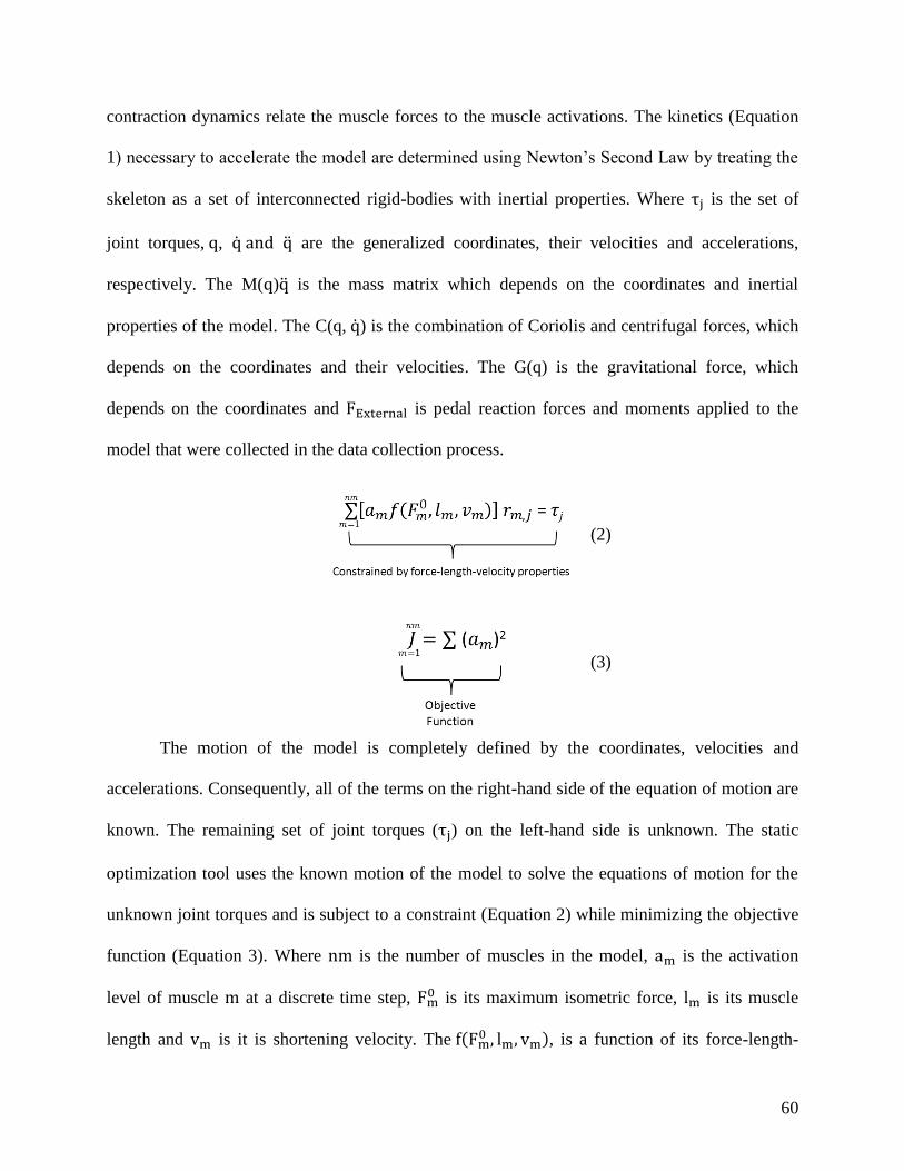
60
contraction dynamics relate the muscle forces to the muscle activations. The kinetics (Equation
1) necessary to accelerate the model are determined using Newton’s Second Law by treating the
skeleton as a set of interconnected rigid-bodies with inertial properties. Where is the set of
joint torques, , are the generalized coordinates, their velocities and accelerations,
respectively. The M( ) is the mass matrix which depends on the coordinates and inertial
properties of the model. The C(q, ) is the combination of Coriolis and centrifugal forces, which
depends on the coordinates and their velocities. The G(q) is the gravitational force, which
depends on the coordinates and is pedal reaction forces and moments applied to the
model that were collected in the data collection process.
The motion of the model is completely defined by the coordinates, velocities and
accelerations. Consequently, all of the terms on the right-hand side of the equation of motion are
known. The remaining set of joint torques ( ) on the left-hand side is unknown. The static
optimization tool uses the known motion of the model to solve the equations of motion for the
unknown joint torques and is subject to a constraint (Equation 2) while minimizing the objective
function (Equation 3). Where is the number of muscles in the model, is the activation
level of muscle at a discrete time step, is its maximum isometric force, is its muscle
length and is it is shortening velocity. The , is a function of its force-length-
(2)
(3)
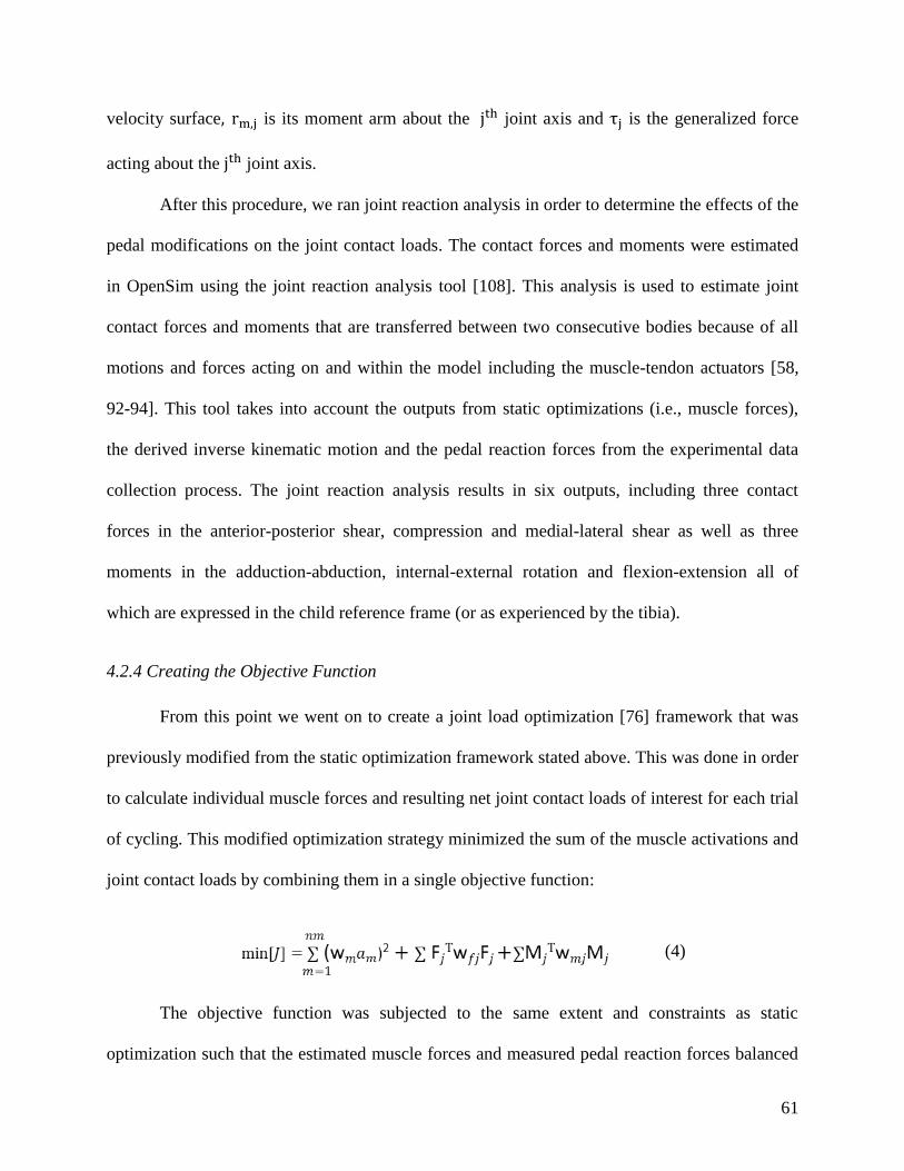
61
velocity surface is its moment arm about the joint axis and is the generalized force
acting about the joint axis.
After this procedure, we ran joint reaction analysis in order to determine the effects of the
pedal modifications on the joint contact loads. The contact forces and moments were estimated
in OpenSim using the joint reaction analysis tool [108]. This analysis is used to estimate joint
contact forces and moments that are transferred between two consecutive bodies because of all
motions and forces acting on and within the model including the muscle-tendon actuators [58,
92-94]. This tool takes into account the outputs from static optimizations (i.e., muscle forces),
the derived inverse kinematic motion and the pedal reaction forces from the experimental data
collection process. The joint reaction analysis results in six outputs, including three contact
forces in the anterior-posterior shear, compression and medial-lateral shear as well as three
moments in the adduction-abduction, internal-external rotation and flexion-extension all of
which are expressed in the child reference frame (or as experienced by the tibia).
4.2.4 Creating the Objective Function
From this point we went on to create a joint load optimization [76] framework that was
previously modified from the static optimization framework stated above. This was done in order
to calculate individual muscle forces and resulting net joint contact loads of interest for each trial
of cycling. This modified optimization strategy minimized the sum of the muscle activations and
joint contact loads by combining them in a single objective function:
The objective function was subjected to the same extent and constraints as static
optimization such that the estimated muscle forces and measured pedal reaction forces balanced
(4)

62
all inertial forces to carry out the measured cycling dynamic motion (Equation 1-3). In the
objective function shown in Equation 4, was the activation of the mth
muscle, which could
vary anywhere between an activation of 0 to an activation of 1. The was a muscle weighting
constant set to penalize a specific muscles, in order to constrain the muscle in order to activate or
not to activate. The joint contact force-weighting constants , and were set to penalize
the vector components of the jth
joint reaction force, . Similar to the joint forces, the joint
contact moment weighting constants, , and were set to penalize the vector
components of the jth
joint reaction moment, . The joint reaction moments and forces represent
the connecting joint contact loads experienced in the connecting joint structures and were
calculated using the joint reaction analysis in OpenSim.
4.2.5 Exploring the Muscle Activation and their Effects on the Joint Contact Loads
We then went on to simulate 10,320 trials (24 subjects x 5 Trials x 43 muscles x 2
different muscle weightings) of neutral cycling with a muscle coordination pattern that aimed to
minimize muscle activations without regard to the joint contact loads then we will examine the
effects on the associated joint contact loads. In this case, the generalized objective function
Equation 4 was reduced to:
This was done in order to determine the sensitivity of the tibiofemoral joint contact loads
due to an adopted variation in muscle activation patterns of individual muscles in the lower limb
by carrying out optimizations that altered the muscle activation weighting constants, , for
each individual lower extremity muscle. In OpenSim, normal static optimization has a default
muscle weighting ( ) of 1 for all associated lower limb muscles. We decided to alter the
(5)

63
weighting of the muscle activations that is shown in Equation 5 with a weighting of 0 or 100 for
one specific muscle at a time while keeping all other . Where a = 0 represents no
penalty and allowing the muscle to activate or be used as much as it wants or needs, conversely,
with a = 100 turns the muscle completely off and penalizes, prohibits activation or use of a
specific muscle.
This was done in order to investigate the variation in tibiofemoral joint contact loads due
to different muscle activation weighting by adopting or altering the activations of lower
extremity muscles, then examining a specific muscle’s influence on the joint contact loads. To do
this we performed two modified optimizations for each muscle and trial of neutral cycling. First,
before we altered the muscle activations of specific individual muscles, we ran normal static
optimization, which has a muscle weighting of 1 in order to observe normal muscle activation in
neutral cycling. Second, we altered the muscle weightings by taking a specific muscle and
altering the to exclude activation of a specific muscles, we executed a modified optimization
with changing previously established muscle weighting from = 1, to = 100 for a specific
muscle while for all other muscles was kept at 1. Third, in order to encourage muscle
activation, we then performed a modified static optimization procedure again with = 0 for a
specific muscle while all others kept it at 1. Performing these two optimizations for each muscle
of the lower limb determined the range of tibiofemoral joint contact loads caused by a variation
in muscle activations. We determined the change in peak tibiofemoral joint contact loads due to
activation of a muscle by computing the change between peak tibiofemoral joint contact loads
obtained from the static optimizations with = 0 and = 100 and the normal static
optimization carried out with all = 1.

64
4.2.6 A Muscle Coordination Pattern Minimizing Compressive Tibiofemoral Force
We simulated the 120 trials of neutral cycling with a muscle coordination pattern that
minimized the important contact loads of the tibiofemoral joint. In this case, the generalized
objective function (Equation 4) simplified to:
The weighting of the muscle activations, , was set to zero for all muscles in order to
not penalize any of the muscles and allow the muscles to adopt a muscle activation pattern that
would minimize the joint contact loads of the tibiofemoral joint. We only examined the joint
contact loads that have been linked to OA progression, which are the compressive contact load
and the adduction-abduction moment. The compressive tibiofemoral contact load was penalized
by setting its weighting constant,
and the adduction-abduction moment was
penalized by setting the weight constant,
, while all other joint contact forces
and moments were set to zero. This strategy determined a muscle coordination pattern that would
minimize the important tibiofemoral joint contact loads that would aid in slowing the progression
of medial compartment tibiofemoral OA.
4.2.5 Change in Tibiofemoral Forces Due to Maximized Muscle Activations
We went on to determine the change in tibiofemoral forces due to adopting a maximized
muscle activation pattern of the lower limb by performing optimizations with muscle activation
weighting constants, , for each muscle. The objective function in equation 4 is
simplified to:
(6)
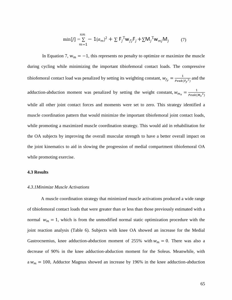
65
In Equation 7, , this represents no penalty to optimize or maximize the muscle
during cycling while minimizing the important tibiofemoral contact loads. The compressive
tibiofemoral contact load was penalized by setting its weighting constant,
and the
adduction-abduction moment was penalized by setting the weight constant,
while all other joint contact forces and moments were set to zero. This strategy identified a
muscle coordination pattern that would minimize the important tibiofemoral joint contact loads,
while promoting a maximized muscle coordination strategy. This would aid in rehabilitation for
the OA subjects by improving the overall muscular strength to have a better overall impact on
the joint kinematics to aid in slowing the progression of medial compartment tibiofemoral OA
while promoting exercise.
4.3 Results
4.3.1Minimize Muscle Activations
A muscle coordination strategy that minimized muscle activations produced a wide range
of tibiofemoral contact loads that were greater than or less than those previously estimated with a
normal , which is from the unmodified normal static optimization procedure with the
joint reaction analysis (Table 6). Subjects with knee OA showed an increase for the Medial
Gastrocnemius, knee adduction-abduction moment of 255% with . There was also a
decrease of 90% in the knee adduction-abduction moment for the Soleus. Meanwhile, with
a Adductor Magnus showed an increase by 196% in the knee adduction-abduction
(7)
66
moment. The Semitendinosus, showed a decrease of 93% in the adduction-abduction moment.
The Sartorius showed an increase of 119% for the knee compressive load with . As well
as Semitendinosus showed a decrease of 70% for the knee compressive load. With a
the Pectineus showed an increase of 287% for the knee compressive load. The Tibialis
Anterior decreased by 20% for the knee compressive load. Consequently, subjects with knee OA
mostly increased their knee adduction-abduction moment and compressive load with a
and a . While there was some success in decreasing the loads, they were mostly seen
in biarticular muscles.
Just like in the OA cohort, the tibiofemoral contact load depended on which muscle was
altered (Table 6). Subjects without knee OA showed an evident increase in the Vastus Medialis’s
knee adduction-abduction moment of 109% with . There was also an evident decrease in
the Tensor Fascia Lata, knee adduction-abduction moment of 33%. While for a
Semimembranosus showed an increase of 104% for the knee adduction-abduction moment. The
Medial Gastrocnemius, showed a decrease of 21% in the adduction-abduction moment. There
were no evident increases in the compressive load with . While the Vastus Medialis
showed a decrease of 18% for the knee compressive load. While the Semimembranosus with
a showed an increase of 51% for the knee compressive load. While Adductor Brevis
showed a decrease of 3% for the knee compressive load. There were many other muscles that
showed to have a noticeable effect on the joint contact loads (Figure 8, Table 6). Consequently
subjects without knee OA mostly increased their knee adduction-abduction moment with a
and Though their compressive load varied differently in both weightings
depending on the muscle and its specific function being altered.

67
4.3.2 Minimize Joint Contact Forces
A muscle coordination strategy that minimized tibiofemoral joint contact loads produced
lower contact loads than the forces that were previously estimated in normal joint reaction
analysis (Figure 9). Subjects with knee OA showed a significant decrease of 73% for the knee
adduction-abduction moment. There was a noticeable decrease in the compressive load of 96%.
For the subjects without knee OA there was a noticeable decrease in the for the knee adduction-
abduction moment of 72%. There was also an evident a decrease of 95% for the compressive
load. In general subject without knee OA with the given kinematics were affected more than
subjects without knee OA. All subjects were able to significantly lower their joint contact loads
as shown in Figure 9.
4.3.3 Maximizing the muscle force to minimize the joint loads
A muscle coordination strategy that maximized muscle activations while minimizing the
tibiofemoral forces, produced results that further decreased the peak and overall knee joint
contact loads while maximizing the muscle forces (Figure 9). Subjects with knee OA portrayed
an evident decrease in the knee adduction-abduction moment of 25%. While the compressive
load showed a decrease of 19%. For the subjects without knee OA there was also a noticeable
decrease in the knee adduction-abduction moment of 81%. However, for the compressive load
there was a decrease of 17% for the compressive load. Overall subject without knee OA with the
given kinematics were affected more than subjects without knee OA. Just like in the minimized
joint contact loads, all subjects reacted differently to the optimization protocol some lowered
their joint contact loads more while other did not as shown in Figure 9.

68
4.4 Discussion
Our results demonstrate that varying muscle activation patterns during stationary cycling
can induce large variations in the joint contact loads at the tibiofemoral joint. Evaluating a varied
set of different in muscle activation weightings was used in order to evaluate whether a strategy
minimizing activations patterns by either turning the muscles on without penalty or turning the
muscles off. This was done in order to examine the effects a specific muscles has on the
tibiofemoral contact loads to see if they was consistent or different (increase or decrease) from
previously estimated joint contact loads. Subjects with knee OA seemed to have achieved more
intense results in the knee adduction-abduction moment and compressive load with both muscle
variation weightings as they mostly increased; although, this largely depended on the specific
muscle function during the cycling motion as well as the joint it spanned. Like the OA cohort
subjects, subjects without OA also experienced mostly increases in contact loads compared to
normal except with a muscle weighting of zero for the compressive load. These results suggest
that treatment interventions meant for retraining muscle coordination should target the specific
muscles in the OA cohort that showed a reduction in the adduction-abduction moment and the
compressive load in order to reduce tibiofemoral contact loads. Alternatively, avoid the use of
these specific muscles, which increase the tibiofemoral loading with an in order to lower
the mechanical loading at the knee.
We determined the potential for subjects with and without OA to decrease tibiofemoral
joint contact loads during cycling by allowing the lower limb to adopt a muscle activation pattern
that would achieve the desirable (i.e. minimized) joint contact loads. Our model achieved
tibiofemoral forces that were lower than previously estimated in joint reaction analysis with a
normal static optimization by adopting different muscle coordination patterns that
69
achieved the desired results. Subjects with knee OA were able to reduce their contact loads more
than the healthy subjects. We determined a maximized muscle activation pattern while
minimizing the joint contact loads for subjects with and without OA. Our model achieved
tibiofemoral forces that were lower in all cases than the normal joint reaction analysis and higher
than the results of the minimized contact loads, which was to be expected since we wanted to
increase the lower extremities muscular strength. This provides a range of feasible joint contact
loads that is different from previously calculated or observed in normal cycling kinematics,
which had a huge effect overall in both cohorts and on the important joint contact loads.
This current study has a few research challenges and our results should be interpreted in
the context of these limitations. First, a modified static optimization framework was used to
estimate muscle forces that were used in simulations over the use of a dynamic optimization
framework. Static optimization was selected due to the large amount of simulations created
(10,560) and the low computational expenditure associated with this approach. In addition, it has
been previously validated by Anderson and Pandy (2001) and found that static and dynamic
optimization is fundamentally equivalent for predicting in vivo quantities such as muscle forces
and joint contact loads for walking [56]. Consequently, the results from static optimization
should not alter the conclusions that we have drawn during this study. Secondly, the data
collection procedure did not include EMG for the 24 subjects. Even though we did not have
EMG data, our data was previously matched with normal static optimization during neutral
cycling to experimentally collected data from a study that was carried out by Neptune et al. [98]
investigating elite male athletes. Additionally, the joint contact load values reported in this study
characterize the whole tibiofemoral joint contact loads, rather than the specific narrowing in
exhibited in the radiographic evidence of the subjects with medial compartment tibiofemoral
70
OA. However, adduction-abduction moment relates to the uneven contact loads that compress
the medial and lateral compartments and contribution to the bone-on-bone contact characteristics
leading to tibiofemoral OA, thus finding any alteration from the normal contact load shows an
improvement or a limitation for subjects with medial tibiofemoral OA. Lastly, the pedal reaction
force data was collected only from one instrument pedal; this constraint will not alter the results
of the analyzed leg would just be used as a comparison within subjects. Although the joint
contact loads may vary if we made different modeling assumptions; however, our conclusions
regarding the relative changes in references to the estimated joint contact loads in cycling would
not be likely to change drastically. This is due to the face that the same modeling assumptions
remained consistent throughout all the simulations and across all subjects.
What has not been answered in previous literature until now is the question of whether or
not subjects with and without knee OA have can further reduce their tibiofemoral contact loads
by adopting varied set of muscle activations patterns. Previous literature focused on walking with
an instrumented knee implant in a healthy subject rather than cycling [76]. For example DeMers
et al, [76] used this modified optimization framework in walking. Our study investigated both
subjects with and without knee OA and none of the subjects were competitive cyclists.
Additionally, previous literature has not examined the use of an optimization framework in knee
OA and healthy cohorts during neutral cycling. This newly found information on muscle
activations may help improve rehabilitation strategies targeting muscle strength and improving
joint function.
This study identified specific muscles that substantially affect tibiofemoral forces during
stationary cycling as well as adopted a muscle activation pattern that minimized the joint contact
loads. This research has provided a range of feasible muscle activations and now the question is

71
to examine these muscle activations that were obtained in minimized knee joint loading and
maximized muscle activations, to figure out how these activations can be obtained as well as
what are different in these muscle activations than actual cycling kinematics. Future work needs
to discover how to target a specific muscle in order to activate or deactivate it in order to obtain
the associated decrease in the contact loads. It should also focus on if and in what pedal
modifications can obtain the desired and calculated minimized joint contact loads and the
maximized muscle activation while minimizing the important joint contact loads (i.e.,
compressive and adduction-abduction moment). Further testing is also needed with modeling and
simulation, in conjunction with biofeedback. To figure out how to activate and deactivate a
specific muscle group to achieve lower tibiofemoral contact loads as well as test the utility of
different pedal modifications in order to be able to achieve or target different muscle groups
that’s have the greatest affect in lowering the joint contact loads.
72
CHAPTER V
CONCLUSION
5.1 Significance of Research
Effective OA rehabilitation training and a proper subject-specific treatment would have
enormous potential to significantly reduce the progression of OA degeneration in not only the
subjects with OA but would also help the subjects without OA as a preventative measure.
However, improving the effectiveness of rehabilitation and treatment of OA programs are
challenging because the cause-effect relationship between muscle function and joint
biomechanics with respect to OA progression is not well understood. Dynamic modeling and
simulations provide the capability to determine the meaningful role of muscles contributions,
which is essential to understanding their effect and relationship with the joint contact loads.
Developing dynamic simulations helps to define the biomechanical aspects that influence knee
motion in order to provide a better treatment protocol. We anticipated that the insights gained
from this work would aid in providing new guidelines for designing subject-specific OA
treatment programs resulting in a significant drop in OA progression and possible pain.
The biomechanical models and computational tools developed, will find broad
application. Numerous studies have been performed to record neuromuscular excitation patterns,
understand muscles contraction dynamics, characterize musculoskeletal geometry and quantified
multijoint movements and kinematics. However, linking the detailed knowledge of these
elements of the neuro-musculoskeletal system to create an integrated understanding of normal
and pathological movement remains a major challenge in the application of biomechanics to a
wide range of clinical problems and basic science research. The long-term goal of this research
was to provide a scientific framework for planning, evaluation and improvement of subject-
specific rehabilitation for individuals with knee OA.
73
This work developed methodologies for interpreting the dynamic functions of muscles
during cycling movement, generated novel biomechanical optimization approaches to
undercover the muscles forces and joint contact loads during movement and a general
computational framework for pursuing further research. The simulations we developed were
implemented in freely available musculoskeletal modeling and simulation software [57] which
enables us to share the results with other research groups. Over the past few years, there have
been more than 165,103 downloads of models, simulations and software from the project’s
website (see simtk.org/home/OpenSim) with over 28,096 active users of these freely available
biomechanics tools. This project will continue this development. Thus, the aims of this proposed
research program were achieved; this work will have a broad impact on the basic studies of
neuromuscular biomechanics and for clinical treatment and prevention for the subjects with and
without knee OA.
5.2 Research Innovation
The main beliefs that direct the relationships between muscles contributions and dynamic
repetitive movement in individuals during stationary cycling have not been discovered until now.
For decades, experimental research and approaches have driven our understanding of
biomechanics of the human body. However, the lack of ability to experimentally measure muscle
forces and ascertain the cause-effect relationships between muscle forces contribution to joint
biomechanics has limited research progress over the years. The trouble in defining an individual
muscles’ contribution to dynamics movement stems from the circumstance that muscles
accelerate and affect joints that they do not span and body segments in which they do not
innervate. Thus in order to determine a muscles influence on the movement required a new
approach motivated by the use of an instrumental set of tools to accomplish this task.

74
Muscle-actuated inverse dynamic simulations provide a scientific framework, in
combination with complementing experimental approaches for estimating important variables
and identifying cause-and-effect relationships. In this study, muscle-actuated inverse dynamic
simulations, optimization and motion capture analysis were used to bridge the gap in providing a
better understanding of dynamic human movement.
This work advanced rudimentary knowledge and understanding in this arena. It engaged
the experimental capabilities of physicians, physical therapist and rehabilitation scientists,
engineers’ use of computer simulations, mathematicians and computer scientist to address the
biomechanical importance of the research questions. An additional advantage of this research
was that the creation of subject-specific musculoskeletal models from a freely available open
source software (OpenSim). The results that this dissertation discovered will advance research
and accelerate the understanding of the underlying fundamental mechanisms behind
biomechanical injuries and treatment of OA.
5.3 Fundamental Contributions
The objective of this research was to determine individual muscle force contributions
during cycling in different pedal modifications, in order to assess how individual muscles, affect
the tibiofemoral joint contact loads. As well as adapting a varied muscle activation patterns
designed to minimize the important joint contact loads. The work presented here was able to
accomplish all of these objectives.
Static and biomechanical optimizations are techniques that have been previously used to
study muscle contribution to walking, running and other treatment modifications. However, the
vital importance was in the development of the subject-specific models and application of these
tools to investigate their effect on subjects with and without OA in order to examine and identify
75
the fundamental differences between and within the cohorts. Subject-specific models, were
customized and tailored based on the subject parameters from data collection and our specific
research question that we posed. Since, OA treatment is often based upon the clinicians, clinical
experience to treatment plans. The models that are commonly used by physicians and
constructed using data from other patients, these clinical outcomes are unreliable. We developed
an approach using computational models based on the engineering mechanics and optimization
to predict subject-specific outcomes. We simulated how cycling modifications that were used as
a possible treatment option for knee OA, which may properly strengthen the lower extremity
muscles while decreasing the subjects important joint contact loads (compressive force and the
knee adduction-abduction contact moment) for specific subjects.
5.4 Future Work
This study identified specific muscles forces and joint contact loads in subjects with and
with tibiofemoral OA during stationary cycling. As well as found a range of feasible muscle
activations that are possible to reduce the joint contact loads. However, the question is to
examine the muscle activations that were obtained in study three, to figure out how these
activations can be obtained through different cycling modifications, see how to effectively
achieve the desired activations and muscle forces mentally and physically receiving biofeedback.
Future work should test other cycling modifications in order to discover how to obtain the
desired muscle activations, the minimized joint contact loads and the maximized muscle
activation. Since our model is lacking biofeedback and does not experience any pain these
activations that were achieved through optimization may not be feasible for all subjects.
Research also needs to obtain a possible way to receive and give biofeedback through simulated
human movement.

76
LIST OF REFERENCES
77
1. C.D.C, Prevalence and most common causes of disability among adults--United States,
2005. MMWR Morb Mortal Wkly Rep, 2009. 58(16): p. 421-6.
2. Hootman, J.M., Helmick, C.G., Brady, T.J., A public health approach to addressing
arthritis in older adults: the most common cause of disability. Am J Public Health, 2012.
102(3): p. 426-33.
3. Gerbrands, T.A., Pisters, M.F., Vanwanseele, B., Individual selection of gait retraining
strategies is essential to optimally reduce medial knee load during gait. Clin Biomech
(Bristol, Avon), 2014. 29(7): p. 828-34.
4. Lawrence, R.C., et al., Estimates of the prevalence of arthritis and other rheumatic
conditions in the United States. Arthritis Rheum, 2008. 58: p. 26-35.
5. Mundermann, A., C.O. Dyrby, and T.P. Andriacchi, Secondary gait changes in patients
with medial compartment knee osteoarthritis: increased load at the ankle, knee, and hip
during walking. ARTHRITIS & RHEUMATISM, 2005. 59(9): p. 2835-44.
6. Lawrence, R.C., et al., Estimates of the Prevalence of Arthritis and Other Rheumatic
Conditions in the United States. Arthritis & Rheumatism, 2008. 58(1): p. 26-35.
7. Hootman, J.M. and C.G. Helmick, Projections of US prevalence of arthritis and
associated activity limitations. ARTHRITIS & RHEUMATISM, 2006. 54(1): p. 226-229.
8. Semanik, P., R.W. Chang, and D.D. Dunlop, Aerobic Activity in Prevention & Symptom
Control of Osteoarthritis. PM & R: the journal of injury, function, and rehabilitation,
2012. 4(5 0): p. S37-S44.
9. Dillon, C.F., et al., Prevalence of knee osteoarthritis in the United States: arthritis data
from the Third National Health and Nutrition Examination Survey 1991–1994. Journal of
Rheumatology, 2006. 33(11): p. 2271-2279.
10. Cheng, Y.J., et al., Prevalence of Doctor-Diagnosed Arthritis and Arthritis-Attributable
Activity Limitation — United States, 2007–2009. 2010. 59(39): p. 2238-2239.
11. Rozen, D.N. Biomechanical aspects of knee osteoarthritis and AposTherapy. Scientific
Approach 2013; Available from: http://apostherapy.com/en/healthcare-
professionals/treatment-packages/biomechanical-aspects.
12. Shull, P.B., et al., Muscle force modification strategies are not consistent for gait
retraining to reduce the knee adduction moment in individuals with knee osteoarthritis.
Journal of Biomechanics, 2015. 48: p. 3163-69.
13. Felson, D.T., et al., Osteoarthritis: new insights. Part 1: the disease and its risk factors.
Ann Intern Med, 2000. 133(8): p. 635-46.
14. Felson, D.T., et al., Evidence for a Mendelian gene in a segregation analysis of
generalized radiographic osteoarthritis: the Framingham Study. Arthritis &
Rheumatism, 1998. 41(6): p. 1064-71.
15. Felson, D.T., The Epidemiology of Knee Osteoarthritis -Results from the Framingham
Osteoarthritis Study. Semin Arthritis Rheum, 1990. 20: p. 42-50.
16. Nevitt, M.C. and D.T. Felson, Sex hormones and the risk of osteoarthritis in women:
Epidemiological evidence. Ann Rheum Dis, 1996. 55: p. 673-6.
17. Srikanth, V.K., et al., A meta-analysis of sex differences prevalence, incidence and
severity of osteoarthritis. Osteoarthritis and Cartilage, 2005. 13(9): p. 769-81.
18. Theis, K.A., C.G. Helmick, and J.M. Hootman, Arthritis burden and impact are greater
among U.S. women than men: intervention opportunities. Journal of Womens Health,
2007. 16: p. 441-53.

78
19. Baker, K., et al., Quadriceps weakness and its relationship to tibiofemoral and
patellofemoral knee osteoarthritis in Chinese: the Beijing osteoarthritis study. Arthritis
Rheum, 2004. 50(6): p. 1815-21.
20. Slemenda, C., et al., Reduced quadriceps strength relative to body weight: a risk factor
for knee osteoarthritis in women? Arthritis & Rheumatism, 1998. 41(11): p. 1951-9.
21. Slemenda, C., et al., Quadriceps weakness and osteoarthritis of the knee. Ann Intern
Med, 1997. 127: p. 97-104.
22. Lohmander, L.S., et al., High prevalence of knee osteoarthritis, pain, and functional
limitations in female soccer players twelve years after anterior cruciate ligament injury.
Arthritis & Rheumatism, 2004. 50: p. 3145-52.
23. Felson, D.T., Occupational physical demands, knee bending, and knee osteoarthritis:
results from the Framingham Study. Journal of Rheumatology, 1991. 18(10): p. 1587-92.
24. Coggon, D., et al., Occupational activities and osteoarthritis of the knee. Arthritis &
Rheumatism, 2000. 43(7): p. 1449-9.
25. Focht, B.C., et al., Exercise, self-efficacy, and mobility performance in overweight and
obese older adults with knee osteoarthritis. Arthritis & Rheumatism, 2005. 53: p. 659-65.
26. Messier, S.P., et al., Do older adults with knee osteoarthritis place greater loads on the
knee during gait? A preliminary study. Arch Phys Med Rehabil, 2005. 86(4): p. 703-9.
27. Messier, S.P., Obesity and osteoarthritis: disease genesis and nonpharmacologic weight
management. Rheum Dis Clin North Am, 2008. 34(3): p. 713-29.
28. Zhang, W., et al., OARSI recommendations for the management of hip and knee
osteoarthritis, Part I: Critical appraisal of existing treatment guidelines and systematic
review of current research evidence. OsteoArthritis and Cartilage, 2007. 15: p. 981-1000.
29. Zhang, W., et al., OARSI recommendations for the management of hip and knee
osteoarthritis, Part II: OARSI evidence-based, expert consensus guidelines. 2008.
30. Hinman, R.S., et al., Lateral Wedges in Knee Osteoarthritis: What Are Their Immediate
Clinical and Biomechanical Effects and Can These Predict a Three-Month Clinical
Outcome? Arthritis & Rheumatism, 2008. 59(3): p. 408–415.
31. Shull, P.B., Silder, A., Shultz, R., Dragoo, J.L., Besier, T.F., Delp, S.L., and Cutkosky,
M.R., Six-Week Gait Retraining Program reduces Knee Adduction Moment, Reduces
Pain, and Improves Function for Individuals with Medial Compartment Knee
Osteoarthritis. Journal of Orthopaedic Resarch, 2013. 31: p. 1020-1025.
32. Shull, P.B., Shultz, R., Silder, A., Dragoo, J.L., Besier, T.F., Cutkosky, M.R., Delp, S.L.,
Toe-in gait reduces the first peak knee adduction moment in patients with medial
compartment knee osteoarthritis. Journal of Biomechanics, 2013. 46: p. 122-128.
33. Mangione, K.K., et al., The effects of high-intensity and low-intensity cycle ergometry in
older adults with knee osteoarthritis. J Gerontol A Biol Sci Med Sci., 1999. 54(4): p.
184-90.
34. Akbarshahi, M., et al., Subject-specific evaluation of patellofemoral joint biomechanics
during functional activity. Medical Engineering Physics, 2014. 36(9): p. 1122-1133.
35. Ericson, M., On the biomechanics of cycling. A study of joint and muscle load during
exercise on the bicycle ergometer. Scand J Rehabil Med Suppl., 1986. 16: p. 1-43.
36. Ericson, M.O., Mechanical muscular power output and work during ergometer cycling at
different work loads and speeds. European Journal of Applied Physiology, 1988. 57: p.
382-7.
79
37. Ericson, M.O., et al., Power output and work in different muscle groups during
ergometer cycling. European Journal of Applied Physiology, 1986. 55: p. 229-235.
38. Ericson, M.O. and R. Nissell, Tibiofemoral joint forces during ergometer cycling. The
American Journal of Sports Medicine, 1986. 14(4): p. 285-90.
39. Sanderson, D.J. and A. Black, The effect of prolonged cycling on pedal forces. Journal of
Sports Sciences, 2003. 21(3): p. 191-99.
40. Bailey, M.P., F.J. Maillardet, and N. Messenger, Kinematics of cycling in relation to
anterior knee pain and patellar tendinitis. J Sport Sci, 2003. 21: p. 649-657.
41. Too, D., Biomechanics of Cycling and Factors Affecting Performance. Sports Medicine,
2012. 10(5): p. 286-302.
42. Schipplein, O.D. and T.P. Andriacchi, Interaction between Active and Passive Knee
Stabilizers during Levelwalking. J Orthopaed Res, 1991. 9: p. 113-9.
43. Miyazaki, T., et al., Dynamic load at baseline can predict radiographic disease
progression in medial compartment knee osteoarthritis. Ann Rheum Dis, 2002. 61: p.
617-22.
44. Chakravarty, E.F., et al., Long distance running and knee osteoarthritis - A prospective
study. Am J Prev Med, 2008. 35: p. 133-8.
45. Ahlbäck, S., Osteoarthrosis of the knee. A radiographic investigation. Acta Radioligica
Diagnosis Supplement, 1968. 277.
46. Cerejo, R., et al., The influence of alignment on risk of knee osteoarthritis progression
according to baseline stage of disease. Arthritis & Rheumatism, 2002. 46: p. 2332-36.
47. Hurley, M.V., THE ROLE OF MUSCLE WEAKNESS IN THE PATHOGENESIS OF
OSTEOARTHRITIS. Rheumatic Disease Clinics in north America, 2005. 25(2): p. 283-
298.
48. Hurley, M.V., Muscle Dysfunction and Effective Rehabilitation of Knee Osteoarthritis:
What We Know and What We Need To Find Out. Arthritis & Rheumatism, 2003. 49(3):
p. 444-52.
49. Walter, J.P., Korkmaz, N, Fregly, B.J., Pandy, M.G., Contribution of Tibiofemoral Joint
Contact to Net Loads at the Knee in Gait. Journal of Orthopaedic Resarch, 2015. 33(7):
p. 1054-60.
50. Baliunas, A.J., Hurwitz, D.E., Ryals, A.B., Karrar, A., Case, J.P., Block, J.A.,
Andriacchi, T.P., Increased knee joint loads during walking are present in subjects with
knee osteoarthritis. Osteoarthritis Cartilage, 2002. 10(7): p. 573-9.
51. Gardner, J.K., et al., Acute effects of lateral shoe wedges on joint biomechanics of
patients with medial compartment knee osteoarthritis during stationary cycling. Journal
of Biomechanics, 2016. 49: p. 2817-23.
52. Gardner, J.K., et al., Effects of Toe-in Angles on Knee Biomechanics in Cycling of
Patients with Medial Knee Osteoarthritis. Clin Biomechanics, 2015. 30: p. 276–282.
53. Kautz, S.A., M.L. Hull, and R.R. Neptune, A Comparison of Muscular Mechanical
Energy-Expenditure and Internal Work in Cycling. Journal of Biomechanics, 1994.
27(12): p. 1459-1467.
54. MacIntosh, B.R., R.R. Neptune, and J.F. Horton, Cadence, power, and muscle activation
in cycle ergometry. Medicine and Science in Sports and Exercise, 2000. 32(7): p. 1281-
1287.
55. Anderson, F.C. and M.G. Pandy, Dynamic optimization of human walking. J Biomech
Eng, 2001. 123(5): p. 381-90.

80
56. Anderson, F.C. and M.G. Pandy, Static and dynamic optimization solutions for gait are
practically equivalent. Journal of Biomechanics, 2001. 34(2): p. 153-61.
57. Delp, S.L., et al., OpenSim: open-source software to create and analyze dynamic
simulations of movement. IEEE Trans Biomed Eng, 2007. 54(11): p. 1940-50.
58. Thelen, D.G., Adjustment of muscle mechanics model parameters to simulate dynamic
contractions in older adults. J Biomech Eng, 2003. 125(1): p. 70-7.
59. Thelen, D.G. and F.C. Anderson, Using computed muscle control to generate forward
dynamic simulations of human walking from experimental data. Journal of Biomechanics,
2006. 39(6): p. 1107-15.
60. Thelen, D.G., F.C. Anderson, and S.L. Delp, Generating dynamic simulations of
movement using computed muscle control. Journal of Biomechanics, 2003. 36(3): p. 321-
8.
61. Hamner, S.R., A. Seth, and S.L. Delp, Muscle contributions to propulsion and support
during running. Journal of Biomechanics, 2010. 43(14): p. 2709-16.
62. Fregly, B.J., et al., Grand challenge competition to predict in vivo knee loads. J Orthop
Res, 2012. 30(4): p. 503-13.
63. Morrison, J.B., The mechanics of the knee joint in relation to normal walking. Journal of
Biomechanics, 1970. 3: p. 51-61.
64. Bei, Y. and B.J. Fregly, Multibody dynamic simulation of knee contact mechanics.
Medical Engineering & Physics, 2004. 26(9): p. 777-89.
65. Lin, Y.C., et al., Simultaneous prediction of muscle and contact forces in the knee during
gait. Journal of Biomechanics, 2010. 43(5): p. 945-52.
66. Hast, M.W. and S.J. Piazza, Dual-joint modeling for estimation of total knee replacement
contact forces during locomotion. Journal of Biomechanical Engineering, 2013. 135(2):
p. 021013.
67. Thelen, D.G., K.W. Choi, and A.M. Schmitz, Co-simulation of neuromuscular dynamics
and knee mechanics during human walking. Journal of Biomechanical Engineering,
2014. 136(2): p. 021033.
68. Yang, N.H., et al., Effect of frontal plane tibiofemoral angle on the stress and strain at
the knee cartilage during the stance phase of gait. Journal of Orthopaedic Research,
2010. 28(12).
69. Gerus, P., et al., Subject-specific knee joint geometry improves predictions of medial
tibiofemoral contact forces. Journal of Biomechanics, 2013. 46: p. 2778-2786.
70. Hsu, R.W., et al., Normal axial alignment of the lower extremity and load-bearing
distribution at the knee. Clinical Orthopaedics and Related Research, 1990. 225: p. 215-
227.
71. Hurwitz, D.E., et al., The knee adduction moment during gait in subjects with knee
osteoarthritis is more closely correlated with static alignment than radiographic disease
severity, toe out angle and pain. Journal of Orthopaedic Research, 2002. 20(1): p. 101-
107.
72. Halder, A., et al., Influence of limb alignment on mediolateral loading in total knee
replacement: in vivo measurements in five patients. Journal of Bone and Joint Surgery:
American Volume, 2012. 94: p. 1023-29.
73. Moreland, J.R., L.W. Bassett, and G.J. Hanker, Radiographic analysis of the axial
alignment of the lower extremity. Journal of Bone and Joint Surgery: American Volume,
1987. 69(5): p. 745-9.

81
74. Winby, C.G., et al., Muscle and external load contribution to knee joint contact loads
during normal gait. Journal of Biomechanics, 2009. 42(14): p. 2294-2300.
75. Kumar, D., K.S. Rudolph, and K.T. Manal, EMG-driven modeling approach to muscle
force and joint load estimations: Case study in knee osteoarthritis. Journal of
Orthopaedic Research, 2011. 30(3): p. 377-83.
76. DeMers, M.S., S. Pal, and S.L. Delp, Changes in Tibiofemoral Forces due to Variations
in Muscle Activity during Walking. Ortho Research Society, 2014: p. 1-8.
77. Bergmann, G., et al., Hip contact forces and gait patterns from routine activities. J
Biomech, 2001. 34(7): p. 859-71.
78. Richards, C. and J.S. Higginson, Knee contact force in subjects with symmetrical OA
grades: differences between OA severities. J Biomech, 2010. 43(13): p. 2595-600.
79. Peat, G., R. McCarney, and P. Croft, Knee pain and osteoarthritis in older adults: a
review of community burden and current use of primary health care. Ann Rheum Dis,
2001. 60: p. 91-7.
80. Aglietti, P. and P.P. Menchetti, Distal femoral varus osteotomy in the valgus
osteoarthritic knee. American Journal of Knee Surgery, 2000. 13(2): p. 89-95.
81. Coventry, M.B., D.M. Ilstrup, and S.L. Wallrichs, Proximal tibial osteotomy. A critical
long-term study of eighty-seven cases. Journal of Bone and Joint Surgery: American
Volume, 1993. 75(2): p. 196-201.
82. Edgerton, B.C., E.M. Mariani, and B.F. Morrey, Distal femoral varus osteotomy for
painful genu valgum. A five-to-11 year follow-up study. Clinical Orthopaedics and
Related Research, 1993. 288(263-9).
83. Naudie, D., et al., The Insall award. Survivorship of the high tibial valgus osteotomy. A
10- to 22-year follow-up study. Clinical Orthopaedics and Related Research, 1999. 367:
p. 18-27.
84. Losina, E., et al., Cost-effectiveness of Total Knee Arthroplasty in the United States:
Patient Risk and Hospital Volume. Archives Internal Medicine, 2009. 169(12): p. 1113-
22.
85. Bachmeier, C.J., et al., A comparison of outcomes in osteoarthritis patients undergoing
total hip and knee replacement surgery. Osteoarthritis and Cartilage, 2001. 9: p. 137-146.
86. Roddy, E., W. Zhang, and M. Doherty, Is gout associated with reduced quality of life? A
case-control study. Rheumatology (Oxford), 2007. 46(9): p. 1441-1444.
87. Hurley, M.V. and N.E. Walsh, Effectiveness and clinical applicability of integrated
rehabilitation programs for knee osteoarthritis. Current Opinion in Rheumatology, 2009.
21: p. 171-6.
88. Bakker, M.F., et al., Tight control in treatment of rheumatoid arthritis efficacy and
feasibility. Ann. Rheum. Dis., 2007. 67: p. 190-195.
89. Fregly, B.J., J.A. Reinbolt, and T.L. Chmielewski, Evaluation of a patient-specific cost
function to predict the influence of foot path on the knee adduction torque during gait.
Computer Methods in Biomechanics and Biomedical Engineering, 2008. 11(1): p. 63-71.
90. Fregly, B.J., et al., Design of patient-specific gait modifications for knee osteoarthritis
rehabilitation. Ieee Transactions on Biomedical Engineering, 2007. 54(9): p. 1687-1695.
91. Rajagopal, A., et al., Full-Body Musculoskeletal Model for Muscle-Driven Simulation of
Human Gait. IEEE TRANSACTIONS ON BIOMEDICAL ENGINEERING,, 2016.
63(10).
82
92. Millard, M., et al., Flexing computational muscle: modeling and simulation of
musculotendon dynamics. J Biomech Eng, 2013. 135(2): p. 021005.
93. Winters, J.M., An improved muscle-reflex actuator for use in large-scale
neuromusculoskeletal models. Annals of Biomedical Engineering, 1995. 23(4): p. 359-74.
94. Zajac, F.E., Muscle and tendon: properties, models, scaling, and application to
biomechanics and motor control. Crit Rev Biomed Eng, 1989. 17(4): p. 359-411.
95. Anderson, F.C. and M.G. Pandy, A Dynamic Optimization Solution for Vertical Jumping
in Three Dimensions. Comput Methods Biomech Biomed Engin, 1999. 2(3): p. 201-231.
96. Delp, S.L., et al., An interactive graphics-based model of the lower extremity to study
orthopaedic surgical procedures. IEEE Trans Biomed Eng, 1990. 37(8): p. 757-67.
97. Hoy, M.G., F.E. Zajac, and M.E. Gordon, A musculoskeletal model of the human lower
extremity: the effect of muscle, tendon, and moment arm on the moment-angle
relationship of musculotendon actuators at the hip, knee, and ankle. Journal of
Biomechanics, 1990. 23(2): p. 157-69.
98. Neptune, R.R., S.A. Kautz, and M.L. Hull, The effect of pedaling rate on coordination in
cycling. Journal of Biomechanics, 1997. 30(10): p. 1051-1058.
99. Hicks, J.L., et al., Is My Model Good Enough? Best Practices for Verification and
Validation of Musculoskeletal Models and Simulations of Movement. Journal of
Biomechanical Engineering, 2015. 137(2): p. 1-24.
100. Neptune, R.R. and M.L. Hull, Evaluation of performance criteria for simulation of
submaximal steady-state cycling using a forward dynamic model. Journal of
Biomechanical Engineering-Transactions of the Asme, 1998. 120(3): p. 334-341.
101. Smak, W., R.R. Neptune, and M.L. Hull, The influence of pedaling rate on bilateral
asymmetry in cycling. Journal of Biomechanics, 1999. 32(9): p. 899-906.
102. Mornieux, G., et al., Influence of cadence, power output and hypoxia on the joint moment
distribution during cycling. Eur J Appl Physiol, 2007. 102: p. 11-18.
103. Neptune, R.R. and M.L. Hull, A theoretical analysis of preferred pedaling rate selection
in endurance cycling. Journal of Biomechanics, 1999. 32(4): p. 409-415.
104. Neptune, R.R. and M.L. Hull, Methods for determining hip movement in seated cycling
and their effect on kinematics and kinetics. Journal of Applied Biomechanics, 1996.
12(4): p. 493-507.
105. Leddy, J.J., et al., Effects of Exercise Rehabilitation on Muscle Function, Activity and
Joint Spaces in Patients with Osteoarthritis. Med Sci Sports Exer, 1994. 26(Suppl): p.
S142.
106. Kutzner, I., et al., Loading of the Knee Joint During Ergometer Cycling: Telemetric In
Vivo Data. Journal of Orthopaedic & Sports Physical Therapy, 2012. 42(12): p. 1032-38.
107. Walter, J.P., et al., Decreased knee adduction moment does not guarantee decreased
medial contact force during gait. J Orthop Res, 2010. 28(10): p. 1348-54.
108. Seth, A., et al., OpenSim: a musculoskeletal modeling and simulation framework for in
silico investigations and exchange. Iutam Symposium on Human Body Dynamics, 2011.
2: p. 212-232.
109. Neptune, R.R. and S.A. Kautz, Knee joint loading in forward versus backward pedaling:
implications for rehabilitation strategies. Clin Biomech (Bristol, Avon), 2000. 15(7): p.
528-35.
110. Kutzner, I., et al., Loading of the knee joint during activities of daily living measured in
vivo in five subjects. J Biomechanics, 2010. 43: p. 2164–2173.
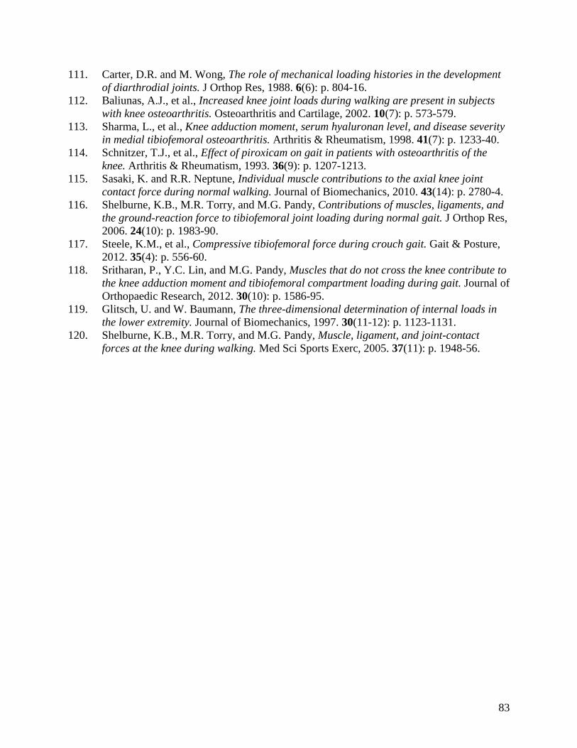
83
111. Carter, D.R. and M. Wong, The role of mechanical loading histories in the development
of diarthrodial joints. J Orthop Res, 1988. 6(6): p. 804-16.
112. Baliunas, A.J., et al., Increased knee joint loads during walking are present in subjects
with knee osteoarthritis. Osteoarthritis and Cartilage, 2002. 10(7): p. 573-579.
113. Sharma, L., et al., Knee adduction moment, serum hyaluronan level, and disease severity
in medial tibiofemoral osteoarthritis. Arthritis & Rheumatism, 1998. 41(7): p. 1233-40.
114. Schnitzer, T.J., et al., Effect of piroxicam on gait in patients with osteoarthritis of the
knee. Arthritis & Rheumatism, 1993. 36(9): p. 1207-1213.
115. Sasaki, K. and R.R. Neptune, Individual muscle contributions to the axial knee joint
contact force during normal walking. Journal of Biomechanics, 2010. 43(14): p. 2780-4.
116. Shelburne, K.B., M.R. Torry, and M.G. Pandy, Contributions of muscles, ligaments, and
the ground-reaction force to tibiofemoral joint loading during normal gait. J Orthop Res,
2006. 24(10): p. 1983-90.
117. Steele, K.M., et al., Compressive tibiofemoral force during crouch gait. Gait & Posture,
2012. 35(4): p. 556-60.
118. Sritharan, P., Y.C. Lin, and M.G. Pandy, Muscles that do not cross the knee contribute to
the knee adduction moment and tibiofemoral compartment loading during gait. Journal of
Orthopaedic Research, 2012. 30(10): p. 1586-95.
119. Glitsch, U. and W. Baumann, The three-dimensional determination of internal loads in
the lower extremity. Journal of Biomechanics, 1997. 30(11-12): p. 1123-1131.
120. Shelburne, K.B., M.R. Torry, and M.G. Pandy, Muscle, ligament, and joint-contact
forces at the knee during walking. Med Sci Sports Exerc, 2005. 37(11): p. 1948-56.

84
APPENDIX
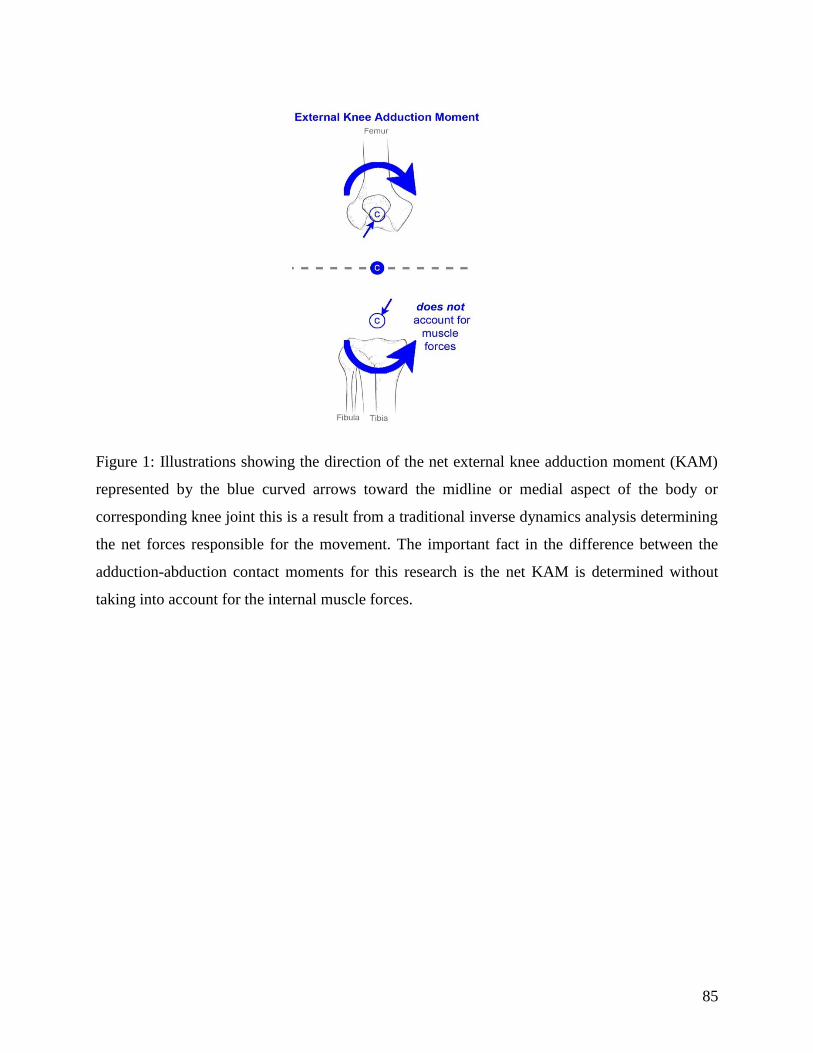
85
Figure 1: Illustrations showing the direction of the net external knee adduction moment (KAM)
represented by the blue curved arrows toward the midline or medial aspect of the body or
corresponding knee joint this is a result from a traditional inverse dynamics analysis determining
the net forces responsible for the movement. The important fact in the difference between the
adduction-abduction contact moments for this research is the net KAM is determined without
taking into account for the internal muscle forces.
86
Figure 2: Showing what the a) lower limb looks like when there is a change in alignments, b)
shows a closer look as to what happens at the knee joint when the joint changes from normal
alignment to a more varus or adducted alignment.
a) b)
Anterior
View
Posterior
View

87
Figure 3: Many factors contribute to knee joint contact loads associated with OA disease
progression and disability during movements. The transformations between experimental EMG
patterns and coordinated multi-joint movement (shaded region) are complicated. Furthermore, to
make exercise prescription decisions, clinicians must predict joint load changes after equipment
adjustments. Exercise prescription alters musculoskeletal geometry and multi-joint dynamics and
these changes are not easily measured. The long-term goal of our work is to provide a scientific
basis for exercise prescription that reduces harmful knee joint contact loads.

88
Figure 4: Four-step procedure to generate a subject-specific inverse dynamic simulation that
reproduces experimental data.
4. Test Solution
3. Solve Tracking Problem – Inverse Dynamic Simulation
Generic Model
1. Modify ModelSize differences
muscle-tendon lengthsmoment arms
2. Process DataPrepare for tracking
Clinical Data
Muscle Excitations
Inverse Dynamic
Simulation
Simulation Data
=?
No
Yes
Kinematics
Pedal Reaction Forces
Alter trackingparameters
and re-solve
Joint MomentsPedal Reaction Forces
Joint MomentsPedal Reaction Forces
Excitations
Use simulation for knee OA rehabilitation
Fm
am
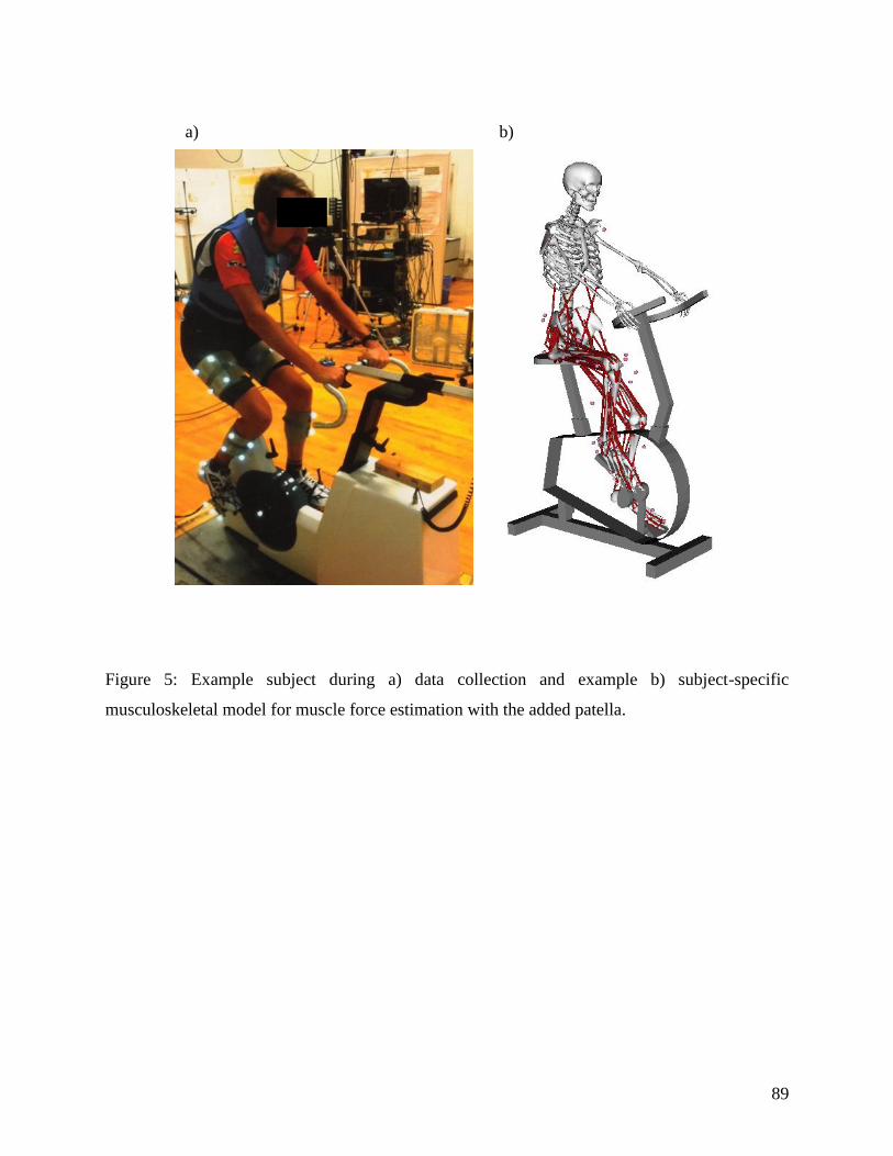
89
Figure 5: Example subject during a) data collection and example b) subject-specific
musculoskeletal model for muscle force estimation with the added patella.
a) b)

90
Table 1: A muscle force change identity grid showing the significant differences within each
cohort in all lower extremity muscles compared to neutral pedal condition, for the mean muscle
forces. Red indicates an increase, while blue indicates a decrease compared to neutral. The
darker the color the greater the increase or decrease.
5° 10° 5° 10° 5° 10° 5° 10°
Adductor Brevis + 7%BW
Adductor Longus
Adductor Magnus 1
Adductor Magnus 2
Gemellus
Gluteus Maximus 1
Gluteus Maximus 2
Gluteus Maximus 3
Gluteus Medius 1
Gluteus Medius 2
Gluteus Medius 3
Gluteus Minimus 1
Gluteus Minimus 2
Gluteus Minimus 3
Iliacus
Pectineus
Piriformis 0%BW
Psoas Major
Quadratus Femoris
Adductor Magnus 3
Biceps Femoris Long Head
Gracilis
Rectus Femoris
Sartorius
Semimembranosus
Semitendinosus
Tensor Fascia Lata
Biceps Femoris Short Head
Vastus Intermedius
Vastus Lateralis
Vastus Medialis
Lateral Gastrocnemius
Medial Gastrocnemius
Extensor Digitorum
Extensor Hallicus
Flexor Digitorum
Flexor Hallicus
Peroneus Brevis
Peroneus Longus
Peroneus Tertius
Soleus
Tibialis Anterior
Tibialis Posterior - 12%BW
Joint
Spanned
Hip
Hip to
Knee
Knee
Knee to
Ankle
Ankle
Muscle
OACohort Healthy Cohort
Lateral Wedge Toe-in Lateral Wedge Toe-in

91
Figure 6: Some of the muscles that showed the most change across the conditions (at least 3
conditions compared to neutral) with a threshold set at greater than 10%BW, observed for a)
mean and b) peak muscle forces normalized by %BW.

92
Table 2: A muscle force change identity grid showing the significant differences within each
cohort in all muscles compared to neutral, for the peak muscle forces. Red indicates an increase,
while blue indicates a decrease compared to neutral; while, the darker the color the greater the
increase or decrease.
5° 10° 5° 10° 5° 10° 5° 10°
Adductor Brevis + 16%BW
Adductor Longus
Adductor Magnus 1
Adductor Magnus 2
Gemellus
Gluteus Maximus 1
Gluteus Maximus 2
Gluteus Maximus 3
Gluteus Medius 1
Gluteus Medius 2
Gluteus Medius 3
Gluteus Minimus 1
Gluteus Minimus 2
Gluteus Minimus 3
Iliacus
Pectineus
Piriformis 0%BW
Psoas Major
Quadratus Femoris
Adductor Magnus 3
Biceps Femoris Long Head
Gracilis
Rectus Femoris
Sartorius
Semimembranosus
Semitendinosus
Tensor Fascia Lata
Biceps Femoris Short Head
Vastus Intermedius
Vastus Lateralis
Vastus Medialis
Lateral Gastrocnemius
Medial Gastrocnemius
Extensor Digitorum
Extensor Hallicus
Flexor Digitorum
Flexor Hallicus
Peroneus Brevis
Peroneus Longus
Peroneus Tertius
Soleus
Tibialis Anterior
Tibialis Posterior - 25%BW
Ankle
Joint
Spanned
Hip
Hip to
Knee
Knee
Knee to
Ankle
OA Cohort Healthy Cohort
Muscle Lateral Wedge Toe-in Lateral Wedge Toe-in
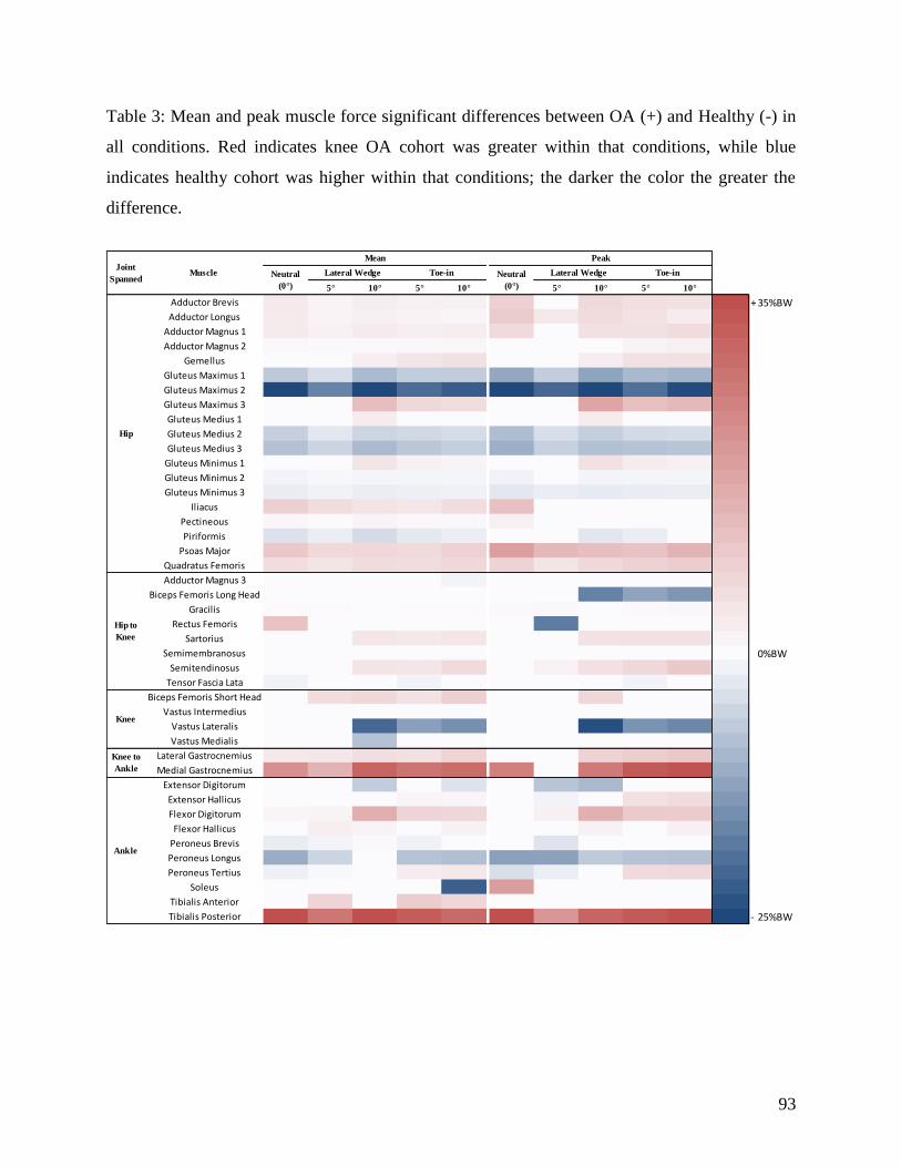
93
Table 3: Mean and peak muscle force significant differences between OA (+) and Healthy (-) in
all conditions. Red indicates knee OA cohort was greater within that conditions, while blue
indicates healthy cohort was higher within that conditions; the darker the color the greater the
difference.
5° 10° 5° 10° 5° 10° 5° 10°
Adductor Brevis + 35%BW
Adductor Longus
Adductor Magnus 1
Adductor Magnus 2
Gemellus
Gluteus Maximus 1
Gluteus Maximus 2
Gluteus Maximus 3
Gluteus Medius 1
Gluteus Medius 2
Gluteus Medius 3
Gluteus Minimus 1
Gluteus Minimus 2
Gluteus Minimus 3
Iliacus
Pectineous
Piriformis
Psoas Major
Quadratus Femoris
Adductor Magnus 3
Biceps Femoris Long Head
Gracilis
Rectus Femoris
Sartorius
Semimembranosus 0%BW
Semitendinosus
Tensor Fascia Lata
Biceps Femoris Short Head
Vastus Intermedius
Vastus Lateralis
Vastus Medialis
Lateral Gastrocnemius
Medial Gastrocnemius
Extensor Digitorum
Extensor Hallicus
Flexor Digitorum
Flexor Hallicus
Peroneus Brevis
Peroneus Longus
Peroneus Tertius
Soleus
Tibialis Anterior
Tibialis Posterior - 25%BW
Ankle
Mean
Neutral
(0°)
Lateral Wedge Toe-in
Peak
Neutral
(0°)
Lateral Wedge Toe-inJoint
SpannedMuscle
Hip
Hip to
Knee
Knee
Knee to
Ankle
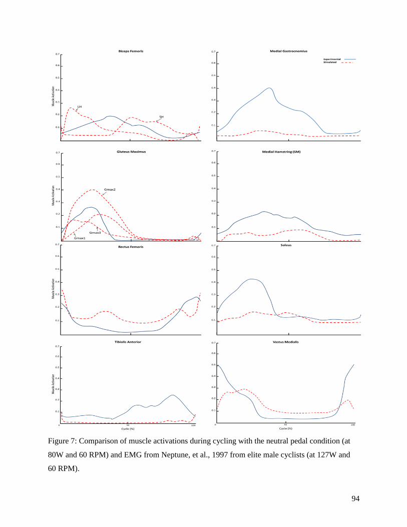
94
Figure 7: Comparison of muscle activations during cycling with the neutral pedal condition (at
80W and 60 RPM) and EMG from Neptune, et al., 1997 from elite male cyclists (at 127W and
60 RPM).
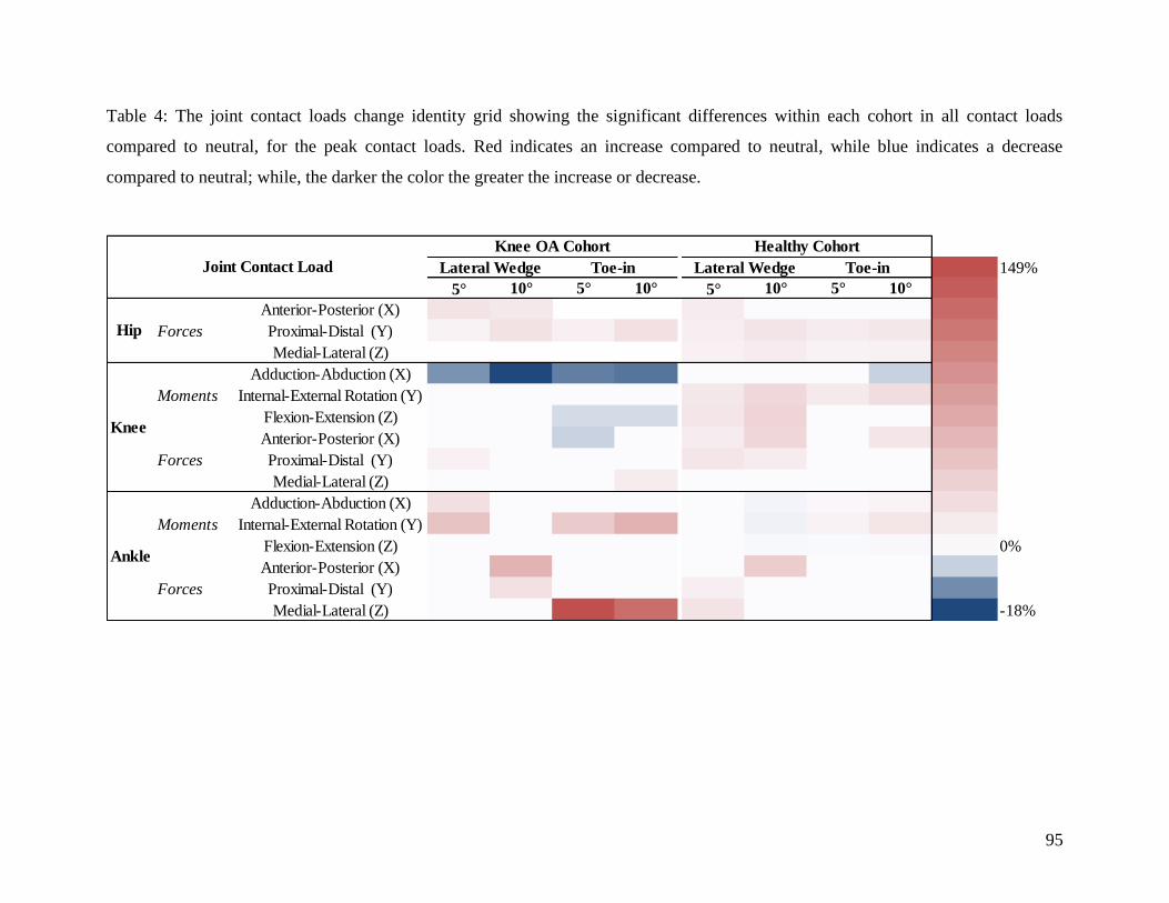
95
Table 4: The joint contact loads change identity grid showing the significant differences within each cohort in all contact loads
compared to neutral, for the peak contact loads. Red indicates an increase compared to neutral, while blue indicates a decrease
compared to neutral; while, the darker the color the greater the increase or decrease.
149%
5° 10° 5° 10° 5° 10° 5° 10°
Anterior-Posterior (X)
Forces Proximal-Distal (Y)
Medial-Lateral (Z)
Adduction-Abduction (X)
Moments Internal-External Rotation (Y)
Flexion-Extension (Z)
Anterior-Posterior (X)
Forces Proximal-Distal (Y)
Medial-Lateral (Z)
Adduction-Abduction (X)
Moments Internal-External Rotation (Y)
Flexion-Extension (Z) 0%
Anterior-Posterior (X)
Forces Proximal-Distal (Y)
Medial-Lateral (Z) -18%
Healthy Cohort
Lateral Wedge Toe-in
Hip
Lateral WedgeJoint Contact Load
Knee
Ankle
Knee OA Cohort
Toe-in

96
Table 5: The peak joint contact loads significant differences between OA (+) and Healthy (-) across all conditions. Red indicates knee
OA cohort was greater within the corresponding condition, while blue indicates healthy cohort was higher within that condition. The
darker the color the greater the difference.
5° 10° 5° 10°
Anterior-Posterior (X) 105%
Forces Proximal-Distal (Y)
Medial-Lateral (Z)
Adduction-Abduction (X)
Moments Internal-External Rotation (Y)
Flexion-Extension (Z)
Anterior-Posterior (X)
Forces Proximal-Distal (Y)
Medial-Lateral (Z) 0%
Adduction-Abduction (X)
Moments Internal-External Rotation (Y)
Flexion-Extension (Z)
Anterior-Posterior (X)
Forces Proximal-Distal (Y)
Medial-Lateral (Z) -89%
Ankle
Joint Contact LoadNeutral
(0°)
Lateral Wedge Toe-in
Hip
Knee

97
Table 6: A change identity grid showing the differences in the joint contact loads for the specific
muscles that changed from the normal joint reaction analysis. Results are shown for each cohort
for step 1’s muscle variation, for the subjects with and without knee OA in all muscles used
stands for a muscle weighting on 1, while don’t stands for. Red indicates an increase compared
to joint reaction analysis, while blue indicates a decrease compared to joint reaction analysis. The
darker the color the greater the increase or decrease.
W=0 W=100 W=0 W=100 W=0 W=100 W=0 W=100
Adductor Brevis 287%
Adductor Longus
Adductor Magnus 1
Adductor Magnus 2
Gemellus
Gluteus Maximus 1
Gluteus Maximus 2
Gluteus Maximus 3
Gluteus Medius 1
Gluteus Medius 2
Gluteus Medius 3
Gluteus Minimus 1
Gluteus Minimus 2
Gluteus Minimus 3
Iliacus
Pectineous
Piriformis
Psoas Major
Quadratus Femoris
Adductor Magnus 3
Biceps Femoris Long Head
Gracilis
Rectus Femoris
Sartorius
Semimembranosus
Semitendinosus
Tensor Fascia Lata
Biceps Femoris Short Head
Vastus Intermedius
Vastus Lateralis
Vastus Medialis
Lateral Gastrocnemius
Medial Gastrocnemius
Extensor Digitorum
Extensor Hallicus
Flexor Digitorum
Flexor Hallicus
Peroneus Brevis
Peroneus Longus
Peroneus Tertius
Soleus
Tibialis Anterior
Tibialis Posterior 90%
Ankle
Musle Names Joint(s)
Spanned
Hip
Hip to
Knee
Knee
Knee to
Ankle
Percent Change
Adduction-
Abduction Contact
Proximal-Distal
Compressive
Adduction-
Abduction Contact
Proximal-Distal
Compressive
Subjects with OA Subjects without OA

98
Figure 8: An example of a few muscle activations for a) subjects with OA and b) Subjects
without OA. This tested the sensitivity of the joint contact loads by altering the muscle
activations weightings from normal static optimization (weighting of 1) shown in blue. The red
line is a weighting of 0 indicating being able to use the muscle as much as it wants to carry out
the cycling movement. The green line is with a weighting of 100 penalizing the muscle with a
and not allowing it to be used. Lastly, the black line is for maximizing the muscle activations.

99
Figure 8 Continued
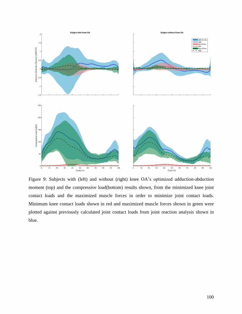
100
Figure 9: Subjects with (left) and without (right) knee OA’s optimized adduction-abduction
moment (top) and the compressive load(bottom) results shown, from the minimized knee joint
contact loads and the maximized muscle forces in order to minimize joint contact loads.
Minimum knee contact loads shown in red and maximized muscle forces shown in green were
plotted against previously calculated joint contact loads from joint reaction analysis shown in
blue.

101
VITA
Rachel L. Thompson was born in Rochester, NY, to the parents of Ann and Brian
Thompson. She is the third child of three: Matthew Thompson and Nicole Ray. She grew up and
attended elementary through high school in Rochester, NY. After her graduation, Rachel went on
to receive her Bachelors of Science degree in 2012 from Slippery Rock University in Health and
Environmental Science with a minor in Adapted Physical Activity. After Graduation, Rachel
went on to an internship at the Canandaigua Veteran Affairs (VA) Medical Center, where she
was introduced to the field of Biomedical Engineering. Then she went on to work for a year at
the University of Rochester Medical Center in the operating room, as a project assistant and then
ultimately a cardiac and vascular implant coordinator. Where her job was to order and receive
medical implants for specific surgeries from here, her passion for implants, prosthetics and
medical devices grew and she ultimately decided to pursue an advanced degree in Biomedical
Engineering. In her quest for knowledge and desire to learn in this field of Biomedical
Engineering, she attended the University of Tennessee where she received her Master’s degree in
2016. Finally, Rachel went on to pursue and ultimately complete a PhD in Biomedical
Engineering at the University of Tennessee. She graduated in May 2017. She is planning on
working in the medical research field.