The Musculoskeletal System
21
Learning Objectives 1. Describe muscle structure in terms of muscle cells, tendons, and bones. (p 76–82) 2. Describe the neuromuscular junction and explain the function(s) for each part. (p 77–78) 3. Describe the structure of a sarcomere. (p 77) 4. Describe the sliding filament theory of muscle contraction. (p 78–80) 5. Explain polarization, depolarization, and repolarization in terms of ions and charges. (p 79–80) 6. Name the energy sources for muscle contraction. (p 80–81) 7. Explain the importance of hemoglobin, myoglobin, oxygen debt, and lactic acid. (p 81) 8. Describe the difference between antagonistic and synergistic muscles. (p 82) 9. State the major muscles of the body and their functions. (p 83–92) The Musculoskeletal System Skidplate: © Photodisc; Cells © ImageSource/age fotostock CHAPTER 4 75 © Jones & Bartlett Learning, LLC. NOT FOR SALE OR DISTRIBUTION.
-
Upload
khangminh22 -
Category
Documents
-
view
4 -
download
0
Transcript of The Musculoskeletal System

Learning Objectives
1. Describe muscle structure in terms of muscle cells, tendons, and bones. (p 76–82)
2. Describe the neuromuscular junction and explain the function(s) for each part. (p 77–78)
3. Describe the structure of a sarcomere. (p 77)
4. Describe the sliding fi lament theory of muscle contraction. (p 78–80)
5. Explain polarization, depolarization, and repolarization in terms of ions and charges. (p 79–80)
6. Name the energy sources for muscle contraction. (p 80–81)
7. Explain the importance of hemoglobin, myoglobin, oxygen debt, and lactic acid. (p 81)
8. Describe the difference between antagonistic and synergistic muscles. (p 82)
9. State the major muscles of the body and their functions. (p 83–92)
The Musculoskeletal System The Musculoskeletal The Musculoskeletal The Musculoskeletal The Musculoskeletal System
Skidplate: © Photodisc; Cells ©
ImageSource/age fotostock
CHAPTER CHAPTER CHAPTER
44
75
9781449642303_CH04_Pass5.indd 75 18/03/14 9:45 AM
© Jones & Bartlett Learning, LLC. NOT FOR SALE OR DISTRIBUTION.
© Jones & Bartlett Learning, LLCNOT FOR SALE OR DISTRIBUTION
© Jones & Bartlett Learning, LLCNOT FOR SALE OR DISTRIBUTION
© Jones & Bartlett Learning, LLCNOT FOR SALE OR DISTRIBUTION
© Jones & Bartlett Learning, LLCNOT FOR SALE OR DISTRIBUTION
© Jones & Bartlett Learning, LLCNOT FOR SALE OR DISTRIBUTION
© Jones & Bartlett Learning, LLCNOT FOR SALE OR DISTRIBUTION
© Jones & Bartlett Learning, LLCNOT FOR SALE OR DISTRIBUTION
© Jones & Bartlett Learning, LLCNOT FOR SALE OR DISTRIBUTION
© Jones & Bartlett Learning, LLCNOT FOR SALE OR DISTRIBUTION
© Jones & Bartlett Learning, LLCNOT FOR SALE OR DISTRIBUTION
© Jones & Bartlett Learning, LLCNOT FOR SALE OR DISTRIBUTION
© Jones & Bartlett Learning, LLCNOT FOR SALE OR DISTRIBUTION
© Jones & Bartlett Learning, LLCNOT FOR SALE OR DISTRIBUTION
© Jones & Bartlett Learning, LLCNOT FOR SALE OR DISTRIBUTION
© Jones & Bartlett Learning, LLCNOT FOR SALE OR DISTRIBUTION
© Jones & Bartlett Learning, LLCNOT FOR SALE OR DISTRIBUTION
© Jones & Bartlett Learning, LLCNOT FOR SALE OR DISTRIBUTION
© Jones & Bartlett Learning, LLCNOT FOR SALE OR DISTRIBUTION
© Jones & Bartlett Learning, LLCNOT FOR SALE OR DISTRIBUTION
© Jones & Bartlett Learning, LLCNOT FOR SALE OR DISTRIBUTION
The median nerve passes through a strong band of connective tissue in the wrist, the carpal tunnel. Many conditions, including glandular diseases, pregnancy, and overuse, can result in irritation and compression of the nerve, or carpal tunnel syndrome. Carpal tunnel syndrome is a common source of occupational disability claims.
The median nerve passes through a strong band of connective tissue in the wrist, the carpal tunnel. Many conditions, including glandular diseases, pregnancy, and overuse, can result in irritation and compression of the nerve, or carpal tunnel syndrome. Carpal tunnel syndrome is a common source of occupational disability claims.
PathophysiologyPathophysiology
6 Introduction
The human body is a well-designed system whose form, upright posture, and movement are provided by the musculo-skeletal system . The term musculoskeletal refers to the bones and voluntary muscles of the body. The musculoskeletal sys-tem also protects the vital internal organs of the body. Muscle is composed of � bers that contract, causing movement. The body contains three types of muscle: skeletal muscle (striated), smooth muscle, and cardiac muscle. Muscles are a form of tis-sue that causes body movement. The understanding of muscle tissue is very important to your clinical � eld practice. Whether the patient is an injured athlete, a victim of a motor vehicle crash, or has runaway tachycardia caused by irritable cardiac muscle tissue, you need to be familiar with how muscles work.
6 Skeletal Muscle
Skeletal muscle , so named because it attaches to the bones of the skeleton, forms the major muscle mass of the body. It is also called voluntary muscle because skeletal muscle can be consciously controlled by the brain Figure Figure Figure Figure Figure Figure 4-14-14-1 . The body contains more than 350 skeletal muscles.
Movement of the body, like waving or walking, results from skeletal muscle contraction or relaxation. Usually a speci� c motion is the result of several muscles contracting and relaxing simultaneously. Individual skeletal muscles are separated from other muscles and held in position by layers of � brous connective tissue known as fascia.
Coverings of Connective Tissue
Fascia surrounds every muscle and may form cordlike tendons beyond each muscle’s end. Tendon � bers may intertwine with bone � bers to attach muscles to bones. Broad sheets of � bers that may attach to bones or to the coverings of other muscles are known as aponeuroses .
Skeletal muscles are closely surrounded by a layer of connective tissue known as an epimysium . The muscle is separated into small compartments by another layer known as the perimysium . Inside these compartments are muscle fascicles ( muscle fasciculus ), which are bundles of skeletal muscle cells bound together by connective tissue and form-ing one of the constituent elements of a muscle � bers. These layers form a thin covering ( endomysium ). The many layers of connective tissue that enclose and separate skeletal mus-cles allow a great deal of independent movement.
Perimysium
Endomysium
Epimysium
Fascia
FasciculusMusclefibers
Sarcolemma
Tendon lacerations, especially in the hand, may result in long-term impairment unless they are properly treated. Unless a tendon is completely transected, the patient often will retain some motion of the fi nger. The examiner must test for pain against resistance to motion rather than simply motion itself. A partially torn or lacerated tendon will allow motion but also causes pain against resistance to motion. If a partially torn tendon is missed and the hand is improperly immobilized, the tear may become complete. If repair is delayed in such a situation, the surgical procedure becomes much more complicated and the results are not as good.
Tendon lacerations, especially in the hand, may result in long-term impairment unless they are properly treated. Unless a tendon is completely transected, the patient often will retain some motion of the fi nger. The examiner must test for pain against resistance to motion rather than simply motion itself. A partially torn or lacerated tendon will allow motion but also causes pain against resistance to motion. If a partially torn tendon is missed and the hand is improperly immobilized, the tear may become complete. If repair is delayed in such a situation, the surgical procedure becomes much more complicated and the results are not as good.
PathophysiologyPathophysiology
Structure of Skeletal Muscle Fibers
A single cell that contracts in response to stimulation and relaxes when the stimulation ceases is known as a skeletal muscle � ber. These � bers are thin, elongated cylinders with rounded ends. The cell membrane ( sarcolemma ) lies above the cytoplasm (also known as sarcoplasm), with many small, oval-shaped mitochondria and nuclei. The sarcoplasm is made up of many threadlike myo� brils arranged parallel to each other.
76 Anatomy & Physiology for the Prehospital Provider, Second Edition
9781449642303_CH04_Pass5.indd 76 18/03/14 9:45 AM
© Jones & Bartlett Learning, LLC. NOT FOR SALE OR DISTRIBUTION.
© Jones & Bartlett Learning, LLCNOT FOR SALE OR DISTRIBUTION
© Jones & Bartlett Learning, LLCNOT FOR SALE OR DISTRIBUTION
© Jones & Bartlett Learning, LLCNOT FOR SALE OR DISTRIBUTION
© Jones & Bartlett Learning, LLCNOT FOR SALE OR DISTRIBUTION
© Jones & Bartlett Learning, LLCNOT FOR SALE OR DISTRIBUTION
© Jones & Bartlett Learning, LLCNOT FOR SALE OR DISTRIBUTION
© Jones & Bartlett Learning, LLCNOT FOR SALE OR DISTRIBUTION
© Jones & Bartlett Learning, LLCNOT FOR SALE OR DISTRIBUTION
© Jones & Bartlett Learning, LLCNOT FOR SALE OR DISTRIBUTION
© Jones & Bartlett Learning, LLCNOT FOR SALE OR DISTRIBUTION
© Jones & Bartlett Learning, LLCNOT FOR SALE OR DISTRIBUTION
© Jones & Bartlett Learning, LLCNOT FOR SALE OR DISTRIBUTION
© Jones & Bartlett Learning, LLCNOT FOR SALE OR DISTRIBUTION
© Jones & Bartlett Learning, LLCNOT FOR SALE OR DISTRIBUTION
© Jones & Bartlett Learning, LLCNOT FOR SALE OR DISTRIBUTION
© Jones & Bartlett Learning, LLCNOT FOR SALE OR DISTRIBUTION
© Jones & Bartlett Learning, LLCNOT FOR SALE OR DISTRIBUTION
© Jones & Bartlett Learning, LLCNOT FOR SALE OR DISTRIBUTION
© Jones & Bartlett Learning, LLCNOT FOR SALE OR DISTRIBUTION
© Jones & Bartlett Learning, LLCNOT FOR SALE OR DISTRIBUTION

Your unit is standing by at the local high school’s regional soccer tournament. Four games have been playing simultaneously for the past 4 hours and there are 2 more hours to go. Suddenly, you notice a commotion over on fi eld 4 and fi nd a girl has been injured and is lying on the fi eld. Witnesses say that she was running after the ball and came to a sudden stop while attempting to make a turn. You determine that the scene is safe, and your general impression of this patient reveals a girl whose right knee is fl exed and swollen. The patient did not strike her head or lose consciousness. She is crying in apparent pain and is attempting to hold her knee still.
You take standard precautions and determine that there are no immediate life-threats. Examination reveals that the patient has no injuries to the spine, head, or body parts other than the knee. The patient states that she heard a pop and now feels that the knee joint is unstable and she cannot bear any weight on the leg.
Case Study Case Study PART 1
Recording Time: 0 MinutesRecording Time: 0 MinutesRecording Time: 0 Minutes
1. What type of muscle is involved in the function of a joint such as the knee?
2. What is the primary function of the type of muscle found around the knee?
Myo� brils have thick protein � laments composed of myosin , and thin protein � laments mostly composed of actin Figure Figure Figure Figure Figure Figure 4-24-24-2 . These � laments are organized so that they appear as striations —areas of alternating colored bands of skeletal muscle � ber. The repeating patterns of striation units that appear along each muscle � ber are referred to as sarcomeres . Muscles are basically considered to be collections of sarco-meres.
There are two main parts of the striation pattern of skel-etal muscle � bers. The light bands (I bands) are made up of thin � laments of actin attached to Z lines. The dark bands (A bands) are made up of thick � laments of myosin that over-lap thin � laments of actin. There is a central region (H zone) of thick � laments, with a thickened area (the M line) that consists of proteins holding them in place. Sarcomeres extend from one Z line to another Z line, as shown in Figure 4- 2 .
Inside the sarcoplasm of a muscle � ber, a network of channels surrounds each myo� bril Figure Figure Figure Figure Figure Figure 4-34-34-3 . These membranous channels form the sarcoplasmic reticulum . Transverse tubules (T-tubules) are other membranous
channels extending inward and passing through the � ber. These tubules open to the outside of the muscle � ber and contain extracellular � uid. Each tubule lies between enlarged structures called cisternae, near the point where actin and myosin � laments overlap. Together, the sarcoplas-mic reticulum and T-tubules activate muscle contraction when stimulated.
Neurologic Structures
Neurons (nerve cells) conduct nerve impulses. Motor neurons control effectors, which include skeletal muscle. Each skeletal muscle � ber is connected in a functional man-ner to the axon of a motor neuron. These pass outward from the brain or spinal cord. Each functional connection is called a synapse . At the synapses, neurons communicate with other cells by releasing neurotransmitters (chemicals that enable communication). Skeletal muscle � bers usually contract when stimulated by motor neurons.
A neuromuscular junction is the connection between a motor neuron and a muscle � ber Figure Figure Figure Figure Figure Figure 4-44-44-4 . A motor
77Chapter 4 The Musculoskeletal System
9781449642303_CH04_Pass5.indd 77 18/03/14 9:45 AM
© Jones & Bartlett Learning, LLC. NOT FOR SALE OR DISTRIBUTION.
© Jones & Bartlett Learning, LLCNOT FOR SALE OR DISTRIBUTION
© Jones & Bartlett Learning, LLCNOT FOR SALE OR DISTRIBUTION
© Jones & Bartlett Learning, LLCNOT FOR SALE OR DISTRIBUTION
© Jones & Bartlett Learning, LLCNOT FOR SALE OR DISTRIBUTION
© Jones & Bartlett Learning, LLCNOT FOR SALE OR DISTRIBUTION
© Jones & Bartlett Learning, LLCNOT FOR SALE OR DISTRIBUTION
© Jones & Bartlett Learning, LLCNOT FOR SALE OR DISTRIBUTION
© Jones & Bartlett Learning, LLCNOT FOR SALE OR DISTRIBUTION
© Jones & Bartlett Learning, LLCNOT FOR SALE OR DISTRIBUTION
© Jones & Bartlett Learning, LLCNOT FOR SALE OR DISTRIBUTION
© Jones & Bartlett Learning, LLCNOT FOR SALE OR DISTRIBUTION
© Jones & Bartlett Learning, LLCNOT FOR SALE OR DISTRIBUTION
© Jones & Bartlett Learning, LLCNOT FOR SALE OR DISTRIBUTION
© Jones & Bartlett Learning, LLCNOT FOR SALE OR DISTRIBUTION
© Jones & Bartlett Learning, LLCNOT FOR SALE OR DISTRIBUTION
© Jones & Bartlett Learning, LLCNOT FOR SALE OR DISTRIBUTION
© Jones & Bartlett Learning, LLCNOT FOR SALE OR DISTRIBUTION
© Jones & Bartlett Learning, LLCNOT FOR SALE OR DISTRIBUTION
© Jones & Bartlett Learning, LLCNOT FOR SALE OR DISTRIBUTION
© Jones & Bartlett Learning, LLCNOT FOR SALE OR DISTRIBUTION

end plate is formed by specialized muscle � ber mem-branes. Motor end plates have abundant mitochondria and nuclei, with greatly folded sarcolemmas.
Motor neurons branch out and project into muscle � ber membrane recesses. Cytoplasm at these distal ends have many mitochondria and tiny synaptic vesicles that con-tain neurotransmitters. On receiving impulses, the vesicles release neurotransmitters into the synaptic cleft between the neuron and motor end plate, stimulating muscle contraction.
Motor Units
Most muscle � bers have a single motor end plate, though motor neuron axons have many branches connecting the motor neuron to various muscle � bers. When an impulse is transmitted, all of the connected muscle � bers contract at the same time. A motor unit is therefore made up of a motor neuron and the muscle � bers that it controls Figure Figure Figure Figure Figure Figure 4-54-54-5 .
Contraction of Skeletal Muscles
Skeletal muscles contract when organelles and molecules bind myosin to actin to cause a pulling action. The myo-� brils then move as the actin and myosin � laments slide, shortening the muscle � ber and pulling on its attachments.
Required Chemicals Myosin molecules are made up of two protein strands with globe-shaped cross-bridges that project outward. Groups of many myosin molecules make up a myosin (thick) � lament.
Actin molecules are globe-shaped with a binding site that attaches to myosin cross-bridges. Groups of many
Certain types of nerve gas, as well as ingredients in cer-tain pesticides, bind to acetylcholinesterase and inhibit its function. Thus, acetylcholine is not degraded in the synaptic cleft and continues to constantly stimulate the muscle fi ber, resulting in spastic paralysis, a condition in which muscles fi re spontaneously but cannot relax, resulting in paralysis, an inability to respond to volun-tary stimuli.
Certain types of nerve gas, as well as ingredients in cer-tain pesticides, bind to acetylcholinesterase and inhibit its function. Thus, acetylcholine is not degraded in the synaptic cleft and continues to constantly stimulate the muscle fi ber, resulting in spastic paralysis, a condition in which muscles fi re spontaneously but cannot relax, resulting in paralysis, an inability to respond to volun-tary stimuli.
PathophysiologyPathophysiology
I band
Bone
Tendon
Skeletal muscle
Myofibril
A band
Bundles ofmuscle fibers
Myofibril
Muscle fiber(a single cell)
Z lineThin
filament(Actin)
Thickfilament(Myosin)
Z line
A Band I Band
M line
H zone
(contracting sarcomere)
78 Anatomy & Physiology for the Prehospital Provider, Second Edition
9781449642303_CH04_Pass5.indd 78 18/03/14 9:45 AM
© Jones & Bartlett Learning, LLC. NOT FOR SALE OR DISTRIBUTION.
© Jones & Bartlett Learning, LLCNOT FOR SALE OR DISTRIBUTION
© Jones & Bartlett Learning, LLCNOT FOR SALE OR DISTRIBUTION
© Jones & Bartlett Learning, LLCNOT FOR SALE OR DISTRIBUTION
© Jones & Bartlett Learning, LLCNOT FOR SALE OR DISTRIBUTION
© Jones & Bartlett Learning, LLCNOT FOR SALE OR DISTRIBUTION
© Jones & Bartlett Learning, LLCNOT FOR SALE OR DISTRIBUTION
© Jones & Bartlett Learning, LLCNOT FOR SALE OR DISTRIBUTION
© Jones & Bartlett Learning, LLCNOT FOR SALE OR DISTRIBUTION
© Jones & Bartlett Learning, LLCNOT FOR SALE OR DISTRIBUTION
© Jones & Bartlett Learning, LLCNOT FOR SALE OR DISTRIBUTION
© Jones & Bartlett Learning, LLCNOT FOR SALE OR DISTRIBUTION
© Jones & Bartlett Learning, LLCNOT FOR SALE OR DISTRIBUTION
© Jones & Bartlett Learning, LLCNOT FOR SALE OR DISTRIBUTION
© Jones & Bartlett Learning, LLCNOT FOR SALE OR DISTRIBUTION
© Jones & Bartlett Learning, LLCNOT FOR SALE OR DISTRIBUTION
© Jones & Bartlett Learning, LLCNOT FOR SALE OR DISTRIBUTION
© Jones & Bartlett Learning, LLCNOT FOR SALE OR DISTRIBUTION
© Jones & Bartlett Learning, LLCNOT FOR SALE OR DISTRIBUTION
© Jones & Bartlett Learning, LLCNOT FOR SALE OR DISTRIBUTION
© Jones & Bartlett Learning, LLCNOT FOR SALE OR DISTRIBUTION

actin molecules twist in double strands (helixes) to form an actin (thin) � lament, which includes the proteins known as troponin and tropomyosin Figure Figure Figure Figure Figure Figure 4-64-64-6 .
Strands of tropomyosin pre-vent actin–myosin interaction. One subunit of the troponin mol-ecule binds to tropomyosin, form-ing the troponin–tropomyosin complex. Another subunit binds to G actin to hold the complex in position. A third subunit has a receptor binding a calcium ion. When the muscle is at rest, intra-cellular calcium is very low, and the binding site is empty. Contractions cannot occur unless the position of the troponin–tropomyosin com-plex changes to expose the active sites on F actin. The position change occurs when calcium ions bind to receptors on the troponin molecules. This binding causes a change in the structure of the actin molecule that allows actin and myosin to react with each other,
forming a troponin-tropomyosin complex, resulting in muscle contraction.
The functional unit of skeletal muscle is the sarcomere. When sarcomeres shorten within a skeletal muscle � ber, a skeletal muscle contracts. This occurs because of the cross-bridges pulling on the thin � laments of actin. The sliding � lament model is so named because of the way sarcomeres shorten, with thick and thin � laments sliding past each other toward the center of the sarcomere, from both ends.
Myosin � laments contain the enzyme adenosine triphos-phatase (ATPase) in their globe-shaped portions. This enzyme catalyzes the breakdown of adenosine triphosphate (ATP) to both adenosine diphosphate (ADP) and phosphate, releasing energy. The myosin cross-bridges assume a “cocked” posi-tion, binding to actin to pull on the thin � lament. After the pulling occurs, the cross-bridge is released from actin before the ATP splits. The cycle repeats as long as there is enough ATP for energy and muscular stimulation occurs.
Contraction Stimulus The impulse that causes contraction of skeletal muscle is transmitted through motor neurons as a nerve impulse . These impulses are also known as action potentials , and are transmitted from one cell to another in the nervous system, causing each successive cell in the chain to “� re.” The process by which cells activate in response to the action potential is known as depolarization . When the cell is at rest, ions are actively transported into and out of the cell to create an elec-trochemical gradient across the cell membrane. This is known
79Chapter 4 The Musculoskeletal System
Muscle
Muscle fiber(a singlemuscle cell)
Tendon
Cell membrane(sarcolemma) T-tubule
Sarcoplasmicreticulum
Nucleus Mitochondrion Myofibril
Motor neuron
Muscle fibers
Synaptic end bulb
Synaptic vesicles
Synaptic cleft
Axon
Neurotransmitter
Action potential
Neurotransmitterreceptors
Voltage-gatedCa++ channel
Sarcolemma
Sarcoplasmicreticulum
T-tubule
Motor end plate
Ca++
9781449642303_CH04_Pass5.indd 79 18/03/14 9:45 AM
© Jones & Bartlett Learning, LLC. NOT FOR SALE OR DISTRIBUTION.
© Jones & Bartlett Learning, LLCNOT FOR SALE OR DISTRIBUTION
© Jones & Bartlett Learning, LLCNOT FOR SALE OR DISTRIBUTION
© Jones & Bartlett Learning, LLCNOT FOR SALE OR DISTRIBUTION
© Jones & Bartlett Learning, LLCNOT FOR SALE OR DISTRIBUTION
© Jones & Bartlett Learning, LLCNOT FOR SALE OR DISTRIBUTION
© Jones & Bartlett Learning, LLCNOT FOR SALE OR DISTRIBUTION
© Jones & Bartlett Learning, LLCNOT FOR SALE OR DISTRIBUTION
© Jones & Bartlett Learning, LLCNOT FOR SALE OR DISTRIBUTION
© Jones & Bartlett Learning, LLCNOT FOR SALE OR DISTRIBUTION
© Jones & Bartlett Learning, LLCNOT FOR SALE OR DISTRIBUTION
© Jones & Bartlett Learning, LLCNOT FOR SALE OR DISTRIBUTION
© Jones & Bartlett Learning, LLCNOT FOR SALE OR DISTRIBUTION
© Jones & Bartlett Learning, LLCNOT FOR SALE OR DISTRIBUTION
© Jones & Bartlett Learning, LLCNOT FOR SALE OR DISTRIBUTION
© Jones & Bartlett Learning, LLCNOT FOR SALE OR DISTRIBUTION
© Jones & Bartlett Learning, LLCNOT FOR SALE OR DISTRIBUTION
© Jones & Bartlett Learning, LLCNOT FOR SALE OR DISTRIBUTION
© Jones & Bartlett Learning, LLCNOT FOR SALE OR DISTRIBUTION
© Jones & Bartlett Learning, LLCNOT FOR SALE OR DISTRIBUTION
© Jones & Bartlett Learning, LLCNOT FOR SALE OR DISTRIBUTION

Musclefibers
Axonterminals
Motor units
Large Small
as being polarized . When the cell is activated by the release of a neurotransmitter, proteins in the cell wall open rapidly, allowing a rapid in� ux of ions that equalizes the charges on either side of the cell wall. When the charges are equal, the cell has depolarized, and the protein channels close. Then the process of repolarization begins, which again creates the electrochemical gradient so the cell can “� re” again.
The neurotransmitter that stimulates skeletal muscle to contract is acetylcholine . Synthesized in the cytoplasm of motor neurons, acetylcholine is released into the synaptic clefts between motor neuron axons and motor end plates. It rapidly diffuses, binding to certain protein receptors in the muscle � ber membrane, increasing permeability to sodium. These charged particles stimulate a muscle impulse that passes in many directions over the muscle � ber membrane. This impulse eventually reaches the sarcoplasmic reticulum.
The sarcoplasmic reticulum has a high calcium ion con-centration, and it responds to the muscle impulse by becom-ing more permeable to calcium. Calcium is then released into the cytoplasm where it binds to troponin, initiating a contraction cycle in which troponin and tropomyosin inter-act to form linkages between actin and myosin � laments. The muscular contraction also requires ATP and continues as long as acetylcholine is released.
Muscle relaxation is caused by the decomposition of ace-tylcholine via the enzyme acetylcholinesterase . It prevents a single nerve impulse from stimulating the muscle � ber con-tinuously. When the stimulus ceases, calcium ions are trans-ported back to the sarcoplasmic reticulum. The actin and myosin linkages break, and the muscle relaxes.
Energy Sources Muscle contraction, regardless of type, requires energy. Muscle � bers have just enough ATP for short-term contrac-tion. ATP must be regenerated when � bers are active, using existing ATP molecules in the cells. ATP is regenerated from ADP and phosphate. Creatine phosphate enables this with high-energy phosphate bonds. It is between four and six
80 Anatomy & Physiology for the Prehospital Provider, Second Edition
ADP P
ADP P
(b)Ca++
Ca++
Step 1: Action potential
ADP P
ADP P
(a)Actin
TroponinTropomyosin
Resting
Myosin
ATP
ATP
(e) Release
Step 4: ATP binding and actin-myosin release
ADP P
ADP P
(f) Recover
Step 5: ATP cleavage
ADP P
ADP P
(c) CatchMyosin binding site
Ca++
Ca++
Step 2: Myosin-actin binding
ADP P
ADP P
(d) Drive
Step 3: Power stroke
9781449642303_CH04_Pass5.indd 80 18/03/14 9:45 AM
© Jones & Bartlett Learning, LLC. NOT FOR SALE OR DISTRIBUTION.
© Jones & Bartlett Learning, LLCNOT FOR SALE OR DISTRIBUTION
© Jones & Bartlett Learning, LLCNOT FOR SALE OR DISTRIBUTION
© Jones & Bartlett Learning, LLCNOT FOR SALE OR DISTRIBUTION
© Jones & Bartlett Learning, LLCNOT FOR SALE OR DISTRIBUTION
© Jones & Bartlett Learning, LLCNOT FOR SALE OR DISTRIBUTION
© Jones & Bartlett Learning, LLCNOT FOR SALE OR DISTRIBUTION
© Jones & Bartlett Learning, LLCNOT FOR SALE OR DISTRIBUTION
© Jones & Bartlett Learning, LLCNOT FOR SALE OR DISTRIBUTION
© Jones & Bartlett Learning, LLCNOT FOR SALE OR DISTRIBUTION
© Jones & Bartlett Learning, LLCNOT FOR SALE OR DISTRIBUTION
© Jones & Bartlett Learning, LLCNOT FOR SALE OR DISTRIBUTION
© Jones & Bartlett Learning, LLCNOT FOR SALE OR DISTRIBUTION
© Jones & Bartlett Learning, LLCNOT FOR SALE OR DISTRIBUTION
© Jones & Bartlett Learning, LLCNOT FOR SALE OR DISTRIBUTION
© Jones & Bartlett Learning, LLCNOT FOR SALE OR DISTRIBUTION
© Jones & Bartlett Learning, LLCNOT FOR SALE OR DISTRIBUTION
© Jones & Bartlett Learning, LLCNOT FOR SALE OR DISTRIBUTION
© Jones & Bartlett Learning, LLCNOT FOR SALE OR DISTRIBUTION
© Jones & Bartlett Learning, LLCNOT FOR SALE OR DISTRIBUTION
© Jones & Bartlett Learning, LLCNOT FOR SALE OR DISTRIBUTION

Many cardiac drugs infl uence the passage of calcium, sodium, or both across cell membranes. These drugs include standard anti-dysrhythmic agents, such as lido-caine or procainamide, and calcium channel blockers, such as verapamil, diltiazem, and amiodarone.
Many cardiac drugs infl uence the passage of calcium, sodium, or both across cell membranes. These drugs include standard anti-dysrhythmic agents, such as lido-caine or procainamide, and calcium channel blockers, such as verapamil, diltiazem, and amiodarone.
Clinical TipClinical Tip
times more abundant in muscle � bers than ATP; however, it does not directly supply energy. It stores excess energy from the mitochondria in the phosphate bonds.
When ATP breaks down, energy from creatine phosphate is transferred to ADP molecules to convert them back into ATP. Creatine phosphate stores are exhausted rapidly when muscles are active; therefore, the muscles use cellular respi-ration of glucose as energy to synthesize ATP.
When a muscle becomes fatigued and cramps, it experiences a sustained, involuntary contraction. Though not fully understood, muscle cramps appear to be caused by changes in the extracellular � uid surrounding muscle � bers and motor neurons.
Production of Heat Most of the energy released in cellular respiration becomes heat. Muscle tissue generates a large amount of heat because muscles form so much of the total body mass. Body tem-perature is partially maintained by the blood transporting heat generated by the muscle to other body tissues.
Muscle Responses Muscle contractions can be observed by using a myogram to “see” muscle twitches. This requires electrical signals that can cause various strengths and frequencies of responses. A mus-cle � ber will remain unresponsive until a certain strength of stimulation (the threshold stimulus) is applied. An action potential is then generated that results in an impulse that spreads throughout the � ber, releasing calcium and activat-ing cross-bridge binding. This causes contraction.
The contractile response of a � ber to an impulse is called a twitch, and it consists of a period of contraction followed by a period of relaxation. A myogram records this pattern of events. There is a brief delay between the stimulation time and the beginning of contraction. This is known as the latent period, and may last less than 2 milliseconds. A myogram results from the combined twitches of muscle � bers taking part in contraction. There are two types of twitches: the fatigue-resistant slow twitch and the fatigable fast twitch.
The force of individual twitches combines via the pro-cess of summation. When sustained contractions have no relaxation at all, they are referred to as either tetanic con-tractions or the condition known as tetany. High intensities of stimulation can activate many motor units (recruitment).
Actions of Skeletal Muscles Skeletal muscles cause unique movements based on the type of joint they attach to and where the attachment points are. When a muscle appears to be at rest, its � bers still undergo some sustained contraction, known as muscle tone or tonus.
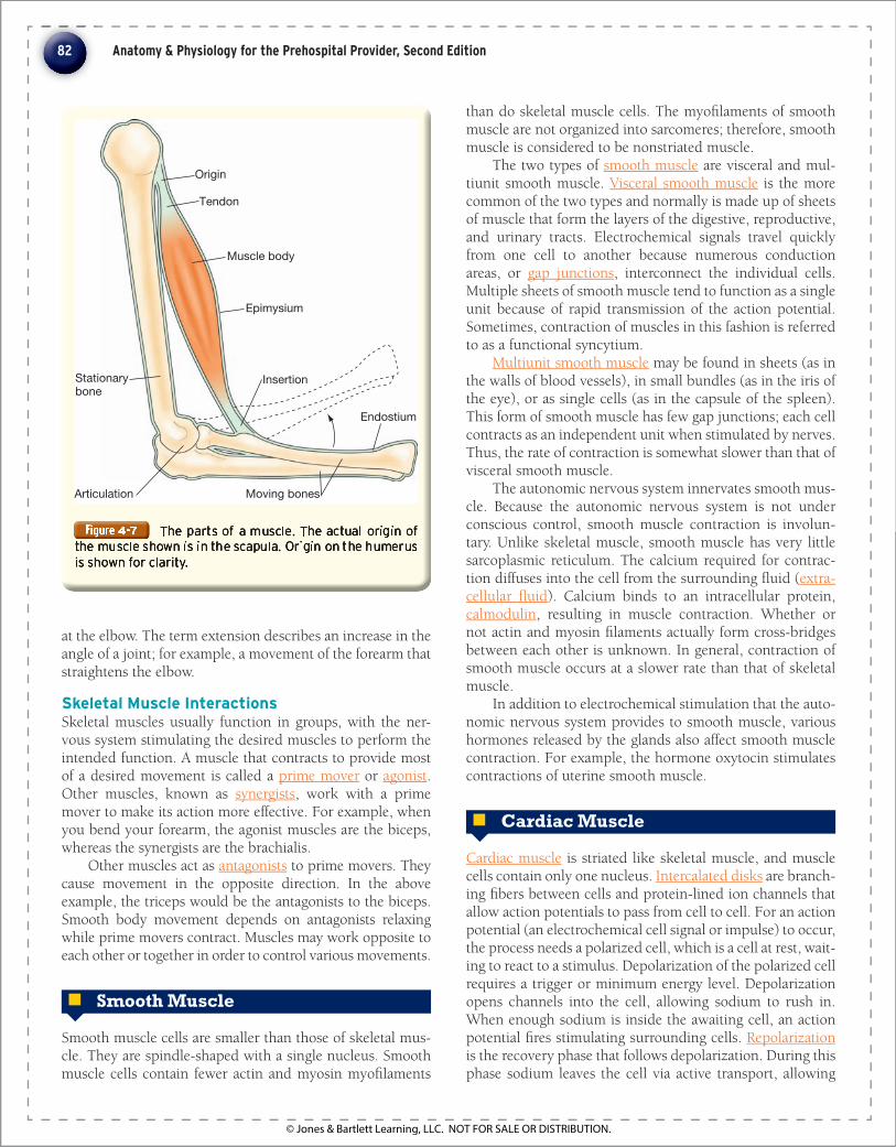
Origins and Insertions One end of a skeletal muscle usually is fastened to a rela-tively immovable part ( origin ) at a moveable joint. The other end connects to a one side of a movable joint ( insertion ) on the other side of the joint. As contraction occurs, the inser-tion is pulled toward the origin. There may be more than one origin or insertion, such as in the biceps brachii muscle of the arm. When this muscle contracts, the insertion being pulled toward its origin causes the forearm to � ex and supi-nate at the elbow Figure Figure Figure Figure Figure Figure 4-74-74-7 .
The head of a muscle is the part closest to its origin. The term � exion describes a decrease in the angle of a joint; for example, a movement of the forearm that causes it to bend
Oxygen Use and Debt Oxygen is required for the breakdown of glucose in the mito-chondria. Red blood cells carry oxygen, bound to hemoglo-bin molecules. Hemoglobin is the pigment that makes blood appear red. The pigment myoglobin is synthesized in the muscles to give skeletal muscles their reddish-brown color. Myoglobin can also combine with oxygen and temporarily store it in order to reduce muscular requirements for contin-uous blood supply during contraction.
When skeletal muscles are used for a more than a min-ute or two, anaerobic respiration is required for energy. In one type of anaerobic respiration, glucose is broken down via glycolysis to yield pyruvic acid, which is converted to lactic acid . Lactic acid can accumulate in muscles, but often diffuses in the bloodstream, reaching the liver, where it is converted back into glucose.
When a person is exercising strenuously, oxygen is used mostly to synthesize ATP. As lactic acid increases, an oxygen debt develops. Oxygen debt is equivalent to the amount of oxygen that liver cells require to convert the lactic acid into glucose, as well as the amount needed by muscle cells to restore ATP and creatine phosphate levels.
It may take several hours for the body to convert lactic acid back into glucose. Muscles may experience a change in their metabolic activity as exercise levels change. Increased exercise raises the muscles’ capacity for glycolysis. Aerobic exercise increases the muscles’ capacity for aerobic respiration.
Muscle Fatigue Prolonged exercise may cause a muscle to become unable to contract. This condition is called fatigue, and it may also occur because of interruption of muscular blood supply, or occasionally a lack of acetylcholine in the motor neuron axons. Lactic acid accumulation is the usual cause of mus-cular fatigue. As lactic acid lowers pH levels, muscle � bers become progressively less able to respond to stimulation.
81Chapter 4 The Musculoskeletal System
9781449642303_CH04_Pass5.indd 81 18/03/14 9:45 AM
© Jones & Bartlett Learning, LLC. NOT FOR SALE OR DISTRIBUTION.
© Jones & Bartlett Learning, LLCNOT FOR SALE OR DISTRIBUTION
© Jones & Bartlett Learning, LLCNOT FOR SALE OR DISTRIBUTION
© Jones & Bartlett Learning, LLCNOT FOR SALE OR DISTRIBUTION
© Jones & Bartlett Learning, LLCNOT FOR SALE OR DISTRIBUTION
© Jones & Bartlett Learning, LLCNOT FOR SALE OR DISTRIBUTION
© Jones & Bartlett Learning, LLCNOT FOR SALE OR DISTRIBUTION
© Jones & Bartlett Learning, LLCNOT FOR SALE OR DISTRIBUTION
© Jones & Bartlett Learning, LLCNOT FOR SALE OR DISTRIBUTION
© Jones & Bartlett Learning, LLCNOT FOR SALE OR DISTRIBUTION
© Jones & Bartlett Learning, LLCNOT FOR SALE OR DISTRIBUTION
© Jones & Bartlett Learning, LLCNOT FOR SALE OR DISTRIBUTION
© Jones & Bartlett Learning, LLCNOT FOR SALE OR DISTRIBUTION
© Jones & Bartlett Learning, LLCNOT FOR SALE OR DISTRIBUTION
© Jones & Bartlett Learning, LLCNOT FOR SALE OR DISTRIBUTION
© Jones & Bartlett Learning, LLCNOT FOR SALE OR DISTRIBUTION
© Jones & Bartlett Learning, LLCNOT FOR SALE OR DISTRIBUTION
© Jones & Bartlett Learning, LLCNOT FOR SALE OR DISTRIBUTION
© Jones & Bartlett Learning, LLCNOT FOR SALE OR DISTRIBUTION
© Jones & Bartlett Learning, LLCNOT FOR SALE OR DISTRIBUTION
© Jones & Bartlett Learning, LLCNOT FOR SALE OR DISTRIBUTION
82 Anatomy & Physiology for the Prehospital Provider, Second Edition
Origin
Muscle body
Epimysium
Insertion
Articulation Moving bones
Tendon
Stationarybone
Endostium
at the elbow. The term extension describes an increase in the angle of a joint; for example, a movement of the forearm that straightens the elbow.
Skeletal Muscle Interactions Skeletal muscles usually function in groups, with the ner-vous system stimulating the desired muscles to perform the intended function. A muscle that contracts to provide most of a desired movement is called a prime mover or agonist . Other muscles, known as synergists , work with a prime mover to make its action more effective. For example, when you bend your forearm, the agonist muscles are the biceps, whereas the synergists are the brachialis.
Other muscles act as antagonists to prime movers. They cause movement in the opposite direction. In the above example, the triceps would be the antagonists to the biceps. Smooth body movement depends on antagonists relaxing while prime movers contract. Muscles may work opposite to each other or together in order to control various movements.
6 Smooth Muscle
Smooth muscle cells are smaller than those of skeletal mus-cle. They are spindle-shaped with a single nucleus. Smooth muscle cells contain fewer actin and myosin myo� laments
than do skeletal muscle cells. The myo� laments of smooth muscle are not organized into sarcomeres; therefore, smooth muscle is considered to be nonstriated muscle.
The two types of smooth muscle are visceral and mul-tiunit smooth muscle. Visceral smooth muscle is the more common of the two types and normally is made up of sheets of muscle that form the layers of the digestive, reproductive, and urinary tracts. Electrochemical signals travel quickly from one cell to another because numerous conduction areas, or gap junctions , interconnect the individual cells. Multiple sheets of smooth muscle tend to function as a single unit because of rapid transmission of the action potential. Sometimes, contraction of muscles in this fashion is referred to as a functional syncytium.
Multiunit smooth muscle may be found in sheets (as in the walls of blood vessels), in small bundles (as in the iris of the eye), or as single cells (as in the capsule of the spleen). This form of smooth muscle has few gap junctions; each cell contracts as an independent unit when stimulated by nerves. Thus, the rate of contraction is somewhat slower than that of visceral smooth muscle.
The autonomic nervous system innervates smooth mus-cle. Because the autonomic nervous system is not under conscious control, smooth muscle contraction is involun-tary. Unlike skeletal muscle, smooth muscle has very little sarcoplasmic reticulum. The calcium required for contrac-tion diffuses into the cell from the surrounding � uid ( extra-cellular � uid ). Calcium binds to an intracellular protein, calmodulin , resulting in muscle contraction. Whether or not actin and myosin � laments actually form cross-bridges between each other is unknown. In general, contraction of smooth muscle occurs at a slower rate than that of skeletal muscle.
In addition to electrochemical stimulation that the auto-nomic nervous system provides to smooth muscle, various hormones released by the glands also affect smooth muscle contraction. For example, the hormone oxytocin stimulates contractions of uterine smooth muscle.
6 Cardiac Muscle
Cardiac muscle is striated like skeletal muscle, and muscle cells contain only one nucleus. Intercalated disks are branch-ing � bers between cells and protein-lined ion channels that allow action potentials to pass from cell to cell. For an action potential (an electrochemical cell signal or impulse) to occur, the process needs a polarized cell, which is a cell at rest, wait-ing to react to a stimulus. Depolarization of the polarized cell requires a trigger or minimum energy level. Depolarization opens channels into the cell, allowing sodium to rush in. When enough sodium is inside the awaiting cell, an action potential � res stimulating surrounding cells. Repolarization is the recovery phase that follows depolarization. During this phase sodium leaves the cell via active transport, allowing
9781449642303_CH04_Pass5.indd 82 18/03/14 9:45 AM
© Jones & Bartlett Learning, LLC. NOT FOR SALE OR DISTRIBUTION.
© Jones & Bartlett Learning, LLCNOT FOR SALE OR DISTRIBUTION
© Jones & Bartlett Learning, LLCNOT FOR SALE OR DISTRIBUTION
© Jones & Bartlett Learning, LLCNOT FOR SALE OR DISTRIBUTION
© Jones & Bartlett Learning, LLCNOT FOR SALE OR DISTRIBUTION
© Jones & Bartlett Learning, LLCNOT FOR SALE OR DISTRIBUTION
© Jones & Bartlett Learning, LLCNOT FOR SALE OR DISTRIBUTION
© Jones & Bartlett Learning, LLCNOT FOR SALE OR DISTRIBUTION
© Jones & Bartlett Learning, LLCNOT FOR SALE OR DISTRIBUTION
© Jones & Bartlett Learning, LLCNOT FOR SALE OR DISTRIBUTION
© Jones & Bartlett Learning, LLCNOT FOR SALE OR DISTRIBUTION
© Jones & Bartlett Learning, LLCNOT FOR SALE OR DISTRIBUTION
© Jones & Bartlett Learning, LLCNOT FOR SALE OR DISTRIBUTION
© Jones & Bartlett Learning, LLCNOT FOR SALE OR DISTRIBUTION
© Jones & Bartlett Learning, LLCNOT FOR SALE OR DISTRIBUTION
© Jones & Bartlett Learning, LLCNOT FOR SALE OR DISTRIBUTION
© Jones & Bartlett Learning, LLCNOT FOR SALE OR DISTRIBUTION
© Jones & Bartlett Learning, LLCNOT FOR SALE OR DISTRIBUTION
© Jones & Bartlett Learning, LLCNOT FOR SALE OR DISTRIBUTION
© Jones & Bartlett Learning, LLCNOT FOR SALE OR DISTRIBUTION
© Jones & Bartlett Learning, LLCNOT FOR SALE OR DISTRIBUTION
83Chapter 4 The Musculoskeletal System
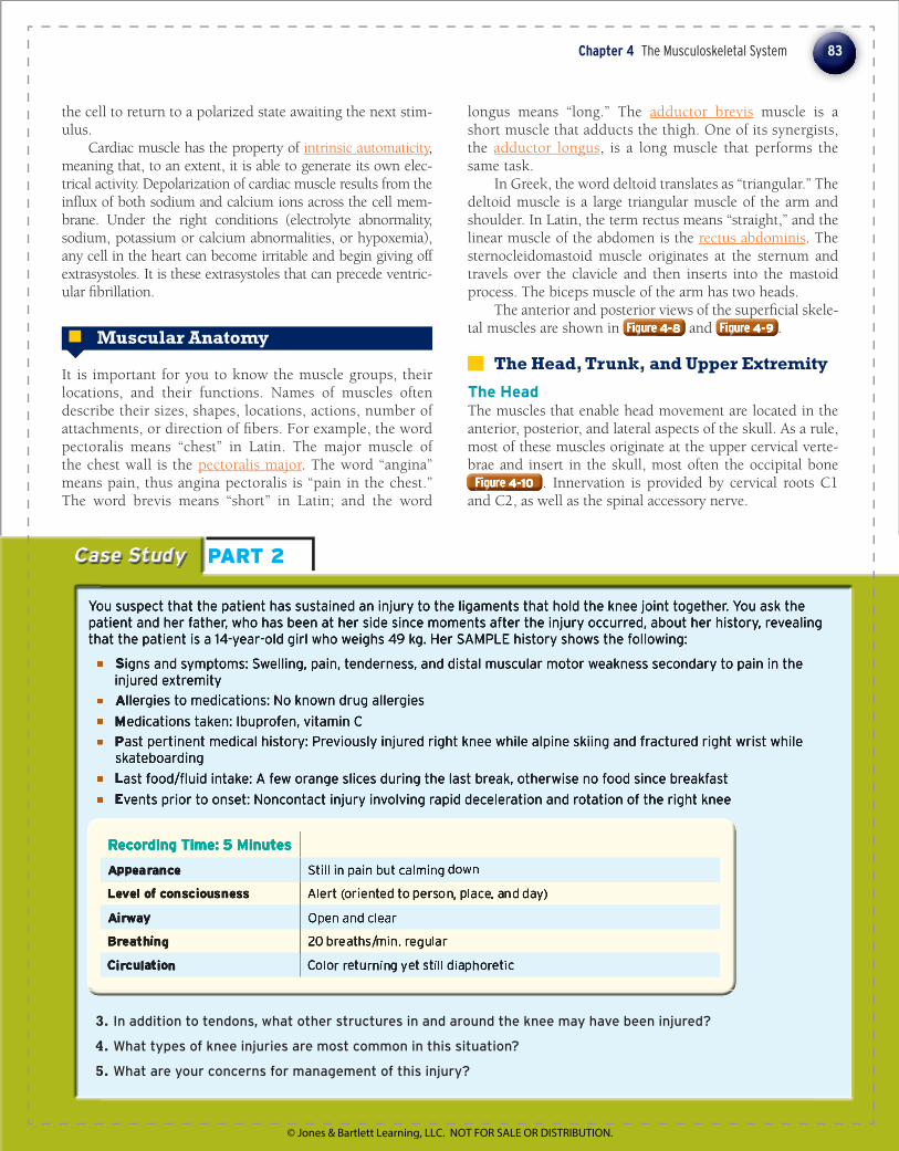
You suspect that the patient has sustained an injury to the ligaments that hold the knee joint together. You ask the patient and her father, who has been at her side since moments after the injury occurred, about her history, revealing that the patient is a 14-year-old girl who weighs 49 kg. Her SAMPLE history shows the following:
■ S igns and symptoms: Swelling, pain, tenderness, and distal muscular motor weakness secondary to pain in the injured extremity
■ A llergies to medications: No known drug allergies ■ M edications taken: Ibuprofen, vitamin C ■ P ast pertinent medical history: Previously injured right knee while alpine skiing and fractured right wrist while
skateboarding ■ L ast food/fl uid intake: A few orange slices during the last break, otherwise no food since breakfast ■ E vents prior to onset: Noncontact injury involving rapid deceleration and rotation of the right knee
Case Study Case Study PART 2
Recording Time: 5 MinutesRecording Time: 5 MinutesRecording Time: 5 Minutes
3. In addition to tendons, what other structures in and around the knee may have been injured?
4. What types of knee injuries are most common in this situation?
5. What are your concerns for management of this injury?
the cell to return to a polarized state awaiting the next stim-ulus.
Cardiac muscle has the property of intrinsic automaticity , meaning that, to an extent, it is able to generate its own elec-trical activity. Depolarization of cardiac muscle results from the in� ux of both sodium and calcium ions across the cell mem-brane. Under the right conditions (electrolyte abnormality, sodium, potassium or calcium abnormalities, or hypoxemia), any cell in the heart can become irritable and begin giving off extrasystoles. It is these extrasystoles that can precede ventric-ular � brillation.
6 Muscular Anatomy
It is important for you to know the muscle groups, their locations, and their functions. Names of muscles often describe their sizes, shapes, locations, actions, number of attachments, or direction of � bers. For example, the word pectoralis means “chest” in Latin. The major muscle of the chest wall is the pectoralis major . The word “angina” means pain, thus angina pectoralis is “pain in the chest.” The word brevis means “short” in Latin; and the word
longus means “long.” The adductor brevis muscle is a short muscle that adducts the thigh. One of its synergists, the adductor longus , is a long muscle that performs the same task.
In Greek, the word deltoid translates as “triangular.” The deltoid muscle is a large triangular muscle of the arm and shoulder. In Latin, the term rectus means “straight,” and the linear muscle of the abdomen is the rectus abdominis . The sternocleidomastoid muscle originates at the sternum and travels over the clavicle and then inserts into the mastoid process. The biceps muscle of the arm has two heads.
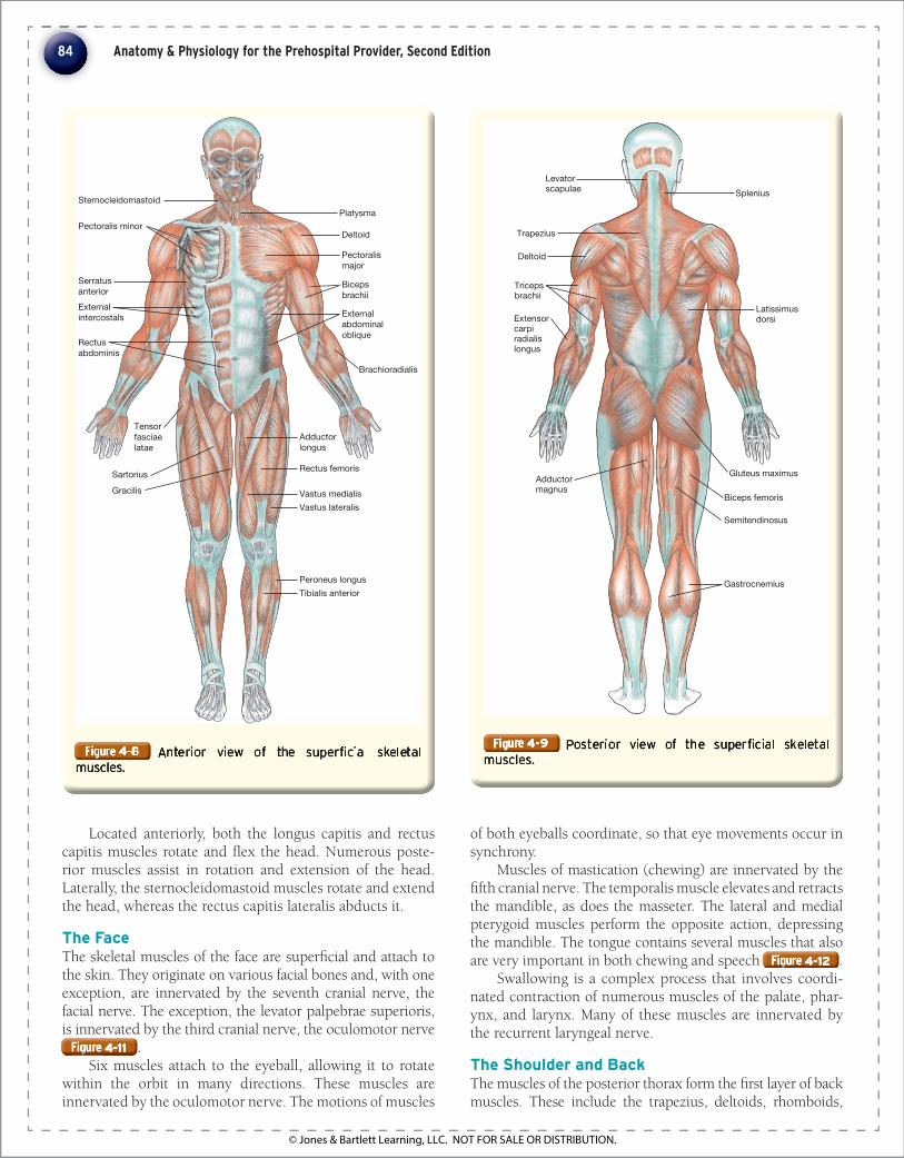
The anterior and posterior views of the super� cial skele-tal muscles are shown in Figure Figure Figure Figure Figure Figure 4-84-84-8 and Figure Figure Figure Figure Figure Figure 4-94-94-9 .
The Head, Trunk, and Upper Extremity
The Head The muscles that enable head movement are located in the anterior, posterior, and lateral aspects of the skull. As a rule, most of these muscles originate at the upper cervical verte-brae and insert in the skull, most often the occipital bone
Figure Figure Figure Figure Figure Figure 4-104-104-10 . Innervation is provided by cervical roots C1 and C2, as well as the spinal accessory nerve.
9781449642303_CH04_Pass5.indd 83 18/03/14 9:45 AM
© Jones & Bartlett Learning, LLC. NOT FOR SALE OR DISTRIBUTION.
© Jones & Bartlett Learning, LLCNOT FOR SALE OR DISTRIBUTION
© Jones & Bartlett Learning, LLCNOT FOR SALE OR DISTRIBUTION
© Jones & Bartlett Learning, LLCNOT FOR SALE OR DISTRIBUTION
© Jones & Bartlett Learning, LLCNOT FOR SALE OR DISTRIBUTION
© Jones & Bartlett Learning, LLCNOT FOR SALE OR DISTRIBUTION
© Jones & Bartlett Learning, LLCNOT FOR SALE OR DISTRIBUTION
© Jones & Bartlett Learning, LLCNOT FOR SALE OR DISTRIBUTION
© Jones & Bartlett Learning, LLCNOT FOR SALE OR DISTRIBUTION
© Jones & Bartlett Learning, LLCNOT FOR SALE OR DISTRIBUTION
© Jones & Bartlett Learning, LLCNOT FOR SALE OR DISTRIBUTION
© Jones & Bartlett Learning, LLCNOT FOR SALE OR DISTRIBUTION
© Jones & Bartlett Learning, LLCNOT FOR SALE OR DISTRIBUTION
© Jones & Bartlett Learning, LLCNOT FOR SALE OR DISTRIBUTION
© Jones & Bartlett Learning, LLCNOT FOR SALE OR DISTRIBUTION
© Jones & Bartlett Learning, LLCNOT FOR SALE OR DISTRIBUTION
© Jones & Bartlett Learning, LLCNOT FOR SALE OR DISTRIBUTION
© Jones & Bartlett Learning, LLCNOT FOR SALE OR DISTRIBUTION
© Jones & Bartlett Learning, LLCNOT FOR SALE OR DISTRIBUTION
© Jones & Bartlett Learning, LLCNOT FOR SALE OR DISTRIBUTION
© Jones & Bartlett Learning, LLCNOT FOR SALE OR DISTRIBUTION
SternocleidomastoidPlatysma
Pectoralis minor
Serratusanterior Externalintercostals
Tensorfasciaelatae
Sartorius
Adductorlongus
Rectus femoris
Vastus medialisVastus lateralis
Tibialis anteriorPeroneus longus
Gracilis
Deltoid
Pectoralismajor
Bicepsbrachii
Externalabdominaloblique
Brachioradialis
Rectusabdominis
Located anteriorly, both the longus capitis and rectus capitis muscles rotate and � ex the head. Numerous poste-rior muscles assist in rotation and extension of the head. Laterally, the sternocleidomastoid muscles rotate and extend the head, whereas the rectus capitis lateralis abducts it .
The Face The skeletal muscles of the face are super� cial and attach to the skin. They originate on various facial bones and, with one exception, are innervated by the seventh cranial nerve, the facial nerve. The exception, the levator palpebrae superioris, is innervated by the third cranial nerve, the oculomotor nerve Figure Figure Figure Figure Figure Figure 4-114-114-11 .
Six muscles attach to the eyeball, allowing it to rotate within the orbit in many directions. These muscles are innervated by the oculomotor nerve. The motions of muscles
of both eyeballs coordinate, so that eye movements occur in synchrony.
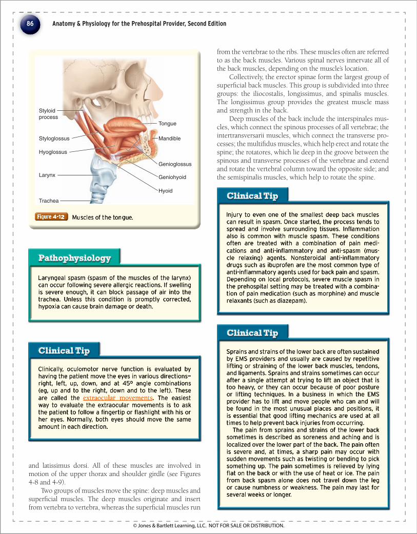
Muscles of mastication (chewing) are innervated by the � fth cranial nerve. The temporalis muscle elevates and retracts the mandible, as does the masseter. The lateral and medial pterygoid muscles perform the opposite action, depressing the mandible. The tongue contains several muscles that also are very important in both chewing and speech Figure Figure Figure Figure Figure Figure 4-124-124-12 .
Swallowing is a complex process that involves coordi-nated contraction of numerous muscles of the palate, phar-ynx, and larynx. Many of these muscles are innervated by the recurrent laryngeal nerve.
The Shoulder and Back The muscles of the posterior thorax form the � rst layer of back muscles. These include the trapezius, deltoids, rhomboids,
84 Anatomy & Physiology for the Prehospital Provider, Second Edition
Latissimusdorsi
Adductor magnus
Deltoid
Triceps brachii
Extensorcarpiradialislongus
Levatorscapulae Splenius
Trapezius
Gluteus maximus
Semitendinosus
Gastrocnemius
Biceps femoris
9781449642303_CH04_Pass5.indd 84 18/03/14 9:46 AM
© Jones & Bartlett Learning, LLC. NOT FOR SALE OR DISTRIBUTION.
© Jones & Bartlett Learning, LLCNOT FOR SALE OR DISTRIBUTION
© Jones & Bartlett Learning, LLCNOT FOR SALE OR DISTRIBUTION
© Jones & Bartlett Learning, LLCNOT FOR SALE OR DISTRIBUTION
© Jones & Bartlett Learning, LLCNOT FOR SALE OR DISTRIBUTION
© Jones & Bartlett Learning, LLCNOT FOR SALE OR DISTRIBUTION
© Jones & Bartlett Learning, LLCNOT FOR SALE OR DISTRIBUTION
© Jones & Bartlett Learning, LLCNOT FOR SALE OR DISTRIBUTION
© Jones & Bartlett Learning, LLCNOT FOR SALE OR DISTRIBUTION
© Jones & Bartlett Learning, LLCNOT FOR SALE OR DISTRIBUTION
© Jones & Bartlett Learning, LLCNOT FOR SALE OR DISTRIBUTION
© Jones & Bartlett Learning, LLCNOT FOR SALE OR DISTRIBUTION
© Jones & Bartlett Learning, LLCNOT FOR SALE OR DISTRIBUTION
© Jones & Bartlett Learning, LLCNOT FOR SALE OR DISTRIBUTION
© Jones & Bartlett Learning, LLCNOT FOR SALE OR DISTRIBUTION
© Jones & Bartlett Learning, LLCNOT FOR SALE OR DISTRIBUTION
© Jones & Bartlett Learning, LLCNOT FOR SALE OR DISTRIBUTION
© Jones & Bartlett Learning, LLCNOT FOR SALE OR DISTRIBUTION
© Jones & Bartlett Learning, LLCNOT FOR SALE OR DISTRIBUTION
© Jones & Bartlett Learning, LLCNOT FOR SALE OR DISTRIBUTION
© Jones & Bartlett Learning, LLCNOT FOR SALE OR DISTRIBUTION

Temporalis
Levator labii superioris
Zygomaticus minor
Zygomaticus major
Levator anguli oris
Depressor anguli oris
Depressor labii inferioris
Platysma
Mentalis
Obicularis oris
Buccinator
Masseter
Levator labiisuperiorisalaeque nasi
Obicularis oculi
Procerus
Occipitofrontalis
Rectus capitisanterior Longus colli
Clavicle
Sternocleidomastoid
Rectus capitislateralis
Longus capitis
Manubrium of sternum
The term whiplash describes a neck injury associated with sudden fl exion or extension and resulting in pain or other evidence of injury. Typically, a hyperexten-sion-hyperfl exion injury of the cervical spine occurs as a result of a rear-end collision. The patient reports neck and head pain, which may radiate down either arm. At times, concomitant stimulation of the sympathetic ner-vous system may result in dizziness, blurred vision, or pain behind the eyeballs. In the absence of fracture, dislocation, or neurologic injury, symptoms typically resolve spontaneously within 6 to 8 weeks, and long-term impairment is rare.
The term whiplashwhiplash describes a neck injury associated with sudden fl exion or extension and resulting in pain or other evidence of injury. Typically, a hyperexten-sion-hyperfl exion injury of the cervical spine occurs as a result of a rear-end collision. The patient reports neck and head pain, which may radiate down either arm. At times, concomitant stimulation of the sympathetic ner-vous system may result in dizziness, blurred vision, or pain behind the eyeballs. In the absence of fracture, dislocation, or neurologic injury, symptoms typically resolve spontaneously within 6 to 8 weeks, and long-term impairment is rare.
PathophysiologyPathophysiology
Bell palsy is a condition that results from dysfunction of the facial nerve, as a result of either trauma or infec-tion. The patient becomes unable to move the facial muscles on the affected side, and the lip sags during speech and attempts at smiling. The most signifi cant problem that results from Bell palsy is that the patient often is unable to close his or her eyelid and loses the protective blinking refl ex. To protect the eye, patients can instill artifi cial tears or keep the eyelid taped shut. Nerve function returns in most persons, at least to a degree, within a few weeks.
Bell palsyBell palsy is a condition that results from dysfunction of the facial nerve, as a result of either trauma or infec-tion. The patient becomes unable to move the facial muscles on the affected side, and the lip sags during speech and attempts at smiling. The most signifi cant problem that results from Bell palsy is that the patient often is unable to close his or her eyelid and loses the protective blinking refl ex. To protect the eye, patients can instill artifi cial tears or keep the eyelid taped shut. Nerve function returns in most persons, at least to a degree, within a few weeks.
PathophysiologyPathophysiology
85Chapter 4 The Musculoskeletal System
9781449642303_CH04_Pass5.indd 85 18/03/14 9:46 AM
© Jones & Bartlett Learning, LLC. NOT FOR SALE OR DISTRIBUTION.
© Jones & Bartlett Learning, LLCNOT FOR SALE OR DISTRIBUTION
© Jones & Bartlett Learning, LLCNOT FOR SALE OR DISTRIBUTION
© Jones & Bartlett Learning, LLCNOT FOR SALE OR DISTRIBUTION
© Jones & Bartlett Learning, LLCNOT FOR SALE OR DISTRIBUTION
© Jones & Bartlett Learning, LLCNOT FOR SALE OR DISTRIBUTION
© Jones & Bartlett Learning, LLCNOT FOR SALE OR DISTRIBUTION
© Jones & Bartlett Learning, LLCNOT FOR SALE OR DISTRIBUTION
© Jones & Bartlett Learning, LLCNOT FOR SALE OR DISTRIBUTION
© Jones & Bartlett Learning, LLCNOT FOR SALE OR DISTRIBUTION
© Jones & Bartlett Learning, LLCNOT FOR SALE OR DISTRIBUTION
© Jones & Bartlett Learning, LLCNOT FOR SALE OR DISTRIBUTION
© Jones & Bartlett Learning, LLCNOT FOR SALE OR DISTRIBUTION
© Jones & Bartlett Learning, LLCNOT FOR SALE OR DISTRIBUTION
© Jones & Bartlett Learning, LLCNOT FOR SALE OR DISTRIBUTION
© Jones & Bartlett Learning, LLCNOT FOR SALE OR DISTRIBUTION
© Jones & Bartlett Learning, LLCNOT FOR SALE OR DISTRIBUTION
© Jones & Bartlett Learning, LLCNOT FOR SALE OR DISTRIBUTION
© Jones & Bartlett Learning, LLCNOT FOR SALE OR DISTRIBUTION
© Jones & Bartlett Learning, LLCNOT FOR SALE OR DISTRIBUTION
© Jones & Bartlett Learning, LLCNOT FOR SALE OR DISTRIBUTION
Clinically, oculomotor nerve function is evaluated by having the patient move the eyes in various directions—right, left, up, down, and at 45º angle combinations (eg, up and to the right, down and to the left). These are called the extraocular movements . The easiest way to evaluate the extraocular movements is to ask the patient to follow a fi ngertip or fl ashlight with his or her eyes. Normally, both eyes should move the same amount in each direction.
Clinically, oculomotor nerve function is evaluated by having the patient move the eyes in various directions—right, left, up, down, and at 45º angle combinations (eg, up and to the right, down and to the left). These are called the extraocular movements . The easiest way to evaluate the extraocular movements is to ask the patient to follow a fi ngertip or fl ashlight with his or her eyes. Normally, both eyes should move the same amount in each direction.
Clinical TipClinical Tip
Injury to even one of the smallest deep back muscles can result in spasm. Once started, the process tends to spread and involve surrounding tissues. Infl ammation also is common with muscle spasm. These conditions often are treated with a combination of pain medi-cations and anti-infl ammatory and anti-spasm (mus-cle relaxing) agents. Nonsteroidal anti-infl ammatory drugs such as ibuprofen are the most common type of anti-infl ammatory agents used for back pain and spasm. Depending on local protocols, severe muscle spasm in the prehospital setting may be treated with a combina-tion of pain medication (such as morphine) and muscle relaxants (such as diazepam).
Injury to even one of the smallest deep back muscles can result in spasm. Once started, the process tends to spread and involve surrounding tissues. Infl ammation also is common with muscle spasm. These conditions often are treated with a combination of pain medi-cations and anti-infl ammatory and anti-spasm (mus-cle relaxing) agents. Nonsteroidal anti-infl ammatory drugs such as ibuprofen are the most common type of anti-infl ammatory agents used for back pain and spasm. Depending on local protocols, severe muscle spasm in the prehospital setting may be treated with a combina-tion of pain medication (such as morphine) and muscle relaxants (such as diazepam).
Clinical TipClinical Tip
Sprains and strains of the lower back are often sustained by EMS providers and usually are caused by repetitive lifting or straining of the lower back muscles, tendons, and ligaments. Sprains and strains sometimes can occur after a single attempt at trying to lift an object that is too heavy, or they can occur because of poor posture or lifting techniques. In a business in which the EMS provider has to lift and move people who can and will be found in the most unusual places and positions, it is essential that good lifting mechanics are used at all times to help prevent back injuries from occurring.
The pain from sprains and strains of the lower back sometimes is described as soreness and aching and is localized over the lower part of the back. The pain often is severe and, at times, a sharp pain may occur with sudden movements such as twisting or bending to pick something up. The pain sometimes is relieved by lying fl at on the back or with the use of heat or ice. The pain from back spasm alone does not travel down the leg or cause numbness or weakness. The pain may last for several weeks or longer.
Clinical TipClinical Tip
from the vertebrae to the ribs. These muscles often are referred to as the back muscles. Various spinal nerves innervate all of the back muscles, depending on the muscle’s location.
Collectively, the erector spinae form the largest group of super� cial back muscles. This group is subdivided into three groups: the iliocostalis, longissimus, and spinalis muscles. The longissimus group provides the greatest muscle mass and strength in the back.
Deep muscles of the back include the interspinales mus-cles, which connect the spinous processes of all vertebrae; the intertransversarii muscles, which connect the transverse pro-cesses; the multi� dus muscles, which help erect and rotate the spine; the rotatores, which lie deep in the groove between the spinous and transverse processes of the vertebrae and extend and rotate the vertebral column toward the opposite side; and the semispinalis muscles, which help to rotate the spine.
Laryngeal spasm (spasm of the muscles of the larynx) can occur following severe allergic reactions. If swelling is severe enough, it can block passage of air into the trachea. Unless this condition is promptly corrected, hypoxia can cause brain damage or death.
Laryngeal spasm (spasm of the muscles of the larynx) can occur following severe allergic reactions. If swelling is severe enough, it can block passage of air into the trachea. Unless this condition is promptly corrected, hypoxia can cause brain damage or death.
PathophysiologyPathophysiology
and latissimus dorsi. All of these muscles are involved in motion of the upper thorax and shoulder girdle (see Figures 4- 8 and 4- 9 ).
Two groups of muscles move the spine: deep muscles and super� cial muscles. The deep muscles originate and insert from vertebra to vertebra, whereas the super� cial muscles run
86 Anatomy & Physiology for the Prehospital Provider, Second Edition
Styloidprocess
Styloglossus
Hyoglossus
Larynx
Trachea
Tongue
Mandible
Genioglossus
Geniohyoid
Hyoid
9781449642303_CH04_Pass5.indd 86 18/03/14 9:46 AM
© Jones & Bartlett Learning, LLC. NOT FOR SALE OR DISTRIBUTION.
© Jones & Bartlett Learning, LLCNOT FOR SALE OR DISTRIBUTION
© Jones & Bartlett Learning, LLCNOT FOR SALE OR DISTRIBUTION
© Jones & Bartlett Learning, LLCNOT FOR SALE OR DISTRIBUTION
© Jones & Bartlett Learning, LLCNOT FOR SALE OR DISTRIBUTION
© Jones & Bartlett Learning, LLCNOT FOR SALE OR DISTRIBUTION
© Jones & Bartlett Learning, LLCNOT FOR SALE OR DISTRIBUTION
© Jones & Bartlett Learning, LLCNOT FOR SALE OR DISTRIBUTION
© Jones & Bartlett Learning, LLCNOT FOR SALE OR DISTRIBUTION
© Jones & Bartlett Learning, LLCNOT FOR SALE OR DISTRIBUTION
© Jones & Bartlett Learning, LLCNOT FOR SALE OR DISTRIBUTION
© Jones & Bartlett Learning, LLCNOT FOR SALE OR DISTRIBUTION
© Jones & Bartlett Learning, LLCNOT FOR SALE OR DISTRIBUTION
© Jones & Bartlett Learning, LLCNOT FOR SALE OR DISTRIBUTION
© Jones & Bartlett Learning, LLCNOT FOR SALE OR DISTRIBUTION
© Jones & Bartlett Learning, LLCNOT FOR SALE OR DISTRIBUTION
© Jones & Bartlett Learning, LLCNOT FOR SALE OR DISTRIBUTION
© Jones & Bartlett Learning, LLCNOT FOR SALE OR DISTRIBUTION
© Jones & Bartlett Learning, LLCNOT FOR SALE OR DISTRIBUTION
© Jones & Bartlett Learning, LLCNOT FOR SALE OR DISTRIBUTION
© Jones & Bartlett Learning, LLCNOT FOR SALE OR DISTRIBUTION
Hiccups occur when there is irritation of the phrenic nerve, resulting in sudden and unpredictable contrac-tions of the diaphragm.
Hiccups occur when there is irritation of the phrenic nerve, resulting in sudden and unpredictable contrac-tions of the diaphragm.
PathophysiologyPathophysiology
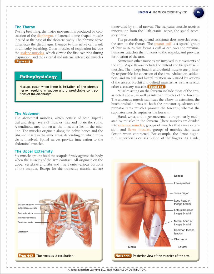
The Thorax During breathing, the major movement is produced by con-traction of the diaphragm , a � attened dome-shaped muscle located at the base of the thoracic cavity. The phrenic nerve innervates the diaphragm. Damage to this nerve can result in dif� culty breathing. Other muscles of respiration include the scalene muscles , which elevate the � rst two ribs during inspiration, and the external and internal intercostal muscles Figure Figure Figure Figure Figure Figure 4-134-134-13 .
innervated by spinal nerves. The trapezius muscle receives innervation from the 11th cranial nerve, the spinal acces-sory nerve.
The pectoralis major and latissimus dorsi muscles attach the arm to the thorax. The rotator cuff is a special group of four muscles that forms a cuff or cap over the proximal humerus, attaches the humerus to the scapula, and provides for rotation of the arm.
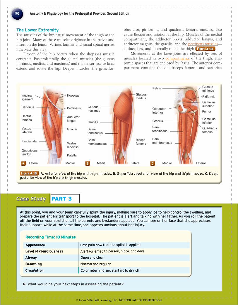
Numerous other muscles are involved in movements of the arm. Major � exors include the deltoid and biceps brachii muscles. The triceps brachii and deltoid muscles are primar-ily responsible for extension of the arm. Abduction, adduc-tion, and medial and lateral rotation are caused by actions of the triceps brachii and deltoid muscles, as well as several other accessory muscles Figure Figure Figure Figure Figure Figure 4-144-144-14 .
Muscles acting on the forearm include those of the arm, as noted above, as well as intrinsic muscles of the forearm. The anconeus muscle stabilizes the elbow in extension; the brachioradialis � exes it. Both the pronator quadratus and pronator teres muscles pronate the forearm, whereas the supinator muscle supinates the forearm.
Hand, wrist, and � nger movements are primarily medi-ated by muscles in the forearm. These muscles are divided into extensor muscles , groups of muscles that cause exten-sion, and � exor muscles , groups of muscles that cause � exion when contracted. For example, the � exor digito-rum super� cialis causes � exion of the � ngers. As a rule,
The Abdomen The abdominal muscles, which consist of both super� -cial and deep layers of muscles, � ex and rotate the spine. A tendinous area known as the linea alba lies in the mid-line. The muscles originate along the pelvic bones and the ribs and insert in the same areas, depending on which mus-cle is involved . Spinal nerves provide innervation to the abdominal muscles.
The Upper Extremity Six muscle groups hold the scapula � rmly against the body when the muscles of the arm contract. All originate on the upper vertebrae and ribs and insert onto various portions of the scapula. Except for the trapezius muscle, all are
Deltoid
Infraspinatus
Teres major
Long head of triceps brachii
Lateral head of triceps brachii
Medial head of triceps brachii
Common triceps tendon
Olecranon
Medial Lateral
87Chapter 4 The Musculoskeletal System
Scalene muscles
Pectoralis minor
Internal intercostals
Serratus anterior
External intercostals
Diaphragm
9781449642303_CH04_Pass5.indd 87 18/03/14 9:46 AM
© Jones & Bartlett Learning, LLC. NOT FOR SALE OR DISTRIBUTION.
© Jones & Bartlett Learning, LLCNOT FOR SALE OR DISTRIBUTION
© Jones & Bartlett Learning, LLCNOT FOR SALE OR DISTRIBUTION
© Jones & Bartlett Learning, LLCNOT FOR SALE OR DISTRIBUTION
© Jones & Bartlett Learning, LLCNOT FOR SALE OR DISTRIBUTION
© Jones & Bartlett Learning, LLCNOT FOR SALE OR DISTRIBUTION
© Jones & Bartlett Learning, LLCNOT FOR SALE OR DISTRIBUTION
© Jones & Bartlett Learning, LLCNOT FOR SALE OR DISTRIBUTION
© Jones & Bartlett Learning, LLCNOT FOR SALE OR DISTRIBUTION
© Jones & Bartlett Learning, LLCNOT FOR SALE OR DISTRIBUTION
© Jones & Bartlett Learning, LLCNOT FOR SALE OR DISTRIBUTION
© Jones & Bartlett Learning, LLCNOT FOR SALE OR DISTRIBUTION
© Jones & Bartlett Learning, LLCNOT FOR SALE OR DISTRIBUTION
© Jones & Bartlett Learning, LLCNOT FOR SALE OR DISTRIBUTION
© Jones & Bartlett Learning, LLCNOT FOR SALE OR DISTRIBUTION
© Jones & Bartlett Learning, LLCNOT FOR SALE OR DISTRIBUTION
© Jones & Bartlett Learning, LLCNOT FOR SALE OR DISTRIBUTION
© Jones & Bartlett Learning, LLCNOT FOR SALE OR DISTRIBUTION
© Jones & Bartlett Learning, LLCNOT FOR SALE OR DISTRIBUTION
© Jones & Bartlett Learning, LLCNOT FOR SALE OR DISTRIBUTION
© Jones & Bartlett Learning, LLCNOT FOR SALE OR DISTRIBUTION

Rotator cuff injuries are a common source of shoulder pain and often occur as a result of tendon degeneration from age and repeated trauma. As the tendons weaken, thickening and chronic infl ammation occur in the over-lying shoulder bursa. Complete ruptures occasionally occur during athletic events involving heavy lifting or a fall on an outstretched hand, but they occur far more frequently as a result of chronic degeneration. Patients typically report pain and tenderness over the shoul-der, which is worsened by abduction of the arm. In the absence of complete tendon rupture, some strength is maintained. With rupture, severe weakness and variable pain are both common. Radiographs often are normal; magnetic resonance imaging can help diagnose com-plete and partial tears. Strains of the rotator cuff or tendinitis respond well to rest and treatment with non-steroidal anti-infl ammatory drugs. Tears may require surgical repair.
Rotator cuff injuries are a common source of shoulder pain and often occur as a result of tendon degeneration from age and repeated trauma. As the tendons weaken, thickening and chronic infl ammation occur in the over-lying shoulder bursa. Complete ruptures occasionally occur during athletic events involving heavy lifting or a fall on an outstretched hand, but they occur far more frequently as a result of chronic degeneration. Patients typically report pain and tenderness over the shoul-der, which is worsened by abduction of the arm. In the absence of complete tendon rupture, some strength is maintained. With rupture, severe weakness and variable pain are both common. Radiographs often are normal; magnetic resonance imaging can help diagnose com-plete and partial tears. Strains of the rotator cuff or tendinitis respond well to rest and treatment with non-steroidal anti-infl ammatory drugs. Tears may require surgical repair.
PathophysiologyPathophysiologythe extensor muscles originate on the lateral aspect of the elbow, and the � exor muscles originate on the medial side Figure Figure Figure Figure Figure Figure 4-154-154-15 .
Movement is also affected by the intrinsic muscles of the hand, the lumbricales and the interossei, as well as the muscles of the thenar and hypothenar eminences, � eshy prominences at the base of the thumb (thenar) and � fth � n-ger (hypothenar). These small muscles are located entirely within the hand Figure Figure Figure Figure Figure Figure 4-164-164-16 . All muscles that cause motion of the hand and � ngers are innervated by the median, ulnar, or radial nerve.
The Pelvis and Lower Extremity
The Pelvic Floor and Perineum The coccygeus muscle and the levator ani muscle form the � oor of the pelvis. The area below these muscles is the perineum . The structures of the urogenital system (some-times called the urogenital triangle ) lie anteriorly; the struc-tures of the anus, or anal triangle , lie posteriorly.
88 Anatomy & Physiology for the Prehospital Provider, Second Edition
Extensor muscles(lateral)
Extensor tendons
Hand
Flexor tendons
Flexor muscles(medial)
Biceps andtendon Medial epicondyle
of humerus
Anconeus
Supinator
Abductor pollicislongus
Extensor pollicislongus
Extensor pollicisbrevis
Extensor indicis
Medial MedialLateral LateralAAA BBB
9781449642303_CH04_Pass5.indd 88 18/03/14 9:46 AM
© Jones & Bartlett Learning, LLC. NOT FOR SALE OR DISTRIBUTION.
© Jones & Bartlett Learning, LLCNOT FOR SALE OR DISTRIBUTION
© Jones & Bartlett Learning, LLCNOT FOR SALE OR DISTRIBUTION
© Jones & Bartlett Learning, LLCNOT FOR SALE OR DISTRIBUTION
© Jones & Bartlett Learning, LLCNOT FOR SALE OR DISTRIBUTION
© Jones & Bartlett Learning, LLCNOT FOR SALE OR DISTRIBUTION
© Jones & Bartlett Learning, LLCNOT FOR SALE OR DISTRIBUTION
© Jones & Bartlett Learning, LLCNOT FOR SALE OR DISTRIBUTION
© Jones & Bartlett Learning, LLCNOT FOR SALE OR DISTRIBUTION
© Jones & Bartlett Learning, LLCNOT FOR SALE OR DISTRIBUTION
© Jones & Bartlett Learning, LLCNOT FOR SALE OR DISTRIBUTION
© Jones & Bartlett Learning, LLCNOT FOR SALE OR DISTRIBUTION
© Jones & Bartlett Learning, LLCNOT FOR SALE OR DISTRIBUTION
© Jones & Bartlett Learning, LLCNOT FOR SALE OR DISTRIBUTION
© Jones & Bartlett Learning, LLCNOT FOR SALE OR DISTRIBUTION
© Jones & Bartlett Learning, LLCNOT FOR SALE OR DISTRIBUTION
© Jones & Bartlett Learning, LLCNOT FOR SALE OR DISTRIBUTION
© Jones & Bartlett Learning, LLCNOT FOR SALE OR DISTRIBUTION
© Jones & Bartlett Learning, LLCNOT FOR SALE OR DISTRIBUTION
© Jones & Bartlett Learning, LLCNOT FOR SALE OR DISTRIBUTION
© Jones & Bartlett Learning, LLCNOT FOR SALE OR DISTRIBUTION

In males, the bulbospongiosus muscle constricts the urethra and aids in the erection of the penis; in females, it results in erection of the clitoris. The ori� ce of the anal canal is kept closed by the sphincter ani externus, whereas the urethral sphincter muscle constricts the ure-thra whereas the urethral sphincter muscle constricts the ure-whereas the urethral sphincter muscle constricts the ure-
Figure Figure Figure 4-174-174-17 .
Bicipital tendinitis is a common cause of shoulder pain in adults older than 40 years; however, it also may occur in younger athletes who use repeated throwing motions. The common denominator is infl ammation of the biceps tendon, as well as its sheath, in the bicipital groove. Patients have pain over the anterolateral aspect of the shoulder, which often radiates down the arm. Palpation reveals tenderness in the bicipital groove of the humerus, and a positive Yergason test , supported by pain in the bicipital groove on supination of the fore-arm against resistance. Radiographic studies most often are normal. Treatment consists of rest, heat, range-of-motion exercises, and nonsteroidal anti-infl ammatory drugs. Sometimes, corticosteroids are injected into the shoulder. Surgery is a last resort.
Bicipital tendinitis is a common cause of shoulder pain in adults older than 40 years; however, it also may occur in younger athletes who use repeated throwing motions. The common denominator is infl ammation of the biceps tendon, as well as its sheath, in the bicipital groove. Patients have pain over the anterolateral aspect of the shoulder, which often radiates down the arm. Palpation reveals tenderness in the bicipital groove of the humerus, and a positive Yergason testYergason testthe humerus, and a positive Yergason testthe humerus, and a positive , supported by pain in the bicipital groove on supination of the fore-arm against resistance. Radiographic studies most often are normal. Treatment consists of rest, heat, range-of-motion exercises, and nonsteroidal anti-infl ammatory drugs. Sometimes, corticosteroids are injected into the shoulder. Surgery is a last resort.
PathophysiologyPathophysiology
89Chapter 4 The Musculoskeletal System
Lumbricales
Interossei
Hypothenareminence
Flexor retinaculum
Thenareminence
Urogenitaltriangle
Penis
Bulbospongiosus
Ischiocavernosus
Analtriangle
Deep transverseperinei
Levator ani
Anus
Gluteus maximus
Sphincter aniexternus
Coccyx
Clitoris
Bulbospongiosus
Urethra
Vagina
Levator ani
Anus
Ischiocavernosus
Deep transverseperinei
Gluteus maximus
Sphincter aniexternus
Coccyx
Urogenitaltriangle
Analtriangle
AAA BBB
9781449642303_CH04_Pass5.indd 89 18/03/14 9:46 AM
© Jones & Bartlett Learning, LLC. NOT FOR SALE OR DISTRIBUTION.
© Jones & Bartlett Learning, LLCNOT FOR SALE OR DISTRIBUTION
© Jones & Bartlett Learning, LLCNOT FOR SALE OR DISTRIBUTION
© Jones & Bartlett Learning, LLCNOT FOR SALE OR DISTRIBUTION
© Jones & Bartlett Learning, LLCNOT FOR SALE OR DISTRIBUTION
© Jones & Bartlett Learning, LLCNOT FOR SALE OR DISTRIBUTION
© Jones & Bartlett Learning, LLCNOT FOR SALE OR DISTRIBUTION
© Jones & Bartlett Learning, LLCNOT FOR SALE OR DISTRIBUTION
© Jones & Bartlett Learning, LLCNOT FOR SALE OR DISTRIBUTION
© Jones & Bartlett Learning, LLCNOT FOR SALE OR DISTRIBUTION
© Jones & Bartlett Learning, LLCNOT FOR SALE OR DISTRIBUTION
© Jones & Bartlett Learning, LLCNOT FOR SALE OR DISTRIBUTION
© Jones & Bartlett Learning, LLCNOT FOR SALE OR DISTRIBUTION
© Jones & Bartlett Learning, LLCNOT FOR SALE OR DISTRIBUTION
© Jones & Bartlett Learning, LLCNOT FOR SALE OR DISTRIBUTION
© Jones & Bartlett Learning, LLCNOT FOR SALE OR DISTRIBUTION
© Jones & Bartlett Learning, LLCNOT FOR SALE OR DISTRIBUTION
© Jones & Bartlett Learning, LLCNOT FOR SALE OR DISTRIBUTION
© Jones & Bartlett Learning, LLCNOT FOR SALE OR DISTRIBUTION
© Jones & Bartlett Learning, LLCNOT FOR SALE OR DISTRIBUTION
© Jones & Bartlett Learning, LLCNOT FOR SALE OR DISTRIBUTION
At this point, you and your team carefully splint the injury, making sure to apply ice to help control the swelling, and prepare the patient for transport to the hospital. The patient is alert and talking with her father. As you roll the patient off the fi eld on your stretcher, all the parents and bystanders applaud. You can see on her face that she appreciates their support, while at the same time, she appears anxious about her injury.
At this point, you and your team carefully splint the injury, making sure to apply ice to help control the swelling, and prepare the patient for transport to the hospital. The patient is alert and talking with her father. As you roll the patient off the fi eld on your stretcher, all the parents and bystanders applaud. You can see on her face that she appreciates their support, while at the same time, she appears anxious about her injury.
Case Study Case Study PART 3
Recording Time: 10 MinutesRecording Time: 10 MinutesRecording Time: 10 Minutes
6. What would be your next steps in assessing the patient?
The Lower Extremity The muscles of the hip cause movement of the thigh at the hip joint. Many of these muscles originate in the pelvis and insert on the femur. Various lumbar and sacral spinal nerves innervate this area.
Flexion of the hip occurs when the iliopsoas muscle contracts. Posterolaterally, the gluteal muscles (the gluteus minimus, medius, and maximus) and the tensor fasciae latae extend and rotate the hip. Deeper muscles, the gemellus,
obturator, piriformis, and quadratis femoris muscles, also cause � exion and rotation at the hip. Muscles of the medial compartment, the adductor brevis, adductor longus, and adductor magnus, the gracilis, and the pectineus muscles —adduct, � ex, and internally rotate the thigh Figure Figure Figure Figure Figure Figure 4-184-184-18 .
Movements at the knee joint are effected by sets of muscles located in two compartments of the thigh, ana-tomic spaces that are enclosed by fascia. The anterior com-partment contains the quadriceps femoris and sartorius
90 Anatomy & Physiology for the Prehospital Provider, Second Edition
Sartorius
Patella
Gracilis
Adductorlongus
Pectineus
IliopsoasInguinalligament
Vastusmedialis
Bicepsfemoris
Gluteusminimus
MedialMedialLateral Lateral Medial Lateral
Rectusfemoris
Vastuslateralis
Fascia lata
Quadricepstendon
Piriformis
Gemellussuperior
Gemellusinferior
Quadratusfemoris
Pelvis
FemurObturatorinternus
Gracilis
Semi-tendinosus
Semi-membranosus
Semi-tendinosus
Semi-membranosus
Gluteusmaximus
Gracilis
Gluteusmedius
AAA CCCBBB
9781449642303_CH04_Pass5.indd 90 18/03/14 9:46 AM
© Jones & Bartlett Learning, LLC. NOT FOR SALE OR DISTRIBUTION.
© Jones & Bartlett Learning, LLCNOT FOR SALE OR DISTRIBUTION
© Jones & Bartlett Learning, LLCNOT FOR SALE OR DISTRIBUTION
© Jones & Bartlett Learning, LLCNOT FOR SALE OR DISTRIBUTION
© Jones & Bartlett Learning, LLCNOT FOR SALE OR DISTRIBUTION
© Jones & Bartlett Learning, LLCNOT FOR SALE OR DISTRIBUTION
© Jones & Bartlett Learning, LLCNOT FOR SALE OR DISTRIBUTION
© Jones & Bartlett Learning, LLCNOT FOR SALE OR DISTRIBUTION
© Jones & Bartlett Learning, LLCNOT FOR SALE OR DISTRIBUTION
© Jones & Bartlett Learning, LLCNOT FOR SALE OR DISTRIBUTION
© Jones & Bartlett Learning, LLCNOT FOR SALE OR DISTRIBUTION
© Jones & Bartlett Learning, LLCNOT FOR SALE OR DISTRIBUTION
© Jones & Bartlett Learning, LLCNOT FOR SALE OR DISTRIBUTION
© Jones & Bartlett Learning, LLCNOT FOR SALE OR DISTRIBUTION
© Jones & Bartlett Learning, LLCNOT FOR SALE OR DISTRIBUTION
© Jones & Bartlett Learning, LLCNOT FOR SALE OR DISTRIBUTION
© Jones & Bartlett Learning, LLCNOT FOR SALE OR DISTRIBUTION
© Jones & Bartlett Learning, LLCNOT FOR SALE OR DISTRIBUTION
© Jones & Bartlett Learning, LLCNOT FOR SALE OR DISTRIBUTION
© Jones & Bartlett Learning, LLCNOT FOR SALE OR DISTRIBUTION
© Jones & Bartlett Learning, LLCNOT FOR SALE OR DISTRIBUTION

muscles. The quadriceps femoris muscle primarily extends the knee when it contracts. (Because portions of the quad-riceps femoris also cross the hip, a secondary function of this muscle is � exion of the hip joint.) The sartorius mus-cle is the longest muscle in the body, and it � exes the knee and the hip when it contracts. In the posterior compart-ment, the biceps femoris muscle � exes and laterally rotates the knee and extends the hip. The semimembranosus and semitendinosus muscles � ex and medially rotate the knee. Together, the biceps, semimembranosus, and semitendino-sus muscles are referred to as the hamstrings.
Muscles located in the leg act on the ankle and foot. These muscles typically originate on the tibia and � bula and insert into the foot Figure Figure Figure Figure Figure Figure 4-194-194-19 . The tibial and pero-neal nerves, terminal divisions of the sciatic nerve, innervate these muscles.
Anatomically and functionally, the leg contains four compartments: the anterior, the super� cial posterior, the deep posterior, and the lateral compartments. The anterior compartment contains muscles that extend (or dorsi� ex) the ankle and toes. The super� cial posterior compartment contains the gastrocnemius, plantaris, and soleus muscles, which are super� cial muscles that plantar � ex the ankle. The Achilles tendon is the strong tendon that attaches these muscles to the calcaneus. The deep posterior compartment contains muscles that � ex the toes and invert the foot and ankle. The lateral compartment contains muscles that pri-marily evert the foot .
The intrinsic foot muscles are located within the foot itself and are arranged similarly to the intrinsic muscles of the hand. They � ex, extend, abduct, and adduct the toes Figure Figure Figure Figure Figure Figure 4-204-204-20 .
91Chapter 4 The Musculoskeletal System
Peroneusmuscles
Patella
Gastrocnemius
Tibialis anterior
Soleus
Extensor musclesand tendons Soleus
Plantaris
Gastrocnemius
Peroneus longus
Achilles tendon
MedialMedial LateralLateralAAAAAA
9781449642303_CH04_Pass5.indd 91 18/03/14 9:46 AM
© Jones & Bartlett Learning, LLC. NOT FOR SALE OR DISTRIBUTION.
© Jones & Bartlett Learning, LLCNOT FOR SALE OR DISTRIBUTION
© Jones & Bartlett Learning, LLCNOT FOR SALE OR DISTRIBUTION
© Jones & Bartlett Learning, LLCNOT FOR SALE OR DISTRIBUTION
© Jones & Bartlett Learning, LLCNOT FOR SALE OR DISTRIBUTION
© Jones & Bartlett Learning, LLCNOT FOR SALE OR DISTRIBUTION
© Jones & Bartlett Learning, LLCNOT FOR SALE OR DISTRIBUTION
© Jones & Bartlett Learning, LLCNOT FOR SALE OR DISTRIBUTION
© Jones & Bartlett Learning, LLCNOT FOR SALE OR DISTRIBUTION
© Jones & Bartlett Learning, LLCNOT FOR SALE OR DISTRIBUTION
© Jones & Bartlett Learning, LLCNOT FOR SALE OR DISTRIBUTION
© Jones & Bartlett Learning, LLCNOT FOR SALE OR DISTRIBUTION
© Jones & Bartlett Learning, LLCNOT FOR SALE OR DISTRIBUTION
© Jones & Bartlett Learning, LLCNOT FOR SALE OR DISTRIBUTION
© Jones & Bartlett Learning, LLCNOT FOR SALE OR DISTRIBUTION
© Jones & Bartlett Learning, LLCNOT FOR SALE OR DISTRIBUTION
© Jones & Bartlett Learning, LLCNOT FOR SALE OR DISTRIBUTION
© Jones & Bartlett Learning, LLCNOT FOR SALE OR DISTRIBUTION
© Jones & Bartlett Learning, LLCNOT FOR SALE OR DISTRIBUTION
© Jones & Bartlett Learning, LLCNOT FOR SALE OR DISTRIBUTION
© Jones & Bartlett Learning, LLCNOT FOR SALE OR DISTRIBUTION

En route to the hospital, you continue to take serial vital signs, which remain stable. You reassess the splint and reevaluate the patient’s distal pulse, motor, and sensory function. The application of ice has helped diminish the patient’s pain somewhat. You fi nd out from the patient’s father that he tore his anterior cruciate ligament about 10 years ago at work and had it reconstructed, so he is very familiar with this type of injury.
En route to the hospital, you continue to take serial vital signs, which remain stable. You reassess the splint and reevaluate the patient’s distal pulse, motor, and sensory function. The application of ice has helped diminish the patient’s pain somewhat. You fi nd out from the patient’s father that he tore his anterior cruciate ligament about 10 years ago at work and had it reconstructed, so he is very familiar with this type of injury.
Case Study Case Study PART 4
The Achilles tendon can be ruptured, usually following jumping or overstretching activities such as racquet-ball. Spontaneous rupture is rare. Following the injury, the patient walks fl at-footed and is unable to stand on the ball of the foot. Active plantar fl exion is lost, although other muscles in the calf may allow some motion to remain. The Thompson test is a test that is used to evaluate the integrity of the Achilles tendon for possible rupture; in the uninjured foot, the test normally is positive—squeezing the calf results in plantar fl exion at the ankle. With a complete tear, the foot remains stationary. Treatment is occasionally surgical.
The Achilles tendon can be ruptured, usually following jumping or overstretching activities such as racquet-ball. Spontaneous rupture is rare. Following the injury, the patient walks fl at-footed and is unable to stand on the ball of the foot. Active plantar fl exion is lost, although other muscles in the calf may allow some motion to remain. The Thompson testThompson test is a test that is used to evaluate the integrity of the Achilles tendon for possible rupture; in the uninjured foot, the test normally is positive—squeezing the calf results in plantar fl exion at the ankle. With a complete tear, the foot remains stationary. Treatment is occasionally surgical.
PathophysiologyPathophysiology
Each anatomic compartment of the leg is enclosed by fascia and is essentially a closed space. Following trauma, blood or fl uid can accumulate within a com-partment, resulting in compression of the blood vessels and tissue damage secondary to ischemia, a condition known as compartment syndrome . If not recognized and promptly treated, compartment syndrome can cause death of muscle and loss of the limb.
Each anatomic compartment of the leg is enclosed by fascia and is essentially a closed space. Following trauma, blood or fl uid can accumulate within a com-partment, resulting in compression of the blood vessels and tissue damage secondary to ischemia, a condition known as compartment syndromecompartment syndrome . If not recognized and promptly treated, compartment syndrome can cause death of muscle and loss of the limb.
PathophysiologyPathophysiology
92 Anatomy & Physiology for the Prehospital Provider, Second Edition
Lumbricales Flexor hallucisbrevis
Abductorhallucis
Flexor digitorumbrevis
Flexor digitiminimi brevis
Abductor digitiminimi
Insertion ofplantar aponeurosis
9781449642303_CH04_Pass5.indd 92 18/03/14 9:46 AM
© Jones & Bartlett Learning, LLC. NOT FOR SALE OR DISTRIBUTION.
© Jones & Bartlett Learning, LLCNOT FOR SALE OR DISTRIBUTION
© Jones & Bartlett Learning, LLCNOT FOR SALE OR DISTRIBUTION
© Jones & Bartlett Learning, LLCNOT FOR SALE OR DISTRIBUTION
© Jones & Bartlett Learning, LLCNOT FOR SALE OR DISTRIBUTION
© Jones & Bartlett Learning, LLCNOT FOR SALE OR DISTRIBUTION
© Jones & Bartlett Learning, LLCNOT FOR SALE OR DISTRIBUTION
© Jones & Bartlett Learning, LLCNOT FOR SALE OR DISTRIBUTION
© Jones & Bartlett Learning, LLCNOT FOR SALE OR DISTRIBUTION
© Jones & Bartlett Learning, LLCNOT FOR SALE OR DISTRIBUTION
© Jones & Bartlett Learning, LLCNOT FOR SALE OR DISTRIBUTION
© Jones & Bartlett Learning, LLCNOT FOR SALE OR DISTRIBUTION
© Jones & Bartlett Learning, LLCNOT FOR SALE OR DISTRIBUTION
© Jones & Bartlett Learning, LLCNOT FOR SALE OR DISTRIBUTION
© Jones & Bartlett Learning, LLCNOT FOR SALE OR DISTRIBUTION
© Jones & Bartlett Learning, LLCNOT FOR SALE OR DISTRIBUTION
© Jones & Bartlett Learning, LLCNOT FOR SALE OR DISTRIBUTION
© Jones & Bartlett Learning, LLCNOT FOR SALE OR DISTRIBUTION
© Jones & Bartlett Learning, LLCNOT FOR SALE OR DISTRIBUTION
© Jones & Bartlett Learning, LLCNOT FOR SALE OR DISTRIBUTION
© Jones & Bartlett Learning, LLCNOT FOR SALE OR DISTRIBUTION

Skidplate: © Photodisc
Prep Kit Prep Kit Prep Kit Prep Kit 6 Chapter Summary
■ Muscles are classi� ed as skeletal muscle (voluntary), smooth muscle (involuntary), or cardiac muscle.
■ Muscle � bers, as well as connective tissue, blood vessels, and nerves, comprise the more than 350 voluntary skeletal muscles in the body.
■ Myo� brils, which are threadlike structures that extend from one end of the muscle � ber to the other, are located within individual muscle cells.
■ A cell membrane surrounds each muscle � ber. As several muscle � bers are bundled with connective tissue, they form a muscle fasciculus. A complete muscle consists of many fasciculi grouped together, surrounded by the perimysium.
■ One end of a skeletal muscle usually is fastened to a relatively immovable part (origin) at a movable joint. The other end connects to one side of a mov-able joint (insertion) on the other side of the joint.
■ Calcium and energy, from ATP, are important to the contraction of muscles.
■ There are two types of smooth muscle: visceral and multiunit. Smooth muscles are nonstriated and are involuntary. The smooth muscles are located in the digestive, reproductive, and urinary tracts, as well as in the linings of blood vessels.
■ The autonomic nervous system innervates smooth muscle throughout the body.
■ The study of muscular anatomy involves under-standing the placement of muscles and their func-tion in moving bones.
■ Synergists are muscles that work together to accom-plish a particular movement; antagonists are mus-cles that work in opposition to one another.
■ Speci� c muscles are key to the movement of the head, face and eyes, back, thorax, abdomen, pelvis, and the upper and lower extremities.
6 Vital Vocabulary
acetylcholine A chemical neurotransmitter that serves as a mediator in both the sympathetic and parasympa-thetic nervous system.
acetylcholinesterase The enzyme that causes muscle relaxation by helping to break down acetylcholine.
Achilles tendon The strong tendon that joins the muscles in the posterior leg to the calcaneus.
actin The component that makes up most of the thin pro-tein � laments of the myo� brils.
action potentials Changes in electrical potential that occur when a cell or tissue has been activated by a stimulus.
adductor brevis The short muscle that adducts the thigh.
adductor longus The long muscle that adducts the hip.
agonist A prime mover; a muscle that contracts to provide most of a desired movement.
anal triangle The area within the pelvis that contains the anus.
antagonists Muscles working in opposition to each other.
aponeuroses Broad sheets of � bers that may attach to bones or to the coverings of other muscles.
Bell palsy A condition caused by damage, either through trauma or infection, to the facial nerve, resulting in an inability to move the facial muscles on the affected side.
biceps femoris Located in the posterior compartment of the leg; � exes and laterally rotates the knee and extends the hip.
calmodulin An intracellular protein to which calcium binds, resulting in muscle contraction.
cardiac muscle Muscle that is found only in the heart, providing the contractions needed to propel the blood through the circulatory system.
compartment syndrome Accumulation of blood or � uid in a fascial compartment, typically following trauma, resulting in compression of blood vessels and tissue damage secondary to ischemia and, if not recognized and promptly treated, death of muscle and loss of the limb.
compartments Anatomic spaces within the body that are enclosed by fascia.
creatine phosphate An organic compound in muscle tis-sue that can store and provide energy for muscle con-traction.
depolarization The rapid movement of electrolytes across a cell membrane that changes the cell’s overall charge. This rapid shifting of electrolytes and cellular charges is the main catalyst for muscle contractions and neu-ral transmissions.
diaphragm A muscular dome that forms the undersur-face of the thorax, separating the chest from the abdominal cavity. Contraction of the diaphragm (and the chest wall muscles) brings air into the lungs. Relaxation allows air to be expelled from the lungs.
endomysium The delicate connective tissue surrounding individual muscular � bers.
epimysium A layer of connective tissue that closely sur-rounds skeletal muscles.
extensor muscles Groups of muscles that cause extension.
93
9781449642303_CH04_Pass5.indd 93 18/03/14 9:46 AM
© Jones & Bartlett Learning, LLC. NOT FOR SALE OR DISTRIBUTION.
© Jones & Bartlett Learning, LLCNOT FOR SALE OR DISTRIBUTION
© Jones & Bartlett Learning, LLCNOT FOR SALE OR DISTRIBUTION
© Jones & Bartlett Learning, LLCNOT FOR SALE OR DISTRIBUTION
© Jones & Bartlett Learning, LLCNOT FOR SALE OR DISTRIBUTION
© Jones & Bartlett Learning, LLCNOT FOR SALE OR DISTRIBUTION
© Jones & Bartlett Learning, LLCNOT FOR SALE OR DISTRIBUTION
© Jones & Bartlett Learning, LLCNOT FOR SALE OR DISTRIBUTION
© Jones & Bartlett Learning, LLCNOT FOR SALE OR DISTRIBUTION
© Jones & Bartlett Learning, LLCNOT FOR SALE OR DISTRIBUTION
© Jones & Bartlett Learning, LLCNOT FOR SALE OR DISTRIBUTION
© Jones & Bartlett Learning, LLCNOT FOR SALE OR DISTRIBUTION
© Jones & Bartlett Learning, LLCNOT FOR SALE OR DISTRIBUTION
© Jones & Bartlett Learning, LLCNOT FOR SALE OR DISTRIBUTION
© Jones & Bartlett Learning, LLCNOT FOR SALE OR DISTRIBUTION
© Jones & Bartlett Learning, LLCNOT FOR SALE OR DISTRIBUTION
© Jones & Bartlett Learning, LLCNOT FOR SALE OR DISTRIBUTION
© Jones & Bartlett Learning, LLCNOT FOR SALE OR DISTRIBUTION
© Jones & Bartlett Learning, LLCNOT FOR SALE OR DISTRIBUTION
© Jones & Bartlett Learning, LLCNOT FOR SALE OR DISTRIBUTION
© Jones & Bartlett Learning, LLCNOT FOR SALE OR DISTRIBUTION

Skidplate: © Photodisc
extracellular � uid Fluid outside of the cells, in which most of the body’s supply of sodium in contained; accounts for 15% of body weight.
extraocular movements Movement of the eyes in various directions.
fascia A layer of � brous connective tissue outside the epimysium that separates individual muscles and individual muscle groups.
� exor muscles Groups of muscles that cause � exion when contracted.
gap junctions Conduction areas between cells (eg, in vis-ceral smooth muscle) that interconnect individual muscle cells.
hemoglobin An iron-containing protein within red blood cells that has the ability to bind to oxygen.
insertion A moveable part of the body to which a skeletal muscle is fastened at a moveable joint.
intercalated disks Branching � bers in cardiac muscle that allow action potentials to pass from cell to cell.
intrinsic automaticity The ability of a muscle to generate its own electrical activity.
lactic acid A metabolic end product of the breakdown of glucose that accumulates when metabolism proceeds in the absence of oxygen.
motor end plate The � attened end of a motor neuron that transmits neural impulses to a muscle.
motor neurons Specialized nerve cells that deliver an impulse to muscle cells, causing them to contract.
motor unit A motor neuron and the muscle � bers that it controls.
multiunit smooth muscle One of the two types of smooth muscle, it is formed into sheets of muscle (as in the walls of blood vessels), small bundles of muscles (as in the iris of the eye), or single cells (as in the capsule of the spleen).
muscle Fibers that contract causing movement; three types of muscle are present in the body: skeletal mus-cle, smooth muscle, and cardiac muscle.
muscle fasciculus A bundle of skeletal muscle cells bound together by connective tissue and forming one of the constituent elements of a muscle.
muscle impulse One that passes in many directions over a muscle � ber membrane after stimulation by acetylcholine.
musculoskeletal system The bones and voluntary muscles of the body.
myo� brils Threadlike structures that extend from one end of the muscle � ber to the other.
myoglobin An iron-containing red pigment, similar to hemoglobin, that is found in muscle � bers.
myosin The component that makes up most of the thick protein � laments of the myo� brils.
nerve impulse Electrochemical changes transmitted by neurons to other neurons and to cells outside the ner-vous system.
neuromuscular junction The junction between a motor neuron and a muscle � ber; one type of a synapse.
neurotransmitters Chemical substances that transmit nerve impulses across a synapse.
origin A relatively immovable part of the body where a skeletal muscle is fastened at a moveable joint.
oxygen debt The amount of oxygen that liver cells need to convert lactic acid into glucose, as well as the amount needed by muscle cells to restore adenosine triphos-phate and creatine phosphate levels.
pectineus muscles Deep muscles of the medial compart-ment that adduct, � ex, and internally rotate the thigh.
pectoralis major The largest muscle of the chest wall; it adducts and internally rotates the shoulder.
perimysium The connective tissue sheath that surrounds a muscle and forms sheaths for the bundles of muscle � bers.
perineum The area below the coccygeus and levator ani muscles, which forms the � oor of the pelvis.
polarized When a cell is at rest, ions are actively trans-ported into and out of the cell to create an electro-chemical gradient across the cell membrane.
prime mover The muscle in a group of muscles that has the major role in movement.
quadriceps femoris Muscle contained in the anterior com-partment of the thigh that extends the knee when contracted.
rectus abdominis The linear muscle of the midline of the abdomen.
repolarization The process by which ions are moved across the cell wall to return to a polarized state.
rotator cuff A special group of four muscles that forms a cap over the proximal humerus and ties the humerus to the scapula; it controls rotation at the shoulder joint.
sarcolemma The thin transparent sheath surrounding a striated muscle � ber.
sarcomeres The repeating patterns of striation units that appear along each skeletal muscle � ber.
sarcoplasmic reticulum A system of membranes that transport materials in muscle cells.
sartorius muscle The longest muscle in the human body, it is located in the anterior compartment of the thigh and � exes both the hip and knee when it contracts.
scalene muscles Muscles of respiration that elevate the � rst two ribs during inspiration.
skeletal muscle Striated muscles that are under direct volitional control of the brain; also called voluntary muscle.
Prep Kit, continuedPrep Kit, Prep Kit, continued
9781449642303_CH04_Pass5.indd 94 18/03/14 9:46 AM
© Jones & Bartlett Learning, LLC. NOT FOR SALE OR DISTRIBUTION.
© Jones & Bartlett Learning, LLCNOT FOR SALE OR DISTRIBUTION
© Jones & Bartlett Learning, LLCNOT FOR SALE OR DISTRIBUTION
© Jones & Bartlett Learning, LLCNOT FOR SALE OR DISTRIBUTION
© Jones & Bartlett Learning, LLCNOT FOR SALE OR DISTRIBUTION
© Jones & Bartlett Learning, LLCNOT FOR SALE OR DISTRIBUTION
© Jones & Bartlett Learning, LLCNOT FOR SALE OR DISTRIBUTION
© Jones & Bartlett Learning, LLCNOT FOR SALE OR DISTRIBUTION
© Jones & Bartlett Learning, LLCNOT FOR SALE OR DISTRIBUTION
© Jones & Bartlett Learning, LLCNOT FOR SALE OR DISTRIBUTION
© Jones & Bartlett Learning, LLCNOT FOR SALE OR DISTRIBUTION
© Jones & Bartlett Learning, LLCNOT FOR SALE OR DISTRIBUTION
© Jones & Bartlett Learning, LLCNOT FOR SALE OR DISTRIBUTION
© Jones & Bartlett Learning, LLCNOT FOR SALE OR DISTRIBUTION
© Jones & Bartlett Learning, LLCNOT FOR SALE OR DISTRIBUTION
© Jones & Bartlett Learning, LLCNOT FOR SALE OR DISTRIBUTION
© Jones & Bartlett Learning, LLCNOT FOR SALE OR DISTRIBUTION
© Jones & Bartlett Learning, LLCNOT FOR SALE OR DISTRIBUTION
© Jones & Bartlett Learning, LLCNOT FOR SALE OR DISTRIBUTION
© Jones & Bartlett Learning, LLCNOT FOR SALE OR DISTRIBUTION
© Jones & Bartlett Learning, LLCNOT FOR SALE OR DISTRIBUTION
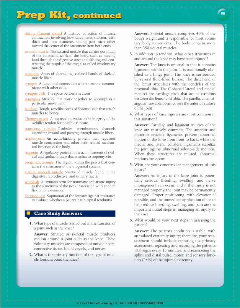
Skidplate: © Photodisc
sliding � lament model A method of action of muscle contraction involving how sarcomeres shorten, with thick and thin � laments sliding past each other toward the center of the sarcomere from both ends.
smooth muscle Nonstriated muscle that carries out much of the automatic work of the body, such as moving food through the digestive tract and dilating and con-stricting the pupils of the eye; also called involuntary muscle.
striations Areas of alternating, colored bands of skeletal muscle � ber.
synapse A functional connection where neurons commu-nicate with other cells.
synaptic cleft The space between neurons.
synergists Muscles that work together to accomplish a particular movement.
tendons Tough, ropelike cords of � brous tissue that attach muscles to bones.
Thompson test A test used to evaluate the integrity of the Achilles tendon for possible rupture.
transverse tubules T-tubules; membranous channels extending inward and passing through muscle � bers.
tropomyosin An actin-binding protein that regulates muscle contraction and other actin-related mechan-ical function of the body.
troponin A regulatory protein in the actin � laments of skel-etal and cardiac muscle that attaches to tropomyosin.
urogenital triangle The region within the pelvis that con-tains the structures of the urogenital system.
visceral smooth muscle Sheets of muscle found in the digestive, reproductive, and urinary tracts.
whiplash A layman’s term for traumatic soft-tissue injury to the structures of the neck, associated with sudden � exion or extension.
Yergason test Supination of the forearm against resistance to evaluate whether a patient has bicipital tendinitis.
6 Case Study Answers
1. What type of muscle is involved in the function of a joint such as the knee?
Answer: Striated or skeletal muscle produces motion around a joint such as the knee. These voluntary muscles are composed of muscle � bers, connective tissue, blood vessels, and nerves.
2. What is the primary function of the type of mus-cle found around the knee?
Answer: Skeletal muscle comprises 40% of the body’s weight and is responsible for most volun-tary body movements. The body contains more than 350 skeletal muscles.
3. In addition to tendons, what other structures in and around the knee may have been injured?
Answer: The knee is unusual in that it contains ligaments within the joint. It is traditionally clas-si� ed as a hinge joint. The knee is surrounded by several � uid-� lled bursae. The distal end of the femur articulates with the condyles of the proximal tibia. The C-shaped lateral and medial menisci are cartilage pads that act as cushions between the femur and tibia. The patella, a � at tri-angular movable bone, covers the anterior surface of the joint.
4. What types of knee injuries are most common in this situation?
Answer: Cartilage and ligament injuries of the knee are relatively common. The anterior and posterior cruciate ligaments prevent abnormal motion of the knee from front to back, while the medial and lateral collateral ligaments stabilize the joint against abnormal side-to-side motions. When these structures are injured, abnormal motions can occur.
5. What are your concerns for management of this injury?
Answer: An injury to the knee joint is poten-tially serious. Bleeding, swelling, and nerve impingement can occur, and if the injury is not managed properly, the joint may be permanently damaged. Proper positioning, with elevation if possible, and the immediate application of ice to help reduce bleeding, swelling, and pain are the important initial steps in managing an injury to the knee.
6. What would be your next steps in assessing the patient?
Answer: The patient’s condition is stable, with an isolated extremity injury; therefore, your reas-sessment should include repeating the primary assessment, repeating and recording the patient’s vital signs every 15 minutes, and reassessing the splint and distal pulse, motor, and sensory func-tion (PMS) of the injured extremity.
Prep Kit, continuedPrep Kit, Prep Kit, continued 95
9781449642303_CH04_Pass5.indd 95 18/03/14 9:46 AM
© Jones & Bartlett Learning, LLC. NOT FOR SALE OR DISTRIBUTION.
© Jones & Bartlett Learning, LLCNOT FOR SALE OR DISTRIBUTION
© Jones & Bartlett Learning, LLCNOT FOR SALE OR DISTRIBUTION
© Jones & Bartlett Learning, LLCNOT FOR SALE OR DISTRIBUTION
© Jones & Bartlett Learning, LLCNOT FOR SALE OR DISTRIBUTION
© Jones & Bartlett Learning, LLCNOT FOR SALE OR DISTRIBUTION
© Jones & Bartlett Learning, LLCNOT FOR SALE OR DISTRIBUTION
© Jones & Bartlett Learning, LLCNOT FOR SALE OR DISTRIBUTION
© Jones & Bartlett Learning, LLCNOT FOR SALE OR DISTRIBUTION
© Jones & Bartlett Learning, LLCNOT FOR SALE OR DISTRIBUTION
© Jones & Bartlett Learning, LLCNOT FOR SALE OR DISTRIBUTION
© Jones & Bartlett Learning, LLCNOT FOR SALE OR DISTRIBUTION
© Jones & Bartlett Learning, LLCNOT FOR SALE OR DISTRIBUTION
© Jones & Bartlett Learning, LLCNOT FOR SALE OR DISTRIBUTION
© Jones & Bartlett Learning, LLCNOT FOR SALE OR DISTRIBUTION
© Jones & Bartlett Learning, LLCNOT FOR SALE OR DISTRIBUTION
© Jones & Bartlett Learning, LLCNOT FOR SALE OR DISTRIBUTION
© Jones & Bartlett Learning, LLCNOT FOR SALE OR DISTRIBUTION
© Jones & Bartlett Learning, LLCNOT FOR SALE OR DISTRIBUTION
© Jones & Bartlett Learning, LLCNOT FOR SALE OR DISTRIBUTION
© Jones & Bartlett Learning, LLCNOT FOR SALE OR DISTRIBUTION