Chapter 1 - Musculoskeletal Signs - Amazon AWS
84
1 CHAPTER 1 MUSCULOSKELETAL SIGNS
-
Upload
khangminh22 -
Category
Documents
-
view
3 -
download
0
Transcript of Chapter 1 - Musculoskeletal Signs - Amazon AWS

1
CHAPTER 1
MUSCULOSKELETAL SIGNS

Anterior drawer test
2
CL
INIC
AL
PE
AR
L
Anterior drawer test
Mechanism/sThe ACL arises from the anterior aspect of the tibial plateau and inserts into the medial aspect of the lateral femoral condyle. It limits anterior movement of the tibia upon the femur. Loss of continuity of the ACL permits inappropriate anterior movement of the tibia and thus knee joint instability.
Sign valueA literature review of six studies reported variable sensitivity of 27–88%, specificity of 91–99%, positive LR of 11.5 and negative LR of 0.5.2 A literature review by Solomon DH et al. of nine studies reported a sensitivity of 9–93% and specificity of 23–100%.1
While a positive anterior drawer sign (+LR 11.5)2 has been suggested to be strong evidence of ACL injury, the results are not uniform, with another study reporting a +LR 2.0 (sensitivity 83%, specificity 57%, –LR 0.3).3 A negative anterior drawer sign cannot reliably exclude ACL injury (sensitivity 27–88%; –LR 0.5).2 When strong clinical suspicion persists, further diagnostic steps are necessary (e.g. interval re-examination, MRI, arthroscopy).
DescriptionWith the patient lying supine, the knee at 90° flexion and the foot immobilised by the examiner, the proximal third of the tibia is pulled towards the examiner. In a positive test, there is anterior (forward) movement of the tibia without an abrupt stop.1
Condition/s associated with•Anteriorcruciateligament(ACL)
injury
FIGURE 1.1 Anterior drawer test for anterior cruciate ligament injury
90º

Apley’s grind test
3
1Apley’s grind test
is increased rotation relative to the unaffected side, this is suggestive of a ligamentous lesion. If rotation plus compression is more painful or there is decreased rotation relative to the unaffected side, this is suggestive of meniscal injury.4
Condition/s associated with•Meniscalinjury
Mechanism/sDirect mechanical force upon the injured meniscus elicits tenderness.
Sign valueA review by Hegedus EJ et al. reported a pooled sensitivity of 60.7% and specificity of 70.2% with an odds ratio of 3.4.5 Significant heterogeneity in the data limits its accuracy. Overall, Apley’s grind test has limited diagnostic utility, limited supporting data and, in the acute setting, the manoeuvre produces severe pain.6
McMurray’s grind test has more robust supporting data.
DescriptionWith the patient lying prone and the knee at 90° flexion, the lower leg is passively internally and externally rotated while axial pressure is applied to the lower leg. The test is considered positive if tenderness is elicited.
The process can also be combined with or without distraction. If rotation plus distraction is more painful or there
FIGURE 1.2 Apley’s grind test

Apley’s scratch test
4
Apley’s scratch test
Condition/s associated withCommon•Rotatorcuffmuscleinjury
•Labraltear
•Anteriorshoulderdislocation
•Bicipitaltendonitis
•Adhesivecapsulitis(frozenshoulder)
•Acromioclavicularjointinjury
Mechanism/sThe shoulder joint is a complex structure. Its components include the humeral head, glenoid fossa, acromion, clavicle, scapula and surrounding soft tissue structures. Under normal circumstances the shoulder joint is capable of a vast range of movement. Apley’s scratch test assesses glenohumeral abduction, adduction, flexion, extension, internal rotation and external rotation. Tenderness or limited range of movement suggests injury to one or more components of the shoulder joint.
Sign valueApley’s scratch test is a useful component of the general shoulder exam but has limited utility for a specific diagnosis. The position of the shoulder at which tenderness or limited range of movement occurs should be noted. In the patient with an abnormal Apley’s scratch test, further diagnostic manoeuvres should be performed to narrow the differential diagnosis.
DescriptionApley’s scratch test is a general range of movement assessment of the shoulder joint (i.e. glenohumeral, acromioclavicular, sternoclavicular and scapulothoracic joints). The patient is instructed to touch the unaffected shoulder anteriorly and posteriorly (behind their head), and touch the inferior scapula posteriorly (behind their back). Tenderness and/or limited range of movement while performing these movements is considered an abnormal test.7
FIGURE 1.3 One of three manoeuvres of Apley’s scratch test
Based on Woodward T, Best TM. The painful shoulder: part 1, clinical evaluation. Am Fam Phys 2000; 61(10): 3079–3088.

Apparent leg length inequality (functional leg length)
5
1Apparent leg length inequality (functional leg length)
Ligamentous laxityThe ligaments on one side (e.g. in the hip joint) may be more flexible or longer than their counterparts, making the femur sit lower in the joint capsule.
Joint contractureA joint contracture impairs full range of movement. If the knee joint is contracted in a flexed position, the length of the affected side will be less than the opposite leg during maximal attempted extension.
Altered foot mechanicsExcessive pronation of the foot eventuates in and/or may be accompanied by a decreased arch height compared to the ‘normal’ foot, resulting in a functionally shorter limb.8
DescriptionA disparity between the relative distance from the umbilicus to the medial malleolus of each leg.8Bydefinition it implies asymmetry of the lower extremities in the absence of a bony abnormality. (See ‘True leg length inequality’ in this chapter.)
Condition/s associated with•Alteredfootmechanics
•Adaptiveshorteningofsofttissues
•Jointcontractures
•Ligamentouslaxity
•Axialmalalignments
Mechanism/sAn apparent or functional leg length inequality may occur at any point from the pelvis to the foot.8
FIGURE 1.4 Measurement of leg lengths A The apparent leg length is the distance from the umbilicus to the medial malleolus; B pelvic rotation causing an apparent leg length discrepancy; C the true leg length is the distance from the anterior superior iliac spine to the medial malleolus.
Based on Firestein GS, Budd RC, Harris ED et al., Kelley’s Textbook of Rheumatology, 8th edn, Philadelphia: WB Saunders, 2008: Fig 42-24.
A B C

Apparent leg length inequality (functional leg length)
6
Sign valueThe distance (anywhere from 3–22 mm) at which apparent leg length inequality results in a clinically
significant effect is controversial.8 The test should be interpreted in relation to the patient’s history and full gait assessment.

Apprehension test
7
1Apprehension test
•Rotatorcuffmuscleinjury
•Glenoidlabruminjury
•Glenoiddefect(e.g.Bankart’sfracture)
•Humeralheaddefect(e.g.Hill–Sachs fracture)
Less common – atraumatic•Connectivetissuedisorder:
Ehlers–Danlos syndrome, Marfan’s syndrome
•Congenitalabsenceofglenoid
Mechanism/sGlenohumeraljointinstabilityiscausedby dysfunction of the bony and/or soft tissue structures that maintain joint stability:glenoid,humeralhead,jointcapsule, capsuloligamentous or glenohumeral ligaments, labrum, and rotator cuff muscles. The shoulder joint is susceptible to instability due to its inherent mobility and complex soft tissue structures responsible for stability.
In the apprehension test, the joint is placed into a position vulnerable to instability. It is the typical position precipitating traumatic anterior shoulder dislocation. For this reason, a significant number of healthy patients will experience apprehension during this manoeuvre.
Sign valueT’Jonck L et al. reported a sensitivity of 88.0%, specificity of 50%, positive likelihood ratio of 1.8 and negative likelihood ratio of 0.23.10
The apprehension test for glenohumeral joint instability is a moderatelyusefulscreeningtest.Basedon available data, the test has limited utility to rule in the diagnosis. It is not used in the setting of acute anterior shoulder dislocation.
DescriptionThe apprehension test is an assessment of glenohumeral joint instability. With the patient sitting or lying supine, the shoulder is placed into 90° abduction, 90° external rotation and 90° elbow flexion. The examiner applies pressure to the posterior aspect of the proximal humerus and attempts to move the humeral head anteriorly (see Figure 1.5). The test is positive if the patient experiences apprehension due to impending subluxation or dislocation of the glenohumeral joint.9
Condition/s associated withMore common – traumatic•Recurrentglenohumeraljoint
subluxation or dislocation
FIGURE 1.5 Apprehension test The arm is abducted and placed in an externally rotated position. Note the right arm of the examiner is providing anterior traction on the humerus, pulling the posterior part of the humeral head forward. The same test can be done from the back, with the patient sitting up and the examiner pushing forward on the posterior head of the humerus.

Apprehension–relocation test (Fowler’s sign)
8
Apprehension–relocation test (Fowler’s sign)
DescriptionThe apprehension–relocation test is an assessment of glenohumeral joint instability. The relocation manoeuvre is typically performed following the apprehension test (see ‘Apprehension test’). With the patient sitting or lying supine, the shoulder is placed into 90° abduction, 90° external rotation and 90° elbow flexion. The examiner applies pressure to the anterior aspect of the proximal humerus and attempts to move the humeral head posteriorly. The test is positive if the patient experiences relief of apprehension (i.e. no longer feels impending shoulder dislocation).
Condition/s associated with•Recurrentglenohumeraljoint
subluxation or dislocation
•Rotatorcuffmuscleinjury
•Glenoidlabruminjury
•Glenoiddefect(e.g.Bankart’sfracture)
•Humeralheaddefect(e.g.Hill–Sachs fracture)
Less common – atraumatic•Connectivetissuedisorder:
Ehlers–Danlos syndrome, Marfan’s syndrome
•Congenitalabsenceofglenoid
Mechanism/sThe underlying anatomy and causes of glenohumeral joint instability are outlined under ‘Apprehension test’ and apply here. In the apprehension–relocation test, symptomatic relief is due to restoration of the normal anatomical relationship of the humeral head in the glenohumeral joint.
Sign valueT’Jonck L et al. reported a sensitivity of 85%, specificity of 87%, positive likelihood ratio of 6.5 and negative likelihood ratio of 0.18.10 Lo et al. reported sensitivity of 32% and specificity of 100%.11 Speer KP et al. reported a sensitivity of 68% and specificity of 100%.12
The apprehension–relocation test is a useful screening manoeuvre for anterior glenohumeral joint instability. It appears to be more specific than the ‘apprehension test’ alone.
FIGURE 1.6 Apprehension–relocation (Fowler) test Note that pressure is applied anteriorly to the proximal humerus.

Bouchard’s and Heberden’s nodes
9
1Bouchard’s and Heberden’s nodes
Mechanism/sA number of studies have implicated bony osteophyte growth as the principal causeofHeberden’sandBouchard’snodes.13 Other contributing factors or theoriesinclude:
•geneticpredisposition
•endochrondralossificationofhypertrophied cartilage as a result of chronic osteoarthritic changes14
•tractionspursgrowingintendonsin response to excessive tension and repetitive strain.15
Sign valueBouchard’sorHeberden’snodesare a classical sign of interphalangeal osteoarthritis15,16 and are associated with generalised osteoarthritis.17,18 ThepresenceofBouchard’sand/orHeberden’s nodes is predictive of the radiographic changes of osteoarthritis.19
DescriptionBouchard’snodesarebonyoutgrowthsor nodules found over the proximal interphalangeal joints of the hands.
Heberden’s nodes are similar but located over the distal interphalangeal joints.
Condition/s associated with•Osteoarthritis
•Familial
FIGURE 1.7 Prominent Heberden’s nodes
Based on Ferri FF, Ferri’s Clinical Advisor, Philadelphia: Elsevier, 2011: Fig 1-223.

10
Boutonnière deformity
Boutonnière deformity
FIGURE 1.8 Digital extensor mechanism A The proximal interphalangeal joint is extended by the central tendon slip (an extension of the hand’s dorsal extensor tendon); B the X is a functional representation of the fibrous interconnections between the two systems.
Based on DeLee JC, Drez D, Miller MD, DeLeeandDrez’sOrthopaedicSportsMedicine, 3rd edn, Philadelphia: Saunders, 2009: Fig 20B2-27.
Central tendon slip
Functional tendinous interconnectionsbetween two extensor tendons
A
Lateral band
B
FIGURE 1.9 Pathoanatomy of boutonnière deformity Thesequenceis:ruptureofthecentraltendonslip,whichthensimultaneouslypullsonthelateral bands, pulling the DIP joint into hyperextension and the PIP into flexion.
Based on DeLee JC, Drez D, Miller MD, DeLeeandDrez’sOrthopaedicSportsMedicine, 3rd edn, Philadelphia: Saunders, 2009: Fig 20B2-28.
3
3
2
2
1
1
4
4
Central tendon slip
Central tendon slip pulls off bone
Retracted central tendon slip pulls on lateral band
The lateral band, in turn, hyperextends the DIP joint
With no central tendon connection the PIP joint flexes,completing the full boutonnière deformity
Lateral band

11
1
Boutonnière deformity
Inflammatory arthropathy (e.g. rheumatoid arthritis)Pannus in the PIP joint (which may be present in rheumatoid arthritis) can damage the central slip tendon.20 Chronic inflammation and synovitis of the joint may result in persistent PIP flexion and gradual elongation of the central slip tendon. Subsequent volar migration of the lateral bands results in the characteristic deformity.21–24
TraumaForced flexion of an extended PIP joint, crush injury or penetrating injury may result in avulsion of the central tendon slip. Typically, the degree of deformity increases in the days following the injury. Acutely, the deformity may be subtle.
Sign valueA boutonnière deformity is classically associated with rheumatoid arthritis occurring in up to 50% of patients with the disease.
In a patient with blunt or penetrating trauma, the presence of a boutonnière deformity should be considered evidence of a central slip extensor tendon injury.
DescriptionUsed to describe a deformity of the resting finger in which the proximal interphalangeal (PIP) joint is flexed and the distal interphalangeal (DIP) joint is hyperextended.
Condition/s associated with•Inflammatoryarthropathy(e.g.
rheumatoid arthritis)
•Centralslipextensortendoninjury
Mechanism/sDisruption or avulsion of the central slip extensor tendon and volar migration of the lateral bands of the extensor tendon mechanism result in PIP flexion and DIP extension. The sign derives its name from the appearance of the central tendon slip, which was thought to resemble a buttonhole, or boutonnière in French, when torn.
The central tendon slip attaches to the dorsal aspect of the middle phalanx. Its main function is to maintain PIP extension and stabilise the extensor tendon apparatus. If the central tendon is disrupted or avulsed (torn off the base of the middle phalanx), the actions of the lateral bands and flexor digitorum profundus are unopposed, resulting in resting PIP flexion and DIP hyperextension.

Bulge/wipe/stroke test
12
Bulge/wipe/stroke test
More common•Osteoarthritis
•Rheumatoidarthritis
•Haemoarthrosis–trauma,coagulopathy
•Gout
•Infection–septicarthritis,gonococcal arthritis, transient synovitis
Less common•Pseudogout(calciumpyrophosphate
deposition disease)
•Tumour
Mechanism/sMechanical manipulation of excess fluid in the synovial joint capsule results in visible fluid shift. The wipe or bulge test displaces synovial fluid from one part of the synovial joint to another, thus suggesting the presence of a joint effusion as the cause of knee swelling.
Sign valueLimited evidence has been gathered on the value of this test as an individual sign. Some authors report that this test may pick up on as little as 4–8 mL of swelling.25 An effusion in the absence of acute traumatic injury or systemic disease is most commonly due to osteoarthritis.26
GogusFetal.27 reported the wipe test as having a sensitivity of 11–33% and specificity of 66–92% for identifying the presence of a knee effusion. Emphasis should be placed upon identifying a joint effusion in the setting of septic arthritis, an orthopaedic emergency.
DescriptionThe bulge, wipe or stroke test is used to assess for knee joint effusion. With the patient supine and their knee extended, the examiner ‘swipes’ the medial aspect of the knee joint to displace fluid into the superolateral aspect of the synovial compartment, and then swipes the lateral side looking for a visible fluid shift. The test is positive if the examiner sees a wave of fluid.
Condition/s associated withAny condition causing a knee effusion.
FIGURE 1.10 Demonstration of the bulge test for a small synovial knee effusion The medial aspect of the knee has been stroked to move the synovial fluid from this area (shaded depressed area in A); B shows a bulge in the previously depressed area after the lateral aspect of the knee has been tapped.
Based on Firestein GS, Budd RC, Harris ED et al., Kelley’s Textbook of Rheumatology, 8th edn, Philadelphia: WB Saunders, 2008: Figs 35-9A and B.
A
B
Butterfly rash (malar rash)
13
1Butterfly rash (malar rash)
Mechanism/sThe exact mechanism is unclear. However, like the underlying disorder in SLE, it is thought to result from an autoimmune reaction caused by genetic, environmental and immunological factors.
Factors shown to be involved include:28
•Ageneticpredispositiontoineffective or deficient complement, leading to a failure to clear immune complexes of apoptotic cells, which in turn increases the chance of the development of autoimmunity.
•Sunlighthasbeenshowntodamage and/or induce apoptosis of keratinocyte proteins in the epidermis and can stimulate autoantibody production. Sunlight may also increase the chance of keratinocytes being destroyed by complement and antibody-dependent mechanisms.
•Alteredcellularandhumoralimmunity reactions have been seen in studies reviewing cutaneous manifestations of lupus.
It is likely that a combination of these factors leads to immune deposition in the skin, damage, oedema and the characteristic malar rash.
DescriptionA red or purple, macular, mildly scaly rash that is seen over the bridge of the nose and cheeks. The shape of the rash can somewhat resemble a butterfly. The rash spares the nasolabial folds, which helps distinguish it from other rashes (e.g. rosacea). It is also photosensitive.
Condition/s associated withCommon•Systemiclupuserythematosus
(SLE)
•Drug-inducedlupuserythematosus
•Dermatomyositis
FIGURE 1.11 Malar rash of SLE
Reproduced, with permission, from Goldman L, Ausiello D, Cecil Medicine, 23rd edn, Philadelphia: Saunders, 2007: Fig 287-3.
Butterfly rash (malar rash)
14
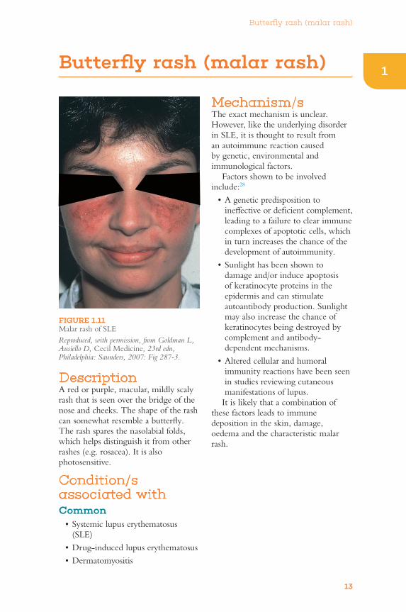
FIGURE 1.12 Mechanism of malar rash
Geneticpredisposition
Deficiency incomplement
Failure to clearcells of
complement
Increasedchance of auto-
immunity
Autoimmune reaction and complex deposition – damage tocollagen and blood vessels
Malar and other cutaneous rashes in SLE
Sunlight
Keratinocytedamage/proteins
Inflammatory cytokineproduction and
autoantigen exposure
Increased auto-antibody
production
EnvironmentAltered cellular andhumoral immunity
Sign valueThe malar rash is seen in approximately 40% of patients with SLE.28 It has a sensitivity of 57% and specificity of 96% for SLE.14 Its absence does not exclude the diagnosis.

Calcinosis/calcinosis cutis
15
1Calcinosis/calcinosis cutis
» Systemic sclerosis
» Burns
•Metastatic» Due to hypercalcaemia or
hyperphosphataemia of any cause
» Chronic renal failure – most common
» Excess vitamin D
» Primary hyperparathyroidism – rare
» Paraneoplastic hypercalcaemia
» Destructive bone disease (e.g. Paget’s disease)
•Iatrogenic» Calcium gluconate injections
» Tumour lysis syndrome
•Idiopathic
•Calciphylaxis» End-stage renal disease
» Altered calcium metabolism
Mechanism/sDystrophic calcinosisDystrophic calcinosis occurs when crystals of calcium phosphate or hydroxyapatite are deposited in the skin secondary to inflammation, tissue damage and degeneration.30 Calcium and phosphate levels are usually normal. Proposedmechanismsinclude:
•Highlocallevelsofalkalinephosphatase break down a pyrophosphate that normally inhibits calcification.31
•Tissuebreakdownmayleadtodenatured proteins that bind to phosphate. These phosphate–protein compounds may react with calcium and thus provide a nidus for calcification.32
FIGURE 1.13 Calcinosis Hard, whitish nodules of the digit representing dystrophic calcinosis in this patient with dermatomyositis.
Reproduced, with permission, from James WD, Berger T, Elston D, Andrews’ Diseases of the Skin:ClinicalDermatology, 11th edn, Philadelphia: Saunders, 2011: Fig 26-12.
DescriptionCalcinosis refers to the formation or deposition of calcium in soft tissue. Calcinosis cutis more specifically refers to calcium deposits in the skin.
Condition/s associated withConditions associated with calcinosis may be classified as dystrophic, metastatic, iatrogenic, idiopathic or calciphylaxis.
•Dystrophiccalcinosis» Scleroderma
» Dermatomyositis
» SLE

Calcinosis/calcinosis cutis
16
•VitaminDdeficiencyowingtorenal failure worsens initial hypocalcaemia and, therefore, further stimulates secondary hyperparathyroidism.
IatrogenicIntravenous administration of calcium or phosphate may cause local extravasation and precipitation of hydroxyapatite in surrounding tissue. Inflammation of the surrounding tissue secondary to the injection may also cause calcium and protein release, contributing to precipitation.
IdiopathicOccurs in the absence of tissue injury or systemic metabolic disturbance.
Sign valueThere is very limited evidence on this sign and it is rarely seen in isolation. If identified, further investigation is warranted.
Metastatic calcinosisAbnormal calcium or phosphate metabolism with high levels of either or both is present. Excess calcium and/or phosphate allows for the formation and precipitation of calcium salts.
In chronic renal failure a number of mechanisms lead to altered phosphate andcalciummetabolism:
•Decreasedrenalexcretion of phosphate leads to hyperphosphataemia.
•Hyperphosphataemiaresultsinacompensatory rise in parathyroid hormone (PTH) in an attempt to excrete phosphate. The rise in PTH results in an increase in phosphate absorption from the gut and also mobilises calcium from the bones, resulting in more calcium being available to precipitate with phosphate.

Charcot foot
17
1
CL
INIC
AL
PE
AR
L
1Charcot foot
DescriptionA progressive destructive arthropathy of the ankle and foot.33 In its early stages, it may present as unilateral foot oedema following minor trauma. In advanced disease, significant destruction of bones and joints may occur (particularly in the midfoot), resulting in collapse of the plantar arch and development of ‘rocker-bottom foot’.
Condition/s associated withConditions resulting in sensory neuropathy:
•Diabetesmellitus–mostcommon
•Syphilis–originaldescriptionbyCharcot
Mechanism/sIn neurotraumatic theory, peripheral neuropathy caused by diabetes leads to decreased pain sensation and impaired proprioception. Thus, if an acute injury occurs (e.g. microfracture, subluxation or fracture), the patient feels little or no pain and does not ‘guard’ the foot
when mobilising. This leads to a destructive cycle of continued loading on the injured foot and progressive damage.34
Under the inflammatory theory, when the same local insult occurs (microfracture, subluxation or fracture), inflammatory cytokines are released, including TNF-α and interleukin-1β. These two cytokines have been shown to increase activation of RANK ligand (RANKL), which in turn increases the transcription factor nuclear factor-κB(NF-κB).Thenetresultofthisisstimulation of the maturation of osteoclasts, which further eat away at bone. This predisposes the patient to engage in another vicious cycle of further fractures, inflammation, abnormal weight loading and osteolysis.34
Regardless of underlying contributing factors, the RANKL/OPG(osteoprotegerin)pathwayisthought to be a common denominator.
RANKL is a member of the tumour necrosis factor (TNF) superfamily, and OPGisthecompetitiveproteinofRANKL. The process of bone
FIGURE 1.14 Charcot foot A, B The classic rocker-bottom Charcot foot, with collapse and then reversal of the longitudinal arch; C loss of the normal calcaneal pitch, or angle relative to the floor, in patients with Charcot collapse of the arch.
Reproduced, with permission, from Mann JA, Ross SD, Chou LB, Chapter 9: Foot and ankle surgery. In: Skinner HB, Current Diagnosis & Treatment in Orthopedics, 4th edn, Fig 9-8. Available: http://proxy14.use.hcn.com.au/content.aspx?aID=2321540 [10 Mar 2011].
A
Achilles Achilles
Normal
Calcaneal pitch Loss ofcalcaneal pitch
C
B

Charcot foot
18
osteopenia due to unrestricted RANKL activity.35
Othercontributingfactorsinclude:
•Sympatheticdenervationindistallimbs leads to increased peripheral blood flow – hyperaemia and more inflammation.36
•Pre-existingosteopaeniahasbeenseen in both type 1 and type 2 diabetes via a number of mechanisms,36 and this predisposes the diabetic patient to microfracture.
•Abnormalloadingmechanics.» Oxidative stress and the
formation of reactive oxidant species (ROS) with local dysregulation of immunoinflammatory processes.
» AGE/RAGE pathway − hyperglycaemia generates AGEs. These promote change in both intra- and extracellular proteins, which become defective and lose functionality. Binding of AGE to receptors for AGE (RAGE) accelerates ROS production, activating NF-κB. An increase in AGE-modified collagen has been shown to affect osteoblastic cell differentiation and function in vitro. This may play a role in the pathogenesis of osteopaenia, which is present in patients with poorly controlled diabetes.
» PI3 kinase pathway − PI3 kinase normally impedes inflammation via enhanced nitric oxide (NO) production. NO may cause apoptosis of osteoclast progenitors as well as inhibit the resorptive action of mature osteoclasts. The PI3 kinase and NO pathway may be compromised in diabetes.
» Altered Wnt/β-catenin pathway (an important bone anabolic pathway).
FIGURE 1.15 Simplified inflammatory and neurotraumatic mechanisms of Charcot foot
Based on Jeffcoate WJ, Game F, Cavanagh PR, Lancet 2005; 366: 2058–2061.
Trauma
Increasedforce
DislocationFracture
Osteopenia
Osteoclastogenesis
Pro-inflammatorycytokines
(TNF-α, interleukin-1β)
RANKLNF-κB
Neuropathy
Inflammation
Abnormalloading
resorption and formation is controlled bythelevelofRANKLandOPG,andmany factors contribute to this pathway.
RANKL has been shown to mediate osteolysis in Charcot foot by stimulating osteoclastic differentiation of monocytes/macrophages and triggering the synthesis of nuclear factor-κβ (NF-κβ), spurring maturation.
Hyperglycaemia can increase the level of advanced glycation end products(AGEs),reactiveoxygenspecies (ROS), and oxidised lipids, which may all enhance the expression of RANKL in diabetes.
The pro-inflammatory state increases cytokine expression (TNF-α, IL-1β and IL-6), which may also increase RANKL. Diabetic neuropathy is associated with exhaustion of calcitonin gene-related peptide (CGRP)storesfromCandAδ nerve fibres.ThedeficiencyofCGRPmayfurther accelerate underlying
Charcot foot
19
1
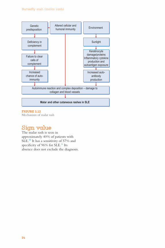
FIGURE 1.16 Detailed mechanism behind the development of Charcot foot
From Zhao H-M, Diao J-Y, Liang X-J et al. Pathogenesis and potential relative risk factors of diabetic neuropathic osteoarthropathy. Journal of Orthopaedic Surgery and Research 2017; 12: 142.
Hyperglycemia
Osteoblast ↓
BMD ↓
ROS ↑
Inflammation
AGE/RAGE ↑, PKC ↑, PI3 kinase ↓
NO ↓ Repetitive trauma CGRP ↓
Neuropathy
BMD ↓
Blood flow ↑
TNF-α, IL-1β, IL-6 ↑oxPTM ↑
FLS activated
Joint destruction
RANKL/OPG ↑
MMPs↑ Activated osteoclast ↑
Osteolysis
IL-4 ↓IL-10 ↓OPG ↓
DKK-1, Wnt-1 ↓sclerostin ↓
NO ↓
InflammationOsteolysis/ligaments injury
Fracture/dislocation
oxPTM: oxidative post-translational modificationsBMD: bone mineral densityFLS: fibroblast-like synoviocytesMMPs: matrix metalloproteinasesDNOAP: diabetic neuropathic osteoarthropathy
Deformity
neuropathy
Repetitive trauma
neuropathy
persistent hyperglycemianeuropathytrauma
Acute DNOAP
Chronic DNOAPstable
Osteoclasts ↑Monocytes ↑
» Altered fibroblast-like synoviocytes (FLS).
» OPG genetic polymorphisms.35
Sign valuePatients with Charcot foot are at higher risk of diabetic foot ulcers (affecting up to 50% of patients)37,38 and amputation.36
The presence of foot pain, heat and/or swelling in the diabetic patient needs immediate attention including referral to diabetic/high-risk foot clinics or orthopaedic and diabetic specialist services.

Crepitus
20
Crepitus
DescriptionGrating,crunching,poppingorcrackling sounds heard and/or felt over joints during passive range of motion examination.
Condition/s associated with•Arthropathy
» Osteoarthritis
» Rheumatoid arthritis
•Trauma» Cartilaginous injury –
meniscal injury, labral injury
» Ligamentous injury – anterior cruciate ligament
» Fracture
General mechanism/sCrepitus of the joints is caused when two rough surfaces grind against one another.
Rheumatoid/osteoarthritisIn both rheumatoid arthritis and osteoarthritis arthritis, degeneration of
the articular cartilage of the joint surfaces occurs, creating erosions and irregularity. Two rough surfaces moving against each other produce crepitus.
In rheumatoid arthritis, the autoimmune response and subsequent inflammation, cytokine release and pannus formation cause destruction of cartilage.
In osteoarthritis, repetitive strain with loss of glycosaminoglycans and activation of matrix metalloproteinases (MMPs) is principally responsible for damage.
Sign valueAltman R et al. reported crepitus had a sensitivity of 89%, specificity of 58%, positive likelihood ratio of 3.0 and negative likelihood ratio of 0.2 for predicting osteoarthritis of the knee.39 Crepitus is common in patients with osteoarthritis. Crepitus alone has limited diagnostic value, due to its presence in other common disease states.

Dropped arm test
21
1Dropped arm test
responsible for the first 15° of motion. The deltoid muscle is responsible for movement beyond 15°.40 Therefore, if a rotator cuff tear (e.g. supraspinatus muscle tear) or subacromial impingement is present, the ability of the arm to maintain abduction is impaired.
Sign valueMurrellGACetal.andDinnesJetal.reported a sensitivity of 10% and specificity of 98%, and a calculated positive likelihood ratio greater than 10 for rotator muscle tear.41,42ParkHBet al. reported a sensitivity of 27%, specificity of 88%, positive likelihood ratio of 2.3 and negative likelihood ratio of 0.8 for subacromial impingement.43
When positive, the dropped arm test significantly increases the probability of rotator cuff muscle tear (supraspinatus muscle tear) or subacromial impingement. A negative test does not reliably exclude the diagnosis.
FIGURE 1.17 Dropped arm test
Based on Multimedia Group LLC, Occupation Orthopedics. Available: http://www.eorthopod.com/eorthopodV2/index.php?ID=7244790ddace6ee8ea5da6f0a57f8b45&disp_type=topic_detail&area =6&topic_id=4357b9903d317fcb3ff32f72b24cb6b6 [28 Feb 2011].
DescriptionWith the patient upright, the examiner passively moves the patient’s arm to 90° of abduction. Then the patient is asked to slowly lower the arm to the anatomical position. A positive test occurs if the patient is unable to perform the action due to pain or if the arm just ‘drops’ to the side.
Condition/s associated with•Rotatorcuffmuscleinjury(e.g.
supraspinatus muscle)
•Subacromialimpingement
•Neurogenicweakness
•Suprascapularnervepalsy
•Axillarynervepalsy
•C5radiculopathy
Mechanism/sAbduction of the arm from 0° to 90° is dependent upon the supraspinatus and deltoid muscles. The supraspinatus is

Eichhoff’s test
22
Eichhoff’s test
DescriptionThe patient places their thumb within the palm of the examiner’s hand, who grasps it tightly. The hand is then abducted towards the ulna by the examiner (see Figure 1.18).
Condition/s associated with•DeQuervain’stenosynovitis
Mechanism/sDeQuervain’stenosynovitisis an inflammatory condition of the
contents of the first extensor synovial compartment:thetendonsofabductorpollicis longus and extensor pollicis brevis.
Repetitive strain injury or inflammatory disorders cause inflammation, leading to swelling over the radial aspect of the wrist. This narrows the space that the abductor pollicis longus and extensor pollicis brevis pass through on their way to the hand.
This manoeuvre and Finkelstein’s test involve generation of a passive distension and shear stress between the tendons and radius on its blunt styloid edge. In essence, the abductor pollicis longus and extensor pollicis brevis tendons are moved into the narrowed compartment and stretched, causing pain.
Sign valueThere is limited data on the evidence of Eichhoff’s test’s diagnostic accuracy indiagnosingdeQuervain’stenosynovitis. One small study suggested Eichhoff’s test was associated with more false positives than Finkelstein’s test.44
FIGURE 1.18 Eichhoff’s test

Finkelstein’s test
23
1Finkelstein’s test
radial aspect of the wrist (at the abductor pollicis longus tendon or extensor pollicis brevis tendon) is considered a positive test result.
Condition/s associated with•DeQuervain’stenosynovitis
Mechanism/sDeQuervain’stenosynovitisisaninflammatory condition of the contents of the 1st extensor synovial compartment:abductorpollicislongusand extensor pollicis brevis tendons.
Repetitive strain injury or inflammatory disorders cause inflammation that, in turn, causes swelling over the radial aspect of the wrist. This narrows the space through which the abductor pollicis longus and extensor pollicis brevis pass on their way to the hand. When performing this manoeuvre, the abductor pollicis longus and extensor pollicis brevis tendons are moved into the narrowed compartment and stretched, causing pain.45
Sign valueThere is limited data on the evidence for Finkelstein’s test in diagnosing De Quervain’stenosynovitis.DeQuervain’stenosynovitisisaclinicaldiagnosis.
FIGURE 1.19 Finkelstein’s test With the thumb inside the hand, the wrist is ulnarly deviated. Pain indicates a positive test.
Based on Frontera WR, Silver JK, Rizzo Jr TD, Essentials of Physical Medicine and Rehabilitation, 2nd edn, Philadelphia: Saunders, 2008: Fig 24-2.
DescriptionThe examiner applies force at the patient’s thumb metacarpal, placing the wrist into forced ulnar deviation. Tenderness with the manoeuvre at the

Gottron’s papules
24
CL
INIC
AL
PE
AR
L
Gottron’s papules
DescriptionViolaceous(violet-coloured)papularrash on the dorsal aspect of the interphalangeal joints.46
Condition/s associated with•Dermatomyositis
Mechanism/sOne histological study47 demonstrated lymphocytic infiltration, epidermal atrophy and vacuoles in the basal layer of the skin, in addition to other findings. The mechanism is unknown.
Sign valueGottron’spapulesaresaidtobepathognomonic for dermatomyositis; however, they are not present in all patients with the disease.48FIGURE 1.20
Gottron’spapulesFoundoverbonyprominences:fingers,elbows and knees. The lesions are slightly elevated, violaceous papules with slight scale.
Reproduced, with permission, from Habif TP, Clinical Dermatology, 5th edn, Philadelphia: Mosby, 2009: Figs 17-20, 17-21.

Hawkins’ impingement test
25
1Hawkins’ impingement test
FIGURE 1.21 Hawkins’ test anatomy
ClavicleAcromion
Glenoid
Scapula
Humerus
Bursa
Rotatorcuff
FIGURE 1.22 Hawkins’ test
DescriptionWith the patient upright, shoulder and elbow both flexed to 90°, the examiner internally rotates the shoulder joint. The sign is positive if tenderness is elicited (see Figure 1.22).
Condition/s associated with•Rotatorcuffmuscleimpingement
– supraspinatus, teres minor, infraspinatus muscles
•Rotatorcufftendonitis
Mechanism/sThe tendons of the rotator cuff muscles pass through a narrow space between the acromion process of the scapula, bursa and the head of the humerus. Hawkins’ impingement test exacerbates narrowing in the coracoacromial space and will worsen pre-existing impingement of the tendons and muscles when present. The position advances the greater tuberosity towards or against the coracoacromial ligament. This manoeuvre will also elicit tenderness when rotator cuff tendonitis

Hawkins’ impingement test
26
is present, due to mechanical forces or compression on the injured tendon or muscle.49
Sign valueCalis M et al. reported a sensitivity of 92% and a specificity of 26–44% for identifying rotator cuff tendonitis.50 MacdonaldPBetal.reportedasensitivity of 83% and a specificity of 51% for NLR of 0.3 for rotator cuff tear.51
A pooled analysis of 1029 patients across six studies by Alqunaee M et al.52 reported a sensitivity of 74% andspecificityofonly57%.Giventhese results, the test is of little value to the examiner, while a negative test has moderate utility. Further physical examination and imaging should be used to confirm the diagnosis.
Heliotrope rash
27
1
CL
INIC
AL
PE
AR
L
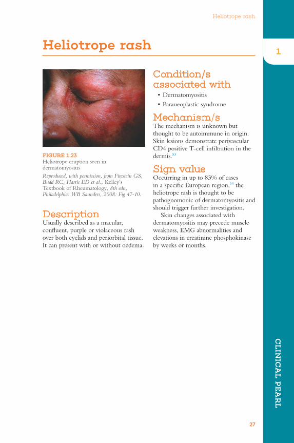
1Heliotrope rash
Condition/s associated with•Dermatomyositis
•Paraneoplasticsyndrome
Mechanism/sThe mechanism is unknown but thought to be autoimmune in origin. Skin lesions demonstrate perivascular CD4 positive T-cell infiltration in the dermis.53
Sign valueOccurring in up to 83% of cases in a specific European region,54 the heliotrope rash is thought to be pathognomonic of dermatomyositis and should trigger further investigation.
Skin changes associated with dermatomyositis may precede muscle weakness,EMGabnormalitiesandelevations in creatinine phosphokinase by weeks or months.
FIGURE 1.23 Heliotrope eruption seen in dermatomyositis
Reproduced, with permission, from Firestein GS, Budd RC, Harris ED et al., Kelley’s Textbook of Rheumatology, 8th edn, Philadelphia: WB Saunders, 2008: Fig 47-10.
DescriptionUsually described as a macular, confluent, purple or violaceous rash over both eyelids and periorbital tissue. It can present with or without oedema.
Kyphosis
28
Kyphosis
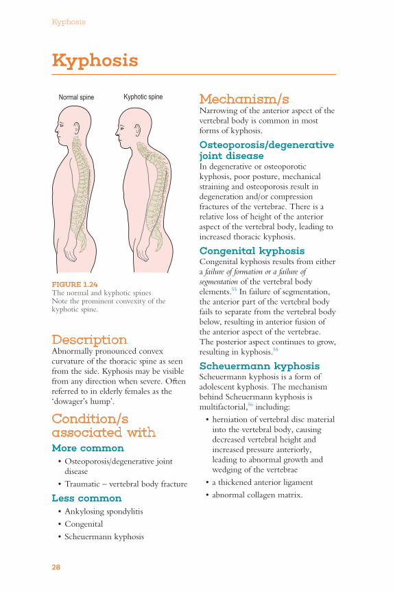
FIGURE 1.24 The normal and kyphotic spines Note the prominent convexity of the kyphotic spine.
Normal spine Kyphotic spine
DescriptionAbnormally pronounced convex curvature of the thoracic spine as seen from the side. Kyphosis may be visible from any direction when severe. Often referred to in elderly females as the ‘dowager’s hump’.
Condition/s associated withMore common•Osteoporosis/degenerativejoint
disease
•Traumatic–vertebralbodyfracture
Less common•Ankylosingspondylitis
•Congenital
•Scheuermannkyphosis
Mechanism/sNarrowing of the anterior aspect of the vertebral body is common in most forms of kyphosis.
Osteoporosis/degenerative joint diseaseIn degenerative or osteoporotic kyphosis, poor posture, mechanical straining and osteoporosis result in degeneration and/or compression fractures of the vertebrae. There is a relative loss of height of the anterior aspect of the vertebral body, leading to increased thoracic kyphosis.
Congenital kyphosisCongenital kyphosis results from either a failure of formation or a failure of segmentation of the vertebral body elements.55 In failure of segmentation, the anterior part of the vertebral body fails to separate from the vertebral body below, resulting in anterior fusion of the anterior aspect of the vertebrae. The posterior aspect continues to grow, resulting in kyphosis.55
Scheuermann kyphosisScheuermann kyphosis is a form of adolescent kyphosis. The mechanism behind Scheuermann kyphosis is multifactorial,56including:
•herniationofvertebraldiscmaterialinto the vertebral body, causing decreased vertebral height and increased pressure anteriorly, leading to abnormal growth and wedging of the vertebrae
•athickenedanteriorligament
•abnormalcollagenmatrix.

Kyphosis
29
1Sign valueKyphosis in paediatric patients may be suggestive of congenital kyphosis, which can have serious complications and lead to significant disability if left
untreated. Acute worsening in the degree of kyphosis in an elderly patient should prompt consideration of pathological fracture.
Lachman’s test
30
CL
INIC
AL
PE
AR
L
Lachman’s test
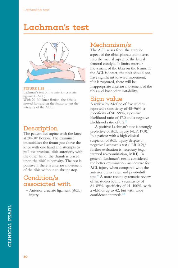
FIGURE 1.25 Lachman’s test of the anterior cruciate ligament (ACL) With 20–30° knee flexion, the tibia is moved forward on the femur to test the integrity of the ACL.
DescriptionThe patient lies supine with the knee at 20–30° flexion. The examiner immobilises the femur just above the knee with one hand and attempts to pull the proximal tibia anteriorly with the other hand; the thumb is placed upon the tibial tuberosity. The test is positive if there is anterior movement of the tibia without an abrupt stop.
Condition/s associated with•Anteriorcruciateligament(ACL)
injury
Mechanism/sThe ACL arises from the anterior aspect of the tibial plateau and inserts into the medial aspect of the lateral femoral condyle. It limits anterior movement of the tibia on the femur. If the ACL is intact, the tibia should not have significant forward movement; if it is ruptured, there will be inappropriate anterior movement of the tibia and knee joint instability.
Sign valueAreviewbyMcGeeoffivestudiesreported a sensitivity of 48–96%, a specificity of 90–99%, a positive likelihood ratio of 17.0 and a negative likelihood ratio of 0.2.2
A positive Lachman’s test is strongly predictive of ACL injury (+LR 17.0).2 In a patient with a high clinical suspicion of ACL injury despite a negative Lachman’s test (–LR 0.2),2 further evaluation is necessary (e.g. interval re-examination, MRI). In general, Lachman’s test is considered the better examination manoeuvre for ACL injury when compared with the anterior drawer sign and pivot-shift test.57 A more recent systematic review of six studies found a sensitivity of 81–89%, specificity of 91–100%, with a +LR of up to 42, but with wide confidence intervals.58
Livedo reticularis
31
1
CL
INIC
AL
PE
AR
L
1Livedo reticularis
Less common•SecondaryLR
Present in numerous disorders including:
•Hypercoagulablestate» Antiphospholipid syndrome
» Cryoglobulinaemia
» Multiple myeloma
» DVT
•Microangiopathy/microangiopathichaemolytic anaemia (MAHA)» Thrombotic/thrombocytopenic
purpura (TTP)
» Haemolytic uraemic syndrome
» Disseminated intravascular coagulation
•Vasculitis/arteriopathy» Snedden’s syndrome
» Calciphylaxis
•Connectivetissuedisorders(e.g.SLE, dermatomyositis)
•Embolisation(e.g.cholesterolembolisation syndrome)
•Drugsideeffect» Amantadine
» Quinine
General mechanism/sArterioles arising from the dermis divide to form a capillary bed. These capillaries then drain into the venules of the venous plexus. Livedo reticularis results from increased visibility of the venules of the skin. Venodilatation of superficial venules and deoxygenation of blood in the plexus are two main factors.59
In general, venodilatation is caused by altered autonomic nervous system function, circulating factors that cause
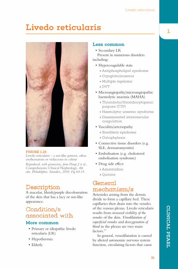
DescriptionA macular, bluish/purple discolouration of the skin that has a lacy or net-like appearance.
Condition/s associated withMore common•Primaryoridiopathiclivedo
reticularis (LR)
•Hypothermia
•Elderly
FIGURE 1.26 Livedo reticularis – a net-like pattern, often erythematous or violaceous in colour
Reproduced, with permission, from Floege J et al., Comprehensive Clinical Nephrology, 4th edn, Philadelphia: Saunders, 2010: Fig 64-13.

Livedo reticularis
32
venodilatation or in response to local hypoxia.Venodilatationresultsinengorged venules, making them larger and thus easier to see through the skin.
Deoxygenation is principally caused by decreased cutaneous perfusion,59 which can be the result of decreased arteriolar inflow60 or decreased venous outflow.Thesearecausedby:
•decreased arteriolar inflow – vasospasm due to cold, autonomic nervous system activity, arterial thrombosis or increased blood viscosity
•decreased venous outflow – venous thrombosis, increased blood viscosity.
Primary or idiopathic livedo reticularisLR without the presence of underlying disease or hypothermia is associated with spontaneous arteriolar vasospasm, which decreases oxygenated blood inflow, causing tissue hypoxia and increased deoxygenation of venous blood.61
Hypothermia (autonomic nervous system)The normal physiological response to hypothermia is arteriolar vasospasm. This decreases arteriolar blood flow, local tissue hypoxia and venous plexus dilatation.
ElderlyThe previous mechanisms apply to elderly patients, but with the added element of thinning of the skin that occurs with old age. This delicate and relatively translucent skin makes it more likely that the venous plexus will be visible.
Anti-phospholipid syndromeAnti-phospholipid syndrome is associated with arterial and venous thrombosis, resulting in increased tissue hypoxia and venule dilatation (due to venous stasis).
FIGURE 1.27 Mechanism of livedo reticularis
Underlying condition:e.g. anti-phospholipid syndrome, polycythaemia
DVT, infection, ANS dysfunction, cryoglobulinaemia etc.
Engorged and enlarged venous plexus + discoloureddeoxygenated blood
Deoxygenation of RBCs
Livedo reticularis
Venodilatation
Arterial thrombosis or vasospasmdecreased arteriolar inflow
Venous thrombosis decreased venous outflow
ANSdysfunction
Local hypoxiaCirculating
venodilators

Livedo reticularis
33
1AmantadineThis is an antiviral and anti-Parkinson’s medication. Its involvement in LR is thought to be related to a combination of catecholamine-induced vasospasm and the effects of amantadine on N-methyl-D aspartic acid receptors in the skin.
CryoglobulinaemiaCryoglobulins are proteins that become insoluble and precipitate when the temperature drops. Increasing viscosity results in stasis and tissue hypoxia. In addition, cryoglobulinaemia is associated with microvascular thrombosis.
Sign valuePrimary or idiopathic LR is a diagnosis of exclusion; a secondary cause should be sought.
•LRhasbeenshowntohaveasignificant relationship with anti-phospholipid syndrome, with up to 40% of patients presenting with LR as the first sign.62
•LivedoreticularisinapatientwithSLE is associated with the development of neuropsychiatric symptoms.

McMurray’s test
34
McMurray’s test
summary of the value of a number of tests and history is provided in Table 1.1.
Condition/s associated with•Meniscalinjury
Mechanism/sByextendingtheflexedkneewhileapplying external or internal rotation of the leg, the femoral condyle is moved over the tibia and meniscus. Crepitus will be present when the femur moves over the torn meniscal fragment.
Sign valueInareviewoftwostudiesMcGeereported a sensitivity of 17–29%, a specificity of 96–98%, a positive likelihood ratio of 8.0 and a negative likelihood ratio of 0.2 for detecting meniscal injury.2 In a meta-analysis Scholten RJPM et al. reported a sensitivity of 10–63% and specificity of 57–98%.63
In the setting of acute knee joint injury this manoeuvre is often very painful. These patients are often instructed to rest, ice, elevate and immobilise the affected knee, and return at a later date for repeat examination.
DescriptionThis test begins with the patient lying supine and knee flexed to 90°. The medial meniscus is palpated with one hand on the posteromedial edge of the joint, while the other hand holds the ankle and performs external rotation. The lateral meniscus is assessed with one hand over the posterolateral aspect of the joint while the leg is internally rotated. The test is positive if ‘clunking’ is felt as the meniscal fragment is moved against the femur.
Physical examination of knee pathology can be challenging, especially in the presence of acute pain. A
FIGURE 1.28 McMurray’s test

McMurray’s test
35
1TABLE 1.1 Key physical examination tests for knee pathology versus MRI116
Location
Test
Thessaly test
McMurray’s test
Apley’s test
Joint line tenderness
testSensitivity 0.62 (0.52 to
0.71)0.63 (0.53 to 0.72) 0.43 (0.34
to 0.52)0.83 (0.75 to 0.89)
Specificity 0.55 (0.44 to 0.66)
0.63 (0.53 to 0.73) 0.72 (0.61 to 0.80)
0.39 (0.29 to 0.49)
LR+ 1.38 (1.05 to 1.81)
1.72 (1.26 to 2.33) 1.52 (1.04 to 2.21)
1.36 (1.14 to 1.62)
LR– 0.69 (0.51 to 0.93)
0.50 (0.44 to 0.78) 0.80 (0.65 to 0.97)
0.44 (0.28 to 0.69)
OR 2.00 (1.14 to 3.50)
2.93 (1.66 to 5.19) 1.91 (1.08 to 3.38)
3.08 (1.67 to 5.67)
PPV 0.55 (0.46 to 0.64)
0.57 (0.48 to 0.66) 0.59 (0.48 to 0.69)
0.50 (0.43 to 0.57)
NPV 0.52 (0.41 to 0.62)
0.57 (0.47 to 0.67) 0.49 (0.41 to 0.58)
0.64 (0.51 to 0.76)
LR– = likelihood ratio for negative test; LR+ = likelihood ratio for positive test; OR = odds ratio.p-values are based on a chi-squared distribution to assess whether or not the sensitivities, specificities, PPV or NPV are equal along the four physical tests and clinical history.

Neer’s impingement test
36
Neer’s impingement test
FIGURE 1.29 Neer’s impingement test
DescriptionThe patient’s shoulder is placed into 90° flexion and internal rotation, with the elbow in full extension. The examiner then stabilises the scapula with one hand and passively moves the shoulder joint to 180° flexion with the other hand. If tenderness is elicited at the anterolateral aspect of the shoulder joint, the test is positive.
Condition/s associated with•Rotatorcuffimpingement/
tendonitis» Supraspinatus
» Infraspinatus
•Subacromialbursitis
Mechanism/sThe supraspinatus tendon and infraspinatus tendon transverse a narrow passage between the acromion, coracoacromial ligament and the humeral head before they insert into
the proximal humerus. Narrowing of this space due to abnormalities of the acromion, acquired weakness in the posterior rotator cuff muscles (supraspinatus, infraspinatus, teres minor), or muscle hypertrophy in overuse, may cause impingement and inflammation.
In Neer’s test, passive shoulder flexion from 90° to 180° exacerbates underlying narrowing of the passage made up by the acromion, coracoacromial ligament and humeral head, resulting in compression of its contents (i.e. the supraspinatus and infraspinatus tendons).
Sign valueCalis M et al. reported a sensitivity of 88.7%, a specificity of 30.5%, a positive predictive value of 75.9% and a negative predictive value of 52.3%.50 MacdonaldPBetal.reportedasensitivity of 75%, a specificity of 47.5%, a positive predictive value of 36% and a negative predictive value

Neer’s impingement test
37
1of 82.9% for the test to identify patients with subacromial bursitis. The same study reported a sensitivity of 83.3%, a specificity of 50.8%, a positive predictive value of 40.0% and a negative predictive value of 88.6% for the test to identify patients with rotator cuff tendon impingement.51
Neer’s impingement test is somewhat useful to exclude rotator cuff tendon impingement with a negative test. The test has limited potential to identify patients with rotator cuff impingement, because many painful shoulder conditions may result in a ‘positive’ test.

Patellar apprehension test
38
Patellar apprehension test
Condition/s associated with•Patellofemoralinstability
Mechanism/sThe patella normally rests in the patellofemoral groove, sliding up and down through this groove during knee flexion and extension. It is kept in place by the quadriceps tendon and patellar ligament, as well as other supporting structures. If these structures are damaged the patella is susceptible to lateral instability.
Bydisplacingthepatellalaterallyduring attempted active knee extension, the examiner is deliberately attempting to displace the patella out of the groove to assess for patellofemoral instability.
Sign valueThere is limited evidence supporting the use of this test. One small study reported a sensitivity of 39%.64 This is in contrast to another small study of 51 patients reporting a sensitivity of 100%,specificityof88%,PPV89%,NPV100%,+LR 8.3 and −LR of 0 for predicting patellar instability.65
FIGURE 1.30 Patellar apprehension test The patient experiences a sensation of the patella dislocating as a lateral force is applied to the medial edge of the patella with the knee slightly flexed.
Reprinted, with permission, from DeLee JC, Drez D, Miller MD, DeLeeandDrez’sOrthopaedic Sports Medicine, 3rd edn, Philadelphia: Saunders, 2009: Fig 22C1-5.
DescriptionWith the patient supine and knee slightly flexed (20–30°), the examiner applies pressure, attempting to displace the patella laterally, while the patient is instructed to straighten the knee. The test is positive if apprehension is elicited due to impending lateral patella instability/dislocation or tenderness.
Patellar tap
39
1Patellar tap
•Gout
•Infection–septicarthritis,gonococcal arthritis, transient synovitis
Less common•Pseudogout(calciumpyrophosphate
deposition disease)
•Tumour
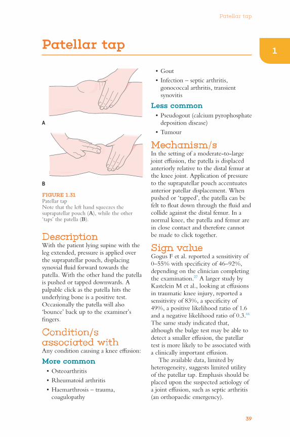
Mechanism/sIn the setting of a moderate-to-large joint effusion, the patella is displaced anteriorly relative to the distal femur at the knee joint. Application of pressure to the suprapatellar pouch accentuates anterior patellar displacement. When pushed or ‘tapped’, the patella can be felt to float down through the fluid and collide against the distal femur. In a normal knee, the patella and femur are in close contact and therefore cannot be made to click together.
Sign valueGogusFetal.reportedasensitivityof0–55% with specificity of 46–92%, depending on the clinician completing the examination.27 A larger study by Kastelein M et al., looking at effusions in traumatic knee injury, reported a sensitivity of 83%, a specificity of 49%, a positive likelihood ratio of 1.6 and a negative likelihood ratio of 0.3.66 The same study indicated that, although the bulge test may be able to detect a smaller effusion, the patellar test is more likely to be associated with a clinically important effusion.
The available data, limited by heterogeneity, suggests limited utility of the patellar tap. Emphasis should be placed upon the suspected aetiology of a joint effusion, such as septic arthritis (an orthopaedic emergency).
DescriptionWith the patient lying supine with the leg extended, pressure is applied over the suprapatellar pouch, displacing synovial fluid forward towards the patella. With the other hand the patella is pushed or tapped downwards. A palpable click as the patella hits the underlying bone is a positive test. Occasionally the patella will also ‘bounce’ back up to the examiner’s fingers.
Condition/s associated withAnyconditioncausingakneeeffusion:
More common•Osteoarthritis
•Rheumatoidarthritis
•Haemarthrosis–trauma,coagulopathy
FIGURE 1.31 Patellar tap Notethatthelefthandsqueezesthesuprapatellar pouch (A), while the other ‘taps’ the patella (B).
A
B
Patrick’s test (FABER test)
40
Patrick’s test (FABER test)
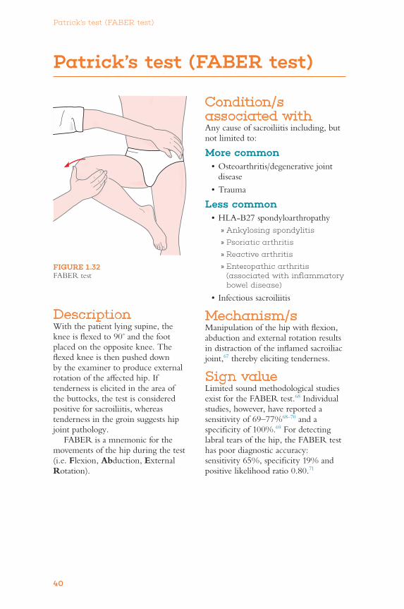
FIGURE 1.32 FABERtest
DescriptionWith the patient lying supine, the knee is flexed to 90° and the foot placed on the opposite knee. The flexed knee is then pushed down by the examiner to produce external rotation of the affected hip. If tenderness is elicited in the area of the buttocks, the test is considered positive for sacroiliitis, whereas tenderness in the groin suggests hip joint pathology.
FABERisamnemonicforthemovements of the hip during the test (i.e. Flexion, Abduction, External Rotation).
Condition/s associated withAny cause of sacroiliitis including, but notlimitedto:
More common•Osteoarthritis/degenerativejoint
disease
•Trauma
Less common•HLA-B27spondyloarthropathy
» Ankylosing spondylitis
» Psoriatic arthritis
» Reactive arthritis
» Enteropathic arthritis (associated with inflammatory bowel disease)
•Infectioussacroiliitis
Mechanism/sManipulation of the hip with flexion, abduction and external rotation results in distraction of the inflamed sacroiliac joint,67 thereby eliciting tenderness.
Sign valueLimited sound methodological studies existfortheFABERtest.68 Individual studies, however, have reported a sensitivity of 69–77%68–70 and a specificity of 100%.69 For detecting labraltearsofthehip,theFABERtesthaspoordiagnosticaccuracy:sensitivity 65%, specificity 19% and positive likelihood ratio 0.80.71
Phalen’s sign
41
1Phalen’s sign
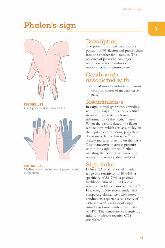
DescriptionThe patient puts their wrists into a position of 90° flexion and presses them into one another for 1 minute. The presence of paraesthesias and/or numbness in the distribution of the median nerve is a positive test.
Condition/s associated with•Carpaltunnelsyndrome(themost
common causes of median nerve palsy)
Mechanism/sIn carpal tunnel syndrome, crowding within the carpal tunnel or repetitive strain injury results in chronic inflammation of the median nerve. When the wrist is flexed, the flexor retinaculum, which acts as a pulley on the digital flexor tendons, pulls them down onto the median nerve72 and acutely increases pressure on the nerve. This manoeuvre increases pressure within the carpal tunnel, further irritating the nerve, thus worsening neuropathic sensory abnormalities.
Sign valueD’Arcy CA et al. reported a wide range of a sensitivity of 10–91%, a specificity of 33–76%, a positive likelihood ratio of 1.1–2.1 and a negative likelihood ratio of 0.3–1.0.73 However, a more recent study, also comparing clinical tests with nerve conduction, reported a sensitivity of 74% across all severities of carpal tunnel syndrome, with a specificity of 74%. The sensitivity in identifying mild to moderate severity CTS was 92%.74
FIGURE 1.33 Hand placement in Phalen’s test
FIGURE 1.34 Median nerve distribution of paraesthesias in the hand

Posterior drawer test
42
Posterior drawer test
DescriptionWith the patient and examiner in the same position as for the anterior drawer test, the examiner pushes the lower leg posteriorly, gently moving the proximal tibia backwards. If there is a tear in the posterior cruciate ligament, there will be significant abnormal posterior movement with a soft endpoint (see Figure 1.35).
FIGURE 1.35 Performing the posterior drawer test
Condition/s associated with•Posteriorcruciateligament(PCL)
tear
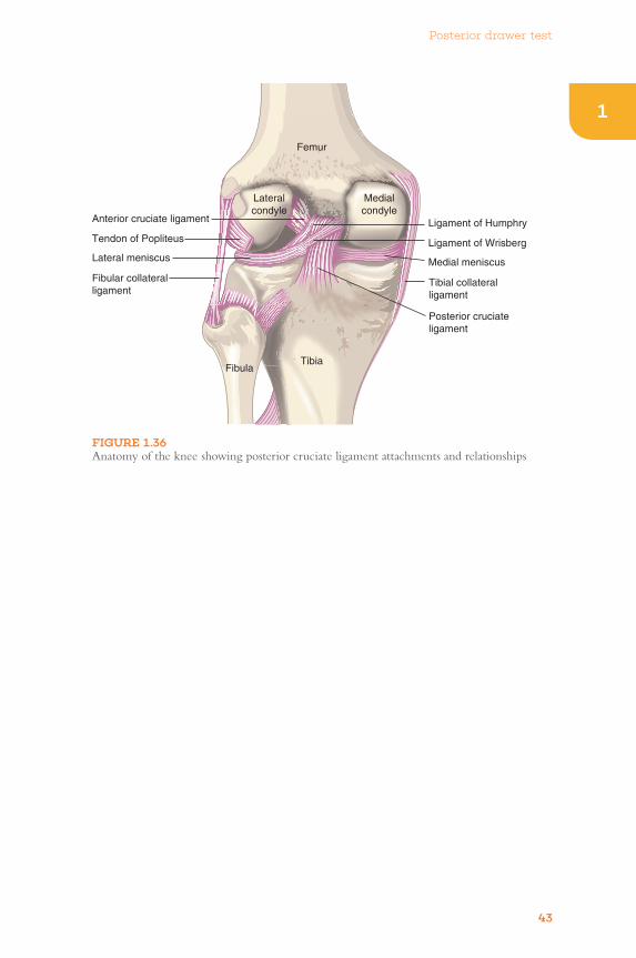
Mechanism/sThe PCL attaches the posterior aspect of the tibia to the lateral medial condyle (see Figure 1.36). It is a short ligament which restricts anterior movement of the femur on the tibia and posterior movement of the tibia. When the posterior cruciate is damaged or deficient, abnormal movement is possible. The posterior drawer test is used to assess the integrity of the PCL.
Sign valueThe posterior drawer test is a useful sign, with reported sensitivity of 90–95%, specificity of 99% and a +LR of 97.8 if present.76
Posterior drawer test
43
1
FIGURE 1.36 Anatomy of the knee showing posterior cruciate ligament attachments and relationships
Anterior cruciate ligament Ligament of Humphry
Lateralcondyle
Femur
TibiaFibula
Medialcondyle
Tendon of Popliteus Ligament of WrisbergLateral meniscus Medial meniscus
Fibular collateralligament
Tibial collateralligament
Posterior cruciateligament

Proximal weakness/proximal myopathy
44
Proximal weakness/proximal myopathy
Mechanism/sInflammatory myopathiesInflammatory myopathies result in immunologically mediated inflammation and destruction of skeletal muscle, causing weakness (see Table 1.2).
Systemic disordersProximal myopathy may present in a number of systemic rheumatological disorders such as SLE and RA. It is thought that circulating antibody complexes, deposited in tissues and/or targeted at muscles, damage muscle fibres, resulting in weakness.
Sign valuePatients with gradual-onset progressive symmetric proximal muscle weakness should be evaluated for a myopathy.
DescriptionProximal myopathy is a muscle disorder which results in proximal muscle group weakness (e.g. shoulder:pectoralis major, deltoid, biceps; hip:gluteal,quadriceps,iliopsoas,adductor). Proximal weakness is rapidly assessed by asking the patient to rise from a seated position and/or perform the motion of hanging washing on a clothesline. A complete assessment of power should be performed.
Condition/s associated with•Inflammatorymyopathy
» Polymyositis
» Dermatomyositis
•Endocrinemyopathy» Hyperthyroidism – see
Chapter 7, ‘Endocrinological signs’
» Hypothyroidism – see Chapter 7, ‘Endocrinological signs’
» Hyperparathyroidism – see Chapter 7, ‘Endocrinological signs’
•Systemicdisorders» Systemic lupus erythematosus
(SLE)
» Rheumatoid arthritis
•Genetic» Myotonic dystrophy
» Spinal muscular atrophy
•Other» Myasthenia gravis
» Polymyalgia rheumatica
TABLE 1.2 Mechanisms of inflammatory myopathies
Disease MechanismPolymyositis T-cell (in particular
CD8) and macrophage destruction of muscle fibres
Dermatomyositis Complement and antibody destruction of microvasculature; the deposition of complement and antibody complexes leads to inflammation and destruction of muscle fibres and hence weakness

Psoriatic nails/psoriatic nail dystrophy
45
1Psoriatic nails/psoriatic nail dystrophy
FIGURE 1.37 Nail dystrophic changes A Nail pitting; B onycholysis; C severe destructive change with nail loss and pustule formation.
Reproduced, with permission, from Firestein GS, Budd RC, Harris ED et al., Kelley’s Textbook of Rheumatology, 8th edn, Philadelphia: WB Saunders, 2008: Fig 72-3.
C
A B
FIGURE 1.38 ‘Oil drops’ under the nail
Reproduced, with permission, from Habif TP, Clinical Dermatology, 5th edn, Philadelphia: Mosby, 2009: Fig 8-23.
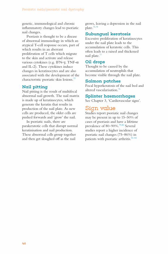
DescriptionPsoriatic nail changes refer to a number of different abnormalities seen in the nails rather than just one sign. Changes include:76
•Pittingofthenailplate
•Subungualhyperkeratosisunderthenail plate
•Onycholysis(naillifting)andchanges in nail shape
•‘Oildrops’and‘salmonpatches’
•Splinterhaemorrhages
Condition/s associated with•Psoriasis
•Psoriaticarthritis
Mechanism/sThe mechanism is poorly understood. It is likely that a combination of
Psoriatic nails/psoriatic nail dystrophy
46
grows, leaving a depression in the nail plate.76,78
Subungual keratosisExcessive proliferation of keratinocytes under the nail plate leads to the accumulation of keratotic cells. This often leads to a raised and thickened nail plate.77
Oil dropsThought to be caused by the accumulation of neutrophils that become visible through the nail plate.
Salmon patchesFocal hyperkeratosis of the nail bed and altered vascularisation.76
Splinter haemorrhagesSee Chapter 3, ‘Cardiovascular signs’.
Sign valueStudies report psoriatic nail changes may be present in up to 15–50% of cases of psoriasis and have a lifetime prevalence of 80–90%.79,80 Several studies report a higher incidence of psoriatic nail changes (75–86%) in patients with psoriatic arthritis.81–84
genetic, immunological and chronic inflammatory changes lead to psoriatic nail changes.
Psoriasis is thought to be a disease of abnormal immunology in which an atypical T-cell response occurs, part of which results in an aberrant proliferation of T cells which migrate to the skin and activate and release various cytokines (e.g. IFN-γ, TNF-α and IL-2). These cytokines induce changes in keratinocytes and are also associated with the development of the characteristic psoriatic skin lesions.77
Nail pittingNail pitting is the result of multifocal abnormal nail growth. The nail matrix is made up of keratinocytes, which generate the keratin that results in production of the nail plate. As new cells are produced, the older cells are pushed forwards and ‘grow’ the nail.
In psoriatic nails, there are parakeratotic cells that disrupt normal keratinisation and nail production. These abnormal cells group together and then get sloughed off as the nail

Raynaud’s syndrome/phenomenon
47
1
CL
INIC
AL
PE
AR
L
1Raynaud’s syndrome/phenomenon
1 white – blanching associated with vasoconstriction of the blood vessels
2 blue – cyanosis3 red – when blood flow is restored
and hyperaemia results.
DescriptionRaynaud’s syndrome/phenomenon occurs in the digits from various stimuli, resulting in peripheral hypoperfusion followed by hyperaemia. Ithasthree‘colour’phases:
FIGURE 1.39 Raynaud’s phenomenon A Sharply demarcated pallor of the distal fingers resulting from the closure of the digital arteries; B cyanosis of the fingertips.
Reproduced, with permission, from Kumar V, Abbas AK, Fausto N, Aster J, Robbins and Cotran PathologicBasisofDisease,ProfessionalEdition, 8th edn, Philadelphia: Saunders, 2009: Fig 11-28.
Altered localvascularfunction
Impairedhabituation
of CVresponse to
stress
Increased sympathetic sensitivity
and activation
Other factorse.g. hormonal,
increased blood viscosity,
endothelial damage
Cold exposure/stress response
Vasoconstriction
Imbalance ofvasoconstrictors vs
vasodilators
Raynaud’s syndrome/phenomenon
A B

Raynaud’s syndrome/phenomenon
48
cell membrane and interact with its ligand.89
2 Impaired habituation of the cardiovascular response to stress is also thought to contribute. Habituation is the gradual extinction of a response to a stimulus over time. In normal individuals, ongoing exposure to a stress results in habituation, and decreasing incidence and duration of the response.85,86
3 Local vascular factors – an imbalance between local vasoconstrictive factors (endothelin, 5-HT, thromboxane [TXA] and other cyclo-oxygenase [COX] pathway products) and vasodilatory factors (nitric oxide [NO])85,86 may also exist in Raynaud’s syndrome.» Local endothelin may not
produce enough NO for vasodilatation.86
» Repeated vasospasm causes oxidative stress and reduced NO production, thus decreasing vasodilatation.85
» Inappropriately greater production of endothelin and thromboxane (TXA2) in response to cold also occurs, leading to marked vasoconstriction.85,86
» In some studies, a higher than normal endothelin-1, a potent vasoconstrictor, was seen in patients with primary Raynaud’s syndrome.86
4 Other factors.Someoftheseinclude:» oestrogen – causing
sensitisation of vessels to vasoconstriction85,86
» increased blood viscosity86
» decreased amounts of calcitonin gene-related peptide (CGRP) neurons – impairing normal nerve sensitivity, activation and vasodilatation86
» endothelial damage.
Condition/s associated withCommon•Raynaud’sphenomenon
Less common•Vasculitis
» Buerger’s disease
•Autoimmune/connectivetissuedisorders» Scleroderma (systemis
sclerosis)
» Systemic lupus erythematosus
» CREST syndrome
» Sjögren’s syndrome
» Dermatomyositis
» Polymyositis
» Rheumatoid arthritis
•Drugs» Beta blockers
Mechanism/sRaynaud’s syndrome occurs due to an exaggerated vasoconstrictive response causing transient cessation of blood flow to the digits.85–88
The cause of this abnormal vasoconstrictive response is multifactorial:
1 Increased sympathetic nerve activation (centrally and peripherally mediated) – in response to cold temperatures or stressful situations, enhanced sympathetic nerve activation leads to vasoconstriction of the arterioles in the digits. Larger numbers of alpha-2-adrenoreceptors may result in more pronounced vasoconstriction.85–88 The increase in alpha-2-adrenoreceptors involves reactive oxygen species, Rho/Rho kinase and the actin cytoskeleton. Cold can induce Rho/Rho kinase, causing more adrenoreceptors to move to the

Raynaud’s syndrome/phenomenon
49
1Secondary Raynaud’s syndromeStructural vascular abnormalities (in addition to the factors outlined above) are thought to play a role in Raynaud’s phenomenon occurring secondary to an underlying disease process.
In scleroderma (systemic sclerosis), abnormal proliferation of intimal cells results in endothelial cell damage. Abnormal endothelial cells then exacerbatevasospasmby:86,88
•perturbingsmoothmusclecells,causing them to proliferate and contract
•enhancingpro-coagulantactivityand inhibitors of fibrinolysis, thus promoting microthrombi
•promotinginflammationthroughrelease of adhesion factors.
Other factors thought to contribute insystemicsclerosisinclude:86
•raisedlevelsofangiotensinII–avasoconstrictor
•lackofcompensatoryangiogenesisto meet the demands of proliferated intima – leading to ischaemia.
Sign valueAlthough not always associated with other rheumatological complaints, a thorough history and examination of a patient presenting with Raynaud’s syndrome needs to be undertaken to assess for other underlying causes. It is said that the majority of patients with scleroderma recall these symptoms many years before the onset of skin induration. Further, Raynaud’s phenomenon is present in 90–99% of patients with diffuse or limited scleroderma.90

Saddle nose deformity
50
Saddle nose deformity
•Cocaineuse,complication
•Congenitalsyphilis–rare
Mechanism/sDestruction of the nasal septum or support cartilage results in the deformity. Direct trauma or prior surgery is the most common aetiology.
Wegener’s granulomatosisWegener’s granulomatosis is an autoimmune vasculitic disorder characterised by necrotising granulomas affecting the small blood vessels of the upper and lower airways. It is thought that immune complex deposition or an autoimmune response results in inflammation and damage/destruction of the vessels and their surrounding structures. In immune disease, the cartilaginous structures of the outer nose and septum are generally more severely involved than the bony nasal dorsum. Severe disease may cause progressive loss of septal support, leading to enlarged anterior septal perforations, resulting in significant collapse of nasal cartilage.91
Relapsing polychondritisRelapsing polychondritis is an autoimmune chronic inflammatory disorder resulting in the destruction of cartilage – in particular auricular and nasal cartilage.25
Sign valueSaddle nose deformity occurs in up to 65% of relapsing polychondritis, and 9–29% of patients with Wegener’s granulomatosis.25
DescriptionCollapse of the middle section of the nose relative to the tip and dorsum, like a saddle.
Condition/s associated withMore common•Trauma
•Iatrogenic–nasalsurgery
Less common•Wegener’sgranulomatosis
•Relapsingpolychondritis
FIGURE 1.40 Saddle nose deformity
Reproduced, with permission, from Firestein GS, Budd RC, Harris ED et al., Kelley’s Textbook of Rheumatology, 8th edn, Philadelphia: WB Saunders, 2008: Fig 82-5.
Sausage-shaped digits (dactylitis)
51
1Sausage-shaped digits (dactylitis)
Uncommon•Tuberculosis
•Gout
•Sarcoidosis
•Disseminatedgonorrhoea
Mechanism/sSpondyloarthropathiesIrritation of the flexor tendons, flexor tendon sheath and surrounding soft tissues due to pro-inflammatory cytokines results in pronounced diffuse inflammation of the digits.93,94 Recent high-resolution MRI studies in psoriatic arthritis reported flexor tendon pulley and sheath-related enthesitis as the more specific cause.95
Tuberculosis dactylitisA variant of tuberculous osteomyelitis wherebyTBgranulomasinvadetheshort tubular bones of the hands and feet and then the surrounding tissues, causing inflammation and swelling.93
Syphilitic dactylitisA manifestation of congenital syphilis where the syphilitic spirochetes invade perichondrium, bone, periosteum and marrow and thus inhibit osteogenesis. Inflammation from the invasion is another contributing factor to pain and swelling of the digits.93
Sarcoid dactylitisSarcoid non-caseating granulomas invade bone and soft tissue, causing swelling and inflammation.93
Sickle cell dactylitisIn sickle cell anaemia, a haemoglobin S-gene mutation results in rigid and ‘sickle’-shaped red blood cells under hypoxic conditions. Acute sickling in
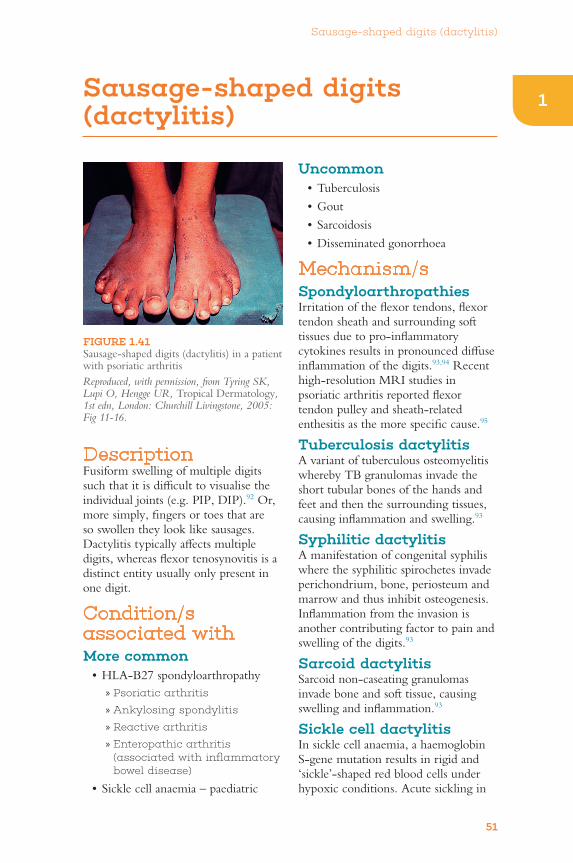
DescriptionFusiform swelling of multiple digits such that it is difficult to visualise the individual joints (e.g. PIP, DIP).92 Or, more simply, fingers or toes that are so swollen they look like sausages. Dactylitis typically affects multiple digits, whereas flexor tenosynovitis is a distinct entity usually only present in one digit.
Condition/s associated withMore common•HLA-B27spondyloarthropathy
» Psoriatic arthritis
» Ankylosing spondylitis
» Reactive arthritis
» Enteropathic arthritis (associated with inflammatory bowel disease)
•Sicklecellanaemia–paediatric
FIGURE 1.41 Sausage-shaped digits (dactylitis) in a patient with psoriatic arthritis
Reproduced, with permission, from Tyring SK, Lupi O, Hengge UR, Tropical Dermatology, 1st edn, London: Churchill Livingstone, 2005: Fig 11-16.
Sausage-shaped digits (dactylitis)
52
the peripheral circulation results in digital ischaemia and painful fusiform digital swelling. It typically occurs in the paediatric population.
Sign valueIn regards to patients with seronegative spondyloarthropathy, sausage-shaped digits have a sensitivity of 17.9% and a specificity of 96.4%.96 The development of dactylitis may be a marker for progression of psoriatic
arthritis,97 being present in 16–24%97 of reported cases, with lifetime incidence and prevalence of 48% and 33%, respectively.98 It is seen in only 4% of tuberculosis93 cases.
Identification of sausage-shaped digits or dactylitis in an adult should prompt an evaluation for a seronegative spondyloarthropathy. Development of dactylitis in a child of African or Mediterranean descent should prompt evaluation for sickle cell disease.

Sclerodactyly
53
1Sclerodactyly
Condition/s associated with•Scleroderma(systemicsclerosis)
•CRESTsyndrome(i.e.Calcinosis, Raynaud’s phenomenon, Oesophageal dysmotility, Sclerodactyly, Telangiectasia)
Mechanism/sIn scleroderma, T cells infiltrate the skin and set in motion a cascade of events including abnormal fibroblast and growth factor stimulation. This in turn leads to increased production of extracellular matrix, fibrillin and type 1 collagen and other factors. Ultimately this results in fibrosis and thickening of the skin.
Sign valueSkin thickening is seen more often in diffuse scleroderma (27%) than in limited disease (5%).99
DescriptionThickening and tightening of the skin covering the digits.
FIGURE 1.42 Sclerodactyly with flexion contractures
Reproduced, with permission, from Firestein GS, Budd RC, Harris ED et al., Kelley’s Textbook of Rheumatology, 8th edn, Philadelphia: WB Saunders, 2008: Fig 47-12.
FIGURE 1.43 Proposed mechanism of sclerodactyly
Genetic factors Environmental Other factors
Immunological reaction – mononuclear cells andcytokines infiltrate layers of skin
Vascular inflammation, fibroblasts stimulated, TGF-βreleased, growth factors, other factors released
Collagen, fibrillin, fibronectin, extracellular matrixsynthesis and deposition
Fibrosis, skin thickening and tightening
Sclerodactyly

Shawl sign
54
Shawl sign
FIGURE 1.44 Shawl sign Note discolouration over the posterior shoulder and neck.
Reproduced, with permission, from Hochberg MC et al., Rheumatology, 5th edn, Philadelphia: Mosby, 2010: Fig 144-7.
DescriptionA confluent, violaceous, macular rash over the posterior shoulders and neck.
Condition/s associated with•Dermatomyositis
Mechanism/sComplement and antibody mediated microvascular injury likely results in the development of the rash.100 Dermatomyositis is a systemic inflammatory disorder primarily of muscle and skin characterised by microvascular damage due to antibody complex and complement deposition. Geneticpredisposition,virusesand UVlightareallthoughttoplay a role.100
The cutaneous changes in dermatomyositisinclude:hyperkeratosis, epidermal basal cell vacuolar degeneration, pathological apoptosis of epidermal basal and suprabasal cells, dyskeratosis, a focally
thinned epidermis, and increased dermal mucin deposition.101 Factors contributingtothesechangesinclude:
•Dysregulatedcytokineproductionand cell-mediated processes. Histopathology of dermatomyositic lesions have shown the principal infiltrating cell in the skin is the CD4+ T lymphocyte, distributed mainly in the perivascular upper dermis.
•Inskinbiopsies,asignificantlyincreased number of both CD-40+ cells (including keratinocytes and mononuclear cells in the dermis), as well as infiltrating CD4+ CD-40L+ T-lymphocytes, are found. Activation of the CD-40/CD-40L system may cause upregulation of several pro-inflammatory molecules, including IL-6, IL-15, IL-8 and MCP-1.
•Dysregulatedapoptosisofkeratinocytes. The exact mechanism for keratinocyte apoptosis is unclear, but includes UVBlight,Fas-FasL,TNFα, and CD8+ T-cell-mediated activation of the apoptosis pathway.
•Photosensitivityisanimportantclinical feature. Exposure to sunlightandspecificallyUVAandUVBradiationmayserveacentralrole in disease onset and persistence.ExposuretoUVBradiation has also been shown to upregulate the pro-inflammatory cytoking TNF-α.
•Excessmucindepositionhasbeenhypothesised to occur secondary to increased hyaluronic acid production by dermal fibroblasts following immunological stimulation.

Shawl sign
55
1Sign valueAlthough not pathognomonic, the shawl sign is strongly associated with dermatomyositis. In up to 30% of cases
of dermatomyositis, skin manifestations occur. However, skin manifestations may occur without the onset of muscle symptoms.102
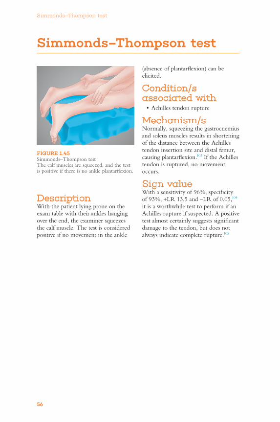
Simmonds–Thompson test
56
Simmonds–Thompson test
(absence of plantarflexion) can be elicited.
Condition/s associated with•Achillestendonrupture
Mechanism/sNormally,squeezingthegastrocnemiusand soleus muscles results in shortening of the distance between the Achilles tendon insertion site and distal femur, causing plantarflexion.103 If the Achilles tendon is ruptured, no movement occurs.
Sign valueWith a sensitivity of 96%, specificity of 93%, +LR 13.5 and −LR of 0.05,104 it is a worthwhile test to perform if an Achilles rupture if suspected. A positive test almost certainly suggests significant damage to the tendon, but does not always indicate complete rupture.105
FIGURE 1.45 Simmonds–Thompson test Thecalfmusclesaresqueezed,andthetestis positive if there is no ankle plantarflexion.
DescriptionWith the patient lying prone on the exam table with their ankles hanging overtheend,theexaminersqueezesthe calf muscle. The test is considered positive if no movement in the ankle
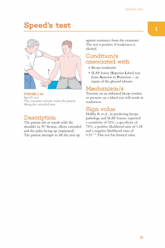
Speed’s test
57
1Speed’s test
against resistance from the examiner. The test is positive if tenderness is elicited.
Condition/s associated with•Bicepstendonitis
•SLAPlesion(Superior Labral tear from Anterior to Posterior) – an injury of the glenoid labrum
Mechanism/sTraction on an inflamed biceps tendon or pressure on a labral tear will result in tenderness.
Sign valueHoltby R et al., in predicting biceps pathology and SLAP lesions, reported a sensitivity of 32%, a specificity of 75%, a positive likelihood ratio of 1.28 and a negative likelihood ratio of 0.91.106 This test has limited value.
DescriptionThe patient sits or stands with the shoulder in 90° flexion, elbow extended and the palm facing up (supinated). The patient attempts to lift the arm up
FIGURE 1.46 Speed’s test The examiner actively resists the patient lifting the extended arm.

Subcutaneous nodules (rheumatoid nodules)
58
Subcutaneous nodules (rheumatoid nodules)
Sign valueSeen in 20–25% of seropositive rheumatoid arthritis. They are the most common extra-articular manifestation of the disease. Frequency of development of nodules is associated with elevated rheumatoid factor titres.108 The presence of nodules is associated with a more severe disease phenotype.109
FIGURE 1.47 Mechanism of rheumatoid nodule formation
Repeated trauma
Local vascular damage
Direct activation of monocytes Complement-mediated activated monocytes
TGF-β, TGF-α, fibronectin, proteases, other cytokines, cells
Pallisading granuloma – rheumatoid nodule
Neoangiogenesis andgranulation tissue formation
Endothelial injury + IgM RF immunecomplex/complement deposited in vessel walls
DescriptionVisibleandpalpablesubcutaneousnodules typically occur over bony prominences and/or extensor surfaces.
Condition/s associated with•Rheumatoidarthritis
Mechanism/sThought to be mediated via Th-1 inflammatory response.107 Trauma over bony prominences causes local vessel damage that leads to new blood vessel growth and granulomatous tissue formation. Endothelial injury results in accumulation of immune complexes and stimulates monocytes to secrete IL-1,TNF,TGF-β, prostaglandins and other factors, including proteases, collagenases and fibronectin. This ultimately leads to angiogenesis, fibrin deposition and formation of the characteristic rheumatoid nodule.107,108
FIGURE 1.48 Large rheumatoid nodules are seen in a classic location along the extensor surface of the forearm and in the olecranon bursa
Reproduced, with permission, from Goldman L, Ausiello D, Cecil Medicine, 23rd edn, Philadelphia: Saunders, 2007: Fig 285-9.
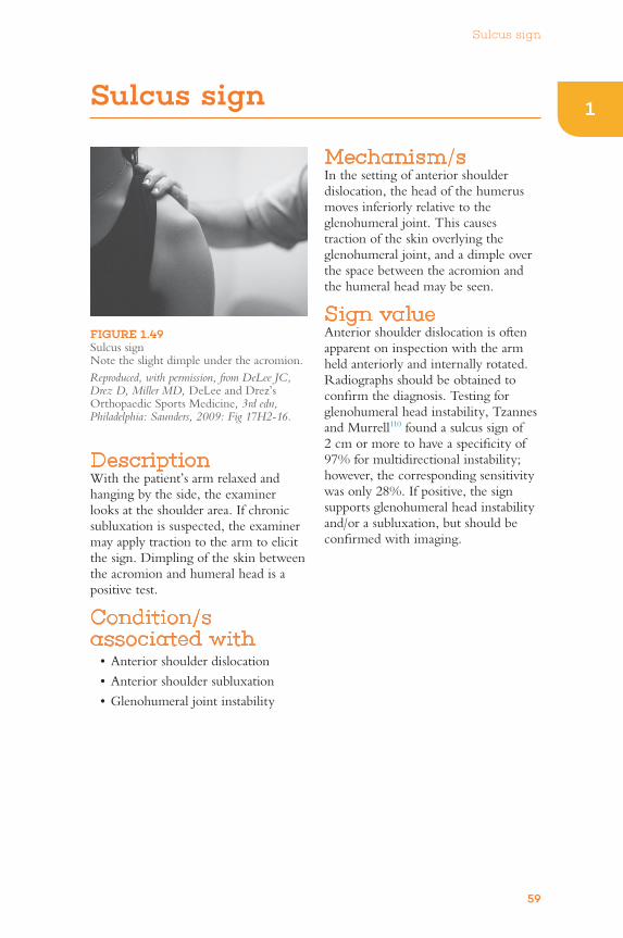
Sulcus sign
59
1Sulcus sign
Mechanism/sIn the setting of anterior shoulder dislocation, the head of the humerus moves inferiorly relative to the glenohumeral joint. This causes traction of the skin overlying the glenohumeral joint, and a dimple over the space between the acromion and the humeral head may be seen.
Sign valueAnterior shoulder dislocation is often apparent on inspection with the arm held anteriorly and internally rotated. Radiographs should be obtained to confirm the diagnosis. Testing for glenohumeralheadinstability,Tzannesand Murrell110 found a sulcus sign of 2 cm or more to have a specificity of 97% for multidirectional instability; however, the corresponding sensitivity was only 28%. If positive, the sign supports glenohumeral head instability and/or a subluxation, but should be confirmed with imaging.
FIGURE 1.49 Sulcus sign Note the slight dimple under the acromion.
Reproduced, with permission, from DeLee JC, Drez D, Miller MD, DeLeeandDrez’sOrthopaedic Sports Medicine, 3rd edn, Philadelphia: Saunders, 2009: Fig 17H2-16.
DescriptionWith the patient’s arm relaxed and hanging by the side, the examiner looks at the shoulder area. If chronic subluxation is suspected, the examiner may apply traction to the arm to elicit the sign. Dimpling of the skin between the acromion and humeral head is a positive test.
Condition/s associated with•Anteriorshoulderdislocation
•Anteriorshouldersubluxation
•Glenohumeraljointinstability

Supraspinatus test (empty-can test)
60
Supraspinatus test (empty-can test)
Mechanism/sThe supraspinatus muscle works in concert with the deltoid muscle during shoulder abduction and stabilises the humeral head in the glenoid fossa. Mechanical strain upon an injured supraspinatus muscle or tendon will result in tenderness and/or weakness during this manoeuvre.
Sign valueMcGee,inareviewoftwostudiesinpatients with rotator cuff muscle tears with painful supraspinatus tests, reported a sensitivity of 63–85%, a specificity of 52–60% and a positive likelihood ratio of 1.7.75 A review of five studies of patients with rotator cuff muscle tears with weakness during testing, reported a sensitivity of 41–84%, a specificity of 58–70%, a positive likelihood ratio of 2.0 and a negative likelihood ratio of 0.5.2
The supraspinatus test has limited utility and may be positive in several other shoulder conditions. Detection of weakness has more diagnostic utility than tenderness alone.
FIGURE 1.50 Supraspinatus or empty-can test
DescriptionThe patient sits or stands with the shoulder in 90° flexion, 30° abduction, with the elbow extended and the thumbs pointing towards the ground, as if emptying two cans. The examiner applies downward pressure as the patient attempts to lift the arms up. The test is positive if the patient experiences tenderness or is unable to hold up their arm.
Condition/s associated with•Supraspinatustear
•Supraspinatustendonitis
•Supraspinatusimpingement

Swan-neck deformity
61
1Swan-neck deformity
Mechanism/sA relative imbalance of flexor and extensor tendons of the digit due to chronic synovial inflammation.111 A variety of changes may result in this deformity, whose basis is inflammatory disruption of the collateral ligaments, volar plates, joint capsule or invasion of the flexor tendons.112 The resulting changesmaybe:
DescriptionA deformity characterised by distal interphalangeal (DIP) joint flexion and proximal interphalangeal (PIP) joint hyperextension in the resting digit, to some extent resembling a swan’s neck.
Condition/s associated withCommon•Rheumatoidarthritis
FIGURE 1.51 Swan-neck deformity pathoanatomy A Terminal tendon rupture may be associated with synovitis of DIP joint, leading to DIP joint flexion and subsequent PIP joint hyperextension. Rupture of flexor digitorum superficialis tendon can be caused by infiltrative synovitis, which can lead to decreased volar support of PIP joint and subsequent hyperextension deformity; B lateral-band subluxation dorsal to axis of rotation of PIP joint. Contraction of triangular ligament and attenuation of transverse retinacular ligament are depicted.
Based on Jupiter JB, Chapter 70: Arthritic hand. In: Canale TS, Beaty JH, Campbell’s Operative Orthopaedics, 11th edn, Philadelphia: Elsevier, 2007: Fig 70-13.
Attenuated or rupturedextensor tendon
Attenuated transverseretinacular ligament
Contractedtriangular ligament
Dorsal subluxationof lateral band
A
B

Swan-neck deformity
62
•attenuationordisruptionoftheextensor tendon on the distal phalanx, leading to unopposed flexion – and thus the flexed DIP joint
•disruptionoftheretinacularligament (which helps hold the finger in flexion), leading to unopposed extensor forces at the PIP joint and PIP joint hyperextension.
Sign valueSwan-neck deformity is classically associated with rheumatoid arthritis. In patients with acute trauma with forced DIP flexion during active extension, mallet finger (i.e. extensor tendon avulsion distal to DIP joint) should be considered.
FIGURE 1.52 Swan-neck deformity
Reproduced, with permission, from Jupiter JB, Chapter 70: Arthritic hand. In: Canale TS, Beaty JH, Campbell’s Operative Orthopaedics, 11th edn, Philadelphia: Elsevier, 2007: Fig 70-14.

Telangiectasia
63
1Telangiectasia
General mechanism/sTelangiectasias are predominantly persistently dilated small capillaries and venules. The exception to this is hereditary haemorrhagic telangiectasia, as these lesions are arteriovenous (AV) malformations.
Hereditary haemorrhagic telangiectasia (HHT)HHT is an autosomal dominant disordercausingdevelopmentofAVmalformations, due to a genetic abnormalityoftheTGF-β receptor. TheTGF-β pathway is known to modulate vascular architecture, matrix formation and basement membrane development.113
SclerodermaThe underlying mechanism for telangiectasia in scleroderma is unknown. It is presumed that there is endothelial injury, leading to aberrant angiogenesis and the development of new vessels. It has been suggested that theTGF-β pathway may be involved.113
FIGURE 1.53 Telangiectasia associated with systemic sclerosis (scleroderma) Note the skin tightening around the lips.
Reproduced, with permission, from Habif TP, Clinical Dermatology, 5th edn, Philadelphia: Mosby, 2009: Fig 17-30.
DescriptionPermanent dilatation of superficial peripheral vessels resulting in blanching red lesions on the skin. Telangiectasia may present as a fine red line or a punctum (dot) with radiating lines.46
Condition/s associated withThere are numerous conditions associated with telangiectasia, including but not limited to those listed in Table 1.3.
TABLE 1.3 Telangiectasia-associated conditions
Systemic diseasesCarcinoid syndrome
Ataxia–telangiectasia
Mastocytosis
Dermatomyositis
Scleroderma – especially periungual telangiectasia
Systemic lupus erythematosus
Hereditary haemorrhagic telangiectasia
Liver cirrhosis
Telangiectasia
64
•Telangiectasiasinadulthoodthatare located around the mucous membranes, extremities and under the nails are associated with hereditary haemorrhagic telangiectasia.
•Thepresenceofincreasednumbersof telangiectasias may indicate the development of pulmonary vascular disease in scleroderma. Telangiectasia may be a clinical marker of more widespread microvascular disease in scleroderma.115
Spider naevusSee ‘Spider naevus’ in Chapter 6, ‘Gastroenterologicalsigns’.
Sign valueLocation and characteristics of telangiectasia can assist in diagnosis.
•Periungualtelangiectasia(telangiectasia next to the nails) is said to be highly suggestive of SLE, scleroderma or dermatomyositis.114
•Broadmaculeswithapolygonal or oval shape, known as mat telangiectasias, are associated with CREST syndrome.114

Thessaly test
65
1Thessaly test
DescriptionThe test is performed with the knee at 5° and 20 of flexion. The examiner supports the patient by the hands as they stand flatfooted. The patient then rotates their knee and body, internally and externally, three times, keeping the knee in slight flexion (5°). The same movement is repeated with the knee flexed to 20°. The test is always performed first on the normal knee so that the patient is comfortable with performing the manoeuvre (i.e. how to keep the knee in 5° and then 20° of flexion). See Figure 1.54.
Condition/s associated with•Meniscaltears
FIGURE 1.54 Thessaly test
Mechanism/sThe knee meniscus (lateral and medial) is fibrocartilaginous tissue that separates the femur from the tibia. The menisci provide rotational stability and act as shock absorbers, among other functions. The Thessaly test creates loading conditions on the meniscus and a rotational stress that may result in reproduction of pain, catching or other reported symptoms.
Sign valueBlythMetal.116 compared the accuracy of physical tests for meniscal tears to MRI-based diagnosis (see Table 1.1 on page 35). The Thessaly test had only moderate value in assessment when compared to MRI. The tests may be useful initially, but realistically, definitive imaging is required.

Thomas’ test
66
Thomas’ test
Mechanism/sDrawing up the knee and flexing one side of the hip rotates the pelvis. In order to keep the alternate leg flat on the bed, the hip flexors and rectus femoris must stretch enough to allow the leg to lie flat. In other words, if the hip flexors are contracted, the affected leg will rise as the pelvis rotates.
Sign valueA systematic review of diagnostic tests for hip pathology found minimal scientific evidence for Thomas’ test due to methodological flaws.117 There is limited value in this sign.
FIGURE 1.55 Performance of Thomas’ test
1
2
3
DescriptionWith the patient lying supine, the knee and hip on the ‘normal’ side are flexed, with the knee held against the chest. A positive test occurs if the opposite leg rises off the table.
Condition/s associated with•Hipflexioncontracture–fixed
flexion deformity
•Iliotibialbandsyndrome
•Normalageing/stiffness

Tinel’s sign
67
1Tinel’s sign
and resulting damage to the median nerve. It is thought that this damage results in altered mechanosensitivity118 of the median nerve, possibly due to an abnormally excitable membrane. So, when lightly struck through the skin, the inflamed nerve functions abnormally.
Sign valueD’Arcy CA et al. reported Tinel’s sign had limited or no value in distinguishing people with carpal tunnel syndrome from those without.73 A review of several studies reported a sensitivity of 25–60%, a specificity of 64–80%, a positive likelihood ratio of 0.7–2.7 and a negative likelihood ratio of 0.5–1.1. Neither Tinel’s sign nor Phalen’s sign reliably rule in or rule out carpal tunnel syndrome.118
FIGURE 1.56 Completing Tinel’s test Tapping over the wrist causes pins and needles in the fingers.
FIGURE 1.57 Mechanism of Tinel’s test
Increased carpal tunnel pressure anddamage to median nerve
Altered membrane excitability
Easier discharge on tapping
Paraesthesias in median nerve distribution
Increased mechanosensitivity
DescriptionParaesthesias in a median nerve distribution occur when the examiner taps with a finger at the distal wrist over the median nerve. It should be noted that Tinel’s original description was not specific for the median nerve but rather for the sensation of ‘pins and needles’ arising from any injured nerve tested in this way.
Condition/s associated with•Carpaltunnelsyndrome
Mechanism/sIn carpal tunnel syndrome, there is increased pressure in the carpal tunnel
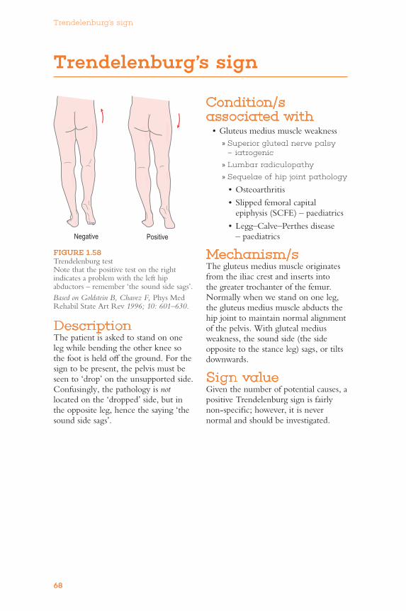
Trendelenburg’s sign
68
Trendelenburg’s sign
Condition/s associated with•Gluteusmediusmuscleweakness
» Superior gluteal nerve palsy – iatrogenic
» Lumbar radiculopathy
» Sequelae of hip joint pathology
•Osteoarthritis
•Slippedfemoralcapitalepiphysis (SCFE) – paediatrics
•Legg–Calve–Perthesdisease– paediatrics
Mechanism/sThe gluteus medius muscle originates from the iliac crest and inserts into the greater trochanter of the femur. Normally when we stand on one leg, the gluteus medius muscle abducts the hip joint to maintain normal alignment of the pelvis. With gluteal medius weakness, the sound side (the side opposite to the stance leg) sags, or tilts downwards.
Sign valueGiventhenumberofpotentialcauses,apositive Trendelenburg sign is fairly non-specific; however, it is never normal and should be investigated.
DescriptionThe patient is asked to stand on one leg while bending the other knee so the foot is held off the ground. For the sign to be present, the pelvis must be seen to ‘drop’ on the unsupported side. Confusingly, the pathology is not located on the ‘dropped’ side, but in the opposite leg, hence the saying ‘the sound side sags’.
FIGURE 1.58 Trendelenburg test Note that the positive test on the right indicates a problem with the left hip abductors – remember ‘the sound side sags’.
Based on Goldstein B, Chavez F, Phys Med Rehabil State Art Rev 1996; 10: 601–630.
Negative Positive

True leg length inequality (anatomic leg length inequality)
69
1True leg length inequality (anatomic leg length inequality)
Mechanism/sTrue, or anatomic, leg length equality relates to the actual length of the bones and anatomical structures making up the hip and the lower limb. Therefore, any problem in the anatomy that constitutes the leg length (from the head of the femur down to the ankle) may cause a discrepancy. For example, abnormalities in growth plates during development may lead to one leg being longer than the other. Aberrant healed fractures can also lead to a shortened leg.
Sign valueA leg length discrepancy is a non-specific sign. It should be interpreted in the context of the patient’s history.
DescriptionThe leg length is measured from the anterior iliac spine to the medial malleolus. There is no clear definition as to what constitutes a significant discrepancy. Some authors suggest that it is not clinically relevant until there is more than 20 mm difference between legs.119
Condition/s associated with•Fracture–hip,femur,tibia
•Dislocation–hip,knee
•Post-surgicalshortening
•Congenitaldisorders

Ulnar deviation
70
Ulnar deviation
stable than interphalangeal joints. Progressive inflammatory changes from rheumatoid arthritis result in stretching of the joint capsule and ligaments, causing instability. Extrinsic forces on the joints tend to pull in a direction of ulnar deviation. Possible factors include:108,111
•thenormaltendencyoffingerstomove towards the ulnar side on flexion
•inflammationofthecarpometacarpal (CMC) joints in the 4th and 5th fingers causes further spread of the metacarpals in flexion, producing an ‘ulnarly’ directed force on the extensor tendons
•stretchingofthecollateralligamentsof the MCP joints, accessory collateral ligaments or flexor tunnels that permits volar displacement of the proximal phalanges.
Radiocarpal ulnar deviationProgressive inflammatory changes lead to progressive synovitis of the wrist joint and carpal bones, including the scaphoid. Abnormal wrist mechanics develop due to translocation of the carpal bones relative to the radius, and imbalance of mechanical forces.108
Sign valueUlnar deviation of the MCP joints is classically associated with rheumatoid arthritis, although other causes should be considered.
DescriptionDisplacement of the metacarpo-phalangeal and/or radiocarpal joint towards the ulnar aspect of the wrist.
Condition/s associated with•Rheumatoidarthritis
•Pigmentedvillonodularsynovitis
Mechanism/sMetacarpophalangeal (MCP) jointMCP joints are condylar and are able to move in two planes. They are less
FIGURE 1.59 Ulnar deviation and subluxation The hand shows typical manifestations of end-stage erosive changes around the metacarpophalangeal joints, with volar and ulnar drift of the fingers.
Reproduced, with permission, from Firestein GS, Budd RC, Harris ED et al., Kelley’s Textbook of Rheumatology, 8th edn, Philadelphia: WB Saunders, 2008: Fig 66-5.

V-sign
71
1V-sign
FIGURE 1.60 Irregular patchy erythema with associated prominent telangiectasias in a woman with dermatomyositis
Reproduced, with permission, from Shields HM et al., ClinGastroenterolHepatol 2007; 5(9): 1010–1017.
DescriptionA confluent, macular, violet/red rash seen over the anterior neck and upper chest.OftenfoundinaV-shapesimilarto the neck of a shirt.
Condition/s associated with•Dermatomyositis
Mechanism/sComplement- and antibody-mediated microvascular injury likely results in the development of the rash.120 Dermatomyositis is an inflammatory myopathy characterised by microvascular damage and destruction of muscle by antibody complex and complementdeposition.Geneticpredisposition,virusesandUVlightareall thought to play a role.100 See Heliotrope rash and Shawl sign.
Sign valueAlthough not pathognomonic, the V-signishighlysuggestiveofdermatomyositis. In up to 30% of cases, skin manifestations including the V-signmayoccurbeforedevelopmentof the characteristic muscle weakness.
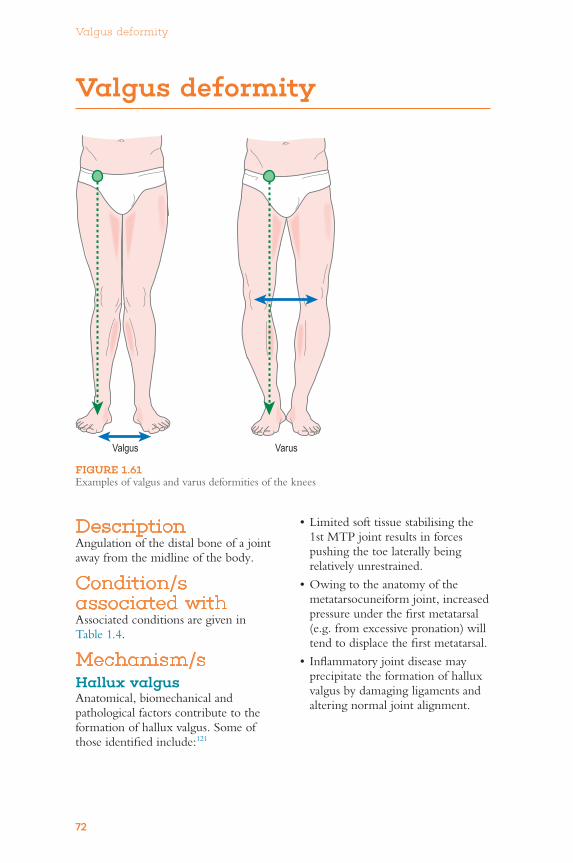
Valgus deformity
72
Valgus deformity
•Limitedsofttissuestabilisingthe1st MTP joint results in forces pushing the toe laterally being relatively unrestrained.
•Owingtotheanatomyofthemetatarsocuneiform joint, increased pressure under the first metatarsal (e.g. from excessive pronation) will tend to displace the first metatarsal.
•Inflammatoryjointdiseasemayprecipitate the formation of hallux valgus by damaging ligaments and altering normal joint alignment.
DescriptionAngulation of the distal bone of a joint away from the midline of the body.
Condition/s associated withAssociated conditions are given in Table 1.4.
Mechanism/sHallux valgusAnatomical, biomechanical and pathological factors contribute to the formation of hallux valgus. Some of thoseidentifiedinclude:121
FIGURE 1.61 Examples of valgus and varus deformities of the knees
Valgus Varus
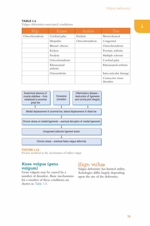
Valgus deformity
73
1TABLE 1.4 Valgusdeformity-associatedconditions
Hip Knee Ankle ToeOsteochrondrosis Cerebral palsy Paralytic Biomechanical
Idiopathic Osteochrondrosis Congenital
Blount’sdisease Osteochrondrosis
Rickets Psoriatic arthritis
Paralytic Multiple sclerosis
Osteochrondrosis Cerebral palsy
Rheumatoid arthritis
Rheumatoid arthritis
Osteoarthritis Intra-articular damage
Connective tissue disorders
FIGURE 1.62 Factors involved in the mechanism of hallux valgus
Anatomical absence ofmuscle stabiliser – frommetatarsal to proximal
great toe
Excessivepronation
Inflammatory disease –destruction of ligamentsand normal joint integrity
Chronic stress on medial ligaments – eventual disruption of medial ligaments
Unopposed adductor ligament action
Chronic stress – eventual hallux valgus deformity
Medial displacement of proximal toe, lateral displacement of distal toe
Knee valgus (genu valgum)Genuvalgummaybecausedbyanumberofdisorders.Basicmechanismsfor a number of these conditions are shown in Table 1.5.
Sign valueValgusdeformityhaslimitedutility.Aetiologies differ largely depending upon the site of the deformity.

Valgus deformity
74
TABLE 1.5 Genuvalgummechanism/s
Condition Basic mechanismVitaminDdeficiency A lack of vitamin D leads to abnormal bone
mineralisation, softer-than-normal bones, abnormal bone regrowth and bowing of the legs. Mechanical forces play a role in bone regrowth
Paget’s disease Invasion with paramyxovirus leads to abnormal activation of osteoclasts and aberrant osteoblast activity. Deformation of the bone and knee can lead to anatomical changes and valgus deformity
Osteochrondrosis Interrupted blood supply, especially to the epiphysis, leads to necrosis and then later bone regrowth – resulting in abnormal formation of femur and knee joint – and eventually a valgus deformity
Neuromuscular disorders Weak quadriceps, gastrocnemius and hip abductors may cause knees to enter valgus position56

Varus deformity
75
1Varus deformity
DescriptionAngulation of the distal bone of a joint towards the midline.
Condition/s associated withAssociated conditions are given in Table 1.6.
Mechanism/sCoxa varaPresent if the angle between the femoral neck and shaft is less than 120°.
CongenitalCongenital coxa vara may present in infancy or later in childhood. It is often bilateral and characterised by progressive bowing of the femur and a defect in the medial part of the neck of the femur.56
RicketsPressure placed on the femoral neck of abnormally mineralised bone distorts its normal architecture.
FIGURE 1.63 BowingofbothlegsininfantileBlount’sdisease
Reproduced, with permission, from Harish HS, Purushottam GA, Wells L, Chapter 674: Torsional and angular deformities. In: Kliegman RM et al., Nelson Textbook of Pediatrics, 18th edn, Philadelphia: Saunders, 2007: Fig 674-8.

Varus deformity
76
TABLE 1.6 Varusdeformity-associatedconditions
Hip Knee Ankle ToeCongenital disorders (e.g. cleidocranial dysplasia, Gaucher’sdisease)
Physiological – common
Trauma Complication from bunion surgery
Perthes’ disease Blount’sdisease Iatrogenic Trauma
Development dysplasia of hip
Rickets Congenital Burninjurywithcontracture
Slipped capital femoral epiphysis (SCFE)
Trauma Rheumatoid arthritis
Rickets Infection Psoriatic arthritis
Osteomyelitis Tumour Charcot–Marie–Tooth (CMT) disease
Paget’s disease Skeletal dysplasia Avascular necrosis
Trauma
Perthes’ diseaseAlthough the underlying cause of Perthes’ disease is unknown, there is a loss of blood supply to the femoral head. Avascular necrosis of the femoral head results in distortion of the normal bony alignment of the femur.
Genu varumGenuvarumor‘bow-leggedness’isnormal in many children up to 2 years.122,123 It should be differentiated fromBlount’sdisease.
Blount’s diseaseThe underlying mechanism of genu varuminBlount’sdiseaseisunknown.Abnormal growth of the medial tibial epiphyseal growth plate causes progressive varus deformity at the knee joint.123
Hallux varusHallux varus is comprised of medial deviation of the first metatarsophalangeal (MTP) joint, supination of phalanx and interphalangeal flexion or claw toe. It results from an imbalance between osseous, tendon and capsuloligamentous structures at the first MTP joint.124
Sign valueAetiologies differ largely depending upon the site of the deformity.
FIGURE 1.64 Metaphyseal chondrodysplasia, type Schmid There is bilateral coxa vara, the metaphyses are splayed and irregular, and there is lateral bowing of the femora.
Reproduced, with permission, from Adam A, Dixon AK (eds), Grainger&Allison’sDiagnostic Radiology, 5th edn, New York: Churchill Livingstone, 2008: Fig 67.13.

Yergason’s sign
77
1Yergason’s sign
FIGURE 1.65 Yergason’s sign
FIGURE 1.66 Yergason’s sign pathoanatomy Overhead view of the subscapularis muscle, longheadofthebicepstendon(LBT)andbicipital groove. A Intact structure depicting normal anatomy; B partial tear of the subscapularis tendon from the attachment onthelessertubercle,withtheLBTsubluxed over the lesser tubercle into the subscapularis muscle; C complete tear of the subscapularis tendon from the attachment onthelessertubercle,withtheLBTsubluxed over the lesser tuberosity and the subscapularis tendon.
Based on Pettit RW et al., Athletic Training Edu J 2008; 3(4): 143–147.
Subscapularis
Scapula
LBT
A
B
C
Humerus
Lessertuberosity
DescriptionThe examiner stands in front of the patient, who has their arms flexed to 90° at the elbow and the palms facing downwards (pronated). The patient then tries to supinate the forearm against resistance from the examiner.
Condition/s associated with•Bicepstendonitis
•SLAPlesion(Superior Labral tear from Anterior to Posterior) – an injury of the glenoid labrum
•Rotatorcuffinjuries
Mechanism/sThe long head of biceps is the main supinator of the arm. With resistance against supination, the muscle and tendon are stressed and any inflammation or damage is exacerbated, resulting in tenderness.
The long head of biceps travels in the bicipital groove of the humerus and
originates on the lip of the glenoid labrum. The fibrous extension of the subscapularis muscle covers the long head of the biceps tendon and holds it in place.125 If this fibrous extension is ruptured, the biceps tendon is susceptible to subluxation. Continuous subacromial impingement can wear away the capsule above the long head

Yergason’s sign
78
ratio of 0.72.106 A more recent meta-analysis and systematic review reported a specificity of 95%, with significant heterogeneity between studies.126
In detecting rotator cuff tendonitis, the reported sensitivity was 37% and specificity 87%, with a +LR of 2.8 and −LR of 0.7.127
of the biceps tendon. This causes ongoing injury.75
Sign valueHoltby R et al., in predicting biceps tendon pathology and SLAP lesions, reported a sensitivity of 43%, a specificity of 79%, a positive likelihood ratio of 2.05 and a negative likelihood

79
1
References
References1. SolomonDH,SimelDL,BatesDW,KatzJN,SchafferJL.Doesthispatienthavea
tornmeniscusofligamentoftheknee?Valueofphysicalexamination.JAMA 2001;286(13):1610–19.
2. McGeeS.Chapter53:Examinationofthemusculoskeletalsystem.In:Evidence Based Physical Diagnosis.2nded.StLouis:Saunders;2007.
3. WagemakersHP,LuijsterburgPA,BoksSS,etal.Diagnosticaccuracyofhistorytaking and physical examination for assessing anterior cruciate ligament lesions of the knee in primary care. Arch Phys Med Rehabil 2010;91(9):1452–9.
4. Magee DJ. Orthopedic Physical Assessment.5thed.StLouis:SaundersElsevier;2008.
5. HegedusEJ,CookC,HasselbladV,GoodeA,McCroryDC.Physicalexaminationtestsforassessingatornmeniscusintheknee:asystematicreviewwithmeta-analysis. J Orthop Sports Phys Ther 2007;37(9):541–50.
6. ScholtenR,DevilleW,OpsteltenW,BijlD,vanderPlasCG,BouterL.Theaccuracyofphysicaldiagnostictestsforassessingmeniscallesionsoftheknee:ameta-analysis. J Fam Pract 2001;50(11):938–44.
7. WoodwardTW,BestTM.Thepainfulshoulder:Part1.Clinicalevaluation.Am Fam Physician 2000;61(10):3079–88.
8. BradyRJ,DeanJB,SkinnerTM,GrossMT.Limblengthinequality:clinicalimplications for assessment and intervention. J Orthop Sports Phys Ther 2003; 33(5):221–34.
9. TennantTD,BeachWR,MeyersJF.Areviewofspecialtestsassociatedwithshoulderexamination.PartII:Laxity,instabilityandsuperiorlabralanteriorandposterior(SLAP) lesions. Am J Sports Med 2003;31:301–7.
10. T’Jonck L, Staes F, Smet L, Lysens R. The relationship between clinical shoulder tests and the findings in arthroscopy examination. Geneeskunde Sport 2001;34:15–24.
11. LoIKY,NonweilerB,WoolfreyM,LitchfieldR,KirkleyA.Anevaluationoftheapprehension, relocation and surprise tests for anterior shoulder instability. Am J Sports Med 2004;32:301.
12. Speer KP, Hannafin JA, Altchek DW, Warren RF. An evaluation of the shoulder relocation test. Am J Sports Med 1994;22:177–83.
13. AlexanderCJ.Heberden’sandBouchard’snodes.Ann Rheum Dis 1999;58:675–8.
14. FassbenderHG.Pathology of Rheumatic Diseases.NewYork:Springer;1975.
15. Collins DH. The Pathology of Articular and Spinal Diseases.London:EdwardArnold;1949. pp. 109–13.
16. SokoloffL.Thepathologyofosteoarthritisandtheroleofageing.In:NukiG, editor. The Aetiopathogenesis of Osteoarthrosis.TunbridgeWells:PitmanMedical;1980.pp. 1–15.
17. KellegranJH,LawrenceJS,BierF.Geneticfactorsingeneralizedosteoarthritis.Ann Rheum Dis 1963;22:237–55.
18. StecherRM,HerschAH.Heberden’snodes:themechanismofinheritanceinhypertrophic arthritis of the fingers. J Clin Invest 1944;23:699–704.
19. ThaperA,ZhangW,WrightG,DohertyM.RelationshipbetweenHeberden’snodesand underlying radiographic changes of osteoarthritis. Ann Rheum Dis 2005;64:1214–16.
20. CoonsMS,GreenSM.Boutonnièredeformity.Hand Clin 1995;11(3):387–402.
21. LikesRL,GhidellaSD.Boutonnièredeformity.eMedicine.Available:http://emedicine. medscape.com/article/1238095-overview [11 Aug 2010].
22. Nalebuff EA, Millender LH. Surgical treatment of the boutonnière deformity in rheumatoid arthritis. Orthop Clin North Am 1975;6(3):753–63.
23. Rosen A, Weiland AJ. Rheumatoid arthritis of the wrist and hand. Rheum Dis Clin North Am 1998;24(1):101–28.
80
References
24. Fox A, Kang N. Reinserting the central slip – a novel method for treating boutonnière deformity in rheumatoid arthritis. J Plast Reconstr Aesthet Surg 2009;62(5):e91–2.
25. FiresteinGS,BuddRC,HarrisED,etal.Kelley’s Textbook of Rheumatology. 8th ed. Philadelphia:WBSaundersCompany;2008.
26. Cibere J, et al. Reliability of the knee examination in osteoarthritis. Arthritis Rheum 2004;50(2):458–68.
27. GogusF,KitchenJ,CollinsR,KaneD.Reliability of physical knee examination for effusion: verification by musculoskeletal ultrasound. Presentation:2008AnnualScientificMeeting of American College of Rheumatology,SanFrancisco,2008.Available:http://acr.comfex.com/acr/2008/webprogram/paper2759.htm[22Nov2010].
28. PatelP,WerthV.Cutaneouslupuserythematosus:areview.Dermatol Clin 2002;20: 373–85.
29. Uva L, Miguel D, Pinheiro C, et al. Cutaneous manifestations of systemic lupus erythematosus. Autoimmune Diseases, 2012; 2012.https://doi.org/10.1155/2012/834291.
30. OrgretmenA,AkayA,BicakciC,BicakciHC.Calcinosiscutisuniversalis.JEADV 2002;16:621–4.
31. NeumanWF,DiStefanoV,MubryanBJ.Thesurfacechemistryofbone.III.Observations of the role of phosphate. J Biol Chem 1951;193:227–36.
32. CousinsMAM,JonesDB,WhyteMP,MonafoWW.Surgicalmanagementofcalcinosis cutis universalis in systemic lupus erythematosus. Arthritis Rheum 1997; 40:570–2.
33. FrykbergRG,ArmstrongDG,GiurnliJ,etal.Diabeticfootdisorders:aclinicalpractice guideline. American College of Foot and Ankle Surgeons. J Foot Ankle Surg 2000;39(Suppl.5):s1–60.
34. JeffcoateWJ,GameF,CavanaghPR.Theroleofproinflammatorycytokinesinthecause of neuropathic osteoarthropathy (acute Charcot foot) in diabetes. Lancet 2005; 366:2058–61.
35. Zhao H-M, Diao J-Y, Liang X-J, et al. Pathogenesis and potential relative risk factors of diabetic neuropathic osteoarthropathy. Journal of Orthopaedic Surgery and Research 2017;12:142.DOI:10.1186/s13018-017-0634-8.
36. Jeffcoate WJ. Theories concerning the pathogenesis of acute Charcot foot suggest future therapy. Curr Diab Rep 2005;5:430–5.
37. NabarroJD.DiabetesintheUnitedKingdom:apersonalseries.Diabet Med 1991;8:59–68.
38. Fabrin J, Larsen K, Holstein PE. Long term follow up in diabetic Charcot feet with spontaneous onset. Diabetes Care 2000;23:796–800.
39. AltmanR,AschE,BlockD,etal.Developmentofcriteriafortheclassificationandreportingofosteoarthritis:classificationofosteoarthritisoftheknee.Arthritis Rheum 1986;29:1039–49.
40. DrakeEL,VoglW,MitchellAW.Gray’s Anatomy for Students.Philadelphia:Elsevier;2005.
41. MurrellGAC,WaltonJR.Diagnosisofrotatorcufftears.Lancet 2001;357:769–70.
42. Dinnes J, Loveman E, McIntyre L, Waugh N. The effectiveness of diagnostic tests for theassessmentofshoulderpainduetosofttissuedisorders:asystematicreview.Health Technol Assess 2003;7(29):1–166.
43. ParkHB,etal.Diagnosticaccuracyofclinicaltestsfordifferentdegreesofsubacromialimpingement syndrome. J Bone Joint Surg Am 2005;87:1446–55.
44. Wu F, Rajpura A, Sandher D. Finkelstein’s test is superior to Eichhoff’s test in the investigationofdeQuervain’sdisease.J Hand Microsurg 2018;10(2):116–8.DOI:10.1055/s-0038-1626690.
45. KutsumiK,AmadioPC,ZhaoC,ZobitzME,TanakaT,AnKN.Finkelstein’stest:abiomechanical analysis. J Hand Surg [Am] 2005;30(1):130–5.

81
1
References
46. Anderson DM. Dorlands Illustrated Medical Dictionary.30thed.Philadelphia:Saunders;2003.
47. MendeseG,MahalingamM.HistopathologyofGottron’spapules–utilityindiagnosing dermatomyositis. J Cutan Pathol 2007;34:793–6.
48. StoneJH,SackKE,McCalmontTH,ConnollyKM.Gottronpapules?Arthritis Rheum 1995;38(6):862–5.
49.McFarlandEG,Muvdi-GarzonJ,XiaofengJ,etal.Clinicalanddiagnostictestsforshoulderdisorders:acriticalreview.Br J Sports Med 2010;44:328–33.
50. CalisM,AkgunK,BirtaneM,etal.Diagnosticvaluesforclinicaldiagnostictestsinsubacromial impingement syndrome. Ann Rheum Dis 2000;59:44–7.
51. MacdonaldPB,ClarkP,SutherlandK.AnanalysisofthediagnosticaccuracyoftheHawkins and Neer subacromial impingement signs. J Shoulder Elbow Surg 2000;9: 299–301.
52. AlqunaeeM,GalvinR,FaheyT.Diagnosticaccuracyofclinicaltestsforsubacromialimpingementsyndrome:asystematicreviewandmeta-analysis.Arch Phys Med Rehabil 2012;93(2):229–36.
53. Iaccarino L, et al. The clinical features, diagnosis and classification of dermatomyositis. J Autoimmun 2014;48–49:122–7.
54. BalciMA,DonmezS,SaritasF,etal.TheepidemiologyofdermatomyositisinnorthwesternThraceregioninTurkey:epidemiologyofdermatomyositisinTurkey.Rheumatol Int 2017;37(9):1519–25.DOI:10.1007/s00296-017-3710-9.
55. Wheeless CR III. Wheeless orthopedics online.Available:http://www.wheelessonline.com/[October 2010].
56. CanaleTS,BeatyJH.Campbell’s Operative Orthopaedics.11thed.Philadelphia:Elsevier;2007.
57. LeeJK,YaoL,PhelpsCT,etal.Anteriorcruciateligamenttears:MRimagingcompared with arthroscopy and clinical test. Radiology 1988;166(3):861–4.
58. Swain MS, Henschke N, Kamper SJ, et al. Accuracy of clinical tests in the diagnosis of anteriorcruciateligamentinjury:asystematicreview.Chiropr Man Therap 2014;22:25.DOI:10.1186/s12998-014-0025-8.
59. GibbsMR,EnglishJC,ZirwasJ.Livedoreticularis:anupdate.J Am Acad Dermatol 2005;52(6):1009–18.
60. SajjanVV,LungeS,SwamyMB,PanditAM.Livedoreticularis:areviewoftheliterature. Indian Dermatol Online J 2015;6(5):315–21.
61. FreemanR,DoverJS.Autonomicneurodermatology(part1):erythromelalgia,reflexsympathetic dystrophy and livedo reticularis. Semin Neurol 1992;12:385–93.
62. KesterS,McCartyDL,McCartyGA.Theantiphospholipidantibodysyndromeintheemergency department setting – livedo reticularis and recurrent venous thrombosis. Ann Emerg Med 1992;21:207–11.
63. ScholtenRJ,DevilléWL,OpsteltenW,BijlD,vanderPlasCG,BouterLM. The accuracy of physical diagnostic tests for assessing meniscal lesions of the knee. A meta-analysis. J Fam Pract 2001;50(11):938–44.
64. SallayPI,PoggiJ,SpeerFP,GarrettWE.Acutedislocationofthepatella.Acorrelativepathoanatomic study. Am J Sports Med 1996;24(1):52–60.
65. AhmadCS,McCarthyM,GomezJA,ShubinSteinBE.Themovingpatellarapprehension test for lateral patellar instability. Am J Sports Med 2009;37(4):791–6.DOI:10.1177/0363546508328113.
66. Kastelein M, Luijsterburg PA, Wagemakers HP, et al. Diagnostic value of history taking and physical examination to assess effusion of the knee in traumatic knee patients in general practice. Arch Phys Med Rehabil 2009;90:82–6.
67. BernardTN.The role of the sacroiliac joints in low back pain: basic aspects of pathophysiology, and management.Available:http://www.kalindra.com/bernard.pdf[28Feb2011].
82
References
68. Stuber KJ. Specificity, sensitivity and predictive values of clinical tests of the sacroiliac joint:asystematicreviewoftheliterature.J Can Chiropr Assoc 2007;51(1):30–41.
69. BroadhurstNA,BondMJ.Painprovocationtestsfortheassessmentofsacroiliacjointdysfunction. J Spinal Disord 1998;11(4):341–5.
70. DreyfussP,MichaelsenM,PauzaK,McLartyJ,BogdukN.Thevalueofmedicalhistory and physical examination in diagnosing sacroiliac joint pain. Spine 1996; 21(22):2594–602.
71. Martin RL, Sekiya JK. The reliability and validity of clinical tests used to assess individuals with potential labral tears of the hip (SS-20). Arthroscopy 2007;23(6):e10–e11.
72. Seror P. Phalen’s test in the diagnosis of carpal tunnel syndrome. J Hand Surg 1988; 13-B(4):383–5.
73. D’ArcyCA,McGeeS.Doesthispatienthavecarpaltunnelsyndrome?JAMA 2000; 283(23):3110–17.
74. KasundraGM,SoodI,BhargavaAN,etal.Carpaltunnelsyndrome:analyzingefficacyand utility of clinical tests and various diagnostic modalities. J Neurosci Rural Pract 2015;6(4):504–10.DOI:10.4103/0976-3147.169867.
75.McGeeS.Evidence Based Physical Diagnosis.4thed.StLouis:Saunders;2017.
76. SzepietowskiJC,SalomonJ.Dofungiplayaroleinpsoriaticnails?Mycoses 2007;50: 437–42.
77. SzepietowskiJC,SalomonJ.Thenailchangesinpsoriasis.In:LiponzencicJ,PasicA,editors. Suvremene Sponznaje o Psorijazi Zagreb. Medicinska Naklada; 2004. pp. 55–9.
78. JiaravuthisanMM,SassevilleD,VenderRB,MurphyF,MuhnCY.Psoriasisofthenail:anatomy,pathology,clinicalpresentation,andareviewoftheliteratureontherapy. J Am Acad Dermatol 2007;57(1):1–27.
79. CrawfordGM.Psoriasisofthenails.Arch Derm Syphilol 1938;38:583–94.
80. Samman PD, Fenton DA. The Nails in Disease.5thed.London:Butterworth-Heineman Ltd; 1994.
81. KaurI,SaraswatA,KumarB.Nailchangesinpsoriasis:astudyof167patients.Int J Dermatol 2001;40:597–604.
82. Faber EM, Nall L. Nail psoriasis. Cutis 1992;50:174–8.
83. LavaroniG,KokeljF,PauluzziP,TrevisanG.Thenailsinpsoriaticarthritis.Acta Derm Venereol Suppl (Stockh) 1994;186:113.
84. SalomanJ,SzeptietowskiJC,ProniewiczA.Psoriaticnails:aprospectiveclinicalstudy. J Cutan Med Surg 2003;7:317–21.
85. Herrick AL. Pathogenesis of Raynaud’s phenomenon. Rheumatology (Oxford) 2005;44: 587–96.
86. Cooke JP, Marshall JM. Mechanisms of Raynaud’s disease. Vasc Med 2005;10:293–307.
87. BakstR,MerolaJF,FranksAGJr,SanchezM.Raynaud’sphenomenon:pathogenesisand management. J Am Acad Dermatol 2008;59(4):633–53.
88. Wigley FM. Pathogenesis of Raynaud phenomenon. Uptodate. Last updated 3 October 2010.Available:http://www.uptodate.com[1Mar2011].
89. FardounMM,NassifJ,IssaK,etal.Raynaud’sphenomenon:abriefreviewoftheunderlying mechanisms. Front Pharmacol 2016;7:438.DOI:10.3389/fphar.2016.00438.
90. GkogkolouP,LugerTA,BöhmM.Cutaneousmanifestationsofrheumaticdiseases.Clinical presentation and underlying pathophysiology. Giornale Italiano di Dermatologia e Venereologia 2014;149(5):483–503.
91. CoordesA,LooseSM,HofmannVM,etal.Saddlenosedeformityandseptalperforation in granulomatosis with polyangiitis. Clin Otolaryngol 2018;43(1):291–9.https://doi.org/10.1111/coa.12977.
92. RothschildBM,PingitoreC,EatonM.Dactylitis:implicationsforclinicalpractice.Semin Arthritis Rheum 1998;28:41–7.

83
1
References
93. OliveriI,ScaranoE,PadulaA,GiassiV,PrioloF.Dactylitis,atermfordifferentdigitdiseases. Scand J Rheumatol 2006;35:333–40.
94. McGonagleD,PeaseC,Marzo-OrtegaH,O’ConnorP,EmeryP.Thecaseofclassification of polymyalgia rheumatica and remitting seronegative symmetrical synovitis with pitting edema as primarily capsular/entheseal based pathologies. J Rheumatol 2000;27:837–40.
95. Tan AL, Fukuba E, Halliday NA, et al. High-resolution MRI assessment of dactylitis in psoriatic arthritis shows flexor tendon pulley and sheath-related enthesitis. Ann Rheum Dis 2015;74:185–9.
96. OliveriA,etal.Editorial:Dactylitisor‘sausage-shaped’digit.J Rheumatol 2007;34(6): 1217–20.
97. OliveriI,BarozziL,PierroA,DeMatteisM,PadulaA,PavlicaP.Toedactylitis inpatientswithspondyloarthropathy:assessmentbymagneticresonanceimaging. J Rheumatol 1997;24:926–30.
98. BrockbankJE,SteinM,SchentagCT,GladmanDD.Dactylitisinpsoriaticarthritis:amarker for disease severity? Ann Rheum Dis 2005;64:188–90.
99. SilverRM,MedsgerTAJr,BolsterMB.Chapter77:Systemicsclerosisandsclerodermavariants:clinicalaspects.In:KoopmanWJ,MorelandLW,editors.Arthritis and Allied Conditions.Philadelphia:LippincottWilliams&Wilkins;2005.
100. SontheimerRD,CostnerMI.Chapter157:Dermatomyositis.In:WolffK,GoldsmithLA,KatzSI,GilchrestB,PallerAS,LeffellDJ,editors.Fitzpatrick’s Dermatology in General Medicine.7thed.Available:http://proxy14.use.hcn.com.au/content.aspx?aID =2992330 [3 Oct 2010].
101. KrathenMS,FiorentinoD,WerthVP.Dermatomyositis.Curr Dir Autoimmun 2008;10:313–32.
102. SontheimerRD.Dermatomyositis:anoverviewofrecentprogresswithemphasisondermatologic aspects. Dermatol Clin 2002;20(3):387–408.
103. ScottBW,AlChalabiA.HowtheSimmonds–Thompsontestworks.J Bone Joint Surg 1992;74-B(2):314–15.
104. MaffulliN.TheclinicaldiagnosisofsubcutaneoustearoftheAchillestendon:aprospective study in 174 patients. Am J Sports Med 1998;26(2):266–70.
105. DouglasJ,KellyM,BlachutP.ClarificationoftheSimmonds-Thompsontestforrupture of an Achilles tendon. Can J Surg 2009;52(3):E40–1.
106. HoltbyR,RazmjouH.AccuracyoftheSpeed’sandYergaon’stestsindetectingbicepspathologyandSLAPlesions:comparisonwitharthroscopicfindings.Arthroscopy 2004;20(3):231–6.
107. Hessian P, Highton J, Kean A, et al. Cytokine profile of the rheumatoid nodule suggests that it is a Th1 granuloma. Arthritis Rheum 2003;24:334–8.
108. Garcia-PatosV.Rheumatoidnodule.Semin Cutan Med Surg 2007;26:100–7.109. Nyhäll-WåhlinBM,TuressonC,JacobssonLT,etal.Thepresenceofrheumatoid
nodules at early rheumatoid arthritis diagnosis is a sign of extra-articular disease and predicts radiographic progression of joint destruction over 5 years. Scand J Rheumatol 2011;40(2):81–7.
110. TzannesA,MurrellGAC.Clinicalexaminationoftheunstableshoulder.Sports Med 2002;32(7):447–57.
111. Rosen A, Weiland AJ. Rheumatoid arthritis of the wrist and hand. Rheum Dis Clin North Am 1998;24(1):101–28.
112. BeatyJH,CanaleTS,etal.Fingerdeformitiescausedbyrheumatoidarthritis.In:CanaleTS,BeatyJH,editors.Campell’s Operative Orthopedics.11thed.Philadelphia:Elsevier; 2007.
113. Mould TL, Roberts-Thomson PJ. Pathogenesis of telangiectasia in scleroderma. Asian Pac J Allergy Immunol 2000;18:195–200.
114. BologniaJL,BravermanIM.Chapter54:Skinmanifestationsofinternaldisease.In:FauciAS,BraunwaldE,KasperDL,etal.,editors.Harrison’s Principles of Internal

84
References
Medicine.17thed.Available:http://proxy14.use.hcn.com.au/content.aspx?aID =2864525 [28 Nov 2010].
115. ShahAA,WigleyFM,HummersLK.Telangiectasesinscleroderma:apotentialclinical marker of pulmonary arterial hypertension. J Rheumatol 2009;37(1):98–104.
116. BlythM,AnthonyI,FrancqB,etal.DiagnosticaccuracyoftheThessalytest,standardised clinical history and other clinical examination tests (Apley’s, McMurray’s and joint line tenderness) for meniscal tears in comparison with magnetic resonance imaging diagnosis. Health Technol Assess 2015;19(62):1–62.
117. PasicN,BryantD,NaudieD,WillitsK.Diagnosticvalidityofthephysicalexaminationmaneuversforhippathology:asystematicreview.Orthopedic Muscul Syst 2014;3:2.
118. UrbanoFL.Tinel’ssignandPhalen’smaneuver:physicalsignsincarpaltunnelsyndrome. Hosp Phys2000;July:39–44.
119. Friberg O. Clinical symptoms and biomechanics of lumbar spine and hip joint in leg length inequality. Spine 1983;8(6):643–51.
120. Crowson N, Magro C. The role of microvascular injury in the pathogenesis of cutaneous lesions in dermatomyositis. Hum Pathol 1996;27(1):15–19.
121. Ferrari J. Hallux valgus deformity (bunion).In:EiffP,editor.http://www.uptodate.com[22 Feb 2010].
122. HolsalkaHS,GholvePA,WellsL.Chapter674:Torsionalandangulardeformities. In:KliegmanRM,etal.,editors.Nelson Textbook of Pediatrics.18thed.Philadelphia:Saunders; 2007.
123. RabGT.Chapter11:Pediatricorthopedicsurgery.In:SkinnerHB,editor.Current Diagnosis & Treatment in Orthopedics.4thed.Available:http://proxy14.use.hcn.com.au/content.aspx?aID=2315794 [14 Oct 2010].
124. BevernageBD,LeemrijseT.Halluxvarus:classificationandtreatment.Foot Ankle Clin N Am 2009;14:51–65.
125. KarlssonJ.PhysicalexaminationtestsarenotvalidfordiagnosingSLAPtears:areview.Clin J Sport Med 2010;20(2):134–5.
126. HegedusEJ,GoodeAP,CookCE,etal.Whichphysicalexaminationtestsprovideclinicians with the most value when examining the shoulder? Update of a systematic review with meta-analysis of individual tests. Br J Sports Med 2012;46(14):964–78.DOI:10.1136/bjsports-2012-091066.
127. CalisM,AkgunK,BirtaneM,etal.Diagnosticvaluesofclinicaldiagnostictestsinsubacromial impingement syndrome. Ann Rheum Dis 2000;59:44–47.