Protocol independent multicast-dense mode (pim-dm): protocol specification
Upload
independentCategory
view
0download
0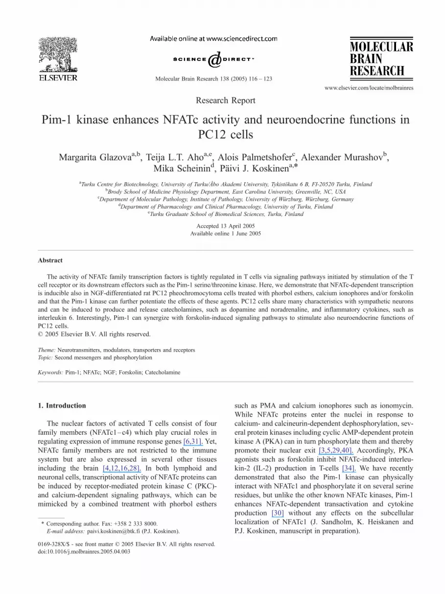
www.elsevier.com/locate/molbrainres
Molecular Brain Research
Research Report
Pim-1 kinase enhances NFATc activity and neuroendocrine functions in
PC12 cells
Margarita Glazovaa,b, Teija L.T. Ahoa,e, Alois Palmetshoferc, Alexander Murashovb,
Mika Scheinind, Paivi J. Koskinena,*
aTurku Centre for Biotechnology, University of Turku/Abo Akademi University, Tykistokatu 6 B, FI-20520 Turku, FinlandbBrody School of Medicine Physiology Department, East Carolina University, Greenville, NC, USA
cDepartment of Molecular Pathology, Institute of Pathology, University of Wurzburg, Wurzburg, GermanydDepartment of Pharmacology and Clinical Pharmacology, University of Turku, Finland
eTurku Graduate School of Biomedical Sciences, Turku, Finland
Accepted 13 April 2005
Available online 1 June 2005
Abstract
The activity of NFATc family transcription factors is tightly regulated in T cells via signaling pathways initiated by stimulation of the T
cell receptor or its downstream effectors such as the Pim-1 serine/threonine kinase. Here, we demonstrate that NFATc-dependent transcription
is inducible also in NGF-differentiated rat PC12 pheochromocytoma cells treated with phorbol esthers, calcium ionophores and/or forskolin
and that the Pim-1 kinase can further potentiate the effects of these agents. PC12 cells share many characteristics with sympathetic neurons
and can be induced to produce and release catecholamines, such as dopamine and noradrenaline, and inflammatory cytokines, such as
interleukin 6. Interestingly, Pim-1 can synergize with forskolin-induced signaling pathways to stimulate also neuroendocrine functions of
PC12 cells.
D 2005 Elsevier B.V. All rights reserved.
Theme: Neurotransmitters, modulators, transporters and receptors
Topic: Second messengers and phosphorylation
Keywords: Pim-1; NFATc; NGF; Forskolin; Catecholamine
1. Introduction
The nuclear factors of activated T cells consist of four
family members (NFATc1–c4) which play crucial roles in
regulating expression of immune response genes [6,31]. Yet,
NFATc family members are not restricted to the immune
system but are also expressed in several other tissues
including the brain [4,12,16,28]. In both lymphoid and
neuronal cells, transcriptional activity of NFATc proteins can
be induced by receptor-mediated protein kinase C (PKC)-
and calcium-dependent signaling pathways, which can be
mimicked by a combined treatment with phorbol esthers
0169-328X/$ - see front matter D 2005 Elsevier B.V. All rights reserved.
doi:10.1016/j.molbrainres.2005.04.003
* Corresponding author. Fax: +358 2 333 8000.
E-mail address: [email protected] (P.J. Koskinen).
such as PMA and calcium ionophores such as ionomycin.
While NFATc proteins enter the nuclei in response to
calcium- and calcineurin-dependent dephosphorylation, sev-
eral protein kinases including cyclic AMP-dependent protein
kinase A (PKA) can in turn phosphorylate them and thereby
promote their nuclear exit [3,5,29,40]. Accordingly, PKA
agonists such as forskolin inhibit NFATc-induced interleu-
kin-2 (IL-2) production in T-cells [34]. We have recently
demonstrated that also the Pim-1 kinase can physically
interact with NFATc1 and phosphorylate it on several serine
residues, but unlike the other known NFATc kinases, Pim-1
enhances NFATc-dependent transactivation and cytokine
production [30] without any effects on the subcellular
localization of NFATc1 (J. Sandholm, K. Heiskanen and
P.J. Koskinen, manuscript in preparation).
138 (2005) 116 – 123
M. Glazova et al. / Molecular Brain Research 138 (2005) 116–123 117
Pim-1 is an oncogenic serine/threonine kinase, whose
expression in hematopoietic cells is transiently induced upon
activation of cytokine or antigen receptors [7,20,32,39]. We
have observed that, during murine embryogenesis, the three
pim family genes are expressed in overlapping patterns in
the central nervous system [8], while in adult brain tissues,
the pim genes are expressed at lower levels but can be
upregulated, e.g. by seizure activity [10,18].
Rat PC12 pheochromocytoma cells are adrenal gland-
derived chromaffin-like cells that are widely used as a
neuronal cell culture model since they can be differentiated
by nerve growth factor (NGF) or interleukin 6 (IL-6) into
cells that resemble sympathetic neurons and that are able to
synthesize and release catecholamines [14,33]. Catechol-
amines can in turn induce production of IL-6 in differ-
entiated PC12 cells [9], suggesting that there is an autocrine
positive feedback loop, which not only promotes differ-
entiation, but in addition helps the cells to resist apoptotic
stimuli [35].
Since Pim and NFATc family proteins are coexpressed
in PC12 cells and within the central nervous system and
since both types of proteins have been implicated in
regulation of synaptic plasticity and memory [12,18], we
hypothesized that, similarly to T cells, Pim kinases might
enhance NFATc activity also in neurons. Here, we
demonstrate that, in NGF-differentiated PC12 cells, Pim-1
potentiates other signaling pathways not only to stimulate
NFATc activity, but also to enhance neuroendocrine
functions of PC12 cells, as evidenced by increased
production of catecholamines.
2. Materials and methods
2.1. Plasmid constructs
The pSV-pim-1 and pSV-pimNT81 expression vectors
have been described previously [2]. The NFAT-LUC
luciferase reporter plasmid kindly provided by G.R.
Crabtree (Stanford University, Stanford, CA) contains three
copies of the composite NFAT sites derived from the IL-2
enhancer binding both NFATc and AP-1 family members.
The NFAT-mut-LUC reporter without functional NFAT sites
was derived from NFAT-LUC, as described previously [30],
and the pSV-h-galactosidase reporter construct was from
Promega (Madison, WI).
2.2. Cell culture
PC12 cells were plated on Primaria six-well plates (BD
Biosciences, San Jose, CA) or on collagen-coated plates
(Collagen type I from rat tail, Sigma-Aldrich, St. Louis,
MO) and grown in DMEM supplemented with 5% fetal calf
serum, 10% horse serum and the antibiotics penicillin and
streptomycin. The cultures were maintained at 37 -C in a
5% CO2/air atmosphere. Differentiation of PC12 cells was
induced by adding NGF (50 ng/ml, Promega) to the culture
medium for 4 to 6 days.
2.3. Transactivation assays
NGF-differentiated PC12 cells were transfected by the
FUGENE 6 transfection reagent (Roche Diagnostics,
Indianapolis, IN) with 1 Ag of reporter plasmids together
with 1 Ag of pim-1 expression vectors. Two days after
transfection, cells were left unstimulated or were stimulated
for 3 to 6 h with 10 ng/ml PMA (Sigma-Aldrich) and 5 AMionomycin (Calbiochem, La Jolla, CA). To stimulate protein
kinase A activity, cells were cotreated with 20 AM forskolin
(Sigma-Aldrich), and to inhibit calcineurin activity, 1 Ag/ml
of cyclosporin A (Sigma-Aldrich) was added. After treat-
ments, cells were collected and analyzed for luciferase
activities using the Labsystems luminometer (Labsystems,
Helsinki, Finland). The transfection efficiencies were
normalized against h-galactosidase activities. Shown in
the figures are means and standard deviations of represen-
tative experiments with at least triplicate samples. Statistical
analyses were carried out using Student’s t test, where
stimulated samples were compared to untreated control
samples; * means P < 0.05 and **P < 0.01.
2.4. Western blot analysis
Cells were lysed into SDS gel loading buffer by heating
them at 95 -C for 10 min, and proteins of the lysates were
separated by SDS-PAGE followed by transfer into nitro-
cellulose membranes (Schleicher and Schuell, Dassel,
Germany). For Western blotting, the membranes were
blocked with 3% nonfat dry milk in Tris-buffered saline
and incubated with monoclonal anti-Pim-1 (19F7, Santa
Cruz Biotechnology, Santa Cruz, CA) or anti-TH (Zymed
Laboratories, San Francisco, CA) antibodies or with affinity-
purified polyclonal antiserum against NFATc1 (Immuno-
globe, Himmelstadt, Germany). The proteins recognized by
primary antisera were visualized by using horseradish-
peroxidase-conjugated secondary antibodies and ECL+Plus
reagents (Amersham Biosciences, Uppsala, Sweden). Equal
protein loading was verified by blotting stripped membranes
with anti-h-actin antibody (Sigma-Aldrich).
2.5. Immunocytochemistry
Cells were fixed in 4% paraformaldehyde and incubated
with primary antisera followed by TRITC-conjugated
secondary antibodies (Zymed Laboratories). Specificities
of immunoreactivities were confirmed by omitting primary
antisera from the reactions.
2.6. Catecholamine assays
Cells were collected in 0.1 M perchloric acid, and the
cellular levels of the catecholamines dopamine (DA) and
M. Glazova et al. / Molecular Brain Research 138 (2005) 116–123118
noradrenaline (NA) and their precursor l-DOPA were
determined by electrochemical detection (ESA Coulochem
5011, Bedford, MA) from samples separated by HPLC on a
reversed-phase C18 column (Ultrasphere ODS, 4.6 � 250
mm, Beckman Instruments, Fullerton, CA).
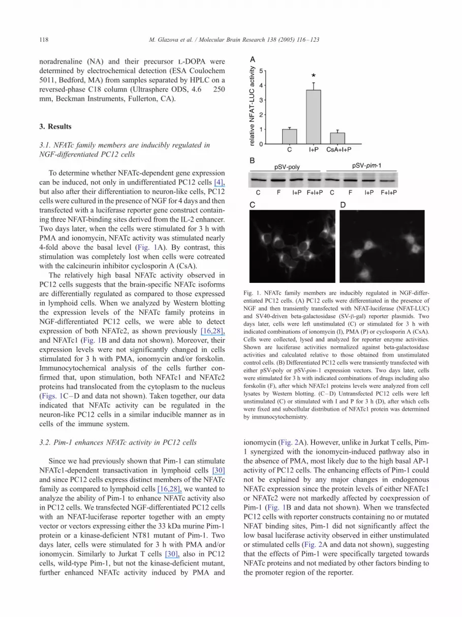
Fig. 1. NFATc family members are inducibly regulated in NGF-differ-
entiated PC12 cells. (A) PC12 cells were differentiated in the presence of
NGF and then transiently transfected with NFAT-luciferase (NFAT-LUC)
and SV40-driven beta-galactosidase (SV-h-gal) reporter plasmids. Two
days later, cells were left unstimulated (C) or stimulated for 3 h with
indicated combinations of ionomycin (I), PMA (P) or cyclosporin A (CsA).
Cells were collected, lysed and analyzed for reporter enzyme activities.
Shown are luciferase activities normalized against beta-galactosidase
activities and calculated relative to those obtained from unstimulated
control cells. (B) Differentiated PC12 cells were transiently transfected with
either pSV-poly or pSV-pim-1 expression vectors. Two days later, cells
were stimulated for 3 h with indicated combinations of drugs including also
forskolin (F), after which NFATc1 proteins levels were analyzed from cell
lysates by Western blotting. (C–D) Untransfected PC12 cells were left
unstimulated (C) or stimulated with I and P for 3 h (D), after which cells
were fixed and subcellular distribution of NFATc1 protein was determined
by immunocytochemistry.
3. Results
3.1. NFATc family members are inducibly regulated in
NGF-differentiated PC12 cells
To determine whether NFATc-dependent gene expression
can be induced, not only in undifferentiated PC12 cells [4],
but also after their differentiation to neuron-like cells, PC12
cells were cultured in the presence of NGF for 4 days and then
transfected with a luciferase reporter gene construct contain-
ing three NFAT-binding sites derived from the IL-2 enhancer.
Two days later, when the cells were stimulated for 3 h with
PMA and ionomycin, NFATc activity was stimulated nearly
4-fold above the basal level (Fig. 1A). By contrast, this
stimulation was completely lost when cells were cotreated
with the calcineurin inhibitor cyclosporin A (CsA).
The relatively high basal NFATc activity observed in
PC12 cells suggests that the brain-specific NFATc isoforms
are differentially regulated as compared to those expressed
in lymphoid cells. When we analyzed by Western blotting
the expression levels of the NFATc family proteins in
NGF-differentiated PC12 cells, we were able to detect
expression of both NFATc2, as shown previously [16,28],
and NFATc1 (Fig. 1B and data not shown). Moreover, their
expression levels were not significantly changed in cells
stimulated for 3 h with PMA, ionomycin and/or forskolin.
Immunocytochemical analysis of the cells further con-
firmed that, upon stimulation, both NFATc1 and NFATc2
proteins had translocated from the cytoplasm to the nucleus
(Figs. 1C–D and data not shown). Taken together, our data
indicated that NFATc activity can be regulated in the
neuron-like PC12 cells in a similar inducible manner as in
cells of the immune system.
3.2. Pim-1 enhances NFATc activity in PC12 cells
Since we had previously shown that Pim-1 can stimulate
NFATc1-dependent transactivation in lymphoid cells [30]
and since PC12 cells express distinct members of the NFATc
family as compared to lymphoid cells [16,28], we wanted to
analyze the ability of Pim-1 to enhance NFATc activity also
in PC12 cells. We transfected NGF-differentiated PC12 cells
with an NFAT-luciferase reporter together with an empty
vector or vectors expressing either the 33 kDa murine Pim-1
protein or a kinase-deficient NT81 mutant of Pim-1. Two
days later, cells were stimulated for 3 h with PMA and/or
ionomycin. Similarly to Jurkat T cells [30], also in PC12
cells, wild-type Pim-1, but not the kinase-deficient mutant,
further enhanced NFATc activity induced by PMA and
ionomycin (Fig. 2A). However, unlike in Jurkat T cells, Pim-
1 synergized with the ionomycin-induced pathway also in
the absence of PMA, most likely due to the high basal AP-1
activity of PC12 cells. The enhancing effects of Pim-1 could
not be explained by any major changes in endogenous
NFATc expression since the protein levels of either NFATc1
or NFATc2 were not markedly affected by coexpression of
Pim-1 (Fig. 1B and data not shown). When we transfected
PC12 cells with reporter constructs containing no or mutated
NFAT binding sites, Pim-1 did not significantly affect the
low basal luciferase activity observed in either unstimulated
or stimulated cells (Fig. 2A and data not shown), suggesting
that the effects of Pim-1 were specifically targeted towards
NFATc proteins and not mediated by other factors binding to
the promoter region of the reporter.
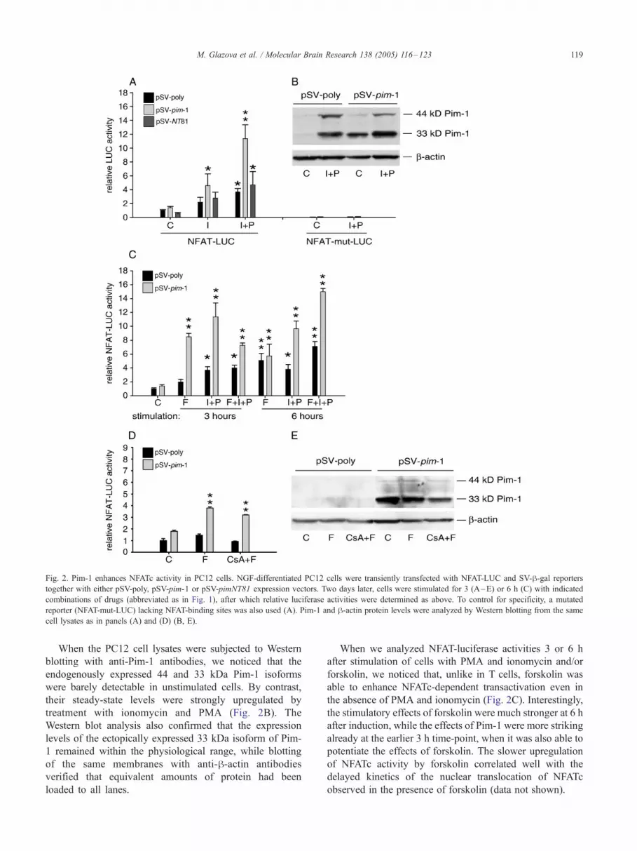
Fig. 2. Pim-1 enhances NFATc activity in PC12 cells. NGF-differentiated PC12 cells were transiently transfected with NFAT-LUC and SV-h-gal reporterstogether with either pSV-poly, pSV-pim-1 or pSV-pimNT81 expression vectors. Two days later, cells were stimulated for 3 (A–E) or 6 h (C) with indicated
combinations of drugs (abbreviated as in Fig. 1), after which relative luciferase activities were determined as above. To control for specificity, a mutated
reporter (NFAT-mut-LUC) lacking NFAT-binding sites was also used (A). Pim-1 and h-actin protein levels were analyzed by Western blotting from the same
cell lysates as in panels (A) and (D) (B, E).
M. Glazova et al. / Molecular Brain Research 138 (2005) 116–123 119
When the PC12 cell lysates were subjected to Western
blotting with anti-Pim-1 antibodies, we noticed that the
endogenously expressed 44 and 33 kDa Pim-1 isoforms
were barely detectable in unstimulated cells. By contrast,
their steady-state levels were strongly upregulated by
treatment with ionomycin and PMA (Fig. 2B). The
Western blot analysis also confirmed that the expression
levels of the ectopically expressed 33 kDa isoform of Pim-
1 remained within the physiological range, while blotting
of the same membranes with anti-h-actin antibodies
verified that equivalent amounts of protein had been
loaded to all lanes.
When we analyzed NFAT-luciferase activities 3 or 6 h
after stimulation of cells with PMA and ionomycin and/or
forskolin, we noticed that, unlike in T cells, forskolin was
able to enhance NFATc-dependent transactivation even in
the absence of PMA and ionomycin (Fig. 2C). Interestingly,
the stimulatory effects of forskolin were much stronger at 6 h
after induction, while the effects of Pim-1 were more striking
already at the earlier 3 h time-point, when it was also able to
potentiate the effects of forskolin. The slower upregulation
of NFATc activity by forskolin correlated well with the
delayed kinetics of the nuclear translocation of NFATc
observed in the presence of forskolin (data not shown).
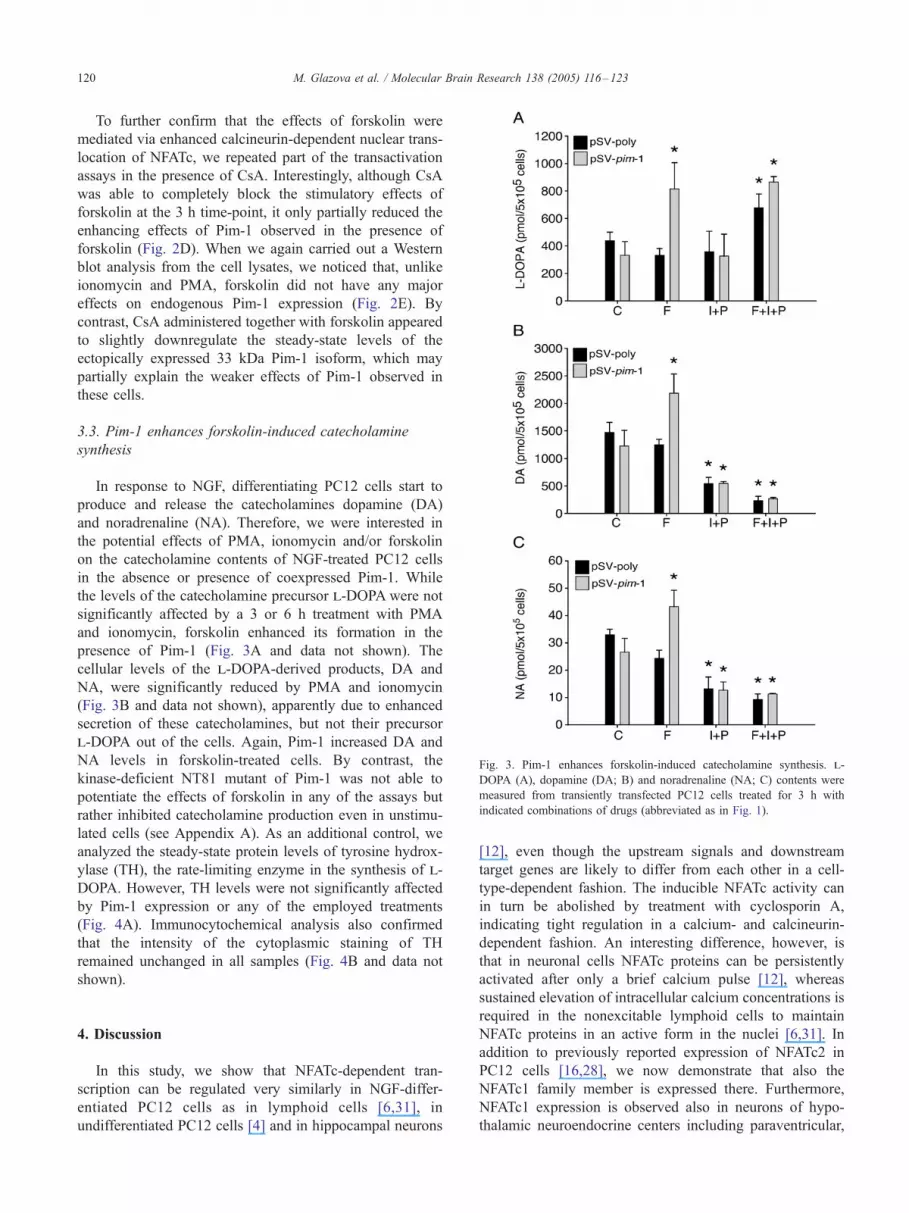
Fig. 3. Pim-1 enhances forskolin-induced catecholamine synthesis. l-
DOPA (A), dopamine (DA; B) and noradrenaline (NA; C) contents were
measured from transiently transfected PC12 cells treated for 3 h with
indicated combinations of drugs (abbreviated as in Fig. 1).
M. Glazova et al. / Molecular Brain Research 138 (2005) 116–123120
To further confirm that the effects of forskolin were
mediated via enhanced calcineurin-dependent nuclear trans-
location of NFATc, we repeated part of the transactivation
assays in the presence of CsA. Interestingly, although CsA
was able to completely block the stimulatory effects of
forskolin at the 3 h time-point, it only partially reduced the
enhancing effects of Pim-1 observed in the presence of
forskolin (Fig. 2D). When we again carried out a Western
blot analysis from the cell lysates, we noticed that, unlike
ionomycin and PMA, forskolin did not have any major
effects on endogenous Pim-1 expression (Fig. 2E). By
contrast, CsA administered together with forskolin appeared
to slightly downregulate the steady-state levels of the
ectopically expressed 33 kDa Pim-1 isoform, which may
partially explain the weaker effects of Pim-1 observed in
these cells.
3.3. Pim-1 enhances forskolin-induced catecholamine
synthesis
In response to NGF, differentiating PC12 cells start to
produce and release the catecholamines dopamine (DA)
and noradrenaline (NA). Therefore, we were interested in
the potential effects of PMA, ionomycin and/or forskolin
on the catecholamine contents of NGF-treated PC12 cells
in the absence or presence of coexpressed Pim-1. While
the levels of the catecholamine precursor l-DOPA were not
significantly affected by a 3 or 6 h treatment with PMA
and ionomycin, forskolin enhanced its formation in the
presence of Pim-1 (Fig. 3A and data not shown). The
cellular levels of the l-DOPA-derived products, DA and
NA, were significantly reduced by PMA and ionomycin
(Fig. 3B and data not shown), apparently due to enhanced
secretion of these catecholamines, but not their precursor
l-DOPA out of the cells. Again, Pim-1 increased DA and
NA levels in forskolin-treated cells. By contrast, the
kinase-deficient NT81 mutant of Pim-1 was not able to
potentiate the effects of forskolin in any of the assays but
rather inhibited catecholamine production even in unstimu-
lated cells (see Appendix A). As an additional control, we
analyzed the steady-state protein levels of tyrosine hydrox-
ylase (TH), the rate-limiting enzyme in the synthesis of l-
DOPA. However, TH levels were not significantly affected
by Pim-1 expression or any of the employed treatments
(Fig. 4A). Immunocytochemical analysis also confirmed
that the intensity of the cytoplasmic staining of TH
remained unchanged in all samples (Fig. 4B and data not
shown).
4. Discussion
In this study, we show that NFATc-dependent tran-
scription can be regulated very similarly in NGF-differ-
entiated PC12 cells as in lymphoid cells [6,31], in
undifferentiated PC12 cells [4] and in hippocampal neurons
[12], even though the upstream signals and downstream
target genes are likely to differ from each other in a cell-
type-dependent fashion. The inducible NFATc activity can
in turn be abolished by treatment with cyclosporin A,
indicating tight regulation in a calcium- and calcineurin-
dependent fashion. An interesting difference, however, is
that in neuronal cells NFATc proteins can be persistently
activated after only a brief calcium pulse [12], whereas
sustained elevation of intracellular calcium concentrations is
required in the nonexcitable lymphoid cells to maintain
NFATc proteins in an active form in the nuclei [6,31]. In
addition to previously reported expression of NFATc2 in
PC12 cells [16,28], we now demonstrate that also the
NFATc1 family member is expressed there. Furthermore,
NFATc1 expression is observed also in neurons of hypo-
thalamic neuroendocrine centers including paraventricular,
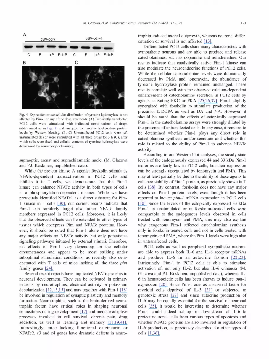
Fig. 4. Expression or subcellular distribution of tyrosine hydroxylase is not
affected by Pim-1 or any of the drug treatments. (A) Transiently transfected
PC12 cells were stimulated with indicated combinations of drugs
(abbreviated as in Fig. 1) and analyzed for tyrosine hydroxylase protein
levels by Western blotting. (B, C) Untransfected PC12 cells were left
unstimulated (B) or were stimulated with all three drugs for 3 h (C), after
which cells were fixed and cellular contents of tyrosine hydroxylase were
determined by immunocytochemistry.
M. Glazova et al. / Molecular Brain Research 138 (2005) 116–123 121
supraoptic, arcuat and suprachiasmatic nuclei (M. Glazova
and P.J. Koskinen, unpublished data).
While the protein kinase A agonist forskolin stimulates
NFATc-dependent transactivation in PC12 cells and
inhibits it in T cells, we demonstrate that the Pim-1
kinase can enhance NFATc activity in both types of cells
in a phosphorylation-dependent manner. While we have
previously identified NFATc1 as a direct substrate for Pim-
1 kinase in T cells [30], our current results indicate that
Pim-1 can similarly target also other NFATc family
members expressed in PC12 cells. Moreover, it is likely
that the observed effects can be extended to other types of
tissues which coexpress Pim and NFATc proteins. How-
ever, it should be noted that Pim-1 alone does not have
any major effects on NFATc activity but only potentiates
signaling pathways initiated by external stimuli. Therefore,
net effects of Pim-1 vary depending on the cellular
circumstances and appear to be most striking under
suboptimal stimulation conditions, as recently also dem-
onstrated with T cells of mice lacking all the three pim
family genes [24].
Several recent reports have implicated NFATc proteins in
neuronal development. They can be activated in primary
neurons by neurotrophins, electrical activity or potassium
depolarization [12,13,15] and may together with Pim-1 [18]
be involved in regulation of synaptic plasticity and memory
formation. Neurotrophins, such as the brain-derived neuro-
trophic factor, have critical roles in shaping neuronal
connections during development [17] and mediate adaptive
processes involved in cell survival, chronic pain, drug
addiction, as well as learning and memory [11,19,41].
Interestingly, mice lacking functional calcineurin or
NFATc2, c3 and c4 genes have dramatic defects in neuro-
trophin-induced axonal outgrowth, whereas neuronal differ-
entiation or survival is not affected [13].
Differentiated PC12 cells share many characteristics with
sympathetic neurons and are able to produce and release
catecholamines, such as dopamine and noradrenaline. Our
results indicate that catalytically active Pim-1 kinase can
also modulate the neuroendocrine functions of PC12 cells.
While the cellular catecholamine levels were dramatically
decreased by PMA and ionomycin, the abundance of
tyrosine hydroxylase protein remained unchanged. These
results correlate well with the observed calcium-dependent
enhancement of catecholamine secretion in PC12 cells by
agents activating PKC or PKA [25,26,37]. Pim-1 slightly
synergized with forskolin to stimulate production of the
precursor l-DOPA as well as DA and NA. However, it
should be noted that the effects of ectopically expressed
Pim-1 in the catecholamine assays were strongly diluted by
the presence of untransfected cells. In any case, it remains to
be determined whether Pim-1 plays any direct role in
catecholamine synthesis and/or secretion and whether this
role is related to the ability of Pim-1 to enhance NFATc
activity.
According to our Western blot analyses, the steady-state
levels of the endogenously expressed 44 and 33 kDa Pim-1
isoforms are fairly low in PC12 cells, but their expression
can be strongly upregulated by ionomycin and PMA. This
may at least partially be due to the ability of these agents to
enhance stability of Pim-1 protein, as previously shown in T
cells [38]. By contrast, forskolin does not have any major
effects on Pim-1 protein levels, even though it has been
reported to induce pim-1 mRNA expression in PC12 cells
[10]. Since the levels of the ectopically expressed 33 kDa
Pim-1 in unstimulated or in forskolin-treated cells were
comparable to the endogenous levels observed in cells
treated with ionomycin and PMA, this may also explain
why exogenous Pim-1 affected catecholamine synthesis
only in forskolin-treated cells and not in cells treated with
ionomycin and PMA, where the Pim-1 levels were high also
in untransfected cells.
PC12 cells as well as peripheral sympathetic neurons
are able to express both IL-6 and IL-6 receptor mRNAs
and produce IL-6 in an autocrine fashion [22,23].
Intriguingly, Pim-1 in PC12 cells is able to stimulate
activation of, not only IL-2, but also IL-6 enhancer (M.
Glazova and P.J. Koskinen, unpublished data), whereas IL-
6 in hematopoietic cells has been shown to induce pim-1
expression [20]. Since Pim-1 acts as a survival factor for
myeloid cells deprived of IL-3 [21] or subjected to
genotoxic stress [27] and since autocrine production of
IL-6 may be equally essential for the survival of neuronal
cells [35], it would be interesting to determine whether
Pim-1 could indeed act up- or downstream of IL-6 to
protect neuronal cells from various types of apoptosis and
whether NFATc proteins are also involved in regulation of
IL-6 production, as previously described for other types of
cells [1,36].
M. Glazova et al. / Molecular Brain Research 138 (2005) 116–123122
Taken together, our results indicate that the Pim-1 kinase
can enhance NFATc activity in a phosphorylation-dependent
manner and collaborate with agents activating the PKA
pathway to modulate the neuroendocrine functions of the
neuron-like PC12 cells and most likely also of primary
neuronal cells.
Acknowledgments
We thank G.R. Crabtree for reagents and K.L. Laine and
R. Pohjola for expert technical assistance. This work was
supported by the Academy of Finland (to PJK) and the
Center for International Mobility (to MG).
Appendix A. Supplementary data
Supplementary data associated with this article can be
found, in the online version, at doi:10.1016/j.molbrainres.
2005.04.003.
References
[1] K.L. Abbott, J.R. Loss, A.M. Robida, T.J. Murphy, Evidence that
Ga(q)-coupled receptor-induced interleukin-6 mRNA in vascular
smooth muscle cells involves the nuclear factor of activated T cells,
Mol. Pharmacol. 58 (2000) 946–953.
[2] T.L.T. Aho, J. Sandholm, K.J. Peltola, H.P. Mankonen, M. Lilly, P.J.
Koskinen, Pim-1 kinase promotes inactivation of the pro-apoptotic
Bad protein by phosphorylating it on the Ser112 gatekeeper site, FEBS
Lett. 571 (2004) 43–49.
[3] C.R. Beals, C.M. Sheridan, C.W. Turck, P. Gardner, G.R. Crabtree,
Nuclear export of NF-ATc enhanced by glycogen synthase kinase-3,
Science 275 (1997) 1930–1933.
[4] V. Boss, D.J. Talpade, T.J. Murphy, Induction of NFAT-mediated
transcription by Gq-coupled receptors in lymphoid and non-lymphoid
cells, J. Biol. Chem. 271 (1996) 10429–10432.
[5] C.W. Chow, M. Rincon, J. Cavanagh, M. Dickens, R.J. Davis, Nuclear
accumulation of NFAT4 opposed by the JNK signal transduction
pathway, Science 278 (1997) 1638–1641.
[6] G.R. Crabtree, Generic signals and specific outcomes: signaling
through Ca2+, calcineurin, and NF-AT, Cell 96 (1999) 611–614.
[7] F. Dautry, D. Weil, J. Yu, A. Dautry-Varsat, Regulation of pim and
myb mRNA accumulation by interleukin 2 and interleukin 3 in murine
hematopoietic cell lines, J. Biol. Chem. 263 (1988) 17615–17620.
[8] A. Eichmann, L. Yuan, C. Breant, K. Alitalo, P.J. Koskinen,
Developmental expression of Pim kinases suggests functions also
outside of the hematopoietic system, Oncogene 19 (2000)
1215–1224.
[9] I.J. Elenkov, R.L. Wilder, G.P. Chrousos, E.S. Vizi, The sym-
pathetic nerve—An integrative interface between two supersystems:
the brain and the immune system, Pharmacol. Rev. 52 (2000)
595–638.
[10] J.D. Feldman, L. Vician, M. Crispino, G. Tocco, M. Baudry, H.R.J.
Herschman, Seizure activity induces PIM-1 expression in brain,
Neurosci. Res. 53 (1998) 502–509.
[11] A. Ghosh, J. Carnahan, M.E. Greenberg, Requirement for BDNF in
activity-dependent survival of cortical neurons, Science 263 (1994)
1618–1623.
[12] I.A. Graef, P.G. Mermelstein, K. Stankunas, J.R. Neilson, K.
Deisseroth, R. Tsien, G.R. Crabtree, L-type calcium channels and
GSK-3 regulate the activity of NF-ATc4 in hippocampal neurons,
Nature 401 (1999) 703–708.
[13] I.A. Graef, F. Wang, F. Charron, L. Chen, J. Neilson, M. Tessier-
Lavigne, G.R. Crabtree, Neurotrophins and netrins require calcineur-
in/NFAT signaling to stimulate outgrowth of embryonic axons, Cell
113 (2003) 657–670.
[14] L.A. Greene, A.S. Tischler, Establishment of a noradrenergic clonal
line of rat adrenal pheochromocytoma cells which respond to nerve
growth factor, Proc. Natl. Acad. Sci. U. S. A. 73 (1976) 2424–2428.
[15] R.D. Groth, P.G. Mermelstein, Brain-derived neurotrophic factor
activation of NFAT (nuclear factor of activated T-cells)-dependent
transcription: a role for the transcription factor NFATc4 in neuro-
trophin-mediated gene expression, J. Neurosci. 23 (2003) 8125–8134.
[16] A.M. Ho, J. Jain, A. Rao, P.G. Hogan, Expression of the transcription
factor NFATp in a neuronal cell line and in the murine nervous system,
J. Biol. Chem. 269 (1994) 28181–28186.
[17] E.J. Huang, L.F. Reichardt, Neurotrophins: roles in neuronal develop-
ment and function, Annu. Rev. Neurosci. 24 (2001) 677–736.
[18] U. Konietzko, G. Kauselmann, J. Scafidi, U. Staubli, H. Mikkers, A.
Berns, M. Schweizer, R. Waltereit, D. Kuhl, Pim kinase expression is
induced by LTP stimulation and required for the consolidation of
enduring LTP, EMBO J. 18 (1999) 3359–3369.
[19] A. Kruttgen, J.C. Moller, J.V. Heymach Jr., E.M. Shooter, Neuro-
trophins induce release of neurotrophins by the regulated secretory
pathway, Proc. Natl. Acad. Sci. U. S. A. 95 (1998) 9614–9619.
[20] M. Lilly, T. Le, P. Holland, S.L. Hendrickson, Sustained expression
of the pim-1 kinase is specifically induced in myeloid cells by
cytokines whose receptors are structurally related, Oncogene 7
(1992) 727–732.
[21] M. Lilly, J. Sandholm, J.J. Cooper, P.J. Koskinen, A. Kraft, The PIM-1
serine kinase prolongs survival and inhibits apoptosis-related mito-
chondrial dysfunction in part through a bcl-2-dependent pathway,
Oncogene 18 (1999) 4022–4031.
[22] M.T. Liu, H.M. Huang, K.C. Jeng, S.C. Ou, J.S. Kuo, Induction of
cytokine genes and IL-1a by chemical hypoxia in PC12 cells, Life Sci.
67 (2000) 2147–2157.
[23] P. Marz, R.A. Gadient, U. Otten, Expression of interleukin-6 receptor
(IL-6R) and gp130 mRNA in PC12 cells and sympathetic neurons:
modulation by tumor necrosis factor alpha (TNF-alpha), Brain Res.
706 (1996) 71–79.
[24] H. Mikkers, M. Nawjin, J. Allen, C. Brouwers, E. Verhoeven, J.
Jonkers, A. Berns, Mice deficient for all PIM-kinases display reduced
body size and impaired responses to hematopoietic growth factors,
Mol. Cell. Biol. 24 (2004) 6104–6115.
[25] H. Oda, T. Murayama, Y. Nomura, Inhibition of protein kinase C-
dependent noradrenaline release by wortmannin in PC12 cells, Arch.
Biochem. Biophys. 337 (1997) 96–102.
[26] T. Ono, I. Matsuoka, S. Ohkubo, J. Kimura, H. Nakanishi, Effects of
YT-146 [2-(1-octynyl) adenosine], an adenosine A2A receptor agonist,
on cAMP production and noradrenaline release in PC12 cells, Jpn.
J. Pharmacol. 78 (1998) 269–277.
[27] T.J. Pircher, S. Zhao, J.N. Geiger, B. Joneja, D.M. Wojchowski, Pim-1
kinase protects hematopoietic FDC cells from genotoxin-induced
death, Oncogene 19 (2000) 3684–3692.
[28] S. Plyte, M. Boncristiano, E. Fattori, F. Galvagni, S.R. Paccani, M.B.
Majolini, S. Oliviero, G. Ciliberto, J.L. Telford, C.T. Baldari,
Identification and characterization of a novel nuclear factor of
activated T-cells-1 isoform expressed in mouse brain, J. Biol. Chem.
276 (2001) 14350–14358.
[29] C.M. Porter, M.A. Havens, N.A. Clipstone, Identification of amino
acid residues and protein kinases involved in the regulation of NFATc
subcellular localization, J. Biol. Chem. 275 (2000) 3543–3551.
[30] E.M. Rainio, J. Sandholm, P.J. Koskinen, Cutting edge: transcriptional
activity of NFATc1 is enhanced by the Pim-1 kinase, J. Immunol. 168
(2002) 1524–1527.
[31] A. Rao, C. Luo, P.G. Hogan, Transcription factors of the NFAT
M. Glazova et al. / Molecular Brain Research 138 (2005) 116–123 123
family: regulation and function, Annu. Rev. Immunol. 15 (1997)
707–747.
[32] C.J. Saris, J. Domen, A. Berns, The pim-1 oncogene encodes two
related protein–serine/threonine kinases by alternative initiation at
AUG and CUG, EMBO J. 10 (1991) 655–664.
[33] T. Satoh, S. Nakamura, T. Taga, T. Matsuda, T. Hirano, T. Kishimoto,
Y. Kaziro, Induction of neuronal differentiation in PC12 cells by B-
cells stimulatory factor 2/interleukin 6, Mol. Cell. Biol. 8 (1988)
3546–3549.
[34] B.S. Skalhegg, K. Tasken, Specificity in the cAMP/PKA signaling
pathway. Differential expression, regulation and subcellular local-
ization of subunits of PKA, Front. Biosci. 5 (2000) D678–D693.
[35] H. Umegaki, K. Yamada, M. Naito, T. Kameyama, A. Iguchi, T.
Nabeshima, Protective effect of interleukin-6 against the death of
PC12 cells caused by serum deprivation or by the addition of a
calcium ionophore, Biochem. Pharmacol. 52 (1996) 911–916.
[36] N.J. Van Wagoner, E.N. Benveniste, Interleukin-6 expression and
regulation in astrocytes, J. Neuroimmunol. 100 (1999) 124–139.
[37] R.H. Westerink, A.A. Klompmakers, H.G. Westenberg, H.P. Vijver-
berg, Signaling pathways involved in Ca2+- and Pb2+-induced
vesicular catecholamine release from rat PC12 cells, Brain Res. 957
(2002) 25–36.
[38] D. Wingett, D. Stone, W.C. Davis, N.S. Magnuson, Expression of the
pim-1 proto-oncogene: Differential inducibility between a/h- and g/y-T cells and B cells, Cell Immunol. 162 (1995) 123–130.
[39] D. Wingett, A. Long, D. Kelleher, N.S. Magnuson, pim-1 proto-
oncogene expression in anti-CD3-mediated T cell activation is
associated with protein kinase C activation and is independent of
Raf-1, J. Immunol. 156 (1996) 549–557.
[40] T.T. Yang, Q. Xiong, H. Enslen, R.J. Davis, C.W. Chow, Phosphor-
ylation of NFATc4 by p38 mitogen-activated protein kinases, Mol.
Cell. Biol. 22 (2002) 3892–3904.
[41] F. Zafra, B. Hengerer, J. Leibrock, H. Thoenen, D. Lindholm, Activity
dependent regulation of BDNF and NGF mRNAs in the rat hippo-
campus is mediated by non-NMDA glutamate receptors, EMBO J. 9
(1990) 3545–3550.
Copyright © 2022 FDOKUMEN