Annexin 1, Glucocorticoids, and the Neuroendocrine-Immune Interface
13
Annexin 1, Glucocorticoids, and the Neuroendocrine–Immune Interface JULIA C. BUCKINGHAM a , CHRISTOPHER D. JOHN a , EGLE SOLITO a , TANYA TIERNEY a , RODERICK J. FLOWER b , HELEN CHRISTIAN c , and JOHN MORRIS c a Division of Neuroscience and Mental Health, Imperial College London, Hammersmith Campus, Du Cane Road, London W12 0NN, UK b Department of Biochemical Pharmacology, The William Harvey Research Institute, London EC1M 6BQ, UK c Department of Human Anatomy and Genetics, The University of Oxford, South Parks Road, Oxford OX1 3QX, UK Abstract Annexin 1 (ANXA1) was originally identified as a mediator of the anti-inflammatory actions of glucocorticoids (GCs) in the host defense system. Subsequent work confirmed and extended these findings and also showed that the protein fulfills a wider brief and serves as a signaling intermediate in a number of systems. ANXA1 thus contributes to the regulation of processes as diverse as cell migration, cell growth and differentiation, apoptosis, vesicle fusion, lipid metabolism, and cytokine expression. Here we consider the role of ANXA1 in the neuroendocrine system, particularly the hypothalamo-pituitary-adrenocortical (HPA) axis. Evidence is presented that ANXA1 plays a critical role in effecting the negative feedback effects of GCs on the release of corticotrophin (ACTH) and its hypothalamic-releasing hormones and that it is particularly pertinent to the early-onset actions of the steroids that are mediated via a nongenomic mechanism. The paracrine/juxtacrine mode of ANXA1 action is discussed in detail, with particular reference to the significance of the secondary processing of ANXA1, the processes that control the intracellular and transmembrane trafficking of the protein of the molecule and the mechanism of ANXA1 action on its target cells. In addition, the role of ANXA1 in the perinatal programming of the HPA axis is discussed. Keywords annexin 1; HPA axis; glucocorticoids; cytokines INTRODUCTION Annexin 1 (ANXA1, formerly known as lipocortin 1) is a 37-kDa member of the annexin superfamily of Ca 2+ and phospholipid binding proteins. Members of this family show a high degree of structural homology, with each including four (or 8 in the case of ANXA6) repeated units of some 70 amino acids within the core and C-terminal regions; these units confer the Ca 2+ and phospholipid binding properties of the proteins. The N-terminal region of each family member is, however, unique and is considered to confer the biological specificity of the individual proteins. The basic structure of ANXA1 is shown in Figure 1. Address for correspondence: Julia C. Buckingham, Division of Neuroscience and Mental Health, Imperial College London, Hammersmith Campus, Du Cane Road, London W12 0NN. Voice: 44-208-383-8034; fax: 44-208-383-8032. e-mail: [email protected]. Europe PMC Funders Group Author Manuscript Ann N Y Acad Sci. Author manuscript; available in PMC 2007 April 25. Published in final edited form as: Ann N Y Acad Sci. 2006 November ; 1088: 396–409. doi:10.1196/annals.1366.002. Europe PMC Funders Author Manuscripts Europe PMC Funders Author Manuscripts
-
Upload
independent -
Category
Documents
-
view
5 -
download
0
Transcript of Annexin 1, Glucocorticoids, and the Neuroendocrine-Immune Interface

Annexin 1, Glucocorticoids, and the Neuroendocrine–ImmuneInterface
JULIA C. BUCKINGHAMa, CHRISTOPHER D. JOHNa, EGLE SOLITOa, TANYA TIERNEYa,RODERICK J. FLOWERb, HELEN CHRISTIANc, and JOHN MORRISc
aDivision of Neuroscience and Mental Health, Imperial College London, Hammersmith Campus,Du Cane Road, London W12 0NN, UKbDepartment of Biochemical Pharmacology, The William Harvey Research Institute, LondonEC1M 6BQ, UKcDepartment of Human Anatomy and Genetics, The University of Oxford, South Parks Road,Oxford OX1 3QX, UK
AbstractAnnexin 1 (ANXA1) was originally identified as a mediator of the anti-inflammatory actions ofglucocorticoids (GCs) in the host defense system. Subsequent work confirmed and extended thesefindings and also showed that the protein fulfills a wider brief and serves as a signalingintermediate in a number of systems. ANXA1 thus contributes to the regulation of processes asdiverse as cell migration, cell growth and differentiation, apoptosis, vesicle fusion, lipidmetabolism, and cytokine expression. Here we consider the role of ANXA1 in the neuroendocrinesystem, particularly the hypothalamo-pituitary-adrenocortical (HPA) axis. Evidence is presentedthat ANXA1 plays a critical role in effecting the negative feedback effects of GCs on the releaseof corticotrophin (ACTH) and its hypothalamic-releasing hormones and that it is particularlypertinent to the early-onset actions of the steroids that are mediated via a nongenomic mechanism.The paracrine/juxtacrine mode of ANXA1 action is discussed in detail, with particular reference tothe significance of the secondary processing of ANXA1, the processes that control theintracellular and transmembrane trafficking of the protein of the molecule and the mechanism ofANXA1 action on its target cells. In addition, the role of ANXA1 in the perinatal programming ofthe HPA axis is discussed.
Keywordsannexin 1; HPA axis; glucocorticoids; cytokines
INTRODUCTIONAnnexin 1 (ANXA1, formerly known as lipocortin 1) is a 37-kDa member of the annexinsuperfamily of Ca2+ and phospholipid binding proteins. Members of this family show a highdegree of structural homology, with each including four (or 8 in the case of ANXA6)repeated units of some 70 amino acids within the core and C-terminal regions; these unitsconfer the Ca2+ and phospholipid binding properties of the proteins. The N-terminal regionof each family member is, however, unique and is considered to confer the biologicalspecificity of the individual proteins. The basic structure of ANXA1 is shown in Figure 1.
Address for correspondence: Julia C. Buckingham, Division of Neuroscience and Mental Health, Imperial College London,Hammersmith Campus, Du Cane Road, London W12 0NN. Voice: 44-208-383-8034; fax: 44-208-383-8032. e-mail:[email protected].
Europe PMC Funders GroupAuthor ManuscriptAnn N Y Acad Sci. Author manuscript; available in PMC 2007 April 25.
Published in final edited form as:Ann N Y Acad Sci. 2006 November ; 1088: 396–409. doi:10.1196/annals.1366.002.
Europe PM
C Funders A
uthor Manuscripts
Europe PM
C Funders A
uthor Manuscripts
Crystallography studies indicate that the four repeated sequences are arranged around a pore,giving the protein a “doughnut” appearance. The N-terminal domain is embedded within thepore at low Ca2+ concentrations, but elevations in [Ca2+] expose this region and maythereby influence the biological activity of the protein.1 The gene encoding ANXA1 is wellcharacterized in rodents (rat and mouse) and man2-4 (Fig. 1). Although the gene ismultiexonic in structure, no spliced variants have been described to date. Sequence analysishas, however, revealed multiple potential sites for secondary processing, including tyrosine,serine, and threonine phosphorylation, acetylation, and lipidation. The protein may thus existin multiple forms.
ANXA1 was first identified as a glucocorticoid (GC)-inducible protein in rat peritonealmacrophages and was heralded as a potential mediator of the powerful anti-inflammatoryactions of these steroid hormones. Subsequent studies have confirmed that ANXA1 doesindeed contribute to the anti-inflammatory actions of GCs. These studies have also revealedthat the protein fulfills wider brief as in intra- and intercellular signaling molecule and that itis concerned with processes as diverse as cell growth and differentiation, apoptosis, vesiclefusion, endocytosis, exocytosis, and neuroprotection. Work in our laboratory has focused onthe role of ANXA1 in the neuroendocrine system, particularly in relation to the negativefeedback actions of GCs within the hypothalamo-pituitary-adrenocortical (HPA) axis.
EXPRESSION AND FUNCTION OF ANXA1 IN THE HPA AXISLocalization
ANXA1 is expressed in abundance in the anterior pituitary gland, where it is localizedspecifically to the S100-positive folliculostellate cells.5,6 It is also found in lesser amountsin the hypothalamus, hippocampus, and other parts of the brain.7 Within the hypothalamus,ANXA1 is concentrated in the median eminence and the supraoptic and paraventricularnuclei.5 It is particularly prevalent in the ependymal cells lining the third ventricle and inactivated glial cells but is not detectable in hypothalamic neurons.
Functional StudiesGCs act within the brain and pituitary gland to maintain the blood levels of GCs withinappropriate limits by modulating the secretion of corticotrophin (ACTH) and itshypothalamic-releasing hormones, corticotrophin-releasing hormone (CRH) and argininevasopressin (AVP). The actions of the steroids are complex and, in addition to acting atseveral different sites (e.g., pituitary gland, hypothalamus, hippocampus, ventral tegmentalarea), they also act via several different molecular mechanisms. GCs thus exert effects thatare slow in onset (>2 h) and are characterized by suppression of the genes encoding ACTH,CRH, and AVP. In addition, they exert more immediate effects (onset 15–60 min) thatinhibit the release of preformed hormone into the blood stream.
The early-onset actions of GCs within the HPA axis can be readily demonstrated in the ratand other species using in vivo and in vitro experimental systems. Thus, for example, therelease of ACTH and corticosterone induced by stressors, such as restraint, endotoxintreatment, or surgical intervention, is readily suppressed by administration of GCs 15 min–1h before the onset of stress. Similarly, in vitro, the stimulus-evoked release of ACTH andCRH/AVP from pituitary and hypothalamic tissue is inhibited by inclusion of GCs in themedium 15 min–1 h prior to addition of a secretagogue. Using these experimental models,we have identified an important role for ANXA1 in mediating the “early-onset” actions ofGC in both the pituitary gland and the hypothalamus. We have thus shown that theinhibitory effects of GCs on the stress-induced release of ACTH and corticosterone in vivoare mimicked by intracerebroventricular (i.c.v.) injection of human recombinant ANXA1(hrANXA1) or peptides derived from its N terminus and antagonized specifically by central
BUCKINGHAM et al. Page 2
Ann N Y Acad Sci. Author manuscript; available in PMC 2007 April 25.
Europe PM
C Funders A
uthor Manuscripts
Europe PM
C Funders A
uthor Manuscripts

(i.c.v.) or peripheral subcutaneous (s.c.) administration of neutralizing anti-ANXA1 antisera(Fig. 2). Similarly in vitro, hrANXA1 mimics the inhibitory effects of GCs on the release ofCRH and ACTH from rodent (rat or mouse) hypothalamic and pituitary tissue, respectively.Furthermore, the effects of the steroids are specifically reversed by neutralizing anti-ANXA1 antisera or antisense oligodeoxynucleotides (ODNs) directed specifically againstANXA1 mRNA.8-10 Interestingly, in vivo, ANXA1 appears to play a particularly importantrole in modulating the HPA responses to inflammatory stimuli.10,11 Similarly, at thehypothalamic level, ANXA1 is particularly effective in suppressing the CRH responses toproinflammatory cytokines in vitro.11
MECHANISM OF ANNEXIN 1 ACTION WITHIN THE HPA AXISThe Paracrine/Juxtacrine Hypothesis
Studies involving cell fractionation techniques and electron microscopicimmunohistochemistry have shown that ANXA1 is distributed between three cellularcompartments, namely, the cytoplasm, within the membranes of cell organelles, and inassociation with the plasma membrane, where a proportion of the protein is localized to theouter cell surface.6,7 Early work in our laboratory revealed that GCs regulate both thesynthesis and the cellular disposition of ANXA1 in the anterior pituitary gland and thehypothalamus.9,11 GCs thus induce de novo ANXA1 synthesis in these tissues and causethe translocation of the protein from the cytoplasm to the outer cell membrane, where it isretained by a Ca2+-dependent mechanism. The transfer of cytoplasmic ANXA1 to the cellsurface is apparent within 15 min of GC contact both in vivo and in vitro. By contrast, theinduction of ANXA1 synthesis is slow in onset (>2 h) and appears to serve primarily as amechanism for replenishing the diminished cytoplasmic pool of the protein.7
The findings that ANXA1 is not expressed by CRH/AVP neurons or corticotrophs, but ispresent in the nonendocrine cells adjacent to them, together with evidence that the protein ispromptly translocated to the cell surface by GCs, led us to propose that ANXA1 acts viaparacrine/juxtacrine mechanism to mediate the inhibitory effects of the steroids on CRH/AVP and ACTH release. Several lines of evidence support this hypothesis. Firstly, specific,high-affinity (Kd = 14 ± 3 nM), proteinaceous ANXA1 binding sites are expressed on thecell surface of the corticotrophs and other endocrine cells in the anterior pituitary gland.12Secondly, enzymatic destruction of these sites ablates ANXA1 binding and the antisecretoryeffects of both GCs and ANXA1 on ACTH secretion in primary cultures of rat pituitarytissue, suggesting that these sites play an important role in mediating ANXA1 actions.12Thirdly, immunogold labeling has revealed that ANXA1 release occurs at focal points onthe FS cell membranes, particularly at loci where the stellar projections of the cells makeclose contact with the endocrine cells13 (Fig. 3). Finally, drugs that prevent the GC-inducedtranslocation of ANXA1 to the cell membrane (e.g., PKC inhibitors, ATP-sensitive K+
channel openers) overcome the acute inhibitory effects of GCs on ACTH secretion in invitro models.14,15
Confirmation that ANXA1 serves as cell–cell mediator of GC action in the pituitary glandwas provided by studies on a co-culture system comprising two cell lines, viz., murinecorticotroph (AtT20, clone D1) and murine FS (TtTGF). TtT/GF cells express ANXA1 inabundance and readily translocate the protein to the cell surface in response to a GCchallenge.16 By contrast, while specific ANXA1 binding sites are readily detected on thecell surface of the AtT20 D1 cells by FACS analysis, the cell line does not express ANXA1mRNA or protein as detected by RT-PCR and Western blot analysis. These two cell linesthus mirror the pattern of expression of ANXA1 and ANXA1 binding observed in vivo and,thus, provide a useful model for exploration of ANXA1 biology. When cultured alone, theAtT20 D1 cells are unresponsive to GCs but respond readily to ANXA1 and peptides
BUCKINGHAM et al. Page 3
Ann N Y Acad Sci. Author manuscript; available in PMC 2007 April 25.
Europe PM
C Funders A
uthor Manuscripts
Europe PM
C Funders A
uthor Manuscripts

derived from it. The CRH-induced release of ACTH from the cells is thus unaffected by theinclusion of dexamethasone in the incubation medium, but is readily blocked by ANXA1and ANXA1Ac2–26. However, when co-cultured with TtT/GF cells, the AtT20 D1 cellsrespond to dexamethasone with a marked reduction in CRH-induced ACTH release;furthermore, the magnitude of the response to dexamethasone is proportional to the ratio ofTtT/GF to AtT20 D1 cells in culture (Fig. 4). The importance of ANXA1 in mediating thisaction of dexamethasone was confirmed by antisense studies that showed clearly that theeffects of the steroid were ablated by antisense ODNs directed against ANXA1 mRNA butnot by corresponding sense- or antisense sequences.16
Translocation of ANXA1 to the Cell SurfaceThe amino acid sequence of ANXA1 does not include a signal sequence. Consequently, theprotein is not packaged into vesicles by the Golgi system for release by exocytosis, and itstranslocation to the cell membrane is unaffected by drugs that interfere with various stepswithin the exocytotic pathway (e.g., nocodazole, brefeldin A).7 Early studies indicated thatthe association of ANXA1 with membranes is strongly influenced by the phosphorylationstatus of the molecule.17 Accordingly, we examined the potential role of phosphorylation inthe GC-induced translocation of ANXA1 to cell membrane. Our initial studies indicated thatcell surface ANXA1 is phosphorylated on both serine and tyrosine residues.14,18 However,while blockade of PKC-dependent serine phosphorylation prevents the GC-inducedtranslocation of ANXA1 to the cell surface of primary FS cells,14 inhibition of tyrosinephosphorylation does not.18 Together these data suggest that serine but not tyrosinephosphorylation is critical to the GC-induced transfer of ANXA1 across the cell membrane.Further evidence to support a role for serine phosphorylation emerged from studies on ahuman FS cell line (PDFS). Work on this line also demonstrated that the actions of GCs aremediated via the glucocorticoid receptor but that downstream signaling is effected by anongenomic mechanism that uses PI3 kinase and MAP kinase as well as PKC.19 TheANXA1 sequence includes multiple serine phosphorylation sites; and we are thereforeexploiting site-directed mutagenesis and green fluorescent protein (GFP) tagging todetermine the relative importance of each. Our initial data suggest that serine-27 isimportant in this regard, but other sites, particularly serine-5, may also have a significantrole. In addition, they have identified a potential role for protein lipidation in the signalingcascade.20
The mechanism by which phosphorylated ANXA1 traverses the membrane is poorlyunderstood. Double-labeling immunohistochemistry has shown that ANXA1 co-localizeswith an ATP binding cassette (ABC) transporter protein (ABCA1) at focal points in themembrane of primary and transformed (TtT/GF) FS cells, thus raising the possibility thatthis protein may be important in this regard.21 This hypothesis was supported by theevidence that glyburide, which blocks ABCA1, inhibits not only the GC-induced exportationof ANXA1 in FS cells and primary pituitary tissue, but also the ANXA1-dependentinhibitory effects of dexamethasone on ACTH release from pituitary tissue.21 However,doubts regarding the role of ABCA1 have emerged, as Tangiers lymphocytes, which lackthe ability to traffic ABCA1 to the cell membrane, readily export ANXA1 in response to asteroid challenge. This unexpected finding could be explained by the development of acompensatory mechanism. However, glyburide is not specific to ABCA1, but also opensATP-sensitive K+ channels (K+
ATP channels). These channels are expressed by FS cells.Moreover, the K+
ATP channel blockers, chromokalim and minoxidil, induce thetranslocation of ANXA1 to the cell surface of TtT/GF cells in a concentration-dependentmanner; furthermore, their effects are blocked by glyburide and other K+
ATP channelopeners. Thus, while the role of ABCA1 is uncertain, it seems likely that K+
ATP channelscontribute to the signaling mechanism involved in the membrane translocation of ANXA1.
BUCKINGHAM et al. Page 4
Ann N Y Acad Sci. Author manuscript; available in PMC 2007 April 25.
Europe PM
C Funders A
uthor Manuscripts
Europe PM
C Funders A
uthor Manuscripts
Actions of ANXA1 on Endocrine CellsAs discussed above, there is good evidence that ANXA1 acts via specific membrane-boundreceptors on the corticotrophs and other pituitary endocrine cells to suppress pituitaryhormone release.12 The nature of these receptors is ill-defined, but data from other systemshave raised the possibility that formyl peptide receptors (FPRs) may be important in thisregard.22 In humans, three members of this G-protein-coupled family of receptors havebeen characterized, namely, FPR, FPR-L1, and FPR-L2. Furthermore, ANXA1 and certainpeptides derived from it have been shown to bind to both FPR and FPR-L1. However, inrodents the family is more extensive with eight or possibly nine family members. Themajority are poorly characterized, but Fpr1 and Fpr-rs1/Fpr-rs2 are mooted to be the murineequivalents of the human FPR and FPR-L1. We have recently shown that Fpr1 is notexpressed in the mouse or rat pituitary gland and that Fpr1 gene deletion does not affect thecapacity of ANXA1 or dexamethasone to suppress ACTH release from murine pituitarytissue. Other members of the FPR family are, however, found in the murine anteriorpituitary gland. Furthermore, the inhibitory effects of dexamethasone and ANXA1 onACTH release are mimicked by lipoxin A4, a lipid mediator purported to be an endogenousligand of FPR-L1, and by high concentrations of the bacterial peptide, formyl-methione-leucine-proline (fMLF), which shows low affinity for FPR-L1. These findings, together withevidence that the effects of GCs, ANXA1, fMLF, and lipoxin A4 on ACTH release areblocked by N-Boc-MLP, an analogue of fMLP, which shows antagonist activity at both FPRand FPR-L1, supports the premise that a member of the FPR family contributes to theregulatory actions of ANXA1 on ACTH release. The signaling mechanism downstream ofthe receptor is largely unexplored. However, other data suggest that ANXA1 disruptssignaling at a point distal to the formation of cyclic AMP and the entry of Ca2+ and that,while the protein readily suppresses adenylyl cyclase-dependent secretagogue signaling, itdoes not affect phospholipase C-driven exocytosis.8-10,14,23
The sequences within the ANXA1 molecule that are required for the inhibition of peptiderelease have been explored using peptide analogues.14 Removal of the third and fourthrepeat units of the core (ANXA11–188) has no effect on either the potency or the efficacy ofthe protein in the pituitary gland. However, additional loss of the second repeat and much ofthe first repeat (ANXA11–50) results in a significant loss of potency but not efficacy.ANXA1Ac2–26 shows a marked reduction in both potency and efficacy, and shorter N-terminal sequences (e.g., ANXA1Ac–12, ANXA1Ac13–26) are without effect.18 These datasupport the premise that the N-terminal plays an essential role in the manifestation ofANXA1 action, as also does our finding that neutralizing antisera directed specificallyagainst the N-terminal domain attenuate the inhibitory effects of GCs on ACTH release invivo (Fig. 2). However, they also reveal marked differences in the activity profile andpotency ratios of the peptides studied in this and other systems (e.g., models ofinflammation; see Fig. 5), where, for example, ANXA11–188 is less potent than the parentmolecule (ANXA11–346 and ANXA1Ac2–26 displays full efficacy.24 Taken together, thesedata raise the possibility that two or more receptor subtypes mediate the actions of ANXA1in the body, a view that is consistent with a role for two or more members of the FPRfamily.
EARLY LIFE PROGRAMMING OF THE ANXA1 SYSTEMThe ontogeny of ANXA1 expression in the developing fetus/neonate has not been studied indetail. However, striking tissue-specific changes in ANXA1 expression occur in the neonateat the time of the stress hyporesponsive period (SHRP). In particular, ANXA1 expression inthe anterior pituitary gland and hippocampus is raised (vs. adult) while thymic ANXA1 isreduced.25 Complementary functional experiments suggest that the upregulation of ANXA1in the neuroendocrine system may be an important factor in the maintenance of the
BUCKINGHAM et al. Page 5
Ann N Y Acad Sci. Author manuscript; available in PMC 2007 April 25.
Europe PM
C Funders A
uthor Manuscripts
Europe PM
C Funders A
uthor Manuscripts

quiescent state of the HPA axis during this period. ANXA1 may thus serve to protect thedeveloping organism from the potentially harmful effects of overexposure to GCs at thistime.25 The regulatory effects of ANXA1 on AVP from the hypothalamus appear to beparticularly important in this regard as, in addition to facilitating the inhibitory actions ofGCs in the release of CRH/AVP and ACTH from the hypothalamus and pituitary gland,respectively, ANXA1 exerts a tonic inhibitory effect on AVP release at this stage andthereby quenches that stimulatory effects of interleukin IL-1α, IL-1β, and IL-6 on therelease of the peptide.25
Interestingly, administration of very low doses of dexamethasone to dams via the drinkingwater in late gestation (E16–19) or post partum (day 1–7) causes tissue-specific changes inANXA1 expression in the brain and pituitary gland of the offspring that persist intoadulthood.26 Furthermore, ANXA1-dependent GC feedback is blunted in animals exposedto exogenous GCs in the perinatal period, apparently because the GC-induced translocationof ANXA1 to cell surface is compromised.26 These and other (unpublished) data raise thepossibility that ANXA1 contributes to the disturbances in HPA function and associatedpathologies that are evident in adults rats treated with GCs at critical stages of development.
CONCLUSIONSSince its discovery in 1979, ANXA1 has been shown to play a diverse role as a signalingmolecule in a number of physiological and pathophysiological systems, including theneuroendocrine system as discussed in this article. Our data suggest that ANXA1 acts via aparacrine/juxtacrine mechanism to suppress the release of ACTH and its hypothalamic-releasing hormones (Fig. 6). New technologies now pave the way for important new insightsto the mechanisms which regulate the secondary processing and trafficking of ANXA1, tothe physiological significance of these processes, and to the molecular mechanisms ofANXA1 action. The knowledge that emerges from these studies should provide new insightinto the role of this protein in health and disease and may ultimately lead to the therapeuticexploitation of ANXA1-mimietics.
AcknowledgmentsOur work on ANXA1 has been generously supported by the Wellcome Trust, BBSRC, MRC, GSK, and the Societyfor Endocrinology.
REFERENCES1. Rosengarth A, Luecke H. A calcium-driven conformational switch of the N-terminal and core
domains of annexin A1. J. Mol. Biol. 2003; 326:1317–1325. [PubMed: 12595246]
2. Horlick KR, et al. Mouse lipocortin I gene structure and chromosomal assignent: gene duplicationand the origins of a gene family. Genomics. 1991; 10:365–374. [PubMed: 1676980]
3. Kovacic RT, et al. Correlation of gene and protein structure of rat and human lipocortin I.Biochemistry. 1991; 30:9015–9021. [PubMed: 1832554]
4. Wallner BP, et al. Cloning and expression of human lipocortin, a phospholipase A2 inhibitor withpotential anti-inflammatory activity. Nature. 1986; 320:77–81. [PubMed: 2936963]
5. Smith T, Flower RJ, Buckingham JC. Lipocortins 1, 2 and 5 in the central nervous system andpituitary gland of the rat: selective induction by dexamethasone of lipocortin 1 in the anteriorpituitary gland. Mol. Neuropharmacol. 1993; 3:45–55.
6. Traverso V, et al. Lipocortin 1 (annexin 1): a candidate paracrine agent localized in pituitaryfolliculo-stellate cells. Endocrinology. 1999; 140:4330–4338.
7. Philip JG, Flower RJ, Buckingham JC. Glucocorticoids modulate the cellular disposition oflipocortin 1 in the rat brain in vivo and in vitro. Neuroreport. 1997; 8:1871–1876. [PubMed:9223068]
BUCKINGHAM et al. Page 6
Ann N Y Acad Sci. Author manuscript; available in PMC 2007 April 25.
Europe PM
C Funders A
uthor Manuscripts
Europe PM
C Funders A
uthor Manuscripts

8. Taylor AD, et al. An antisense oligodeoxynucleotide to lipocortin 1 reverses the inhibitory actionsof dexamethasone on the release of adrenocorticotropin from rat pituitary tissue in vitro.Endocrinology. 1997; 138:2909–2918. [PubMed: 9202235]
9. Taylor AD, et al. Lipocortin 1 mediates an early inhibitory action of glucocorticoids on the secretionof ACTH by the rat anterior pituitary gland in vitro. Neuroendocrinology. 1993; 58:430–439.[PubMed: 7506818]
10. Taylor AD, Flower RJ, Buckingham JC. Dexamethasone inhibits the release of TSH from the ratanterior pituitary gland in vitro by mechanisms dependent on de novo protein synthesis andlipocortin 1. J. Endocrinol. 1995; 147:533–544. [PubMed: 8543924]
11. Loxley HD, et al. Modulation of the hypothalamo-pituitary-adrenocortical responses to cytokinesin the rat by lipocortin 1 and glucocorticoids: a role for lipocortin 1 in the feedback inhibition ofCRF-41 release? Neuroendocrinology. 1993; 57:801–814. [PubMed: 8413817]
12. Christian HC, et al. Characterization and localization of lipocortin 1-binding sites on rat anteriorpituitary cells by fluorescence-activated cell analysis/sorting and electron microscopy.Endocrinology. 1997; 138:5341–5351. [PubMed: 9389519]
13. Chapman LP, et al. Externalization of annexin I from a folliculo-stellate-like cell line.Endocrinology. 2002; 147:4330–4338. [PubMed: 12399429]
14. John C, et al. Annexin 1-dependent actions of glucocorticoids in the anterior pituitary gland: rolesof the N-terminal domain and protein kinase C. Endocrinology. 2002; 143:3060–3070. [PubMed:12130572]
15. Payne JP, et al. Modulators of the SUR2B/Kir6.1 ATP-sensitive K+ channel regulate annexin 1release in the TtT/GF folliculostellate cell line. Endocrine Abstracts. 2005; 9:OC22.
16. Tierney T, et al. Evidence from studies on co-cultures of TtT/GF and AtT20 cells that Annexin 1acts as a paracrine or juxtacrine mediator of the early inhibitory effects of glucocorticoids onACTH release. J. Neuroendocrinol. 2003; 15:1134–1143. [PubMed: 14636175]
17. Creutz CE, et al. Identification of chromaffin granule-binding proteins. Relationship of thechromobindins to calelectrin, synhibin, and the tyrosine kinase substrates p35 and p36. J. Biol.Chem. 1987; 262:1860–1868. [PubMed: 2948960]
18. John CD, et al. Kinase-dependent regulation of the secretion of thyrotrophin and luteinizinghormone by glucocorticoids and annexin 1 peptides. J. Neuroendocrinol. 2003; 15:946–957.[PubMed: 12969239]
19. Solito E, et al. Dexamethasone induces rapid serine-phosphorylation and membrane translocationof annexin 1 in a human folliculostellate cell line via a novel nongenomic mechanism involvingthe glucocorticoid receptor, protein kinase C, phosphatidylinositol 3-kinase, and mitogen-activatedprotein kinase. Endocrinology. 2003; 144:1164–1174. [PubMed: 12639897]
20. Solito E, et al. Post translational modification plays an essential role in the translocation of annexinA1 from the cytoplasm to the cell surface. FASEB J. 2006; 20:1498–1500. [PubMed: 16720734]
21. Chapman LP, et al. Evidence for a role of the ATP-binding cassette transporter A1 (ABCA1) in theexternalisation of annexin 1 from pituitary folliculostellate cells. Endocrinology. 2003; 144:1062–1073. [PubMed: 12586783]
22. Gerke V, Moss SE. Annexins: from structure to function. Physiol. Rev. 2002; 82:331–371.[PubMed: 11917092]
23. Taylor AD, et al. Annexin 1 (lipocortin 1) mediates the glucocorticoid inhibition of cyclicadenosine 3′,5′-monophosphate-stimulated prolactin secretion. Endocrinology. 2000; 141:2209–2219. [PubMed: 10830310]
24. John CD, et al. Annexin 1 and the regulation of endocrine function. Trends Endocrinol. Metab.2004; 15:103–109. [PubMed: 15046738]
25. Buckingham JC, Flower RJ. Lipocortin-1: a second messenger of glucocorticoid action in thehypothalamo-pituitary-adrenocortical axis. Mol. Med. Today. 1997; 3:196–302.
26. Theogaraj E, et al. Perinatal glucocorticoid treatment produces molecular, functional, andmorphological changes in the anterior pituitary gland of the adult male rat. Endocrinology. 2005;146:4804–4813. [PubMed: 16099861]
BUCKINGHAM et al. Page 7
Ann N Y Acad Sci. Author manuscript; available in PMC 2007 April 25.
Europe PM
C Funders A
uthor Manuscripts
Europe PM
C Funders A
uthor Manuscripts
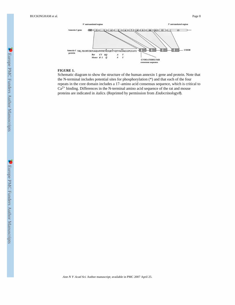
FIGURE 1.Schematic diagram to show the structure of the human annexin 1 gene and protein. Note thatthe N-terminal includes potential sites for phosphorylation (*) and that each of the fourrepeats in the core domain includes a 17–amino acid consensus sequence, which is critical toCa2+ binding. Differences in the N-terminal amino acid sequence of the rat and mouseproteins are indicated in italics. (Reprinted by permission from Endocrinology8).
BUCKINGHAM et al. Page 8
Ann N Y Acad Sci. Author manuscript; available in PMC 2007 April 25.
Europe PM
C Funders A
uthor Manuscripts
Europe PM
C Funders A
uthor Manuscripts

FIGURE 2.Reversal of the inhibitory effects of corticosterone on the IL-1β-stimulated release of ACTHin adult male rats by administration of an antiannexin 1 polyclonal antiserum raised in sheepagainst ANXA1Ac2–26. Corticosterone 500 μg/kg, i.p in a volume of 1 mL/kg; IL-1β 10 ng/rat in a volume of 3 μL, i.c.v. Controls received equivalent volumes of the sterile saline(Sal) vehicle. Antiannexin 1 antiserum (anti-ANXA1 pAb) 1mL/kg, s.c. or nonimmunesheep serum (NSS, control) 1mL/kg, s.c. was administered 15 min before the steroid. Valuesrepresent mean ± SEM (n = 6–7). **P < 0.01 versus Sal–Sal treated control; †† P < 0.01versus Sal–IL-1-β treated group; NS = not significant (P > 0.05). (ANOVA plus Scheffé'stest).
BUCKINGHAM et al. Page 9
Ann N Y Acad Sci. Author manuscript; available in PMC 2007 April 25.
Europe PM
C Funders A
uthor Manuscripts
Europe PM
C Funders A
uthor Manuscripts

FIGURE 3.Electron micrograph showing immunogold detection of annexin 1 in a folliculostellate celladjacent to three endocrine (E) cells in freeze-substituted mouse anterior pituitary tissue.Gold particles (15 nm) are scattered over the cytoplasm and adjacent to the plasmamembrane of the cell; they are also localized on the FS cell surface at the ends of theprocesses contacting endocrine cells (see enlarged inset). Arrows indicate intercellularjunctions. Scale bar: 1 μm. (Reprinted by permission from Endocrinology13).
BUCKINGHAM et al. Page 10
Ann N Y Acad Sci. Author manuscript; available in PMC 2007 April 25.
Europe PM
C Funders A
uthor Manuscripts
Europe PM
C Funders A
uthor Manuscripts

FIGURE 4.Effects of corticotrophin-releasing hormone (CRH 1 μm) and dexamethasone (Dex 100 nm)on the secretion of adrenocorticotrophic hormone (ACTH) by AtT20 D1 cells (200,000 perwell) cultured alone and in the presence of increasing numbers of TtT/GF cells (5,000–50,000 cells per well). (A) basal and CRH-stimulated release of ACTH. ***P < 0.001 basalversus CRH. †P < 0.05, ††P < 0.01 co-cultures versus AtT20 D1 cells alone. (B) The effectsof Dex on CRH-stimulated ACTH release; Dex had no effect on basal ACTH release (datanot shown). ***P < 0.001 Basal versus CRH. †††P < 0.001 CRH + Dex versuscorresponding group treated with CRH alone. Data shown are mean ± SEM of six wells pergroup. Statistical analysis was carried out by ANOVA and Tukey's post hoc test. The datashown are representative of three separate experiments. (Reprinted by permission from theJournal of Neuroendocrinology16).
BUCKINGHAM et al. Page 11
Ann N Y Acad Sci. Author manuscript; available in PMC 2007 April 25.
Europe PM
C Funders A
uthor Manuscripts
Europe PM
C Funders A
uthor Manuscripts

FIGURE 5.Comparison of the biological activity of full-length human recombinant annexin 1(hrANXA11–346), a truncated protein (ANXA11–188), and an N-terminal peptide(ANXA1Ac2–26) on (a) pituitary function and (b) in a model of inflammation. (A)demonstrates the inhibitory effects of graded concentrations of (i) ANXA11–346, (ii)ANXA11–188, and (iii) ANXA1Ac2–26 on the release of ir-ACTH from rat anterior pituitarysegments in in vitro data. Mean ± SEM (n = 6) are expressed as percentage of the secretoryresponse to forskolin alone (100 μM). *P < 0.05, **P < 0.01 versus forskolin alone group(ANOVA and Duncan's multiple range test). (B) demonstrates the effects of the peptides invivo on the IL-1 β-induced migration of neutrophils into a mouse air pouch migration(elicited by in vivo injection of IL-1-β into a small air pouch in the mouse). ANXA1 protein/peptides were co-injected with IL-1-β; the data are expressed as the mean ± SEM, n-4–10.Note (a) that in the pituitary gland, ANXA11–364 and ANXA11–188 are roughly equipotent,whereas in the air pouch model ANXA11–346 is approximately two orders of magnitudemore potent than ANXA11–188, (b) that ANXA1Ac2–26 shows the full efficacy of the parentprotein in the air pouch model but not in the pituitary gland, where at best it produces a 60%inhibition of peptide release and (c) that while ANXA1Ac2–26 is less potent than the parentprotein in both models, the potency difference is considerably greater in the pituitary gland.(Reprinted by permission from Trends in Endocrinology and Metabolism24).
BUCKINGHAM et al. Page 12
Ann N Y Acad Sci. Author manuscript; available in PMC 2007 April 25.
Europe PM
C Funders A
uthor Manuscripts
Europe PM
C Funders A
uthor Manuscripts
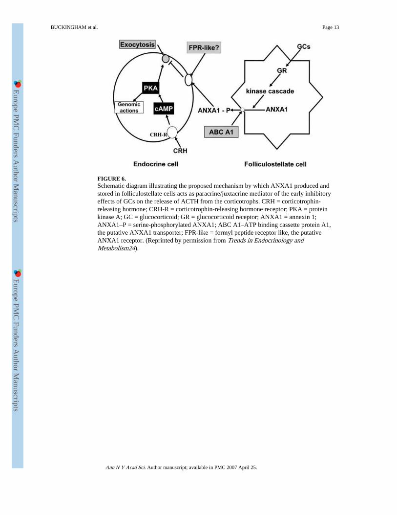
FIGURE 6.Schematic diagram illustrating the proposed mechanism by which ANXA1 produced andstored in folliculostellate cells acts as paracrine/juxtacrine mediator of the early inhibitoryeffects of GCs on the release of ACTH from the corticotrophs. CRH = corticotrophin-releasing hormone; CRH-R = corticotrophin-releasing hormone receptor; PKA = proteinkinase A; GC = glucocorticoid; GR = glucocorticoid receptor; ANXA1 = annexin 1;ANXA1–P = serine-phosphorylated ANXA1; ABC A1–ATP binding cassette protein A1,the putative ANXA1 transporter; FPR-like = formyl peptide receptor like, the putativeANXA1 receptor. (Reprinted by permission from Trends in Endocrinology andMetabolism24).
BUCKINGHAM et al. Page 13
Ann N Y Acad Sci. Author manuscript; available in PMC 2007 April 25.
Europe PM
C Funders A
uthor Manuscripts
Europe PM
C Funders A
uthor Manuscripts