Jak2 Is Necessary for Neuroendocrine Control of Female Reproduction
21
Jak2 is Necessary for Neuroendocrine Control of Female Reproduction Sheng Wu 1,4 , Sara Divall 1 , Gloria E. Hoffman 3 , Wei Wei Le 3 , Kay-Uwe Wagner 2 , and Andrew Wolfe 1 1 Department of Pediatrics, Johns Hopkins University School of Medicine, Baltimore, MD 2 Eppley Institute for Research in Cancer and Allied Diseases and Department of Pathology and Microbiology, University of Nebraska Medical Center, Omaha, Nebraska 3 Department of Biology, Morgan State University, Baltimore, MD Abstract GnRH neurons represent the final common output of signals from the brain that regulates reproductive function. A wide range of environmental factors impact GnRH neuron activity including disease, stress, nutrition, and seasonal cues, as well as gonadal steroid hormones. The CNS response is thought to be mediated, at least in part, through intermediate signaling molecules that affect GnRH neuronal activity. In vitro, GnRH neuronal cell lines respond to a variety of ligands which activate the Jak/STAT intracellular signaling pathway. In order to determine its biological function in reproduction, we used Cre/LoxP technology to generate GnRH neuron specific Jak2 conditional knockout (Jak2 G−/−) mice. GnRH mRNA levels were reduced in Jak2 G−/− mice when compared to controls, while the number of GnRH neurons was equivalent, indicating a reduction in GnRH gene expression. Secretion of GnRH is also reduced as basal serum LH levels were significantly lower in female Jak2 G−/− mice while the pituitary responded normally to exogenous GnRH. Preovulatory LH surge levels were blunted in Jak2 G−/− mice, which was correlated with reduced GnRH neuronal activation as assessed by c-Fos. However the activation of GnRH neurons following release from estrogen negative feedback is retained. Female Jak2 G−/− mice exhibited significantly delayed puberty and first estrus, abnormal estrous cyclicity and impaired fertility. These results demonstrate an essential role for Jak2 signaling in GnRH neurons for normal reproductive development and fertility in female mice. Keywords GnRH; Jak2; Cre/LoxP; fertility; estrous cycle; ovary Introduction Gonadotropin releasing hormone (GnRH) is the key regulator of reproduction and sexual behavior. GnRH travels via the portal vasculature to the anterior pituitary stimulating the synthesis and secretion of luteinizing hormone (LH) and follicle stimulating hormone (FSH) from the gonadotroph. LH and FSH are released into the circulation and stimulate the maturation and development of the gonads and the synthesis and secretion of the gonadal steroid hormones. GnRH neurons integrate signals that regulate reproduction; for example 4 Corresponding author and to whom reprint requests should be sent: Sheng Wu, Department of Pediatrics, Johns Hopkins University School of Medicine, Baltimore, MD, 21287 Phone: 410-614-0055; Fax: 410-502-7580; [email protected]. NIH Public Access Author Manuscript J Neurosci. Author manuscript; available in PMC 2011 July 5. Published in final edited form as: J Neurosci. 2011 January 5; 31(1): 184–192. doi:10.1523/JNEUROSCI.2974-10.2011. NIH-PA Author Manuscript NIH-PA Author Manuscript NIH-PA Author Manuscript
Transcript of Jak2 Is Necessary for Neuroendocrine Control of Female Reproduction

Jak2 is Necessary for Neuroendocrine Control of FemaleReproduction
Sheng Wu1,4, Sara Divall1, Gloria E. Hoffman3, Wei Wei Le3, Kay-Uwe Wagner2, andAndrew Wolfe11 Department of Pediatrics, Johns Hopkins University School of Medicine, Baltimore, MD2 Eppley Institute for Research in Cancer and Allied Diseases and Department of Pathology andMicrobiology, University of Nebraska Medical Center, Omaha, Nebraska3 Department of Biology, Morgan State University, Baltimore, MD
AbstractGnRH neurons represent the final common output of signals from the brain that regulatesreproductive function. A wide range of environmental factors impact GnRH neuron activityincluding disease, stress, nutrition, and seasonal cues, as well as gonadal steroid hormones. TheCNS response is thought to be mediated, at least in part, through intermediate signaling moleculesthat affect GnRH neuronal activity. In vitro, GnRH neuronal cell lines respond to a variety ofligands which activate the Jak/STAT intracellular signaling pathway. In order to determine itsbiological function in reproduction, we used Cre/LoxP technology to generate GnRH neuronspecific Jak2 conditional knockout (Jak2 G−/−) mice. GnRH mRNA levels were reduced in Jak2G−/− mice when compared to controls, while the number of GnRH neurons was equivalent,indicating a reduction in GnRH gene expression. Secretion of GnRH is also reduced as basalserum LH levels were significantly lower in female Jak2 G−/− mice while the pituitary respondednormally to exogenous GnRH. Preovulatory LH surge levels were blunted in Jak2 G−/− mice,which was correlated with reduced GnRH neuronal activation as assessed by c-Fos. However theactivation of GnRH neurons following release from estrogen negative feedback is retained.Female Jak2 G−/− mice exhibited significantly delayed puberty and first estrus, abnormal estrouscyclicity and impaired fertility. These results demonstrate an essential role for Jak2 signaling inGnRH neurons for normal reproductive development and fertility in female mice.
KeywordsGnRH; Jak2; Cre/LoxP; fertility; estrous cycle; ovary
IntroductionGonadotropin releasing hormone (GnRH) is the key regulator of reproduction and sexualbehavior. GnRH travels via the portal vasculature to the anterior pituitary stimulating thesynthesis and secretion of luteinizing hormone (LH) and follicle stimulating hormone (FSH)from the gonadotroph. LH and FSH are released into the circulation and stimulate thematuration and development of the gonads and the synthesis and secretion of the gonadalsteroid hormones. GnRH neurons integrate signals that regulate reproduction; for example
4Corresponding author and to whom reprint requests should be sent: Sheng Wu, Department of Pediatrics, Johns Hopkins UniversitySchool of Medicine, Baltimore, MD, 21287 Phone: 410-614-0055; Fax: 410-502-7580; [email protected].
NIH Public AccessAuthor ManuscriptJ Neurosci. Author manuscript; available in PMC 2011 July 5.
Published in final edited form as:J Neurosci. 2011 January 5; 31(1): 184–192. doi:10.1523/JNEUROSCI.2974-10.2011.
NIH
-PA Author Manuscript
NIH
-PA Author Manuscript
NIH
-PA Author Manuscript

nutrition, stress, developmental cues, and seasonal and circadian information which in turnregulate the expression and secretion of GnRH (Porkka-Heiskanen et al., 1997;Bucholtz etal., 2000;Nagatani et al., 1996;Pitteloud et al., 2007;Popa et al., 2008;Smith and Grove,2002). The cytokine family of hormones, growth factors and chemokines interact with thecytokine class of receptors and have been implicated in the central regulation ofreproduction (Gao et al., 2004;Bates et al., 2003;Stanley et al., 2000;Dozio et al.,2005;Watanobe and Yoneda, 2001;Argetsinger et al., 1993;Danilovich et al., 1999;Bartke etal., 1999;Shillingford et al., 2002), although the precise mechanisms of action are still beingexplored. Jak2, STAT3 and STAT5 are expressed in models of GnRH neurons, such as theGN11 and GT1-7 cells (Argetsinger et al., 1993;Magni et al., 2007), and activation of Jak2has been suggested to mediate the effects of cytokines such as LIF and CNTF through theirreceptors on GnRH neurons (Magni et al., 2007;Dozio et al., 2005).
The cytokine receptors lack intrinsic kinase activity and require interaction with members ofthe Janus-activated-kinase (Jak) family of intracellular signaling molecules. Jak2 belongs tothe non-receptor tyrosine kinase family (Ihle, 1995). It is the predominant Jak kinasemediating the responses of single chain cytokine receptors and plays an important role insignaling via the gp130 receptor family or the Class II cytokine receptors (Kisseleva et al.,2002). Receptor activation causes Jak2 autophosphorylation which phosphorylates itsassociated receptor. The phosphorylated receptor provides multiple docking sites that recruitSignal Transducers and Activator of Transcription (STAT) proteins of which there are sixfamily members (STAT1-6) (Pellegrini and Dusanter-Fourt, 1997). STAT is phosphorylatedby Jak2 resulting in dimerization and translocation to the nucleus where it acts as atranscription factor to regulate gene expression (Ihle, 1995;Parganas et al., 1998). Acomplete KO of the Jak2 gene in vivo has been produced and results in embryonic lethality(Parganas et al., 1998).
Since in vitro models provide evidence that Jak/STAT signaling may mediated importantsignals to the GnRH neurons, but in vivo evidence for a specific role for GnRH neuronalJak2 is lacking, therefore, we generated mice lacking Jak2 in GnRH neurons using the Cre/LoxP binary recombination system (Singh et al., 2009;Hamilton and Abremski, 1984) andreport here a role for the Jak2 signaling molecule in the regulation of reproductivedevelopment and function in female mice.
Materials and MethodsGeneration of GnRH neuron specific Jak2 conditional knockout (Jak2G−/−) mice
GnRH promoter driven Cre recombinase (GnRH-Cre) transgenic mice were created in ourlaboratory on a CD1 background (Wolfe et al., 2008). Jak2 fl/fl mice were produced aspreviously described and were in a 129SvJ strain (Krempler et al., 2004). Exon 2 of Jak2 isflanked by loxP sites. Excision of exon 2 results in complete disruption of Jak2 expression(Krempler et al., 2004). The GnRH-Cre; Jak2fl/fl conditional knockout (KO) mice,designated as Jak2 G−/−, were generated by first mating female (GnRH-Cre+) with male(Jak2fl/fl) and then crossing a heterozygous (GnRH-Cre+; Jak2fl/wt) female with aheterozygous (GnRH-Cre-; Jak2fl/wt) male to generate 6 genotype combinations. GnRH-Cre+; Jak2fl/fl mice represent the homozygous conditional KO mice. GnRH-Cre−; Jak2fl/fl
littermates were used as controls for all studies except for the mating and pubertyassessments, which also used GnRH-Cre-; Jak2wt/wt, GnRH-Cre−; Jak2fl/wt, and GnRH-Cre+; Jak2wt/wt littermates due to limited litter sizes of our early matings. No difference wasobserved among these genotypes.
Wu et al. Page 2
J Neurosci. Author manuscript; available in PMC 2011 July 5.
NIH
-PA Author Manuscript
NIH
-PA Author Manuscript
NIH
-PA Author Manuscript

Animal HousingMice were maintained with food and water ad libitum in a 14:10hr light:dark cycle at 24°Cin the Broadway Research Building animal facility at the Johns Hopkins University Schoolof Medicine. All procedures were approved by the Johns Hopkins University Animal Careand Use Committee.
Genotyping and DNA extractionFor genotyping, two pairs of primers were used: GnRH-Cre specific primers: sense 5′-GGTAGCTTCAGCTGTGAAAG-3′; antisense 5′-CATCTTCAGGTTCTGCGGGAAACC-3′, and Jak2fl/fl primers: sense 5′-ATTCTGAGATTCAGGTCTGAGC-3′; antisense 5′-CTCACAACCATCTGTATCTCAC-3′
To obtain genomic DNA of pups, a clipping from the ear or tail was collected from the miceand put into 10% chelex-100 resin (Bio-Rad, Hercules, CA) with 0.1% tween-20 and0.15mg/mL proteinase K. Samples were incubated at 50°C for 90 min, proteinase K wasinactivated at 95°C for 20 min, and the solution was cooled to 10°C. One μL of supernatantwas removed for PCR. The PCR reaction was as follows: 94°C for 3 min, 35 cycles with94°C for 30sec, 58°C for 30sec, 72°C for 30sec and last cycle at 72°C for 7 min. Foridentifying tissue-specific Jak2 allele recomination, different tissues were collectedincluding hypothalamus, cerebellum, cortex, pituitary, liver, heart, ovary and testes. DNAwas obtained from these tissues by phenol-chloroform extraction and isopropanolprecipitation. Primers used for detection of Jak2 G−/− : sense 5′-GTCTATACACCACCACTCCTG-3′ and antisense 5′-CGAGCTGGAAAGATAGGTCAGC-3′. The sequences of the primers related to Jak2 arethe same as in (Krempler et al., 2004). The locations of primers are labelled in Fig. 1A.
Puberty and fertility examinationFemale vaginal opening was assessed daily after 21days of life. Following vaginal opening,daily vaginal cytology was performed in the morning by collecting vaginal cells using 0.9%saline lavage. Cells were dried on slides, fixed in ethanol and stained with Diff-Quikstaining kit (IMEB INC, California). Estrous cycle staging was assessed using the methodpreviously described (Nelson et al., 1982). Proestrus was assigned as predominantly basaland cornified nucleated cells, estrus was assigned as predominantly cornified epithelial cells,metestrus was assigned as mixed cornified epithelial cells and leukocytes, and diestrus wasassigned as predominantly leukocytes.
Adult female (2.5 months old) were mated with proven fertile adult male mice for a periodof 90 days. Time to each litter and litter size for each pair was recorded.
Total RNA extraction, Reverse transcription and Real Time PCRHypothalamic RNA was extracted by Trizol (Invitrogen) according to the manufactor’sprotocol. One μg of total RNA was reverse transcribed (iScript cDNA Synthesis Kit,BioRad, Hercules, CA) to cDNA. For GnRH, Taqman quantitative PCR (BioresearchTechnologies, Novato, CA) was performed and GAPDH was used as the internal control.Primers for GnRH: sense 5′-CCAACGGAAGCTCGAGATCC-3′, and antisense 5′-TGCCGGCCATCAGTTTGAG-3′ with the probe5′TGACTTTCACATCCAAACAGAGTGGACA-3′ labeled with FAM and BHQ-1. Primersfor GAPDH: sense 5′-GGGCATCTTGGGCTACACT-3′ and antisense 5′-GGCATCGAAGGTGGAAGAGT-3′ with the probe 5′-AGGACCAGGTTGTCTCCTGCGA-3′ labeled with Cal fluoro red-610 and BHQ-2.
Wu et al. Page 3
J Neurosci. Author manuscript; available in PMC 2011 July 5.
NIH
-PA Author Manuscript
NIH
-PA Author Manuscript
NIH
-PA Author Manuscript

Reactions were performed using an iCycler iQ5 real time PCR machine (BioRad, CA). PCRconditions were optimized to generate >95% PCR efficiency and only those reactionsbetween 95% and 105% efficiency were included in subsequent analysis. Cycle threshold(Ct) was obtained for each sample. A corrected Ct (delta Ct) was calculated by subtractingthe GAPDH Ct from the unknown sample Ct for each sample. Relative differences from thecontrol sample were then calculated by using the formula: fold change=2^(control delta Ctminus sample delta Ct).
Hormonal assayBlood from female mice was obtained by mandibular vein puncture in the morning andvaginal lavage was performed at the same time for cytological assessment. Blood wascentrifuged at 4000×g for 15 min at 4°C and serum was collected and stored at −80°C untilneeded. LH and FSH were measured using a Milliplex MAP immunoassay (Rat Pituitarypanel; Millipore, Billerica, MA) in the Luminex200 (Austin, Texas). Analysis wasperformed using the XPonent 3.0 software program with Logistic 5D Weighted analyses.Further analysis details are described in (Singh et al., 2009). Estradiol was measured with anestradiol enzyme immunoassay kit (Cayman Chemical co. Ann Arbor, MI) according to themanufacturer’s directions.
The mouse surge protocol was adapted from (Christian et al., 2005) with modifications.Mice were ovariectomized (OVX) in the morning (day 0) and diluted 17-β estradiol(Cayman Chemical Company) filled alzet micro-osmotic pumps (DURECT™, model1007D) were inserted under the skin dorsally at the neck. 17-β estradiol was dissolved inethanol at a concentration of 750ng/μl. This solution was further diluted with PBS to a finalconcentration of 7.5ng/μl and injected into the mini-pump. Estradiol is released at 90ng/mlper day by the pump, with a 0.5μl per hour rate. On day 2, blood was taken (~50μl) fromeach individual OVX mouse by mandibular vein puncture at 10:00, 16:00 and one hourbefore lights off at 20:00. Intact control mice and their Jak2 G−/− litter mates were bled at20:00 for 7 consecutive days and vaginal lavage for estrous cycle assessment was performedat the same time.
Another group of mice treated as above were perfused at 20:00 and brains were preservedfor colocalization of GnRH and c-Fos.
HistologyOvaries were collected at metestrus or diestrus and fixed in 10% buffered formalinphosphate (Fisher Scientific) solution and stored at 4°C. Paraffin embedded ovaries weresectioned at 7 microns thickness (Phenotype Core Facility, Johns Hopkins University Schoolof Medicine). Ovarian sections were stained with hematoxylin and eosin, examined with aZeiss microscope and photographed with an AxioCamICc1 camera and exported toAxioVision Software.
Perfusion and immunostainingMice were anaesthetized with ketamine and xylazine, and perfused transcardially with 0.9%saline followed by 4% paraformaldehyde in 1xPBS. Brains were post fixed in 4%paraformaldehyde overnight and dehydrated in 30% sucrose for 24h or until they sank to thebottom. Brains were then embedded in OCT at −20°C and sections were cut coronally usinga cryostat (MICROM, HM550) beginning rostrally at the olfactory bulb and ending caudallyat the median eminence.
For the purpose of counting GnRH neurons, GnRH promoter driven GFP (GnRH-GFP) miceobtained from Dan Spergel (University of Chicago, (Spergel et al., 1999)) were used. Jak2 G
Wu et al. Page 4
J Neurosci. Author manuscript; available in PMC 2011 July 5.
NIH
-PA Author Manuscript
NIH
-PA Author Manuscript
NIH
-PA Author Manuscript

−/− mice were produced on the background of the GnRH-GFP mice. Forty micron sectionswere mounted onto slides with Vectashield (Vector laboratories). All GnRH neurons werecounted directly by GFP fluorescence by analyzing sequential section from the rostralolfactory tissue through the median eminence using a Zeiss microscope with X-Cite 120fluorescence Illumination System and photographed with an AxioCamMR camera andexported to AxioVision Software. All images were exposed for 1sec with the samebrightness and contrast parameters.
For the purpose of examining colocalization of GnRH and Jak2, brains of GnRH-GFP micewere coronally cut at 10 microns, mounted onto slides and stored at −20°C until needed.Slides were thawed at room temperature for 30 min and sections were washed 3 times with1xPBS for 5 min each. Sections were then permeablized with 0.25% Triton-100 in 1xPBSfor 6 min and washed 2 times with 1xPBS quickly and blocked with 8% goat serum in1xPBS for 1h at room temperature. Rabbit-anti Jak2 C-terminal polyclonal antibody (SantaCruz Biotechnology, CA) was diluted 1:100 in 0.02% Tween-20 of 1% BSA PBS. Sectionswere incubated with primary antibodies for 48h and washed 4–5 times with PBS for 10 mineach. Then sections were incubated with goat anti-rabbit IgG Alexa fluro-594 (Invitrogen) at1:400 dilution in 0.02% Tween-20 of 1% BSA PBS for 1.5 h. Sections were then washed 5times with 1xPBS for 10 min per wash. Sections were mounted with Vectashield andimaged as for GnRH-GFP neuronal counts.
For colocalizing c-Fos and GnRH, sections were cut at 30 microns and stored incryoprotectant antifreeze until use. Staining for c-Fos and GnRH used the same protocol andantibodies described in (Hoffman et al., 2005). Stained sections were photographed with anAxioCamICc1 camera and exported to AxioVision Software.
Statistical analysisAll data was analyzed by unpaired student’s t test using GraphPad Prism, (GraphPadSoftware, Inc. San Diego, California) and expressed as means ± S.E.M. P<0.05 were definedas statistically significant.
ResultsJak2 specific conditional knockout in GnRH neurons (Jak2 G−/−)
Floxed Jak2 mice with LoxP sites flanking exon 2 (Fig. 1A) were mated with GnRH-Cremice which express the Cre recombinase gene in GnRH neurons. As expected, the KO allele(400bp) was only found in the hypothalamus and not in other tissues of Jak2 G−/− mice(Fig. 1B). The Jak2fl/fl allele (320bp) was identified in all collected tissues from the samegenomic DNA pool that was used for the Jak2 G−/− PCR reaction (Fig. 1B). To confirmcell-specific deletion of Jak2 in GnRH neurons, immunostaining for Jak2 was performed on10μm brain sections. Fig. 1C shows GnRH expressing neurons localized in the vertical limbdiagonal band (VDB) in green, reflecting the expression of GFP under the direction of themurine GnRH promoter (top panel). Binding of the Jak2 antibody is visualized by redfluorescence (Alexa594; middle panel). Overlaying the images shows Jak2 colocalization inhypothalamic GnRH neurons of control GFP mice (bottom left panel) but no colocalizationin GnRH neurons of Jak2 G−/− GFP mice (bottom right panel). About 35% of the GnRHcell bodies colocalized with Jak2 in control mice examined from the rostral olfactory bulbthrough the median eminence. We found no regional differences in colocalization within thehypothalamus.
To investigate whether the absence of Jak2 signaling in GnRH neurons affects GnRHneuronal development and migration, the number of GnRH neurons and their distributionwere examined. There were a similar number of GnRH cell bodies (602.0±13.0 (control) vs
Wu et al. Page 5
J Neurosci. Author manuscript; available in PMC 2011 July 5.
NIH
-PA Author Manuscript
NIH
-PA Author Manuscript
NIH
-PA Author Manuscript

611.7±11.3 (Jak2 G−/−); Fig. 2A)) counted across the entire basal forebrain, and nosignificant difference in the number of GnRH cell bodies was observed in each successivelycounted region across the hypothalamus (Fig. 2B). A normal distribution of GnRH neuronswas observed between Jak2 G−/− and control groups (representative regions of thehypothalamus are shown in Figs. 2C-F for control and H-K for Jak2 G−/−). GnRH terminaldistribution at the median eminence was examined to determine whether Jak2 signalingcontributed to the axonal migration, a critical step in GnRH neuronal development, and wasnot found to differ between control and Jak2 G−/− mice (Figs 2G and L respectively).Interestingly, results from quantitative real-time PCR showed that hypothalamic GnRHmRNA was reduced by more than 50% of controls (P<0.001) (Fig. 2M).
Jak2 G−/− mice have reduced serum LHBasal serum LH levels from female Jak2 G−/− mice were significantly reduced whencompared to controls (0.10 ng/mL ±0.02, n=16 (Jak2 G−/−) vs 0.25 ng/mL ±0.06, n=15(control), P<0.05; Fig. 3A). In contrast, female basal FSH levels were similar between Jak2G−/− mice and control mice (26.34 ng/mL ±5.07, n=16 (Jak2 G−/−) vs 30.83 ng/mL ±5.17,n=15 (control); Fig. 3B). To confirm that this difference in serum LH levels was not due tochanges in pituitary responsiveness, a GnRH stimulation test was performed. The pituitariesfrom control and Jak2G−/− mice exhibited equal responses to 100ng/kg body weight GnRH(Fig. 3A, B). No increase in FSH was observed at 10 minutes following GnRH stimulationin either Jak2 G−/− or control mice.
Hypothalamic-pituitary gonadotropic activity is influenced by estrogen negative feedbackduring most of the estrous cycle. When LH and FSH levels were measured followingremoval of negative feedback by OVX there was no difference between Jak2 G−/− andcontrol litter mates (Fig. 3A, B).
Jak2 G−/− mice exhibit delayed pubertyVaginal opening has been shown to be an estrogen-dependent process and is used as anindicator of the onset of puberty (Safranski et al., 1993). The age of vaginal opening wassignificantly delayed in the Jak2 G−/− mice compared to control litter mates (29 day of life(dol) ± 1.11, n=14 (Jak2 G−/−) vs 24 dol ±1.9, n=20 (control), P<0.001; Fig. 4). The day offirst estrus implies progressive sexual maturation and is widely used as an indicator ofprogression through puberty (Safranski et al., 1993). Daily vaginal smears were obtained toidentify the age of first estrus. Jak2 G−/− mice exhibited a significant delay of first estruscompared to control litter mates (39.3 dol±1.6, n=7 (Jak2 G−/−) vs 33dol±1.1, n=7(control), P<0.01; Fig. 4). However, these data do not include the Jak2 G−/− mice that hadno first estrus when evaluated for up to 50 dol (2 of 9 examined).
Jak2 G−/− mice have abnormal estrous cycles and impaired ability to generate an LHsurge
Vaginal cytology was analyzed from adult female mice for 15 consecutive days anddemonstrated that Jak2 G−/− mice (n=8) exhibited irregular estrous cycles when comparedto their control litter mates (n=17) (Fig. 5A-C). During the 15 days of analysis only 50% ofJak2 G−/− mice had one complete cycle, compared to 95% in the control group.Furthermore, only 2 of 8 Jak2 G−/− mice (25%) had two complete cycles in the 15 dayperiod, whereas 82% of the control litter mates exhibited two complete cycles (Fig. 5A). Theduration spent in the proestrus phase in Jak2 G−/− mice was significantly shortenedcompared to control litter mates (Fig. 5B). Acyclic mice mainly remained predominantly inpersistent metestrus/diestrus (Fig. 5C).
Wu et al. Page 6
J Neurosci. Author manuscript; available in PMC 2011 July 5.
NIH
-PA Author Manuscript
NIH
-PA Author Manuscript
NIH
-PA Author Manuscript

The gonadotropic LH surge stimulates ovulation in female mice which is an essentialprocess in fertility and is induced by positive feedback of estradiol on the hypothalamus andpituitary. To explore whether positive feedback regulation is disrupted in Jak2 G−/− mice, asurge induction paradigm was used (Christian et al., 2005) that produces LH surgegeneration within an hour before lights off on day 2 of the paradigm. Chronic treatment withestradiol in OVX females results in levels of LH that were near the limit of detection in bothJak2 G−/− and control females at 10:00 and were elevated but not significantly different at16:00 (Data not shown). Surge LH levels in control mice at 20:00 on day 2 in our study werenearly identical to the levels observed by Christian et al. (Christian et al., 2005). However,LH levels were significantly blunted in Jak2 G−/− mice (Fig. 5D). The estrogen levels afterOVX+E replacement in all genotypes were found to be 29.5±7.9pg/ml n=7, which is similarto estrogen levels in proestrus (Wu et al., 2010;Christian et al., 2005;Nelson et al., 1982) andis nearly three-fold higher than the basal morning circulating levels in intact female mice(around 10pg/ml for both WT and Jak2 G−/− mice; data not shown).
While Jak2 G−/− mice frequently exhibit persistent diestrus a number of KO females wentthrough proestrus (Fig. 5A-C). To explore the magnitude of the LH surge in cycling females,evening blood samples were obtained in control and Jak2 G−/− mice during the estrouscycle and proestrus LH levels were found to be more than 6-fold lower in Jak2 G−/− micecompared to WT mice (Data not shown).
Jak2 G−/− mice demonstrated attenuated GnRH neuronal activation during the surgeIn order to determine whether changes in GnRH neuronal activation correlated with thereduced LH levels during the surge, c-Fos immunostaining in GnRH neurons was assessedby dual labeling immunohistochemistry after mice underwent the surge induction paradigm.All sections with immunostained GnRH neurons were counted and colocalization wasdocumented in two regions as has been described previously (Herbison et al.,2010;d’Anglemont, X et al., 2010). In both medial septum/diagonal band of Broca (MS/DBB) and organum vasculosum of the lamina terminalis/rostral preoptic area (OVLT/rPOA)regions we observed a significantly higher percentage of GnRH neurons with c-Fos incontrol mice than Jak2 G−/− mice (104% and 115% more dual labeled neurons for the MS/DBB and VOLT/rPOA, respectively; Fig. 6A-C).
Jak2 G−/− mice exhibit impaired fertilityFertility was also examined in control and Jak2 G−/− mice in a continuous mating protocol.Jak2 G−/− female mice demonstrated an impaired ability to produce offspring, as shown inthe mating paradigm in Fig. 7A. The birth of each litter is represented by a black dot andlitter sizes are indicated by numbers above each line for every successful mating event. Jak2G−/− mice exhibited decreased fertility compared to control littermates (Fig. 7A). None ofthe 8 Jak2 G−/− females had 4 litters during the 90 days mating period where 4 of the 6control mice had 4 litters in this period. Quantification of these data indicate that femaleJak2 G−/− mice bore their first litter significantly later after introduction to males thancontrol female mice (33.63 days ±6.71 (control), n=8 vs 22.00 days ±0.47, n=14 (Jak2 G−/−), P<0.05; Fig. 7B). During 90 days of mating, female Jak2 G−/− mice had a significantlyfewer number of litters per female (2.5 ±0.33, n=8 (Jak2 G−/−) vs 3.67 ±0.21, n=6 (control),P<0.05) and a reduced number of pups per litter (5.6 ±0.6 (Jak2 G−/−), n=20 vs 12.0 ±1.76,n=27 (control), P<0.01) compared to control females (Fig. 7C and D).
Since impaired neuroendocrine function is expected to result in fewer ovulations, we nextcounted corpora lutea from ovaries obtained from 4 month old mice. A dramatic decrease inthe number of corpora lutea from Jak2 G−/− mice compared to controls (7.25±1.3 (Jak2 G−/−), n=4 vs 17±2.3, n=4 (control), P<0.01; Fig. 8A, B) directly reflects a reduction in the
Wu et al. Page 7
J Neurosci. Author manuscript; available in PMC 2011 July 5.
NIH
-PA Author Manuscript
NIH
-PA Author Manuscript
NIH
-PA Author Manuscript

number of follicles that have ovulated. The ovarian weights were also found to besignificantly reduced in Jak2 G−/− mice compared to control mice in diestrus (7.68 mg±0.89, n=6 (Jak2 G−/−) vs 11.79 mg ±0.10, n=8 (control), P<0.01; Fig. 8C) and is likelydue to the reduced number of corpora lutea.
DiscussionThe regulation of reproductive function by the brain is mediated by the pulsatile secretion ofGnRH from the hypothalamus. Numerous signals are integrated by the brain and ultimatelyresult in changes in GnRH neuronal activity which may activate or inhibit reproductivefunction. The signaling pathways in GnRH neurons that mediate these cues, includingregulation by cytokines such as LIF and CNTF, have been characterized to some extentusing in vitro cell culture models of transformed GnRH neurons (Dozio et al., 2005; Magniet al., 2007). Here we show that the Jak2 signaling molecule within the GnRH neuron playsan important role in the regulation of puberty and reproduction in vivo.
Few in vivo studies have explored the role of Jak2 in reproductive neuroendocrine functionalthough recent work has shown that specific disruption of STAT3 in the neurons of thecentral nervous system (STAT3N−/−) mimics leptin receptor disrupted (db/db) and leptindeficient (ob/ob) mice in the regulation of energy homeostasis and reproduction (Gao et al.,2004). However, the role of neuronal STAT3 signaling on reproduction is leptin receptorindependent (Bates et al., 2003), therefore STAT3 is mediating non-leptinergic signals in thebrain to impact reproductive function. The present studies were undertaken to determinewhether GnRH neurons are a locus in which disrupted Jak2/STAT3 signaling maycontribute to the infertility observed in the STAT3N−/− mouse. A central nervous systemSTAT5 KO mouse has also been produced (Lee et al., 2008) and the mice are fertilealthough specific parameters of reproductive function were not analyzed (Personalcommunication with Martin Myers). While the phenotypic analyses of the STAT KO miceprovides important information about the role of STAT in the regulation of energy balanceand reproduction we sought to specifically explore the role of Jak2 in GnRH neuronalfunction.
We used Cre-LoxP technology to generate mice lacking Jak2 in GnRH neurons withoutinterfering with peripheral Jak2 function (Jak2 G−/− mice). The GnRH-CRE mouse (Wolfeet al., 2008) has been validated to target CRE expression to GnRH neurons in thehypothalamus and was crossed with a floxed Jak2 mouse which has been shown to be viableand fertile and to effectively exhibit recombination in cells expressing CRE (Krempler et al.,2004;Wagner et al., 2004). We demonstrate that CRE recombinase expression in the GnRHneurons of the hypothalamus results in ablation of Jak2 expression (Fig. 1). Hypothalamus-specific deletion of Jak2 was demonstrated by PCR analysis of genomic DNA fromperipheral tissues such as heart, liver, pituitary, ovary, and non-hypothalamic brainstructures such as cerebellum and cortex (Fig. 1B) and GnRH neuron-specific deletion wasconfirmed by immunohistochemical analysis of the brain (Fig. 1C).
Phenotypic analysis of the Jak2 G−/− mice indicated that there was a significant delay inpuberty in females (Fig. 4). Onset of puberty can be changed by genetic and external factorsand various markers can be used such as vaginal opening, first estrus and cyclicity. Vaginalopening is steroid dependent and therefore indirectly assays the age of GnRH neuronalactivation that underlies puberty. While the age of puberty varies widely in different mousestrains (Pinter et al., 2007), the examination of litter mates in these analyses revealed asignificant delay in puberty in female Jak2G−/− mice (Fig. 4). The delay in puberty is notthe result of reduced numbers of GnRH cell bodies or terminals (Fig. 2A-L) as has beenshown in another mouse model demonstrating delayed puberty (Herbison et al., 2008),
Wu et al. Page 8
J Neurosci. Author manuscript; available in PMC 2011 July 5.
NIH
-PA Author Manuscript
NIH
-PA Author Manuscript
NIH
-PA Author Manuscript

suggesting that an intrinsic change in function of the GnRH neuron underlies the delay inpuberty. A number of factors have been proposed to regulate the elaboration of pubertyincluding, leptin (Ahima et al., 1997), kisspeptin (Seminara et al., 2003) and IGF-1 (Divallet al., 2010;Hiney et al., 1991). Although leptin and its receptor have been reported toinfluence puberty, this is most likely not at the level of the GnRH neuron as we (data notshown) and others (Quennell et al., 2009) do not detect the functional form of leptin receptormRNA (long form). Moreover, the leptin receptor has been conditionally deleted in GnRHneurons and shown to have no effect on female puberty (Quennell et al., 2009). Kisspeptin(Kiss-1) has recently been implicated as playing an essential role in controlling puberty inmice (Seminara et al., 2003). The Kiss-1 receptor (Kiss-1R) has not been found to signal viaJak/STAT, so the delay in puberty observed in the Jak2 G−/− mice suggests thatoverlapping signaling mechanisms exist. The contribution of Jak2 mediated signaling topubertal development may explain the low level activation of the reproductive axis observedin the Kiss-1 and Kiss-1R KO mice (Chan et al., 2009) which exhibit less severereproductive impairment when compared to mice lacking GnRH signaling (Mason et al.,1986;Wu et al., 2010). IGF-1 contributes to the elaboration of puberty in rats (Hiney et al.,1991;Hiney et al., 1996) and mice (Divall et al., 2010) and, interestingly, has been shown toactivate Jak/STAT signaling in 293T cells (Zong et al., 2000). While the Jak2 G−/− mouserecapitulates the phenotype of the GnRH-IGF1RKO mouse with regard to a delay inpuberty, the Jak2 G−/− exhibits significantly more severe reproductive defects. Thissupports a model in which Jak/STAT is a common downstream pathway for multipleligands.
Female Jak2 G−/− mice exhibited impaired reproductive axis function and fertility. GnRHmRNA and LH hormone levels were significantly decreased in female Jak2 G−/− micesuggesting that the reproductive dysfunction is due to attenuated GnRH function. Thedecrease in GnRH mRNA suggests that Jak/STAT signaling regulates GnRH geneexpression. The decreased serum LH observed in the Jak2 G−/− mice is likely due to adecrease in GnRH secretion and/or pulsatility. There were no recombination events detectedin the pituitary (Fig 1B) and GnRH stimulation testing revealed similar LH levels in the Jak2G −/− mice and control mice (Figure 3A), implying intact pituitary function. Reducedactivation of GnRH neurons, as assessed by c-Fos labeling (Hoffman et al., 2005), wasobserved in Jak2 G−/− mice during the LH surge (Fig. 6) further demonstrating thedysfunction of the GnRH neuron.
Estrogen exerts both negative and positive feedback regulation of the hypothalamic-pituitarygonadotropic axis. Release from negative feedback results in stimulation of GnRH neuronalactivity. Elevated GnRH secretion, coupled with reduced negative feedback at the level ofthe pituitary (Singh et al., 2009) produces an increase in LH secretion. We find noimpairment in this process in the Jak2 G−/− females as LH levels are equivalent to controlmice (Fig. 3A, B). Thus, while there is reduced baseline secretion of GnRH in Jak2 G−/−mice, this is not observed following release of negative feedback. It has been shown thatkisspeptin likely mediates the stimulation of GnRH neuronal activity when estrogennegative feedback is removed (Kauffman et al., 2007) and our results suggest that Jak/STATmediated signaling may not be contributing to this process or that kisspeptin stimulation ofthe GnRH neurons can compensate for disrupted Jak/STAT signaling in the completeabsence of estradiol. Morning LH levels in estradiol replaced OVX females were near thelimit of detection of the assay in both Jak2 G−/− and control mice (Fig. 5D) resulting fromcirculating estradiol levels that were designed to mimic proestrus levels.
Positive feedback by estradiol is essential for generating the preovulatory LH surge. Weobserved a significant attenuation in the LH surge in Jak2 G−/− mice both in an inducedsurge paradigm (Fig. 5D) and in cycling females, suggesting that Jak2 mediated signaling
Wu et al. Page 9
J Neurosci. Author manuscript; available in PMC 2011 July 5.
NIH
-PA Author Manuscript
NIH
-PA Author Manuscript
NIH
-PA Author Manuscript

pathways contribute to the development of the LH surge. Further evidence for Jak/STATmediated activation of GnRH neurons during the surge is demonstrated by reduced levels ofc-Fos observed in GnRH neurons in Jak2 G−/− mice when compared to controls (Fig. 6).Kisspeptin is proposed to play an important role in estrogen positive feedback effects onGnRH neuron function (Clarkson et al., 2008), although non-kisspeptin mediated activationmay also contribute to the generation of the LH surge (Dungan et al., 2007). Thus, multiplesignaling pathways, including the Jak/STAT pathway, may contribute to the generation ofthe preovulatory surge.
Female Jak2 G−/− mice also had a reduced number of litters and additionally exhibited areduced number of pups per litter (Fig. 7C and D). LH levels influence follicle maturation,luteinization and ovulation in female ovaries. Attenuated LH surge levels (Fig 5D) result insmaller litters in Jak2 G−/− female mice and contribute to reduced ovarian weights anddisrupted estrous cyclicity. The reduced ovarian weight is correlated with a significantreduction in numbers of corpora lutea (Fig. 8) which serve as a confirmation that there arereduced numbers of ovulations.
In conclusion, we report the development of a novel mouse model of Jak2 ablation in GnRHneurons. A role for Jak2 signaling in puberty and in the expression of the preovulatorygonadotropin surge has been demonstrated while Jak2 signaling does not appear to play arole in the regulation of estradiol negative feedback. This work demonstrates the critical roleof GnRH Jak2 in the integration of cellular signaling pathways that control mammalianreproduction.
AcknowledgmentsThe authors gratefully acknowledge the advice and critical analysis of Fred Wondisford and Sally Radovick. Thetechnical assistance of Lan Su and Dan Diaczok is also greatly appreciated. Supported by research grants from theNational Institutes of Health (R01 HD44608 to A.W.) and the Eunice Kennedy Shriver NICHD/NIH throughcooperative agreement (U54 HD58820) as part of the Specialized Cooperative Centers Program in Reproductionand Infertility Research.
Reference ListAhima RS, Dushay J, Flier SN, Prabakaran D, Flier JS. Leptin accelerates the onset of puberty in
normal female mice. J Clin Invest. 1997; 99:391–395. [PubMed: 9022071]Argetsinger LS, Campbell GS, Yang X, Witthuhn BA, Silvennoinen O, Ihle JN, Carter-Su C.
Identification of JAK2 as a growth hormone receptor-associated tyrosine kinase. Cell. 1993;74:237–244. [PubMed: 8343952]
Bartke A, Chandrashekar V, Turyn D, Steger RW, Debeljuk L, Winters TA, Mattison JA, DanilovichNA, Croson W, Wernsing DR, Kopchick JJ. Effects of growth hormone overexpression and growthhormone resistance on neuroendocrine and reproductive functions in transgenic and knock-outmice. Proc Soc Exp Biol Med. 1999; 222:113–123. [PubMed: 10564535]
Bates SH, Stearns WH, Dundon TA, Schubert M, Tso AW, Wang Y, Banks AS, Lavery HJ, Haq AK,Maratos-Flier E, Neel BG, Schwartz MW, Myers MG Jr. STAT3 signalling is required for leptinregulation of energy balance but not reproduction. Nature. 2003; 421:856–859. [PubMed:12594516]
Bucholtz DC, Chiesa A, Pappano WN, Nagatani S, Tsukamura H, Maeda KI, Foster DL. Regulation ofpulsatile luteinizing hormone secretion by insulin in the diabetic male lamb. Biol Reprod. 2000;62:1248–1255. [PubMed: 10775173]
Chan YM, Broder-Fingert S, Wong KM, Seminara SB. Kisspeptin/Gpr54-independent gonadotrophin-releasing hormone activity in Kiss1 and Gpr54 mutant mice. J Neuroendocrinol. 2009; 21:1015–1023. [PubMed: 19840236]
Wu et al. Page 10
J Neurosci. Author manuscript; available in PMC 2011 July 5.
NIH
-PA Author Manuscript
NIH
-PA Author Manuscript
NIH
-PA Author Manuscript

Christian CA, Mobley JL, Moenter SM. Diurnal and estradiol-dependent changes in gonadotropin-releasing hormone neuron firing activity. Proc Natl Acad Sci U S A. 2005; 102:15682–15687.[PubMed: 16230634]
Clarkson J, d’Anglemont dT X, Moreno AS, Colledge WH, Herbison AE. Kisspeptin-GPR54signaling is essential for preovulatory gonadotropin-releasing hormone neuron activation and theluteinizing hormone surge. J Neurosci. 2008; 28:8691–8697. [PubMed: 18753370]
d’Anglemont dT X, Ackroyd KJ, Chatzidaki EE, Colledge WH. Kisspeptin signaling is required forperipheral but not central stimulation of gonadotropin-releasing hormone neurons by NMDA. JNeurosci. 2010; 30:8581–8590. [PubMed: 20573904]
Danilovich N, Wernsing D, Coschigano KT, Kopchick JJ, Bartke A. Deficits in female reproductivefunction in GH-R-KO mice; role of IGF-I. Endocrinology. 1999; 140:2637–2640. [PubMed:10342852]
Divall SA, Williams TR, Carver SE, Koch L, Bruning JC, Kahn CR, Wondisford F, Radovick S,Wolfe A. Divergent roles of growth factors in the GnRH regulation of puberty in mice. J ClinInvest. 2010; 120:2900–2909. [PubMed: 20628204]
Dozio E, Watanobe H, Ruscica M, Maggi R, Motta M, Magni P. Expression of functional ciliaryneurotrophic factor receptors in immortalized gonadotrophin-releasing hormone-secretingneurones. J Neuroendocrinol. 2005; 17:286–291. [PubMed: 15869563]
Dungan HM, Gottsch ML, Zeng H, Gragerov A, Bergmann JE, Vassilatis DK, Clifton DK, SteinerRA. The role of kisspeptin-GPR54 signaling in the tonic regulation and surge release ofgonadotropin-releasing hormone/luteinizing hormone. J Neurosci. 2007; 27:12088–12095.[PubMed: 17978050]
Franklin, KBJ.; Paxinos, G. The Mouse Brain in Stereotaxic Coordinates. San Diego, California:Academic Press; 1997.
Gao Q, Wolfgang MJ, Neschen S, Morino K, Horvath TL, Shulman GI, Fu XY. Disruption of neuralsignal transducer and activator of transcription 3 causes obesity, diabetes, infertility, and thermaldysregulation. Proc Natl Acad Sci U S A. 2004; 101:4661–4666. [PubMed: 15070774]
Hamilton DL, Abremski K. Site-specific recombination by the bacteriophage P1 lox-Cre system. Cre-mediated synapsis of two lox sites. J Mol Biol. 1984; 178:481–486. [PubMed: 6333513]
Herbison AE, de TX, Doran J, Colledge WH. Distribution and postnatal development of Gpr54 geneexpression in mouse brain and gonadotropin-releasing hormone neurons. Endocrinology. 2010;151:312–321. [PubMed: 19966188]
Herbison AE, Porteous R, Pape JR, Mora JM, Hurst PR. Gonadotropin-releasing hormone neuronrequirements for puberty, ovulation, and fertility. Endocrinology. 2008; 149:597–604. [PubMed:18006629]
Hiney JK, Ojeda SR, Dees WL. Insulin-like growth factor I: a possible metabolic signal involved inthe regulation of female puberty. Neuroendo. 1991; 54:420–423.
Hiney JK, Srivastava V, Nyberg CL, Ojeda SR, Dees WL. Insulin-like growth factor I of peripheralorigin acts centrally to accelerate the initiation of female puberty. Endocrinology. 1996; 137:3717–3728. [PubMed: 8756538]
Hoffman GE, Le WW, Schulterbrandt T, Legan SJ. Estrogen and progesterone do not activate Fos inAVPV or LHRH neurons in male rats. Brain Res. 2005; 1054:116–124. [PubMed: 16084918]
Ihle JN. Cytokine receptor signalling. Nature. 1995; 377:591–594. [PubMed: 7566171]Kauffman AS, Gottsch ML, Roa J, Byquist AC, Crown A, Clifton DK, Hoffman GE, Steiner RA,
Tena-Sempere M. Sexual differentiation of Kiss1 gene expression in the brain of the rat.Endocrinology. 2007; 148:1774–1783. [PubMed: 17204549]
Kisseleva T, Bhattacharya S, Braunstein J, Schindler CW. Signaling through the JAK/STAT pathway,recent advances and future challenges. Gene. 2002; 285:1–24. [PubMed: 12039028]
Krempler A, Qi Y, Triplett AA, Zhu J, Rui H, Wagner KU. Generation of a conditional knockoutallele for the Janus kinase 2 (Jak2) gene in mice. Genesis. 2004; 40:52–57. [PubMed: 15354294]
Lee JY, Muenzberg H, Gavrilova O, Reed JA, Berryman D, Villanueva EC, Louis GW, LeinningerGM, Bertuzzi S, Seeley RJ, Robinson GW, Myers MG, Hennighausen L. Loss of cytokine-STAT5signaling in the CNS and pituitary gland alters energy balance and leads to obesity. PLoS One.2008; 3:e1639. [PubMed: 18286195]
Wu et al. Page 11
J Neurosci. Author manuscript; available in PMC 2011 July 5.
NIH
-PA Author Manuscript
NIH
-PA Author Manuscript
NIH
-PA Author Manuscript

Magni P, Dozio E, Ruscica M, Watanobe H, Cariboni A, Zaninetti R, Motta M, Maggi R. Leukemiainhibitory factor induces the chemomigration of immortalized gonadotropin-releasing hormoneneurons through the independent activation of the Janus kinase/signal transducer and activator oftranscription 3, mitogen-activated protein kinase/extracellularly regulated kinase 1/2, andphosphatidylinositol 3-kinase/Akt signaling pathways. Mol Endocrinol. 2007; 21:1163–1174.[PubMed: 17299136]
Mason AJ, Hayflick JS, Zoeller RT, Young WS, Phillips HS, Nikolics K, Seeburg PH. A deletiontruncating the gonadotropin-releasing hormone gene is responsible for hypogonadism in the hpgmouse. Science. 1986; 234:1366–1371. [PubMed: 3024317]
Nagatani S, Bucholtz DC, Murahashi K, Estacio MA, Tsukamura H, Foster DL, Maeda KI. Reductionof glucose availability suppresses pulsatile luteinizing hormone release in female and male rats.Endocrinology. 1996; 137:1166–1170. [PubMed: 8625885]
Nelson JF, Felicio LS, Randall PK, Sims C, Finch CE. A longitudinal study of estrous cyclicity inaging C57BL/6J mice: I. Cycle frequency, length and vaginal cytology. Biol Reprod. 1982;27:327–339. [PubMed: 6889895]
Parganas E, Wang D, Stravopodis D, Topham DJ, Marine JC, Teglund S, Vanin EF, Bodner S,Colamonici OR, van Deursen JM, Grosveld G, Ihle JN. Jak2 is essential for signaling through avariety of cytokine receptors. Cell. 1998; 93:385–395. [PubMed: 9590173]
Pellegrini S, Dusanter-Fourt I. The structure, regulation and function of the Janus kinases (JAKs) andthe signal transducers and activators of transcription (STATs). Eur J Biochem. 1997; 248:615–633. [PubMed: 9342212]
Pinter O, Beda Z, Csaba Z, Gerendai I. Differences in the onset of puberty in selected inbred mousestrains. Endocrine Abstracts. 2007; 14:P617.
Pitteloud N, et al. Digenic mutations account for variable phenotypes in idiopathic hypogonadotropichypogonadism. J Clin Invest. 2007; 117:457–463. [PubMed: 17235395]
Popa SM, Clifton DK, Steiner RA. The role of kisspeptins and GPR54 in the neuroendocrineregulation of reproduction. Annu Rev Physiol. 2008; 70:213–238. [PubMed: 17988212]
Porkka-Heiskanen T, Khoshaba N, Scarbrough K, Urban JH, Vitaterna MH, Levine JE, Turek FW,Horton TH. Rapid photoperiod-induced increase in detectable GnRH mRNA-containing cells inSiberian hamster. Am J Physiol. 1997; 273:R2032–R2039. [PubMed: 9435658]
Quennell JH, Mulligan AC, Tups A, Liu X, Phipps SJ, Kemp CJ, Herbison AE, Grattan DR, AndersonGM. Leptin indirectly regulates gonadotropin-releasing hormone neuronal function.Endocrinology. 2009; 150:2805–2812. [PubMed: 19179437]
Safranski TJ, Lamberson WR, Keisler DH. Correlations among three measures of puberty in mice andrelationships with estradiol concentration and ovulation. Biol Reprod. 1993; 48:669–673.[PubMed: 8452942]
Seminara SB, Messager S, Chatzidaki EE, Thresher RR, Acierno JS Jr, Shagoury JK, Bo-Abbas Y,Kuohung W, Schwinof KM, Hendrick AG, Zahn D, Dixon J, Kaiser UB, Slaugenhaupt SA,Gusella JF, O’Rahilly S, Carlton MB, Crowley WF Jr, Aparicio SA, Colledge WH. The GPR54gene as a regulator of puberty. N Engl J Med. 2003; 349:1614–1627. [PubMed: 14573733]
Shillingford JM, Miyoshi K, Robinson GW, Grimm SL, Rosen JM, Neubauer H, Pfeffer K,Hennighausen L. Jak2 is an essential tyrosine kinase involved in pregnancy-mediated developmentof mammary secretory epithelium. Mol Endocrinol. 2002; 16:563–570. [PubMed: 11875116]
Singh SP, Wolfe A, Ng Y, Divall SA, Buggs C, Levine JE, Wondisford FE, Radovick S. ImpairedEstrogen Feedback and Infertility in Female Mice with Pituitary-Specific Deletion of EstrogenReceptor Alpha (ESR1). Biol Reprod. 2009; 81:488–496. [PubMed: 19439729]
Smith MS, Grove KL. Integration of the regulation of reproductive function and energy balance:lactation as a model. Front Neuroendocrinol. 2002; 23:225–256. [PubMed: 12127305]
Spergel DJ, Kruth U, Hanley DF, Sprengel R, Seeburg PH. GABA- and glutamate-activated channelsin green fluorescent protein-tagged gonadotropin-releasing hormone neurons in transgenic mice. JNeurosci. 1999; 19:2037–2050. [PubMed: 10066257]
Stanley SA, Todd JF, Small CJ, Kim MS, Heath MM, Anand P, Ghatei MA, Bloom SR. The effects ofciliary neurotrophic factor on the hypothalamo-pituitary gonadal axis in vitro in female rats. JNeuroendocrinol. 2000; 12:1009–1013. [PubMed: 11012842]
Wu et al. Page 12
J Neurosci. Author manuscript; available in PMC 2011 July 5.
NIH
-PA Author Manuscript
NIH
-PA Author Manuscript
NIH
-PA Author Manuscript

Wagner KU, Krempler A, Triplett AA, Qi Y, George NM, Zhu J, Rui H. Impaired alveologenesis andmaintenance of secretory mammary epithelial cells in Jak2 conditional knockout mice. Mol CellBiol. 2004; 24:5510–5520. [PubMed: 15169911]
Watanobe H, Yoneda M. A significant participation of leukemia inhibitory factor in regulating thereproductive function in rats: a novel action of the pleiotropic cytokine. Biochem Biophys ResCommun. 2001; 282:643–646. [PubMed: 11401509]
Wolfe A, Divall S, Singh SP, Nikrodhanond AA, Baria AT, Le WW, Hoffman GE, Radovick S.Temporal and spatial regulation of CRE recombinase expression in gonadotrophin-releasinghormone neurones in the mouse. J Neuroendocrinol. 2008; 20:909–916. [PubMed: 18445125]
Wu S, Wilson MD, Busby ER, Isaac ER, Sherwood NM. Disruption of the Single Copy Gonadotropin-Releasing Hormone Receptor in Mice by Gene Trap: Severe Reduction of Reproductive Organsand Functions in Developing and Adult Mice. Endocrinology. 2010; 151:1142–1152. [PubMed:20068010]
Zong CS, Chan J, Levy DE, Horvath C, Sadowski HB, Wang LH. Mechanism of STAT3 activation byinsulin-like growth factor I receptor. J Biol Chem. 2000; 275:15099–15105. [PubMed: 10747872]
Wu et al. Page 13
J Neurosci. Author manuscript; available in PMC 2011 July 5.
NIH
-PA Author Manuscript
NIH
-PA Author Manuscript
NIH
-PA Author Manuscript

Fig. 1. Generation of GnRH neuron specific Jak2 knockout mouseA, Exon 2 of Jak2 is flanked by two LoxP sites and Cre recombinase expression wasregulated by the mouse GnRH promoter. Primers used in the PCR reactions are marked byarrows. B, PCR of DNA isolated from various tissues of control and Jak2 G−/− mice. C,Immunostaining of Jak2 in GnRH-GFP mice. Brain was sectioned at 10 μm thick fromcontrol (left) and Jak2 G−/− mice (right). GnRH neurons shown here are localized in thevertical limb of the diagonal band. Red is Jak2 immunostaining. Green is GnRH neuron.Yellow is colocalization (arrow) of Jak2 and GnRH. Scale bar is 20 microns.
Wu et al. Page 14
J Neurosci. Author manuscript; available in PMC 2011 July 5.
NIH
-PA Author Manuscript
NIH
-PA Author Manuscript
NIH
-PA Author Manuscript

Fig. 2. GnRH neuron cell number, distribution and expressionA, Brain of GnRH-GFP mice was sectioned at a thickness of 40μm and GnRH cell bodies(green fluorescence) were counted in control (n=4) and Jak2 G−/− mice (n=4). Total GnRHneuron number is expressed as mean ± S.E.M. B, GnRH cell body distribution across thehypothalamus. OVLT is labeled as zero. Each interval counted from four successive slices(160μm). C-L, Images from control and a KO litter mate at the indicated stereotaxic positionfrom interaural (indicated at the left bottom corner, (Franklin and Paxinos, 1997)). M,Relative GnRH mRNA levels as assessed by Taqman Real Time PCR assay for control(n=8) and Jak2 G−/− (n=10) mice expressed as fold change ± S.E.M. of normalized GnRHmRNA levels. Significance is indicated.
Wu et al. Page 15
J Neurosci. Author manuscript; available in PMC 2011 July 5.
NIH
-PA Author Manuscript
NIH
-PA Author Manuscript
NIH
-PA Author Manuscript

Fig. 3. Hormonal assayA, Serum LH levels are graphed for basal, 10 minutes after GnRH stimulation or afterovariectomy (OVX). B, FSH levels for basal, 10 minutes after GnRH stimulation or afterOVX. Values are mean ± S.E.M and significant differences are noted with brackets abovebars.
Wu et al. Page 16
J Neurosci. Author manuscript; available in PMC 2011 July 5.
NIH
-PA Author Manuscript
NIH
-PA Author Manuscript
NIH
-PA Author Manuscript
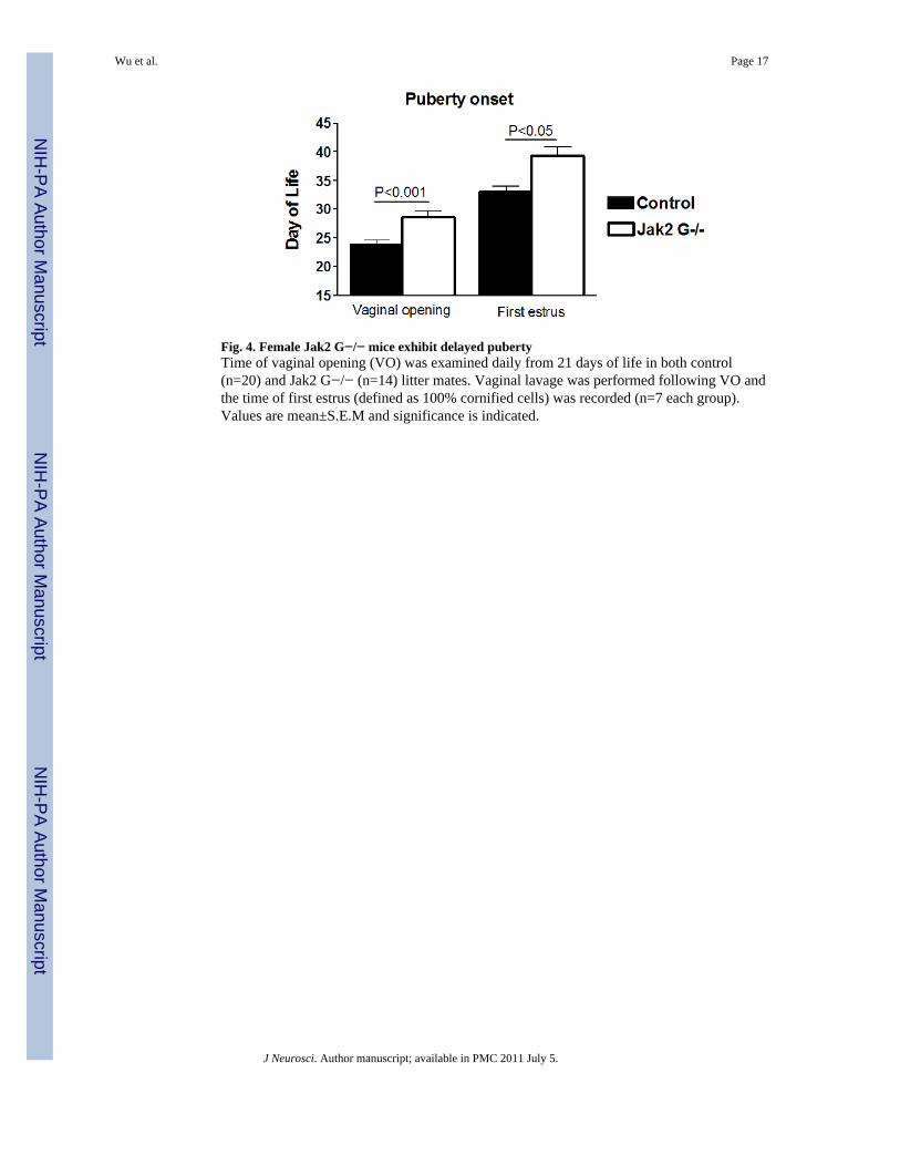
Fig. 4. Female Jak2 G−/− mice exhibit delayed pubertyTime of vaginal opening (VO) was examined daily from 21 days of life in both control(n=20) and Jak2 G−/− (n=14) litter mates. Vaginal lavage was performed following VO andthe time of first estrus (defined as 100% cornified cells) was recorded (n=7 each group).Values are mean±S.E.M and significance is indicated.
Wu et al. Page 17
J Neurosci. Author manuscript; available in PMC 2011 July 5.
NIH
-PA Author Manuscript
NIH
-PA Author Manuscript
NIH
-PA Author Manuscript

Fig. 5. Estrous cycle pattern in control and Jak2 G−/− miceVaginal cytology was assessed for 15 days in control and Jak2 G−/− mice. A, Plotted arethe percent of females that exhibited one complete cycle during the 15 days (Left two bars)or two complete cycles during the 15 days (right two bars). Numbers of mice used tocalculate the percentage are at the top of the bars. B, Time in each estrous cycle phase. E:estrus P: proestrus M/D, metestrus/diestrus. C, Representative estrous cycles of control, Jak2G−/− with irregular cycles, and Jak2 G−/− without cycles. D, OVX mice were implantedwith an estradiol pump on day 0 and bled on day 2 (control n=4 vs Jak2 G−/− n=7). Valuesare mean±S.E.M and significance is indicated.
Wu et al. Page 18
J Neurosci. Author manuscript; available in PMC 2011 July 5.
NIH
-PA Author Manuscript
NIH
-PA Author Manuscript
NIH
-PA Author Manuscript

Fig. 6. Colocalization of GnRH and c-Fos after after surge generationImages of dual labeling of GnRH and c-Fos in control (A) and Jak2 G−/− mice (B) at thelevel of the OVLT. Black arrows point to neurons with labeling of c-Fos (black/gray dot)and GnRH neuron (brown color). White arrows point to neurons labeled only for GnRH. Aneuron with dual labeling is bordered by a solid line, with higher magnification to the right.A neuron with only GnRH staining is bordered by a dashed line, with higher magnificationto the right. Scale bar: 20μm. C, Percentage of GnRH neurons with c-Fos at two differentregions (n=4 per group). Values are mean ± S.E.M and significance and numbers of animalsexamined is indicated.
Wu et al. Page 19
J Neurosci. Author manuscript; available in PMC 2011 July 5.
NIH
-PA Author Manuscript
NIH
-PA Author Manuscript
NIH
-PA Author Manuscript

Fig. 7. Fertility in control and Jak2 G−/− female miceA, Female mice were mated with wild type male mice for 90 days. Each line represents anindividual female mouse. The black dot represents the day that each litter was born afterintroduction to male. Number on the top of the line represents how many pups in each litter.Top panel is female control mice. Bottom panel is Jak2G−/− female mice. Data from A, aresummarized in B-D. B, After introduction with wild type male, the day of first litter wasrecorded in both groups. C, Total numbers of litters per female was significantly reduced inJak2 G−/− mice compared to control mice during the 90 days. D, Number of pups per litterwas significantly reduced in Jak2 G−/− mice compared to controls. For B-D, Values aremean ± S.E.M and significance and numbers of animals examined is indicated.
Wu et al. Page 20
J Neurosci. Author manuscript; available in PMC 2011 July 5.
NIH
-PA Author Manuscript
NIH
-PA Author Manuscript
NIH
-PA Author Manuscript

Fig. 8. Ovarian morphologyA, Ovary was sectioned at 7μm and corpora lutea (CL) were counted every 10th section perovary (n=4 per group). B, 7 μm ovary sections with H&E staining. CL was labeled in thecontrol ovary. C, Ovarian weights (mg) of control and Jak2 G−/− mice (n=8, control; n=6,Jak2 G−/−). Values are mean ± S.E.M and significant differences are noted with bracketsabove bars.
Wu et al. Page 21
J Neurosci. Author manuscript; available in PMC 2011 July 5.
NIH
-PA Author Manuscript
NIH
-PA Author Manuscript
NIH
-PA Author Manuscript