Prolactin-induced Jak2 phosphorylation of RUSH: A key element in Jak/RUSH signaling
15
Prolactin-induced Jak2 phosphorylation of RUSH: a key element in Jak/RUSH signaling Rebecca A. Helmer 1 , Marlyn Panchoo 1 , Janet S. Dertien 2 , Suhani M. Bhakta 1 , Aveline Hewetson 2 , and Beverly S. Chilton 1,† 1 Department of Cell Biology & Biochemistry, Texas Tech University Health Sciences Center, Lubbock, TX 79430 2 Department of Pharmacology & Neuroscience, Texas Tech University Health Sciences Center, Lubbock, TX 79430 Abstract Jak2/Stat-mediated prolactin signaling culminates in Stat5a-DNA-binding. However, not all Jak2- dependent genes have Stat5 sites. Western analysis with inhibitors showed Jak2 is a proximal intermediate in prolactin-induced RUSH phosphorylation. Transfection assays with HRE-H9 cells showed the RUSH-binding site mediated the ability of prolactin to augment progesterone- dependent transcription of the RUSH gene. Jak2 inhibitors or targeted RUSH-site mutation blocked the prolactin effect. RUSH co-immunoprecipitated with phospho-Jak2 from nuclear extracts. Jak2 inhibitors abolished the nuclear pool of phospho-RUSH not the nuclear content of RUSH in HRE-H9 cells. Nucleolar-affiliated partners, e.g. nucleolin, were identified by μLC/MS/ MS analysis of nuclear proteins that co-immunoprecipitated with RUSH/GST-RING. RUSH did not exclusively co-localize with fibrillarin to the nucleolus. MG-132 (proteasomal inhibitor) failed to block Tyrene CR4-mediated decrease in phospho-RUSH, and did not promote RUSH accumulation in the nucleolus. These studies authenticate prolactin-dependent Jak2 phosphorylation of RUSH, and provide functional implications on the RUSH network of nuclear interactions. Keywords Jak2; prolactin; HLTF; nucleolin; actin; HRE-H9 cells (rabbit endometrium) 1. Introduction Prolactin initiates intracellular signaling by binding to cell-surface receptors with subsequent activation of associated kinases (Binart et al., 2010). Major signaling cascades include Jak2/ Stat5a, Ras-Raf-MAPK, and PI-3K (Clevenger et al., 2003). In Jak2/Stat5a signaling, prolactin receptor binding triggers trans- and/or auto-phosphorylation of two Jak2 molecules constitutively associated with the membrane-proximal proline-rich Box 1 motif of the receptor (Hennighausen and Robinson, 2008). Phospho-Jak2 coordinates the tyrosine- © 2010 Elsevier Ireland Ltd. All rights reserved † Beverly S. Chilton Department of Cell Biology & Biochemistry Texas Tech University Health Sciences Center 3601 4 th Street – MS6540 Lubbock, TX 79430 Telephone: 806-743-2709 Fax: 806-743-2990 [email protected]. Publisher's Disclaimer: This is a PDF file of an unedited manuscript that has been accepted for publication. As a service to our customers we are providing this early version of the manuscript. The manuscript will undergo copyediting, typesetting, and review of the resulting proof before it is published in its final citable form. Please note that during the production process errors may be discovered which could affect the content, and all legal disclaimers that apply to the journal pertain. NIH Public Access Author Manuscript Mol Cell Endocrinol. Author manuscript; available in PMC 2011 August 30. Published in final edited form as: Mol Cell Endocrinol. 2010 August 30; 325(1-2): 143–149. doi:10.1016/j.mce.2010.05.010. NIH-PA Author Manuscript NIH-PA Author Manuscript NIH-PA Author Manuscript
-
Upload
independent -
Category
Documents
-
view
0 -
download
0
Transcript of Prolactin-induced Jak2 phosphorylation of RUSH: A key element in Jak/RUSH signaling
Prolactin-induced Jak2 phosphorylation of RUSH: a key elementin Jak/RUSH signaling
Rebecca A. Helmer1, Marlyn Panchoo1, Janet S. Dertien2, Suhani M. Bhakta1, AvelineHewetson2, and Beverly S. Chilton1,†
1Department of Cell Biology & Biochemistry, Texas Tech University Health Sciences Center,Lubbock, TX 794302Department of Pharmacology & Neuroscience, Texas Tech University Health Sciences Center,Lubbock, TX 79430
AbstractJak2/Stat-mediated prolactin signaling culminates in Stat5a-DNA-binding. However, not all Jak2-dependent genes have Stat5 sites. Western analysis with inhibitors showed Jak2 is a proximalintermediate in prolactin-induced RUSH phosphorylation. Transfection assays with HRE-H9 cellsshowed the RUSH-binding site mediated the ability of prolactin to augment progesterone-dependent transcription of the RUSH gene. Jak2 inhibitors or targeted RUSH-site mutationblocked the prolactin effect. RUSH co-immunoprecipitated with phospho-Jak2 from nuclearextracts. Jak2 inhibitors abolished the nuclear pool of phospho-RUSH not the nuclear content ofRUSH in HRE-H9 cells. Nucleolar-affiliated partners, e.g. nucleolin, were identified by μLC/MS/MS analysis of nuclear proteins that co-immunoprecipitated with RUSH/GST-RING. RUSH didnot exclusively co-localize with fibrillarin to the nucleolus. MG-132 (proteasomal inhibitor) failedto block Tyrene CR4-mediated decrease in phospho-RUSH, and did not promote RUSHaccumulation in the nucleolus. These studies authenticate prolactin-dependent Jak2phosphorylation of RUSH, and provide functional implications on the RUSH network of nuclearinteractions.
KeywordsJak2; prolactin; HLTF; nucleolin; actin; HRE-H9 cells (rabbit endometrium)
1. IntroductionProlactin initiates intracellular signaling by binding to cell-surface receptors with subsequentactivation of associated kinases (Binart et al., 2010). Major signaling cascades include Jak2/Stat5a, Ras-Raf-MAPK, and PI-3K (Clevenger et al., 2003). In Jak2/Stat5a signaling,prolactin receptor binding triggers trans- and/or auto-phosphorylation of two Jak2 moleculesconstitutively associated with the membrane-proximal proline-rich Box 1 motif of thereceptor (Hennighausen and Robinson, 2008). Phospho-Jak2 coordinates the tyrosine-
© 2010 Elsevier Ireland Ltd. All rights reserved†Beverly S. Chilton Department of Cell Biology & Biochemistry Texas Tech University Health Sciences Center 3601 4th Street –MS6540 Lubbock, TX 79430 Telephone: 806-743-2709 Fax: 806-743-2990 [email protected]'s Disclaimer: This is a PDF file of an unedited manuscript that has been accepted for publication. As a service to ourcustomers we are providing this early version of the manuscript. The manuscript will undergo copyediting, typesetting, and review ofthe resulting proof before it is published in its final citable form. Please note that during the production process errors may bediscovered which could affect the content, and all legal disclaimers that apply to the journal pertain.
NIH Public AccessAuthor ManuscriptMol Cell Endocrinol. Author manuscript; available in PMC 2011 August 30.
Published in final edited form as:Mol Cell Endocrinol. 2010 August 30; 325(1-2): 143–149. doi:10.1016/j.mce.2010.05.010.
NIH
-PA Author Manuscript
NIH
-PA Author Manuscript
NIH
-PA Author Manuscript
phosphorylation of Stat5a (Li, 2008). Jak2/Stat5a-mediated prolactin signal transductionculminates in sequence-selective binding of Stat5a to previously silent milk protein genes inmammary gland. In this paradigm, Stat5a is an important mediator of prolactin actionbecause Stat5a-deficient mice have reduced alveolar development and fail to lactate (Liu etal., 1997; Teglund et al., 1998).
In rabbit endometrium, prolactin augments the progesterone-dependent increase intranscription of the uteroglobin gene (Chilton et al. 1988; Kleis-SanFrancisco et al., 1993),the founding member of the SCGB1A1 family (Mukherjee et al., 2007). The search forresponsible mediators culminated in the cloning and characterization of the RUSHtranscription factors — a full length, progesterone-dependent, alpha isoform, and atruncated, estrogen-dependent, beta isoform (Hayward-Lester et al., 1996). RUSH-1α hasseven (I, Ia, II–VI) motifs characteristic of SWI/SNF ATPases (Racki and Narlikar, 2008), aC3HC4-RING finger motif characteristic of E3 ubiquitin ligases (Deshaies and Joazeiro,2009; Nagy and Dikic, 2010), and a HIRAN domain purported to recognize damaged DNAand stalled replication forks (Iyer et al., 2006). RUSH-1α protein is 95% similar and 91%identical to its human ortholog, HLTF, previously named SMARCA3. RUSH-1β protein isidentical to full-length RUSH-1α through the RING-finger and for 33 amino acidsthereafter. Then, because of an alternative splicing event, the protein extends for five uniqueamino acids and stops. Thus RUSH-1β is missing SWI/SNF-motifs IV–V–VI, but retains itsprotein interaction potential through the RING-finger motif.
Cyclic amplification and selection of targets identified the RUSH binding site (−126/−121)in the SCGB1A1 gene (Hewetson et al., 2002), and chromatin immunoprecipitationconfirmed site-specific binding of RUSH-1α to that site in the transcriptionally activepromoter in the absence of Stat5a binding sites (Hewetson et al., 2002). Transienttransfection assays with mutated constructs and HRE-H9 cells, a rabbit uterine epithelial cellline (Li et al., 1989), showed the RUSH-binding site mediated the ability of prolactin toaugment progesterone-dependent transcriptional activation of the uteroglobin gene. Theidentification of RUSH-1α as a nuclear effector of prolactin signals prompted speculationabout a Jak/RUSH alternative to Jak/Stat signaling. The fact that RUSH is a phosphonuclearprotein, and RUSH-DNA binding is mediated by tyrosine-phosphorylation (Hewetson et al.,2004) supported the hypothesis. Moreover, not all Jak2 regulated genes contain Stat5binding sites (Dawson et al., 2009; Sattler and Griffin, 2009).
The C3HC4-RING finger of RUSH is a protein interaction domain (Mansharamani et al.,2001; Hewetson et al., 2008). It binds the transcription factors Egr-1 and c-Rel, andcatalyzes DNA looping through its affiliation with these protein partners (Hewetson andChilton, 2008). RUSH-1α mediates progesterone-dependent transcription of its ownpromoter through this DNA-looping mechanism. Northern blotting showed prolactinaugments progesterone-dependent transcription of the RUSH gene in addition to theSCGB1A1 gene (Hayward-Lester et al., 1996).
In this study, Jak2 inhibitors — AG490, TG101209, Staurosporine, Jak inhibitor 1, Jak 2inhibitor 2 and Tyrene CR4 — were used in conjunction with the PI-3 kinase inhibitor,Wortmannin, and the MAP kinase inhibitor, PD98059, in HRE-H9 cells to show thatRUSH-1α is phosphorylated by Jak2 as a direct consequence of prolactin treatment. Westernanalysis, immunofluorescence microscopy, and transient transfection assays confirmed afunctional Jak/RUSH pathway.
Co-immunoprecipitation of Jak2 with RUSH-1α confirmed a physical interaction betweenthese phosphoproteins in nuclear extract (Hewetson et al., 2002). Additional nuclear proteinpartners, such as nucleolin (C23), were identified by μLC/MS/MS analysis of nuclear extract
Helmer et al. Page 2
Mol Cell Endocrinol. Author manuscript; available in PMC 2011 August 30.
NIH
-PA Author Manuscript
NIH
-PA Author Manuscript
NIH
-PA Author Manuscript
proteins that co-immunoprecipitated with RUSH. Like nucleolin, some partners also co-immunoprecipitated with GST-RING, confirming the RING motif mediates RUSH-proteinbinding, and some were identified as phosphonuclear proteins by μLC/MS/MS analysis ofnuclear extract proteins that immunoprecipitated with antiphosphotyrosine antibodies.Confocal immunofluorescence images of HRE-H9 cells showed an even nuclear distributionof RUSH and phospho-Jak2. RUSH did not exclusively co-localize with fibrillarin to thenucleolus, and the proteasome inhibitor, MG-132, failed to promote nucleolar accumulationof RUSH. These new findings provide functional implications for the RUSH network ofnuclear and phospho-nuclear proteins.
2. Material and Methods2.1 Reagents and Tools
Ready Gel 5 % and 7.5% Tris-HCl precast polyacrylamide gels (161–1213 and 161–1154,respectively) were purchased from Bio-Rad (Hercules, CA). Gradipure Coomassie BrilliantBlue R-250 stain reagent was purchased from VWR (Sugar Land, TX). Thermo ScientificPierce silver stain kit (formerly called SilverSNAP Silver Stain Kit II) was purchased FisherThermo Scientific (Rockford, IL). Promegestone (R5020) was purchased from Perkin ElmerLife Science Products, Inc. (Waltham, MA). Dr. A.F. Parlow, Scientific Director, NationalHormone and Pituitary Program (Torrance, CA), provided the ovine prolactin (oPRL). SantaCruz Biotechnology, Inc. (Santa Cruz, CA) was the commercial source of Protein A/GPLUS-Agarose (sc-2003) beads, and antibodies for phosphorylated (p) Jak2 (sc-21870).SMARCA3 antibodies (ab17984), and two nucleolar markers — mouse monoclonal [38F3]to fibrillarin (ab4566) and rabbit polyclonal to fibrillarin (ab5821) — were purchased fromAbcam Inc. (Cambridge, MA). Mouse anti-phosphotyrosine, 4G10® Platinum antibodies(05–1050) were purchased from Millipore (Billerica, MA). Invitrogen Corp. (Carlsbad, CA)was the source of Alexa-conjugated secondary antibodies — Alexa Fluor® 594 chickenanti-mouse IgG (A-21201), Alexa Fluor® 594 chicken anti-goat IgG (A-21468), AlexaFluor® 488 chicken anti-rabbit IgG (A-21441), and ProLong Gold antifade reagent withDAPI.
The following Jak2 inhibitors were purchased from the EMD Biosciences (San Diego, CA):Tyrphostin AG490 (658401); Jak inhibitor 1 (420097); Jak 2 inhibitor II (420132); TyreneCR4 (655230); the PI-3 kinase inhibitor, Wortmannin (681675); the MAK kinase inhibitorPD98059 (513000); and the proteasome inhibitor MG-132 (474790). Two additional Jak2inhibitors included TG101209 purchased from Symansis (Auckland, New Zealand), andStaurosporine (S6942) purchased from Sigma-Aldrich (St. Louis, MO).
2.2 Animal treatmentsAll studies with virgin, adult New Zealand white rabbits were conducted according to theNIH Guidelines for the Care and Use of Laboratory Animals, as reviewed and approved bythe Animal Care and Use Committee at Texas Tech University Health Sciences Center.Estrous animals were given sc injections of progesterone (3 mg/kg/day) for 5 days.
2.3 Nuclear extractsNuclei were isolated from the endometrium from progesterone treated rabbits (Kleis-SanFrancisco et al., 1993), and from R5020-treated HRE-H9 cells (Hewetson and Chilton,2003) to induce RUSH-1α expression. Nuclear extract proteins were immunoprecipitatedwith anti-phosphotyrosine 4G10 Platinum antibodies (4 μl/reaction) or anti-phospho Jak2 (5μl/reaction), resolved by SDS-PAGE, and processed for Western blotting with anti-SMARCA3 (1:1000) or anti-phospho Jak2 (1:1000), respectively, and species appropriateHRP-conjugated secondary antibodies.
Helmer et al. Page 3
Mol Cell Endocrinol. Author manuscript; available in PMC 2011 August 30.
NIH
-PA Author Manuscript
NIH
-PA Author Manuscript
NIH
-PA Author Manuscript
2.4 Protein Sequence AnalysisNuclear extract proteins were co-immunoprecipitated with RUSH (n = 3), GST-RING (n =1), or anti-phosphotyrosine antibodies (n = 2). The use of the RING-motif in GST-pulldownassays has been described (Mansharamani et al., 2001; Hewetson and Chilton, 2008;Hewetson et al., 2008). Immunoprecipitated nuclear proteins were resolved by SDS/PAGE,and either Silver- or Coomassie-stained. One-dimensional gel slices were excised,dehydrated with acetonitrile, and subject to in-gel tryptic digestion. Protein sequenceanalysis was performed at the Harvard Microchemistry Facility and Proteomics AnalysisFacility by microcapillary reverse-phase high performance liquid chromatography (HPLC)nano-electrospray tandem mass spectrometry (μLC/MS/MS) on a Finnigan LCQ DECA XPPlus quadrupole ion trap mass spectrometer (RUSH/HLTF immunoprecipitated samples,plus one 4-hour gradient to further identify peptides; anti-phosphotyrosineimmunoprecipitated samples), or a Thermo LTQ-Orbitrap mass spectrometer (GST-RINGsamples). These instrument configurations are capable of acquiring individual sequence(MS/MS) spectra on-line at high sensitivity (<<<1 femtomole) for multiple peptides in thechromatographic run. These MS/MS spectra (or sequence) are then correlated with knownsequences using the algorithm Sequest developed at the University of Washington (Eng etal., 1994), and programs developed in the Harvard Microchemistry laboratory (Chittum etal., 1998). MS/MS peptide sequences were reviewed for consensus with known proteins,and the results manually confirmed for fidelity.
2.5 Transient Transfection AssaysCloning the RUSH fragment (−712/+90) into the luciferase reporter plasmid pGL-3-Basic,targeted mutagenesis of the RUSH site, and differential expression of the constructs in HRE-H9 cells has been described (Hewetson and Chilton, 2003, 2008). HRE-H9 cells are anSV-40 transformed rabbit uterine epithelial cell line derived from the endometrium of 3-month-old female rabbits treated with hCG (Li et al., 1989). Transient transfections wereperformed in 24-well dishes a minimum of 2 times in quadruplicate. Cells were treated(Hewetson and Chilton, 2003, 2008) with R5020 (10−8 M) in dimethyl sulfoxide (0.1% v/v)for 16–20 h ± Tyrene CR4 (800 nM) for 16–20 hours ± prolactin (10−8 M) for 5 min.pRUSH-LUC expression was normalized to co-transfected pRL-TK-LUC expression tocontrol for transfection efficiency. Multiple ratio data were ranked (Conover and Iman,1981) and the ranks between the groups were analyzed by Kruskal-Wallis (Nonparametric)ANOVA followed by Dunn's Multiple Comparisons Test (P < 0.05 significance level) usingGraphPad InStat Version 3 for Macintosh (GraphPad Software, Inc.). The P value for theKruskal-Wallis Test was P < 0.0001.
2.6 Immunofluorescence microscopyImmunofluorescence microscopy was as previously described (Hewetson and Chilton,2008). Briefly, HRE-H9 cells were grown on poly-D-lysine coated coverslips and treated ±R5020 (10−8 M) in dimethyl sulfoxide (0.1% v/v) for 16–20 h ± prolactin (10−8 M) for 2 or5 min. Cells were fixed in cold methanol (−20 C) for 10 min followed by cold acetone (−20C) for 1 min; rinsed in PBS [20 mM sodium phosphate, pH 7.5, and 0.9% sodium chloride];quenched in 0.1 M glycine for 1 hr at room temperature; and blocked in 10% normalchicken serum with 0.3% Triton X-100. Cells were incubated with primary antibodies(1:500) overnight at 4 C. After three rinses in PBS, cells were incubated with speciesrelevant Alexa Fluor® conjugated secondary antibodies (1:400) for 60 min at roomtemperature in the dark. Cells were rinsed in PBS and mounted in ProLong Gold antifadereagent with DAPI. Controls included cells that were not treated with hormones, andlabeling reactions that excluded either primary or secondary antibodies.
Helmer et al. Page 4
Mol Cell Endocrinol. Author manuscript; available in PMC 2011 August 30.
NIH
-PA Author Manuscript
NIH
-PA Author Manuscript
NIH
-PA Author Manuscript

Immunofluorescence was observed with an Olympus IX71 inverted microscope equippedfor laser scanning confocal microscopy (LSCM). Images were acquired using OlympusFluoview 500 software and multi — Argon, red HeNe, green HeNe, and blue diode —lasers. Initially, differential interference contrast (DIC) images were acquired in order todemarcate cell boundaries. Images were acquired for phospho-Jak2, RUSH, and DAPI. Allimages were imported into Metamorph 6.3 image analysis software (Molecular Devices,Sunnyvale, CA). Cell boundaries were traced over the DIC images using the region toolsand transferred to the phospho-Jak2 and RUSH images. Phospho-Jak2 and RUSH imageswere thresholded. Nuclei were isolated using the “create regions around objects” function.The percent threshold area was determined using the “Show Region Statistics” menu.Values [n = 6–13; power calculation required 5 replicates per treatment (p<0.05)] wereanalyzed by Kruskal-Wallis (Nonparametric) ANOVA followed by Dunn's MultipleComparisons Test (P < 0.05 significance level) using GraphPad InStat Version 3 forMacintosh (GraphPad Software, Inc.). The P value for the Kruskal-Wallis Test was P <0.0002.
2.7 Immunoprecipitation and Western analysisHRE-H9 cells from 12-well dishes were harvested in 1 ml RIPA lysis buffer [0.1% sodiumdodecyl sulfate, 1% Nonidet P-40, 1% sodium deoxycholate, 2 mM EDTA, 0.15 M NaCl,0.01 M sodium phosphate, 50 mM NaF, 2 mM sodium orthovanadata, 1 mMphenylmethylsulfonylfluoride, 5 μg/ml aprotinin, 1 μg/ml pepstatin A, and 2 μg/mlleupeptin] for 1 h at 4 C with constant tumbling, i.e. end-over-end mixing with a Lab-Quakerotator. Lysates were cleared of cellular debris by centrifugation (14,000 × g at 4 C for 30minutes). Resultant supernatants were tumbled with primary antibodies for 1 h at 4 C, andProtein A/G Plus-Agarose beads (20μl/per reaction) were added for overnight (≥16 h)tumbling. Beads were washed 8 times in PBS. Proteins were extracted from beads, resolvedon either 5% or 7.5% SDS-PAGE (minigels), and transferred to nitrocellulose. Molecular-mass markers were separated by electrophoresis and transferred with the samples.Membranes were incubated in blocking buffer, i.e. 1× TBST [20 mM Tris-HCl (pH 7.6),150 mM NaCl, and 0.05% Tween 20] with 3% Carnation non-fat dry milk, for 60 min.Membranes were incubated with antibodies in antibody dilution buffer (1× TBST with 1%Carnation non-fat dry milk) overnight at 4°C. Membranes were washed 8 × 15 min in 1×TBST. Membranes were incubated with appropriate HRP-conjugated secondary antibodiesin antibody dilution buffer for 1 hr at room temperature. Membranes were subsequentlywashed 8 times in 1× TBST. Specific signals were detected by chemiluminescence with theImmun-Star™ Western™ kit.
3. Results3.1 Western analysis
Previously we reported (Hewetson et al., 2004) RUSH is a tyrosine-phosphorylated proteinonly capable of binding DNA when phosphorylated. To test the hypothesis that RUSH isphosphorylated as a direct consequence of prolactin treatment, HRE-H9 cells were givenR5020 (16–20 hours) to induce RUSH-1α and then exposed to prolactin for 2 minutes in thepresence of individual Jak2 inhibitors — AG490, TG101209, Staurosporine, Jak inhibitor 1,Jak 2 inhibitor 2 and Tyrene CR4 — plus the PI-3 kinase inhibitor, Wortmannin, and theMAP kinase inhibitor, PD98059. Western analysis of whole cell extracts revealed that allinhibitors of Jak2 blocked phosphorylation of RUSH (Figure 1A). Inhibitors of MAPK andPI-3K pathways that can be activated by prolactin stimulation had no measurable effect onRUSH expression (data shown as positive controls in other figures).
Helmer et al. Page 5
Mol Cell Endocrinol. Author manuscript; available in PMC 2011 August 30.
NIH
-PA Author Manuscript
NIH
-PA Author Manuscript
NIH
-PA Author Manuscript
3.2 Transient transfection assaysTo show that Jak2-dependent phosphorylation of RUSH mediates the action of prolactin,control and hormone-dependent transcriptional activities were measured with either an intactRUSH construct (−712/+90) containing the promoter (−162/+90), included PRE-half site(−138/−133), and authentic RUSH site (−616/−611), or the same RUSH construct with atargeted mutation (Δ) in the RUSH site. The complete absence of Stat5 binding sites fromthis construct (−712/+90) was confirmed with MatInspector (Genomatix) ReleaseProfessional 8.01 (Cartharius et al., 2005), and with BIOBASE BKL TRANSFAC Suite. Asshown in Figure 2A, prolactin increased (p < 0.001) progesterone-dependent transcription ofthe RUSH gene. The Jak2-inhibitor Tyrene CR4 completely blocked the prolactin-treatmenteffect. Progesterone-dependent transcription of ΔRUSH was comparable (p > 0.05) to theintact RUSH construct, but mutation of the RUSH site was highly deleterious (p < 0.001) tothe prolactin effect. Either treatment with Tyrene CR4 or mutation of the RUSH siteeliminated (p > 0.05) the prolactin-mediated increase in progesterone-dependenttranscription of the RUSH gene.
3.3 Nuclear RUSH and phospho-Jak2We previously reported nuclear co-localization of RUSH and Jak2 (Hewetson et al., 2002)in nuclear extracts from endometrium. Subcellular fractionation experiments demonstratedcatalytically active Jak2 in nuclei isolated from endometrium (Figure 3A) and HRE-H9 cells(Figure 1B), and confirmed a physical affiliation between RUSH and catalytically activeJak2 (Figure 3A, 3B). Confocal immunofluorescence imaging corroborates a significantpool of phosphonuclear Jak2 in HRE-H9 cells (Figure 4A). Treatment of HRE-H9 cells withTyrene CR4 abolished the nuclear pools of catalytically active Jak2 (Figure 1B) andphospho-RUSH (Figure 1C), but not the nuclear content of RUSH (Figure 2B) as evaluatedby quantitative LSCM. As shown in Figure 1C, PD 98059 had no effect on the nuclear poolof phospho-RUSH. Moreover, neither Tyrene CR4 nor PD 98059 has any effect (p > 0.05)on the nuclear pool of RUSH (Figure 2B) confirming that nuclear Jak2 phosphorylates aprogesterone-dependent pool of RUSH.
3.4 μLC/MS/MS analysis of nuclear proteinsRUSH-interacting proteins, and their posttranslational modifications, were identified byμLC/MS/MS analysis. Nuclear extract proteins that co-immunoprecipitated with RUSHwere resolved by SDS-PAGE. One-dimensional gel slices were in gel digested with trypsin,and resulting peptide mixtures were evaluated by μLC/MS/MS analysis. Authentic RUSHprotein partners were identified (Table I) in at least two of three independent experimentswith high stringency, i.e. a minimum of two high-scoring peptides per protein to achieve afalse positive rate for protein identification < 0.1%. In actuality, the average number of high-scoring peptides per RUSH partner was nine. When the list of high-stringency bindingpartners was compared with proteins that also bound the RING domain, three candidateswith 2–3 high-scoring peptides were identified (Table I). Eight RUSH protein partners wereidentified as phosphonuclear proteins, and nine were recognized members of the nucleolarproteome database (Ahmad et al., 2009;Andersen et al., 2005). Table I contains conclusiveevidence that RUSH affiliates with nuclear actin.
3.5 RUSH and nucleolinThe affiliation of RUSH with nucleolin, which is found in nucleoli and nucleoplasm,prompted an inquiry about the intracellular availability of RUSH. As shown in Figure 4B,endogenous RUSH is uniformly distributed throughout the nucleus and cytoplasm. Mergedconfocal immunofluorescence images of RUSH with the nucleolar marker protein fibrillarinshowed RUSH in the nucleolus as well as the nucleoplasm (Figure 4C).
Helmer et al. Page 6
Mol Cell Endocrinol. Author manuscript; available in PMC 2011 August 30.
NIH
-PA Author Manuscript
NIH
-PA Author Manuscript
NIH
-PA Author Manuscript
Proteasome inhibitors such as MG-132 are known to extend the activity of Jak2/Statsignaling by protecting tyrosine-phosphorylated proteins from degradation by theproteasome (Ungureanu et al., 2002; Yu and Burakoff, 1997; Nilsson et al., 2006). However,as shown in Figure 1D, MG-132 failed to block a Tyrene CR4-mediated decrease in thelevel of nuclear phospho-RUSH. Not surprising, MG-132 also failed to promote thenucleolar accumulation of RUSH (data not shown).
4. DiscussionThe current model of RUSH regulation integrates RUSH phosphorylation by Jak2 as a directconsequence of prolactin signaling with the previous finding that RUSH is a phosphonuclearprotein whose DNA binding ability is mediated by tyrosine-phosphorylation (Hewetson etal., 2004). Transfection assays with RUSH reporter constructs allowed us to correlate theloss of Jak2-mediated RUSH phosphorylation with the inability of prolactin to augmentprogesterone-dependent transcription. Jak/RUSH signaling independent of the Jak/Statpathway is supported by the Dawson et al (2009) report that ~ 35% or 14 of 40 Jak2-regulated genes do not contain Stat5 binding sites.
A nuclear pool of catalytically active Jak2 means Jak2 is available to tyrosine phosphorylateprotein targets in close proximity to their nuclear sites of action. In general,phosphorylation-mediated signaling is characterized by a dynamic relationship betweenprotein participants in which site-specific and time-specific phosphorylation/dephosphorylation events occur sequentially to achieve an outcome (Zhang et al., 2005).Compelling examples of functions controlled by nuclear Jak2 include: 1) phosphorylation ofNF1-C2 prevents its proteasomal degradation in mammary epithelial cells (Nilsson et al.,2006); 2) phosphorylation of histone H3 at tyrosine residue 41 (H3Y41) releasestranscriptional repressor heterochromatin protein 1α (HP1α) resulting in transcriptionalactivation of Imo2 in hematopoietic cells (Dawson et al., 2009).
The NetPhos 2.0 Server predicts seven tyrosine phosphorylation sites in RUSH. Two ofthem (Y93 and Y195) are in the DNA-binding domain (amino acids 40–231). Since tyrosinephosphorylation is required for DNA-binding, these sites are the most likely candidates forsite-specific validation/quantification of tyrosine-phosphorylation, and the generation ofsite-specific-phospho-antibodies with which to evaluate tyrosine phosphorylation by Jak2.Although it is unknown if the human (HLTF) and mouse (Hltf) orthologs are Jak2 targets, acontext-dependent search of the sequences containing Y93 and Y195 with the BLAST2.2.23 (SwissProt) program (Altschuel et al., 1997) showed the sites are 100% conserved inall three species.
Understanding RUSH function is found in the context-dependent interactions of RUSH withits protein partners. However rudimentary, our knowledge of those partners — identity,criteria for partner choice, and function — was advanced by the use of μLC/MS/MS analysisof nuclear proteins that co-immunoprecipitated with RUSH. The human ortholog HLTF isone of 489 proteins known to flux through the nucleolus (Andersen et al., 2005). RUSH, likenucleolin, lacks the nucleolar localization (NoLS) signal – [(R/K)(R/K)X(R/K)] – thatappears once or as multiple copies in peptides directed to or retained in the nucleolus(Emmott and Hiscox, 2009). Moreover, like nucleolin, RUSH physically affiliates withnucleophosmin (Table 1), which contains a NoLS, and is purported to target nucleolin to thenucleolus (Li et al., 1996). Physical interactions between RUSH and nucleolin raise thequestion of whether or not nucleolin, which possesses a histone chaperone activity (Angelovet al., 2006; Mongelard and Bouvet, 2007), enhances the chromatin remodeling activity ofRUSH.
Helmer et al. Page 7
Mol Cell Endocrinol. Author manuscript; available in PMC 2011 August 30.
NIH
-PA Author Manuscript
NIH
-PA Author Manuscript
NIH
-PA Author Manuscript
The C3HC4-RING motif in RUSH is a protein interaction domain (Mansharamani et al.,2001; Hewetson and Chilton, 2008; Hewetson et al., 2008). It binds the transcription factorsEgr-1 and c-Rel, and catalyzes DNA looping through its affiliation with these proteinpartners (Hewetson and Chilton, 2008). RUSH mediates progesterone-dependenttranscription of its own promoter through this DNA-looping mechanism. Moreover, theC3HC4-RING motif in RUSH binds a rabbit-specific ATP11B isoform (an atypical Type-IVP-type ATPase) purported to have a role in subnuclear trafficking of transcription factorswith RING motifs (Mansharamani et al., 2001). The discontinuous PVITHC-HAKCPLsequence in the RING-finger binds a sequence conserved in all ATP11B isoforms(Hewetson et al., 2008). Fine mapping CLIPS™-constrained RING peptides that maintainthe 3D-conformation of individual peptides showed the sequence HAKAPL-TH is essentialfor binding, and 100% conserved in all orthologs. Whether or not the discontinuous bindingsite is a universal protein-binding site with functional relevance to other species, and humandisease, is unknown at this time.
This report of a physical affiliation between RUSH and actin in the nucleus is the firstaccount of a physical affiliation between any HLTF ortholog and actin. As bona fidemembers of the nuclear compartment (Louvet and Percipalle, 2009), actin and actin-bindingproteins are involved in gene transcription (Farrants, 2008) in association with ATP-dependent chromatin-remodeling (SWI/SNF) complexes (Zheng et al., 2009). A majorfunction of actin is to behave as an allosteric regulator in the remodeling of macromolecularassemblies such as chromatin remodeling factors or transcription complexes (Zheng et al.,2009).
Collectively these studies focus our attention on previously unrecognized RUSH nuclearpartners known to have important cellular functions. Identifying both stable and transientlyinteracting protein partners will help us to better understand the importance of the RINGdomain. The extent to which RING-protein interactions are evolutionarily conservedhighlights their functional importance.
AcknowledgmentsWe thank Larry Starr, Medical Photography, TTUHSC, for artwork; and the NIH (HD29457) for support.
ReferencesAhmad Y, Boisvert FM, Gregor P, Cobley A, Lamond AI. NOPdb: Nucleolar proteome database-2008
update. Nucleic Acids Res. 2009; 37:D181–D184. [PubMed: 18984612]Altschul SF, Madden TL, Schaffer AA, Zhang J, Zhang Z, Miller W, Lipman DJ. Gapped BLAST and
PSI-BLAST: a new generation of protein database search programs. Nucleic Acids Res. 1997;25:3389–3402. [PubMed: 9254694]
Andersen JS, Lam YW, Leung AK, Ong SE, Lyon CE, Lamond AI, Mann M. Nucleolar proteomedynamics. Nature. 2005; 433:77–83. [PubMed: 15635413]
Angelov D, Bondarenko VA, Almagro S, Menoni H, Mongélard F, Hans F, Mietton F, Studitsky VM,Hamiche A, Dimitrov S, Bouvet P. Nucleolin is a histone chaperone with FACT-like activity andassists remodeling of nucleosomes. EMBO J. 2006; 25:1669–1679. [PubMed: 16601700]
Binart, N.; Bachelot, A.; Bouilly, J. Trends Endocrinol. Metab. Vol. 21. 2010. Impact of prolactinreceptor isoforms on reproduction; p. 362-368.
Cartharius K, Frech K, Grote K, Klocke B, Haltmeier M, Klingenhoff A, Frisch M, Bayerlein M,Werner T. MatInspector and beyond: promoter analysis based on transcription factor binding sites.Bioinformatics. 2005; 21:2933–2942. [PubMed: 15860560]
Chilton BS, Mani SK, Bullock DW. Servomechanism of prolactin and progesterone in regulatinguterine gene expression. Mol. Endocrinol. 1988; 2:1169–1175. [PubMed: 3216859]
Helmer et al. Page 8
Mol Cell Endocrinol. Author manuscript; available in PMC 2011 August 30.
NIH
-PA Author Manuscript
NIH
-PA Author Manuscript
NIH
-PA Author Manuscript
Chittum HS, Lane WS, Carlson BA, Roller PP, Lung FT, Lee BJ, Hatfield DL. Rabbit β-globin isextended beyond its UGA stop codon by multiple suppressions and translational reading gaps.Biochemistry. 1998; 37:10866–10870. [PubMed: 9692979]
Clevenger CV, Furth PA, Hankinson SE, Schuler LA. The role of prolactin in mammary carcinoma.Endocr. Rev. 2003; 24:1–27. [PubMed: 12588805]
Conover WJ, Iman RL. Rank transformation as a bridge between parametric and nonparametricstatistics. Am. Stat. 1981; 35:124–129.
Dawson MA, Bannister AJ, Göttgens B, Foster SD, Bartke T, Green AR, Kouzarides T. JAK2phosphorylates histone H3Y41 and excludes HP1alpha from chromatin. Nature. 2009; 461:819–822. [PubMed: 19783980]
Deshaies RJ, Joazeiro CA. RING domain E3 ubiquitin ligases. Annu. Rev. Biochem. 2009; 78:399–434. [PubMed: 19489725]
Emmott E, Hiscox JA. Nucleolar targeting: the hub of the matter. EMBO Rep. 2009; 10:231–238.[PubMed: 19229283]
Eng JK, McCormick AL, Yates JR III. An approach to correlate tandem mass spectral data of peptideswith amino acid sequences in a protein database. J. Am. Soc. Mass. Spectrom. 1994; 5:976–989.
Farrants AK. Chromatin remodeling and actin organization. FEBS Lett. 2008; 582:2041–2050.[PubMed: 18442483]
Hayward-Lester A, Hewetson A, Beale EG, Oefner PJ, Doris PA, Chilton BS. Cloning,characterization and steroid-dependent posttranscriptional processing of RUSH-1α and β, twouteroglobin promoter-binding proteins. Mol. Endocrinol. 1996; 10:1335–1349. [PubMed:8923460]
Hennighausen L, Robinson GW. Interpretation of cytokine signaling through the transcription factorsSTAT5A and STAT5B. Genes & Dev. 2008; 22:711–721. [PubMed: 18347089]
Hewetson A, Hendrix EC, Mansharamani M, Lee VH, Chilton BS. Identification of the RUSHconsensus-binding site by cyclic amplification and selection of targets: demonstration that RUSHmediates the ability of prolactin to augment progesterone-dependent gene expression. Mol.Endocrinol. 2002; 16:2101–2112. [PubMed: 12198246]
Hewetson A, Chilton BS. An Sp1-NF-Y/Progesterone receptor DNA binding-dependent mechanismregulates progesterone-induced transcriptional activation of the rabbit RUSH/SMARCA3 gene. J.Biol. Chem. 2003; 278:40177–40185. [PubMed: 12890680]
Hewetson A, Moore SL, Chilton BS. Prolactin signals through RUSH/SMARCA3 in the absence of aphysical association with Stat5a. Biol. Reprod. 2004; 71:1907–1912. [PubMed: 15306550]
Hewetson A, Wright-Pastusek AE, Helmer RA, Wesley KA, Chilton BS. Conservation of inter-proteinbinding sites in RUSH and RFBP, an ATPIIB isoform. Mol. Cell. Endocrinol. 2008; 292:79–86.[PubMed: 18584949]
Hewetson A, Chilton BS. Progesterone-dependent DNA looping between RUSH/SMARCA3 andEgr-1 mediates repression by c-Rel. Mol. Endocrinol. 2008; 22:813–822. [PubMed: 18174357]
Iyer LM, Babu MM, Aravind L. The HIRAN domain and recruitment of chromatin remodeling andrepair activities to damaged DNA. Cell Cycle. 2006; 5:775–782. [PubMed: 16627993]
Kleis-SanFrancisco S, Hewetson A, Chilton BS. Prolactin augments progesterone-dependentuteroglobin gene expression by modulating promoter-binding proteins. Mol. Endocrinol. 1993;7:214–223. [PubMed: 8469234]
Li WI, Chen CL, Chou JY. Characterization of a temperature-sensitive β-endorphin-secretingtransformed endometrial cell line. Endocrinology. 1989; 125:2862–2867. [PubMed: 2555129]
Li WX. Canonical and non-canonical JAK-STAT signaling. Trends Cell Biol. 2008; 18:545–551.[PubMed: 18848449]
Li YP, Busch RK, Valdez BC, Busch H. C23 interacts with B23, a putative nucleolar-localization-signal-binding protein. Euro. J. Biochem. 1996; 237:153–158.
Liu X, Robinson GW, Wagner KU, Garrett L, Wynshaw-Boris A, Hennighausen L. Stat5a ismandatory for adult mammary gland development and lactogenesis. Genes Dev. 1997; 11:179–186. [PubMed: 9009201]
Louvet E, Percipalle P. Chapter 3 Transcriptional control of gene expression by actin and myosin. Int.Rev. Cell Mol. Biol. 2009; 272:107–147. [PubMed: 19121817]
Helmer et al. Page 9
Mol Cell Endocrinol. Author manuscript; available in PMC 2011 August 30.
NIH
-PA Author Manuscript
NIH
-PA Author Manuscript
NIH
-PA Author Manuscript
Mansharamani M, Hewetson A, Chilton BS. Cloning and characterization of an atypical type IV P-type ATPase that binds to the RING motif of RUSH transcription factors. J. Biol. Chem. 2001;276:3641–3649. [PubMed: 11058586]
Mongelard F, Bouvet P. Nucleolin: a multiFACeTed protein. Trends Cell Biol. 2007; 17:80–86.[PubMed: 17157503]
Mukherjee AB, Zhang Z, Chilton BS. Uteroglobin: A steroid-inducible immunomodulatory proteinthat founded the secretoglobin superfamily. Endocr. Rev. 2007; 28:707–725. [PubMed: 17916741]
Nagy, V.; Dikic, I. Biol. Chem. Vol. 391. 2010. Ubiquitin ligase complexes: from substrate selectivityto conjugational specificity; p. 163-169.
Nilsson J, Bjursell G, Kannius-Janson M. Nuclear Jak2 and transcription factor NF1-C2: a novelmechanism of prolactin signaling in mammary epithelial cells. Mol. Cell. Biol. 2006; 26:5663–5674. [PubMed: 16847321]
Racki LR, Narlikar GJ. ATP-dependent chromatin remodeling enzymes: two heads are not better, justdifferent. Curr. Opin. Genet. Dev. 2008; 18:137–144. [PubMed: 18339542]
Sattler M, Griffin JD. JAK2 gets histone H3 rolling. Cancer Cell. 2009; 16:365–366. [PubMed:19878867]
Teglund S, McKay C, Schuetz E, van Deursen JM, Stravopodis D, Wang D, Brown M, Bodner S,Grosveld G, Ihle JN. Stat5a and Stat5b proteins have essential and nonessential, or redundant,roles in cytokine responses. Cell. 1998; 93:841–850. [PubMed: 9630227]
Ungureanu D, Saharinen P, Junttila I, Hilton DJ, Silvennoinen O. Regulation of Jak2 through theubiquitination-proteasome pathway involves phosphorylation of Jak2 on Y1007 and interactionwith SOCS-1. Mol. Cell. Biol. 2002; 22:3316–3326. [PubMed: 11971965]
Yu, C.l.; Burakoff, SJ. Involvement of proteasomes in regulating Jak-STAT pathways uponinterleukin-2 stimulation. J. Biol. Chem. 1997; 272:14017–14020. [PubMed: 9162019]
Zhang Y, Wolf-Yadlin A, Ross PL, Pappin DJ, Rush J, Lauffenburger DA, White FM. Time-resolvedmass spectrometry of tyrosine phosphorylation sites in the epidermal growth factor receptorsignaling network reveals dynamic modules. Mol. Cell. Proteomics. 2005; 4:1240–1250.[PubMed: 15951569]
Zheng B, Han M, Bernier M, Wen JK. Nuclear actin and actin-binding proteins in the regulation oftranscription and gene expression. FEBS J. 2009; 276:2669–2685. [PubMed: 19459931]
Helmer et al. Page 10
Mol Cell Endocrinol. Author manuscript; available in PMC 2011 August 30.
NIH
-PA Author Manuscript
NIH
-PA Author Manuscript
NIH
-PA Author Manuscript
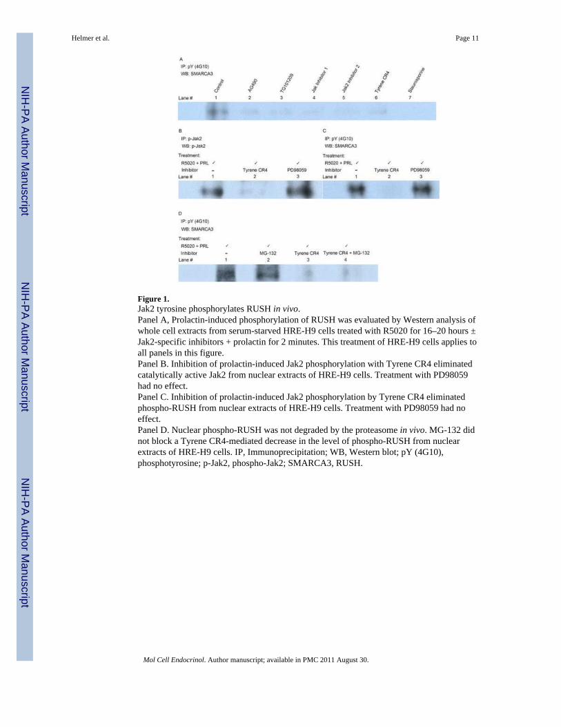
Figure 1.Jak2 tyrosine phosphorylates RUSH in vivo.Panel A, Prolactin-induced phosphorylation of RUSH was evaluated by Western analysis ofwhole cell extracts from serum-starved HRE-H9 cells treated with R5020 for 16–20 hours ±Jak2-specific inhibitors + prolactin for 2 minutes. This treatment of HRE-H9 cells applies toall panels in this figure.Panel B. Inhibition of prolactin-induced Jak2 phosphorylation with Tyrene CR4 eliminatedcatalytically active Jak2 from nuclear extracts of HRE-H9 cells. Treatment with PD98059had no effect.Panel C. Inhibition of prolactin-induced Jak2 phosphorylation by Tyrene CR4 eliminatedphospho-RUSH from nuclear extracts of HRE-H9 cells. Treatment with PD98059 had noeffect.Panel D. Nuclear phospho-RUSH was not degraded by the proteasome in vivo. MG-132 didnot block a Tyrene CR4-mediated decrease in the level of phospho-RUSH from nuclearextracts of HRE-H9 cells. IP, Immunoprecipitation; WB, Western blot; pY (4G10),phosphotyrosine; p-Jak2, phospho-Jak2; SMARCA3, RUSH.
Helmer et al. Page 11
Mol Cell Endocrinol. Author manuscript; available in PMC 2011 August 30.
NIH
-PA Author Manuscript
NIH
-PA Author Manuscript
NIH
-PA Author Manuscript

Figure 2.Jak2 affects transcription of the RUSH gene.Panel A. Prolactin increased (p < 0.001) progesterone-dependent transcription of a RUSHreporter construct (compare lanes 1 and 2). Inhibition of Jak2 phosphorylation by TyreneCR4 (compare lanes 2 and 3), or mutation of the RUSH binding site (compare lanes 4 and5), completely blocked (p < 0.001) the ability of prolactin to augment progesterone-dependent transcription of the same construct. Mean (± SEM) values with the same letterdesignation are not significantly different (p > 0.05). Serum-starved HRE-H9 cells weretreated with ± R5020 for 16–20 hours ± Inhibitor ± prolactin for 5 minutes. This treatmentof HRE-H9 cells applies to all outcomes in this figure.Panel B. Phosphorylation of Jak2 had no effect (p > 0.05) on the total nuclear content ofprogesterone-induced RUSH evaluated by quantitative LSCM (details in section 2.6 of theMaterials and Methods. Mean (± SEM) values with the same letter designation are notsignificantly different (p > 0.05).
Helmer et al. Page 12
Mol Cell Endocrinol. Author manuscript; available in PMC 2011 August 30.
NIH
-PA Author Manuscript
NIH
-PA Author Manuscript
NIH
-PA Author Manuscript
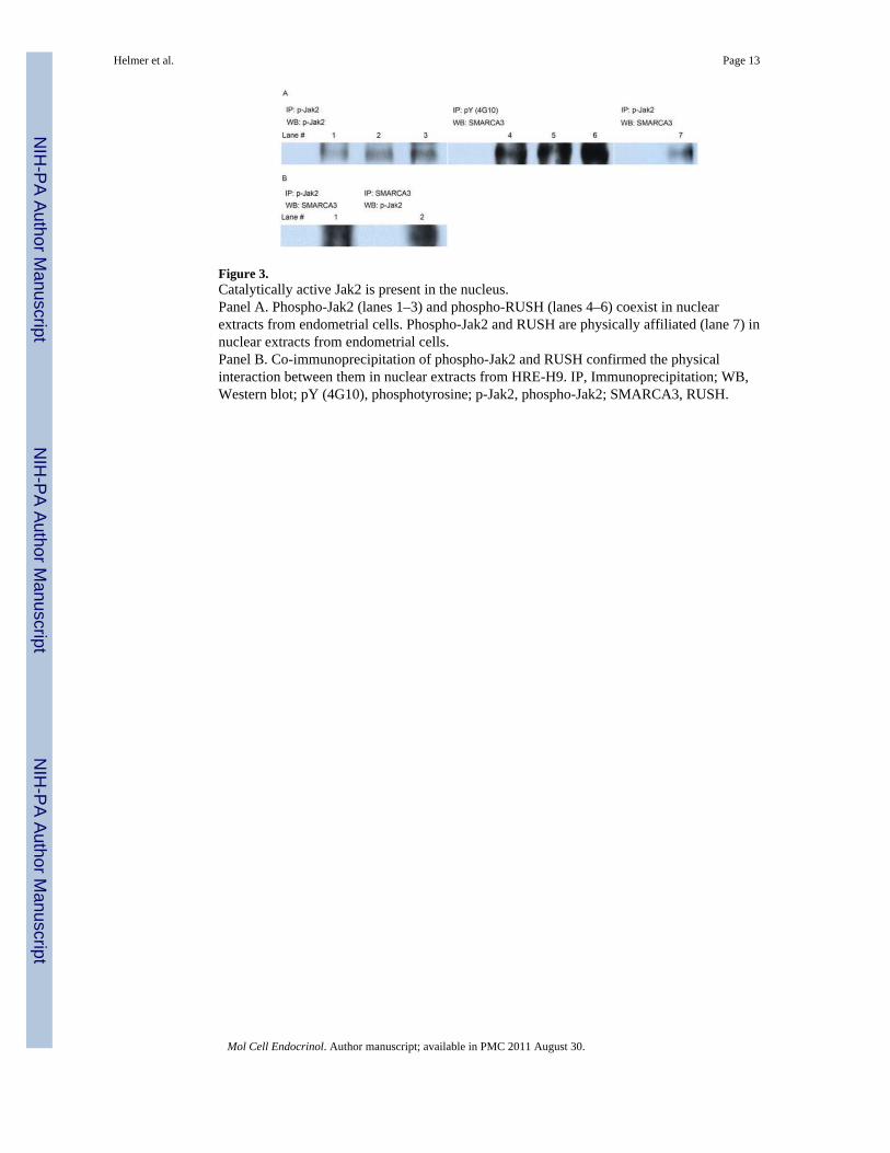
Figure 3.Catalytically active Jak2 is present in the nucleus.Panel A. Phospho-Jak2 (lanes 1–3) and phospho-RUSH (lanes 4–6) coexist in nuclearextracts from endometrial cells. Phospho-Jak2 and RUSH are physically affiliated (lane 7) innuclear extracts from endometrial cells.Panel B. Co-immunoprecipitation of phospho-Jak2 and RUSH confirmed the physicalinteraction between them in nuclear extracts from HRE-H9. IP, Immunoprecipitation; WB,Western blot; pY (4G10), phosphotyrosine; p-Jak2, phospho-Jak2; SMARCA3, RUSH.
Helmer et al. Page 13
Mol Cell Endocrinol. Author manuscript; available in PMC 2011 August 30.
NIH
-PA Author Manuscript
NIH
-PA Author Manuscript
NIH
-PA Author Manuscript

Figure 4.Catalytically active Jak2 resides in the nucleus with RUSH.Panel A. Confocal immunofluorescence images identify phospho-Jak2 (red) in both thenuclear and cytoplasmic compartments of serum-starved HRE-H9 cells treated with R5020for 16–20 hours ± Jak2-specific inhibitors + prolactin for 2 minutes. This treatment of HRE-H9 cells applies to all panels in this figure. DAPI (4,6-diamidino-2-phenylindole) nuclearDNA staining is blue. This image corroborates the detection of nuclear phospho-Jak2 byWestern analysis shown in Figure 1B. Scale bar, 50 μm, applies to all images in this figure.Panel B. Confocal immunofluorescence images identify RUSH (green) in both the nuclearand cytoplasmic compartments of HRE-H9 cells.Panel C. Merged confocal immunofluorescence images of RUSH (green) with fibrillarin(red) show nuclear RUSH does not co-localize (yellow) exclusively to nucleoli. Results arerepresentative of seven independent experiments performed in duplicate.
Helmer et al. Page 14
Mol Cell Endocrinol. Author manuscript; available in PMC 2011 August 30.
NIH
-PA Author Manuscript
NIH
-PA Author Manuscript
NIH
-PA Author Manuscript

NIH
-PA Author Manuscript
NIH
-PA Author Manuscript
NIH
-PA Author Manuscript
Helmer et al. Page 15
Table 1
RUSH-protein partners identified by μLC/MS/MS analysis.
Tyrosine-phosphorylated Nucleolar-affiliated
A. Partners that bind the RING-finger motif
Nucleolin √ √
Ribosomal protein L10 √
HSP 70kDa protein 9B
B. Heterogeneous nuclear ribonucleoproteins
hnRNP U isoform √
hnRNP A1 √
hnRNP A3 isoform 1
C. Histones
Core Histone macro-H2A.1 √
Histone H1.1 √
D. Chaperones
HSP 90α √ √
HSP 90β √ √
Endoplasmin (GRP94) √
Nucleophosmin √ √
E. RNA Helicases
RNA Helicase II/Gu protein √
DEAD-box polypeptide 17 isoform p82 √ √
F. DNA Replication
DNA Topoisomerase I √
G. Splicing Factors
Splicing factor 3a, subunit 1 √
H. Transcription regulator
Actin
Mol Cell Endocrinol. Author manuscript; available in PMC 2011 August 30.